Embed Size (px)
Citation preview
Journal of Neurology, Neurosurgery, and Psychiatry, 1980, 43, 725-729
Haemorrhage associated with meningioma: a casereport and review of the literatureT L HELLE AND F K CONLEY
From the Section of Neurosurgery, Palo Alto Veterans Administration Medical Center, Palo Alto, Californiaand the Division of Neurosurgery, Department of Surgery, Stanford University Medical School,Stanford, California
SUMMARY A case of haemorrhage into a parasagittal meningioma treated by surgical resection ispresented. A review of the literature found 43 additional cases of meningioma associated withhaemorrhage. By correlating these cases with those from another large series of meningiomasclassified by histological type and location, an estimation of the "relative bleeding tendency" ofcertain classes of meningiomas was made. Haemorrhage associated with meningioma is a seriouscomplication and proper treatment consists of evacuation of the haematoma with resection oftumour.
Although much is known about the natural history ofmeningiomas, the behaviour of such tumours in agiven patient depends on many factors: location,size, histology, and cytology.' There is little inform-ation about haemorrhage associated with meningio-mas and the influence of such haemorrhage uponpatient outcome. We present a patient who bledinto a parasagittal meningioma, and review theavailable literature on haemorrhage associated withmeningiomas.
Case report
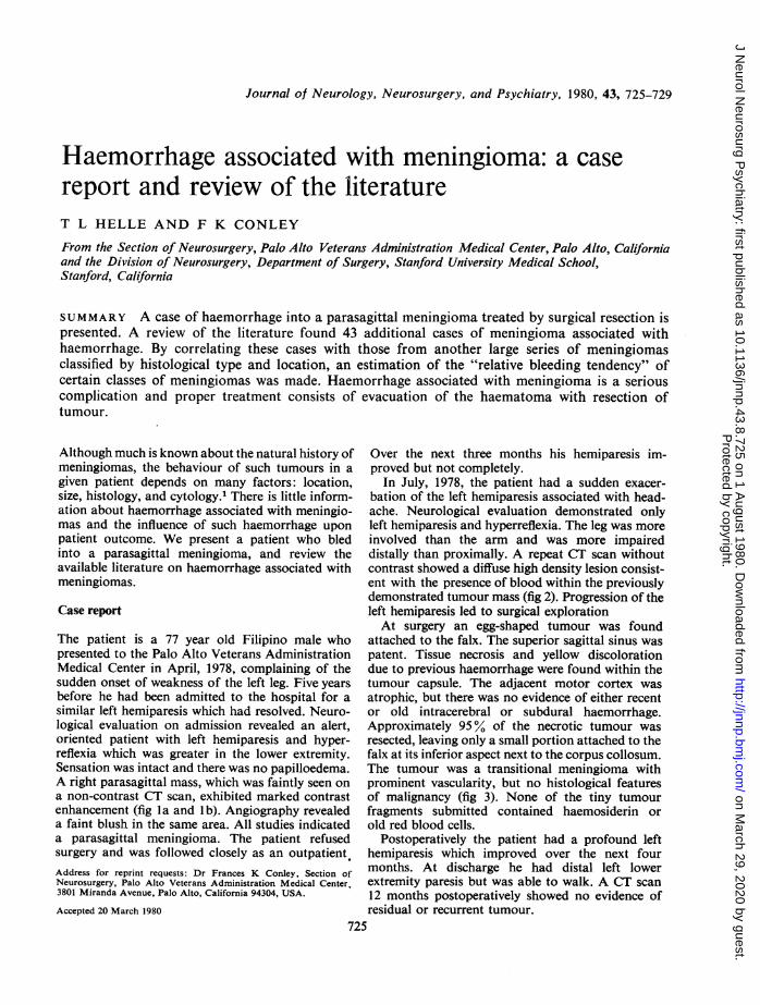
The patient is a 77 year old Filipino male whopresented to the Palo Alto Veterans AdministrationMedical Center in April, 1978, complaining of thesudden onset of weakness of the left leg. Five yearsbefore he had been admitted to the hospital for asimilar left hemiparesis which had resolved. Neuro-logical evaluation on admission revealed an alert,oriented patient with left hemiparesis and hyper-reflexia which was greater in the lower extremity.Sensation was intact and there was no papilloedema.A right parasagittal mass, which was faintly seen ona non-contrast CT scan, exhibited marked contrastenhancement (fig la and lb). Angiography revealeda faint blush in the same area. All studies indicateda parasagittal meningioma. The patient refusedsurgery and was followed closely as an outpatient.Address for reprint requests: Dr Frances K Conley, Section ofNeurosurgery, Palo Alto Veterans Administration Medical Center,3801 Miranda Avenue, Palo Alto, California 94304, USA.
Accepted 20 March 1980
Over the next three months his hemiparesis im-proved but not completely.
In July, 1978, the patient had a sudden exacer-bation of the left hemiparesis associated with head-ache. Neurological evaluation demonstrated onlyleft hemiparesis and hyperreflexia. The leg was moreinvolved than the arm and was more impaireddistally than proximally. A repeat CT scan withoutcontrast showed a diffuse high density lesion consist-ent with the presence of blood within the previouslydemonstrated tumour mass (fig 2). Progression of theleft hemiparesis led to surgical explorationAt surgery an egg-shaped tumour was found
attached to the falx. The superior sagittal sinus waspatent. Tissue necrosis and yellow discolorationdue to previous haemorrhage were found within thetumour capsule. The adjacent motor cortex wasatrophic, but there was no evidence of either recentor old intracerebral or subdural haemorrhage.Approximately 95% of the necrotic tumour wasresected, leaving only a small portion attached to thefalx at its inferior aspect next to the corpus collosum.The tumour was a transitional meningioma withprominent vascularity, but no histological featuresof malignancy (fig 3). None of the tiny tumourfragments submitted contained haemosiderin orold red blood cells.
Postoperatively the patient had a profound lefthemiparesis which improved over the next fourmonths. At discharge he had distal left lowerextremity paresis but was able to walk. A CT scan12 months postoperatively showed no evidence ofresidual or recurrent tumour.
725
Protected by copyright.
on March 29, 2020 by guest.
http://jnnp.bmj.com
/J N
eurol Neurosurg P
sychiatry: first published as 10.1136/jnnp.43.8.725 on 1 August 1980. D
ownloaded from

T L Helle and F K Coniley
Fig 1 a and b CT scans ofpatient in April 1978, showing the right parasagittal mass. Ia is withoutcontrast enhancement; I b is with contrast enhancement.
S~~~~~~~~~A
¾-
Fig 2 Noncontrast CT scan ofpatient in July 1978,showing a high density lesion consistent with bloodwithin the previously demonstrated mass. Fig 3 Photomicrograph of transitional meningioma
renmoved from ouir patient. H & Ex 320.Discussion
This is an unusual case because haemorrhageassociated with an intracranial neoplasm is a rareevent.2 Mutlu, et a13 reported a 09% (2 of 225cases) incidence of intracerebral haemorrhagesecondary to brain tumour, and Yasargil4 reporteda 1-2% incidence of brain tumour in patientspresenting with subarachnoid haemorrhage. Intra-cranial haemorrhage in association with meningiomais very infrequent as is demonstrated by examinationof two large series of meningiomas collected by
Cushing and Eisenhardt5 (313 cases), and Hoesslyand Olivecronaf (280 cases of parasagittal menin-ioma). Neither series reported any case of massivehaemorrhage. By contrast, in our case a transitionalcell parasagittal meningioma underwent spon-taneous, symptomatic, intratumoral haemorrhage.Mechanisms responsible for haemorrhage within
tumours are not completely understood. Glio-blastoma multiforme and oligodendroglioma arethe primary brain tumours which bleed most
726
'k
:10
-.9.:.! .:.
............
W" -
:irf. Z-1....
Protected by copyright.
on March 29, 2020 by guest.
http://jnnp.bmj.com
/J N
eurol Neurosurg P
sychiatry: first published as 10.1136/jnnp.43.8.725 on 1 August 1980. D
ownloaded from

Haemorrhage associated with meningioma: a case report and review of the literature
frequently.7 Haphazard endothelial proliferationand tumour necrosis within glioblastomas may befactors which cause this haemorrhagic tendency.8Hypernephroma and malignant melanoma are themetastatic brain tumours which bleed most fre-quently, and the incompletely formed, friable bloodvessels associated with the rapid growth of meta-static tumours may be responsible for their bleedingtendency.7 Factors responsible for haemorrhagewithin benign intracranial tumours, such as mening-iomas, are less obvious. Angioblastic meningiomascharacteristically are composed of abnormal bloodvessels9 and meningiomas of other cell types can
contain foci of abnormal vessels; this abnormal
vascularity could be related to tumour-associatedhaemorrhage.10 It is possible that blood vesselssupplying a meningioma undergo compensatoryenlargement with weakening of their wall and createthe potential for tumour associated haemorrhage.11It has also been postulated that cerebral oedemaand venous obstruction, which are commonlyassociated with meningiomas, can cause infarctionand subsequent haemorrhage.10 12Only 43 other cases of haemorrhage associated
with meningioma were found in a review of theliterature; these cases as well as our case are
tabulated in table 1. The type of tumour mostfrequently associated with haemorrhage was the
Table 1 Data on 44 patients with meningioma associated haemorrhage
Reference Age Sex Haemorrhage Tumour Tumour Patient's Operationli Symptom(yr) type* siuet histologyt outcome§ onset T
Present Case 77 M IT R parasag Transitional Morb Resection SlowAskenasy and Behmoaram,
196011 1 34 F SA L lat vent Syncytial Died Resection Sudden2 38 F SA L lat vent Fibrous Died Resection Sudden3 32 M IV R lat vent Transitional Morb Resection Slow
Bilodeau and Beraud, 196613 46 M IC, IT R convex Syncytial Died Evacuation SlowBingas and Meese, 196614 65 F SD R convex Syncytial NI Resection SuddenBudny et at, 197715 60 M IC, IT R convex Malignant Died Resection SlowCusick and Bailey, 197216 47 F SD R convex Nonspecified Died None SlowEl-Banhawy and Walter, 196212 20 M SA L parasag Syncytial Morb Resection SlowEverett et al, 197917 65 M IC, IT, IV R convex Syncytial Died Resection SuddenFlynn and Karpas, 197118 78 F IT, SA L convex Nonspecified Died None SlowGoran et al, 1965,19 1 65 M IC, IT R convex Syncytial Died Evacuation Sudden
2 68 F IT, SA R sphenoid Syncytial Died Resection Sudden3 46 M IT R parasag Syncytial Died None Slow4 55 M IC, SA R lat vent Malignant Morb Resection Sudden5 42 F IT L convex Syncytial Died None Sudden
Gruszki-wicz et al, 1969 10 18 M IC, SA R convex Fibrous Morb Resection SuddenHung et al, 197220 42 F IT L convex Fibrous Morb Resection SlowModesti et al, 197621 1 59 M IC, SA, SD L sphenoid Syncytial Nl Resection Sudden
2 49 F IC, SA, SD L parasag Syncytial Morb Resection Slow3 72 F SA, SD L parasag Angioblastic Died Resection Slow4 69 M SD L convex Syncytial N1 Resection Slow
Moore, 195422 1 72 M SA Bilat parasag Fibrous Died None Sudden2 66 F IV, SA R convex Angioblastic Died None Slow
Nakao et al, 19779 21 M IC, SA R convex Angioblastic Morb Resection SlowNas,ar and Correll, 196823 34 M IV, SA Spinal Angioblastic Died None SlowOhaegbulam, 197724 40 M SA L sphenoid Fibrous Died None SuddenRosenberg et al, 197525 1 44 M SA R Meckel's Cv Angioblastic Morb Resection Slow
2 48 F SA L Meckel's Cv Nonspecified Morb Resection SlowSmith rt al, 197526 14 F SA L lat vent Fibrous NI Resection SuddenWalsh et al, 197727 77 F IT, SD Bilat convex Syncytial Died Resection SlowYasargil and So, 197628 1 50 F IT, SA, SD L post fossa Transitional Morb Resection Sudden
2 47 F IT, SA L post fossa Nonspecified Morb Resection SlowSkultety, 196829 58 M IC, IT, SA, SD R sphenoid Fibrous - Resection SlowFukumitsu et al, 1973 30 49 F IC Parasag Syncytial - - -Therkelsen, 196131 - - SD Convex Nonspecified - Nonspecified -Locksley et al, 196632 - - SA Parasag Nonspecified - -
Globus and Sapirstein, 19428 - - SA - Nonspecified - -
McLaurin and Helmer, 196233 - - SA - Angioblastic - -
Russell and Rubinstein,1977341 - - IC, SD - Nonspecified - -
2 - - IC - Nonspecified - - -Zimmerman, 196335 1 - - IC - Angioblastic - -
2 - - IC - Angioblastic - -
Drake and McGee 196136 - - - - Nonspecified - -
* IC-intracerebral; IT-intratumoral; IV-intraventricular; SD-subdural; SA-3ubarazhnDid.t Convexity refers to tumours located on either the frontal, parietal, temporal or ozz:ipital lob2.t Classification according to Russell and Rubinstein (1977).§ Morbidity-any permanent neurologic deficit; Normal-complete rezovery.Resection refers to total or subtotal resection of tumour and evacuation of hematoma; Evacuation refers to only evacuation of h-mitons.
1¶ Slow refers to symptoms relating to the tumour and/or hematoma being present more than 24 hours prior to hospitalization; Sudden refersto symptoms being present less than 24 hours prior to hospitalization.
727
Protected by copyright.
on March 29, 2020 by guest.
http://jnnp.bmj.com
/J N
eurol Neurosurg P
sychiatry: first published as 10.1136/jnnp.43.8.725 on 1 August 1980. D
ownloaded from

728
syncytial meningioma (32 %), followed by angio-blastic (18 %). The greatest number of meningiomaswere located on the convexities, but were also foundinfratentorially, intraventricularly, and in the spinalcanal. Tumours occurred with equal frequency onthe right and left sides. Clinical data revealed thatthe average age at the time of haemorrhage was50 years, and there was an equal sex incidence.Symptoms from the haemorrhage were slow inonset in just over 50% of patients; in the othersthe event was rapid in onset and usually catastrophic.Many patient had more than one type of haemor-rhage associated with their meningioma; however,the most frequently occurring haemorrhage wassubarachnoid (35 %). Of the 35 patients whereoperative data is available, 24 had craniotomy forresection of tumour. Of the 33 patients whereoutcome is known, only 4 (1 %) recovered com-pletely and all these patients had had tumourresection. 520% of these patients died. Interestingly,those patients who had no surgery or only haema-toma evactuation (10 cases) all died, whereas theoutcome was more favourable when tumourresection was accomplished at the time of thecraniotomy (table 2).By relating data from this series of meningiomas
associated with haemorrhage to data obtainedfrom a large series of meningiomas which reportsthe incidence of histological types, it is possible tocalculate an approximate "relative bleeding ten-dency" for a given histological type of tumour.Angioblastic meningiomas comprise 8% of the 1197surgically verified intracranial and intraspinal
Table 2 Correlation ofpatient outcome versus type ofoperation performed
Patient outcome Operation performed
Resection Evacuation None
Died 7 2 8Morbidity 12 0 0Normal 4 0 0
Table 3 Relative bleeding tendency of meningiomas totumour histology calculated by relating the present serieswith Jellinger and Slowik (1975)37
Histology of Distribution of ApproxHistolgy bleeding meningiomas* meningiomast bleeding index
Angioblastic 8(18) 97(8) 2Transitional 3(7) 255(21) 0-3Syncytial 14(32) 749(63) 0 5Fibrous 7(16) 82(7) 2Malignant 2(4) 14(1) 4Nonspecified 10(23) -
TOTAL 44 1,197 -
* Present series.t Jellinger & Slowik (1975).37Percentages in parentheses
T L Helle and F K Conley
Table 4 Relative bleeding tendency ofmeningiomasaccording to tumour location
Location Distribution of Distribution of Approx relativebleeding meningiomast bleeding indexmeningiomas*
Parasagittal 8(22) 194(16-5) 1 3Convexity 15(40) 460(38-5) 1Sphenoid 4(11) 107(9) 1-2Posterior Fossa 2(5) 90(7 5) 0 7Ventricular 5(14) 6(0 5) 27Meckel's Cave 2(5) none -
Spinal 1(3) 208(17) 0-16Others none 132(11) -
TOTAL 37 1197 -
* Present series.t Jellinger and Slowik (1975)37Percentages in parentheses
meningiomas reported by Jellinger and Slowik ;37however, 18% of the meningiomas associated withhaemorrhage reported in the present study wereangioblastic. This suggests that angioblastic men-ingiomas have a tendency to bleed more than twotimes more frequently than all other meningiomascombined. By using the same method of calculation,the relative bleeding tendency for transitionalmeningioma is 0 3, for syncytial, 0 5, for fibrous, 2,and for malignant meningioma, 4 (table 3). Iftumour location is considered, the greatest relativebleeding tendency is 27 for intraventricular mening-ioma and the least, 016 for spinal meningioma(table 4). Parasagittal, convexity, sphenoid, andposterior fossa meningiomas all have a relativebleeding tendency of approximately 1. While thesmall number of cases of haemorrhage in associationwith meningioma does not allow one to place greatsignificance on these calculations, if they are appliedto our patient, his meningioma had a relatively lowbleeding potential because of location (parasagittal)and histology (transitional). His recovery withminimal morbidity relates both to the type ofhaemorrhage (intratumoural only) and to thefact that he had definitive surgery. However, hisbenign clinical course must be considered somewhatunusual because among the conclusions which canbe drawn from this literature review are thathaemorrhage associated with a meningioma is avery serious event and is associated with a highmortality. To our knowledge, the present case isthe first reported where haemorrhage into a men-ingioma has been documented by serial CT scans.CT scans may allow earlier diagnosis of a haemor-rhagic event in association with a benign intracranialtumour and thus improve the ultimate prognosisin patients with these tumours. From our study it isapparent that definitive surgery with combinedevacuation of the haematoma and tumour resectionprovides the best chance for recovery with the leastmorbidity.
Protected by copyright.
on March 29, 2020 by guest.
http://jnnp.bmj.com
/J N
eurol Neurosurg P
sychiatry: first published as 10.1136/jnnp.43.8.725 on 1 August 1980. D
ownloaded from

Haemorrhage associated with meningioma: a case report and review of the literature
The authors thank Leslie Zatz, MD for reviewingthe manuscript and making constructive criticism,Palo Alto Veterans Administration Medical Center'sMedical Media Department for the photographs,and to Ruth Uhrhammer for preparation of themanuscript.
References
1 Skullerud K, Loken AC. The prognosis in menin-giomas. Acta Neuropathol (Berl) 1974; 29:337-44.
2 Scott M. Spontaneous intracerebral hematomacaused by cerebral neoplasms. J Neurosurg 1975;42:338-42.
3 Mutlu N, Berry RG, Alpers BJ. Massive cerebralhemorrhage. Arch Neurol 1963; 8:644-61.
4 Yasargil MG. Die subarachnoidale Blutung. SchweizMed Wochenschr 1969; 99:1629-32.
S Cushing H, Eisenhardt L. Meningiomas. Springfield,Charles C Thomas, 1938.
6 Hoessly GF, Olivecrona H. Report on 280 cases ofverified parasagittal meningioma. J Neurosurg 1955;12:614-26.
7 Zulch KJ. Neuropathology of intracranial haemorr-hage. Prog Brain Res 1968; 30:151-65.
8 Globus JH, Sapirstein M. Massive hemorrhage intobrain tumour. JAMA 1942; 120:348-52.
9 Nakao S, Sato S, Ban S, Inutsuka N, Yamamoto T,Ogata M. Massive intracerebral hemorrhage causedby angioblastic meningioma. Surg Neurol 1977;7:245-8.
10 Gruszkiewicz J, Doron Y, Gellei B, Peyer E.Massive intracerebral bleeding due to supratentorialmeningioma. Neurochirurgia (Stuttg) 1969;12:107-11.
11 Askenasy HM, Behmoaram AD. Subarachnoidhemorrhage in meningiomas of the lateral ventricle.Neurology (Minneap.) 1960; 10:484-9.
12 El-Banhawy A, Walter W. Meningiomas with acuteonset. Acta Neurochir (Wien) 1962; 10:194-206.
13 Bilodeau B, Beraud R. Hemorrhgie dans unmeningiome. Can Med Assoc J 1960; 95:682-4.
14 Bingas B, Meese M. Subdurales Hamatom seltenerAtiologie (Fallmitteilung). Nevenarzt 1966; 37:175-7.
15 Budny JL, Glasauer FE, Sil R. Rapid recurrence ofmeningioma causing intracerebral hemorrhage.Surg Neurol 1977; 8:323-5.
16 Cusick JF, Bailey OT. Association of ossifiedsubdural hematomas and a meningioma. J Neurosurg1972; 37:731-4.
17 Everett BA, Kusske JA, Pribram HW. Anticoag-ulants and intracerebral hemorrhage from anunsuspected meningioma. Surg Neurol 1979;11 :233-5.
18 Flynn JT, Karpas CM. Intracranial lesion withhaemorrhage. NY State J Med 1971; 71:1951-9.
19 Goran A, Ciminello VJ, Fisher RG. Haemorrhageinto meningiomas. Arch Neurol 1965; 13:65-9.
20 Hung C, Chang W, Yao Y. Post-traumatic intra-cranial meningioma. J Formosan Med Assoc 1972;71 :214-9.
21 Modesti LM, Binet EF, Collins GH. Meningiomascausing spontaneous intracranial hematomas. JNeurosurg 1976; 45:437-41.
22 Moore MT. The fate of clinically unrecognizedintracranial meningiomas. Neurology (Minneapolis)1954; 4:837-56.
23 Nassar SI, Correll JW. Subarachnoid haemorrhagedue to spinal cord tumours. Neurology (Minneapolis).1968; 18:87-94.
24 Ohaegbulam SC. Sudden death from an asympto-matic sphenoid ridge meningioma. J Neurol 1977;215:291-4.
25 Rosenberg GA, Herz DA, Leeds N, Strully K.Meckel's cave meningiomas with subarachnoidhaemorrhage. Surg Neurol 1975; 3:333-6.
26 Smith VR, Stein PS, MacCarty CS. Subarachnoidhaemorrhage due to lateral ventricular meningiomas.Surg Neurol 1975; 4:241-3.
27 Walsh JW, Winston KR, Smith T. Meningiomawith subdural hematoma. Surg Neurol 1977;8:293-5.
28 Yasargil MG, So SC. Cerebellopontine anglemeningioma presenting as subarachnoid haemor-rhage. Surg Neurol 1976; 6:3-6.
29 Skultety FM. Meningioma simulating rupturedaneurysm. J Neurosurg 1968; 28:380-2.
30 Fukumitsu T, Yoshida Y, Yamashita J. Massiveintracerebral haemorrhage due to parasagittalmeningioma. Brain Nerve (Tokyo) 1973;25:911-4.
31 Therkelsen J. The diagnostic value of cerebralangiography in patients with apoplectic symptoms.Acta Psychiatr Scand (suppl 150), 1961; 36:129-32.
32 Locksley HB, Sahs AL, Sandler R. Report on thecooperative study of intracranial aneurysms andsubarachnoid haemorrhage. Section III. J Neurosurg1966; 24:1034-56.
33 McLaurin RL, Helmer FA. Errors in diagnosis ofintracranial tumours. JAMA 1962; 180:1011-6.
34 Russell DS, Rubinstein LS. Pathology of tumours ofthe nervous system, 4th Ed., Baltimore: Williamsand Wilkins, 1977.
35 Zimmerman HM. Personal communication inGoran, et al.'9
36 Drake CG, McGee D. Apoplexy associated withbrain tumours. Can Med Assoc J 1961; 84:303-5.
37 Jellinger K, Slowik F. Histological subtypes andprognostic problems in meningiomas. J Neurol1975; 208:279-98.
729
Protected by copyright.
on March 29, 2020 by guest.
http://jnnp.bmj.com
/J N
eurol Neurosurg P
sychiatry: first published as 10.1136/jnnp.43.8.725 on 1 August 1980. D
ownloaded from