Embed Size (px)
Citation preview
GRISEOFULVIN
This substance was considered by previous working groups, in 1975 (IARC, 1976)and 1987 (IARC, 1987). Since that time, new data have become available, and thesehave been incorporated into the monograph and taken into consideration in the presentevaluation.
1. Exposure Data
1.1 Chemical and physical data
1.1.1 Nomenclature
Chem. Abstr. Serv. Reg. No.: 126-07-8Deleted CAS Nos: 8027-03-0; 8055-10-5; 3426-54-8; 11103-62-1; 24659-79-8 Chem. Abstr. Name: (1′S,6′R)-7-Chloro-2′,4,6-trimethoxy-6′-methylspiro[benzo-furan-2(3H),1′-[2]cyclohexene]-3,4′-dioneIUPAC Systematic Name: 7-Chloro-2′,4,6-trimethoxy-6′β-methylspiro[benzo-furan-2(3H),1′-[2]cyclohexene]-3,4′-dioneSynonyms: (2S,4′R)-7-Chloro-2′,4,6-trimethoxy-4′-methylspiro[benzofuran-2-(3H),3′-cyclohexene]-3,6′-dione; (1′S-trans)-7-chloro-2′,4,6-trimethoxy-6′-methyl-spiro[benzofuran-2(3H),1′-[2]cyclohexene]-3,4′-dione; (+)-griseofulvin; (+)-7-chloro-4,6-dimethoxycoumaran-3-one-2-spiro-1′-(2′-methoxy-6′-methylcyclohex-2′-en-4′-one)
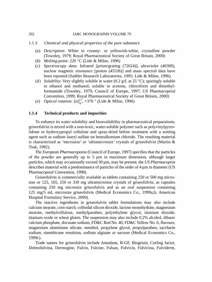
1.1.2 Structural and molecular formulae and relative molecular mass
C17H17ClO6 Relative molecular mass: 352.77
–291–
O
OOCH3
Cl
H3COH CH3
OCH3
O
1.1.3 Chemical and physical properties of the pure substance
(a) Description: White to creamy- or yellowish-white, crystalline powder(Townley, 1979; Royal Pharmaceutical Society of Great Britain, 2000)
(b) Melting-point: 220 °C (Lide & Milne, 1996)(c) Spectroscopy data: Infrared [prism/grating (72624)], ultraviolet (40309),
nuclear magnetic resonance [proton (45536)] and mass spectral data havebeen reported (Sadtler Research Laboratories, 1995; Lide & Milne, 1996).
(d ) Solubility: Very slightly soluble in water (0.2 g/L at 25 °C); sparingly solublein ethanol and methanol; soluble in acetone, chloroform and dimethyl-formamide (Townley, 1979; Council of Europe, 1997; US PharmacopeialConvention, 1999; Royal Pharmaceutical Society of Great Britain, 2000)
(e) Optical rotation: [α]1D7, +376 ° (Lide & Milne, 1996)
1.1.4 Technical products and impurities
To enhance its water solubility and bioavailability in pharmaceutical preparations,griseofulvin is mixed with a non-toxic, water-soluble polymer such as polyvinylpyrro-lidone or hydroxypropyl cellulose and spray-dried before treatment with a wettingagent such as sodium lauryl sulfate on benzalkonium chloride. The resulting materialis characterized as ‘microsize’ or ‘ultramicrosize’ crystals of griseofulvin (Martin &Tsuk, 1982).
The European Pharmacopoeia (Council of Europe, 1997) specifies that the particlesof the powder are generally up to 5 μm in maximum dimension, although largerparticles, which may occasionally exceed 30 μm, may be present; the US Pharmacopeiadescribes material with a predominance of particles of the order of 4 μm in diameter (USPharmacopeial Convention, 1998).
Griseofulvin is commercially available as tablets containing 250 or 500 mg micro-size or 125, 165, 250 or 330 mg ultramicrosize crystals of griseofulvin, as capsulescontaining 250 mg microsize griseofulvin and as an oral suspension containing125 mg/5 mL microsize griseofulvin (Medical Economics Co., 1999a,b; AmericanHospital Formulary Service, 2000).
The inactive ingredients in griseofulvin tablet formulations may also includecalcium stearate, corn starch, colloidal silicon dioxide, lactose monohydrate, magnesiumstearate, methylcellulose, methylparaben, polyethylene glycol, titanium dioxide,titanium oxide or wheat gluten. The suspension may also include 0.2% alcohol, dibasiccalcium phosphate, docusate sodium, FD&C Red No. 40, FD&C Yellow No. 6, flavours,magnesium aluminium silicate, menthol, propylene glycol, propylparaben, saccharinsodium, simethicone emulsion, sodium alginate or sucrose (Medical Economics Co.,1999c).
Trade names for griseofulvin include Amudane, B-GF, Biogrisin, Curling factor,Delmofulvina, Dermogine, Fulcin, Fulcine, Fulsan, Fulvicin, Fulvicina, Fulviderm,
IARC MONOGRAPHS VOLUME 79292
Fulvina, Fulvinil, Fulvistatin, Fungivin, Gefulvin, Greosin, Gricin, Grifulin, Grifulvin,Gris-PEG, Grisactin, Griséfuline, griseo von ct, Griseo, Griseoderm, Griseofort,Griseoful, Griseofulvin Capsules USP 23, Griseofulvin Leo, Griseofulvin Oral Suspen-sion USP 23, Griseofulvin Tablets BP 1999, Griseofulvin Tablets USP 23, GriseofulvinUltra, Griseofulvin Vetag, Griseofulvina, Griseomed, Griseostatin, Grisfulvin, Griso-fulvin, Grisol, Grisomicon, Grisovin, Grisovina, Grivate, Grivin, Grizeofulvin, Grysio,Idifulvin, Lamoryl, Likuden, Microcidal, Neo-fulcin, Norofulvin, Polygris, Poncyl,Spirofulvin, Sporostatin, Sulvina, Ultragris, Ultramicrosize Griseofulvin TabletsUSP 23, Vetmix Griseofulvin, Viro Griseo M and Walavin (Royal PharmaceuticalSociety of Great Britain, 2000; Swiss Pharmaceutical Society, 2000)
1.1.5 Analysis
Several international pharmacopoeias specify infrared absorption spectrophoto-metry with comparison to standards and high-performance liquid chromatography(HPLC) with ultraviolet detection as the methods for identifying griseofulvin; ultra-violet absorption spectrophotometry and HPLC with ultraviolet detection are used toassay its purity. In pharmaceutical preparations, griseofulvin is identified by infraredand ultraviolet absorption spectrophotometry and HPLC with ultraviolet detection;ultraviolet absorption spectrophotometry and HPLC with ultraviolet detection are usedto assay for griseofulvin content (British Pharmacopoeia Commission, 1993; Councilof Europe, 1997; US Pharmacopeial Convention, 1999).
1.2 Production
Griseofulvin is an antifungal substance typically produced by the growth ofcertain strains of Penicillium griseofulvum (Royal Pharmaceutical Society of GreatBritain, 2000). A method for the synthesis of griseofulvin from dimethoxyphenol hasbeen reported (Pirrung et al., 1991).
Information available in 2000 indicated that griseofulvin was manufactured by sixcompanies in China, three in Japan and one each in India and the United Kingdom(CIS Information Services, 2000a) and that it was used in the formulation ofpharmaceuticals by 44 companies in India, eight companies each in Germany and theUnited Kingdom, six companies each in Argentina, Japan and the USA, fivecompanies each in Singapore, Switzerland, Taiwan and Thailand, four companies eachin China, Indonesia, Italy and Malaysia; three companies each in Australia, Canada,Chile, Ecuador and the Netherlands, two companies each in Brazil, Egypt, Hong Kong,Mexico, New Zealand, Peru, the Philippines, Portugal, South Africa, Spain, Turkeyand Viet Nam and one company each in Austria, Finland, Ireland, the Islamic Republicof Iran, Israel, Malta, Norway, Sweden and Venezuela (CIS Information Services,2000b).
GRISEOFULVIN 293

1.3 Use
Griseofulvin is an antibiotic fungistatic drug administered orally in the treatment ofdermatophyte and ringworm infections. It is fungistatic against various species ofMicrosporum, Epidermophyton and Trichophyton in vitro. It is generally given forinfections that involve the scalp, hair, nails and skin (e.g. tinea corporis (ringworm ofthe body), tinea pedis (athlete’s foot), tinea cruris (ringworm of the groin or thigh), tineabarbae (barber’s itch), tinea capitis (ringworm of the scalp), tinea unguium (onycho-mycosis; ringworm of the nails)) and which do not respond to topical treatments;infections of the soles of the feet, the palms of the hands and the nails respond slowly(Medical Economics Co., 1999a,b,c,d; Royal Pharmaceutical Society of Great Britain,2000).
Because griseofulvin has some vasodilatatory activity, its use has resulted in someimprovement in a small number of patients with Raynaud’s disease and anginapectoris. Because it is structurally similar to colchicine and shares its activity as ametaphase inhibitor, griseofulvin has been used in the treatment of gout (AmericanHospital Formulary Service, 2000).
The dosage of griseofulvin varies depending on whether the drug is administeredas a microsize or ultramicrosize preparation. In addition, the recommended doses ofultramicrosize griseofulvin vary slightly depending on the manufacturer and theformulation of the drug. Therapy with griseofulvin is generally maintained for at least2–4 weeks for the treatment of tinea corporis; at least 4–12 weeks for the treatment oftinea capitis; 4–8 weeks for tinea pedis; and from 4–6 months to 1 year or longer fortinea unguium (American Hospital Formulary Service, 2000).
The usual adult dose of ultramicrosize griseofulvin for the treatment of tineacorporis, tinea cruris or tinea capitis is 330–375 mg/day in single or divided doses,depending on the manufacturer and formulation of the drug; the usual adult dose ofultramicrosize griseofulvin for the treatment of infections that are more difficult to era-dicate, such as tinea pedis and tinea unguium, is 660–750 mg/day, depending on themanufacturer and formulation. The usual adult dose of microsize griseofulvin for thetreatment of tinea corporis, tinea cruris, or tinea capitis is 500 mg/day and 1 g daily forthe treatment of infections that are more difficult to eradicate, such as tinea pedis andtinea unguium (Gennaro, 1995; American Hospital Formulary Service, 2000; RoyalPharmaceutical Society of Great Britain, 2000).
The usual dose of ultramicrosize griseofulvin for children > 2 years of age isapproximately 7.3 mg/kg bw per day, although doses up to 10–15 mg/kg bw daily havebeen used. The manufacturers suggest that children weighing approximately 14–23 kgcan receive 82.5–165 mg of ultramicrosize griseofulvin daily and those weighing> 23 kg can receive 165–330 mg/day. Alternatively, the manufacturers suggest thatchildren weighing 16–27 kg can receive 125–187.5 mg of ultramicrosize griseofulvindaily and those weighing > 27 kg can receive 187.5–375 mg daily. For the treatmentof tinea capitis and tinea corporis, the American Academy of Pediatrics recommends
IARC MONOGRAPHS VOLUME 79294
that children receive ultramicrosize griseofulvin at a single daily dose of 5–10 mg/kgbw (maximum dose, 750 mg). The usual paediatric dose of microsize griseofulvin is10–11 mg/kg bw per day, although doses up to 20–25 mg/kg bw per day have beenused. The manufacturers suggest that children weighing approximately 14–23 kg canreceive 125–250 mg microsize griseofulvin daily and that children weighing > 23 kgcan receive 250–500 mg daily. Alternatively, some clinicians suggest that children begiven microsize griseofulvin at a dose of 300 mg/m2 daily. The American Academy ofPediatrics recommends that children receive microsize griseofulvin at a daily dose of10–20 mg/kg bw (maximum dose, 1 g) given in up to two divided doses (Gennaro,1995; American Hospital Formulary Service, 2000; Royal Pharmaceutical Society ofGreat Britain, 2000).
When preparations, available in some countries, containing ultramicrocrystallineor ultramicrosize griseofulvin are used, the doses are reduced by one-third to one-halfof the recommended doses of microcrystalline or microsize griseofulvin. Griseofulvinis probably best given with or after meals (Royal Pharmaceutical Society of GreatBritain, 2000).
The duration of treatment depends on the thickness of the keratin layer: 2–6 weeksfor infections of the hair and skin, up to 6 months for infections of the fingernails and12 months or more for infections of the toenails (Royal Pharmaceutical Society ofGreat Britain, 2000).
Although griseofulvin is usually given systemically, beneficial responses in fungalskin infections have been reported with some topical formulations (Royal Pharmaceu-tical Society of Great Britain, 2000).
Griseofulvin is also used as a veterinary antifungal drug (US Pharmacopeial Con-vention, 1998; Budavari, 2000; Food and Drug Administration, 2000).
1.4 Occurrence
1.4.1 Occupational exposure
According to the 1981–83 National Occupational Exposure Survey (National Insti-tute for Occupational Safety and Health, 2000), about 1700 pharmacists in the USAwere potentially exposed to griseofulvin.
1.4.2 Environmental occurrence
No data were available to the Working Group.
1.5 Regulations and guidelines
Griseofulvin is listed in the pharmacopoeias of China, the Czech Republic,France, Germany, Italy, Japan, Poland, the United Kingdom and the USA and in the
GRISEOFULVIN 295

European and International pharmacopoeias (British Pharmacopeia Convention, 1993;Society of Japanese Pharmacopoeia, 1996; Royal Pharmaceutical Society of GreatBritain, 2000; Swiss Pharmaceutical Society, 2000; Vidal, 2000). It is also registeredfor human use in Ireland, Norway, Portugal, Spain and Sweden (Instituto Nacional deFarmacia e do Medicamento, 2000; Irish Medicines Board, 2000; Medical ProductsAgency, 2000; Norwegian Medicinal Depot, 2000; Spanish Medicines Agency, 2000).
2. Studies of Cancer in Humans
2.1 Case report
A 48-year-old woman with a history of gastric ulcers was admitted to a dermato-logical clinic in Essen, Germany, with tinea affecting the skin and nails of the feet andleft hand. She was treated orally with a total dose of 31 g of griseofulvin over a periodof about 1.5 months. Analysis of the patient’s peripheral blood did not show alterationssuggestive of haematological disorders, and there was no apparent splenomegaly.Seven months later, when the woman was hospitalized after a minor traffic accident,analysis of the blood strongly suggested chronic granulocytic leukaemia. The patientwas subsequently followed and treated for this condition (König et al., 1969/70).
2.2 Cohort studies
Griseofulvin was included in a hypothesis-generating cohort study designed toscreen a large number (215) of drugs for possible carcinogenicity, which covered morethan 140 000 subscribers enrolled between July 1969 and August 1973 in a prepaidmedical care programme in northern California (USA). Computer records of persons towhom at least one drug prescription has been dispensed were linked to the cancerrecords of hospitals covered by the medical care programme and the regional cancerregistry. The observed numbers of cancers were compared with those expected,standardized for age and sex, for the entire cohort. Three publications summarized thefindings for follow-up periods of up to 7 years (Friedman & Ury, 1980), 9 years(Friedman & Ury, 1983) and 15 years (Selby et al., 1989). Griseofulvin was includedonly in the two most recent reports. In the 9-year follow-up, an excess of thyroid cancerwas reported among 744 griseofulvin users (two observed cases versus 0.2 expected;p < 0.05), while no excess was seen for cancers at all sites (23 observed cases versus22.7 expected). In the 15-year follow-up, no results were reported for griseofulvin,implying that no significant association was observed for any of the 56 cancer sitesconsidered. [The Working Group noted, as did the authors, that, since some 12 000comparisons were made in this hypothesis-generating study, the associations should beverified independently. Data on duration of use were not provided.]
IARC MONOGRAPHS VOLUME 79296
3. Studies of Cancer in Experimental Animals
Griseofulvin has been evaluated previously (IARC, 1976). One new report hasbecome available (Rustia & Shubik, 1978), and a selection of the most relevant studiesfrom the previous monograph were re-analysed.
3.1 Oral administration
Mouse: Groups of male and female Charles River mice, 5–6 weeks of age, were feddiets containing 1% (w/w) griseofulvin of various particle sizes (regular, microcrys-talline and milled) with specific surface areas of 0.41, 1.3 and 1.52 m2/g, respectively,for 12–16 months. ‘Hepatomas’ developed in 4/8 male and 0/9 female mice fed regular-size griseofulvin, 4/4 male and 4/5 female mice fed microcrystalline griseofulvin and 1/1male and 3/3 female mice treated with milled griseofulvin. No tumours occurred in fourmale or four female controls (De Matteis et al., 1966).
In the study published since the previous evaluation, groups of 30–40 male and30–40 female Swiss mice, 7 weeks of age, were fed diets containing 0.1, 0.3, 1.5 or 3%(w/w) griseofulvin [purity unspecified] for life. The diets with the three higher concen-trations were given daily for alternate 5-week periods (5 weeks on, 5 weeks off). A groupof 100 male and 100 female controls received basal diet. The study was terminated at120 weeks. A dose-related decrease in survival rate was seen in the treated groups, and0/98, 1/38, 2/25, 20/29 and 15/18 males and 0/98, 0/38, 0/28, 15/28 and 20/23 femalesdeveloped ‘hepatomas’ at the concentrations of 0 (control), 0.1, 0.3, 1.5 and 3%,respectively (Rustia & Shubik, 1978). [The Working Group noted that some of thetumours were described as ‘less differentiated trabecular’, which would be consideredcarcinomas under current histological criteria.]
Rat: In the study published since the previous evaluation, groups of 30 male and 30female Wistar rats, 7 weeks of age, were fed diets containing 0.2, 1 or 2% (w/w) griseo-fulvin [purity unspecified] for life. Treatment was given daily for alternate 5-weekperiods (5 weeks on, 5 weeks off). A group of 100 male and 100 female controlsreceived basal diet. The survival rate of treated animals was slightly higher than that ofcontrols up to the end of the study of 160 weeks. Follicular-cell adenomas andcarcinomas of the thyroid (both follicular and papillary) were found in 1/98, 4/30,11/30 and 16/30 males and 2/99, 2/30, 8/30 and 7/30 females given the diets containing0 (control), 0.2, 1 and 2% griseofulvin, respectively. The increases at the two higherdoses were statistically significant (p < 0.001) (Rustia & Shubik, 1978).
Hamster: In the study published since the previous evaluation, groups of 30 maleand 30 female Syrian hamsters, 7 weeks of age, were fed diets containing 0.3, 1.5 or3% (w/w) griseofulvin [purity unspecified] for life. A group of 49 male and 49 femalecontrols received basal diet. The study was terminated at 120 weeks, when thesurvival rate of treated animals was similar to that of controls. Most female hamsters
GRISEOFULVIN 297
had died by week 90. No increase in tumour incidence was observed (Rustia &Shubik, 1978).
3.2 Subcutaneous administration
Mouse: Random-bred infant Swiss (ICR/Ha) mice were injected subcutaneouslywith suspensions of griseofulvin. Doses of griseofulvin in excess of 0.25 mg on day 1 oflife produced acute toxicity. After administration of 0.5, 0.5, 1.0 and 1.0 mg on days 1,7, 14 and 21 of age, respectively (total dose, 3 mg), a higher incidence of ‘hepatomas’was found in male mice alive at 49 weeks (7/16; 44%) than in solvent controls (4/48,8%). No liver tumours were found in females (Epstein et al., 1966). [The WorkingGroup noted the inadequate survival in this study.]
3.3 Administration with known carcinogens
Mouse: Groups of female Swiss mice, 6 weeks of age, were given topical appli-cations of 240 μg benzo[a]pyrene followed by acetone, griseofulvin, croton oil orcroton oil preceded 4 or 24 h earlier by griseofulvin. Griseofulvin had no promotingactivity in skin carcinogenesis when given alone but reduced the skin tumourpromoting activity of croton oil (Vesselinovitch & Mihailovich, 1968).
4. Other Data Relevant to an Evaluation of Carcinogenicity and Its Mechanisms
4.1 Absorption, distribution, metabolism and excretion
4.1.1 Humans
The plasma–concentration time curve was bi-exponential in five male volunteersgiven 90–180 mg of griseofulvin intravenously, with a half-time of 0.7–1.7 h for the firstexponent and 9.5–21 h for the second. Absorption was found to occur up to 30 h afteroral ingestion of 500 mg griseofulvin, with 27–72.5% of the dose absorbed (Rowlandet al., 1968). Particle size, fat intake, dissolution rate, formulation and dosage all affectedthe degree of griseofulvin absorption (Lin & Symchowicz, 1975).
In humans, 6-desmethylgriseofulvin was the major urinary metabolite after oraladministration of griseofulvin, with 48% of an oral dose recovered as the free formand 37.4% as the glucuronide conjugate. Very little was excreted as unmetabolizedgriseofulvin, and only 2% appeared as the glucuronide of 4-desmethylgriseofulvin(Lin et al., 1973a).
IARC MONOGRAPHS VOLUME 79298
4.1.2 Experimental systems
Oral administration of [3H]griseofulvin (9.2 μCi; dose not specified) to Sprague-Dawley rats [sex not specified] resulted in excretion of 18.2% of the radiolabel, 82%of which was during the first 48 h. A similar excretion pattern was seen after topicaladministration of griseofulvin, 17.6% appearing in the urine (Nimni et al., 1990).
In bile-cannulated male CD rats, 77% of an intravenous dose of 7.5 mg/kg bw[14C]griseofulvin appeared in the bile and 12% in the urine. In contrast, in male NewZealand rabbits, only 11% of the dose was found in the bile and 78% in the urine. Inthe urine of intact and cannulated rats, two major metabolites were present, the 4- and6-desmethyl derivatives of griseofulvin. Rabbit urine contained 6-desmethylgriseo-fulvin as the predominant metabolite. In rats, most of the 4-desmethylgriseofulvinappeared as the glucuronide conjugate, whereas 6-desmethylgriseofulvin occurredonly in its free form (Symchowicz et al., 1967).
In mice given griseofulvin orally, 34% appeared in the urine as 4-desmethylgriseo-fulvin and its glucuronide and 23% as free 6-desmethylgriseofulvin (Lin et al., 1972).
4.1.3 Comparison of animals and humans
In mice and rats, 4-desmethylgriseofulvin glucuronide and unconjugated 6-des-methylgriseofulvin are the major metabolites of griseofulvin, but in humans (as inrabbits), 6-desmethylgriseofulvin, in both its conjugated and unconjugated forms, isthe major metabolite.
4.2 Toxic effects
4.2.1 Humans
Griseofulvin therapy can disturb porphyrin metabolism in humans (Knasmülleret al., 1997). A clear indication of a porphyrinogenic effect was found in a study of 84patients, of whom 52 were receiving griseofulvin at a dose of 0.5 g three times dailyfor 1 month, followed by 0.5 g twice daily for 23 months. Forty-two of the patients hadcompleted their course or were completing it during the study and had therefore beenoff treatment for 2–80 weeks. The total faecal porphyrin concentrations of patientscurrently receiving the drug were more than 2.5-fold higher than those of untreatedcontrols, and those of patients who had finished their course were more than twofoldhigher (Rimington et al., 1963).
4.2.2 Experimental systems
(a) Effects on thyroid functionAdministration of griseofulvin by gavage at a dose of 100 or 2000 mg/kg bw per
day to groups of 10 male and 10 female Wistar rats for 30 days resulted in a significant
GRISEOFULVIN 299

reduction in serum thyroxine concentrations in males at both doses. In females, a cleareffect was restricted to the higher dose. Serum triiodothyronine concentrations werereduced only in the females at 2000 mg/kg bw per day. Serum thyroid-stimulatinghormone concentrations were increased in both males and females at the highest dose,paralleled by a pronounced increase in thyroid gland weight (again only at the highestdose). Histopathological examination indicated the presence of follicles with highprismatic epithelial cells, but no hyperplastic changes (Sandow, J. & Rechberg, W.cited by Knasmüller et al., 1997).
(b) Effects on liverFeeding mice a diet containing 1% griseofulvin for 5–8 days resulted in liver
enlargement, porphyria and hypercholesterolaemia (De Matteis, 1966). The accumu-lation of protoporphyrin in mouse liver is due to decreased conversion of proto-porphyrin to haem caused by inhibition of mitochondrial ferrochelatase. After feedingof 1% griseofulvin to mice, only 25% of the initial ferrochelatase activity in the liverwas present after 3 days. Concomitantly, hepatic 5-aminolaevulinate synthetase activitywas enhanced 6.6-fold. The effects seen in rats were much less pronounced (De Matteis& Gibbs, 1975). Griseofulvin-induced accumulation of porphyrins in mouse liver wasfollowed by cell damage and necrotic and inflammatory processes (Gschnait et al.,1975). A green pigment that inhibited ferrochelatase was isolated from the livers ofmice treated with griseofulvin, which had chromatographic characteristics identical tothose of N-methyl protoporphyrin (Holley et al., 1990). In a detailed study of the dose–response relationship of the porphyrinogenic action of griseofulvin given to mice[strain not specified] at 0.1, 0.5 or 1.0% in the diet for 38–450 days, serum proto- andcycloprophyrins as well as liver protoporphyrin and liver weight were clearly increasedat the two highest feed concentrations (Shimoyama & Nonaka, 1987).
Administration of a diet containing 2.5% griseofulvin to three random-bred albinomice [sex not specified] for up to 194 days resulted in accumulation of hyalin(Mallory) bodies in hepatocytes (Denk et al., 1975).
Feeding a diet containing 0.5% griseofulvin for 10 days to partially hepatectomizedmale Sprague-Dawley rats resulted in a 26% stimulation of liver-weight gain over theregenerative response in hepatectomized controls (Gershbein & Pedroso, 1985).
Protoporphyria was also induced in CF1 mice by topical application of griseo-fulvin (dose not given) every other day for up to 52 days (Polo et al., 1997).
(c) Other effectsGriseofulvin has anti-mitotic properties, which were shown to be associated with
binding to tubulin both in a cell-free system and in intact cells in culture. It wastherefore concluded that it interfered with the normal polymerization of microtubuleprotein (Weber et al., 1976; Wehland et al., 1977). Griseofulvin was shown to interactdirectly with the tubulin dimer (Sloboda et al., 1982). Microtubules have beensuggested to play a role in thyroid secretion through an effect on colloid endocytosis
IARC MONOGRAPHS VOLUME 79300
(Williams & Wolff, 1970). Contraction of microtubules is important in the process offusion of colloid droplets and lysosomal bodies in follicular cells essential for thyroid-stimulating hormone-stimulated release of thyroxine and triiodothyronine fromcolloid and subsequent diffusion into the circulation (Capen, 2000).
4.3 Reproductive and prenatal effects
4.3.1 Humans
Fourteen volunteers were given 2 g of griseofulvin daily for 3 months. No changesin semen quality (motility or morphology) were detected. In eight men examined, nochanges in the histological appearance of the testes were seen (MacLeod & Nelson,1959).
Although there have been a few case reports, no adequate epidemiological studieson the teratogenic potential of griseofulvin in humans were available to the WorkingGroup.
4.3.2 Experimental systems
High doses of griseofulvin (200–2000 mg/kg bw) given to mice simultaneouslywith human chorionic gonadotropin to induce ovulation caused mitotic arrest of theoocytes in metaphase I. The arrested cells could overcome the division block and formzygotes that were often polyploid. When griseofulvin was given 2 h after humanchorionic gonadotropin, cell division was less affected, but the frequency of hyper-ploid cells was substantially increased. These effects were due to an action of the drugon the spindle apparatus (Knasmüller et al., 1997).
High incidences of skeletal defects were reported in rats, mice, cats and dogs afteradministration of griseofulvin (summarized by Schardein, 1993). Some of thesestudies are reviewed below.
When female rats were given griseofulvin (microsize particles) orally at a dose of125, 250, 750, 1250 or 1500 mg/kg bw per day on days 6–15 of gestation, mal-formations were observed in the offspring of dams at doses ≥ 250 mg/kg bw per day,and survival was decreased. The malformations included tail anomalies, anophthalmia,anal atresia and exencephaly (Klein & Beall, 1972). Slonitskaya (1969) made similarobservations in rats given an oral dose of 50 or 500 mg/kg bw per day on days 11–14 ofgestation.
Administration of micronized griseofulvin dissolved in polyethylene glycol 300 torats at a dose of 50, 250 or 500 mg/kg bw per day [route not stated but probably oral]on days 6–15 of gestation caused a dose-related reduction in pup birth weight and inthe number of live pups, while the incidence of resorptions was increased. A variety ofsevere vertebral and rib malformations were reported at the two higher doses [abstractonly, numbers of malformations and of animals involved not specified] (Steelman &Kocsis, 1978).
GRISEOFULVIN 301
A review of a number of case reports and a small study of four cats treated withgriseofulvin for ring worm suggested that griseofulvin may be teratogenic in cats,causing a variety of defects in the central nervous system, the eye and soft tissue (Scottet al., 1975).
4.4 Effects on enzyme induction/inhibition and gene expression
4.4.1 Humans
Griseofulvin affects microsomal enzymes in humans (Lapina et al., 1989; Hammond& Strobel, 1990).
4.4.2 Experimental systems
Six mice [strain not specified] fed a diet containing 1% griseofulvin for 5–8 dayshad a hexobarbital sleeping time that was 55% that of 12 controls (De Matteis, 1966).Feeding of griseofulvin to mice led to hypertrophy of the endoplasmic reticulum, butno enhancement of the total cytochrome P450 (CYP) content, so that the CYP contentper milligram of microsomal protein was decreased (Lin et al., 1973b).
Administration of griseofulvin induced a 126-fold increase in CYP2A5 mRNAand a 10-fold increase in 7-hydroxylation of coumarin in the liver of DBA/2 mice; inC57BL/6 mice, the increases were ninefold and sevenfold, respectively (Salonpääet al., 1995).
Feeding male BALB/c mice a diet containing 2% griseofulvin for 3 weeks resultedin a more than fourfold increase in liver cytosolic glutathione S-transferase activity(Vincent et al., 1989).
Male Swiss albino mice given a diet containing 2.5% griseofulvin for 12 daysshowed a 50% reduction in total CYP content and a twofold increase in cytochrome b5(Denk et al., 1977). When the same dose was given for 10 days to male CD1 mice,similar effects were seen (Cantoni et al., 1983). Denk et al. (1977) also showed thatNADH and NADPH ferricyanide reductase activities (expressed per mg microsomalprotein) were increased by 30% and 90%, as were NADH and NADPH cytochrome creductase activities (by 150% and 275%). Decreases of 25–40% in CYP (expressed permg microsomal protein) were seen in hepatic microsomal preparations of hyperplasticnodules that were induced by administration of a diet containing 2.5% griseofulvin forat least 6 months. These nodules also had increased activity of NADH and NADPH-cytochrome c reductase (4-fold and 1.4-fold, respectively), NADPH-ferricyanidereductase (1.5-fold) and stearoyl coenzyme A desaturase (nearly twofold). However,NADPH-supported lipid peroxidation was decreased (58%) (Denk et al., 1980). Otherstudies in which male Swiss mice were given a diet containing 2.5% griseofulvin for6–8 months followed by a standard diet for an additional 2 months showed inductionof liver nodules, and the surrounding tissue had higher 5-aminolaevulinate synthaseactivity than the nodules (Denk et al., 1981). The changes were more marked in control
IARC MONOGRAPHS VOLUME 79302

liver than in the nodules. The authors were further able to show that, with the samedose, route of administration and mouse strain, increased transglutaminase activityoccurred from day 14 of treatment, continuing to day 79. The activity returned tonormal when the griseofulvin-containing diet was replaced by a normal diet. Neo-plastic nodules in the same livers showed similar increases in transglutaminase activity(Denk et al., 1984).
Increased 5-aminolaevulinate synthetase (six- to sevenfold) was found when a dietcontaining 1% griseofulvin was given to mice [strain not specified] for 3 days. Ratsreceiving the same treatment had more than a twofold increase in the activity of thisenzyme within 10 days (De Matteis & Gibbs, 1975).
Sprague-Dawley rats given a diet containing 2.5% griseofulvin for 12 days showeda 40% decrease in CYP, a twofold increase in NADPH-cytochrome c reductase, a 50%decrease in NADH-cytochrome c reductase, a 56% decrease in aryl hydroxylase and a56% decrease in benzphetamine demethylase activity (NADPH and NADH), whereasthe activity of glutathione S-transferase was increased twofold. Complexation ofmetyrapone with CYP was increased by 40% (Williams & Simonet, 1986). Rats alsoshowed decreased activity of microsomal stearyl coenzyme A desaturase (75%) whengiven a diet containing 2.5% griseofulvin (Williams & Simonet, 1988).
When expression of multi-drug resistance genes was examined in male Swissalbino mice given griseofulvin at 2.5% in diet for up to 12 weeks, increased P-glyco-protein production was observed until 8 weeks. As treatment progressed, the expressionbegan to decrease, and at 12 weeks complete loss of expression of P-glycoprotein wasseen in affected cells. Northern blotting revealed increased expression of mdr2 (multi-drug resistant gene 2) and, to a lesser extent, increased mdr1a mRNA (Preisegger et al.,1996).
Male Swiss albino mice ‘intoxicated with griseofulvin’ (given in feed, amount notspecified) showed increased Tau (a microtubule-associated protein) mRNA expressionin the liver. At 4.5 months, the expression was 30-fold higher than that in controls. Theincreased Tau mRNA expression was due to preferential splicing to yield isoform 1.Expression of isoforms 2 and 3 eventually became undetectable. The increase in liverTau protein did not match the increased mRNA expression. Recovery of Tau splicingpatterns occurred within 30 days of withdrawal (Kenner et al., 1999).
A diet containing 0.5% griseofulvin was given to dd-Y mice for 2, 4, 6, 8 or 16days, and the mRNA levels of selected liver, skin and peripheral blood cell enzymeswere studied. In the liver, mRNA expression of δ-aminolaevulinic acid synthase andhaem oxygenase-1 was increased. Similar increases were reported in peripheral bloodcells. The changes in expression of these mRNAs in the skin were not significant. Inliver, peripheral blood cells and skin ferrochelatase, mRNA expression remained lessaffected or unchanged, suggesting that inhibition of ferrochelatase by griseofulvin ispost-transcriptional. The expression increased rapidly during the first 4 days, andwhen treament was stopped, expression began to decline to control levels. Erythrocyteprotoporphyrin concentrations had increased by fivefold at 4 days and were 25-fold
GRISEOFULVIN 303
higher by 16 days of treatment. When treatment was stopped, the concentrationsreturned to control values (Inafuku et al., 1999).
Genes thought to be important in hepatocellular proliferation were studied todetermine their expression after exposure to griseofulvin. Male C3H mice given a dietcontaining 2.5% griseofulvin for 5 or 14 months had increased expression of c-fos,AP-1, NFκB, PPARβ, PPARγ, RARα, RARβ and RARγ. Expression of catalase,AOX, CYP4a1, the activated receptor α (PPARα) and the retinoid X receptor-α and γ(RXR) were down-regulated (Nagao et al., 1998).
4.5 Genetic and related effects
The genotoxicity of griseofulvin has been reviewed (Knasmüller et al., 1997).
4.5.1 Humans
No data were available to the Working Group.
4.5.2 Experimental systems (see Table 1 for references)
Griseofulvin did not induce SOS repair in Escherichia coli or a response in theBacillus subtilis rec test, nor did it induce reverse mutation in various Salmonellatyphimurium strains when tested either in the absence or in the presence of an endo-genous metabolic system. Griseofulvin did not induce recombination or mutation inSaccharomyces cerevisiae, but induced DNA damage, somatic mutation and mitoticrecombination in Drosophila melanogaster. It did not induce unscheduled DNAsynthesis in primary rat hepatocytes in vitro or gene mutation in mouse lymphoma orChinese hamster V79 cells. Griseofulvin induced micronucleus formation in a numberof rodent cell lines and in human lymphocytes in vitro. In a study of micronucleusformation in isolated human lymphocytes, 99% of the micronuclei contained wholechromosomes (kinetochore-positive), indicating an aneuploidic event. In addition,griseofulvin altered the cell cycle of lymphocytes, thereby increasing the percentageof triploid cells. It induced aneuploidy in R3-5 cells in vitro and in mouse germ cellsin vivo. Griseofulvin induced transformation of Syrian hamster embryo cells. It alsoinduced sister chromatid exchange in bone-marrow cells and chromosomal aberrationsin spermatocytes of mice treated in vivo, but it did not induce micronucleus formationin the bone-marrow cells of mice treated in vivo. Griseofulvin induced abnormalsperm morphology in one mouse strain but not in another.
Evidence has been obtained that griseofulvin interacts with the formation ofmicrotubuli (Sloboda et al., 1982) and can therefore disturb the correct distribution ofchromosomes between daughter cells during cell division (colchicine-like effect)(Sehgal et al., 1990).
IARC MONOGRAPHS VOLUME 79304

GRISEO
FULV
IN305
Table 1. Genetic and related effects of griseofulvin
ResultaTest system
Withoutexogenousmetabolicsystem
Withexogenousmetabolicsystem
Doseb
(LED/HID)Reference
Escherichia coli PQ37, SOS repair test – NT 1000 μg/test Venier et al. (1989)Bacillus subtilis H17/M45, rec test – NT 100 μg/disc Ueno & Kubota (1976)Bacillus subtilis H17/M45, HLL3g/HJ-15, rec test NT – NR Suter & Jaeger (1982)Salmonella typhimurium TA100, TA1537, TA98, reverse mutation – – 500 μg/plate Bruce & Heddle (1979)Salmonella typhimurium TA100, TA1535, TA1537, TA98, reverse mutation
– – 400 μg/plate Wehner et al. (1978)
Salmonella typhimurium TA100, TA1530, TA1532, TA1535, TA1537, TA1538, TA98, TA1950, TA1975, TA1978, G46, reverse mutation
NT – 500 μg/plate Léonard et al. (1979)
Salmonella typhimurium TA100, TA1535, TA1937, TA98, TA97, reverse mutation
– – 333 μg/plate Zeiger et al. (1992)
Saccharomyces cerevisiae D61.M, recombination or mutation – NT 1600 Albertini et al. (1993)Drosophila melanogaster, DNA damage + 3000 in feed Inoue et al. (1995)Drosophila melanogaster, somatic mutation or mitotic recombination
+ 3000 in feed Inoue et al. (1995)
Drosophila melanogaster, eye, mitotic recombination + 35.3 in feed Rodriguez-Arnaiz &Aranda (1994)
Unscheduled DNA synthesis, primary Fischer 344 rat hepatocytes in vitro
– NT 353 Williams et al. (1989)
Gene mutation, mouse lymphoma L5178Y cells, trifluorothymidine resistance in vitro
– NT 150 Stopper et al. (1994)
Gene mutation, Chinese hamster V79 cells, 6-thioguanine resistance, forward mutation in vitro
– NT 10 Kinsella (1982)
Sister chromatid exchange, Chinese hamster V79 cells in vitro – NT 10 Kinsella (1982)

IARC M
ON
OG
RAPH
S VO
LUM
E 79306
Table 1 (contd)
ResultaTest system
Withoutexogenousmetabolicsystem
Withexogenousmetabolicsystem
Doseb
(LED/HID)Reference
Micronucleus formation, Chinese hamster V79 cells in vitro + + 10 Seelbach et al. (1993)Micronucleus formation, mouse lymphoma L5178 cells in vitro + NT 12.5 Stopper et al. (1994)Micronucleus formation, Chinese hamster lung V79 cells in vitro + + 10 Kalweit et al. (1999)Aneuploidy, R3-5 hybrid cell line in vitro + NT 15 Bourner et al. (1998)Cell transformation, Syrian hamster embryo cells – NT 1 Amacher & Zelljadt
(1983)Cell transformation, Syrian hamster embryo cells + NT 8 Gibson et al. (1995)Cell transformation, rat 3T3 cells –c NT 25 Seif (1980)Micronucleus formation, human lymphocytes in vitrod + NT 5 Kolachana & Smith
(1994)Micronucleus formation, human lymphocytes in vitrod + NT 15.2 Migliore et al. (1996)Gap junction intercellular communication, Chinese hamster V79 cells in vitro
– NT 5.0 Kinsella (1982)
Sister chromatid exchange, Swiss albino mouse bone-marrow cells in vivo
+ 100 ip × 1 Curry et al. (1984)
Micronucleus formation, (C57BL/6×C3H/He)F1 hybrid female mouse bone-marrow cells in vivo
– 8000 ip × 5 Bruce & Heddle (1979)
Micronucleus formation, BALB/c mouse bone-marrow cells in vivo
– 2000 ip × 1 Léonard et al. (1979)
Chromosomal aberrations, BALB/c mouse bone-marrow cells in vivo
– 2000 ip × 1 Léonard et al. (1979)
Chromosomal aberrations, Swiss mouse spermatocytes in vivo + 500 po × 1 Fahmy & Hassan(1996)

GRISEO
FULV
IN307
Table 1 (contd)
ResultaTest system
Withoutexogenousmetabolicsystem
Withexogenousmetabolicsystem
Doseb
(LED/HID)Reference
Aneuploidy, ICR mouse oocytes in vivo + 1000 po × 1 Mailhes et al. (1993)Aneuploidy, (102/El×C3H/E1)F1 hybrid mouse sperm in vivo + 1000 po × 1 Qinghua et al. (1999)Sperm morphology, (C57BL/6×C3H/He)F1 hybrid mice in vivo + 8000 ip × 5 Bruce & Heddle (1979)Sperm morphology, BALB/c mice in vivo – 1500 ip × 1 Léonard et al. (1979)
NR, not reporteda +, positive; –, negative; NT, not testedb LED, lowest effective dose; HID, highest ineffective dose; in-vitro tests, μg/mL; in-vivo tests, mg/kg bw per day; ip, intraperitoneal injection;po, oral gavagec A 40-fold stimulation was reported of polyoma-virus A2-induced cell transformation at a concentration of 2 ng/mL.d Micronuclei were 94–99% kinetochore-positive in these assays.
4.6 Mechanistic considerations
Griseofulvin is reported to alter thyroid hormone homeostasis in rats. The under-lying mechanism for this effect is unknown, but it could be related to enzyme inductionor to its anti-mitotic activity through tubulin binding. Chronic liver damage associatedwith porphyria, Mallory body formation, enhanced cell proliferation, liver enlargementand enzyme induction may all contribute to the hepatocarcinogenic effect ofgriseofulvin in mice.
Griseofulvin can be considered genotoxic by virtue of its ability to induce micro-nuclei and aneuploidy in rodent cells in vitro and in vivo and in human cells in vitro.It did not induce gene mutation in bacteria or cultured mammalian cells.
5. Summary of Data Reported and Evaluation
5.1 Exposure data
Griseofulvin is an antifungal drug given orally for the treatment of dermatophyteand ringworm infections of the scalp, hair, nails and skin. It is also used as an anti-fungal agent in veterinary medicine.
5.2 Human carcinogenicity data
Griseofulvin was mentioned in the report of a cohort study designed to screen 215drugs for carcinogenicity. Although an excess of thyroid cancer was reported amongusers of griseofulvin in a 9-year follow-up, no results for this drug were reported in a15-year follow-up, implying that no significant association was observed for cancer atany site.
5.3 Animal carcinogenicity data
Griseofulvin was tested by oral administration in two studies in mice and in onestudy each in rats and hamsters. It produced hepatocellular adenomas and carcinomasin mice and thyroid follicular-cell adenomas and carcinomas in rats. The incidence oftumours was not increased in hamsters.
5.4 Other relevant data
Griseofulvin induces hepatic enlargement and accumulation of protoporphyrin inmice by inhibiting ferrochelatase. Hepatic porphyria is accompanied by cell damage,necrosis and inflammation. Administration of griseofulvin to mice increased P-glyco-protein in hepatic membranes and resulted in the formation of Mallory bodies. Griseo-
IARC MONOGRAPHS VOLUME 79308

fulvin induced the cytochrome P450 (CYP) 2A5 enzyme concentration in mouse liver.These effects may be related to its hepatocarcinogenic effects. Short-term treatment ofrats by gavage caused thyroid gland enlargement, decreased serum thyroxine concen-trations and increased serum concentrations of thyroid-stimulating hormone. Griseo-fulvin binds to tubulin, thereby interfering with the normal polymerization of micro-tubule protein.
Griseofulvin was teratogenic in rats and cats.No data were available on the genetic and related effects of griseofulvin in humans.
Griseofulvin induced sister chromatid exchange in bone-marrow cells and chromo-somal aberration in spermatocytes, but it did not cause micronucleus formation orchromosomal aberrations in bone-marrow cells of mice. It induced aneuploidy in vivoand in vitro and micronucleus formation in cells in vitro. Griseofulvin did not inducerecombination or mutation in fungi, but it induced DNA damage and somatic mutationor mitotic recombination in insects. Griseofulvin was not mutagenic and did not induceDNA damage in bacteria.
5.5 Evaluation
There is inadequate evidence in humans for the carcinogenicity of griseofulvin.There is sufficient evidence in experimental animals for the carcinogenicity of
griseofulvin.
Overall evaluation
Griseofulvin is possibly carcinogenic to humans (Group 2B).
6. References
Albertini, S., Brunner, M. & Würgler, F.E. (1993) Analysis of the six additional chemicals forin vitro assays of the European Economic Communities’ EEC aneuploidy programmeusing Saccharomyces cerevisiae D61.M and in vitro porcine brain tubulin assembly assay.Environ. mol. Mutag., 21, 180–192
Amacher, D.E. & Zelljadt, I. (1983) The morphological transformation of Syrian hamsterembryo cells by chemicals reportedly nonmutagenic to Salmonella typhimurium. Carcino-genesis, 4, 291–295
American Hospital Formulary Service (2000) AHFS Drug Information® 2000, Bethesda, MD,American Society of Health-System Pharmacists [AHFSfirst CD-ROM]
Bourner, R.D.P., Parry, E.M. & Parry, J.M. (1998) Chemically induced aneuploidy: Investi-gations into chromosome specific effects in mitosis. Mutat. Res., 404, 191–197
British Pharmacopoeia Commission (1993) British Pharmacopoeia 1993, Vols I & II, London,Her Majesty’s Stationery Office, pp. 316–317, 933–934
GRISEOFULVIN 309

Bruce, R.W. & Heddle, J.A. (1979) The mutagenic activity of 61 agents as determined by themicronucleus, Salmonella, and sperm abnormality assay. Can. J. Genet. Cytol., 21, 319–334
Budavari, S., ed. (2000) The Merck Index, 12th Ed., version 12:3, Whitehouse Station, NJ,Merck & Co. & Boca Raton, FL, Chapman & Hall/CRC [CD-ROM]
Cantoni, L., di Padova, C., Rovagnati, P., Ruggieri, R., Dal Fiume, D. & Tritapepe, R. (1983) Bilesecretion and liver microsomal mixed function oxidase system in mice with griseofulvin-induced hepatic protoporphyria. Toxicology, 27, 27–29
Capen, C.C. (2000) Comparative anatomy and physiology. In: L.E. Braverman & Utiger, R.D.,eds, Werner and Inbars, The Thyroid. A Fundamental and Clinical Text, 8th Ed., LippincottWilliams & Wilkins, Philadelphia, pp. 20–42
CIS Information Services (2000a) Directory of World Chemical Producers (Version 2000.1),Dallas, TX [CD-ROM]
CIS Information Services (2000b) Worldwide Bulk Drug Users Directory (Version 2000),Dallas, TX [CD-ROM]
Council of Europe (1997) European Pharmacopoeia, 3rd Ed., Strasbourg, pp. 916–917Curry, P.T., Reed, R.N., Martino, R.M. & Kitchin, R.M. (1984) Induction of sister-chromatid
exchanges in vivo in mice by the mycotoxins sterigmatocystin and griseofulvin. Mutat. Res.,137, 111–115
De Matteis, F. (1966) Hypercholesterolemia and liver enlargement in experimental hepaticporphyria. Biochem. J., 98, 23C–25C
De Matteis, F. & Gibbs, A.H. (1975) Stimulation of the pathway of porphyrin synthesis in theliver of rats and mice by griseofulvin, 3,5-diethoxycarbonyl-1,4-dihydrocollidine and relateddrugs: Evidence for two basically different mechanisms. Biochem. J., 146, 285–287
De Matteis, F., Donnelly, A.J. & Runge, W.J. (1966) The effect of prolonged administration ofgriseofulvin in mice with reference to sex differences. Cancer Res., 26, 721–726
Denk, H., Gschnait, F. & Wolff, K. (1975) Hepatocellular hyalin (Mallory bodies) in long termgriseofulvin-treated mice: A new experimental model for the study of hyalin formation.Lab. Invest., 32, 773–776
Denk, H., Eckerstorfer, R., Talcott, R.E. & Schenkman, J.B. (1977) Alteration of hepatic micro-somal enzymes by griseofulvin treatment of mice. Biochem. Pharmacol., 26, 1125–1130
Denk, H., Abdelfattah-Gad, M., Eckerstorfer, R. & Talcott, R.E. (1980) Microsomal mixed-function oxidase and activities of some related enzymes in hyperplastic nodules inducedby long-term griseofulvin administration in mouse liver. Cancer Res., 40, 2568–2573
Denk, H., Kalt, R., Abdelfattah-Gad, M. & Meyer, U.A. (1981) Effect of griseofulvin on 5-aminolevulinate synthase and on ferrochelatase in mouse liver neoplastic nodules. CancerRes., 41, 1535–1538
Denk, H., Bernklau, G. & Krepler, R. (1984) Effect of griseofulvin treatment and neoplastictransformation on transglutaminase activity in mouse liver. Liver, 4, 208–213
Esptein, S.S., Andrea, J., Joshi, S. & Mantel, N. (1966) Hepatocarcinogenicity of griseofulvinfollowing parental administration to infant mice. Cancer Res., 27, 1900–1906
Fahmy, M.A. & Hassan, N.H.A. (1996) Cytogenetic effect of griseofulvin in mouse sper-matocytes. J. appl. Toxicol., 16, 177–183
Food and Drug Administration (2000) USFDA Greenbook of FDA Approved Animal Products,Rockville, MD
IARC MONOGRAPHS VOLUME 79310
Friedman, G.D. & Ury, H.K. (1980) Initial screening for carcinogenicity of commonly useddrugs. J. natl Cancer Inst., 65, 723–733
Friedman, G.D. & Ury, H.K. (1983) Screening for possible drug carcinogenicity: Second reportof findings. J. natl Cancer Inst., 71, 1165–1175
Gennaro, A.R. (1995) Remington: The Science and Practice of Pharmacy, 19th Ed., Vol. II,Easton, PA, Mack Publishing Co., p. 1329
Gershbein, L.L. & Pedroso, A.F. (1985) Action of drugs and chemical agents on rat liver regene-ration. Drug chem. Toxicol., 8, 125–143
Gibson, D.P., Aardema, M.J., Kerckaert, G.A., Carr, G.J., Brauninger, R.M. & LeBoeuf, R.A.(1995) Detection of aneuploidy-inducing carcinogens in the Syrian hamster embryo (SHE)cell transformation assay. Mutat. Res., 343, 7–24
Gschnait, F., Konrad, K., Hönigsmann, H., Denk, H. & Wolff, K. (1975) Mouse model forprotoporphyria. I. The liver and hepatic protoporphyrin crystals. J. invest. Dermatol., 65,290–299
Hammond, D.K. & Strobel, H.W. (1990) Human colon tumor cell line LS174T drug meta-bolizing system. Mol. Cell Biochem., 93, 95–105
Holley, A., King, L.J., Gibbs, A.H. & De Matteis, F. (1990) Strain and sex differences in theresponse of mice to drugs that induce protoporphyria: Role of porphyrin biosynthesis andremoval. J. biochem. Toxicol., 5, 175–182
IARC (1976) IARC Monographs on the Evaluation of Carcinogenic Risk of Chemicals to Man,Vol. 10, Some Naturally Occurring Substances, Lyon, IARCPress, pp. 153–161
IARC (1987) IARC Monographs on the Evaluation of Carcinogenic Risks to Humans, Suppl. 7,Overall Evaluations of Carcinogenicity: An Updating of IARC Monographs Volumes 1 to42, Lyon, IARCPress, pp. 64, 391
IARC (1999) IARC Monographs on the Evaluation of Carcinogenic Risks to Humans, Vol. 71,Re-evaluation of Some Organic Chemicals, Hydrazine and Hydrogen Peroxide, Lyon,IARCPress, pp. 1181–1187
Inafuku, K., Takamiyagi, A., Oshiro, M., Kinjo, T., Nakashima, Y. & Nonaka S., (1999) Alte-ration of mRNA levels of δ-aminolevulinic acid syntase, ferrochelatase and hemeoxygenase-1 in griseofulvin induced protoporphyria mice. J. dermatol. Sci., 19, 189–198
Inoue, H., Baba, H., Awano, K. & Yoshikawa, K. (1995) Genotoxic effect of griseofulvin insomatic cells of Drosophila melanogaster. Mutat. Res., 343, 229–234
Instituto Nacional de Farmacia e do Medicamento (2000) Lisbon Irish Medicines Board (2000) Dublin Kalweit, S., Utesch, D., von der Hude, W. & Madle, S. (1999) Chemically induced micronucleus
formation in V79 cells — Comparison of three different test approaches. Mutat. Res., 439,183–190
Kenner, L., Zatloukai, K., Stumptner, C., Eferl, R. & Denk, H. (1999) Altered microtubule-asso-ciated Tau messenger RNA isoform expression in livers of griseofulvin- and 3,5-diethoxy-carbonyl-1,4-dihydrocollidine-treated mice. Hepatology, 29, 793–800
Kinsella, A.R. (1982) Elimination of metabolic co-operation and the induction of sisterchromatid exchanges are not properties common to all promoting or co-carcinogenicagents. Carcinogenesis, 3, 499–503
Klein, M.F. & Beall, J.R. (1972) Griseofulvin: A teratogenic study. Science, 175, 1483–1484
GRISEOFULVIN 311

Knasmüller, S., Parzefall, W., Helma, C., Kassie, F., Ecker, S. & Schulte-Hermann, R. (1997)Toxic effects of griseofulvin: Disease models, mechanisms, and risk assessment. Crit. Rev.Toxicol., 27, 495–537
Kolachana, P. & Smith, M.T. (1994) Induction of kinetochore-positive micronuclei in humanlymphocytes by the anti-fungal drug griseofulvin. Mutat. Res., 322, 151–159
König, E., Berthold, K., Hienz, H.A. & Brittinger, G. (1969/70) Griseofulvin and chronicgranulocytic leukaemia. Helvet. med. Acta, 35, 103–107
Lapina, I.Z., Leshchenko, V.M., Bendikov, E.A. & Petrakov, A.V. (1989) [Detoxifying functionof the liver in patients with rubromycosis during treatment with antimycotics.] Vestn.Dermatol. Venerol., 4, 51–54 (in Russian)
Léonard, A., Poncelet, F., Grutman, G., Carbonelle, E. & Fabry, L. (1979) Mutagenicity testswith griseofulvin. Mutat. Res., 68, 225–234
Lide, D.R. & Milne, G.W.A. (1996) Properties of Organic Compounds, Version 5.0, BocaRaton, FL, CRC Press, Inc. [CD-ROM]
Lin, C.-C. & Symchowicz, S. (1975) Absorption, distribution, metabolism and excretion ofgriseofulvin in man and animals. Drug Metab. Rev., 4, 75–95
Lin, C., Chang, R., Magat, J. & Symchowicz, S. (1972) Metabolism of [14C]griseofulvin in themouse. J. Pharm. Pharmacol., 24, 911–913
Lin, C.-C., Magat, J., Chang, R., McGlotten, J. & Symchowicz, S. (1973a) Absorption, meta-bolism and excretion of 14C-griseofulvin in man. J. Pharmacol. exp. Ther., 187, 415–422
Lin, C.-C., Chang, R., Casmer, C. & Symchowicz, S. (1973b) Effects of phenobarbital, 3-methyl-cholanthrene and griseofulvin on the O-demethylation of griseofulvin by liver microsomesof rats and mice. Drug Metab. Disposition, 1, 611–618
MacLeod, J. & Nelson, W.O. (1959) Griseofulvin and human spermatogenesis. Proc. Soc. exp.Med. Biol., 102, 259–260
Mailhes, J.B., Marchetti, F. & Aardema, M.J. (1993) Griseofulvin-induced aneuploidy andmeiotic delay in mouse oocytes: Effect of dose and harvest time. Mutat. Res., 300, 155–163
Martin, F.H. & Tsuk, A.G. (1982) Therapeutic Compositions with Enhanced Bioavailability(Patent No. 4,344,934). US Patent assignee: New York, American Home ProductsCorporation
Medical Economics Co. (1999a) Fulvicin P/G 165 & 330 tablets (Schering Corp.). In: PDR®:Physicians’ Desk Reference, 53rd Ed., Montvale, NJ, Medical Economics Data ProductionCo. [MicroMedex Online]
Medical Economics Co. (1999b) Fulvicin P/G tablets (Schering Corp.). In: PDR®: Physicians’Desk Reference, 53rd Ed., Montvale, NJ, Medical Economics Data Production Co. [Micro-Medex Online]
Medical Economics Co. (1999c) Grifulvin V tablets microsize and oral suspension microsize(ortho dermatological). In: PDR®: Physicians’ Desk Reference, 53rd Ed., Montvale, NJ,Medical Economics Data Production Co. [MicroMedex Online]
Medical Economics Co. (1999d) Gris-PAG tablets (Allergan). In: PDR®: Physicians’ DeskReference, 53rd Ed., Montvale, NJ, Medical Economics Data Production Co. [Micro-Medex Online]
Medical Products Agency (2000) Uppsala
IARC MONOGRAPHS VOLUME 79312
Migliore, L., Cocchi, L. & Scarpato, R. (1996) Detection of the centromere in micronuclei byfluorescence in situ hybridisation: Its application to the human lymphocyte micronucleusassay after treatment with four suspected aneugens. Mutagenesis, 11, 285–290
Nagao, Y., French, B.A., Cai, Y., French, S.W. & Wan, Y.-J.Y. (1998) Inhibition ofPPARα/PXRα-mediated direct hyperplasia pathways during griseofulvin-induced hepato-carcinogenesis. J. cell. Biochem., 69, 189–200
National Institute for Occupational Safety and Health (2000) National Occupational ExposureSurvey 1981–83, Cincinnati, OH, Department of Health and Human Services, PublicHealth Service
Nimni, M.E., Ertl, D. & Oakes, R.A. (1990) Distribution of griseofulvin in the rat: comparisonof the oral and topical route of administration. J. pharm. Pharmacol., 42, 729–731
Norwegian Medicinal Depot (2000) Oslo Pirrung, M.C., Brown, W.L., Rege, S. & Laughton, P. (1991) Total synthesis of (+)-griseo-
fulvin. J. Am. chem. Soc., 113, 8561–8562Polo, C.F., Buzaleh, A.M., Vazquez, E.S., Afonso, S.G., Navone, N.M. & Del Carmen Batlle,
A.M. (1997) Griseofulvin-induced hepatopathy due to abnormalities in heme pathway.Gen. Pharmacol., 29, 207–210
Preisegger, K.H., Stumptner, C., Riegelnegg, D., Brown, P.C., Silverman, J.A., Thorgeirsson,S.S. & Denk, H. (1996) Experimental Mallory body formation is accompanied by modu-lation of the expression of multidrug-resistance genes and their products. Hepatology, 24,248–252
Qinghua, S., Schmid, T.E. & Adler, I.-D. (1999) Griseofulvin-induced aneuploidy and meioticdelay in male mouse germ cells: Detected by using conventional cytogenetics and three-color FISH. Mutat. Res., 441, 181–190
Rimington, C., Morgan, P.N., Nicholls, K., Everall, J.D. & Davies, R.R. (1963) Griseofulvinadministration and porphyrin metabolism. A survey. Lancet, 13, 318–322
Rodriguez-Arnaiz, R. & Aranda, J.H. (1994) Metabolic activation of four drugs in the eyemosaic assay measuring principally mitotic recombination in Drosophila melanogaster:Differences in strain susceptibility and route of exposure. Mutat. Res., 305, 157–163
Rowland, M., Riegelman, S. & Epstein, W.L. (1968) Absorption kinetics of griseofulvin inman. J. pharm. Sci., 57, 984–989
Royal Pharmaceutical Society of Great Britain (2000) Martindale, The Extra Pharmacopoeia,13th Ed., London, The Pharmaceutical Press [MicroMedex Online]
Rustia, M. & Shubik, P. (1978) Thyroid tumours in rats and hepatomas in mice after griseo-fulvin treatment. Br. J. Cancer, 38, 237–249
Sadtler Research Laboratories (1995) Sadtler Standard Spectra, 1981–1995 SupplementaryAlphabetical Index, Philadelphia, PA, p. 388
Salonpää, P., Krause, K., Pelkonen, O. & Raunio, H. (1995) Up-regulation of CYP2A5expression by porphyrinogenic agents in mouse liver. Arch. Pharmacol., 351, 446–452
Schardein, J.L. (1993) Chemically Induced Birth Defects, 2nd Ed., New York, Marcel Dekker,p. 374
Scott, F.W., De LaHunta, A., Schultz, R.D., Bistner, S.I. & Riis, R.C. (1975) Teratogenesis incats associated with griseofulvin therapy. Teratology, 11, 79–86
GRISEOFULVIN 313
Seelbach, A., Fissler, B., Strohbusch, A. & Madle, S. (1993) Development of a modified micro-nucleus assay in vitro for detection of aneugenic effects. Toxicol. In Vitro, 7, 185–193
Sehgal, A., Osgood, C. & Zimmering, S. (1990) Aneuploidy in Drosophila. III. Aneuploidogensinhibit in vitro assembly of Taxol-purified Drosophila microtubules. Environ. Mol. Muta-genesis, 16, 217–224
Seif, R. (1980) Factors which disorganize microtubules or microfilaments increase the frequencyof cell transformation by polyoma virus. J. Virol., 36, 421–428
Selby, J.V., Friedman, G.D. & Fireman, B.H. (1989) Screening prescription drugs for possiblecarcinogenicity: Eleven to fifteen years of follow-up. Cancer Res., 49, 5736–5747
Shimoyama, T. & Nonaka, S. (1987) Biochemical studies on griseofulvin induced proto-porphyria. Ann. N.Y. Acad. Sci., 514, 160–169
Sloboda, R.D., Van Blaricom, G., Creasey, W.A., Rosenbaum, J.L. & Malawista, S.E. (1982)Griseofulvin: Association with tubulin and inhibition of in vitro microtubule assembly.Biochem. biophys. Res. Comm., 105, 882–888
Slonitskaya, N.N. (1969) Teratogenic effect of griseofulvin-forte on rat foetus. Antibiotiki, 14,44–48
Society of Japanese Pharmacopoeia (1996) The Japanese Pharmacopoeia JP XIII, 13th Ed.,Tokyo, p. 414
Spanish Medicines Agency (2000) Madrid Steelman, R.L. & Kocsis, J.J. (1978) Determination of the teratogenic and mutagenic potential
of griseofulvin (Abstract). Toxicol. appl. Pharmacol., 45, 343–344 Stopper, H., Eckert, I., Schiffmann, D., Spencer, D.L. & Caspary, W.J. (1994) Is micronucleus
induction by aneugens an early event leading to mutagenesis? Mutagenesis, 9, 411–416Suter, W. & Jaeger, I. (1982) Comparative evaluation of different pairs of DNA repair-deficient
and DNA repair-proficient bacterial tester strains for rapid detection of chemical mutagensand carcinogens. Mutat. Res., 97, 1–18
Swiss Pharmaceutical Society, ed. (2000) Index Nominum, International Drug Directory, 16thEd., Stuttgart, Medpharm Scientific Publishers [MicroMedex Online]
Symchowicz, S., Staub, M.S. & Wong, K.K. (1967) A comparative study of griseofulvin-14Cmetabolism in the rat and rabbit. Biochem. Pharmacol., 16, 2405–2411
Townley, E.R. (1979) Griseofulvin. Anal. Profiles Drug Subst., 8, 219–249Ueno, Y. & Kubota, K. (1976) DNA-attacking ability of carcinogenic mycotoxins in recombi-
nation-deficient mutant cells of Bacillus subtilis. Cancer Res., 36, 445–451US Pharmacopeial Convention (1998) USP Dictionary of USAN and International Drug
Names, 1998, Rockville, MDUS Pharmacopeial Convention (1999) The 2000 US Pharmacopeia, 24th Rev./The National
Formulary, 19th Rev., Rockville, MD, pp. 788–791Venier, P., Montini, R., Zordan, M., Clonfero, E., Paleologo, M. & Levis, A.G. (1989) Induction
of SOS response in Escherichia coli strain PQ37 by 16 chemical compounds and humanurine extracts. Mutagenesis, 4, 51–57
Vesselinovitch, S.D. & Mihailovich, N. (1968) The inhibitory effect of griseofulvin on the‘promotion’ of skin carcinogenesis. Cancer Res., 28, 2463–2465
Vidal (2000) Le Dictionnaire, 76th Ed., Paris, Editions du Vidal
IARC MONOGRAPHS VOLUME 79314
Vincent, S.H., Smith, A.G. & Muller-Eberhard, U. (1989) Modulation of hepatic heme-bindingZ protein in mice by the porphyrogenic carcinogens griseofulvin and hexachlorobenzene.Cancer Lett., 45, 109–114
Weber, K., Wehland, J. & Herzog, W. (1976) Griseofulvin interacts with microtubules bothin vivo and in vitro. J. mol. Biol., 102, 817–829
Wehland, J., Herzog, W. & Weber, K. (1977) Interaction of griseofulvin with microtubules,microtubule protein and tubulin. J. mol. Biol., 111, 329–342
Wehner, F.C., Thiel, P.G., Van Rensburg, S.J. & Demasius, I.P.C. (1978) Mutagenicity toSalmonella typhimurium of some Aspergillus and Penicillium mycotoxins. Mutat. Res.,58, 193–203
Williams, M. & Simonet, L. (1986) Effects of griseofulvin on enzymes associated with phaseI and II of drug metabolism. Biochem. Pharmacol., 35, 2630–2632
Williams, M. & Simonet, L. (1988) In vivo suppression of stearyl CoA desaturase activity bygriseofulvin: Evidence against the involvement of lipid peroxidation. Toxicol. appl.Pharmacol., 96, 541–549
Williams, J.A. & Wolff, J. (1970) Possible role of microtubules in thyroid secretion. Proc. natlAcad. Sci. USA, 67, 1901–1908
Williams, G.M., Mori, H. & McQueen, C.A. (1989) Structure–activity relationships in the rathepatocyte DNA-repair test for 300 chemicals. Mutat. Res., 221, 263–286
Zeiger, E., Anderson, B., Haworth, S., Lawlor, T. & Mortelmans, K. (1992) Salmonella muta-genicity tests. V. Results from the testing of 311 chemicals. Environ. mol. Mutag., 19(Suppl. 21), 2–141
GRISEOFULVIN 315