Embed Size (px)
Citation preview

Fluorescence and Scanning Electron Microscopy of Chitosan/DNA Nanoparticles for Biological Applications
A. Masotti*, F. Marino, G. Ortaggi and Cleofe Palocci Department of Chemistry, University of Rome “La Sapienza”, P.za Aldo Moro 5, 00185 Rome, Italy Polysaccharides and other cationic polymers have been recently used in pharmaceutical research and industry for their properties to control the release of antibiotics, DNA, proteins, peptide drugs or vaccines and have been also extensively studied as non viral DNA carriers for gene delivery and therapy. Among them, chitosan is one of the most used because it promotes long term release of incorporated drugs. The preparation of chitosan and chitosan/DNA nanoparticles of definite size and shape by using a novel and simple osmosis-based method is reported. The average diameter (measured by DLS) of chitosan nanoparticles is 45 ± 9 nm while that of chitosan/DNA is 38 ± 4 nm. The chitosan/DNA nanoparticles have a spherical morphology as also revealed by scanning electron microscopy (SEM). Varying solvent, temperature and membrane cut-off several nanostructured systems with different size and shape may be obtained for further use in biotechnological applications.
Keywords Nanoparticles; Chitosan-DNA polyplex; Encapsulation; DNA Release
1. Introduction
Important efforts and advances in biotechnology have facilitated the production of macromolecules like polypeptides, proteins and polysaccharides. Chitosan is a biodegradable polysaccharide obtained from deacetylated chitin (66% to 95% deacetylation) and the commercial product has an average molecular weight ranging between 4 and 20 kDa. Chitosan contains several amino groups that in acidic pH may undergo protonation leading to its solubilization in water. Moreover, it may electrostatically interact with the negatively charged DNA to form complexes (polyplexes) and being non toxic, chitosan is also widely used in pharmaceutical research and in industry for the controlled release of antibiotics, DNA, proteins, peptide drugs or vaccines [1,2]. In the field of gene therapy, the development of efficient and safe gene carrier systems able to transfer DNA into cell is the major goal [3-6]. Viral systems are the most widely used owing to their high gene transfer efficiency but the main disadvantages are their immunogenic responses [7,8]. Consequently, different non-viral systems were developed in the past: cationic liposomes [9-12], polylysine and its conjugates [13-15], DEAE-dextran [16], dextran-spermine polycations [17], polyethyleneimine (PEI) [18,19], polyamidoamine dendrimers [20], lipopolyamines [21-23] and chitosan [24]. However, these polymers or their DNA complexes (polyplexes) have never been obtained in a well-defined nanostructured morphology. Size and shape of these systems are important factors for several medical application in order to maximise bioavailabilities (i.e., overcoming enzymatic or adsorption barriers and in the case of nasal administration the mucociliary clearance) and to prolong the residence time of drug delivery systems at the site of drug absorption [25]. Moreover, polymeric matrices provide the potential to control the ratio dose/duration of DNA delivery. For these reasons, we devised a novel and simple osmosis-based method to obtain well-defined chitosan and chitosan/DNA nanoparticles [26] that may be employed in various biotechnological applications.
* Corresponding author: e-mail: [email protected], Phone: +39 3470181867, Fax: +39 06233234321
690
©FORMATEX 2007Modern Research and Educational Topics in Microscopy.
A. Méndez-Vilas and J. Díaz (Eds.) __________________________________________________________________________________________________________

2. Results and Discussion
The patented methodology may be easily modulated by varying the solvent/non–solvent couple, temperature, membrane cut-off and also polymer type, affording nanoparticles with different size (Fig.1) and morphology (Fig.2) that may be used in biomedicine (i.e., delivery vehicles). Figure 1 shows scanning electron microscopy (SEM) images of polymer nanoparticles of different size obtained after treatment with a selected couple of solvents (solvent/non-solvent). The solvent is used to dissolve the polymer while the non-solvent is the precipitating agent under the experimental condition used. Fig. 1 SEM images of different polymer nanoparticles obtained after the osmosis-based method. A) poly(DL-lactic acid) nanoparticles obtained after treatment with (THF-H2O) solvent/non-solvent couples. B) hyaluronic acid nanoparticles obtained after treatment with (DMSO/EtOH) solvent/non-solvent couples. C) polymethylmetacrylate nanoparticles obtained after treatment with (THF-H2O) solvent/non-solvent couples.
Different morphologies have been obtained following this method by employing different polysaccharides like chitosan, dextran and pullulan or glycosaminoglycanes like hyaluronic acid (Figures 2 and 3).
Fig. 2 SEM images of chitosan (left) and dextran (right) particles of different shape obtained with different solvent/non-solvent couples.
A
CB
691
Modern Research and Educational Topics in Microscopy.A. Méndez-Vilas and J. Díaz (Eds.) ©FORMATEX 2007__________________________________________________________________________________________________________

Fig. 3 SEM images of pullulan (left) and hyaluronic acid (right) particles of different shape obtained with different solvent/non-solvent couples. Preparation of chitosan and chitosan/DNA nanoparticles was carried out by using a new patented methodology [26]. Briefly, 2.5 mg of chitosan (400 kDa) was dissolved in 5 ml of distilled water acidified with acetic acid (pH=5.5). The polymer solution was transferred in a dialysis bag (cellulose acetate, MWCO=12 kDa) and immersed into methanol (100 ml), selected as a proper non-solvent for the precipitation of the polymer. The osmotic equilibrium was reached after approximately 72 h, after which the precipitated polymer was recovered, washed several times with methanol and the pellet freeze-dried. For chitosan-DNA nanoparticles preparation, to a solution of 2.5 mg of chitosan dissolved in 2.5 ml of water (pH=5.5) was added a solution of 0.625 mg of deoxyribonucleic acid from Herring sperm (hsDNA) preheated at 50°C under vigorous stirring. The mixture was then dialyzed as described above for chitosan nanoparticle preparation. In order to determine the DNA payload, freeze-dried chitosan/DNA nanoparticles (1 mg) were incubated in PBS (1 ml, pH=5.5) under magnetic stirring up to their complete dissolution. DNA concentration was determined by spectrophotometric measurements at 260 nm following standard procedures. A DNA payload of 30% (±0.5%) was calculated from the initial amount of hsDNA added to the solution. Size and size distribution of chitosan and chitosan/DNA nanoparticles were characterized by dynamic light scattering (DLS) measurements. For all DLS measurements, an optical fiber probe (Brookhaven FOQELS) has been employed, in conjunction with a Brookhaven 9000 AT logarithmic correlator. In this fiber-optic probe, the scattering volume is illuminated by a Gaussian laser beam through a mono-mode optical fiber, a second fiber, positioned at a fixed angle of 137.5, collects the scattered light. The main advantage of this apparatus, when compared to more traditional ones, consists in its inherent larger insensitiveness to multiple scattering effects [27], that can affect the measurements at quite large volume fractions. Moreover, since large particles mainly scatter in the forward direction, the effects of large irregular aggregates, that typically form in polymeric systems close to the precipitation condition, can be greatly reduced by using a backscattering detection geometry. For DLS measurements chitosan and chitosan/DNA samples were prepared by taking a small amount (≈1 ml) of the suspension directly from the\ dialysis tube employed for the nanoparticle preparation after 48 hours from the beginning of the process. All DLS experiments were carried out at a temperature of 25.0 ± 0.2 °C and were repeated several times to check the reproducibility. Both the distribution of chitosan and chitosan/DNA samples appear bimodal. The peak centered at 45 nm (accounting for the 99% of particles in the suspension) was attributed to the small nanoparticles that are also clearly distinguishable in the SEM micrographs. The broader peak at 400 nm is probably due to the presence of aggregates. The average diameter of chitosan nanoparticles is 45 ± 9 nm, while for chitosan/DNA nanoparticles a value of 38 ± 4 nm is obtained. Nanoparticles morphology was also investigated by scanning electron microscopy (SEM) in both the secondary and the backscattered electron modes using a LEO 1450 VP microscope coupled with an EDX microanalysis system INCA 300 to obtain the elemental analysis of chitosan-DNA nanoparticles.
692
©FORMATEX 2007Modern Research and Educational Topics in Microscopy.
A. Méndez-Vilas and J. Díaz (Eds.) __________________________________________________________________________________________________________

A monolayer of dry nanospheres was mounted on an aluminium stab using double-sided carbon tape. The sample was coated with a 10 nm thick gold film using a sputter coater. Coated samples were examined using an electron acceleration voltage of 20 KeV. Size distribution and average particle diameter were determined analyzing 5-10 images, representing a population of more than 2000 particles. Figure 4 reports the SEM images of chitosan/DNA nanoparticles at two different magnifications. Macroscopically (Fig. 4, left) chitosan/DNA particles appear as a long chain of interacting particles but at a higher magnification (Fig. 4, right) these chains appear to be composed of small nanoparticles with a calculated diameter of 45 (±10) nm.
Fig. 4 SEM images of chitosan/DNA nanoparticles obtained with H2O/EtOH solvent/non-solvent couple at two different magnifications. The amount of phosphorous obtained from SEM microanalysis and elemental analysis was almost 4% with respect to other investigated elements (C,N,O), indicating a calculated ratio DNA/Chitosan of almost one phosphate group every four D-glucosamine monomers. The incorporation of DNA into chitosan nanoparticles was also monitored by following the fluorescence emission of ethidium bromide intercalated into the DNA double helix. Chitosan-DNA nanoparticles were examined with a ZEISS Axioskop fluorescence microscope provided with a Zeiss Axiocam. To discriminate between outer DNA adhesion and actual incorporation, extracellular fluorescence (254 nm wavelength) was quenched by addition of Trypan Blue (0.2 mg/ml, pH=4.4) [28], the non-quenched fraction thus representing encapsulated DNA. Figure 5 shows the fluorescence of chitosan-DNA aggregated nanoparticles under UV light (Fig. 5B) and the slight quenching effect induced by Trypan Blue (Fig. 5C) suggesting that most of DNA is encapsulated inside the nanoparticles. A visible light image was also acquired (Fig. 5A) for comparison purpose.
Fig. 5 Chitosan/DNA nanoparticles under A) Visible, UV light before B) and after C) the addition of the quencher Trypan Blue to the solution.
693
Modern Research and Educational Topics in Microscopy.A. Méndez-Vilas and J. Díaz (Eds.) ©FORMATEX 2007__________________________________________________________________________________________________________
DNA fluorescence is substantially uniform within the polymeric material indicating the absence of large DNA aggregates. Results of encapsulation efficiency obtained by varying chitosan/DNA ratio are reported below.
Chitosan/DNA
ratio (w/w)
DNA encapsulation
(%)
2:1 19 (±0.5)
4:1 30 (±0.5)
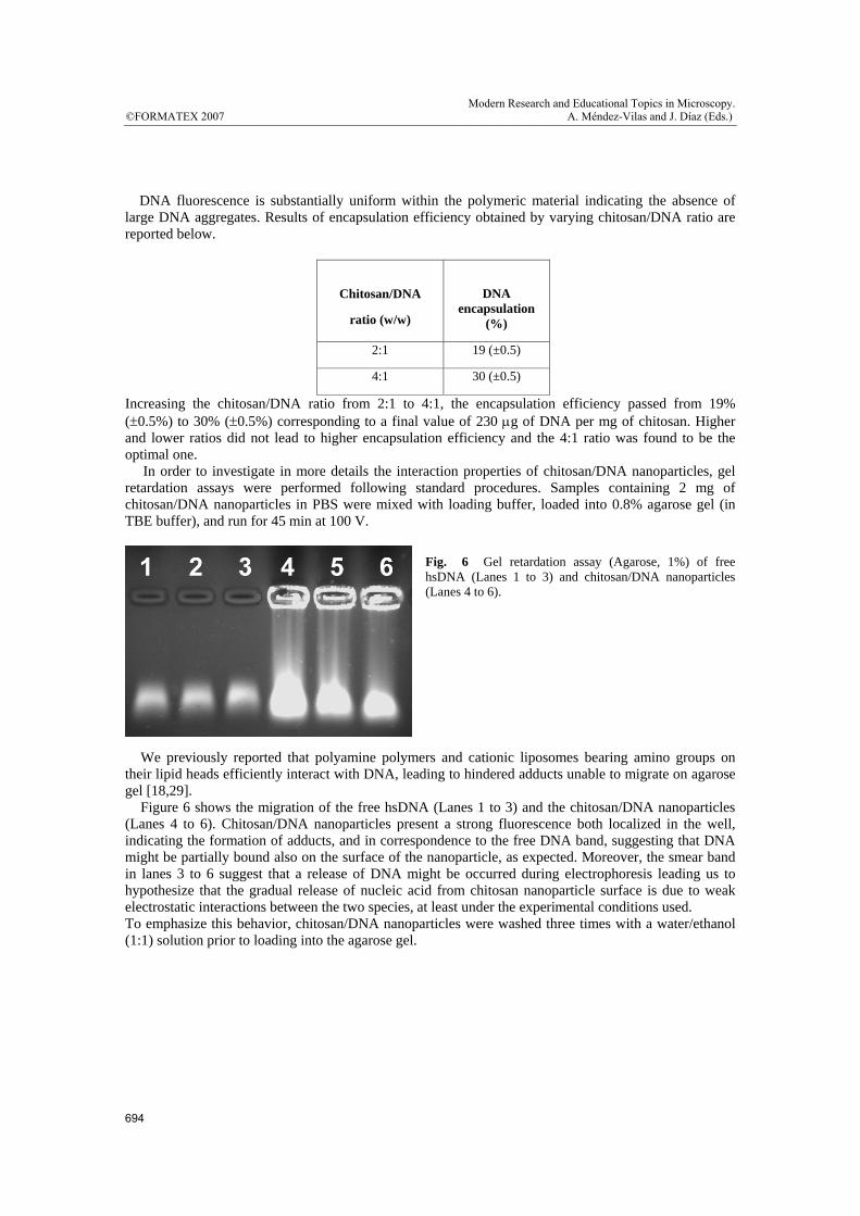
Increasing the chitosan/DNA ratio from 2:1 to 4:1, the encapsulation efficiency passed from 19% (±0.5%) to 30% (±0.5%) corresponding to a final value of 230 μg of DNA per mg of chitosan. Higher and lower ratios did not lead to higher encapsulation efficiency and the 4:1 ratio was found to be the optimal one. In order to investigate in more details the interaction properties of chitosan/DNA nanoparticles, gel retardation assays were performed following standard procedures. Samples containing 2 mg of chitosan/DNA nanoparticles in PBS were mixed with loading buffer, loaded into 0.8% agarose gel (in TBE buffer), and run for 45 min at 100 V.
Fig. 6 Gel retardation assay (Agarose, 1%) of free hsDNA (Lanes 1 to 3) and chitosan/DNA nanoparticles (Lanes 4 to 6).
We previously reported that polyamine polymers and cationic liposomes bearing amino groups on their lipid heads efficiently interact with DNA, leading to hindered adducts unable to migrate on agarose gel [18,29]. Figure 6 shows the migration of the free hsDNA (Lanes 1 to 3) and the chitosan/DNA nanoparticles (Lanes 4 to 6). Chitosan/DNA nanoparticles present a strong fluorescence both localized in the well, indicating the formation of adducts, and in correspondence to the free DNA band, suggesting that DNA might be partially bound also on the surface of the nanoparticle, as expected. Moreover, the smear band in lanes 3 to 6 suggest that a release of DNA might be occurred during electrophoresis leading us to hypothesize that the gradual release of nucleic acid from chitosan nanoparticle surface is due to weak electrostatic interactions between the two species, at least under the experimental conditions used. To emphasize this behavior, chitosan/DNA nanoparticles were washed three times with a water/ethanol (1:1) solution prior to loading into the agarose gel.
694
©FORMATEX 2007Modern Research and Educational Topics in Microscopy.
A. Méndez-Vilas and J. Díaz (Eds.) __________________________________________________________________________________________________________

Figure 7 shows the agarose gel electrophoresis of chitosan/DNA nanoparticles before (lane 2) and after (lane 3) the washing step. A significant reduction of free DNA band was observed, indicating that an effective removal of DNA bound to the external surface of chitosan nanoparticles had take place. On the contrary, the fluorescence signal relative to encapsulated DNA-chitosan nanoparticles remained unaltered. Fig. 7 Gel retardation assay (Agarose, 1%) of chitosan/DNA nanoparticles before (lane 2) and after (lane 3) a water/ethanol (1:1) washing step. A molecular weight ladder (1 Kb) was run on lane 1.
To quantify the DNA release from the obtained nanostructured systems, 4 mg of chitosan/DNA nanoparticles were incubated in PBS (pH = 7.4, 37 °C) under gentle magnetic stirring for 72 hours. Samples were centrifuged for 10 min at 16,000g then the supernatant was removed and replaced with fresh buffer. At fixed time points, supernatant was collected, analyzed by spectrophotometry and the kinetic of DNA release from chitosan-DNA nanoparticles was obtained. All measurements were collected in triplicate.
Figure 8 reports the DNA release values at different time points and the sigmoid curve obtained after data fitting (R2=0.9586). Fig. 8 DNA release (%) from chitosan/DNA nanoparticles at 4:1 ratio (w/w) after incubation in PBS. Values and standard deviations are reported together with the sigmoid fitting curve.
The release of DNA from chitosan/DNA nanoparticles showed an initial burst release within the first 3 hours of incubation. Then, DNA was constantly released up to 72 hours with more than 60% of the encapsulated DNA released within 24 hours. These results suggest that chitosan/DNA nanoparticles, prepared following the proposed methodology, are able to encapsulate DNA with improved efficiency in comparison with polyethylene vinyl co-acetate (EVAc) polymeric nanoparticles employed by Y.S.Jong et al. [30]. In our hands the maximum release of DNA was 86% of the initial feeding amount. The achieved kinetic profile account for the existence of two different DNA release mechanisms from nanoparticles. The first one, occurring within 3 hours is likely due to DNA release from the nanoparticle surface, while, at a later stage DNA was constantly released from the core of nanoparticles as a consequence of chitosan hydration and swelling. The kinetic follows a pseudo first order law according to the diffusion through the porous polymeric matrix. Kinetic data are in agreement with gel retardation assays that showed both the migration of DNA present on the surface and the presence of DNA incorporated into the nanoparticles.
Hours
0 20 40 60 80
DN
A R
elea
se (%
)
40
50
60
70
80
90
695
Modern Research and Educational Topics in Microscopy.A. Méndez-Vilas and J. Díaz (Eds.) ©FORMATEX 2007__________________________________________________________________________________________________________

In conclusion, we devised and reported a novel and simple osmosis-based method to obtain chitosan and chitosan/DNA nanoparticles of definite size and shape with peculiar release properties. This process may be easily modified by varying the solvent/non–solvent couple, temperature, membrane cut-off and polymer type, affording useful nanostructured systems to employ in several biomedical and biotechnological applications.
Acknowledgement Authors thank MIUR (Ministero dell’Università e della Ricerca) (PRIN 2005 - prot. 2005039758) and SAPIENZA - Università di Roma (Progetto di Ateneo 2006) for financial support.
References
[1] Dai, H. ; Jiang, X. ; Tan, GC. ; Chen, Y. ; Torbenson, M. ; Leong, KW. ; Mao, HQ. Int J Nanomedicine, 1(4), 507-522, (2006).
[2] Kang, ML. ; Jiang, HL. ; Kang, SG. ; Guo, DD. ; Lee, DY. ; Cho, CS. ; Yoo, HS. Vaccine, 2007, [Epub ahead of print].
[3] Kay, M.A. ; Liu, D.; Hoogerbrugge, P.M. Proc. Natl. Ac. Sci. USA, 1997, 94, 12744-12746. [4] Andreson, W.F. Nature, 1998, 392, 25-30. [5] Verma, I.M.; Somia, N. Nature, 1997, 389, 239-242. [6] Wilson, J. M.; New Engl. J. Med., 1996, 334, 1185-1187. [7] Jooss, K.; Yang, Y.; Fisher, K.J.; Wilson J.M. J. Virol., 1998, 72, 4212-4223. [8] Fisher, K.J.; Jooss, K.; Alston, J.; Yang, Y.; Haecker, SE.; High, K.; Pathak, R.; Raper, SE., Wilson, JM.
Nature Med., 1997, 3, 306-312. [9] Gao, X.; Huang, L. Gene Therapy 1995; 2: 710–722. [10] Meyer, O.; Kirpotin, D.; Hong, K.; Sternberg, B.; Park, JW.; Woodle, MC.; Papahadjopoulos, D. J Biol Chem
1998; 273: 15621–15627. [11] Behr, JP. Bioconj Chem 1994; 5: 382–389. [12] Vigneron, JP.; Oudrhiri, N.; Fauquet, M.;, Vergely, L.; Bradley, JC.; Basseville, M.; Lehn, P.; Lehn, JM. Proc
Natl Acad Sci USA 1996; 93: 9682–9686. [13] Curiel, D.T.; Ann. N. Y. Acad. Sci., 1994, 716, 36-56. [14] Wagner, E.; Cotten, M.; Foisner, R.; Birnstiel, M. L.; Proc. Natl. Acad. Sci. U.S.A.,1991, 88, 4255-4259. [15] Stankovics; J.; Crane, A.M.; Andrews, E.; Wu, C.H.; Wu, G.Y.; Ledley, F. D. Human Gene Therapy, 1994, 5,
1095-104. [16] Takai, T.; Ohmori, H. Biochim. Biophys. Acta, 1990, 1048, 105-109. [17] Hosseinkhani, H.; Azzam, T.; Tabata, Y.; Domb, A.J. Gene Therapy, 2004, 11, 194-203. [18] Masotti, A.; Moretti, F.; Mancini, F.; Russo, G.; Di Lauro, N.; Checchia, P.; Marianecci, C.; Carafa, M.;
Santucci, E.; Ortaggi, G. Bioorg. Med. Chem., 2007, 15(3), 1504-1515. [19] Boussif, O.; Lezoualc’h, F.; Zanta, M. A.; Mergny, M. D.; Scherman, D.; Demeneix, B.; Behr, J.-P. Proc. Natl.
Acad. Sci. U.S.A.,1995, 92, 7297-7301. [20] Tang, M.X.; Redemann, C.T.; Szoka, F.C. Bioconj Chem., 1996, 7, 703-714. [21] Behr, J.P.; Demeneix, B.; Loeffler, J.P.; Perez-Mutul, J. Proc. Natl. Acad. Sci. U.S.A., 1989, 86, 6982-6986. [22] Remy, J.S.; Sirlin, C.; Vierling, P.; Behr, J.P. Bioconj. Chem., 1994, 5, 647-654. [23] Klotz, I.M.; Royer, G.P.; Sloniewsky, A.R. Biochemistry, 1969, 8, 4752-4756. [24] MacLaughlin, F.C.; Mumper, RJ.; Wang, J.; Tagliaferri, JM.; Gill, I.; Hinchcliffe, M.; Rolland, AP. J. Control
Release, 1998, 56, 259-272. [25] Soane, RJ.; Frier, M.; Perkins, AC.; Jones, NS.; Davis, SS.; Illum, L. Int J Pharm., 1999, 178(1), 55-65. [26] Palocci, C.; Russo, M.V.; Belsito, C.M.A.; Cernia, E.; D'Amato, R.; Fratoddi, I.; Panzavolta, F.; Soro, S.;
Venditti, I. PCT Int. Appl. 2006, WO2006051572, 1-23 [27] Dhadwal, H.S.; Ansari, R.R.; Meyer, W.V. Rev.Sci. Instrum. 1991, 62, 2963. [28] Hed, J.; Hallden, G.; Johanson, S. G. O. and Larsson, P. J. Immunol. Methods, 1987, 101, 119–125. [29] Esposito, C.; Masotti, A.; Del Grosso, N.; Malizia, D.; Bianco, A.; Bonadies, F.; Napolitano, R.; Ortaggi, G.;
Mossa, G. Comptes Rendus Chimie., 2003, 5–6, 617–622. [30] Jong, Y.S.; Jacob, J.S.; Yip, K-P; Gardner, G.; Seitelman, E.; Whitney, M.; Montgomery, S.; Mathiowitz, E.; J
Control Release, 1997, 47, 123-134.
696
©FORMATEX 2007Modern Research and Educational Topics in Microscopy.
A. Méndez-Vilas and J. Díaz (Eds.) __________________________________________________________________________________________________________