Embed Size (px)
Citation preview
(Page: 18, Words: 3,787, References: 26, Figures: 6)
Efficacy of Cardiopulmonary Resuscitation in the Microgravity Environment
Submitted by
XSmi th L. Johnston, M.D.; Mark R. Campbell, M.D.; XRoger D: Billica, M.D.; ''Stevan M. Gilmore, M.D.
XNASA Lyndon B. Johnson Space Center Medical Operations Branch Mail Code 5D26 2101 NASA Road Houston, Texas 77058-3696
Wyle Laboratories, Inc. Life Sciences Division (Formerly KRUG Life Sciences) 1290 Hercules Drive Houston, TX 77058-2787
'Wright State University Department of Emergency Medicine 3525 Southern Blvd. Kettering, OH 45429-1221
Address Reprints
NASA Lyndon B. Johnson Space renter Medical Operations Brandi Mail Code SD26 2101 NASA Road Houston, Texas 77058-3696 (281)483-0453
https://ntrs.nasa.gov/search.jsp?R=20150020975 2020-06-12T20:31:58+00:00Z

Background: End tidal carbon dioxide (EtCO 2) has been previously shown to be an effective non-invasive
tool for estimating cardiac output during cardiopulmonary resuscitation (CPR). Animal models have
shown that this diagnostic adjunct can be used as a predictor of survival when EtCO 2 values are maintained
above 25% of prearrest values.
Hypothesis: CPR can be administered in the microgravity environment.
Methods: Eleven anesthetized Yorkshire swine were flown under the simulated microgravity conditions
on a KC-135. Physiologic parameters including EtCO2 were monitored. Standard Advance Cardiac Life
Support protocols were used to resuscitate these models after induction of cardiac arrest. Chest
compressions were administered using conventional body positioning with restraint and unconventional
vertical-inverted body positioning.
Results: EtCO, values were maintained above 25% of prearrest values in the microgravity environment
\, (33%± 3 ground and 41%± 3 flight controls). No significant difference in CPR, as monitored by EtCO2,
administered in 1G or OG on these models was noted (Ground Control : 35±3% 1-g vs 33± 3% O-g; Flight
Control: 44± 3% 1-g vs 41± 3% O-g) Effective CPR was delivered in both body positions although
conventional body positioning was found to be fatiguing
Conclusions: Cardiopulmonary pulmonary resuscitation can be effectively administered in microgravity.
Validation of this model hds demonstrated that EtCO 2 levels were maintained above a level previously
reported to be predictive of survival. The unconventional vertical-inverted position provided effective CPR
and was less fatiguing as compared to the conventional body positioning.
Key Words: Cardiopulmonary. Resuscitation, End-tidal CO2. Space Medicine, Advanced Cardiac Life
Support, Weightlessness -

Introduction
The inherent risks of space flight are significant. Recently, Johnston et a! characterized the
medical risks associated with long duration space flight (1). In this analysis, data was abstracted from the
Russian Space Program, the Longitudinal Study of Astronaut Health (LSAH), and analog populations such
as winter-over groups in Antarctica and military submarine crews. These data showed that for a crew of
seven astronauts the incidence of a significant medical event was once every 2.4 years and the incidence of
an incapacitating medicil emergency was once every 14 years.
Although routinely used, cardiopulmonary resuscitation (CPR) and Advanced Cardiac Life
Support (ACLS) (2) are relatively new techniques when viewed from the perspective of the history of
medicine (3). Even under optimum circumstances, cardiac and cerebral perfusion pressures are but a
fraction of pre-arrest values (4, 5). For this reason, survivability decreases precipitously when the initiation
of CPR is delayed. The primary motive force behind the generation of perfusion pressures is related to the.
increase in intra-thoracic pressures during CPR as opposed to direct cardiac compression (6).
Animal models and human studies have shown that end-tidal CO 2 (EtCO 2) monitoring is an
accurate adjunct with which to monitor the efficacy of CPR (7-14). These studies showed that during the
low blood-flow states associated with CPR, EtCO 2 monitoring is a reliable detector of pulmonary blood
flow and therefore cardiac output (CO). Although preliminary studies suggested this relationship tobe
linear (14), more recent investigations have shown a near-linear relationship (11). This relationship to CO
is important because coronary perfusion, which is a predictor of the return of spontaneous circulation
(ROSC), is directly related\to CO (15, 16). It is through this link to CO that EtCO 2 has been shown to
correlate with cerebral perfusion pressure as well (10).
Recently, human trials have attempted to define an EtCO 2 level that is predictive of survivability
after the initiation of CPR (8, 12, 13, 17, 18). In their porcine model, Gudipati eta! showed that the ability
to maintain EtCO 2 values at greater than 25% of prearrest values was an accurate predictor of survivability
(9). The objective therefore of this study was to demonstrate that CPR could be effectively administered
in a microgravity envjronment. EtCO 2 monitoring in addition to monitoring of blood pressure and
oximeter levels were used to monitor the effectiveness of CPR in this microgravity model.
3

4
CPR is difficult to deliver in the weightless environment, as chest compressions have to be counter
acted by the CPR provider. This usually done by gravity in the normal 1-g environment, but in the O-g
weightless environment must be performed by either restraint, bracing, or active muscular counteraction by
the CPR provider. Depending on how much active muscular reaction is required, this can be extremely
fatiguing. -
In addition, in the O-g environment, CPR can only be performed if there is a method of restraint of
the patient, CPR provider, and all equipment. A floor level restraint system was developed for medical
restraint in weightlessness and this project was a demonstration of its effectiveness. This Crew Medical
Restraint System (CMRS) provided restraint of the animal model, all ACLS equipment and hardware, and
methods for CPR provider restraint.
Materials and Methods
Experimentation was conducted as part of a program to assess ACLS and Advanced Trauma Life
Support (ATLS) protocols in the microgravity environment. As part of this program, a total of eleven pigs
were flown in a modified KC-135, which is able to simulate microgravity. By using parabolic flight, a KC-
135 is able to provide forty-second intervals of zero gravity (0-g) followed by a 2-g pullout. A total of
eleven flights, one for each porcine model, were conducted with 40 parabolas on each flight. These models
were run in groups of three and experimentation was conducted in 1992, 1993, 1996, and 1998.
Yorkshire swine were chosen as the porcine test model. The models had not undergone previous
testing. The initial three models were 20-23 kg while the remaining eight weighed 50 kg. Strict adherence
to the Standard Anesthetic Regimen, Surgical Training Lab Protocols, and Guide to Care and Use of
Laboratory Animals, NIH Publication #86-23 was maintained. In accordance with KC-135 flight rules that
prohibit the use of inhalational anesthetics, intravenous pentobarbital was used as the general anesthetic.
This study received review, approval, and monitoring by the St. Joseph Hospital Surgical Training Lab
Institutional Animal Care and Use Committee (Houston, Texas), by the NASA-JSC Institutional Animal
Care and Use Committee (Houston, Texas), and the NASA-JSC Institutional Review Board (Houston,
Texas).

Animals were initially prepared at the Surgical Training Lab at St. Joseph's Hospital. After
receiving an intramuscular dose of Ketamine (5cc/kg), the swine were orotracheally intubated and initiated
on halothane gas general anesthesia. The animal was ventilated with 12-15 breaths per minute with a tidal
volume of 10 mI/kg. Each specimen was prepped and draped in a sterile fashion and the central vasculature
of the neck mobilized surgically. An arterial line in the carotid artery was placed for blood pressure
monitoring followed by the placement of a central line in the jugular vein for the administration of fluids
and medications. Peripheral venous access was also obtained via percutaneous introduction of an
angiocatheter into an'eat vein. The method of general anesthesia was then changed to intravenous with
pentobarbital (0.25cc/kg) and paralysis was maintained with Pancuronium (0.09 mg/kg/hour).
Maintenance of adequate anesthesia was titrated based on continuous monitoring of vital signs. The
porcine models were then transported Ellington Field (Houston, Texas) with a total time for prepping to
arrival of 90 minutes. At the completion of all testing, the animals were euthanized using IV Pentothal and
disposed of by the Surgical Training Lab at St. Joseph's Hospital.
Each animal was monitored continuously throughout experimentation. A physiologic monitoring
module (Spacelabs Medical mc, Redmond, WA) was used to monitor intra-arterial blood pressure, cardiac
rhythm strip, rectal temperature, oximetry (via a tongue-probe), and end-tidal CO2 (Novarnetrix Medical
Systems, Inc., Wallingford, CT). A pneumatically powered ventilator (LAMA), which had been previously
•rated for microgravity flight was used throughout the study. Due to safety restrictions, compressed air
instead of 100% oxygen was used when aboard the KC-135. This provided a Sa02 of 94 ± 1% on the
ground and 88 ± 2 % at a flight cabin pressure of 6000-8000 feet. Ventricular fibrillation was induced with
an intravenous bolus of potssium chloride (KCI). After verifying induction of ventricular fibrillation
through cardiac monitoring and drop in arterial blood pressure, chest compressions were performed by
several CPR providers. A commercially available defibrillator/pacer unit (Physio-Control Lifepak- 10,
Redmond, WA), weight-adjusted ACLS medications, and standard ACLS algorithms were then used to
regain spontaneous rhythm.
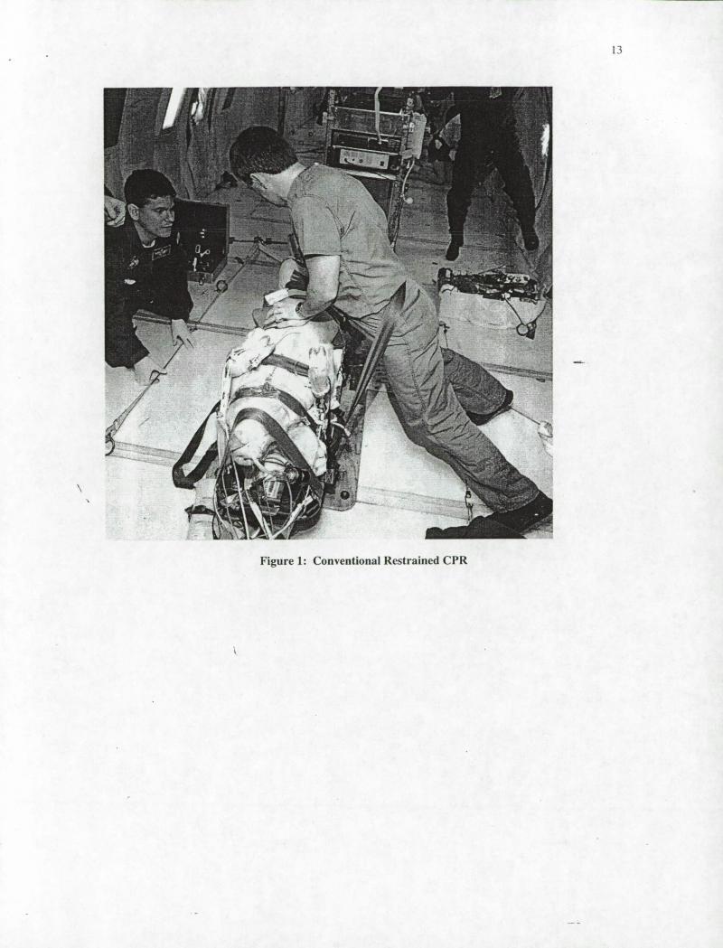
- Two methods were used by CPR providers to perform manual chest compressions. The first
method was a conventional body position with the CPRprovider to the side of the animal model and
restrained by waist belts and/or restraint cords across the lower legs (Figure 1). This method required a

6
large amount of effort by the CPR provider to counter act the force of the chest compressions and was
known to be quickly fatiguing from earlier parabolic flights utilizing manikins. The second method was
with the CPR provider in the unconventional vertical-inverted position with his feet on the ceiling which
allowed for bracing to counter act the force of the chest compressions (Figure 2). Obviously, this position
can only be used during weightlessness. This position was also known from previous parabolic manikin
studies to allow for the performance of CPR with only minimal effort.
Physiologic parameters were measured under the following conditions: 1).ground controls were
obtained preflight at.I-g during normal sinus rhythm (NSR) 2) flight controls obtained in flight at 1-g and
with NSR 3) at altitude during 1-g CPR 4) at altitude during O-g CPR. Five data points were obtained
under each of these conditions and averaged. The EtCO 2 data is expressed as % of EtCO 2 . Data is
presented graphically with 95% confidence intervals included. T-tests are used where P-values are
computed.
Results
Figure 3 contains a sample graph of the physiologic parameters monitored during each KC-135
flight. Note that the onset of ventricular fibrillation, which was induced by intravenous potassium chloride,
was immediately reflected in the vital signs. During each flight, the porcine model was oriented in parallel
with the long axis of the airplane.
Mean oximeter readings are reported in Figure 4. As was documented previously, due to flight
rules supplemental oxygen was not available during the KC-135 flight. This was reflected in the oximeter
readings that are taken at an effective altitude of 6-8000 feet above sea level. This resulted in a significant
change in the Sa0 2 readings with a mean ground control reading of 94 ± 1% and a mean flight control
reading of 89 ± 2% (P= 0.000 1). No statistically significant differences were noted between the oximeter
readings taken during CPR in the micro- or normogravity. The oximeter readings for both CPR groups
were significantly lower than both controls.
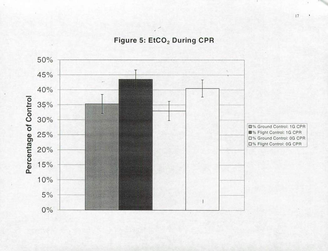
Figure 5 presents the EtCO2 results in terms of percentage of control. Both the percentage of
groufld and flight controls are presented for analysis. These data show that CPR in the microgravity
environment was able to sustain EtCO 2 values well above 25% of prearrest values when compared to both
the ground and flight controls. No significant differences in mean EtCO 2 readings were noted between
7
CPR performed under 1-g or 0-g when comparing the ground (35 ± 3% 1-g vs 33 ± 3% O-g) and flight (44
± 3% 1-g vs 41± 3% 0-g) control results separately. There was a trend towards statistical significance
noted between ECO 2 readings of the ground versus flight CPR results. Although statistically significant,
the difference between these two values would be of questionable clinical significance as Gudipati was not
able to demonstrate anj relationship between survivability and EtCO 2 readings beyond an increased
likelihood of survival above the 25% of prearrest value (9).
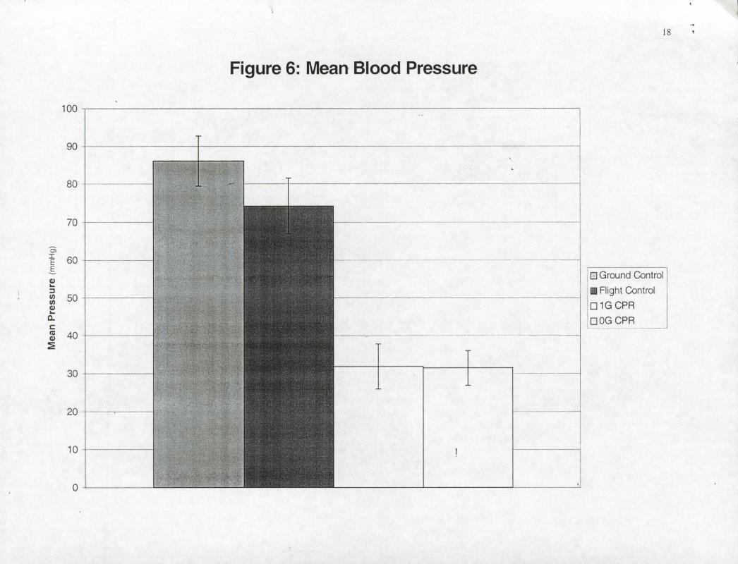
The mean intra-arterial blood pressure results are presented in Figure 6. As expected, the onset of
ventricular fibritlatidn roduced a significant change in pressure. No other significant trends in the mean
blood pressure were noted during CPR conducted in either the micro or normogravity settings.
There was no difference in any of the monitored parameters (Sa02, EtCO2, and intra-arterial BP)
between the two methods of chest compression.
Discussion
This study has several limitations. First, while the KC-135 provides the most representative
\ microgravity experience without actually being in orbit, the duration of 0-g exposure during each parabola
is only 30 seconds. As noted previously, one KC-135 flight typically consists of forty such parabolas.
Interposed between each 0-g parabola of periods of up to 2-g. To maximize the effect of microgravity on
this model, all readings taken under microgravity conditions were recorded 25 seconds into each parabola
to maximize the effect of 0-g. Classically, physiology texts teach that carbon dioxide diffusion across the.
lung parenchyma is essentially instantaneous (19). It is not clear how the low flow states associated with
CPR impact this relationslip. Some studies have suggested that the diffusing capacity of CO 2 is delayed.
It is not clear to what extent this impacts our investigation.
As noted previously, oxygen supplementation was not provided during in-flight experimentation
due to airplane safety restrictions. Administration of high-flow oxygen is one of the basics of resuscitation
and is standard of care in terrestrial medical operations. Additionally, our investigations were performed
undçr the hypobaric environment (10.7-11.7 psi) provided in the KC-135. Despite the absence of
optimum oxygenation, the ability to maintain EtCO 2 levels above a prescribed level was-demonstrated.

The International Space Station is designed with 14.7 psi or 1 atmosphere of pressure and supplemental
oxygen will be available.
A final limitation is that these porcine models were also used to test ATLS techniques. All of
these procedures, including chest tubes and peritoneal lavage, were completed prior to ACLS Mega-code
testing. This sequence is routinely performed in medical residency animal training procedures. It is not
clear what if any affect these other investigations had on experimental results. However, with
consideration to the presented results, it would suggest that our results weren't adversely biased.
Furthermore, although these models were not deconditioned as would an analogous astronaut be, it does
suggest that this can be successfully performed on an individual with significant trauma.
This study demonstrated several important findings. First, as demonstrated by decrements in
oximeter results during CPR, supplemental oxygen would be an important adjunct for any scenario
requiring these procedures. Furthermore, should subsequent transport be required. several studies have
documented further decrements in oximeter readings and arterial Pa02 values in healthy individual
exposed to the hyper-gravity conditions associated with orbital reentry(20, 21). These parameters would be
further compromised in a medically incapacitated patient and oxygen supplementation would assist in
optimizing outcome.
Second and most importantly, with respect to the predictive capabilities of EtCO 2 and
survivability, Gudipati reported that EtCO 2 values less than 25% of prearrest were predictive of
mortality(9). Tervino, in a similar porcine study, showed comparable results (22). This study demonstrates
no significant differences between EtCO 2 values obtained during CPR under 1-g and O-g conditions. More
importantly, in both of thes,e experimental CPR conditions, the EtCO 2 percent of control was maintained
well above the 25% described previously. This was demonstrated both in our ground and flight control
conditions. Due to the relationships between EtCO 2, CO, and cardiac perfusion pressures as well as link
between the percentage EtCO 2 and survivability, we believe that this supports the hypothesis that effective
CPR can be delivered in the microgravity environment.
Both methods of chest compressions, the conventional body position with restraints and
unconventional vertical-inverted, were found to provide adequate and equivalent CPR. However, the
conventional method was quickly fatiguing and the unconventional vertical-inverted method required

minimal effort by the CPR providers. As the dimensions of the KC-135 cabin are similar to the interior
dimensions of the ISS modules, the vertical inverted method is a suitable technique for the administration
of chest compressions during CPR in space flight. Current designs, however, will utilize the conventional
restrained configuration in conjunction with the crew medical restraint system so as to place the health-care
provider in close proximity to the patient for concerns regarding maintenance of airway, drug
administration and other therapeutic considerations.
Although beyond the scope of this discussion, this investigation has resulted in the adaptation of
ACLS Mega-code prcdures and validation of space-rated hardwarefor inclusion in the International
Space Station's Health Maintenance System (HMS). During the course of this investigation a defibrillator,
intravenous fluids and medications, and a ventilator were reliably used and manifested for space flight. A
floor level Crew Medical Restraint System (CMRS) was demonstrated to be able to provide adequate
restraint for the performance of ACLS by providing for patient, hardware, and CPR provider restraint. The
inclusion of these devices in the HMS will extend the medical capabilities to provide care for the critically
ill andlor injured.
Finally, validation of this microgravity CPR model has allowed for its' use as a training model.
Currently, the crew medical officer (CMO) is trained to have at least emergency medical technician
capabilities and may in some cases be a physician depending on the individual. Two individuals have
received training on this model prior to their shuttle flights. Additionally, investigations into the utility of
EtCO 2 during CPR have also shown that it can be effectively used to monitor fatigue and effectiveness of
compression being administered (23-25). Preflight training on this model would allow a CMO to become
familiar with the actual mehanics of performing cardiac compressions in microgravity as well as providing
exposure to and familiarization with on-orbit hardware. This in combination with proposed on-orbit
computer training modules (26), could provide a means for future long-duration crews,to obtain and
maintain medical proficiencies.
In conclusion, this investigation into CPR in microgravity has demonstrated 1) the ability of
maintain EtCO 2 values above previously reported values predictive of survivability in both the IG and 00
models 2) no change in mean intra-arterial pressures generated by external cardiac compression during

CPR in microgravity 3) decrements in oxygen saturation related to our experimental conditions and 4)
demonstrated effective administration of CPR in both the conventional and vertical-inverted positions.
Acknowledgements:
The authors would like to recognize the following institutions for their significant contributions
without which completion of this project would not have been possible: 1) St. Joseph Hospital, Surgical
Training Lab 2) NASA Reduced Gravity Flight, KC-135 Flight Office.
Additionally, we would like to recognize the following individual for their significant
contributions to the completion of this project: from St. Joseph's Hospital, Amado Ruiz, M.D., Leah
Davis, Amy Mills, Leslie Yarborough, D.V.M., F. Eichstadt; from the Lyndon B. Johnson Space Center
William Norfleet, M.D., Brad Beck, M.D., Ron Robinson, Michael Barrat, M.D., and Richard Jennings,
M.D.; from Wyle Life Science (formerly KRUG) Dave Ward, M.D.; and Ezra Krucharz, Brenda Rouse,
Scott Simmons, Charles Lloyd, Ph.D., and Doug Ball, M.D.
References:
1. Johnston SL, Marshburn TH, Lindgren K. Predicted Incidence of Evacuation-Level Illness/Injury
During Space Station Operation. In: Proceedings of the7 1st Annual Scientific Meeting of the Aerospace
Medicine Association; 2000 May 14-18, 2000; Houston, Tx: Aerospace Medicine Association; 2000. p.
105.
2. Advanced cardiac life support. Dallas, Tex: American Heart Association; 1997.
3. -. Safar P. On the hitory of modern resuscitation. Crit Care Med 1996;24(2 Suppl):S3-1 1.
4. Koehler RC, Micheal JR. Cardiopulmonary Resuscitation, Brain Blood Flow, and Neurologic
Recovery. Crit Care Clin 1985;1(1):205-222.
5. Lindner KH, Pfenninger EG, Lurie KG, Schurmann W, Lindner IM, Ahnefeld FW. Effects of
active compression-decompression resuscitation on myocardial and cerebral blood flow in pigs. Circulation
1993;88(3): 1254-63.

11
6. Deshmukh HG, Weil MH, Gudipati CV, Trevino RP, Bisera J, Rackow EC. Mechanism of blood
flow generated by precordial compression during CPR. 1. Studies on closed chest precordial compression.
Chest 1989;95(5): 1092-9.
7. Callaham M, Barton C. Prediction of outcome of cardiopulmonary resuscitation from end-tidal
carbon dioxide concentration [see comments]. Crit Care Med 1990,18(4):358-62.
8. Cantineau JP, Lambert Y, Merckx P, Reynaud P, Porte F, Bertrand C. et al. End-tidal carbon
dioxide during cardiopulmonary resuscitation in humans presenting mostly with asystole: a predictor of
outcome. Crit Care Med 1996;24(5):791-6.
9. Gudipati CV, Well MH, Bisera J, Deshmukh HG. Rackow EC. Expired carbon dioxide: a
noninvasive monitor of cardiopulmonary resuscitation. Circulation 1 98877( 1) :234-9.
10. Lewis LM, Stothert J, Standeven J, Chandel B, Kurtz M, Fortney J. Correlation of end-tidal CO2
to cerebral perfusion during CPR. Ann Emerg Med 1992;21(9):1 131-4.
11. Ornato JP, Garnett AR, Glauser FL. Relationship between cardiac output and the end-tidal carbon
dioxide tension. Ann Emerg Med 1990;19(10):1 104-6.
12. Paradis NA. Objective measurements for guiding initiation, sequencing, and discontinuation of
life-support intervention. New Horiz 1997;5(2):158-63.
13. Ward KR, Yealy DM. End-tidal carbon dioxide monitoring in emergency medicine, Part 2:
Clinical applications [see comments]. Acad Emerg Med 1998;5(6):637-46.
14. Weil MH, Bisera J, Trevino RP, Rackow EC. Cardiac output and end-tidal carbon dioxide. Crit
Care Med 1985;13(11):907-9.
15. Sanders AB, Atla M, Ewy GA, Kern KB, Bragg S. Expired pCo2 as an index of coronary
perfusion pressure. Am J Emerg Med 1985(3): 147-9.
16. Lindberg L, Liao Q, Steen S. The effects of epinephrine/norepinephrine on end-tidal carbon
dioxide concentration, coronary perfusion pressure and pulmonary arterial blood flow during
cardiopulmonary resuscitation. Resuscitation 2000;43(2): 129-40.
17. Koetter KP, Maleck WH. End-tidal carbon dioxide monitoring in cardiac arrest [letter; comment].
Acad Emerg Med l999;6(1):88.
12
18. Levine RL, Wayne MA, Miller CC. End-tidal carbon dioxide and outcome of out-of-hospital
cardiac arrest [see comments]. N Engi J Med 1997;337(5):301-6.
19. Guyton AC. Textbook of Medical Physiology:8th ed. Philadelphia: W.B. Saunders Co.; 1991.
20. Vil-Viliams IF, Kotovskaya AR. Changes of pulmonary function in humans during exposure to
+Gx acceleration after simulated and real microgravity. J Gravit Physiol 1994;1(1):129-32.
21. Little VZ, Leverett SD, Hartman BO. Psychomotor and physiologic changes during accelerations
of 5, 7, and 9+Gx. Aerosp Med 1968;39(1 1): 1190-7.
22. Trevino RI, Bisera J, Weil MH, Ráckow EC, Grundler WG. End-tidal CO2 as a guide to
successful cardiopulmonary resuscitation: a preliminary report. Crit Care Med 1985(13):910-11.
23. Kalenda Z. The capnogram as a guide to the efficacy of cardiac massage. Resuscitation
1978(6):259-63.
24. Ward KR, Menegazzi JJ, Zelenak RR, al e. A comparison of chest compressions between
mechanical and manual CPR by monitoring end-tidal PCO2 during human cardiac arrest. Ann Emerg Med
1993(22):669-74.
25. Kern KB, Sanders AB, Vorhees WD, at e. Changes in expired end-tidal carbon dioxide during
cardiopulmonary resuscitation in dogs: a prognostic guide for resuscitation efforts. J Am Colt Cardiol
1989(13):! 184-9.
26. Gonzalez MA, Chen JO, Oswald R. An integrated logistics support system for training crew
medical officers in advanced cardiac life support management. Comput Methods Programs Biomed
1999;59(2): 115-29.
13
,--
Figure 1: Conventional Restrained CPR

14
--...
Figure 2: Vertical-Inverted CPR

o U
U
- -
U U
C
LU
2:
U
cJ U
.— C
—I
U
OO
Sf
oc
00
- 0z
Sr9
00.
-
oo.o
cL
E F-
ooz
oz
oo.oz
oczt
ooct
o o 0 0 0 0 0 0 0
0
c1 —
,-
—
—
—
uiw
/srq
jU
'S
'% 'H
mlU
UI S
JU
IeIJ
)OjO
SAqJ

0
0
0
0
0
0
0
0
-' (0
it)cv)
CJ
Ua0Ja
0
0
0
0
0)
O DO
D.c —
cr a 00 u
oU
-O
0
,-D
UO
D
LH0
0 C)
C
C
0
C,)
C
C) C
) >
x 0 C)
C)
I
0 0 C, c.,J 0 0 w L() C) I-C) U
-
OQ
-oQ
-0000
.-000
e 2
00
cC
L. DO
DO
-
C •
r 2 c, 0
0)
0U
0LL
0U
00
o io
0
0 IC
) 0
IC) C
) IC
) 0
10
r r
cc
ci
cj
-
IOJ4U
O3 O
36B
jUa3Ja
C)
(1) Cl) C)
0 V 0 0 C C)
C)
0)
cO
O
2 •) o
o
Lp.0
0
0
0
0
0
0
0
0
0
0
0
0
0
0)
CO
N-
(0
It)C
)(\J
(6H
ww
) ain
ssai u
aj