Embed Size (px)
Citation preview

8/7/2019 Cardiopulmonary Resuscitation in Trauma
http://slidepdf.com/reader/full/cardiopulmonary-resuscitation-in-trauma 1/21
Chapter 27 / CPR in Trauma 455
455
From: Contemporary Cardiology: Cardiopulmonary Resuscitation
Edited by: J. P. Ornato and M. A. Peberdy © Humana Press Inc., Totowa, NJ
27 Cardiopulmonary Resuscitation
in Trauma
Rao R. Ivatury, MD and Kevin R. Ward, MD
C ONTENTS
INTRODUCTION
CARDIOPULMONARY SUPPORT IN THE EMERGENCY DEPARTMENT
EMERGENCY DEPARTMENT THORACOTOMY AND OPEN-CHEST CARDIAC
COMPRESSION
REFERENCES
INTRODUCTION
Cardiopulmonary resuscitation (CPR) in a patient with multiple injuries involves a
different approach than in a nontrauma patient. Although the basic principles are the same
as dealt with in other chapters of this book, CPR in the trauma victim has to address
prevention of cardiopulmonary failure from problems exclusive to the injured patient.
This chapter concentrates on these issues and highlights some of the recent developments
in the field.
CARDIOPULMONARY SUPPORTIN THE EMERGENCY DEPARTMENT
Prompt resuscitation of the trauma patient in the emergency department (ED) includescontrol and/or maintenance of the airway, reversal of life-threatening events (e.g., ten-sion pneumothorax,cardiac tamponade), maintenance of cellular aerobic metabolism bysupplemental oxygenation and assisted ventilation, and restoration of normovolemia.One important caveat has developed in volume replacement in recent years: in a bleedingtrauma patient, especially after penetrating trauma, aggressive attempts at stabilizationof the cardiovascular state by fluid infusion before definitive control of bleeding isachieved may lead to a higher morbidity and mortality. This is not a new concept but aresurrection of Cannon’s observations after World War I. He pointed out that “hemor-rhage in the case of shock may not have occurred to a marked degree because bloodpressure has been too low and flow too scant to overcome the obstacle offered by a clot.If the pressure is raised before the surgeon is ready to check any bleeding that may takeplace, blood that is sorely needed may be lost” (1). The effect of massive, early fluidresuscitation was recently examined critically in hypotensive prehospital trauma patients.Kaweski et al. (2) reviewed the records of 6855 hypotensive trauma patients. Fifty-six

8/7/2019 Cardiopulmonary Resuscitation in Trauma
http://slidepdf.com/reader/full/cardiopulmonary-resuscitation-in-trauma 2/21
456 Cardiopulmonary Resuscitation
percent of these patients received prehospital fluid resuscitation. Fluid challenge in thisgroup of patients did not improve survival. Bickell et al. (3) conducted a prospective trialcomparing immediate and delayed fluid resuscitation in 598 adults with penetrating torsoinjuries who presented with a prehospital systolic blood pressure of less than 90 mmHg.
Patients assigned randomly to the immediate-resuscitation group received standard fluidresuscitation before and after they reached the hospital. Those assigned to the delayed-resuscitation group received intravenous cannulation but no fluid resuscitation until theyreached the operating room (OR). When fluid resuscitation was delayed, 203 (70%)survived and were discharged from the hospital, as compared with 193 of the 309 patients(62%) surviving when immediate fluid resuscitation was provided (p = 0.04). In delayed-resuscitation patients who survived to the postoperative period, 55 (23%) had one or morecomplications (adult respiratory distress syndrome, sepsis syndrome, acute renal failure,coagulopathy, wound infection, or pneumonia), as compared with 69 of the 227 patients(30%) in the immediate-resuscitation group, a difference that approached statistical sig-
nificance. The duration of hospitalization was shorter in the delayed-resuscitation group.The authors concluded that delay of aggressive fluid resuscitation until operative controlof bleeding is accomplished improves the outcome of hypotensive patients with penetrat-ing torso injuries. These clinical data are supported by animal models of uncontrolledhemorrhagic shock, induced either by intra-abdominal large-vessel injury to the ileocolicartery, or by tail resection. In these models, infusion of hypertonic saline (HTS) or largevolumes of normal saline increases rebleeding, hemodynamic collapse, and increasedshort-term mortality (4–11). The concept appears to be applicable even in blunt traumaanimal models (12,13). Vigorous boluses of crystalloid infusion after “massive” or“moderate” splenic injury in rats also increases bleeding and shortens survival time (14–
17). Thus, excessive early crystalloid infusion only increases bleeding and shortenssurvival time in the early critical “golden hour” after injury. These data support theconcept that avoiding fluid resuscitation until definitive control of bleeding is achievedor deliberate “hypotensive resuscitation” with a limited volume of crystalloid or colloidsolutions until bleeding can be controlled surgically results in better survival (18).
EMERGENCY DEPARTMENT THORACOTOMY AND OPEN-CHEST CARDIAC COMPRESSION
It is crucial to provide the most efficient adequate cardiac and cerebral perfusion in a
hypovolemic patient with traumatic hemorrhagic shock. Multiple rib fractures and flail
chest can interfere with effective external chest compression. The past decade has seenan ever-increasing enthusiasm for ED thoracotomy (EDT) in trauma centers because it
can optimize blood flow using direct cardiac massage, relieve traumatic pericardial tam-
ponade, and allow control of intrathoracic hemorrhage. Closed-chest compression oftenresults in poor cardiac index and other hemodynamic parameters even in nontraumatic
arrest patients. Babbs (19) developed an electrical model of the human circulatory systemwith heart and blood vessels modeled as resistive-capacitive networks, pressures in the
chest, abdomen, and vascular compartments as voltages, blood flow as electric current,
blood inertia as inductance, and the cardiac and venous valves as diodes. Simulationsincluded two modes: the cardiac pump mechanism, in which the atria and ventricles of
the model were pressurized simultaneously, as occurs during open chest cardiac mas-sage; and the thoracic pump mechanism, in which all intrathoracic elements of the modelwere pressurized simultaneously, as is likely to occur in closed chest compression. The
two mechanisms were compared for the same peak applied pressure (80 mmHg). Pure

8/7/2019 Cardiopulmonary Resuscitation in Trauma
http://slidepdf.com/reader/full/cardiopulmonary-resuscitation-in-trauma 3/21
Chapter 27 / CPR in Trauma 457
cardiac pump CPR generated near normal systemic perfusion pressures throughout thecompression cycle. Pure thoracic pump CPR generated much lower systemic perfusion
pressure, only during the diastolic phase of the compression cycle. Cardiac pump CPR
produced total flows of 2500–3300, myocardial flows of 150–250 and cranial flows of
600–800 mL per minute, depending on the compression rate. In contrast, thoracic pumpCPR produced a total flow of approx 1200-myocardial flow of 70, and cranial flow of 450
mL per minute, independently of the compression rate. The author concluded that directcardiac compression is an inherently superior hemodynamic mechanism because it can
generate greater perfusion pressure throughout the compression cycle. Similar results
were reported by Sanders et al. (20). Reider et al. (21) compared the hemodynamiceffectiveness of closed-chest cardiac massage (CCCM) with closed subdiaphragmatic
massage (CSDM) and four open transdiaphragmatic cardiac massage techniques duringcardiac arrest (CA) with an open abdomen in dogs. CCCM resulted in the lowest cardiac
index (CI), mean arterial pressure (MBP), and carotid blood flow (CBF) of all cardiac
massage techniques tested. CSDM was not statistically superior to CCCM but did resultin a 23% increase in CI and a 54% increase in CBF. Transdiaphragmatic retrocardiac
massage through an incision in the diaphragm resulted in the highest hemodynamic
parameters of the four open transdiaphragmatic techniques and had significantly highervalues than those for CCCM. Open-chest manual compression was also found to be as
effective as open-chest compression-active-decompression (CAD). Therefore, open-
chest compression is vital in the trauma patient who is in extremis and is the rationale forEDT in urban centers. The objectives of EDT for the “agonal” trauma patient are as
follow: (a) maintenance of coronary and cerebral perfusion by relief of cardiac tampon-
ade and/or restoration of efficient cardiac contractility; and (b) control of hemorrhage by
cardiorrhaphy, compression of bleeding intrathoracic vessels, and/or reduction of intra-abdominal blood loss by temporary occlusion of the thoracic aorta. The ultimate objec-tive is to improve survival in these desperate patients, a goal that has been achieved with
variable success in different series (22).EDT has not improved survival in the majority of patients, even though it appears to
have value in important subgroups (e.g., patients with penetrating cardiac injuries). Thereare additional concerns with EDT: the cost of indiscriminate, futile resuscitative attemptsin patients already dead and the risk for disease transmission to the surgical team. Theseissues demand a critical analysis of patient selection for the procedure (23).
In a recent collective review of 111 stab wound (SW) and 239 gunshot wound (GSW)
patients who had EDT, Boyd and associates (24) noted a survival of 18% for SW and 2%for GSW of chest. The survival was 10% for abdominal SW and 6% for abdominal GSW.When multiple sites were injured, survival was 5–6%. Rhee et al. (25) reviewed 24
studies that included 4620 cases of EDT for both blunt and penetrating trauma over the
past 25 years. The overall survival rate was 7.4%. Normal neurological outcomes werenoted in 92.4% of surviving patients. Survival rates were 8.8% for penetrating injuries
(6.8% for SW and 4.3% for GSW) and 1.4% for blunt injuries. Survival rates were 10.7%
for thoracic injuries, 4.5% for abdominal injuries, and 0.7% for multiple injuries. Cardiacinjuries had the highest survival rate (19.4%). If signs of life were present on arrival at
the hospital, survival rate was 11.5% in contrast to 2.6% if none were present. Absence
of signs of life in the field yielded a survival rate of 1.2%. Similar data were provided bya recent series (26). EDT, therefore, plays an important in the CPR of selected patients
with trauma, particularly in penetrating wounds of the chest. The technical details andpotential pitfalls of the procedure have been described in detail elsewhere (23).

8/7/2019 Cardiopulmonary Resuscitation in Trauma
http://slidepdf.com/reader/full/cardiopulmonary-resuscitation-in-trauma 4/21
458 Cardiopulmonary Resuscitation
Hypertonic Saline for Resuscitating Trauma Patients
Hypertonic solutions are the new, potentially beneficial tools for shock/trauma resus-
citation. Compared with isotonic fluids, the lesser volumes of hypertonic solutions are
associated with equivalent or improved systemic blood pressure, cardiac output, andsurvival in experimental animals. A positive cardiac inotropic effect is documented, as
is a decrease in systemic vascular resistance. Restoration of normal cellular transmem-
brane potential is enhanced, indicating a reversal of the cellular abnormalities induced by
hemorrhagic shock. As long as 24 hours after the shock episode, blood pressure is main-
tained more effectively than with conventional crystalloid solutions. A solution of 7.5%
saline has been shown to be more effective with respect to survival than 0.9, 5, or 10%
saline solutions. Improved tissue perfusion occurs as indicated by reduced lactate values.
An early increase in urine output, decreased fluid retention, and improved late pulmonary
function are also seen (27–33). Possible mechanisms by which hypertonic saline-dextran
(HSD) maintains circulation in hemorrhagic shock include rapid shift of fluid fromintracellular to extracellular space, improved peripheral perfusion, and increased cardiac
contractility.
Despite the abundance of animal studies in support of HTS resuscitation, only a few
clinical trials are available to establish its role. Bunn et al. (34) from the Cochrane group
reviewed the available literature data on all randomized trials comparing hypertonic to
isotonic crystalloid in patients with trauma, burns, or undergoing surgery. Seventeen
trials were identified with 869 participants. The pooled relative risk for death in trauma
patients was 0.84 (95% CI 0.61–1.16), in patients with burns 1.49 (95% CI 0.56–3.95),
and in patients undergoing surgery 0.62 (95% CI 0.08–4.57). The authors concluded that
there are not enough data to argue for the superiority of hypertonic crystalloid overisotonic crystalloid for the resuscitation of patients with trauma, burns, or those under-
going surgery. The final recommendations must await further trials, large enough to
detect a clinical difference.
HTS is on a firmer ground in the resuscitation and maintenance in head-injured
patients (35). Two recent reviews summarize the current status (36,37). Although the
exact mechanisms by which HTS acts on the injured brain remain unclear, animal human
studies suggest that HTS possesses osmotic, vasoregulatory, hemodynamic, neurochemi-
cal, and immunologic properties. HTS improves and maintains mean arterial pressure
(MAP) better than the high volumes required of isotonic resuscitation and the consequent
increase in intracranial pressure (ICP). Cerebral perfusion pressure (CPP) may beimproved with HTS resuscitation, leading to better perfusion of injured areas of brain.
Unfortunately, these increases in CPP and cerebral oxygen delivery (CDO2) are transient,
with a rebound rise in ICP or fall in CPP to pre-infusion levels (38–41). HTS also appears
to counteract hypoperfusion and vasospasm via an increase in vessel diameter and through
plasma volume expansion. Additionally, HTS can attenuate the rise in ICP experienced
with hyperemia. The endothelial cell edema that is well documented after trauma may be
reversed by HTS, improving perfusion to multiple organs including the brain (37,42).
Animal models of brain injury suggest that HTS decreases leukocyte adherence and
migration and may alter production of certain prostaglandins. It has been demonstrated
to increase circulating levels of cortisol and adrenocorticotropic hormone (44,45). Neu-trophil margination and trafficking are also decreased with HTS, possibly via alterations
in chemo-attractant production (46 –50). As a result, HTS appears to afford some degree
of protection against serious bacterial illness (51,52).

8/7/2019 Cardiopulmonary Resuscitation in Trauma
http://slidepdf.com/reader/full/cardiopulmonary-resuscitation-in-trauma 5/21
Chapter 27 / CPR in Trauma 459
Numerous animal models demonstrate the efficacy of HTS in reducing ICP. There arefew human trials, generally limited to patients who have failed conventional manage-
ment. Worthley et al. (53) and Einhaus and associates (54) documented small case series
of patients with intractable intracranial hypertension who were treated successfully with
HTS. Suarez et al. (41) described eight patients (one with brain injury, one with braintumor, and others with subarachnoid hemorrhage) in whom HTS was used for ICP control
after failure of mannitol. Schatzmann et al. (55) observed similar effects of a single100-mL bolus of 10% HTS to treat 42 separate episodes of intracranial hypertension
refractory to standard therapy in six patients with severe brain injury. Simma et al. (56)
were the first to perform a prospective, randomized trial in severely head-injured pedi-atric patients to receive either 1.7% HTS or Ringer’s Lactate (LR) as maintenance fluid
for the first 72 hours after admission. They observed that patients receiving HTS hadlower ICP values and required fewer interventions to manage ICP elevations. These
patients also required less fluid to maintain blood pressure and had a decreased incidence
of respiratory distress syndrome. Survival was improved for patients receiving HTS.Similar results were reported by Horn et al. (57) in a prospective study of patients with
traumatic subarachnoid .
The role of HTS as a resuscitation fluid was studied by Vassar and associates in a seriesof studies (29,58,59). Dextran was added to HTS on the basis of its potential to augment
the favorable hemodynamic effects of HTS. They observed higher systolic blood pressure
with smaller fluid volumes of HTS in their prospective study involving 166 traumapatients. The improvement in survival to discharge in patients treated with HTS vs con-
trols did not reach statistical significance for the entire population but was statistically
significant for the subgroup of patients with severe head injury. The same group of
investigators performed a multicenter trial to compare 7.5% HTS, 7.5% HTS/6% dextran,7.5% HTS/12% dextran, and LR (250 mL of each) in hypotensive trauma patients, andagain observed improvements in systolic blood pressure with HTS (29). There was no
difference in overall survival. Survival was significantly higher than predicted in patients
receiving HTS but not LR. Subgroup analysis of patients with an initial Glasgow ComaScore of 8 or less revealed significant improvements in survival to hospital discharge with
use of HTS. Dextran appeared to confer no additional benefit over HTS alone.
The side effects of hypertonic saline therapy are more theoretical than real. Osmoticdemyelination syndrome (ODS), acute renal insufficiency, and hematologic abnormali-
ties including increased hemorrhage, coagulopathy, and red cell lysis have been described
but have not been linked directly to HTS treatment (36). In summary, hypertonic salineresuscitation of trauma victims is a concept with considerable promise but larger studies
are needed to establish its ultimate role.
Cardiopulmonary Support in the OR: The Concept of “Damage Control”
The trauma patient with massive injuries faces many potential landmines in the OR.
In addition to the ongoing bleeding from injuries, the patient rapidly faces the “triad of
death”: acidosis, hypothermia, and coagulopathy, all intertwined and contributing to one
another. The concept of abbreviating operations, also termed “damage-control” sur-
gery has evolved in recent times (60–68) in an effort to break this vicious cycle of
complications.“Damage control” was a term originally coined by the US Navy in reference to “the
capacity of a ship to absorb damage and maintain mission integrity.” First discussed by
Stone in 1983 (60), the technique involved “saving the day for another day in battle” by

8/7/2019 Cardiopulmonary Resuscitation in Trauma
http://slidepdf.com/reader/full/cardiopulmonary-resuscitation-in-trauma 6/21
460 Cardiopulmonary Resuscitation
truncation of laparotomy, intra-abdominal packing for tamponade of nonsurgical bleed-
ing from coagulopathy, and subsequent completion of definitive surgical repair when the
patient is in a better physiological state. Damage control consists of three separate phases:
• Rapid control of hemorrhage and contamination; intra-abdominal packing and tempo-
rary abdominal closure (phase I)• Correction of hypothermia by rewarming; correction of coagulopathy; fluid resuscitation
and optimization of tissue perfusion (phase II)
• When normal physiology has been restored, re-exploration for definitive management of
injuries and abdominal closure (phase III).
PHASE I
The indication for damage control is, in general, a severity of anatomic and physiologi-
cal injury that is beyond the ability of the patient or the surgeon to handle in a time frame
that would likely result in patient survival. The triggers for abbreviating the laparotomy
are (67,68):• Massive blood loss (10–15 units of packed red blood cells),
• Injury Severity Score greater than 35, hypotension, hypothermia (temperature <34°C),
clinical coagulopathy, and acidosis (pH <7.2)
• Inadequate resources in terms of personnel, equipment, and specialty backup.
Occasionally, with injury to the liver, pelvis, or large muscle beds, packing must be
done and prompt angiography performed to embolize and control bleeding from
intraparenchymal or intramuscular vessels. In the case of major vascular injuries, the
patient may need resection of the injured vessel and/or temporary intraluminal shunting
to accomplish distal perfusion; definitive vascular reconstruction is performed at a later
stage. Closure of the packed abdomen is best accomplished by temporary measures;leaving the fascia open to prevent abdominal compartment syndrome (discussed in greater
length below).
PHASE II
The second phase of damage control consists of resuscitation in the intensive care unit
(ICU) to optimize tissue perfusion, correct hypothermia, and correct coagulopathy.
Acidosis associated with hypovolemic shock contributes to coagulopathic bleeding,
worsening the shock state. The goal is complete restoration of aerobic metabolism, as
indicated by normalization of serum lactate levels, base deficit, mixed venous oxygen
saturation, and in some patients, tissue end-points such as gastric mucosal pH (as dis-cussed elsewhere in this volume).
Correction of hypothermia is crucial to break the vicious cycle of triad of death
(61,67,68). Passive external rewarming techniques include simple covering of the patient
to minimize convective heat loss. Active external rewarming techniques include fluid-
circulating heating blankets, convective warm air blankets, and radiant warmers. Active
core rewarming techniques include warmed airway gases, heated peritoneal or pleural
lavage, warmed intravenous fluid infusion, and extracorporeal rewarming. Countercur-
rent heat exchange mechanisms are excellent for rapid infusion of warmed banked blood
products. Continuous arteriovenous rewarming is an excellent technique that is driven by
the patient’s blood pressure and is currently the procedure of choice in massively injuredpatients.
Dilution of coagulation issues and platelets by fluid resuscitation, decreased totaland ionized calcium concentration, hypothermia, severity of injury, shock, and meta-

8/7/2019 Cardiopulmonary Resuscitation in Trauma
http://slidepdf.com/reader/full/cardiopulmonary-resuscitation-in-trauma 7/21
Chapter 27 / CPR in Trauma 461
bolic acidosis may all contribute to coagulopathy. Replacement of clotting issues andplatelets based on clinical coagulopathy rather than laboratory values are the acceptedapproach in these desperate circumstances.
PHASE III
This consists of a return to the OR for definitive organ repair, and fascial closure if
possible. The operation should be undertaken when the patient is on his or her way tocorrection of hypothermia, acidosis, and coagulopathy. A complete correction is not
always necessary. However, continuing transfusion needs, uncorrectable acidosis, orincreasing bladder pressures suggest ongoing bleeding and the need for reexploration.
If the patient is on the way to correct the acidosis and is, at least, improving the
coagulopathy, he or she is ready for phase III of the damage control. At reoperation,hemostasis is secured, the peritoneal cavity is irrigated thoroughly, and the bowel
anastomoses or repair are completed. Definitive vascular repair, if needed, is accom-
plished. Persistent visceral edema may limit abdominal closure in many patients.Usually it is necessary to continue with prosthetic (plastic material) closure until favor-
able circumstances permit skin or fascial closure at a subsequent stage. Some access
to providing enteral feeding is desirable and must be weighed against the dangers of opening a thick, edematous bowel. The patient is returned to the ICU for continued
resuscitation, gradual ventilator weaning, aggressive nutritional support, and antibi-
otic therapy, as indicated.
The open abdomen management has undergone significant refinements recently with
the advent of a “vacuum-pack” technique as described by Barker and associates (69).
After the completion of abdominal exploration, a polyethylene sheet is placed over the
peritoneal viscera and beneath the peritoneum of the abdominal wall to prevent adhesionsbetween the bowel and the fascial edges. Next, a moist sterile surgical towel(s) is folded
to fit the abdominal wall defect and is placed over the polyethylene sheet. The edges
of the towel are positioned below the skin edges. Two large drains are placed on top of
the towel. The wound is then covered with a plastic drape backed with iodophor-impreg-
nated adhesive. Each drain tube is connected to bulb suction. Each bulb suction is con-
nected to a limb of a Y-adapter. The Y-adapter is connected to a suction source at 100–
150 mmHg continuous negative pressure. Suction to the drains is maintained until
reexploration is required. At reexploration, the wound will be considerably smaller and
the fascial edges may be approximated in a significant number of patients. If there is
still tension between the fascial edges, the process is repeated and multiple explorationsmay be necessary to close the fascia. An example of vacuum pack is shown in Fig. 1.
Barker and associates (69) reported on 216 vacuum packs performed in 112 trauma
patients. Sixty-two patients (55.4%) went on to primary closure and 25 patients (22.3%)
underwent polyglactin mesh repair of the defect followed by wound granulation and
eventual skin grafting. Similar excellent results with some variant of vacuum-pack tech-
nique were reported by other authors (70–72). Damage control has become an important
tool in the management of the severely injured patient. The concept is being extended to
other phases of trauma care (prehospital), other injuries (orthopedic and vascular), and other
populations (pediatric). A recent cumulative analysis has collected about 1000 patients with
a 50% survival (64). Rotondo et al. (61) found a remarkable salvage rate of more than 70%in a subset of major vascular injuries. The challenges for the future are to define the
indications better, to reduce the morbidity of repeated operations and advance the man-
agement of open abdomen.

8/7/2019 Cardiopulmonary Resuscitation in Trauma
http://slidepdf.com/reader/full/cardiopulmonary-resuscitation-in-trauma 8/21
462 Cardiopulmonary Resuscitation
Cardiopulmonary Support in the ICU:
The Increasing Problem of “Abdominal Compartment Syndrome”
Increased intra-abdominal pressure (IAP) occurs in a variety of clinical situations such
as accumulation of ascites, bowel distension from ileus or mechanical obstruction, fol-
lowing the reduction into the peritoneal cavity of large, chronic hernia contents that have“lost their domain” and excessive crystalloid resuscitation of patients with burns, mul-
tiple trauma, abdominal catastrophes. Intra-abdominal hypertension (IAH), or markedly
increased IAP, is common after extensive abdominal trauma from accumulation of blood
and clot, bowel edema or congestion from injury to mesenteric vessels, excessive crys-
talloid resuscitation and perihepatic or retroperitoneal packing after “damage-control”
laparotomy. IAH can lead to the classic abdominal compartment syndrome (ACS), char-
acterized by a tensely distended abdomen, elevated intra-abdominal and peak airway
pressures, inadequate ventilation with hypoxia and hypercarbia, disturbed renal function,
and an improvement of these features after abdominal decompression. The adverse physi-
ological sequelae of increased abdominal pressure are becoming increasingly commonin ICU patients. It is imperative to monitor IAP in severely ill patients who are at the brink
of physiological exhaustion (73–80).
IAP can be monitored indirectly by using bladder pressure, either continuously or
intermittently. A simple technique consists of instilling 50 mL of saline into the urinary
bladder through the Foley catheter. The tubing of the collecting bag is clamped and a
needle is inserted into the specimen-collecting port of the tubing proximal to the clamp
and is attached to a manometer. Bladder pressure measured in cm H2O is the height at
which the level of the saline column stabilizes with the symphysis pubis as the 0 point.
The IAP can be measured either in mmHg or cm of H2O (1 mmHg = 1.36 cm of H2O).
The exact level at which IAP should be called IAH that requires treatment has not beendefined. Burch and associates (77) described a grading system of elevated IAP: grade I
(10–15 cm of H20), grade II (15–25 cm of H20), grade III (25–35 cm of H20), and grade
IV (>35 cm of H20). They suggested that most of the patients with grade III and all of the
Fig. 1. Illustration of a method of “vacuum-pack” technique of open abdomen management.

8/7/2019 Cardiopulmonary Resuscitation in Trauma
http://slidepdf.com/reader/full/cardiopulmonary-resuscitation-in-trauma 9/21
Chapter 27 / CPR in Trauma 463
patients with grade IV elevations in IAP should have abdominal decompression. As is
discussed in greater detail below, splanchnic hypoperfusion is noted at an IAP level of
15 mmHg (20.4 cm of H2O). Therefore, our practice is to consider a persistent elevation
of IAP beyond 20 to 25 cm H2O as IAH and institute therapy.
Hemodynamic and Respiratory Consequences of IAH
Venous return and cardiac output fall despite a normal arterial pressure as the IAP risesabove 10 mmHg. Beyond an IAP of 25 mmHg, a marked increase in end-inspiratory
pressures is noted (73–80). Barnes et al. (81) showed that the compliance of the peritoneal
cavity fell as IAP increased from 0 to 40 mmHg. Intrathoracic pressures increased.
Cardiac output and stroke volume were reduced by 36% after an IAP elevation to 40 mmHg.
Flow in the celiac, superior mesenteric, and renal arteries fell by 42, 61, and 70%, respec-
tively, possibly related to neural, hormonal or intrinsic influences. Whole-body O2 con-
sumption, pH, and arterial pO2 decreased. Since these reports, multiple reports recorded
the changes in hemodynamic parameters with increased IAP and the dramatic benefits
of decompression on the cardiovascular status (Fig. 2).
Renal Effects of IAH
Anuria can be produced in animal models by increasing the IAP above 30 mmHg
without a significant drop in systemic blood pressure. This is a reversible phenomenon
and the urine output increases with a drop in IAP. Other observed effects are a decrease
in renal plasma flow, glomerular filtration rate, and glucose reabsorption, independent of
the effect on cardiac output. In a prospective study of postoperative patients, Sugrue et
al. (82) noted renal impairment (defined as a serum creatinine >1.3 mg/L or an increase
in serum creatinine of >1mg/L within 72 hours of surgery) was observed in 33% of the
patients, of whom 20 of 29 or 69% had raised IAP.
IAH and Splanchnic Flow
Caldwell and Ricotta (83) documented a reduction in blood flow to all abdominal
viscera except the adrenal glands using radio-labeled microspheres in an animal model.
Fig. 2. Effects of abdominal decompression on intra-abdominal pressure (IAP), urine output (UO),
mean arterial pressure (MAP), and peak inspiratory pressure (PIP). (Data from ref. 88a.)

8/7/2019 Cardiopulmonary Resuscitation in Trauma
http://slidepdf.com/reader/full/cardiopulmonary-resuscitation-in-trauma 10/21
464 Cardiopulmonary Resuscitation
Fig. 3. Effect of increasing intra-abdominal pressure (IAP) on cardiac output (CO), Superiormesenteric artery flow (SMA) and laser Doppler flow (LDF) in intestinal mucosa. (Modified from
ref. 88a.)
As noted above, Barnes et al. (81) observed a marked decrease in the blood flow
through renal, celiac, and superior mesenteric vessels at an IAP of 40 mmHg. Diebel
and associates showed that the mesenteric and mucosal blood flow in anesthetized pigs
declined progressively to 61% of the base line with an IAP above 20 mmHg and 28%of the baseline at an IAP of 40 mmHg (Fig. 3). Corresponding to these changes, the
intestinal mucosa (as studied by the tonometer) developed severe acidosis (Fig. 3).
Similar reductions were observed in hepatic arterial, portal and hepatic microcircula-
tory blood flow (84,85). Using fluorescence quenching optodes in the submucosa of the
ileum in ventilated swine to measure mucosal partial pressure of oxygen, Bongard et
al. (86) demonstrated a progressive fall in bowel tissue oxygen partial pressure (TPO2)
as the IAP was increased although the subcutaneous TPO2 remained unchanged. Ab-
dominal decompression in patients with IAH reduced the IAP and reversed these
changes (87).
A severe systemic inflammatory response corresponds with this splanchnic hypoper-fusion. Rezende-Neto and associates induced IAH in Sprague-Dawley rats. As compared
with controls, IAH caused a significant decrease in mean arterial pressure. After abdomi-
nal decompression the pressure returned to baseline levels. A significant decrease in
arterial pH was also noted. Increase in the levels of tumor necrosis factor-and interleukin
(IL)-6 was noted 30 minutes after abdominal decompression. Plasma concentration of
IL-1b was elevated after 60 minutes of IAH. Lung neutrophil accumulation was signifi-
cantly elevated only after abdominal decompression. Histopathological findings showed
intense pulmonary inflammatory infiltration including atelectasis and alveolar edema.
Doty and colleagues (89) observed in an experimental study that hemorrhage followed
by reperfusion and a subsequent insult of IAH caused significant gastrointestinal mucosalacidosis, hypoperfusion, as well as systemic acidosis. These changes, however, were not
associated with a significant bacterial translocation as judged by polymerase chain reac-
tion measurements, tissue, or blood cultures.

8/7/2019 Cardiopulmonary Resuscitation in Trauma
http://slidepdf.com/reader/full/cardiopulmonary-resuscitation-in-trauma 11/21
Chapter 27 / CPR in Trauma 465
Oda et al. (90) hypothesized that sequential hemorrhagic shock (HS) and ACS would
result in a greater cytokine activation and polymorphonuclear neutrophil (PMN)-medi-
ated lung injury than with either insult alone. Twenty Yorkshire swine (20–30 kg) were
studied. Group 1 (n = 5) was hemorrhaged to a mean arterial pressure of 25–30 mmHg
for 60 minutes and resuscitated to baseline mean arterial pressure. Intra-abdominal pres-sure was then increased to 30 mmHg above baseline and maintained for 60 minutes.
Group 2 (n = 5) was subjected to hemorrhagic shock alone and group 3 (n = 5) to
abdominal compartment syndrome alone. Group 4 (n = 5) had sham experiment without
either of these insults. Portal and central vein cytokine levels were equivalent but were
significantly higher in group 1 (hemorrhagic shock + abdominal compartment syndrome)
than in other groups. baseline lung lavage (BAL) PMNs were higher (p < 0.05) in group
1 (4.1 ± 2.0 × 10⁶) than in the other groups (0.6 ± 0.5, 1.4 ± 1.3, and 0.1 ± 0.0 times 106,
respectively) and lung myeloperoxidase activity was higher (p < 0.05) in group 1 (134.6 ±
57.6 × 10⁶/g) than in the other groups (40.3 ± 14.7, 46.1 ± 22.4, and 7.73 ± 4.4 × 10⁶/g,
respectively). BAL protein was higher (p < 0.01) in group 1 (0.92±0.32 mg/mL) comparedwith the other groups (0.22 ± 0.08, 0.29 ± 0.11, and 0.08 ± 0.06 mg/mL, respectively).
The authors concluded that, in this clinically relevant model, sequential insults of ischemia-
reperfusion (hemorrhagic shock and resuscitation) and ACS were associated with signifi-
cantly increased portal and central venous cytokine levels and more severe lung injury
than caused by either insult alone.
These experimental reports and the clinical series, described below establish unrecog-
nized, untreated IAH as a major contributor to the classic and complete ACS and contrib-
ute to systemic inflammatory syndrome and multiorgan failure.
IAH and Intracranial Pressure
Josephs and associates (91) noted that elevated IAP during laparoscopy caused a
significant elevation in ICP. Bloomfield and coworkers (92–94) confirmed the effect of
elevated IAP on ICP in animals without head injury as well as in a patient with head
injury. The precise mechanism of the effects of increased IAP on ICP and CPP are not
yet elucidated. Bloomfield and colleagues suggest (94), based on their porcine model,
that elevated central venous pressure as a result of elevated IAP may interfere with venous
drainage from cerebral venous outflow, increase the size of the intracranial vascular bed
and raise the ICP.
Frequency of IAH and ACS
Ertel and associates (95) presented a combined prospective and retrospective study of
311 patients who had severe abdominal and pelvic trauma and had “damage-control”
laparotomy. They defined ACS as significant respiratory compromise, renal dysfunction
or hemodynamic instability, and in a small number of patients, bladder pressures more
than 25 mmHg. The syndrome developed in 5.5% of patients from intra-abdominal
bleeding or visceral edema. In a series of penetrating trauma patients undergoing “dam-
age-control” laparotomy, Ivatury and associates (74) noted that 33% of the patients
developed IAH (IAP >20 cms of H2O). Meldrum and associates (96) noted a 14% preva-
lence in 145 patients with abdominal injuries. ACS was defined as IAP greater than 20mmHg with dysfunction of cardiovascular, respiratory, or renal systems. It is, therefore,
evident that the frequency of the complication varies with the definition. In a prospective
study from Miami, Florida (97), 15 (2%) of 706 patients had intra-abdominal hyperten-

8/7/2019 Cardiopulmonary Resuscitation in Trauma
http://slidepdf.com/reader/full/cardiopulmonary-resuscitation-in-trauma 12/21
466 Cardiopulmonary Resuscitation
sion. Six of the 15 patients with intra-abdominal hypertension had abdominal compart-
ment syndrome. Half of the patients with abdominal compartment syndrome died, as did
two of the remaining nine patients with intra-abdominal hypertension.
Secondary Abdominal Compartment SyndromeACS can occur in the absence of abdominal injury. Maxwell and associates (98)
reported on six patients with secondary “hemorrhagic shock” in the absence of abdominal
injuries in 46 patients who had visceral edema. Bladder pressures in this group averaged
33 ± 3 mmHg. The syndrome is probably related to excessive resuscitation volumes
(average 19 liters of crystalloid and 29 units of packed cells). We have noted this phenom-
enon in patients with blunt nonabdominal injuries as well as in burn patients. The term
secondary ACS has been applied to describe patients who develop ACS but do not have
abdominal injuries. Secondary ACS appears to be a highly lethal event, as substantiated
by the series from Denver (99). Fourteen patients (13 male, aged 45 ± 5 years) developed
ACS 11.6 ± 2.2 hours following resuscitation from shock. Eleven (79%) required vaso-
pressors. The worst base deficit was 14.1 ± 1.9. Resuscitation included 16.7 ± 3.0 L
crystalloid and 13.3 ± 2.9 red blood cell units. Decompressive laparotomy improved
intra-abdominal, systolic, and peak airway pressures, as well as urine output. Mortality
was 38% among trauma, and 100% among nontrauma, patients. In another recent study
(100), 11 (9%) of 128 standardized shock resuscitation patients developed secondary
ACS. All presented in severe shock (systolic blood pressure 85 ± 5 mmHg, base deficit
8.6 ± 1.6 mEq/L), with severe injuries (injury severity score 28 ± 3) and required aggres-
sive shock resuscitation (26 ± 2 units of blood, 38 ± 3 L crystalloid within 24 hours). The
mortality rate was 54%. These data reinforce the notion that secondary ACS is an earlybut, if appropriately monitored, recognizable complication in patients with major
nonabdominal trauma who require aggressive resuscitation.
Abdominal Compartment Syndrome and IAH
This substantial volume of experimental and clinical data supports the hypothesis that
IAH is associated with a significant adverse effect on splanchnic perfusion that is further
aggravated by the unfavorable systemic cardiorespiratory consequences of IAH. Current
studies suggest that IAH (defined as IAP >20–25 cm of H2O) and ACS may not be
synonymous, as was suggested in the past (73,74). IAH may be an earlier phenomenon
that, when uncorrected, leads to the full manifestations of ACS. Splanchnic hypoperfusionand gut mucosal acidosis commence at much lower abdominal pressures, long before the
manifestations of ACS become clinically evident. For example, Ivatury et al. (74) ana-
lyzed 70 patients who had catastrophic penetrating abdominal trauma. Of these, 42 patients
had their gut mucosal pH monitored and 11 of them developed IAH; 7 of the 11(64%) had
acidotic pHi (7.15± 0.2) with IAH, despite having a high CI (3.8 ± 1.2), DO2I (646 ±250)
and VO2I (174 ± 44) and normal PaO2/FiO2 (289 ± 98) and PaCO2 (40 ± 9). The pHi
improved after abdominal decompression in five and none developed ACS. Only two
patients with IAH and low pHi had established ACS. Diebel and associates (85) noted a
similar phenomenon of IAH and gastric mucosal acidosis without the other manifesta-
tions of ACS. Sugrue et al. (101) evaluated postoperative patients prospectively with IAPand pHi monitoring. Patients with a pHi less than 7.32 were 11.3 times more likely to have
an IAP greater than 20 mmHg compared to patients with normal pHi. Abnormal pHi was
also associated with a poor outcome.
8/7/2019 Cardiopulmonary Resuscitation in Trauma
http://slidepdf.com/reader/full/cardiopulmonary-resuscitation-in-trauma 13/21
Chapter 27 / CPR in Trauma 467
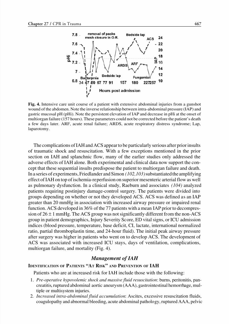
The complications of IAH and ACS appear to be particularly serious after prior insults
of traumatic shock and resuscitation. With a few exceptions mentioned in the prior
section on IAH and splanchnic flow, many of the earlier studies only addressed the
adverse effects of IAH alone. Both experimental and clinical data now support the con-
cept that these sequential insults predispose the patient to multiorgan failure and death.In a series of experiments, Friedlander and Simon (102,103)substantiated the amplifying
effect of IAH on top of ischemia-reperfusion on superior mesenteric arterial flow as well
as pulmonary dysfunction. In a clinical study, Raeburn and associates (104) analyzed
patients requiring postinjury damage-control surgery. The patients were divided into
groups depending on whether or not they developed ACS. ACS was defined as an IAP
greater than 20 mmHg in association with increased airway pressure or impaired renal
function. ACS developed in 36% of the 77 patients with a mean IAP prior to decompres-
sion of 26 ± 1 mmHg. The ACS group was not significantly different from the non-ACS
group in patient demographics, Injury Severity Score, ED vital signs, or ICU admission
indices (blood pressure, temperature, base deficit, CI, lactate, international normalizedratio, partial thromboplastin time, and 24-hour fluid). The initial peak airway pressure
after surgery was higher in patients who went on to develop ACS. The development of
ACS was associated with increased ICU stays, days of ventilation, complications,
multiorgan failure, and mortality (Fig. 4).
Management of IAH
IDENTIFICATION OF PATIENTS “AT RISK” AND PREVENTION OF IAH
Patients who are at increased risk for IAH include those with the following:
1. Pre-operative hypovolemic shock and massive fluid resuscitation: burns, peritonitis, pan-
creatitis, ruptured abdominal aortic aneurysm (AAA), gastrointestinal hemorrhage, mul-tiple or multisystem injuries.
2. Increased intra-abdominal fluid accumulation: Ascites, excessive resuscitation fluids,
coagulopathy and abnormal bleeding, acute abdominal pathology, ruptured AAA, pelvic
Fig. 4. Intensive care unit course of a patient with extensive abdominal injuries from a gunshot
wound of the abdomen. Note the inverse relationship between intra-abdominal pressure (IAP) and
gastric mucosal pH (pHi). Note the persistent elevation of IAP and decrease in pHi at the onset of multiorgan failure (157 hours). These parameters could not be corrected before the patient’s death
a few days later. ARF, acute renal failure; ARDS, acute respiratory distress syndrome; Lap,
laparotomy.

8/7/2019 Cardiopulmonary Resuscitation in Trauma
http://slidepdf.com/reader/full/cardiopulmonary-resuscitation-in-trauma 14/21
468 Cardiopulmonary Resuscitation
and retroperitoneal hematomas, intestinal obstruction and hemoperitoneum from
nonoperative management of solid organ injury.3. Mechanical increase in pressure: Pelvic and retroperitoneal hematoma, “damage-con-
trol” surgery with intra-abdominal packing, sudden intra-abdominal reduction of long-
standing hernial contents, tension pneumothorax, massive hemothorax, “chronic”abdominal compartment syndrome from morbid obesity, intestinal obstruction.
It is important to anticipate IAH and attempt prophylaxis by “open abdomen” in these
patients. The “open abdomen” approach offers several advantages. It provides a rapid
method of abbreviating the laparotomy and transporting the patient to the ICU for resus-citation. In a significant number of patients, it may actually prevent IAH. In recent
reports, Mayberry and associates (105,106)analyzed 73 consecutive patients who had anabsorbable mesh closure of the abdomen, 47 at the initial celiotomy (group 1) and 26 at
a subsequent celiotomy (group 2). The two groups had similar injury severity but group
2 had a higher incidence of postoperative ACS (35 vs 0%). A similar statistically signifi-
cant reduction in IAH was also noted by Ivatury and colleagues (74).It is also important to keep in mind that prophylactic open abdomen and nonclosure
of fascia does not always prevent IAH and ACS, as has been observed by several inves-tigators (74,97, 99,104,107). These patients, therefore, should have close IAP monitoring
in the postoperative period.
Treatment of IAH and ACS
We suggest that a critical level of 25 cm of H2O (18.3 mmHg) should trigger careful
monitoring of IAP and prompt treatment if it continues to increase. The first step in theevaluation of an increased IAP, especially in the presence of agitation and restlessness,
is to sedate and, if necessary, chemically paralyze the patient. If the bladder pressures arestill high and/or systemic manifestations of IAH (as described above) are evident, theappropriate treatment, in most instances, is abdominal decompression by laparotomy.
Recently, two studies emphasized the nonoperative approach to ACS in burn patients
(108,109). The first study (108) evaluated the utility of percutaneous drainage (PD) of
peritoneal fluid compared with decompressive laparotomy in burn patients. Nine of 13
(69%) study patients developed IAH that progressed to abdominal compartment syn-
drome in five (31%). All were treated with PD using a diagnostic peritoneal lavage
catheter. Five patients underwent PD successfully, and their IAH did not progress to
ACS. Four patients with greater than 80% total body surface area burns and severe
inhalation injury did not respond to PD and required decompressive laparotomy. Therewas no evidence of bowel edema, ischemia, or necrosis. All patients requiring decom-
pressive laparotomies died either from sepsis or respiratory failure. The second study
(109) reported similar success with percutaneous drainage in ACS in three burn victims.
Similar paracentesis treatment of IAH was described in patients with liver injury being
treated nonoperatively by Yang et al. (110).
In summary, the current evidence suggests that routine use of IAP monitoring is indi-
cated in all patients “at risk” (includes most of the massively injured patients or criticallyill patients in the ICU). The critical level of IAP that becomes IAH is around 20–25 cm
of H2O. IAH may be an earlier phenomenon that, when persistent or neglected, may lead
to the complete manifestations of ACS. ACS may become manifest at much lowerpressures than recognized previously. Prophylaxis and aggressive and prompt treatment
of IAH is recommended to prevent ACS from becoming an irreversible syndrome and
culminate in cardiopulmonary arrest in the severely ill or the multiply injured patient.

8/7/2019 Cardiopulmonary Resuscitation in Trauma
http://slidepdf.com/reader/full/cardiopulmonary-resuscitation-in-trauma 15/21
Chapter 27 / CPR in Trauma 469
REFERENCES
1. Cannon WB, Faser J, Cowell EM. The preventive treatment of wound shock. JAMA 1918; 47:618.
2. Kaweski SM, Sise MJ, Virgilio RW. The effect of prehospital fluids on survival in trauma patients.
J Trauma 1990; 30:1215–1219.
3. Bickell WH, Wall MJ Jr, Pepe PE, et al. Immediate versus delayed fluid resuscitation for hypotensivepatients with penetrating torso injuries. N Engl J Med 1994; 331:1105–1109.
4. Gross D, Landau EH, Assalia A, Krausz MM. Is hypertonic saline resuscitation safe in “uncontrolled”
hemorrhagic shock? J Trauma 1988; 28:751–756.
5. Bickell WH, Bruttig SP, Wade CE. Hemodynamic response to abdominal aortotomy in the anesthetized
swine. Circ Shock 1989; 28:321–332.
6. Krausz MM, Horn Y, Gross D. The combined effect of small volume hypertonic saline and normal saline
solutions in uncontrolled hemorrhagic shock. Surg Gynecol Obstet. 1992; 174:363–368.
7. Kowalenko T, Stern SA, Dronen SC, Wang X. Improved outcome with hypotensive resuscitation of
uncontrolled hemorrhagic shock in a swine model. J Trauma 1992; 33:349–353.
8. Gross D, Landau EH, Klin B, Krausz MM. Treatment of uncontrolled hemorrhagic shock with hyper-
tonic saline solution. Surg Gynecol Obstet 1990; 170:106–112.
9. Bickell WH, Bruttig SP, Millnamow GA, O’Benar J, Wade CE. The detrimental effects of intravenouscrystalloid after aortotomy in swine. Surgery 1991; 110:529–536.
10. Krausz MM, Bar-Ziv M, Rabinovici R, Gross D. “Scoop and run” or stabilize hemorrhagic shock with
normal saline or small-volume hypertonic saline? J Trauma 1992; 33:6–10.
11. Stern SA, Dronen SC, Birrer P, Wang X. The effect of blood pressure on hemorrhage volume and survival
in a near-fatal hemorrhage model incorporating a vascular injury. Ann Emerg Med 1993; 22:155–163.
12. Matsuoka T, Hildreth J, Wisner DH. Liver injury as a model of uncontrolled hemorrhagic shock:
resuscitation with different hypertonic regimens. J Trauma 1995; 39:674–680.
13. Matsuoka T, Hildreth J, Wisner DH. Uncontrolled hemorrhage from parenchymal injury: is resuscita-
tion helpful? J Trauma 1996; 40:915–922.
14. Solomonov E, Hirsh M, Yahiya A, Krausz MM. The effect of vigorous fluid resuscitation in uncontrolled
hemorrhagic shock following massive splenic injury. Crit Care Med 2000; 28:749–754.
15. Krausz MM, Bashenko Y, Hirsh M. Crystalloid or colloid resuscitation of uncontrolled hemorrhagicshock after moderate splenic injury. Shock 2000; 13:230–235.
16. Krausz MM, Bashenko Y, Hirsh M. Crystalloid and colloid resuscitation of uncontrolled hemorrhagic
shock following massive splenic injury. Shock 2001; 16:383–388.
17. Abu-Hatoum O, Bashenko Y, Hirsh M, Krausz MM. Continuous fluid resuscitation and splenectomy for
treatment of uncontrolled hemorrhagic shock after massive splenic injury. J Trauma 2002; 52:253–258.
18. Kaweski SM, Sise MJ, Virgilio RW. The effect of prehospital fluids on survival in trauma patients.
J Trauma 1990; 30:1215–1219.
19. Babbs CF, Thelander K, Engoren M, Severyn F, Fenn-Buderer N, DeFrank M. Theoretically optimal
duty cycles for chest and abdominal compression during external cardiopulmonary resuscitation: Car-
diac output, coronary blood flow, and blood gases during open-chest standard and compression-active-
decompression cardiopulmonary resuscitation. Resuscitation 2002; 55:309–316.
20. Sanders AB, Kern KB, Ewy GA, Atlas M, Bailey L. Improved resuscitation from cardiac arrest withopen-chest massage. Ann Emerg Med 1984; 13(Pt 1):672–675.
21. Rieder CF, Crawford BG, Iliopoulos JI, Thomas JH, Pierce GE, Hermreck AS. A study of the techniques
of cardiac massage with the abdomen open. Surgery 1985; 98:824–830.
22. Ivatury RR. Emergency Room Thoracotomy. In: Ivatury RR, Cayten CG, eds. The Textbook of Penetrat-
ing Trauma. Baltimore: Williams & Wilkins, 1996.
23. Ivatury RR, Rohman M. Emergency department throacotomy for trauma: a collective review. Resusci-
tation 1987; 15:23.
24. Boyd M, Vaneck VW, Bourguet CC. Emergency room thoracotomy: when is it indicated? J Trauma
1992; 33:714-721.
25. Rhee PM, Jordan MR, Rich N. Survival after emergency department thoracotomy: review of published
data from the past 25 years. J Am Coll Surg 2000; 190:288–298.
26. Ladd AP, Gomez GA, Jacobson LE, Broadie TA, Scherer LR III, Solotkin KC. Emergency roomthoracotomy: updated guidelines for a level I trauma center. Am Surg 2002; 68:421–424.
27. Wade CEJ, Grady JJ, Fabian T, Younas RN, Kramer GC. Efficacy of hypertonic 7.5% saline and
6% dextran-70 in treating trauma: a meta-analysis of controlled clinical studies. Surgery 1997; 122:
609–616.

8/7/2019 Cardiopulmonary Resuscitation in Trauma
http://slidepdf.com/reader/full/cardiopulmonary-resuscitation-in-trauma 16/21
470 Cardiopulmonary Resuscitation
28. Holcroft JW, Vassar MJ, Turner JE, Derlet RW, Kramer GC. 3% NaCl and 7.5% NaCl/dextran 70 in the
resuscitation of severely injured patients. Ann Surg 1987; 206:279–288.
29. Vassar MJ, Fischer RP, O’Brien PE, et al. A multicenter trial for resuscitation of injured patients with
7.5% sodium chloride. Arch Surg 1993; 128:1003–1013.
30. Krausz MM. Controversies in shock research: hypertonic resuscitation pros and cons. Shock 1995;
3:69–72.
31. Dronen SC, Stern SA, Wang X, Stanley M. A comparison of the response of near-fatal acute hemor-
rhage models with and without a vascular injury to rapid volume expansion. Am J Emerg Med 1993;
11:331–335.
32. Sindlinger JF, Soucy DM, Greene SP, Barber AE, Illner H, Shires T. The effects of isotonic saline
volume resuscitation in uncontrolled hemorrhage. Surg Gynecol Obstet 1993; 177:545–550.
33. Krausz MM, David M, Amstislavsky T. Hypertonic saline treatment of uncontrolled hemorrhagic shock
in awake rats. Shock 1994; 2:267–270.
34. Bunn F, Roberts I, Tasker R, Akpa E. Hypertonic versus isotonic crystalloid for fluid resuscitation in
critically ill patients. Cochrane Database Syst Rev 2002; (1):CD002045.
35. Novak L, Shackford SR, Bourguignon P, et al. Comparison of standard and alternative prehospital
resuscitation in pressure driven hemorrhagic shock and head injury. J Trauma. 1999; 47:834–844.
36. Doyle JA, DavisDP, Hoyt DB. The use of hypertonic saline in the treatment of traumatic brain injury.
J Trauma 2001; 50:367–83.
37. Shackford S. Effect of small-volume resuscitation on intracranial pressure and related cerebral vari-
ables. J Trauma 1997; 42(Suppl):S48–S53.
38. Ducey J, Lamiell J, Gueller G. Cerebral electrophysiologic effects of resuscitation with hypertonic
saline-dextran after hemorrhage. Crit Care Med 1990; 18:744–749.
39. Gemma M, Cozzi S, Piccoli S, Magrin S, De Vitis A, Cenzato M. Hypertonic saline fluid therapy
following brain stem trauma. J Neurosurg Anesthesiol 1996; 8:137–141.
40. Qureshi A, Wilson D, Traystman R. Treatment of elevated intracranial pressure in experimental
intracerebral hemorrhage: comparison between mannitol and hypertonic saline. Neurosurgery 1999;
44:1055–1063.
41. Suarez J, Qureshi A, Bhardwaj A, et al. Treatment of refractory intracranial hypertension with 23.4%
saline. Crit Care Med 1998; 26:1118–1122.
42. Corso CO, Okamoto S, Leiderer R, Messmer K. Resuscitation with hypertonic saline dextran reduces
endothelial cell swelling and improves hepatic microvascular perfusion and function after hemorrhagic
shock. J Surg Res 998; 80:210–220.
43. Cudd TA, Purinton S, Patel NC, Wood CE. Cardiovascular, adrenocorticotropin, and cortisol responses
to hypertonic saline in euvolemic sheep are altered by prostaglandin synthase inhibition. Shock 1998;
10:32–36.
43. Corso CO, Okamoto S, Reuttinger D, Messmer K. Hypertonic saline dextran attenuates leukocyte
accumulation in the liver after hemorrhagic shock and resuscitation. J Trauma 1999; 46:417–423.
44. Angle N, Hoyt DB, Cabello-Passini R, Herdon-Remelius C, Loomis W, Junger WG. Hypertonic saline
resuscitation reduces neutrophil margination by suppressing neutrophil L selectin expression. J Trauma
1998; 45:7–13.45. Angle N, Hoyt DB, Coimbra R, et al. Hypertonic saline resuscitation diminishes lung injury by suppress-
ing neutrophil activation after hemorrhagic shock. Shock 1998; 9:164–170.
46. Junger WG, Hoyt DB, Davis RE, et al. Hypertonicity regulates the function of human neutrophils by
modulating chemoattractant receptor signaling and activating mitogen-activated protein kinase p38. J
Clin Invest 1998; 101:2768–2779.
47. Patrick DA, Moore EE, Offner PJ, Johnson JL, Tamura DY, Silliman CC. Hypertonic saline activates
lipid-primed human neutrophils for enhanced elastase release. J Trauma 1998; 44:592–598.
48. Coimbra R, Junger WG, Hoyt DB, Liu FC, Loomis WH, Evers MF. Hypertonic saline resuscitation
restores hemorrhage-induced immunosuppression by decreasing prostaglandin E2 and interleukin-4
production. J Surg Res 1996; 64:203–209.
49. Tokyay R, Zeigler ST, Kramer GC, et al. Effects of hypertonic saline dextran resuscitation on oxygen
delivery, oxygen consumption, and lipid peroxidation after burn injury. J Trauma 1992; 32:704–713.50. Coimbra R, Hoyt DB, Junger WG, et al. Hypertonic saline resuscitation decreases susceptibility to sepsis
after hemorrhagic shock. J Trauma 1997; 42:602–607.
51. Worthley LI, Cooper DJ, Jones N. Treatment of resistant intracranial hypertension with hypertonic
saline. Report of two cases. J Neurosurg 1988; 68:478–481.

8/7/2019 Cardiopulmonary Resuscitation in Trauma
http://slidepdf.com/reader/full/cardiopulmonary-resuscitation-in-trauma 17/21
Chapter 27 / CPR in Trauma 471
52. Einhaus S, Croce M, Watridge C, Lowery R, Fabian T. The use of hypertonic saline for the treatment
of increased intracranial pressure. J Tenn Med Assoc 1996; 89:81–82.
53. Schatzmann C, Heissler H, Kˆnig K, et al. Treatment of elevated intracranial pressure by infusions of
10% saline in severely head injured patients. Acta Neurochir Suppl (Wien) 1998; 71:31–33.
54. Simma B, Burger R, Falk M, Sacher P, Fanconi S. A prospective, randomized, and controlled study of
fluid management in children with severe head injury: lactated Ringer’s solution versus hypertonicsaline. Crit Care Med 1998; 26:1270.
55. Horn P, Meunch E, Vajkoczy P, et al. Hypertonic saline solution for control of elevated intracranial pressure
in patients with exhausted response to mannitol and barbiturates. Neurol Res 1999; 21:758–764.
56. Vassar MJ, Perry CA, Gannaway WL, Holcroft JW. 7.5% sodium chloride/dextran for resuscitation of
trauma patients undergoing helicopter transport. Arch Surg 1991; 126:1065–1072.
57. Vassar M, Perry C, Holcroft J. Prehospital resuscitation of hypotensive trauma patients with 7.5% NaCl
versus 7.5% NaCl with added dextran: a controlled trial. J Trauma 1993; 34:622–632.
58. Stone HH, Strom PR, Mullins RJ. Management of the major coagulopathy with onset during laparotomy.
Ann Surg 1983; 197:532–535.
59. Rotondo MF, Schwab CW, McGonigal MD, et al. Damage control: an approach for improved survival
in exsanguinating penetrating abdominal injury. J Trauma 1993; 35:375–383.
60. Burch JM, Ortiz VB, Richardson RJ, et al. Abbreviated laparotomy and planned re-operation for criti-cally injured patients. Ann Surg 1992; 215:476–484.
61. Moore EE. Staged laparotomy for the hypothermia, acidosis and coagulopathy syndrome. Am J Surg
1996; 172:405–410.
62. Shapiro MB, Jenkins DH, Schwab CW, Rotondo MF. Damage control: collective review. J Trauma
2000; 49:969–978.
63. Vargo DJ, Battistella FD. Abbreviated thoracotomy and temporary chest closure: an application of
damage control after thoracic trauma. ArchSurg 2001; 136:21–24.
64. Johnon JW, Gracias VH, Schwab CW, et al. Evolution in damage control for exsanguinating penetrating
abdominal injury. J Trauma 2001; 51:261–269.
65. Asensio JA, McDuffie L, Petrone P, et al. Reliable variables in the exsanguinated patient which indicate
damage control and predict outcome. Am J Surg 2001; 182:743–751.
66. Hoey BA, Schwab CW. Damage control surgery. Scand J Surg 2002; 91:92–103.67. Barker DE, Kaufman HJ, Smith LA, Ciraulo DL, Richart CL, Burns RP. Vacuum pack technique of
temporary abdominal closure: a 7-year experience with 112 patients. J Trauma 2000; 48:201–206.
68. Miller PR, Thompson JT, Faler BJ, Meredith JW, Chang MC. Late fascial closure in lieu of ventral
hernia: the next step in open abdomen management. J Trauma 2002; 53:843–849.
69. Garner GB, Ware DN, Cocanour CS, et al. Vacuum-assisted wound closure provides early fascial
reapproximation in trauma patients with open abdomens. Am J Surg 2001; 182:630–638.
70. Markley MA, Mantor PC, Letton RW, Tuggle DW. Pediatric vacuum packing wound closure for dam-
age-control laparotomy. J Pediatr Surg 2002; 37:512–514.
71. Ivatury RR, Diebel L, Porter JM et al. Intra-abdominal hypertension and the abdominal compartment
syndrome. Surg Clin North Am 1997; 77:783–800.
72. Ivatury RR, Porter JM, Simon RJ et al. Intra-abdominal hypertension after life threatening abdominal
trauma: incidence, prophylaxis and clinical relevance to gastric mucosal pH and abdominal compart-ment syndrome. J Trauma 1998; 44:1016–1021.
73. Saggi BH, Sugerman HJ, Ivatury RR et al. Abdominal compartment syndrome. J Trauma 1998; 45:
597–609.
74. Schein M, Wittmann DH, Aprahamian CC et al. The abdominal compartment syndrome: the physiological
and clinical consequences of elevated intra-abdominal pressure. J Amer Coll Surg 1995; 180:747–753.
75. Burch JM, Moore EE, Moore FA et al. The abdominal compartment syndrome. Surg Clin North Amer
1996; 76:833–842.
76. Sugerman HJ, Windsor A, Bessos M et al. Intra-abdominal pressure, sagittal abdominal diameter, and
obesity comorbidity. J Intern Med 1997; 241:71–79.
77. Morris JA Jr, Eddy VA, Blinman TA et al. The staged celiotomy for trauma. Issues in unpacking and
reconstruction. Ann Surg 1993; 217:576–586.
78. Loi P, De Backer D, Vincent JL. Abdominal compartment syndrome. Acta Chir Belg 2001; 101:59–64.79. Barnes GE, Laine GA, Gaim PY, et al. Cardiovascular responses to elevation of intra-abdominal hydro-
static pressure. Am J Physiol 1985; 248:R208–R213.
80. Sugrue M, Jones F, Janjua KJ, Deane SA, Bristow P, Hillman K. Temporary abdominal closure: a
prospective evaluation of its effects on renal and respiratory physiology. J Trauma 1998; 45; 914–921.

8/7/2019 Cardiopulmonary Resuscitation in Trauma
http://slidepdf.com/reader/full/cardiopulmonary-resuscitation-in-trauma 18/21
472 Cardiopulmonary Resuscitation
81. Caldwell CB, Ricotta JJ. Changes in visceral blood flow with elevated intra-abdominal pressure. J Surg
Res 1987; 43:14–20.
82. Diebel LN, Wilson RF, Dulchavshy SA. Effect on increased intra-abdominal pressure on hepatic
arterial, portal venous, and hepatic microcirculatory blood flow. J Trauma 1992; 33:279–283.
83. Diebel LN, Dulchavshy SA, Wilson RF. Effect on increased intra-abdominal pressure on mesenteric
arterial and intestinal mucosal blood flow. J Trauma 1992; 33:45–49.84. Bongard F, Pianim N, Dubecz S, Klein S. Adverse consequences of increased intra-abdominal pressure
on bowel tissue oxygenation. J Trauma 1995; 39:519–525.
85. Chang MC, Miller PR, D’Agostino R Jr, Meredith JW. Effects of abdominal decompression on car-
diopulmonary function and visceral perfusion in patients with intra-abdominal hypertension. J of
Trauma 1998; 44:440–445.
86. Rezende-Neto JB, Moore EE, Melo De Andrade MV, et al. Systemic inflammatory response secondary
to abdominal compartment syndrome: stage for multiple organ failure. J. Trauma 2002; 53:1121–1128.
87. Doty JM, Oda J, Ivatury RR, et al. The effects of hemodynamic shock and increased intra-abdominal
pressure on bacterial translocation. J Trauma 2002; 52:13–17.
88. Oda J, Ivatury RR, Blocher CR, Malhotra AJ, Sugerman HJ. Amplified cytokine response and lung
injury by sequential hemorrhagic shock and abdominal compartment syndrome in a laboratory model
of ischemia-reperfusion. J Trauma 2002; 52:625–631.88a. Ivatury RR, Cayten CG. (eds.) The Textbook of Penetrating Trauma. Baltimore, MD: Williams &
Wilkens, 1996.
89. Josephs LG, Este-McDonald JR, Birkett DH et al. Diagnostic laparoscopy increases intracranial pres-
sure. J Trauma 1994; 36:815–819.
90. Bloomfield GL, Ridings PC, Blocher CR, Marmarou A, Sugerman HJ. Effects of increased intra-
abdominal pressure upon intracranial and cerebral perfusion pressure before and after volume expan-
sion. J Trauma 1996; 40:936–943.
91. Bloomfield GL, Ridings PC, Blocher CR, Marmarou A, Sugerman HJ. A proposed relationship between
increased intra-abdominal, intra-thoracic, and intracranial pressure. Crit Care Med 1997; 25:496–503.
92. Ertel W, Oberholzer A, Platz A, et al. Incidence of Abdominal compartment syndrome after “damage-
control” laparotomy in 311 patients with severe abdominal and/or pelvic trauma. Crit Care Med 2000;
28:1747–1753.93. Meldrum DR, Moore FA, Moore EE, Franciose RJ, Sauaia A, Burch JM. Prospective characterization
and selective management of the abdominal compartment syndrome. Am J Surg 1997; 174:667–672.
94. Hong JJ, Cohn SM, Perez JM, Dolich MO, Brown M, McKenney MG. Prospective study of the
incidence and outcome of intra-abdominal hypertension and the abdominal compartment syndrome.
Br J Surg 2002; 89:591–596.
95. Maxwell RA, Fabian TC, Croce MA et al. Secondary abdominal compartment syndrome: an
underappreciated manifestation of severe hemorrhagic shock. J Trauma 1999; 47:995–999.
96. Biffl WL, Moore EE, Burch JM, Offner PJ, Franciose RJ, Johnson JL. Secondary abdominal compart-
ment syndrome is a highly lethal event. Am J Surg 2001; 182:645–648.
97. Balogh Z, McKinley BA, Cocanour CS, Kozar RA, Holcomb JB, Ware DN, Moore FA. Secondary
abdominal compartment syndrome is an elusive early complication of traumatic shock resuscitation.
Am J Surg 2002; 184:538–543.98. Sugrue M, Jones F, Lee A, Buist MD, Deane S, Bauman A, Hillman K, Intraabdominal pressure and
gastric intramucosal pH: is there an association? World J Surg 1996; 20:988–991.
99. Friedlander MH, Simon RJ, Ivatury R, DiRaimo R, Machiedo GW. Effect of hemorrhage on superior
mesenteric artery flow during increased intra-abdominal pressures. J Trauma 1998; 45:433–439.
100. Simon RJ, Friedlander MH, Ivatury RR, DiRaimo R, Machiedo GW. Haemorrhage lowers the thresh-
old for intra-abdominal hypertension-induced pulmonary dysfunction. J Trauma 1997; 42:398–403.
101. Raeburn CD, Moore EE, Biffl WL, et al. The abdominal compartment syndrome is a morbid compli-
cation of postinjury damage control surgery. Am J Surg 2001; 182:542–546.
102. Mayberry JC, Mullins RJ, Crass RA et al. Prevention of abdominal compartment syndrome by absorb-
able mesh prosthesis closure. Arch Surg 1997; 132:957–961.
103. Mayberry JC. Prevention of the abdominal compartment syndrome. Lancet 1999; 354:1749–1750.
104. Offner PJ, de Souza AL, Moore EE, et al. Avoidance of abdominal compartment syndrome in damage-control laparotomy after trauma. Arch Surg 2001; 136:676–681.
105. Latenser BA, Kowal-Vern A, Kimball D, Chakrin A, Dujovny N. A pilot study comparing percutane-
ous decompression with decompressive laparotomy for acute abdominal compartment syndrome in
thermal injury. J Burn Care Rehabil. 2002; 23:190–195.

8/7/2019 Cardiopulmonary Resuscitation in Trauma
http://slidepdf.com/reader/full/cardiopulmonary-resuscitation-in-trauma 19/21
Chapter 27 / CPR in Trauma 473
106. Corcos AC, Sherman HF. Percutaneous treatment of secondary abdominal compartment syndrome. J
Trauma 2001; 51:1062–1064.
107. Yang EY, Marder SR, Hastings G, Knudson MM. The abdominal compartment syndrome complicat-
ing nonoperative management of major blunt liver injuries: recognition and treatment using
multimodality therapy. J Trauma 2002; 52:982–986.

8/7/2019 Cardiopulmonary Resuscitation in Trauma
http://slidepdf.com/reader/full/cardiopulmonary-resuscitation-in-trauma 20/21
CARDIOPULMONARY
R ESUSCITATION
Edited by
JOSEPH P. ORNATO, MD, FACP, FACC, FACEP
MARY A NN PEBERDY , MD, FACC
Departments of Emergency Medicine and Internal Medicine (Cardiology), Virginia Commonwealth University HealthSystem, Richmond, VA

8/7/2019 Cardiopulmonary Resuscitation in Trauma
http://slidepdf.com/reader/full/cardiopulmonary-resuscitation-in-trauma 21/21
© 2005 Humana Press Inc.999 Riverview Drive, Suite 208Totowa, New Jersey 07512
humanapress.com
All rights reserved. No part of this book may be reproduced, stored in a retrieval system, or transmitted in any form orby any means, electronic, mechanical, photocopying, microfilming, recording, or otherwise without written permissionfrom the Publisher.
The content and opinions expressed in this book are the sole work of the authors and editors, who have warranted duediligence in the creation and issuance of their work. The publisher, editors, and authors are not responsible for errorsor omissions or for any consequences arising from the information or opinions presented in this book and make nowarranty, express or implied, with respect to its contents.
Due diligence has been taken by the publishers, editors, and authors of this book to assure the accuracy of the informationpublished and to describe generally accepted practices. The contributors herein have carefully checked to ensure that
the drug selections and dosages set forth in this text are accurate and in accord with the standards accepted at the timeof publication. Notwithstanding, as new research, changes in government regulations, and knowledge from clinicalexperience relating to drug therapy and drug reactions constantly occurs, the reader is advised to check the productinformation provided by the manufacturer of each drug for any change in dosages or for additional warnings andcontraindications. This is of utmost importance when the recommended drug herein is a new or infrequently used drug.It is the responsibility of the treating physician to determine dosages and treatment strategies for individual patients.Further it is the responsibility of the health care provider to ascertain the Food and Drug Administration status of eachdrug or device used in their clinical practice. The publisher, editors, and authors are not responsible for errors oromissions or for any consequences from the application of the information presented in this book and make no warranty,express or implied, with respect to the contents in this publication.
Production Editor: Robin B. WeisbergCover design by Patricia F. Cleary
For additional copies, pricing for bulk purchases, and/or information about other Humana titles, contact Humana atthe above address or at any of the following numbers: Tel.: 973-256-1699; Fax: 973-256-8341, E-mail:[email protected]; or visit our Website: www.humanapress.com
This publication is printed on acid-free paper. ANSI Z39.48-1984 (American National Standards Institute) Permanence of Paper for Printed Library Materials.
Photocopy Authorization Policy:
Authorization to photocopy items for internal or personal use, or the internal or personal use of specific clients, is grantedby Humana Press Inc., provided that the base fee of US $25.00 is paid directly to the Copyright Clearance Center at 222Rosewood Drive, Danvers, MA 01923. For those organizations that have been granted a photocopy license from theCCC, a separate system of payment has been arranged and is acceptable to Humana Press Inc. The fee code for users
of the Transactional Reporting Service is: [1-58829-283-5/05 $25.00].
Printed in the United States of America. 10 9 8 7 6 5 4 3 2 1
eISBN 1-59259-814-5
Library of Congress Cataloging-in-Publication Data
Cardiopulmonary resuscitation / edited by Joseph P. Ornato, Mary Ann Peberdy.p. ; cm. -- (Contemporary cardiology)
Includes bibliographical references and index.ISBN 1-58829-283-5 (alk. paper)
1. CPR (First aid)[DNLM: 1. Cardiopulmonary Resuscitation. WG 205 C2677 2005] I. Ornato, Joseph P.
II. Peberdy, Mary Ann. III. Series: Contemporary cardiology (Totowa, N.J. : Unnumbered)RC87.9.C3732 2005616.1'025--dc22
2004002774