Embed Size (px)
Citation preview

Effect of Mobilization of the Anterior Hip Capsule on GluteusMaximus Strength
Address all correspondence and request for reprints to:Scott Yerys2442 Ellsworth St.Seaford, NY [email protected]
Abstract: Loss of hip extension is often compensated for by extension of the lumbar spine. Thiscompensation can result in hypermobility and ultimately be a source of low back dysfunction andpain. Joint mobilizations have been known to return physiologic and accessory motion tohypomobile structures. Mobilization has also been demonstrated to improve muscular strengthwhen secondary to joint hypomobility. The purpose of this study was to determine the usefulnessof posteroanterior (P-A) hip-joint mobilization in improving strength of the gluteus maximusmuscle. Forty subjects were randomly assigned to a control group (Grade I P-A mobilization) andan experimental group (Grade IV P-A mobilization). The subjects performed a pretest/posttest setof five isometric repetitions on the Cybex NormTM isokinetic machine. The peak torque was deter-mined for both pretest and posttest measurements. The data collected were analyzed using anindependent t-test with a significance level of p < .05. The results demonstrated a statisticallysignificant difference between the experimental and control groups (t=1.68, p=0.002). This studydemonstrated a significant increase in gluteus maximus strength in response to Grade IV P-Amobilizations performed on the anterior hip capsule. Clinicians can utilize these findings in ev-eryday practice to improve muscle strength by integrating manual therapy with therapeutic exer-cise.
Key Words: Arthrokinetic reflex (AKR), Mobilization/manipulation, Hypomobility, Hip Joint,Gluteus Maximus, Muscle Strength, Manual Therapy
Scott Yerys, PT, MSHoward Makofsky, PT, DHSc, OCSCharles Byrd, PT, MSJoseph Pennachio, PT, MSJonathan Cinkay, PT, MS
In the geriatric population, one of the first motionslost is hip extension. This is evident by a shuffling
gait pattern or decreased step length during ambulation1.As a result of decreased extension, there is an associateddecrease in gluteus maximus strength. Although thisweakness has many factors, one often-neglected cause isthe neurally mediated inhibition arising from the articularmechanoreceptors. Although it is established2 that the
articular receptor system contributes significantly to jointposition sense (i.e., static and dynamic aspects of propriocep-tion), the contribution of this same system to muscleperformance is still under appreciated. Recently, Liebleret al2 demonstrated a relationship between increasing lowerthoracic spine extension and lower trapezius strength.They postulated that the improvement in muscle strengthwas due to the removal of neural inhibition generatedfrom the mechanoreceptors of the apophyseal joints.
In this study, the authors investigated a similar re-lationship between hip-joint extension and gluteus maximusstrength. We hypothesized that improving the mobilityof hip extension would remove the inhibitory influenceof the arthrokinetic reflex (AKR) on the gluteus maxi-mus muscle, resulting in enhanced muscle performance3.The authors hoped to underscore with sound evidence
The Journal of Manual & Manipulative TherapyVol. 10 No. 4 (2002), 218 - 224218 / The Journal of Manual & Manipulative Therapy, 2002
what manual therapists have known for years, namelythat mobilization helps to restore all aspects of a hypomobilejoint’s function, including normalization of strength inthe muscles related to it.
Review of LiteratureThe primary function of the hip joint is to support
the weight of the head, arms, and trunk in static anddynamic postures such as ambulating, running, and stair-climbing. Abnormal joint mobility is an important fac-tor in movement dysfunction. Roach and Mills reportedthat hip extension demonstrated the only significant losswith age. This loss of motion is evident as early as 40years of age and may play a role in the development ofosteoarthritis4-5. The head of the femur and the acetabu-lum are in greatest contact at full extension5; with lossof extension, the area of contact will diminish progres-sively as the deformity increases. Consequently, the bodyweight will be carried through a smaller area of the ar-ticular cartilage resulting in degenerative changes, de-tachment of debris, and the development of capsular fibrosis5.Even though Norkin and Levange6 state that a combina-tion of flexion, abduction and slight lateral rotation willimprove articular contact between the femur and acetabu-lum, loss of hip extension is still considered a significantetiologic factor in the development of degenerative hipdisease4-5.
The primary extensors of the hip include the glu-teus maximus, semimembranosus, semitendinosus andbiceps femoris7. The gluteus maximus muscle, however,is the strongest extensor of the hip8-9. In addition to hipextension, it helps to straighten the leg when walking,running, or climbing. The gluteus maximus is also usuallyactive simultaneously with the paraspinal muscles dur-ing back extension, and fatigue of the gluteus maximusis often associated with chronic low back pain8.
Kanakaanpaa et al9 reported that the gluteus maxi-mus fatigued faster in women with chronic low back painthan in control subjects. Patients who suffer from chroniclow back pain very often have hypoactive, hypotonic, andweak gluteus maximus muscles10. In patients with lowback pain, the hip-spine interaction is disturbed11. Theweakness of the gluteus maximus muscle may be anunderlying cause of chronic low back pain; therefore,strengthening this muscle may reduce its incidence.
Restricted hip extension is often compensated for byextension of the lumbar spine12. This compensation canresult in lumbar hypermobility and ultimately be a sourceof low back dysfunction and pain. Hip extension is alsoassociated with anterior rotation of the innominate bone13.Therefore, a loss of hip extension can also result in com-pensation at the iliosacral joint and be another source oflow back pain13-14.
Minimal research has been performed in the man-agement of decreased hip range of motion (ROM)15.
Mobilization is one of the most commonly recommendedtreatments for this condition. The goal of mobilizationis to restore the normal arthrokinematics of a joint, includingspins, rolls and glides, by improving the extensibility ofthe ligamento-capsular tissue. Joint mobilization hasbeen known to return physiologic and accessory motionsto hypomobile structures16-18. Mobilizations/manipula-tions are defined by the Guide to Physical Therapy Prac-tice as “a manual therapy technique comprising a con-tinuum of skilled passive movements to the joints and/or related soft tissues that are applied at varying speedsand amplitudes, including a small-amplitude/high-velocitytherapeutic movement”19. This technique is used on softtissues and joints for the dual purpose of evaluating andtreating somatic impairment. Mobilizations are oftencombined with traditional physical therapy modalities aswell20.
Arthrokinematically, translational motion of the femoralhead on the acetabulum is believed to occur during normalfunction of the hip6,21. During hip extension, specifically,the femoral head demonstrates a slight anterior glidingmotion on the acetabulum19-20. However, according toParis and Loubert22, the depth of the concave acetabu-lum limits the amount of translation that occurs in thehip. Norkin and Levangie report normal ROM for hipextension to be 10-30˚6, whereas Palmer and Eples re-port the normal extension to be 10-15˚23.
Joint mobilization also causes physical loading andunloading of joint cartilage to facilitate the flow of syn-ovial fluid within the joint. This flow of fluid ensuresadequate nutrition to the articular cartilage. When com-pression is combined with mobilization, there is thoughtto be even greater stimulation of synovial fluid flow24.
The muscle-firing pattern of active hip extension inthe prone position is as follows: ipsilateral lumbar erec-tor spinae, ipsilateral hamstrings, contralateral lumbarerector spinae, ipsilateral tensor fascia latae, and ipsilat-eral gluteus maximus4. Visible muscle wasting, particu-larly in the gluteal muscles, is often seen when tightnessis present in the anterior muscle group (iliopsoas) or whenpain is present25.
The muscles about the pelvis that are most commonlyaffected by hip pathologies are the gluteal muscles11.Effective treatment of muscle weakness relies on addressingits cause. Muscles become weak from myogenic causes,diminished use, aging, or disorders of nerve conductionof either peripheral or central origin. Muscle weaknessmay also be caused by inhibition of a muscle related tocapsular hypomobility of the underlying joint3.
Throughout our body, there are three different typesof articulating joints: 1) synarthroses, 2) amphiarthroses,and 3) diarthroses. Manual therapy is geared toward thediarthrodial joints, which have four varieties of mecha-noreceptors26-27. Type-I mechanoreceptors are located onthe superficial aspect of the joint capsule and have staticand dynamic proprioceptive functions. Type-II mechan-
Effect of Mobilization of the Anterior HipCapsule on Gluteus Maximus Strength / 219
oreceptors are located in the deeper layers of the capsuleand have only dynamic proprioceptive function. Type-IIImechanoreceptors are much larger and are found prima-rily on the surface of joint ligaments and, to a lesser de-gree, within joint capsules28. The aforementioned mecha-noreceptors (Types I, II, and III) are corpuscular mecha-noreceptors (i.e., biological transducers) that are stimu-lated by increases in tension in the tissues in which theyare embedded. The Type-IV variety, is represented by anon-encapsulated, unmyelinated plexus of nerve fibers. TheType-IV system is normally inactive but is triggered intoaction when abnormally high tension or inflammationdevelops in the articular tissues27.
The four articular mechanoreceptors discussed abovealso exert reciprocally coordinated, reflexogenic influenceson muscle tone27. Through the arthrokinetic reflex mecha-nism, joint mobilization/manipulation not only affects themotor unit activity in the muscles operating over the jointbeing manipulated, but it also affects more remote musclesas well, including the muscles on the contralateral side ofthe body. This results from the multi-segmental organi-zation of the mechanoreceptor afferents within theneuroaxis27. Consequently, in addition to its traditionalrole as an intervention for stiff and painful joints, manualtherapy also has the potential to achieve reflexogenic changesin muscle tone (e.g., facilitation and inhibition) locallyand to some degree, globally as well.
Herzog et al29 findings confirmed the principle of reflexactivation of muscles after mechanical intervention of thespine. Similarly, Wyke demonstrated that a distraction ofthe cervical facet joints in cats produced a simultaneousonset of electromyographic (EMG) activity in selected forelimbmuscles, which was attributed to a capsular mechanore-ceptor reflex response27. Cibulka et al16 reported thatmobilization of a dysfunctional sacroliac joint restored thenormal length-tension relationship of the hamstrings, thusincreasing its torque production. McNair et al30 demon-strated the inhibitory effects of experimentally-induced kneeswelling on the quadriceps muscle, then reversed thisinhibition/muscle weakness with submaximal exercise. Asmentioned previously, Liebler et al demonstrated a sig-nificant increase in bilateral lower trapezius strength inresponse to Grade-IV posteroanterior (P-A) mobilizationsperformed on asymptomatic thoracic vertebrae (T6-T12)2.
The purpose of this study was to determine the effectof hip-joint mobilization directed to the anterior capsuleon the strength of the gluteus maximus muscle. If arelationship exists between hip-joint hypomobility and aweakness of the gluteus maximus, it follows that any at-tempt to restore strength to the gluteus maximus shouldinclude joint mobilization. As discussed previously, glu-teus maximus function is a crucial component in lowerextremity, pelvic girdle, and lumbar spine mechanics9-10,12.The ability to improve its functional strength in any waypossible, including manual therapy, is therefore worthinvestigating.
Methods
SubjectsA convenience sample of 40 asymptomatic students from
the New York Institute of Technology (NYIT) in Old Westbury,NY, volunteered to take part in the study. Volunteers rangedin age from 19 to 39 years; they were permitted to partici-pate in the study unless meeting one or more exclusioncriteria governing selection. The first exclusion criterioneliminated subjects presenting with hypo/hypermobility atthe hip joint. Hypomobility was defined as hip extensionROM < 10˚, whereas hypermobility was defined as hip ex-tension ROM >20˚ as determined by standard goniometry.The second exclusion criterion eliminated subjects with hippathology, a history of trauma, low back pain, and/or pastsurgery of the hip. The Institutional Review Board at NYITgranted approval for this research; informed consent wasobtained from each subject.
ProcedureSubjects underwent a standardized interview, mus-
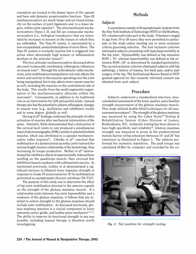
culoskeletal assessment of the lower quarter, and a baselinestrength measurement of the gluteus maximus muscle.This study utilized double-blind techniques for all mea-surement procedures31. The strength of the gluteus maximuswas measured by using the Cybex NormTM Testing &Rehabilitation System (Cybex Division of Lumex,Ronkonkoma, NY). Isokinetic testing has been shown tohave high specificity and reliability32. Gluteus maximusstrength was measured in prone at the predeterminedmotion barrier of hip extension (between 10˚ and 20˚ hipextension) as illustrated in Figure 1. The subjects per-formed five isometric repetitions. The peak torque wascalculated (ft⋅lbs) by computer and recorded by the ex-
Fig. 1: Test position for strength testing.
220 / The Journal of Manual & Manipulative Therapy, 2002
amining clinician.Each subject underwent a familiarization trial on the
Cybex Norm prior to baseline data collection. This pro-cedure eliminated any false positive results that may haveoccurred due to the learning curve. The familiarizationtrial consisted of finding and recording the settings onthe Cybex Norm for each subject followed by a 2-3 minutetrial. The subject then performed five isometric repeti-tions with maximum effort. The familiarization trial wasperformed on the same day but before actual experimen-tal testing and data collection began. There was a five-minute rest period between the familiarization trial andthe actual experimental testing to allow for adequate recoverytime and avoid skewing data due to fatigue.
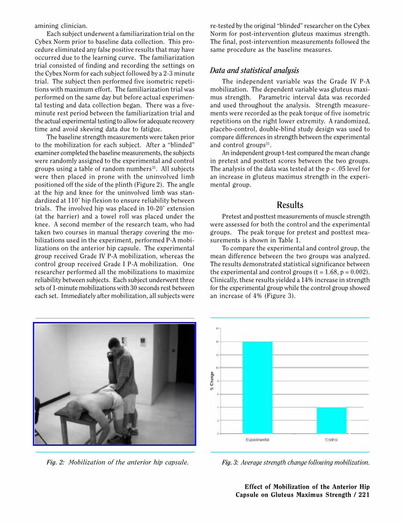
The baseline strength measurements were taken priorto the mobilization for each subject. After a “blinded”examiner completed the baseline measurements, the subjectswere randomly assigned to the experimental and controlgroups using a table of random numbers31. All subjectswere then placed in prone with the uninvolved limbpositioned off the side of the plinth (Figure 2). The angleat the hip and knee for the uninvolved limb was stan-dardized at 110˚ hip flexion to ensure reliability betweentrials. The involved hip was placed in 10-20˚ extension(at the barrier) and a towel roll was placed under theknee. A second member of the research team, who hadtaken two courses in manual therapy covering the mo-bilizations used in the experiment, performed P-A mobi-lizations on the anterior hip capsule. The experimentalgroup received Grade IV P-A mobilization, whereas thecontrol group received Grade I P-A mobilization. Oneresearcher performed all the mobilizations to maximizereliability between subjects. Each subject underwent threesets of 1-minute mobilizations with 30 seconds rest betweeneach set. Immediately after mobilization, all subjects were
re-tested by the original “blinded” researcher on the CybexNorm for post-intervention gluteus maximus strength.The final, post-intervention measurements followed thesame procedure as the baseline measures.
Data and statistical analysisThe independent variable was the Grade IV P-A
mobilization. The dependent variable was gluteus maxi-mus strength. Parametric interval data was recordedand used throughout the analysis. Strength measure-ments were recorded as the peak torque of five isometricrepetitions on the right lower extremity. A randomized,placebo-control, double-blind study design was used tocompare differences in strength between the experimentaland control groups31.
An independent group t-test compared the mean changein pretest and posttest scores between the two groups.The analysis of the data was tested at the p < .05 level foran increase in gluteus maximus strength in the experi-mental group.
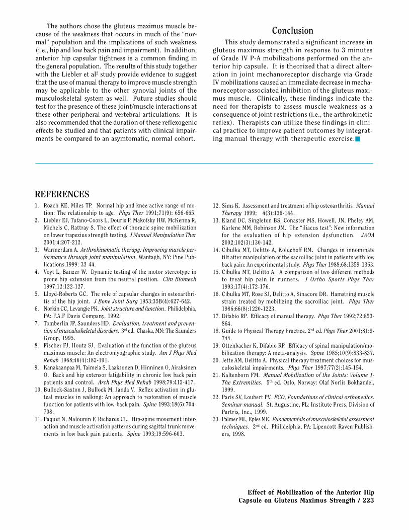
ResultsPretest and posttest measurements of muscle strength
were assessed for both the control and the experimentalgroups. The peak torque for pretest and posttest mea-surements is shown in Table 1.
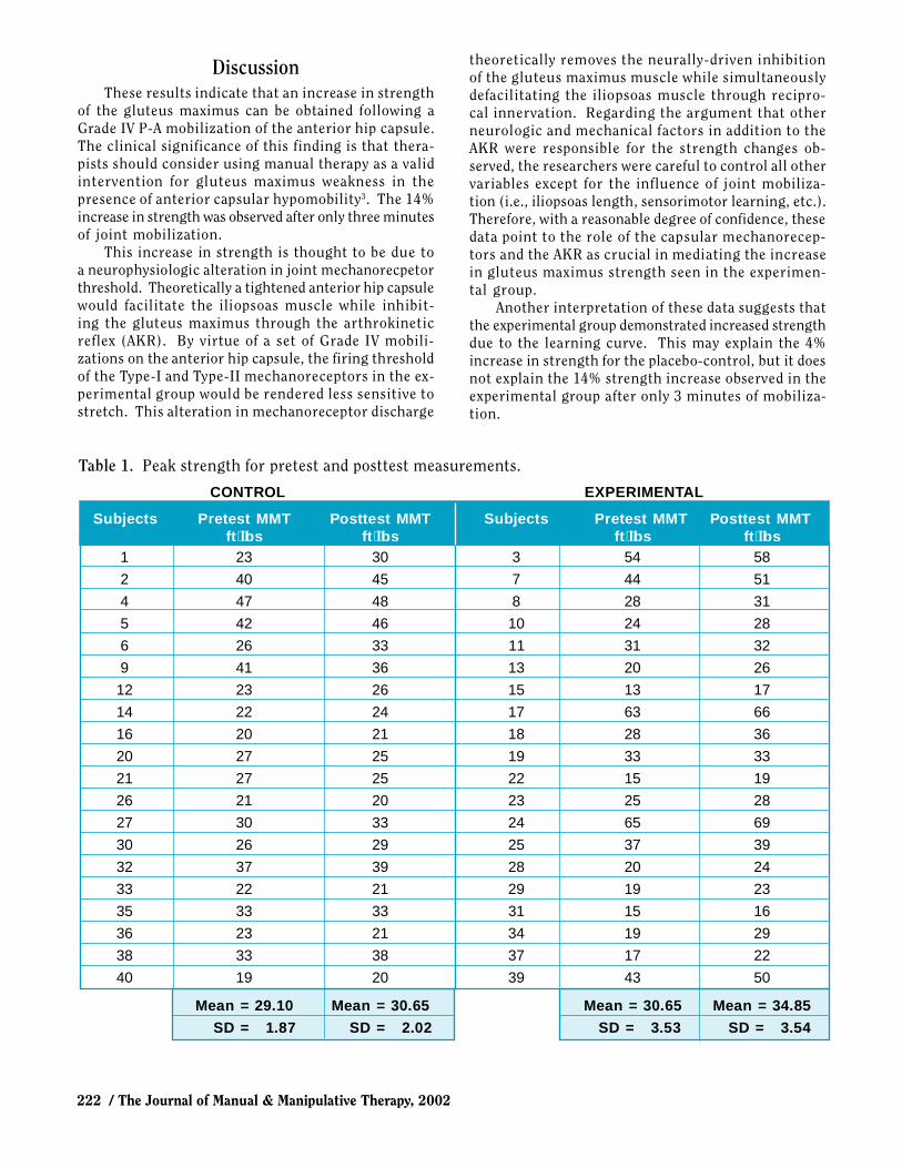
To compare the experimental and control group, themean difference between the two groups was analyzed.The results demonstrated statistical significance betweenthe experimental and control groups (t = 1.68, p = 0.002).Clinically, these results yielded a 14% increase in strengthfor the experimental group while the control group showedan increase of 4% (Figure 3).
Fig. 2: Mobilization of the anterior hip capsule. Fig. 3: Average strength change following mobilization.
Effect of Mobilization of the Anterior HipCapsule on Gluteus Maximus Strength / 221
DiscussionThese results indicate that an increase in strength
of the gluteus maximus can be obtained following aGrade IV P-A mobilization of the anterior hip capsule.The clinical significance of this finding is that thera-pists should consider using manual therapy as a validintervention for gluteus maximus weakness in thepresence of anterior capsular hypomobility3. The 14%increase in strength was observed after only three minutesof joint mobilization.
This increase in strength is thought to be due toa neurophysiologic alteration in joint mechanorecpetorthreshold. Theoretically a tightened anterior hip capsulewould facilitate the iliopsoas muscle while inhibit-ing the gluteus maximus through the arthrokineticreflex (AKR). By virtue of a set of Grade IV mobili-zations on the anterior hip capsule, the firing thresholdof the Type-I and Type-II mechanoreceptors in the ex-perimental group would be rendered less sensitive tostretch. This alteration in mechanoreceptor discharge
CONTROL EXPERIMENTAL
Subjects Pretest MMT Posttest MMT Subjects Pretest MMT Posttest MMTft⋅lbs ft⋅lbs ft⋅lbs ft⋅lbs
1 23 30 3 54 58
2 40 45 7 44 51
4 47 48 8 28 31
5 42 46 10 24 28
6 26 33 11 31 32
9 41 36 13 20 26
12 23 26 15 13 17
14 22 24 17 63 66
16 20 21 18 28 36
20 27 25 19 33 33
21 27 25 22 15 19
26 21 20 23 25 28
27 30 33 24 65 69
30 26 29 25 37 39
32 37 39 28 20 24
33 22 21 29 19 23
35 33 33 31 15 16
36 23 21 34 19 29
38 33 38 37 17 22
40 19 20 39 43 50
Mean = 29.10 Mean = 30.65 Mean = 30.65 Mean = 34.85
SD = 1.87 SD = 2.02 SD = 3.53 SD = 3.54
theoretically removes the neurally-driven inhibitionof the gluteus maximus muscle while simultaneouslydefacilitating the iliopsoas muscle through recipro-cal innervation. Regarding the argument that otherneurologic and mechanical factors in addition to theAKR were responsible for the strength changes ob-served, the researchers were careful to control all othervariables except for the influence of joint mobiliza-tion (i.e., iliopsoas length, sensorimotor learning, etc.).Therefore, with a reasonable degree of confidence, thesedata point to the role of the capsular mechanorecep-tors and the AKR as crucial in mediating the increasein gluteus maximus strength seen in the experimen-tal group.
Another interpretation of these data suggests thatthe experimental group demonstrated increased strengthdue to the learning curve. This may explain the 4%increase in strength for the placebo-control, but it doesnot explain the 14% strength increase observed in theexperimental group after only 3 minutes of mobiliza-tion.
Table 1. Peak strength for pretest and posttest measurements.
222 / The Journal of Manual & Manipulative Therapy, 2002
REFERENCES1. Roach KE, Miles TP. Normal hip and knee active range of mo-
tion: The relationship to age. Phys Ther 1991;71(9): 656-665.2. Liebler EJ, Tufano-Coors L, Douris P, Makofsky HW, McKenna R,
Michels C, Rattray S. The effect of thoracic spine mobilizationon lower trapezius strength testing. J Manual Manipulative Ther2001;4:207-212.
3. Warmerdam A. Arthrokinematic therapy: Improving muscle per-formance through joint manipulation. Wantagh, NY: Pine Pub-lications,1999: 32-44.
4. Voyt L, Banzer W. Dynamic testing of the motor stereotype inprone hip extension from the neutral position. Clin Biomech1997;12:122-127.
5. Lloyd-Roberts GC. The role of capsular changes in osteoarthri-tis of the hip joint. J Bone Joint Surg 1953;35B(4):627-642.
6. Norkin CC, Levangie PK. Joint structure and function. Philidelphia,PA: F.A.F Davis Company, 1992.
7. Tomberlin JP, Saunders HD. Evaluation, treatment and preven-tion of musculoskeletal disorders. 3rd ed. Chaska, MN: The SaundersGroup, 1995.
8. Fischer FJ, Houtz SJ. Evaluation of the function of the gluteusmaximus muscle: An electromyographic study. Am J Phys MedRehab 1968;46(4):182-191.
9. Kanakaanpaa M, Taimela S, Laaksonen D, Hinninen O, AiraksinenO. Back and hip extensor fatigability in chronic low back painpatients and control. Arch Phys Med Rehab 1998;79:412-417.
10. Bullock-Saxton J, Bullock M, Janda V. Reflex activation in glu-teal muscles in walking: An approach to restoration of musclefunction for patients with low-back pain. Spine 1993;18(6):704-708.
11. Paquet N, Malounin F, Richards CL. Hip-spine movement inter-action and muscle activation patterns during sagittal trunk move-ments in low back pain patients. Spine 1993;19:596-603.
12. Sims K. Assessment and treatment of hip osteoarthritis. ManualTherapy 1999; 4(3):136-144.
13. Eland DC, Singleton BS, Conaster MS, Howell, JN, Pheley AM,Karlene MM, Robinson JM. The “iliacus test”: New informationfor the evaluation of hip extension dysfunction. JAOA2002;102(3):130-142.
14. Cibulka MT, Delitto A, Koldehoff RM. Changes in innominatetilt after manipulation of the sacroiliac joint in patients with lowback pain: An experimental study. Phys Ther 1988;68:1359-1363.
15. Cibulka MT, Delitto A. A comparison of two different methodsto treat hip pain in runners . J Ortho Sports Phys Ther1993;17(4):172-176.
16. Cibulka MT, Rose SJ, Delitto A, Sinacore DR. Hamstring musclestrain treated by mobilizing the sacroiliac joint. Phys Ther1986;66(8):1220-1223.
17. Difabio RP. Efficacy of manual therapy. Phys Ther 1992;72:853-864.
18. Guide to Physical Therapy Practice. 2nd ed. Phys Ther 2001;81:9-744.
19. Ottenbacher K, Difabio RP. Efficacy of spinal manipulation/mo-bilization therapy: A meta-analysis. Spine 1985;10(9):833-837.
20. Jette AM, Delitto A. Physical therapy treatment choices for mus-culoskeletal impairments. Phys Ther 1997;77(2):145-154.
21. Kaltenborn FM. Manual Mobilization of the Joints: Volume 1-The Extremities. 5th ed. Oslo, Norway: Olaf Norlis Bokhandel,1999.
22. Paris SV, Loubert PV. FCO, Foundations of clinical orthopedics.Seminar manual. St. Augustine, FL: Institute Press, Division ofPartris, Inc., 1999.
23. Palmer ML, Eples ME. Fundamentals of musculoskeletal assessmenttechniques. 2nd ed. Philidelphia, PA: Lipencott-Raven Publish-ers, 1998.
The authors chose the gluteus maximus muscle be-cause of the weakness that occurs in much of the “nor-mal” population and the implications of such weakness(i.e., hip and low back pain and impairment). In addition,anterior hip capsular tightness is a common finding inthe general population. The results of this study togetherwith the Liebler et al2 study provide evidence to suggestthat the use of manual therapy to improve muscle strengthmay be applicable to the other synovial joints of themusculoskeletal system as well. Future studies shouldtest for the presence of these joint/muscle interactions atthese other peripheral and vertebral articulations. It isalso recommended that the duration of these reflexogeniceffects be studied and that patients with clinical impair-ments be compared to an asymtomatic, normal cohort.
ConclusionThis study demonstrated a significant increase in
gluteus maximus strength in response to 3 minutesof Grade IV P-A mobilizations performed on the an-terior hip capsule. It is theorized that a direct alter-ation in joint mechanoreceptor discharge via GradeIV mobilizations caused an immediate decrease in mecha-noreceptor-associated inhibition of the gluteus maxi-mus muscle. Clinically, these findings indicate theneed for therapists to assess muscle weakness as aconsequence of joint restrictions (i.e., the arthrokineticreflex). Therapists can utilize these findings in clini-cal practice to improve patient outcomes by integrat-ing manual therapy with therapeutic exercise.
Effect of Mobilization of the Anterior HipCapsule on Gluteus Maximus Strength / 223
24. Torzilli PA, Dethmers DA, Rose DE, Schryuer HF. Movement ofinterstitial water through loaded articular cartilage. J Biomech1983;16(3):169-179.
25. Janda V. Muscles, central nervous motor regulation and backproblems. In: Korr I: The neurobiologic mechanism in manipu-lative therapy. New York, NY: Plenum Press, 1978:27-41.
26. Granit R. The functional role of the muscle spindle: Facts andhypotheses. Brain 1975; 98:531-556.
27. Wyke BD. Aspects of manipulative therapy. In: Glasgow EF, TwoneyLT, Scull ER, Kleynhans AM, eds. Aspects of Manipulative Therapy.New York, NY: Churchill Livingstone, 1985:72-77.
28. Mclain, RF. Mechanoreceptor findings in human cervical facet
joints. Spine 1994;19:495-501.29. Herzog W, Scheele D, Conway PJ. Electromyographic responses
of back and limb muscles associated with spinal manipulativetherapy. Spine 1999;24(2):146-152.
30. McNair PJ, Marshall RN, Maguire K. Swelling of the knee joint:Effects of exercise on quadriceps muscle strength. Arch PhysMed Rehab 1996;77(9):896-899
31. Portney LG, Watkins MP. Foundations of clinical research: Ap-plications to practice. Stamford, CT: Appleton and Lange, 1993.
32. Lenaerts A, Verbruggen LA, Duquet W. Reproducibility and re-liability of measurements using a linear isokinetic dynamom-eter. J Sports Med Phys Fitness 2001;41(3):362-370.
224 / The Journal of Manual & Manipulative Therapy, 2002