Embed Size (px)
Citation preview
RESEARCH Open Access
Dietary fiber intake, the gut microbiome,and chronic systemic inflammation in acohort of adult menWenjie Ma1,2, Long H. Nguyen1,2,3, Mingyang Song1,2,4, Dong D. Wang5, Eric A. Franzosa3, Yin Cao6,7,8, Amit Joshi1,2,David A. Drew1,2, Raaj Mehta1,2, Kerry L. Ivey5,9,10, Lisa L. Strate11, Edward L. Giovannucci12, Jacques Izard13,14,15,Wendy Garrett16,17, Eric B. Rimm4,5,12, Curtis Huttenhower3,16,18† and Andrew T. Chan1,2,12,16,18*†
Abstract
Background: A higher intake of dietary fiber is associated with a decreased risk of chronic inflammatory diseases suchas cardiovascular disease and inflammatory bowel disease. This may function in part due to abrogation of chronicsystemic inflammation induced by factors such as dysbiotic gut communities. Data regarding the detailed influences oflong-term and recent intake of differing dietary fiber sources on the human gut microbiome are lacking.
Methods: In a cohort of 307 generally healthy men, we examined gut microbiomes, profiled by shotgunmetagenomic and metatranscriptomic sequencing, and long-term and recent dietary fiber intake in relation to plasmalevels of C-reactive protein (CRP), an established biomarker for chronic inflammation. Data were analyzed usingmultivariate linear mixed models.
Results: We found that inflammation-associated gut microbial configurations corresponded with higher CRP levels. Agreater intake of dietary fiber was associated with shifts in gut microbiome composition, particularly Clostridiales, andtheir potential for carbohydrate utilization via polysaccharide degradation. This was particularly true for fruit fibersources (i.e., pectin). Most striking, fiber intake was associated with significantly greater CRP reduction in individualswithout substantial Prevotella copri carriage in the gut, whereas those with P. copri carriage maintained stable CRPlevels regardless of fiber intake.
Conclusions: Our findings offer human evidence supporting a fiber-gut microbiota interaction, as well as a potentialspecific mechanism by which gut-mediated systemic inflammation may be mitigated.
Keywords: Fiber, Chronic inflammation, Gut microbiome, Prevotella copri, Diet-microbiota interaction, Pectin,Metagenomics, Cohort
© The Author(s). 2021 Open Access This article is licensed under a Creative Commons Attribution 4.0 International License,which permits use, sharing, adaptation, distribution and reproduction in any medium or format, as long as you giveappropriate credit to the original author(s) and the source, provide a link to the Creative Commons licence, and indicate ifchanges were made. The images or other third party material in this article are included in the article's Creative Commonslicence, unless indicated otherwise in a credit line to the material. If material is not included in the article's Creative Commonslicence and your intended use is not permitted by statutory regulation or exceeds the permitted use, you will need to obtainpermission directly from the copyright holder. To view a copy of this licence, visit http://creativecommons.org/licenses/by/4.0/.The Creative Commons Public Domain Dedication waiver (http://creativecommons.org/publicdomain/zero/1.0/) applies to thedata made available in this article, unless otherwise stated in a credit line to the data.
* Correspondence: [email protected]†Curtis Huttenhower and Andrew T. Chan contributed equally to this work.1Clinical and Translational Epidemiology Unit, Massachusetts General Hospitaland Harvard Medical School, Boston, MA, USA2Division of Gastroenterology, Massachusetts General Hospital and HarvardMedical School, Boston, MA, USAFull list of author information is available at the end of the article
Ma et al. Genome Medicine (2021) 13:102 https://doi.org/10.1186/s13073-021-00921-y

BackgroundChronic low-grade systemic inflammation has been asso-ciated with several diseases that collectively comprisethe leading causes of death worldwide, including cardio-vascular disease, cancer, diabetes mellitus, and chronickidney disease [1]. Emerging evidence from animalmodels has demonstrated that diet plays a critical role indriving host inflammatory responses, predominantlythrough alterations in the composition and metabolicactivities of the gut microbiome, which in turn influencedownstream physiology such as intestinal permeability[2, 3]. However, with scant human population data, ourunderstanding of the complex interplay between diet,specific microbes, and how they affect chronic systemicinflammation remains limited.Numerous epidemiological studies have linked higher
dietary fiber intake to reduced risk for several chronicinflammatory diseases [4], with the beneficial effects offiber varying according to fiber types and their foodsources [5–7]. For example, in a meta-analysis of severalprospective cohort studies, insoluble fiber and fiber fromcereals showed the strongest inverse association withrisk of coronary heart disease [5]. Conversely for Crohn’sdisease and diverticulitis, the greatest benefit came fromfruit fiber [6, 7]. Furthermore, fibers are not mechanis-tically equivalent. Both in vitro and in vivo studies haveshown that discrete structural differences of dietary fibermay induce distinct anti-inflammatory effects [8–10]. Inone study, compared to inulin, apple-derived pectin hada highly specific influence on the gut microbiome com-position and strongly promoted Eubacterium eligens [8].However, despite these compelling findings, the mecha-nisms underlying these heterogeneous associations havenot been fully explored.Independent of health outcomes, decreased dietary
fiber intake has also been associated with gut microbialresponses. These include overall lower gut microbial di-versity [11], specific taxonomic changes (particularlyamong microbes involved in fermentation and produc-tion of short-chain fatty acids (SCFAs)) [12], and theproduction of these metabolites themselves [13–15].Meanwhile, recent experimental evidence has furtherdemonstrated interactions between dietary fiber and gutmicrobiota composition in the pathogenesis of inflammationby impairing intestinal barrier function and increasing per-meability [3, 16, 17]. For instance, mice fed a low-fiber diethad greater Proteobacteria carriage, increased permeability,and a reduced growth rate of the inner mucus layer, whereasBifidobacterium or fiber could prevent these mucus aberra-tions [16]. In healthy individuals, dietary fiber interventions,particularly involving fructans and galacto-oligosaccharidesthat are highly fermentable, results in enriched abundanceof Bifidobacteria and increased fecal concentrations of bene-ficial SCFAs, such as butyrate [9, 18].
We thus performed a population-based study to exam-ine the relationship between dietary fiber intake, the gutmicrobiome, and chronic systemic inflammation. Thisemployed a subcohort of 307 men, the Men’s LifestyleValidation Study (MLVS), nested within the Health Pro-fessionals Follow-Up Study (HPFS). We assessed stoolmetagenomes and metatranscriptomes in associationwith long-term and recent dietary fiber intake from vari-ous food sources. This served to elucidate the mecha-nisms underlying the direct effects of fiber intake on themicrobiome and their potential direct and indirect ef-fects on circulating C-reactive protein (CRP), a validatedand widely accepted marker of systemic inflammation.Mildly elevated CRP levels have been associated withrisk of cardiovascular disease and other chronicinflammatory diseases among initially healthy population[19–21]. We found that dietary fiber from fruit sourcesin particular (e.g., pectin) was associated with gut micro-biome composition and function, and CRP reductionwith fiber intake was greater in individuals withoutPrevotella copri carriage.
MethodsStudy population and stool sample collectionThe Health Professionals Follow-Up Study (HPFS) is anongoing prospective cohort study of 51,529 US malehealth professionals aged 40 to 75 years at enrollment in1986 [22]. Participants have been followed bienniallyquerying lifestyle, medical, and other health-related in-formation, with a follow-up rate greater than 90% ofavailable person-time. For this analysis, we used datafrom a sub-study of the HPFS, the Men’s Lifestyle Valid-ation Study (MLVS), which consisted of 908 men aged65 to 80 years who were free from coronary heart dis-ease, stroke, cancer (except squamous or basal cell skincancer), or major neurological diseases.From 2012 to 2013, we recruited 307 men in the
MLVS and collected longitudinal stool samples [23].Participants were asked to provide stool samples fromtwo consecutive bowel movements 24–72 h apart,followed approximately 6 months later by collection of asecond, similar paired samples. Participants placed eachbowel movement into a container with RNAlater andcompleted a questionnaire detailing the date and time ofevacuation, Bristol stool scale, and other relevant informa-tion. Paired specimens were stored in the RNAlater fixa-tive at ambient temperature, shipped overnight to theBroad Institute of MIT and Harvard, and stored immedi-ately in −80°C freezers until nucleic acid extraction. Thestool collection method has been detailed and validatedpreviously and showed that self-collection stool using thefixatives provided statistically near-identical multi’omicsdata to fresh frozen samples according to the HumanMicrobiome Project-validated protocol [23–25].
Ma et al. Genome Medicine (2021) 13:102 Page 2 of 13

As previously described [23], we used Illumina HiSeqsequencing paired-end (2 × 101 nucleotides) shotgunsequencing platform. DNA was extracted from 925 sam-ples, in addition to RNA from a subset of 372 samplesfrom 96 participants who provided stool samples duringboth sampling periods and did not report the use ofantibiotics within the past year.
Assessment of long-term and recent intake of dietaryfiberIn the HPFS, dietary intake was assessed every 4 yearssince enrollment in 1986 with a validated, semi-quantitative Food Frequency Questionnaire (FFQ) [26].Participants were asked how often they typically con-sumed each food of a standard portion size (e.g., 1 freshapple or pear) during the past year, ranging from “al-most never” to “≥ 6 times per day. Daily intake of eachnutrient was calculated by multiplying the reported fre-quency of each food item by its nutrient content andsumming across foods, followed by curator quality con-trol. Fiber intake was calculated using the Associationsof Official Analytical Chemists method (accepted by theUS FDA and the World Health Organization for nutri-tion labeling purposes) [27]. We adjusted fiber intake fortotal caloric intake using the nutrient residual method[28]. FFQs have demonstrated good reproducibility andvalidity for assessing habitual intake; the correlation co-efficient comparing dietary fiber assessment from FFQwith multiple 7-day dietary records is 0.68 [26]. To rep-resent long-term intake, we calculated the cumulativeaverage of dietary fiber intake based on seven FFQs priorto the stool collection from 1986 through 2010.Participants in the MLVS were also administered 7-
day dietary records recording recent diet contemporan-eously with stool sample collections. Participants mea-sured and reported gram weights for foods using aPrimo Multifunction Kitchen Scale and ruler before andafter eating, provided recipes of home-prepared foods,and returned labels of store-brand products. The Nutri-tion Data System for Research was used to derive over150 nutrients and dietary constituents including dietaryfiber intake [29].
Taxonomic and functional profilingTaxonomic and functional profiles were generated byusing the bioBakery meta’omics workflow [30]. Sequen-cing reads were passed through the KneadData 0.3 qual-ity control pipeline (http://huttenhower.sph.harvard.edu/kneaddata) to remove low-quality read bases and readsof human origin. Taxonomic profiling was performedusing MetaPhIAn 2.6.0 (http://huttenhower.sph.harvard.edu/metaphlan2) [31], which classifies the metagenomicreads to taxonomies and yields relative abundances oftaxa identified in the sample based on approximately 1
million clade-specific marker genes derived from 17,000microbial genomes (corresponding to >13,500 bacterialand archaeal). We excluded microbial species that didnot surpass minimum prevalence (10% of samples) andrelative abundance (0.01%) threshold.Metagenomes and metatranscriptomes were function-
ally profiled using HUMAnN 2.11.0 (http://huttenhower.sph.harvard.edu/humann) [32]. Briefly, for each sample,taxonomic profiling is used to identify detectable organ-isms. Reads are recruited to sample-specific pangenomesincluding all gene families in any detected microorgan-isms using Bowtie2 [33]. Unmapped reads are alignedagainst UniRef90 [34] using DIAMOND translatedsearch [35]. Hits are counted per gene family and nor-malized for length and alignment quality. For calculatingabundances from reads that map to more than one ref-erence sequence, search hits are weighted by significance(alignment quality, gene length, and gene coverage). Uni-Ref90 abundances from both the nucleotide and proteinlevels were then (1) mapped to level 4 enzyme commis-sion (EC) nomenclature, (2) combined into structuredpathways from MetaCyc [36], and (3) regrouped tocarbohydrate-active enzymes (CAZy) [37]. More detailsabout functional profiling in the MLVS have been de-scribed previously [23, 25].Metatranscriptomic functional activity was assessed in
372 samples with both RNA and DNA data using RNA/DNA ratios. Owing to the compositionality of RNA andDNA measurements, the resulting ratio is relative to themean transcript abundance of the entire microbial com-munity. Thus, a ratio of 1 implies that the pathway istranscribed at the mean transcription abundance of allpathways in the microbial community. Infinite values ofRNA/DNA ratios were imputed using 99% percentile ofa given feature and values of 0 were imputed using halfof the 1% percentile before log2 transformation. Weexcluded metagenomes and metatranscriptomes that didnot surpass a minimum prevalence (10% for DNA and20% for RNA/DNA ratio) and relative abundance(0.001% for DNA) threshold.
Blood sample collection and measurement of CRPThe MLVS collected two fasting blood samples in bloodtubes with liquid sodium heparin during the same periodas fecal sample collection. After collection, tubes wereplaced in styrofoam containers with ice packs, returnedto the laboratory via overnight courier, and centrifugedand aliquoted for storage in liquid nitrogen freezers(−130°C or colder). Plasma levels of high-sensitivity CRPwere measured at the Franke lab at University of Hawaiiusing a Cobas MiraPlus clinical chemistry analyzer(Roche Diagnostics, Indianapolis, IN) and a latex particleenhanced immunoturbidimetry-based kit from PointeScientific (Lincoln Park, MI). We excluded samples with
Ma et al. Genome Medicine (2021) 13:102 Page 3 of 13

high-sensitivity CRP levels below (<0.01 mg/L) or above(>20 mg/dL), the detection limits.
Data analysisWe paired longitudinal stool microbiome with concur-rent blood measurements and dietary intake, resulting in925 samples for taxonomic and metagenomic analysesand 372 samples for metatranscriptomic analyses. Weused the Bray-Curtis dissimilarity metric to determinethe differences of the taxonomic composition at the spe-cies level, functional potential (DNA), and functional ac-tivity (RNA/DNA ratio). We performed omnibus testingwith permutational multivariate analysis of variance(PERMANOVA) of Bray-Curtis dissimilarities (999permutations) to quantify the percentage of variance ex-plained by age, lifestyle, diet, and clinical biomarkers.We identified microbial species and functions associatedwith dietary fiber intake and CRP using multivariate lin-ear mixed model in MaAsLin 2 0.99.12 [38]. All modelsincluded each participant’s membership as a random ef-fect to account for within-person variation and were alsoadjusted for covariates including age, recent antibioticuse, and total calorie intake:
microbiome features � fiberþ ageþ antibiotic useþ calorie intakeþ 1 j participantð Þ
We additionally adjusted for body mass index (BMI)in the model for CRP to delineate microbial speciesassociated with chronic inflammation independent ofadiposity. To examine an interaction between fiberintake and gut microbiome composition, we applied anequivalent multivariate linear mixed model that includeda product term of fiber and presence of P. copri (fiber: P.copri) in addition to their main effects and evaluated thestatistical significance of the product term:
logCRP � fiberþ P:copriþ fiber: P:copriþ ageþ antibiotic useþ calorie intakeþ 1 j participantð Þ
Overall type I error was controlled using theBenjamini-Hochberg false discovery rate (FDR). A cor-rected p value of <0.25 was considered as statisticallysignificant, with some results limited to a subset forvisualization and full results in the supplement.
ResultsStudy design and populationWe assessed diet-microbiome-inflammation interactionsusing existing data from the MLVS, a cohort of 307generally healthy men nested in the HPFS (Fig. 1A), asdescribed previously [23, 25]. The mean age of MLVSparticipants was 70.6±4.3 years. At the first collection,
participants had a mean intake of dietary fiber of 25.3±8.1 g/d and a mean CRP level of 1.75 mg/dL (Fig. 1B).Recent intake of dietary fiber was inversely correlatedwith BMI as expected (r =−0.24), but not correlated withage (r =−0.02) (Fig. 1C). The source of dietary fiber was33% from cereals, 21% from fruits, and 31% from vegeta-bles (Fig. 1D). The majority of participants were physic-ally active, did not smoke, and had normal stoolconsistency as represented by Bristol score; interestingly,these characteristics did not differ by overall fiber intake(Table 1).
Baseline inter-individual variation in the gut microbiomedominates effects relative to dietary fiber intakeWe included 925 metagenomes and 372 metatranscrip-tomes in our analyses [23]. A total of 139 microbial spe-cies were retained after quality control from MetaPhlAn2 [31] and gene, transcript, and pathway functional pro-files from DNA and RNA using HUMAnN 2 [32].We first tested for associations between overall micro-
biome structure and our main variables of interest (fibersubsets and CRP) and covariates (Fig. 1E, Additional file1: Figures S1 and S2). Using omnibus testing with PERMANOVA of Bray-Curtis dissimilarities, individual factorsincluding age, lifestyle, diet, and clinical biomarkers onlyexplained a minimal amount of the variation of the gutmicrobiome profile (all R2<0.01; Additional file 1: FigureS2). Among them, recent dietary fiber intake was theleading factor, explaining small but significant variancein taxonomic composition (R2=0.0095, p = 0.005) andfunctional potential (R2=0.0085, p = 0.001). Thus,neither fiber intake nor CRP levels alone were the maindrivers of overall microbiome configurations, which wereinstead dominated by baseline inter-individual differ-ences [25].
Microbiome structure, primarily via Prevotella copricarriage, modifies the association between fiber intakeand plasma CRPWe examined the possibility that the relationship be-tween recent dietary fiber intake and CRP levels was notuniform across the population. Specifically, across thelandscape of gut microbiome configurations (beta-diver-sity differences), we observed that individuals in the out-group of high Prevotella copri carriers (a commonsubset of “typical” adult Westernized gut populations)did not appear to maintain the expected protective rela-tionship between increased fiber and lower CRP. Con-sistent with other findings [39], 24.1% of samples in ourstudy reliably carried P. copri. We tested this interactionquantitatively and found that the inverse association be-tween dietary fiber and CRP was significantly strongeramong participants who did not have P. copri, comparedto those with P. copri carriage (P-interaction=0.01, using
Ma et al. Genome Medicine (2021) 13:102 Page 4 of 13

a model with a product term of the two in addition tocovariates; Fig. 2). We tested the 10 most abundant spe-cies and found that P. copri was the only species whoseabundance modified the association between fiber intakeand CRP. We also found similar modulation effects of P.copri at each timepoint separately. For example, theSpearman correlation between recent fiber intake andCRP was −0.08 and −0.24 among samples with and with-out P. copri carriage at timepoint 1, and −0.04 and −0.31at timepoint 2.These results support that microbiome structure, pri-
marily via P. copri carriage, likely modifies the effects offiber intake in alleviating chronic inflammation. Relat-edly, P. copri has previously shown both positive andnegative influences on human health. Some studies havelinked P. copri to improved glucose tolerance and insulinresponses in fiber-rich diets [40, 41]. Others, in contrast,associated P. copri with insulin resistance and glucoseintolerance as well as inflammatory diseases [42–44]. Incombination with recent evidence that Westernizationleads to reduced prevalence and genetic diversity of P.
copri [39] and the much greater amount and diversity ofplant-based dietary fiber sources in global diets, our re-sults provide compelling novel evidence for chronic, sys-temic health consequences of gut microbial metabolismof dietary compounds.
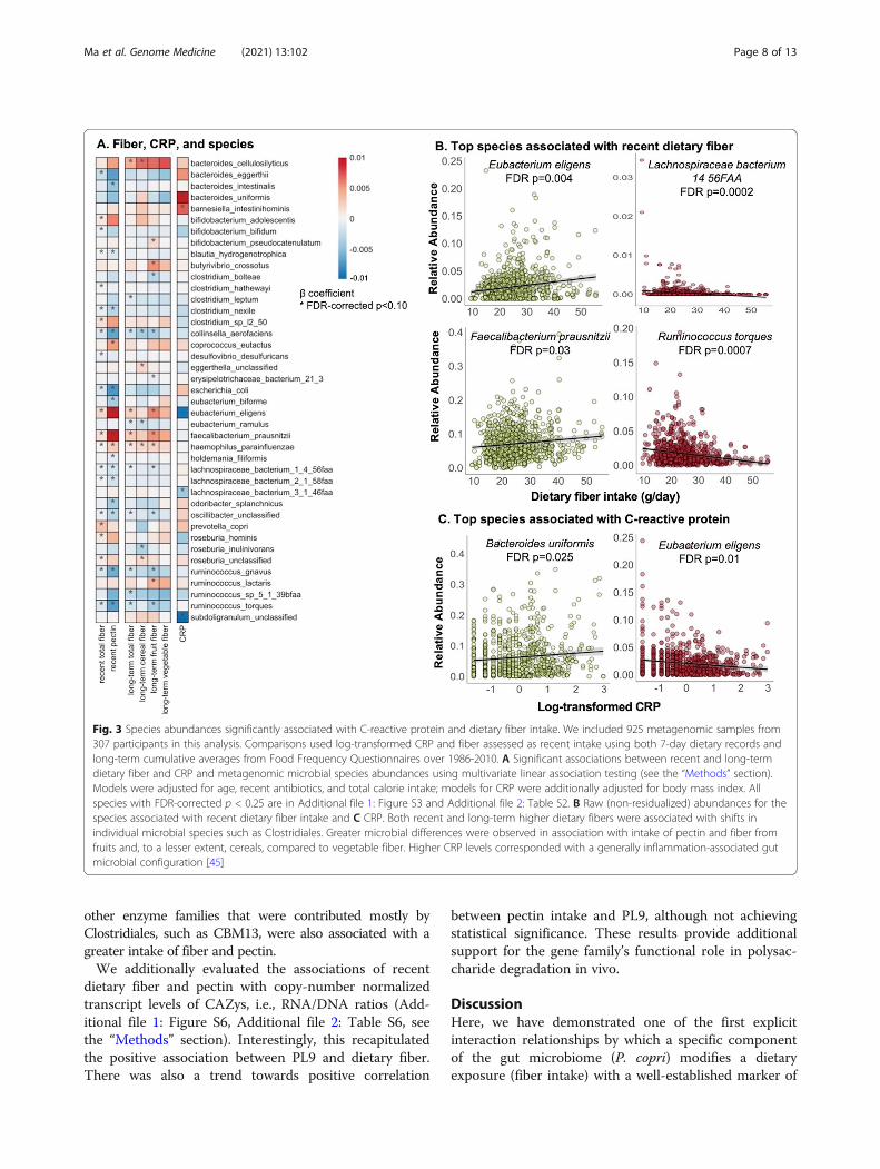
Individual members and functions in the microbiome areassociated with recent and long-term fiber intakequantities and CRPIn the absence of overall microbiome shifts with fiber in-take or CRP, we next identified individual microbial spe-cies associated with these variables using multivariatelinear mixed model in MaAsLin 2 [38] (Fig. 3). Allmodels included one fiber or inflammation outcome ofinterest, each participant’s membership as a random ef-fect, and were also adjusted for covariates including age,recent antibiotic use, and total calorie intake. Consistentwith previous studies [46], both recent and long-termhigher dietary fiber were associated with shifts inClostridiales, the major butyrate producers, includingincreases of Eubacterium eligens, Faecalibacterium
Fig. 1 Linking the gut microbiome, dietary fiber, and systemic inflammation in a cohort of adult males. A 307 participants nested within theHealth Professionals Follow-Up Study [23, 25] provided up to four stool samples with concurrent blood samples over a 6-month study period,generating 925 metagenomes from all participants and 372 metatranscriptomes from a subset of 96 selected because they provided stool atboth sampling periods and did not report antibiotic use during the past year. B Overall recent dietary fiber intake and C-reactive protein (as abiomarker of systemic inflammation) levels were distributed representatively across this population. C Recent dietary fiber intake was inverselycorrelated with body mass index as expected (r =−0.24), but not correlated with age (r =−0.02). D Major food sources of fiber intake includedcereals, vegetables, and fruits. E Principal coordinate analysis based on species-level Bray-Curtis dissimilarity decorated by quartiles of C-reactiveprotein and continuous fruit fiber intake suggested that fiber intake and CRP levels were not the overall largest sources of microbial communityvariability (other fiber subsets in Additional file 1: Figure S1)
Ma et al. Genome Medicine (2021) 13:102 Page 5 of 13

prausnitzii, and genus Roseburia, but also decreases inClostridium, Lachnospiraceae, and Ruminococcus spp.Increased fiber intake was also associated with increasedrelative abundances of Haemophilus parainfluenzae andBacteroides cellulosilyticus. These associations remainedrobust despite additional adjustment for Bristol scoreand other lifestyle factors including alcohol intake andphysical activity.Greater microbial differences were observed in associ-
ation with intake of pectin and fiber from fruits and, to alesser extent, cereals, compared to vegetable fiber. Forexample, the findings mentioned above includingpositive associations with E. eligens and F. prausnitziiand inverse associations with Lachnospiraceae andRuminococcus were largely driven by pectin and fruitfiber. The distinct chemical structures of dietary fiberslead to substantial variations in solubility and ferment-ability and subsequent effects on the microbial compos-ition and functions [9, 10]. Pectin is a soluble dietary
fiber rich in apples, pears, plums, and citrus fruits. Itcomprises a highly complex set of plant cell wall polysac-charides including homo-polygalacturonan, rhamnogalac-turonan I, and rhamnogalacturonan II [47]. Our resultswere in line with reports from in vitro and animal studiesthat pectin induced influences on the gut microbiota com-position, including increases of Clostridiales such as F.prausnitzii and a highly selective promotion of E. eligensas well as depletion of Bacteroidetes [48–50]. Associationsof soluble and insoluble fiber with microbial species weresimilar (Additional file 1: Figure S3, Additional file 2:Table S2), possibly due to the challenge of differentiatingthe soluble vs. insoluble subtypes through available dietinstruments [51].In multivariate models for CRP, higher CRP was asso-
ciated with enrichment of B. uniformis, B. salyersale,Barnesiella intestinihominis, and Parabacteroides inde-pendent of adiposity. In a previous analysis of 178 olderadult subjects, CRP was positively associated with a
Table 1 Characteristics of the 307 MLVS study participants according to quartiles of recent dietary fiber intake
Quartiles of recent dietary fiber intake
1 (n = 77) 2 (n = 77) 3 (n = 77) 4 (n = 76)
Age, years 70.0 (4.1) 71.9 (4.5) 70.0 (4.0) 70.2 (4.1)
Body mass index, kg/m2 26.1 (3.2) 26.9 (4.5) 25.2 (2.9) 24.1 (3.3)
Physical activity, MET-hrs/week 48.2 (39.9) 39.3 (31.2) 52.4 (42.3) 51.1 (32.5)
Total energy intake, kcal/d 2363 (475) 2231 (481) 2312(453) 2337 (473)
Total fiber intake, g/d 16.6 (2.6) 22.0 (1.4) 26.4 (1.4) 36.2 (6.5)
Cereal fiber, g/d 6.2 (1.9) 6.9 (1.9) 7.7 (2.3) 9.5 (3.4)
Fruit fiber, g/d 3.1 (1.5) 4.4 (2.0) 5.4 (2.2) 6.7 (3.1)
Vegetable fiber, g/d 6.2 (2.4) 7.3 (2.2) 7.1 (2.2) 8.2 (2.1)
Alcohol intake, %
Never 4.3 12.5 13.2 18.9
Rarely 17.0 14.3 15.1 17.0
1–6 times/week 38.3 37.5 43.4 34.0
Daily 36.2 32.1 26.4 30.2
More than daily 4.3 3.6 1.9 0
Antibiotic use, % 29.2 28.6 27.8 21.8
Probiotic use, % 4.3 3.6 11.5 5.8
Current smoker, % 2.6 0 1.3 0
Bristol score, %
1–2, hard stool 14.6 18.2 7.4 12.7
3–5, normal stool 79.2 78.2 87.0 81.8
6–7, loose stool 6.3 3.6 5.6 5.5
Total cholesterol, mg/dL 186 (38) 182 (41) 186 (42) 178 (33)
HDL cholesterol, mg/dL 59.9 (13.7) 55.1 (15.0) 56.2 (15.3) 55.5 (12.9)
Total/HDL cholesterol ratio 3.2 (0.8) 3.4 (0.9) 3.4 (0.8) 3.3 (0.7)
C-reactive protein, mg/dL 2.1 (2.8) 1.9 (2.9) 1.3 (2.1) 1.7 (2.5)
Values are means (SD) for continuous variables and percentages for categorical variables. Variables that differed across quartiles of recent intake of dietary fiberare bolded (general linear model with F test for continuous variables and Mantel-Haenszel chi-squared test for categorical variables; p < 0.05)
Ma et al. Genome Medicine (2021) 13:102 Page 6 of 13

metagenomically assembled Bacteroides co-abundancegroup [52]. However, unlike the positive associationobserved here with P. distasonis and P. johnsonii, theformer was shown to alleviate obesity and metabolicdysfunctions via production of succinate and secondarybile acids in mice [53]. Higher CRP levels were alsoassociated with depletion of Lachnospiraceae bacterium3 1 46FAA, E. eligens, and Bifidobacterium bifidum, con-sistent with their anti-inflammatory effects as shown inexperimental studies [50, 54, 55].Dietary fiber intake in particular recent intake from
pectin was also significantly associated with a large num-ber of metagenomic functional pathways (Additional file1: Figure S4, Additional file 2: Table S3) and features(Additional file 1: Figure S5, Additional file 2: Table S4)involved in the metabolism of carbohydrates and aminoacids. A greater total fiber intake was associated withsignificant enrichment of PWY-7456 β-(1,4)-mannandegradation, P124 Bifidobacterium shunt, PWY-5104 L-isoleucine biosynthesis IV, and PWY-6305 putrescinebiosynthesis, whereas the rest of pathways were generallydepleted. A higher intake of total fiber and pectin wasalso associated with significantly enriched expression ofEC 3.2.1.4, an endoglucanase (hydrolyzing of (1,4)-beta-D-glucan linkages in cellulose) and EC 2.4.1.1 (glycogenphosphorylase), both of which play a role in catalyzingthe degradation of glycans or polysaccharides.
Potential biochemical contributors to microbe-specificselection pressures from dietary fiberTo specifically investigate fiber intake in relation tofunctional capacity and activity relating to carbohydrateutilization, we further mapped gene families into
carbohydrate-active enzymes (CAZy) [37]. CAZy coversenzymes catalyzing the breakdown, biosynthesis, ormodification of carbohydrates and glycoconjugates in-cluding glycoside hydrolases (GHs), glucosylTransferases(GTs), polysaccharide lyases (PLs), carbohydrate ester-ases (CEs), auxiliary activities (AAs), and non-catalyticcarbohydrate-binding modules (CBMs). We excludedenzymes that did not surpass a minimum prevalence(10% of samples for CAZy DNA and 20% for RNA/DNAratio) and relative abundance (0.001% for CAZy DNA)threshold, resulting in 134 DNA CAZys and 121 RNA/DNA features in the analysisWe identified a total of 84 CAZys metagenomically as-
sociated with dietary fiber—again, particularly pectinand fruit fiber—using multivariate linear testing (Fig. 4A,Additional file 2: Table S5). Concordant with the chem-ical structure of pectin consisting of repeated units of α-(1-4)-linked D-galacturonic acid, and the fermentationrequirement of pectinase, we detected an enrichment ofPL9 strongly positively correlated with pectin intake.PL9 covers enzymes including pectate lyase (EC 4.2.2.2),exopolygalacturonate lyase (EC 4.2.2.9), thiopeptidoglycanlyase (EC 4.2.2.-), and rhamnogalacturonan endolyase (EC4.2.2.23) and participates in the degradation of homogalac-turonan. In our samples, expression of PL9 was primarilycontributed by E. eligens, followed by B. thetaiotaomicron,F. prausnitzii, and B. sp. 1_1_6 (Fig. 4B). An increase ofGH25, carried by diverse species of Eubacterium, Bacter-oides, and Faecalibacterium, was also strongly associatedwith fiber and pectin intake. Meanwhile, fiber and pectinalso showed inverse associations with some features, suchas GH29, contributed largely by Bacteroides species and in-volved in degradation of other glycan targets. Finally, some
Fig. 2 Prevotella copri carriage abrogates the protective effects of recent dietary fiber intake on C-reactive protein. Multivariate linear mixedmodels of log-transformed CRP were fit including recent fiber intake and P. copri carriage (binary), their interaction, accounting for participantmembership as a random effect, and adjusting for age, recent antibiotic use, and total calorie intake. We excluded 137 samples with CRP valuesbelow or above the detection limits, and thus, 788 samples from 277 participants were included in this analysis. The relationship between dietaryfiber and plasma CRP was significantly stronger among participants who did not carry P. copri (0 abundance)
Ma et al. Genome Medicine (2021) 13:102 Page 7 of 13
other enzyme families that were contributed mostly byClostridiales, such as CBM13, were also associated with agreater intake of fiber and pectin.We additionally evaluated the associations of recent
dietary fiber and pectin with copy-number normalizedtranscript levels of CAZys, i.e., RNA/DNA ratios (Add-itional file 1: Figure S6, Additional file 2: Table S6, seethe “Methods” section). Interestingly, this recapitulatedthe positive association between PL9 and dietary fiber.There was also a trend towards positive correlation
between pectin intake and PL9, although not achievingstatistical significance. These results provide additionalsupport for the gene family’s functional role in polysac-charide degradation in vivo.
DiscussionHere, we have demonstrated one of the first explicitinteraction relationships by which a specific componentof the gut microbiome (P. copri) modifies a dietaryexposure (fiber intake) with a well-established marker of
Fig. 3 Species abundances significantly associated with C-reactive protein and dietary fiber intake. We included 925 metagenomic samples from307 participants in this analysis. Comparisons used log-transformed CRP and fiber assessed as recent intake using both 7-day dietary records andlong-term cumulative averages from Food Frequency Questionnaires over 1986-2010. A Significant associations between recent and long-termdietary fiber and CRP and metagenomic microbial species abundances using multivariate linear association testing (see the “Methods” section).Models were adjusted for age, recent antibiotics, and total calorie intake; models for CRP were additionally adjusted for body mass index. Allspecies with FDR-corrected p < 0.25 are in Additional file 1: Figure S3 and Additional file 2: Table S2. B Raw (non-residualized) abundances for thespecies associated with recent dietary fiber intake and C CRP. Both recent and long-term higher dietary fibers were associated with shifts inindividual microbial species such as Clostridiales. Greater microbial differences were observed in association with intake of pectin and fiber fromfruits and, to a lesser extent, cereals, compared to vegetable fiber. Higher CRP levels corresponded with a generally inflammation-associated gutmicrobial configuration [45]
Ma et al. Genome Medicine (2021) 13:102 Page 8 of 13

Fig. 4 (See legend on next page.)
Ma et al. Genome Medicine (2021) 13:102 Page 9 of 13

systemic inflammation (plasma C-reactive proteinlevels). In addition to this interaction effect, direct effectsof dietary fiber intake on the gut microbiome generallyincreased Clostridiales, which also play a pivotal role inregulating localized and systemic inflammation [56].These microbial alterations also varied among specificfiber sources, with the greatest effects deriving from pec-tin and fruit fiber. For instance, abundances of E. eligensand F. prausnitzii as well as their functions in the deg-radation of polysaccharides were enriched in participantswith greater dietary fiber and, especially, pectin intake.We also linked individual microbial signatures tochronic systemic inflammation. These findings collect-ively offer novel human evidence supporting a variety offiber-gut-microbiome interactions relevant to chronicsystemic inflammation.Specifically, in our population, P. copri carriage elimi-
nated the strongly protective effects of increased fiberintake on systemic inflammation, with P. copri carriersdistributed across a range of generally modest CRP levelsand non-carriers varying between higher and lower ex-tremes according to fiber intake. The impact of P. coprion human health overall is still controversial, as conflict-ing results have been reported among different popula-tions and phenotypes. As a fiber-degrader, Prevotellawas positively associated with production of SCFAs (e.g.,propionate) [57, 58] and could, for example, providehost benefit by improved glucose metabolism in re-sponse to a high-fiber diet [40, 41]. Conversely, P. coprihas been associated with chronic inflammatory condi-tions such as rheumatoid arthritis [42, 43] and insulinresistance and glucose intolerance [44]. Strain-level het-erogeneity and distinct clades of the P. copri complexmay contribute to its functional diversity and some ofthese apparent phenotypic contradictions [59, 60]. Forinstance, genetically diverse P. copri isolates utilizedistinct sets of polysaccharides from dietary plantsources [59]. The combination of P. copri diversity, fibertype and amount diversity, and the gradual, multi-generational loss of P. copri clades from Westernizedpopulations could account for the complexity of thisinteraction [39]. Our findings suggest that P. copri couldin principle have both direct effects on systemic inflam-mation, as well as opposing, indirect effects caused by
reduced bioavailability of fermentable fibers or other fer-mentation products to other microbes. Additional inves-tigation is thus needed to functionally characterize theinfluence of P. copri on modulating dietary effects on in-flammation, health, and host-microbe coevolution.An additional intriguing result from this study was the
specificity of many fiber-microbiome influences to fruitfibers and pectin. As a major soluble fiber component inthe plant cell wall, particularly in fruits and vegetables,pectin serves as the nutritional niche for some groups ofbacteria, such as B. thetaiotaomicron [61], F. prausnitzii[49], and E. eligens [8]. It is likely that the chemical com-plexity of pectin relative to other fiber sources facilitatesits capacity to nourish diverse microbial communities[62]. Polysaccharide utilization loci that orchestrate thedetection, sequestration, enzymatic digestion, and trans-port of complex carbohydrates have been identified inmost gut-resident species, especially among the Bacteroi-detes [63]. However, knowledge of the impact of pectinin particular on the gut microbial communities is stilllimited and has been restricted to in vitro and animalstudies. To our knowledge, we for the first time identi-fied pectin-induced alterations in gut microbiota com-position and functional capabilities and subsequentimpact in inflammation in a human population study.These results suggest that pectin intake may exert a se-lection pressure on the gut microbiota leading to thepredominance of organisms that degrade pectic polysac-charides and an enhancement of functional activitiesspecifically based on their utilization. This supports thenotion that gut microbial strains are highly specialized,particularly with respect to carbon source utilization andproducts, and can evolve and adapt over the course ofan adult lifetime to utilize a unique subset of complexpolysaccharides in a personalized, individual-specificmanner [61].At least one additional recent study, using a distinct
population and methodology, found potentially similarbetween-subject variation in fiber sources with respectto the microbiome. There, significant agreementbetween microbiome composition and fiber-sourcediversity was observed for fruits and grains, but not forvegetables or legumes [64]. Such heterogeneity accord-ing to fiber sources might be explained not only by the
(See figure on previous page.)Fig. 4 CAZy and dietary fiber intake. A Significant associations between recent and long-term dietary fiber and CAZy DNA abundances usingmultivariate linear association testing (see the “Methods” section, Additional file 2: Table S5). Comparisons used fiber assessed as recent intakeusing both 7-day dietary records and long-term cumulative averages from Food Frequency Questionnaires over 1986–2010. Models wereadjusted for age, recent antibiotics, and total calorie intake. B Abundances of metagenomes and metatranscriptomes of polysaccharide lyasefamily 9 (PL9), glycoside hydrolase family 29 (GH29), and carbohydrate-binding module family 13 (CBM13) by contributing species and samples,with species ranked by mean relative abundance, and samples ranked by pectin intake. We included 925 metagenomes and 341metatranscriptomes in this analysis. A total of 84 CAZys metagenomically associated with dietary fiber in particular pectin and fruit fiber. Positiveassociations were observed particularly for several CAZy DNA and RNA/DNA ratios contributed by Clostridiales species
Ma et al. Genome Medicine (2021) 13:102 Page 10 of 13

distinct chemical structures of fibers in each type of food[10], but also by other fruit-specific bioactive com-pounds such as polyphenols (flavonoids, phenolic acids,and carotenoids [65]) and even cooking (raw vs. cookedplant foods) [66]. Our population-based investigationdoes not distinguish between these potential mecha-nisms, for which in vitro studies and randomized con-trolled trials of specific fibers are better suited (althoughthese cannot, conversely, assess the long-term effects ofdietary fiber). Likewise, as an observational study, wecannot be definitive about causality and although thefiber-microbiome-inflammation association was robustdespite adjustment for many variables, we cannot ruleout the potential for residual confounding. Finally, sinceour study only included older adult men in the US, weare cautious about generalizability to other populations,especially, younger and non-Western populations inwhom relevant dietary or microbial components may bequite distinct). Thus, we plan to validate these findingsin additional cohorts with information on diet, the gutmicrobiome, and health outcomes.
ConclusionsAs one of the only sustainable long-term influences onthe gut microbiome and chronic health, dietary interac-tions and interventions are a key strategy to mitigatechronic inflammation. Our findings will benefit from fur-ther investigation of the specific mechanisms by which P.copri mediates dietary biochemistry and host inflamma-tion, as well as the specific routes by which pectin directlyinfluences other gut microbiome members. An under-standing of these distinct effects of dietary fibers, pectin,and how they are transformed and utilized by microbialcommunities would pave the way forward for develop-ment of personalized fiber-based interventions for theprevention of chronic inflammatory diseases.
AbbreviationsCRP: C-reactive protein; SCFA: Short-chain fatty acids; MLVS: Men’s LifestyleValidation Study; HPFS: Health Professionals Follow-Up Study; FFQ: FoodFrequency Questionnaire; EC: Enzyme commission; CAZy: Carbohydrate-active enzymes; FDR: False discovery rate
Supplementary InformationThe online version contains supplementary material available at https://doi.org/10.1186/s13073-021-00921-y.
Additional file 1: Figures S1-S6.
Additional file 2: Tables S1-S6.
AcknowledgementsWe would like to thank the participants in the Health Professionals Follow-Up Study for their continuing outstanding level of cooperation and the stafffor their valuable contributions. The authors assume full responsibility foranalyses and interpretation of these data.
Authors’ contributionsWM, CH, and ATC conceived and designed the study; EBR, JI, CH, and ATCcollected data; WM, LHN, CH, and ATC performed the analysis. All authorsinterpreted the data with critical revision of the manuscript for importantintellectual content, and read and approved the final manuscript.
FundingThis work was supported by the National Institutes of Health (NIH) grantsU54DE023798, U01CA152904, U01CA167552, R01CA202704, R01HL35464,R01DK101495, R24DK110499, K24DK098311, and R21AA027608 and by theStarr Cancer Consortium. WM was supported by the MGH ECOR Tostesonand Fund for Medical Discovery Postdoctoral Fellowship Award. JI waspartially supported by the Hatch Multistate Research capacity fundingprogram W4122 from the USDA National Institute of Food and Agriculture.ATC is a Stuart and Suzanne Steele MGH Research Scholar. The fundingbodies had no role in the design and conduct of the study; collection,analysis, and interpretation of data; and writing the manuscript. The contentis solely the responsibility of the authors and does not necessarily representthe official views of the funders.
Availability of data and materialsAll the metadata from the Health Professionals Follow-Up Study are availablethrough a request for external collaboration and upon approvals of a letterof intent and a research proposal. Details for how to request an external col-laboration with the Health Professionals Follow-Up Study can be found athttps://sites.sph.harvard.edu/hpfs/for-collaborators/. Source code that gener-ates the figures and tables is available at https://github.com/biobakery/Fiber-Microbiome-Inflammation [67].
Declarations
Ethics approval and consent to participateThe study was approved by the Harvard T.H. Chan School of Public HealthInstitutional Review Board (# HSPH 22067-102). Written informed consentwas obtained from all participants. The research conformed to the Principlesof the Helsinki Declaration.
Consent for publicationNot applicable.
Competing interestsThe authors declare that they have no competing interests.
Author details1Clinical and Translational Epidemiology Unit, Massachusetts General Hospitaland Harvard Medical School, Boston, MA, USA. 2Division of Gastroenterology,Massachusetts General Hospital and Harvard Medical School, Boston, MA,USA. 3Department of Biostatistics, Harvard T.H. Chan School of Public Health,Boston, MA, USA. 4Department of Epidemiology, Harvard T.H. Chan School ofPublic Health, Boston, MA, USA. 5Department of Nutrition, Harvard T.H. ChanSchool of Public Health, Boston, MA, USA. 6Division of Public Health Sciences,Department of Surgery, Washington University School of Medicine, St Louis,MO, USA. 7Alvin J. Siteman Cancer Center, Washington University School ofMedicine, St Louis, MO, USA. 8Division of Gastroenterology, Department ofMedicine, Washington University School of Medicine, St Louis, MO, USA.9Microbiome and Host Health Programme, South Australian Health andMedical Research Institute, North Terrace, Adelaide, SA 5000, Australia.10Department of Nutrition and Dietetics, College of Nursing and HealthSciences, Flinders University, Adelaide, Australia. 11Division ofGastroenterology, University of Washington School of Medicine, Seattle, WA,USA. 12Channing Division of Network Medicine, Department of Medicine,Brigham and Women’s Hospital and Harvard Medical School, Boston, MA,USA. 13Department of Food Science and Technology, University ofNebraska-Lincoln, Lincoln, NE, USA. 14Fred and Pamela Buffett Cancer Center,University of Nebraska Medical Center, Omaha, NE, USA. 15School ofBiological Sciences, University of Nebraska, Lincoln, NE, USA. 16Department ofImmunology and Infectious Diseases, Harvard T.H. Chan School of PublicHealth, Boston, MA, USA. 17Department of Medicine, Harvard Medical School,Boston, MA, USA. 18Broad Institute of MIT and Harvard, Cambridge, MA, USA.
Ma et al. Genome Medicine (2021) 13:102 Page 11 of 13

Received: 1 December 2020 Accepted: 8 June 2021
References1. Collaborators GBDCoD. Global, regional, and national age-sex-specific
mortality for 282 causes of death in 195 countries and territories, 1980–2017: a systematic analysis for the Global Burden of Disease Study 2017.Lancet. 2018;392(10159):1736–88. https://doi.org/10.1016/S0140-6736(18)32203-7.
2. Cani PD, Possemiers S, Van de Wiele T, Guiot Y, Everard A, Rottier O, et al.Changes in gut microbiota control inflammation in obese mice through amechanism involving GLP-2-driven improvement of gut permeability. Gut.2009;58(8):1091–103. https://doi.org/10.1136/gut.2008.165886.
3. Llewellyn SR, Britton GJ, Contijoch EJ, Vennaro OH, Mortha A, Colombel JF,et al. Interactions between diet and the intestinal microbiota alter intestinalpermeability and colitis severity in mice. Gastroenterology. 2018;154(4):1037–46 e2. https://doi.org/10.1053/j.gastro.2017.11.030.
4. Reynolds A, Mann J, Cummings J, Winter N, Mete E, Te Morenga L.Carbohydrate quality and human health: a series of systematic reviews andmeta-analyses. Lancet. 2019;393(10170):434–45. https://doi.org/10.1016/S0140-6736(18)31809-9.
5. Threapleton DE, Greenwood DC, Evans CE, Cleghorn CL, Nykjaer C,Woodhead C, et al. Dietary fibre intake and risk of cardiovascular disease:systematic review and meta-analysis. BMJ. 2013;347(dec19 2):f6879. https://doi.org/10.1136/bmj.f6879.
6. Ananthakrishnan AN, Khalili H, Konijeti GG, Higuchi LM, de Silva P, KorzenikJR, et al. A prospective study of long-term intake of dietary fiber and risk ofCrohn’s disease and ulcerative colitis. Gastroenterology. 2013;145(5):970–7.https://doi.org/10.1053/j.gastro.2013.07.050.
7. Ma W, Nguyen LH, Song M, Jovani M, Liu PH, Cao Y, et al. Intake of dietaryfiber, fruits, and vegetables and risk of diverticulitis. Am J Gastroenterol.2019;114(9):1531–8. https://doi.org/10.14309/ajg.0000000000000363.
8. Chung WS, Walker AW, Louis P, Parkhill J, Vermeiren J, Bosscher D, et al.Modulation of the human gut microbiota by dietary fibres occurs at thespecies level. BMC Biol. 2016;14(1):3. https://doi.org/10.1186/s12915-015-0224-3.
9. Baxter NT, Schmidt AW, Venkataraman A, Kim KS, Waldron C, Schmidt TM.Dynamics of human gut microbiota and short-chain fatty acids in responseto dietary interventions with three fermentable fibers. MBio. 2019;10(1).
10. Deehan EC, Yang C, Perez-Munoz ME, Nguyen NK, Cheng CC, Triador L,et al. Precision microbiome modulation with discrete dietary fiber structuresdirects short-chain fatty acid production. Cell Host Microbe. 2020;27(3):389–404 e6.
11. Sonnenburg ED, Smits SA, Tikhonov M, Higginbottom SK, Wingreen NS,Sonnenburg JL. Diet-induced extinctions in the gut microbiota compoundover generations. Nature. 2016;529(7585):212–5. https://doi.org/10.1038/nature16504.
12. Koh A, De Vadder F, Kovatcheva-Datchary P, Backhed F. From dietary fiberto host physiology: short-chain fatty acids as key bacterial metabolites. Cell.2016;165(6):1332–45. https://doi.org/10.1016/j.cell.2016.05.041.
13. Segain JP, de la Bletiere DR, Bourreille A, Leray V, Gervois N, Rosales C, et al.Butyrate inhibits inflammatory responses through NFkappaB inhibition:implications for Crohn's disease. Gut. 2000;47(3):397–403. https://doi.org/10.1136/gut.47.3.397.
14. Inan MS, Rasoulpour RJ, Yin L, Hubbard AK, Rosenberg DW, Giardina C. Theluminal short-chain fatty acid butyrate modulates NF-kappaB activity in ahuman colonic epithelial cell line. Gastroenterology. 2000;118(4):724–34.https://doi.org/10.1016/S0016-5085(00)70142-9.
15. Smith PM, Howitt MR, Panikov N, Michaud M, Gallini CA, Bohlooly YM, et al.The microbial metabolites, short-chain fatty acids, regulate colonic Treg cellhomeostasis. Science. 2013;341(6145):569–73. https://doi.org/10.1126/science.1241165.
16. Schroeder BO, Birchenough GMH, Stahlman M, Arike L, Johansson MEV,Hansson GC, et al. Bifidobacteria or fiber protects against diet-inducedmicrobiota-mediated colonic mucus deterioration. Cell Host Microbe. 2018;23(1):27–40 e7. https://doi.org/10.1016/j.chom.2017.11.004.
17. Desai MS, Seekatz AM, Koropatkin NM, Kamada N, Hickey CA, Wolter M,et al. A dietary fiber-deprived gut microbiota degrades the colonic mucusbarrier and enhances pathogen susceptibility. Cell. 2016;167(5):1339–53 e21.https://doi.org/10.1016/j.cell.2016.10.043.
18. So D, Whelan K, Rossi M, Morrison M, Holtmann G, Kelly JT, et al. Dietaryfiber intervention on gut microbiota composition in healthy adults: asystematic review and meta-analysis. Am J Clin Nutr. 2018;107(6):965–83.https://doi.org/10.1093/ajcn/nqy041.
19. Koenig W, Sund M, Frohlich M, Fischer HG, Lowel H, Doring A, et al. C-Reactive protein, a sensitive marker of inflammation, predicts future risk ofcoronary heart disease in initially healthy middle-aged men: results from theMONICA (Monitoring Trends and Determinants in Cardiovascular Disease)Augsburg Cohort Study, 1984 to 1992. Circulation. 1999;99(2):237–42.https://doi.org/10.1161/01.cir.99.2.237.
20. Aleksandrova K, Jenab M, Boeing H, Jansen E, Bueno-de-Mesquita HB,Rinaldi S, et al. Circulating C-reactive protein concentrations and risks ofcolon and rectal cancer: a nested case-control study within the EuropeanProspective Investigation into Cancer and Nutrition. Am J Epidemiol. 2010;172(4):407–18. https://doi.org/10.1093/aje/kwq135.
21. Lochhead P, Khalili H, Ananthakrishnan AN, Richter JM, Chan AT. Associationbetween circulating levels of c-reactive protein and interleukin-6 and risk ofinflammatory bowel disease. Clin Gastroenterol Hepatol. 2016;14(6):818–24e6. https://doi.org/10.1016/j.cgh.2016.01.016.
22. Nguyen LH, Ma W, Wang DD, Cao Y, Mallick H, Gerbaba TK, et al.Association between sulfur-metabolizing bacterial communities in stool andrisk of distal colorectal cancer in men. Gastroenterology. 2020;158(5):1313–25. https://doi.org/10.1053/j.gastro.2019.12.029.
23. Abu-Ali GS, Mehta RS, Lloyd-Price J, Mallick H, Branck T, Ivey KL, et al.Metatranscriptome of human faecal microbial communities in a cohort ofadult men. Nat Microbiol. 2018;3(3):356–66. https://doi.org/10.1038/s41564-017-0084-4.
24. Franzosa EA, Morgan XC, Segata N, Waldron L, Reyes J, Earl AM, et al.Relating the metatranscriptome and metagenome of the human gut. ProcNatl Acad Sci U S A. 2014;111(22):E2329–38. https://doi.org/10.1073/pnas.1319284111.
25. Mehta RS, Abu-Ali GS, Drew DA, Lloyd-Price J, Subramanian A, Lochhead P,et al. Stability of the human faecal microbiome in a cohort of adult men.Nat Microbiol. 2018;3(3):347–55. https://doi.org/10.1038/s41564-017-0096-0.
26. Rimm EB, Giovannucci EL, Stampfer MJ, Colditz GA, Litin LB, Willett WC.Reproducibility and validity of an expanded self-administeredsemiquantitative Food Frequency qQuestionnaire among male healthprofessionals. Am J Epidemiol. 1992;135(10):1114–26; discussion 27-36.https://doi.org/10.1093/oxfordjournals.aje.a116211.
27. Prosky L, Asp NG, Furda I, DeVries JW, Schweizer TF, Harland BF.Determination of total dietary fiber in foods and food products:collaborative study. J Assoc Off Anal Chem. 1985;68(4):677–9.
28. Willett W. Nutritional Epidemiology Third ed: Oxford University Press; 2012.https://doi.org/10.1093/acprof:oso/9780199754038.001.0001.
29. Schakel SF, Sievert YA, Buzzard IM. Sources of data for developing andmaintaining a nutrient database. J Am Diet Assoc. 1988;88(10):1268–71.
30. McIver LJ, Abu-Ali G, Franzosa EA, Schwager R, Morgan XC, Waldron L, et al.bioBakery: a meta’omic analysis environment. Bioinformatics. 2018;34(7):1235–7. https://doi.org/10.1093/bioinformatics/btx754.
31. Truong DT, Franzosa EA, Tickle TL, Scholz M, Weingart G, Pasolli E, et al.MetaPhlAn2 for enhanced metagenomic taxonomic profiling. Nat Methods.2015;12(10):902–3. https://doi.org/10.1038/nmeth.3589.
32. Franzosa EA, McIver LJ, Rahnavard G, Thompson LR, Schirmer M, WeingartG, et al. Species-level functional profiling of metagenomes andmetatranscriptomes. Nat Methods. 2018;15(11):962–8. https://doi.org/10.1038/s41592-018-0176-y.
33. Langmead B, Salzberg SL. Fast gapped-read alignment with Bowtie 2. NatMethods. 2012;9(4):357–9. https://doi.org/10.1038/nmeth.1923.
34. Suzek BE, Huang H, McGarvey P, Mazumder R, Wu CH. UniRef: comprehensiveand non-redundant UniProt reference clusters. Bioinformatics. 2007;23(10):1282–8. https://doi.org/10.1093/bioinformatics/btm098.
35. Buchfink B, Xie C, Huson DH. Fast and sensitive protein alignment usingDIAMOND. Nat Methods. 2015;12(1):59–60. https://doi.org/10.1038/nmeth.3176.
36. Caspi R, Billington R, Ferrer L, Foerster H, Fulcher CA, Keseler IM, et al. TheMetaCyc database of metabolic pathways and enzymes and the BioCyccollection of pathway/genome databases. Nucleic Acids Res. 2016;44(D1):D471–80. https://doi.org/10.1093/nar/gkv1164.
37. Lombard V, Golaconda Ramulu H, Drula E, Coutinho PM, Henrissat B. Thecarbohydrate-active enzymes database (CAZy) in 2013. Nucleic Acids Res.2014;42(Database issue):D490–5. https://doi.org/10.1093/nar/gkt1178.
Ma et al. Genome Medicine (2021) 13:102 Page 12 of 13

38. Mallick H, McIver LJ, Rahnavard A, Ma S, Zhang Y, Nguyen LH, et al.Multivariable association discovery in population-scale meta-omicsstudies. 2020.
39. Tett A, Huang KD, Asnicar F, Fehlner-Peach H, Pasolli E, Karcher N, et al. ThePrevotella copri complex comprises four distinct clades underrepresented inwesternized populations. Cell Host Microbe. 2019;26(5):666–79 e7. https://doi.org/10.1016/j.chom.2019.08.018.
40. Kovatcheva-Datchary P, Nilsson A, Akrami R, Lee YS, De Vadder F, Arora T,et al. Dietary fiber-induced improvement in glucose metabolism isassociated with increased abundance of Prevotella. Cell Metab. 2015;22(6):971–82. https://doi.org/10.1016/j.cmet.2015.10.001.
41. De Vadder F, Kovatcheva-Datchary P, Zitoun C, Duchampt A, Backhed F,Mithieux G. Microbiota-produced succinate improves glucose homeostasisvia intestinal gluconeogenesis. Cell Metab. 2016;24(1):151–7. https://doi.org/10.1016/j.cmet.2016.06.013.
42. Scher JU, Sczesnak A, Longman RS, Segata N, Ubeda C, Bielski C, et al.Expansion of intestinal Prevotella copri correlates with enhancedsusceptibility to arthritis. Elife. 2013;2:e01202. https://doi.org/10.7554/eLife.01202.
43. Pianta A, Arvikar S, Strle K, Drouin EE, Wang Q, Costello CE, et al. Evidenceof the immune relevance of prevotella copri, a gut microbe, in patientswith rheumatoid arthritis. Arthritis Rheumatol. 2017;69(5):964–75. https://doi.org/10.1002/art.40003.
44. Pedersen HK, Gudmundsdottir V, Nielsen HB, Hyotylainen T, Nielsen T,Jensen BA, et al. Human gut microbes impact host serum metabolome andinsulin sensitivity. Nature. 2016;535(7612):376–81. https://doi.org/10.1038/nature18646.
45. Lloyd-Price J, Arze C, Ananthakrishnan AN, Schirmer M, Avila-Pacheco J,Poon TW, et al. Multi-omics of the gut microbial ecosystem in inflammatorybowel diseases. Nature. 2019;569(7758):655–62. https://doi.org/10.1038/s41586-019-1237-9.
46. Lin D, Peters BA, Friedlander C, Freiman HJ, Goedert JJ, Sinha R, et al.Association of dietary fibre intake and gut microbiota in adults. Br J Nutr.2018;120(9):1014–22. https://doi.org/10.1017/S0007114518002465.
47. Caffall KH, Mohnen D. The structure, function, and biosynthesis of plant cellwall pectic polysaccharides. Carbohydr Res. 2009;344(14):1879–900. https://doi.org/10.1016/j.carres.2009.05.021.
48. Licht TR, Hansen M, Bergstrom A, Poulsen M, Krath BN, Markowski J, et al.Effects of apples and specific apple components on the cecal environmentof conventional rats: role of apple pectin. BMC Microbiol. 2010;10(1):13.https://doi.org/10.1186/1471-2180-10-13.
49. Lopez-Siles M, Khan TM, Duncan SH, Harmsen HJ, Garcia-Gil LJ, Flint HJ.Cultured representatives of two major phylogroups of human colonicFaecalibacterium prausnitzii can utilize pectin, uronic acids, and host-derived substrates for growth. Appl Environ Microbiol. 2012;78(2):420–8.https://doi.org/10.1128/AEM.06858-11.
50. Chung WSF, Meijerink M, Zeuner B, Holck J, Louis P, Meyer AS, et al.Prebiotic potential of pectin and pectic oligosaccharides to promote anti-inflammatory commensal bacteria in the human colon. FEMS MicrobiolEcol. 2017;93(11).
51. Marlett JA, Chesters JG, Longacre MJ, Bogdanske JJ. Recovery of solubledietary fiber is dependent on the method of analysis. Am J Clin Nutr. 1989;50(3):479–85. https://doi.org/10.1093/ajcn/50.3.479.
52. Claesson MJ, Jeffery IB, Conde S, Power SE, O'Connor EM, Cusack S, et al.Gut microbiota composition correlates with diet and health in the elderly.Nature. 2012;488(7410):178–84. https://doi.org/10.1038/nature11319.
53. Wang K, Liao M, Zhou N, Bao L, Ma K, Zheng Z, et al. Parabacteroidesdistasonis alleviates obesity and metabolic dysfunctions via production ofsuccinate and secondary bile acids. Cell Rep. 2019;26(1):222–35 e5. https://doi.org/10.1016/j.celrep.2018.12.028.
54. Duranti S, Gaiani F, Mancabelli L, Milani C, Grandi A, Bolchi A, et al.Elucidating the gut microbiome of ulcerative colitis: bifidobacteria as novelmicrobial biomarkers. FEMS Microbiol Ecol. 2016;92(12).
55. Truax AD, Chen L, Tam JW, Cheng N, Guo H, Koblansky AA, et al. Theinhibitory innate immune sensor NLRP12 maintains a threshold againstobesity by regulating gut microbiota homeostasis. Cell Host Microbe. 2018;24(3):364–78 e6. https://doi.org/10.1016/j.chom.2018.08.009.
56. Sokol H, Pigneur B, Watterlot L, Lakhdari O, Bermudez-Humaran LG,Gratadoux JJ, et al. Faecalibacterium prausnitzii is an anti-inflammatorycommensal bacterium identified by gut microbiota analysis of Crohn
disease patients. Proc Natl Acad Sci U S A. 2008;105(43):16731–6. https://doi.org/10.1073/pnas.0804812105.
57. De Filippo C, Cavalieri D, Di Paola M, Ramazzotti M, Poullet JB, Massart S,et al. Impact of diet in shaping gut microbiota revealed by a comparativestudy in children from Europe and rural Africa. Proc Natl Acad Sci U S A.2010;107(33):14691–6. https://doi.org/10.1073/pnas.1005963107.
58. De Filippis F, Pellegrini N, Vannini L, Jeffery IB, La Storia A, Laghi L, et al.High-level adherence to a Mediterranean diet beneficially impacts the gutmicrobiota and associated metabolome. Gut. 2016;65(11):1812–21. https://doi.org/10.1136/gutjnl-2015-309957.
59. Fehlner-Peach H, Magnabosco C, Raghavan V, Scher JU, Tett A, Cox LM,et al. Distinct polysaccharide utilization profiles of human intestinalprevotella copri isolates. Cell Host Microbe. 2019;26(5):680–90 e5. https://doi.org/10.1016/j.chom.2019.10.013.
60. De Filippis F, Pasolli E, Tett A, Tarallo S, Naccarati A, De Angelis M, et al.Distinct genetic and functional traits of human intestinal prevotella copristrains are associated with different habitual diets. Cell Host Microbe. 2019;25(3):444–53 e3. https://doi.org/10.1016/j.chom.2019.01.004.
61. Martens EC, Lowe EC, Chiang H, Pudlo NA, Wu M, McNulty NP, et al.Recognition and degradation of plant cell wall polysaccharides by twohuman gut symbionts. PLoS Biol. 2011;9(12):e1001221. https://doi.org/10.1371/journal.pbio.1001221.
62. Chung WSF, Walker AW, Vermeiren J, Sheridan PO, Bosscher D, Garcia-Campayo V, et al. Impact of carbohydrate substrate complexity on thediversity of the human colonic microbiota. FEMS Microbiol Ecol. 2019;95(1).
63. Grondin JM, Tamura K, Dejean G, Abbott DW, Brumer H. Polysaccharideutilization loci: fueling microbial communities. J Bacteriol. 2017;199(15).
64. Johnson AJ, Vangay P, Al-Ghalith GA, Hillmann BM, Ward TL, Shields-CutlerRR, et al. Daily sampling reveals personalized diet-microbiome associationsin humans. Cell Host Microbe. 2019;25(6):789–802 e5. https://doi.org/10.1016/j.chom.2019.05.005.
65. Padayachee A, Day L, Howell K, Gidley MJ. Complexity and healthfunctionality of plant cell wall fibers from fruits and vegetables. Crit RevFood Sci Nutr. 2017;57(1):59–81. https://doi.org/10.1080/10408398.2013.850652.
66. Carmody RN, Bisanz JE, Bowen BP, Maurice CF, Lyalina S, Louie KB, et al.Cooking shapes the structure and function of the gut microbiome. NatMicrobiol. 2019;4(12):2052–63. https://doi.org/10.1038/s41564-019-0569-4.
67. Ma W. 2021. Available from: https://github.com/biobakery/Fiber-Microbiome-Inflammation.
Publisher’s NoteSpringer Nature remains neutral with regard to jurisdictional claims inpublished maps and institutional affiliations.
Ma et al. Genome Medicine (2021) 13:102 Page 13 of 13