Embed Size (px)
Citation preview
ATTENUATION OF THE
HEMODYNAMIC RESPONSE TO
TRACHEAL INTUBATION
A COMPARATIVE STUDY BETWEEN
INTRA VENOUS LIGNOCAINE
HYDROCHLORIDE AND INTRAVENOUS
MAGNESIUM SULPHATE
By
DR NIK MARZUKI MOHAMJ.AAD
Dissertation Submitted in Partial Fulfillment of the •• ' f l ~ f C . ' I,', \ l.\·,'
Requirements for the Degree of
Master Of Medicine
(Anesthesiology)
UNNERSITI SAINS MALAYSIA
NOVEMBER 2001
, ACKNOWLEDGEMENT
I would like to extend my deepest thanks and appreciation to the
followings that had been kind enough in assisting me with their advice and support
towards the completion of my dissertation.
Professor Madya Dr. Nik . Abdullah bin Nik Mohamad, Clinical
Lecturer and Head of Department of Anesthesiology and Critical Care Medicine,
Hospital Universiti Sains Malaysia and supervisor for this dissertation.
Dr. Mahamarowi Omar, Clinical Lecturer, Department of ~
Anesthesiology and Critical Care Medicine, Hospital Universiti Sains Malaysia.
All medical officers and colleagues of Department of Anesthesiology
and Critical Care Medicine, Hospital Universiti Sains Malaysia.
All staffs of the general operation theatre, intensive care unit of
Hospital Universiti Sains Malaysia.
Last but not least, my parents Mohammad bin Y unus and Tuan
Hasmah binti Tuan Abdullah, my loving wife, Azrina binti Hashim and my eldest son
Nik Abdul Hakim.
the administration of both study drug and observing the said cardiovascular
responses.
A standardized anesthetic technique was used for both groups
of patients. An indwelling venous cannula was inserted under local anesthesia
and a running intravenous drip was set. Vital signs monitoring were monitored
and chartered before induction which served as a baseline to the hemodynamic
changes expected to be seen at different stages of the induction period.
Induction of anesthesia was perfonned by uSing sodium
thipentone 2.5% solution titrated to the loss of the eye lash reflex, the study
drugs of each allocated popUlation sample was administered accordingly and a
single attempt at cannulation of the endotracheal tube by a single operator
through out the study was performed 3 minutes after the neuromuscular
blocking agent was given.
Anesthesia was maintained with oxgen:nitrous oxide in a 1:2
flow ratio and expired isoflurane mean alveolar concentration (MAC) was
kept at I % while pharmacological paralysis was maintained via imtermittent
bolus doses of intravenous atracurium.Patients were then reversed with
intravenous neostigmine 2.5 mg and intravenous atropine at 1 mg.
Data collection consisted of pulse rate, systolic, diastolic and
mean arterial blood pressure (via a non invasive lllood pressure monitoring
III
device) at selected intervals of pre induction, post induction, post study drug
adminstration and post intubation attempt.
Findings of this study when companng the hemodynamic
variables in question between the two groups revealed that although there
were increases in the systolic, diastolic, mean arterial pressure, and heart rate
parameters in each of the two groups these changes were statistically not
significant Within the respective group and also changes in the cardiovascular
response varaible were not statistically significant comparing the two groups.
Thus the results of this study pointed to the fact that both
intravenous lignocaine and intravenous magnesium were able to minimize the
hemodynamic changes occuring during the act of intubation and more
importantly, that intravenous magnesium was able to blunt the
sympathoadrenomedullary responses to stimulation of the upper respiratory
tract and tracheobronchial tree by the act of endotracheal cannulation.
IV
v
ABSTRAK
Doktor bius telah mencuba berbagai-bagai regim farmakologi dalam
usaha mereka untuk menghalang penghantaran impuls yang menyalurkan isyarat
untuk respons hemodinamik tidak kira sarna ada dari sal uran afferen atau efferen atau
pun kedua-duanya sekali walaupun tidak ada satu pun cara yang boleh di katakan
sempuma kerana ke semua Iangkah-Iangkah yang telah di ilhamkan mempunyai kesan
sampingannya yang tertentu.
Peningkatan tekanan darah aterial, peningkatan tekanan intra kranial,
peningkatan kadar denyut nadi dan berlakunya komplikasi aritmia adalah antara
kesan-kesan langsung komplikasi dari percubaan laringoskopi dan cubaan intubasi.
Sungguh pun perubahan yang di kesani berlaku di dalam sirkulasi
manusia adalah bersifat sementara dan kesan langsung ini ke atas pesakit dari segi
perubatan di klasifikasikan sebagai sihat adalah tidak memudaratkan tetapi kesan
perubahan hemodinamik ini jika berlaku ke atas pesakit yang menghidapi sakit
jantung koronari, penghidap penyakit darah tinggi, dan pesakit yang mengalami
kompromi saluran darah otak adalah amat berbahaya.
SejumJah 50 pesakit ASA I, di pilih secara rambang yang di jadualkan
untuk pembedahan minor elektif ie. "excision biopsy, herniotomy, split skin grafting,
removal of implant, subtotal thyroidectomy", dan memerIukan pembiusan secara total
yang mana intubasi adalah satu kemestian telah di bahagikan kepada 2 kumpulan
berasingan di mana masing-masing akan menerima sarna ada lignocaine intravena 1.5
mglkg berat badan (Kumpulan 1, n=25) atau pun menerima intravena magnesium
VI
kepekatan 50% pada kadar 40 mg/kg berat badan (Kumpulan 2, n=25). Personel yang
terlibat di dalam pembahagian kumpulan dan penyediaan ubatan yang berkenaan tidak
terIibat di dalam pemberian ubat berikut secara intravena dan juga tidak terlibat di
dalam merekod perubahan hemodinamik yang di perhatikan.
Teknik pembiusan adalah di seragamkan kepada satu standard dan di
gunakan kepada kedua-dua kumpulan. yang terlibat. Saluran darah vena di
kanulasikan secara pemberian bius setempat dan aliran cecair di pasangkan. Tanda
tanda vital di rekodkan sebelum bennulanya induksi bius yang mana ini juga di ambil
kira sebagai tanda vital asas sebelum perubahan hemodinamik di Iihat berlaku.
Induksi bius di teruskan dengan pemberian sodium thiopentone pada
kepekatan 2.5% sehingga tanda refleks kelipan mata tidak lagi di kesan, ubat yang
telah di jadualkan untuk kajian di berikan ke dalan vena pesakit dan cubaan intubasi
dari hanya seorang doktor bius yang sarna sepanjang tempoh kajian di jalankan
selepas 3 minit ubat penghalang neuromuskular di berikan.
Pembiusan di teruskan dengan penggunaan pembiusan gas isoflurane
pada kepekatan ekspirasi MAC 1 % dan oksigen : nitrous oksida pada 1:2 kadar aliran
gas dan pengekalan kelumpuhan pesakit adalah dengan pemberian ubat
atracurium.Setelah selesai pembedahan yang di jalanlcan, pesakit di sedarkan dan
diberi neostigmine 2.5 mg dan atropine 1 mg.
Data yang di kumpulkan terdiri dari kadar denyut nadi, tekanan darah
sistolik, tekanan darah diastolik, dan kadar tekanan darah aterial purata (MAP) pada
Vll
jangkamasa yang berlainan iaitu sebelum induksi, selepas ubatan standard induksi di
berikan, selepas ubat kajian di berikan dan selepas percubaan intubasi selesai.
Perolehan keputusan hasil dari kajian yang di jalankan dalam
membandingkan kedua-dua kumpulan menunjukkan bahawa walaupun kelihatan
peningkatan dalam tekanan darah sistolik, diastolik dan kadar tekanan darah purata
serta kadar denyut nadi di dalam kedua-dua kumpulan pesakit tetapi peningkatan ini
adalah tidak penting dan segi statistik.
Kesimpulan yang boleh di ambil menunjukkan kepada kita kedua-dua
intravena lignocaine dan intravena magnesium boleh meminimakan kesan
peningkatan hemodinamik hasil dari kesan cubaan intubasi.
TABLE OF CONTENTS
Acknowledgements
Abstract
Abstrak
Table of contents
List of tables
List of figures
INTRODUCTION
OBJECTIVES
3. LITERATURE REVIEW
3.1 Endotracheal anesthesia
3.1.1 Historical Development
3.1.2 Early Contributors To Endotracheal Anesthesia
3.2 The Respiratory System
3.2.1 The Pharynx
3.2.2 Innervation Of The Pharynx
3.2.3 The Larynx
3.3 Funtional Neuroanatomy
3.3.1 Afferent System
3.3.2 Efferent System
3.3.3 Laryngeal Reflexes
3.3.4 Glottic Closure Reflex
3.4 The Stress Response-Endocrine And Metabolic Alterations
11
v
IX
Xl
XU
1
7
8
8
8
10
13
13
14
17
20
20
21
21
21
23
viii
3.4.1 Peri operative Stress
3.4.2 Neuroendocrine And Metabolic Manifestations
3.5 Pathophysiologic Responses To Intubation
3.5.1 Cardiovascular Responses
3.5.2 Central Nervous System
3.5.3 Respiratory
3.6 Lignocaine
3.6.1 Local Anesthetic
3.6.2 Structure Of lignocaine
3.6.3 Administration And Dosage
3.6.4 Metabolism
3.6.5 Preparation And Clinical Uses
3.6.6 Toxicity
3.6.7 Obtundation Of Hemodynamic Response
3.7 Magnesium: Physiology, Pharmacology, And Its Therapeutic Uses
3.7.1 Physiology And Metabolism
3.7.2 Magnesium Therapy
3.7.3 Magnesium In Obstetrics
3.7.4 Magnesium And Phaechromocytoma
3.7.5 Magnesium And Cardiovascular Anesthesia
3.7.6 Magnesium And Tracheal Intubation
4. :tv1ETHODOLOGY
5. RESULTS
IX
24
26
29
30
31
32
33
33
35
36
37
38
39
39
43
43
47
49
51
52
53
55
58

. 6. DISCUSSION
7. CONCLUSION
REFERENCES
67
13
74
x
LIST OF TABLES
3.2.1 Somatic Innervation Of The Larynx
3.3.2 Somatic Innervation Of The Pharynx
5.1 Demographic Characteristics Of The Two Study Group
5.2 Hemodynamic Changes Systolic, Diastolic,
Mean Arterial Pressure, Pulse Rate At Preinduction
And At 1 Minute Postintubation
Xl
16
16
59
60

XlI
LIST OF FIGURES
3.2.1 Divisions of The Pharynx 15
3.2.2 View of The Larynx At Laryngoscopy 15
3.2.3 Nerve Supply Of The Larynx 15
5.1 Systolic Blood Pressure Changes Over Time 61
5.2 Diastolic Blood Pressure Changes Over Time 62
5.3 Mean Arterial Pressure Changes Over Time 63
5.4 Pulse Rate Changes Over Time 64

1
1. INTRODUCTION
The pressor response to laryngoscopy and endotracheal intubation has
been recognized since the early 1950's. It has been described as a sympbatatic reflex
provoked by stimulation of the epipharynx and laryngopharynx (Tomori Z et aI1969).
The increases in hemodynamic variables such as blood pressure and pulse rate are
usually transitory, variable, and unpredictable (King BD et al1951; Forbes AM et al
1970; Siedlecki J et al 1975). Hypertensive patients are more prone to have significant
increases in blood pressure whether they have been treated beforehand or not (Prys
Roberts C et al 1971). Transitory hypertension and tachycardia are of probably little
consequences in the healthy individuals but either undesirable hemodynamic
complications or both may be hazardous to those with hypertension, myocardial
ischemia disease, or cerebrovascular insufficiency.
The frequent occurrences of cardiovascular reactions to laryngoscopy
and tracheal intubation has attracted the attention of anesthetists the world over for
many, many years (Burstein CL, Lo Pinto et at 1950~ Burstein CL, Woloshin G et al
1950; Noble MJ et a11959; DeVault Met al1960; WycoffCC et a11960; Takeshima
K et al 1964; Saganninga J et al 1963; Gibbs JM et al 1967; Dottori 0 et al 1970;
Forbes AM et al1970; Prys-Roberts C, Greene LT et a11971; Prys-Roberts C, Foex P
et al 1973). The reason for this particular interest is the occasional report of sudden
death following immediately on intubation (Gibbs JM et al 1967) and increasing
awareness that the common appearance of hemodynamic variables such as
tachycardia, hypertension, and arrhythmia after this routine procedure of modem
anesthesia may be potentially hazardous to the patient population. This is especially
2
true in patients with a premorbid status of suffering from hypertension (Forbes Am et
al 1970) and in patients with cardiovascular compromise and cerebral vascular
insufficiency (Dalton B et al 1972). An array of preventive measures recommended
includes among others deep anesthesia, intravenous lignocaine, beta-blockers, and
administration of topical anesthesia. However all the measures mentioned are only
partially effective and each may have its own set of undesirable side effect.
A study done by Robert K Stoelting in 1977 specifically looking into
the aspect of the duration of performing the laryngoscopy sequence and completing
the endotracheal intubation step had observed that the implementation of the various
pharmacological attempts to attenuate these blood pressure and heart rate elevations
(although partially successful) may be negated due to fact of prolonged laryngoscopy
attempt.In this study it was shown that there were progressive increases in the mean
arterial blood pressure above baseline levels and that these increase of mean arterial
blood pressure were maximal after the first 45 seconds of laryngoscopy, and that
prolonging the act of intubation to 60 seconds produced less than a 5 mmHg increase
in mean arterial blood pressure above the value obtained after 45 seconds.
Studies of anesthesia in relation to the hemodynamic consequences of
induction and endotracheal intubation done by Prys-Roberts et al in 1971 had
observed that most of the patients experienced three episodes of circulatory instability
that is: during induction, during and after tracheal intubation, and during the
immediate period surrounding the awakening from the effects of anesthesia phase.
3
Reflex cardiovascular response to mechanical stimulation of the upper
airway respiratory tract have been described in cats by Tomori and Widdicombe in
1969 and to the aspiration of secretions from the trachea in patients with normal
cardiovascular reflexes (Corbett; Prys-Roberts et al 1969) and in patients with
exaggerated cardiovascular reflexes due to autonomic over activity (Corbett et al
1969). The predominant response in man is the tachycardia produced clinically and
arterial hypertension, the latter being as a result of increased cardiac output rather than
increase in systemic vascular resistance (Corbett et al 1969). That these reflex
responses were attributed to and mediated by increased symphatatic nervous activity
is based on circumstantial evidence in man although in has been shown in cats by
Tomori and Widdicombe 1969.
They observed that mechanical stimulation of four areas of the upper
respiratory tract, the nose, the epipharynx, the laryngopharynx, and the
tracheobronchial tree, induced reflex cardiovascular responses associated with
enhanced neuronal activity in the symphatatic nerve fibres.
The cardiovascular responses, tachycardia and arterial hypertension,
and the enhanced symphatatic neuronal activity were most pronounced during
stimulation of the epipharynx whereas those arising from the mechanical stimulation
of the tracheobranchial tree were the least marked.
Once the endotracheal tube was in position and the laryngoscope
withdrawn the hypertension and tachycardia quickly subsided but the dysryhthmia
tended to persist for up to 2-3 minutes.
4
The introduction of a foreign object into the upper respiratory has been
almost always associated with cardiovascular disturbances (Siedlecki et al 1975). As
mentioned before the commonly encountered responses are arterial hypertension,
tachycardia and cardiac dysrhythmias. Although these cardiac manifestations during
anesthesia are recognized there is little documented evidence on the mechanism
giving rise to its production. Alpha and Beta-adrenergic receptor blockade have been
used to minimize these changes and successful results have been reported following
ganglion blockade (Siedlecki 1975).
The basis for adrenergic blockade is the assumption that increases in
plasma catecholamine concentrations could contribute to the cardiovascular changes
seen during intubation. A study done by Russel WJ 1981 and a replication study by
Derbyshire DR et al 1983, with variations in the collection of blood sample sites, and
the use of differing neuromuscular blocking agents revealed that there were increases
in the arterial pressure and plasma norepinephrine concentrations although the serum
levels of epinephrine and dopamine did not change significantly. On this basis it can
be postulated that intubation is associated with a significant increase in symphatatic
neuronal activity.
5
Studies of young normotensive subjects ( Takeshima, Noda, and
Higaki, 1964; Forbes and Dally, 1970 ) have established that the mean increase in
arterial blood pressure due to laryngoscopy and intubation was of the order of 20-25
mmHg, with maximum changes of about 40-45 mmHg.Hypertensive patients whether
treated or not, are prone to much greater changes in arterial blood pressure than
normotensive patients within the same age group. Dingle ( 1966 ), in a series of 19 .
hypertensive patients, found a mean increase in systolic arterial pressure of 35 mmHg.
The endeavor to perform this comparative study between the use of
intravenous lignocaine 1.5 mglkg which has been shown by a number of studies to be
able to obtund the hemodynamic consequences of intubation and the possible
alternative of using intravenous magnesium 40 mg/kg in attenuating the pressor
response, to endotracheal cannulation stems from the fact that a myriad of
pharmacological methods had been studied and advocated in minimizing the
cardiovascular reflex response but none appears to be satisfactory. Although
lignocaine is advocated widely (also being practiced in the Anesthetic Department 'of
University Sains Malaysia) and was chosen as the standard agent for the attenuation
of the pressor response, its effectiveness has been questioned in more severe cases
(Connell H et al 1980) and may be hannful to the fetus in the pregnant lady (Ralston
DH et al 1978); with the opioids there the risk of fetal depression: the vasodilators
may produce serious hypotension; and the beta blockers may produce fetal
hypoglycemia.

6
The properties of intravenous magnesium among others that is the
ability to inhibit catecholamine release (Lishajko F 1970) and to control both
catecholamine release and pressor response make it a potential alternative in our
continuing efforts to find the most ideal agent to minimize the hemodynamic response
to tracheal intubation.
2. OBJECTIVE OF THE STUDY
The aim of this study was:
1. To compare the effectiveness between intravenous magnesium sulphate 50% at a
dose of 40 mg/kg and intravenous lignocaine hydrochloride at a dose of 1.5 mg/kg in
attenuating the hemodynamic responses to endotracheal intubation.
7
It is hoped that this study would be helpful in eliciting the clinical use of magnesium
in terms of its ability to obtund the pressor response to the manipulation of the upper
respiratory tract during the act of endotracheal intubation.
Development of the endotracheal technique was stimulated by the need for providing
of safe anesthesia during operations of the neck, head, and oral cavity particularly
manifested by European surgeons and the controls of respiration during thoracic
surgery at time coming in particular from surgeons from the United States.
The first period of development (1900-1910) was largely influenced by the work of
Franz Kahn who used a tube of coiled flat metal 12 to 15 cm in length that he inserted
into the trachea with a curved introducer by the sense of touch. After insertion of the
tube Franz Kahn used gauze soaked in oil to pack of the passage of the pharynx.
The second period of endotracheal anesthetic development revolved around the so
called insufflation method of anesthesia administration, which originated from the
need to maintain an expanded lung during surgical pneumothorax. In New York in
1909 Meltzer and Auer advanced this technique and later showed when a catheter is
inserted into the trachea as far as the bifurcation of the trachea and if air is blown into
the catheter full oxygenation can be maintained and that the lung would remain
expanded when the chest was opened. This was called a high pressure technique
whereby gas exchange actually depended on the flushing principle.
A third period of development ensued with the break of World War I not only in
technique but also in physiology. It was found that the insuffiation technique
obviously lacked a great deal in maintaining nonnal respiratory physiology and there
was a return to true endotracheal reparation stimulated by the efforts of Magill and
Rowbotham anesthetists for the British Army Plastic Unit. They inserted a wide bore
10
rubber tube into the trachea through which the patient not only inhaled but exhaled
thus the anesthetic system employed was semi closed.
Until 1912 the development of endotracheal anesthesia proceeded independently of
progress being made in the field of laryngoscopy especially so in the invention of the
laryngoscope. The work of one named Chevalier Jackson revolutionized laryngologic
and endoscopic procedures but the ane.sthesiologists had neglected the aid of the
laryngoscope. In the same year of 1912 Elsberg reported his use of the Jackson's
instrument in endotracheal anesthesia and laryngeal intubation and ushered in the
modem endotracheal anesthesia. The laryngoscope now served to reduce the
uncertainty inaccuracy and trauma of performing blind intubation.
3.1.2 Early Contributors To Endotracheal Anestbesia
lOOO-Avicenna.First reported human tracheal intubation (Arabian).
1543-A. Vesalius performed intubation of animal trachea.
1661-Robert Hooke demonstrated the technique of intubation to the Royal Society of
London.
1792-Curry perfonned the first endotracheal intubation.
1858-John Snow used the tracheotomy technique in the rabbit with the wide bore tube
and to and fro breathing from the reservoir bag.
11
1871-Friedrich Trendelunberg used Snows technique in humans but added an
inflatable cuff to prevent aspiration of blood during the operations on the upper
aIrway.
1880-William Mac Ewan perfonned endotracheal as it is known today. He inserted a
metal tube through the mouth into the trachea by the sense of touch.
1887-J. O'Dwyer and G. E. Fell performed their classic work on oral intubation of the
. trachea for diphteria membrane obstruction and treatment of apnea from opium
poisoning.
1893-V. Eisenmonger modified a metal orotracheal tube to have an inflatable cuff
with a pilot balloon to indicate degree of inflation.
1895-R.Kristen originated the laryngoscope and the art of direct laryngosopy but the
tactile method and insuIDation prevailed until 1910.
1900-1910-Franz Kuhn of Cassel, Germany a surgeon used a flexible coil of metal to
provide an orotracheal airway for surgery of upper airway passages. He
employed and taught most principles of inhalation endotracheal anesthesia but
the insuillation technique remained the order of the day at
that particular time.
1910-C. A. Elsberg of Mount Sinai Hospital, New York taught and practiced direct
laryngoscopy and intubated the trachea with a metal endotracheal tube but
continued the insufflation"technique of anesthesia.
12
1910-G. M. Dorrence reintroduced the cuff. He constructed a metal tube with a cuff
for treatment of injuries of the chest and lungs and used the inhalation
anesthesia technique.
1911-Paluel Flagg developed a coiled metal spiral endotracheal tube covered with a
sheath of penrose drain. He developed a laryngoscope for anesthetist for direct
laryngoscopy. The inhalation technique as we know it today in contrast to the
insufflation technique prevailing was described in his text "The art of
Anesthesia". Subsequently the insufflation technique was displaced by the
inhalational technique.
1914-1918-1. W. Magill and E. S. Rowbotham developed a single wide bore
endotracheal tube and the technique of blind nasotracheal intubation for the
British Army Plastic Unit during the World War I.Magill originated an
intubating forceps to guide the tubes into the trachea.
1928-A. Guedel and R. Waters designed an endotracheal tube and a balloon cuff with
a piolet tube to seal off the trachea from the upper airway.
13
3.2 THE RESPIRATORY SYSTEM
The respiratory system is separated into an upper airway tract and a
lower respiratory tract with the former consisting of two main anatomical structures
namely the nose and the pharynx and the latter consisting of the larynx, trachea, and
the lungs.
3.2.1 The Pharynx
The pharynx is a midline, funnel shaped structure, longitudinal,
muscular tube whose dual function is to convey air to and from the larynx and
ingested food to the oesophagus.
It extends from the base of the skull above, passes down behind the
nose, mouth, and the larynx and becomes continuous with the oesophagus at the level
of the sixth cervical vertebra.
The roof of the pharynx is limited above by the body of the sphenoid
and the basilar region of the occipital bones. The posterior wall is separated from the
anterior surfaces of the bodies of the upper 6 cervical vertebrae by the retropharyngeai
space and prevertebral fascia, which pennits free movement during deglutition and
phonation. The anterior wall is deficient, with openings into the nasal cavity, oral
cavity and the larynx.
14
The pharynx is topographically and functionally separated into thirds
an upper nasopharynx, a middle oropharynx, and a lower laryngopharynx.
3.2.2 Innervation Of The Pbarynx
The principal innervations of the pharynx are via the pharyngeal
plexus, which is formed on the external surface of the middle constrictor muscle,
opposite the greater hom of the hyoid bone. It arises from the pharyngeal branches the
cranial nerves IX, X, and XI along with branches from the superior cervical
symphatatic ganglion. From this plexus, branches penetrate into the substance of the
pharynx and are responsible for much of the innervations to this region. The
glossopharyngeal (IX) is the principal sensory nerve, and the cranial part of the
accessory (XI) via the vagus (X) is the main motor supply. The symphatatic
component is largely vasomotor in nature.
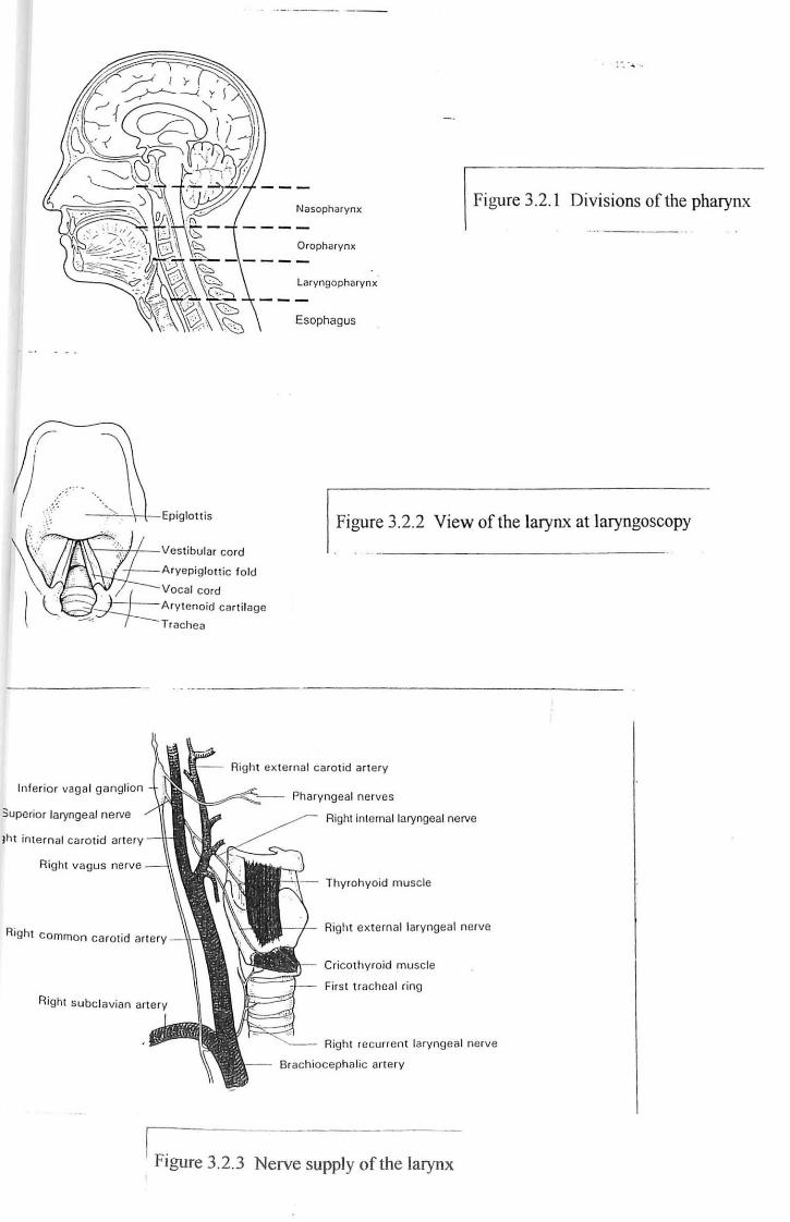
~ . 4 ' .
Nasopharynx Figure 3.2. 1 Divisions of the pharynx
Oropharynx
Esophagus
J -~'-"---'\\-, ·\-Epiglott is Figure 3.2.2 View of the larynx at laryngoscopy
\\~:--jtH--Vestibular cord
Aryepiglottic fold
Right extern al caro tid artery
Inferi or vaga l ganglion
3uperior laryngeal nerve
Jht in ternal caro tid ar tery
Right vagus nerve
Right common carot id art ery
Pharyngeal nerves
Right internal laryngeal nerve
Thyrohyoid muscle
Right ex ternal laryngeal nerve
Cricothyroid muscle
First tracheal ring
Right recurrent laryngeal nerve
Brachiocephalic art ery
I Figure 3.2.3 Nerve supply of the larynx

16
:' .. ·:SOM,A.!"C:~~VA t~ON:::OF~:.~.El.~·:fl~R~ .• t·:· . ... . '.. . .... :.;,::::; .:::: ... .... . ........... . .:
Nasopharynx
Sensory Trigenninal (V) via pterygopalatine ganglion
Glossopharyngeal (IX) to lower regions
Oropharynx
Sensory Glossopharyngeal (IX)
Taste Glossopharyngeal (IX) to region of palatoglossal and oropharynx
Facial (VIll) and glossopharyngeal (IX) to soft palate
Vagus (X) via branch of superior laryngeal nerve to region of epiglottis
Laryngopharynx
Sensory Glossopharyngeal (IX)
Motor Innervation
Pharyngeal plexus to all muscles of pharynx and soft palate except:
Stylopharyngeus via glosopharyngeal (IX)
Tensor veti palatini via trigenninal (V)
Table 3.3.2 Somatic innervation of the pharynx
SOMATIC INNERVATION OF THE· LARYNX . ::
Sensory Vagus (X) via the internal laryngeal nerve to mucosa above vocal folds and
recurrent laryngeal nerve-mucosa below vocal folds.
Motor Accesory (XI) distributed through vagus (X) via recurrent laryngeal nerve to all
Intrinsic muscles except cricithyroid (external laryngeal nerve to X)
Taste Vagus (x) to epiglottis and back of tongue
Table 3.2.1 Somatic innervation of the larynx

17
3.2.3 The Larynx
The larynx is the first part of the lower respiratory tract and is formed
from a complex arrangement of cartilages, muscles, ligaments and membranes. It is a
midline structure, which extends the third and the sixth cervical vertebrae in the adult,
although in the infant it may only extend as far as the inferior border of the fourth
cervical vertebrae. It is superficial and readily palpated, as it is covered only by skin,
by the platysma muscle and superficial and deep layers of the fascia.
Posteriorly it is separated from the cervical column by the pharynx and
the thin prevertebral muscles. It communicates with the laryngopharynx behind and
with the trachea below. Its rigid skeletal structure ensures that it remains patent at all
times, which is essential for its primary role as a respiratory passage. It has also
developed a protective valve me~hanism at the inlet to prevent entry of foreign
material from the laryngopharyngeal passageway.
In view of the complex function of the larynx it is not surprising that
its anatomy is somewhat intricate and highly specialized. The larynx has had to
compromise between the need for rigidity for its respiratory purpose and the need for
mobility to fulfil its function as a valve and the mechanism for phonation.
The larynx itself is fonned from 9 cartilages, 3 of which are midline
and singular and 6, which come in pairs and are more laterally placed. The 3 midline
cartilages are the epiglottis (elastic), the thyroid (hyaline), and the cricoid (hyaline),
and the 3 more lateral pairs are the arytenoids (hyaline and elastic), the cuneifonns
(elastic), and the corniculate cartilages (elastic).

18
The thyroid cartilage is the largest of the laryngeal cartilages and is a
midline structure composed of 2 quadrilateral laminae fused anteriorly in the midline
to form the laryngeal prominence. The posterior border of each lamina is free and
receives the fibres of the stylopharyngeus and the palatopharyngeus muscles. This
border is extended upwards to form the superior horn and downwards to form the
inferior hom. The superior border of the thyroid cartilage is connected to the superior
border of the hyoid bone by the fibrous thyrohyoid membrane, which is pierced
laterally by the internal laryngeal nerve and the superior laryngeal artery. The inferior
horn of the thyroid articulates via synovial joints with the cricoid cartilage below.
The cricoid cartilage is somewhat smaller than the thyroid cartilage
but is thicker, stronger and more fixed in position. It lies at the level of the 6 cervical
vertebrae and is the only rigid region of the larynx. It articulates with the thyroid
cartilage above and is connected below to the tracheal ring via the cricotracheal
membrane. It is shaped like a signet ring with a broad posterior lamina and a narrower
anterior arch. The upper border of the cricoid cartilage is connected to the thyroid
cartilage by the cricothyroid membrane, which is also known as the conus elasticus
and fonns part of the important fibro elastic membrane of the larynx.
The epiglottis is shaped like a leaf or perhaps more realistically like a
bicycle seat. It stands vertically behind the hyoid bone and the root of the tongue and
overhangs the laryngeal inlet. It is broad superiorly with a free border, which tapers
inferiorly to attached to the inner surface of the thyroid cartilage by the thyroepiglottic
ligament. The anterior surface of the epiglottis is attached to the hyoid bone via the

19
hyoepiglottic ligament. The mucous membrane covering this surface is reflected onto
the root and sides of the tongue as one median and two lateral glosso epiglottic folds
and the depressions that form between them are the valleculae.
The laryngeal cavity is divided into 3 topographical regions:
1. An upper vestibule or supraglottic region, which extends from the
laryngeal inlet to the vestibular folds or cords.
2. The ventricle or glottic region, which extends between the vestibular
and the vocal folds.
3. An infraglottic region, which extends from below the vocal folds to
the beginning of the trachea.
The laryngeal inlet is the opemng into the larynx from the
laryngopharynx. It is bounded anteriorly by the upper edge of the epiglottis,
posteriorly by a membrane between the arytenoids cartilages, and laterally by the
aryepiglottic folds containing the cuneiform and corniculate cartilages.

20
3.3 FUNCTIONAL NEUROANATOMY
Function of the larynx is dependant on the complex and timely
interactions of all the structures described as above. This activity is mediated by the
ever present central nervous system and the peripheral neurologic connections of the
upper respiratory tract. The functional neuroanatomy of the larynx would be more
placed into emphasis in the discussions below whereby its functions are profoundly
affected by and are intimately associated with those of the entire upper aero digestive
tract.
3.3.1 Afferent System
Sensation from the supraglottic structures reaches the central nervous
system via the internal branch of the superior laryngeal nerve found on both sides of
the normal human anatomy. Afferent impulses arising in the glottis and subglottic
regions are transmitted by way of the recurrent laryngeal nerves. Proprioceptive and
sensory nerve endings are situated most densely on the laryngeal surface of the
epiglottis and less is to be found on the true vocal cords. More touch receptors are
concentrated toward the posterior commissure than near the anterior and temperature
control receptors are almost found in the supraglottic larynx. Information thus
obtained reaches the central nervous system through the nodal ganglion to the tractis
solitarius where further central and integrative connections take place.
21
3.3.2 Efferent System
Efferent nerve fibers destined for the larynx arise from the nucleus
ambigus of the medulla. All the intrinsic laryngeal muscles are innervated via the
recurrent laryngeal nerves. Only the interarytenoids muscles are bilaterally innervated.
The cricothyroideus muscles, which are not intrinsic to the larynx, are innervated via
the external branch of the superior laryngeal nerves.
3.3.3 Laryngeal Reflexes
The protective, respiratory and the phonatory functions as were
mentioned above are mediated through several polysynaptic reflexes at the level of
the brainstem. Whereas the glottic closure reflex, which is essential for the protection
of the airway is entirely automatic, the other reflexes can at least be initiated , and to
some degree be modified voluntarily.
3.3.4 Glottic Closure Reflex
Touch and chemical or thennal stimulation of the supraglottic mucosa
subtended by the superior laryngeal nerves results in the involuntary forceful closure
of the entire larynx. The sequence of events is anatomically the same as that described
for effort closure. In man unilateral superior laryngeal stimulation does not result in
22
the contra lateral adductor activity seen in most lower animals. This may account for
at least in part for the tendency of unexpected aspiration of saliva associated with
unilateral superior nerve palsy. If the laryngeal closure reflex becomes overly
sensitive laryngospasm may result. It is characterized by electrical activity in the
adductor fibers long after instigating mucosal stimulus has ceased. It can be abolished
by heavy sedation particularly with barbiturates and also decreases with hypoxia. This
may explain why patients are rarely in danger of asphyxia since in very severe cases
the patient in question simply faints from hypoxia at which the spasm breaks.

23
3.4 THE STRESS RESPONSE-ENDOCRINE AND METABOLIC
AL TERA TIONS
The human body naturally will try its very best to maintain its nonnal
environment. Hormones mediate much of the body's homeostatic response to various
stressful stimulus. Physical injury resulting in differing levels of tissue destruction
and reduction in the circulating blood volume constitutes a threat to life and the
outcome is dependent on the efficacy of the human body's defense mechanism. It was
thought (Selye H 1950) that a number of different stimuli otherwise known as
stressors induce similar homeostatic reactions with the sole aim of trying to maintain
and contributing towards the maintenance of the 'milieu interieur'. The metabolic and
honnonal response to trauma is an important component of the protective alarm
reaction or the "general adaptation syndrome".
Recent studies have shown that it has become obvious that a complex
interplay between the various systemic and local factors is taking place (Minnear et al
1983). Some of the many factors influencing quantitively and qualitatively the
metabolic response and alterations seen in patients experiencing tissue trauma. The
neuroendocrine activation induced by tissue trawna is considered the most important
stimulus for the metabolic alterations (Brizio et al 1984).
As hormones play a much important role in mediating body's
homeostatic response to stress the circulating concentrations of the so called flight or
flight hormones norepinephrine (noradrenaline), cortisol, epinephrine (adrenaline) and
growth hormone increase in response to most acute stress. The plasma levels and
14
activities of these and other minerals involved vary following different stressful
situations. Although the stressors have existed for quite some time albeit for centuries
the availability of sensitive and specific hormonal assays are relatively new.
Apparently for this reason the endogenous stress responses continue to be defined.
Evidence is accumulating (Anad et a11987) to support the concept that
modulation of the various stress response may to an extent improve outcome from
surgery. Newer anesthetic analgesic approaches have permitted a reduction in the
peri operative experienced by most patients. In this study the stress response is used to
mean the hemodynamic exhibition presented clinically as a result of the stress induced
changes in the circulating hormone and mineral concentrations during the
peri operative period.
3.4.1 Perioperative Stress
Perioperative anxiety, induction of anesthesia, endotracheal intubation,
vascular cannulation, anesthetic related and surgically induced tissue trauma, visceral
pain, and the process of recovering from surgery are each stressful in its own way and
very much contributes to the responses as described below. In addition to the expected
peri operative events the spectre of the unwanted complications such as sepsis,
hemorrhage, hypothermia, pneumonia, pneumothorax and a number of other
complications may also alter the measurable stress response.
The level or degree of each stressful component is probably central to
the amount of the observed hormonal and metabolic changes observed. Higher grades
of surgical and anesthetic stress are reflected perioperatively by increased circulating