Embed Size (px)
Citation preview
HAL Id: hal-03178895https://hal.univ-angers.fr/hal-03178895
Submitted on 24 Mar 2021
HAL is a multi-disciplinary open accessarchive for the deposit and dissemination of sci-entific research documents, whether they are pub-lished or not. The documents may come fromteaching and research institutions in France orabroad, or from public or private research centers.
L’archive ouverte pluridisciplinaire HAL, estdestinée au dépôt et à la diffusion de documentsscientifiques de niveau recherche, publiés ou non,émanant des établissements d’enseignement et derecherche français ou étrangers, des laboratoirespublics ou privés.
Aqueous core nanocapsules: a new solution forencapsulating doxorubicin hydrochloride
Sandy Vrignaud, Nicolas Anton, Catherine Passirani-Malleret, Jean-PierreBenoit, Patrick Saulnier
To cite this version:Sandy Vrignaud, Nicolas Anton, Catherine Passirani-Malleret, Jean-Pierre Benoit, PatrickSaulnier. Aqueous core nanocapsules: a new solution for encapsulating doxorubicin hydrochlo-ride. Drug Development and Industrial Pharmacy, Taylor & Francis, 2013, 39 (11), pp.1706-11.�10.3109/03639045.2012.730526�. �hal-03178895�

1
Introduction
The development of nanodrug-delivery systems irremediably implies an optimized compatibility between the physicochemical properties of the encapsulated drugs and the structure and nature of the carriers. This is particularly true for hydrophilic drugs, since the nanocarriers encapsulating the drug are themselves dispersed in a water-continuous phase. It is precisely the main difficulty related to the nanoencapsulation of hydrophilic compounds, in contrast with lipophilic drugs more stable when encapsulated. During the formulation process, the hydrophilic ones exhibit a strong trend to be fast released toward the external aqueous phase.
Aqueous core nanocapsules (ACNs) were emphasized in literature as efficient solution for this purpose1–6. ACNs basically consist of a nanoparticle exhibiting a core-shell structure, the core is composed of water solubilizing hydrophilic drugs and the shell is a polymeric capsule. Compared with conventional nanocarriers (e.g. nanospheres), the main advantages of ACNs lies (i) in the high drug loading in the core, (ii) in the low polymer content, and (iii) in the drug confinement and protection within the capsule. However, such a complex structure involves complexes formulation methods4. Several strategies of formulations are reported in literature, using polyisobutylcyanoacry late ACNs encapsulating
ReseaRch aRtIcle
Aqueous core nanocapsules: a new solution for encapsulating doxorubicin hydrochloride
Sandy Vrignaud1,2, Nicolas Anton3, Catherine Passirani1, Jean-Pierre Benoit1,2,4, and Patrick Saulnier1
1LUNAM Université, Université d’Angers, INSERM U1066-MINT - Micro et Nanomédecines Biomimétiques, IBS-CHU Angers, 4 rue Larrey, F-49933 Angers, France, 2Pharmacy, Academic Hospital, Angers, France, 3University of Strasbourg, Department of Pharmacy, CNRS UMR 7199, Laboratoire de Conception et Application de Molécules Bioactives, équipe de Pharmacie Biogalénique, 74 route du Rhin, F-67400 Illkirch, France, and 4Ecole Pratique des Hautes Etudes (EPHE) 12 rue Cuvier, F-75005, Paris, France
abstractIn this study, we propose a new solution for the nanoencapsulation of hydrophilic anticancer drug, doxorubicin hydrochloride (DOX). The drug molecules are solubilized in the core of aqueous nanoreservoirs, so-called aqueous core nanocapsules (ACN) recently developed by our team, and dispersed in aqueous bulk media. Since it is well acknowledged that the nanoencapsulation of DOX has many advantages, like reducing the sides effects (e.g. cardiac toxicity), we propose through the present study a novel formulation solution for this purpose. After focusing on the formulation process for optimizing the drug encapsulation yield, the DOX-release profiles were followed up and analyzed. Different physicochemical and in vitro characterization were performed, and complement activation experiments. ACN were shown efficient to encapsulate DOX reaching yields as high as 80%, followed by a sustained release governed by a diffusion-controlled mechanism. The loaded nanocarriers showed low levels of complement activation, compatible with stealth properties. To summarize, this study brings out a new tool for the nanoencapsulation of hydrophilic anticancers and could open new doors for the administration of this particular class of drugs.Keywords: Doxorubicin hydrochloride, aqueous core, nanocapsule, nanoemulsion, nanoparticle, release
Address for Correspondence: Nicolas Anton, Department of Pharmacy, University of Strasbourg, CNRS UMR 7199, Laboratoire de Conception et Application de Molécules Bioactives, équipe de Pharmacie Biogalénique, 74 route du Rhin, F-67400 Illkirch, France. E-mail: [email protected]
(Received 27 April 2012; revised 11 September 2012; accepted 12 September 2012)
Drug Development and Industrial Pharmacy, 2012; Early Online: 1–6© 2012 Informa Healthcare USA, Inc.ISSN 0363-9045 print/ISSN 1520-5762 onlineDOI: 10.3109/03639045.2012.730526
Drug Development and Industrial Pharmacy
00
00
1
6
27April2012
11September2012
12September2012
0363-9045
1520-5762
© 2012 Informa Healthcare USA, Inc.
10.3109/03639045.2012.730526
2012
Aqueous core nanocapsules
S. Vrignaud et al.
Dru
g D
evel
opm
ent a
nd I
ndus
tria
l Pha
rmac
y D
ownl
oade
d fr
om in
form
ahea
lthca
re.c
om b
y 80
.82.
238.
62 o
n 01
/08/
13Fo
r pe
rson
al u
se o
nly.
2 S. Vrignaud et al.
Drug Development and Industrial Pharmacy
oligonucleotides1,2, azidothymidine-triphosphate or cidofovir7. Many molecules, fluorescein, sulforhodamine, DNA, chlorhexidine, magnetite, have been encapsulated in these ACNs5,8–10. Herein we focus on the encapsulation of hydrophilic anticancer in ACNs. This technology appears highly beneficial and adapted for the administration of such class of cytotoxic molecules, since ACNs can insure a protection not only for the drug but also for the healthy tissues, reducing the potential toxic side effects.
The present study deals the nanoencapsulation of doxorubicin hydrochloride (DOX), an anthracycline anticancer drug, which has been shown to be efficient in the treatment of a wide range of cancers (leukemia and solid tumors11). Due to its irreversible cardiotoxic-ity12, the clinical use of DOX remains limited. However, when nanoencapsulated, DOX toxicity can be reduced or prevented. This is actually the main reason why much effort has been dedicated to the development of pharmaceutical and colloidal forms encapsulating this drug. DOX is protected against in vivo degradation, the toxic side effects are reduced, the repetitive use of bolus injection or the use of perfusion pumps is avoided, hence increasing patient comfort. Finally, it provides favorable pharmacokinetic results. In addition, encap-sulating DOX into nanoparticles has been shown to improve its biodistribution13 since it limits its accumu-lation in the heart. Currently, most work involving DOX entrapment into nanocarriers have been performed using polymer nanoparticles. The DOX molecules are linked to the nanoparticles through electrostatic com-plexation13–15, or by a chemical conjugation between the drug and the polymer16. To date, the nanoencap-sulation of hydrophilic anticancers still remains a sig-nificant technical challenge, and the research efforts ongoing in this topics, even only related to DOX, are still at present highly sustained. Liposomal forms of doxorubicin hydrochloride (DOXIL® and CAELYX®) are marketed for several years, and have led to reduc-tions of the related drug toxicity17. It is actually impor-tant to consider the polymeric nanocapsules, such as ACN, as a possible alternative to the existing technolo-gies for DOX nanoencapsulation. Indeed, compared with liposomes, they offer some original aspects, nota-bly regarding their formulation process (i.e. feasibility and repeatability), the fine control of their physico-chemical properties and surface functionalization, low cost, and finally the large panel of biodegradable polymers that can be used in their formulation. We can find various recently published formulation strategies for that purpose, for instance, polymeric nanoassem-blies18, silica nanoparticles19 or even lipid nanoemul-sions20. However, the DOX is generally entrapped in the polymer matrix or is adsorbed onto the nanoparticle surface, and thus is not protected from the external environment, being released with a burst. However, when the DOX is solubilized in the aqueous core of ACNs, their core-shell structure appears particularly appropriated to the drug protection.
We have recently developed a new solution regard-ing the formulation of ACNs5, based on low-energy nanoemulsification methods, organic solvent-free, and avoiding the use of high temperatures or mechanical stresses. This original process appears highly suitable for the nanoencapsulation of fragile or thermosensitive hyrophilic molecules with high encapsulation yields (EY) around 90%. Through the present study, we propose the proof of concept of the adaptability of this new technol-ogy on the nanoencapsulation of DOX in ACNs. After focusing on the formulation process for optimizing the drug EY, the DOX-release profiles were followed up and analyzed. Different physicochemical and in vitro charac-terization were performed, and complement activation experiments. This study may bring out a new tool for the nanoencapsulation of hydrophilic anticancer drugs and could open new doors for the administration of this par-ticular class of drugs.
Materials and methods
MaterialsTechnical grade polyethoxylated surfactant C18E6, was kindly supplied by Stearinerie-Dubois (Boulogne, France). It is a typical commercial product with a Poisson distribution level of about six for ethylene oxide (EO). Light mineral oil, such as, paraffn oil (the standardized denomination to refer to a mixture of saturated hydro-carbons), was purchased from Cooper (Melun, France). Ultrapure water was obtained by the MilliQ filtration system (Millipore, Saint-Quentin-en Yve
68 lines, France).
Sodium chloride was purchased from Prolabo (Fontenay-sous-Bois, France). 2-Methylbutane was provided by Riedel-de-Haën (Seelze, Germany). Finally, tolylene-2,4-diisocyanate (TDI), the monomer used for interfacial polycondensation, and DOX, were obtained from Sigma (Saint-Quentin-Fallavier, France).
MethodsThe fabrication of ACNsThe fabrication process of ACNs is a patented process (#WO2009037310 (A2) – 2009-03-26), described else-where5. Briefly, a (DOX + water)-in-oil nanoemulsion is first formulated by the phase inversion temperature method (DOX-loaded water nanodroplets dispersed in (paraffin oil + 2-methylbutane), stabilized by the non-ionic surfactant). Formulation parameters: water-to-oil weight ratio = 0.4 and surfactant concentration = 10 wt.%. These water nanodroplets sizing around 30 nm are dis-persed in the oil phase. Then, we synthesize a polymer (polyurea) shell at the water/oil interface of the droplets, through the polycondensation of TDI, by adding the monomer in the oil-bulk phase. The final step consists in substituting the bulk oily phase for another water phase (dispersing water). To do this, dispersing water is added in the formulation and oil (2-methylbutane) is simulta-neously evaporated. The result is a suspension of ACNs dispersed in water encapsulating the DOX molecules. A
Dru
g D
evel
opm
ent a
nd I
ndus
tria
l Pha
rmac
y D
ownl
oade
d fr
om in
form
ahea
lthca
re.c
om b
y 80
.82.
238.
62 o
n 01
/08/
13Fo
r pe
rson
al u
se o
nly.
Aqueous core nanocapsules 3
© 2012 Informa Healthcare USA, Inc.
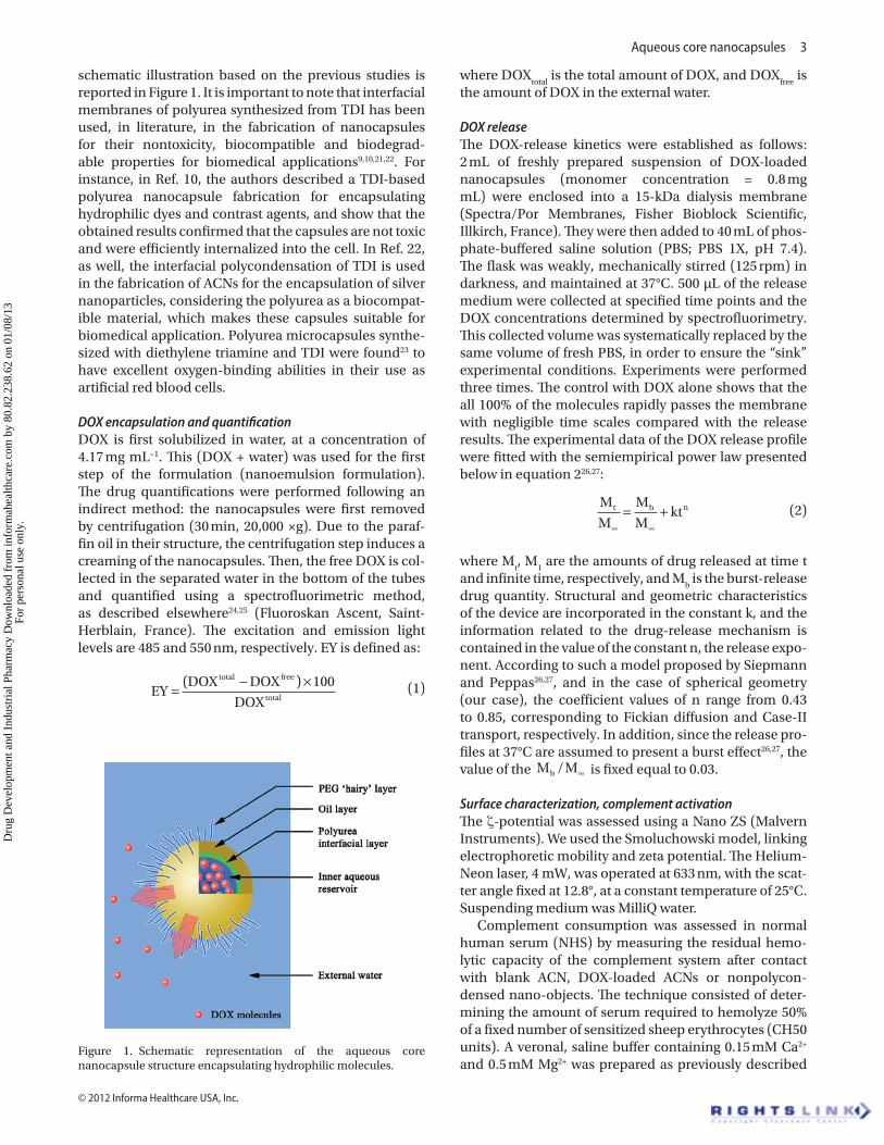
schematic illustration based on the previous studies is reported in Figure 1. It is important to note that interfacial membranes of polyurea synthesized from TDI has been used, in literature, in the fabrication of nanocapsules for their nontoxicity, biocompatible and biodegrad-able properties for biomedical applications9,10,21,22. For instance, in Ref. 10, the authors described a TDI-based polyurea nanocapsule fabrication for encapsulating hydrophilic dyes and contrast agents, and show that the obtained results confirmed that the capsules are not toxic and were efficiently internalized into the cell. In Ref. 22, as well, the interfacial polycondensation of TDI is used in the fabrication of ACNs for the encapsulation of silver nanoparticles, considering the polyurea as a biocompat-ible material, which makes these capsules suitable for biomedical application. Polyurea microcapsules synthe-sized with diethylene triamine and TDI were found23 to have excellent oxygen-binding abilities in their use as artificial red blood cells.
DOX encapsulation and quantificationDOX is first solubilized in water, at a concentration of 4.17 mg mL−1. This (DOX + water) was used for the first step of the formulation (nanoemulsion formulation). The drug quantifications were performed following an indirect method: the nanocapsules were first removed by centrifugation (30 min, 20,000 ×g). Due to the paraf-fin oil in their structure, the centrifugation step induces a creaming of the nanocapsules. Then, the free DOX is col-lected in the separated water in the bottom of the tubes and quantified using a spectrofluorimetric method, as described elsewhere24,25 (Fluoroskan Ascent, Saint-Herblain, France). The excitation and emission light levels are 485 and 550 nm, respectively. EY is defined as:
EYDOX DOX 100
DOX
total free
total=− ×( )
(1)
where DOXtotal
is the total amount of DOX, and DOXfree
is the amount of DOX in the external water.
DOX releaseThe DOX-release kinetics were established as follows: 2 mL of freshly prepared suspension of DOX-loaded nanocapsules (monomer concentration = 0.8 mg mL) were enclosed into a 15-kDa dialysis membrane (Spectra/Por Membranes, Fisher Bioblock Scientific, Illkirch, France). They were then added to 40 mL of phos-phate-buffered saline solution (PBS; PBS 1X, pH 7.4). The flask was weakly, mechanically stirred (125 rpm) in darkness, and maintained at 37°C. 500 µL of the release medium were collected at specified time points and the DOX concentrations determined by spectrofluorimetry. This collected volume was systematically replaced by the same volume of fresh PBS, in order to ensure the “sink” experimental conditions. Experiments were performed three times. The control with DOX alone shows that the all 100% of the molecules rapidly passes the membrane with negligible time scales compared with the release results. The experimental data of the DOX release profile were fitted with the semiempirical power law presented below in equation 226,27:
M
M
M
Mktt b n
∞ ∞
= +
(2)
where Mt, M
1 are the amounts of drug released at time t
and infinite time, respectively, and Mb is the burst-release
drug quantity. Structural and geometric characteristics of the device are incorporated in the constant k, and the information related to the drug-release mechanism is contained in the value of the constant n, the release expo-nent. According to such a model proposed by Siepmann and Peppas26,27, and in the case of spherical geometry (our case), the coefficient values of n range from 0.43 to 0.85, corresponding to Fickian diffusion and Case-II transport, respectively. In addition, since the release pro-files at 37°C are assumed to present a burst effect26,27, the value of the M Mb / ∞ is fixed equal to 0.03.
Surface characterization, complement activationThe ζ-potential was assessed using a Nano ZS (Malvern Instruments). We used the Smoluchowski model, linking electrophoretic mobility and zeta potential. The Helium-Neon laser, 4 mW, was operated at 633 nm, with the scat-ter angle fixed at 12.8°, at a constant temperature of 25°C. Suspending medium was MilliQ water.
Complement consumption was assessed in normal human serum (NHS) by measuring the residual hemo-lytic capacity of the complement system after contact with blank ACN, DOX-loaded ACNs or nonpolycon-densed nano-objects. The technique consisted of deter-mining the amount of serum required to hemolyze 50% of a fixed number of sensitized sheep erythrocytes (CH50 units). A veronal, saline buffer containing 0.15 mM Ca2+ and 0.5 mM Mg2+ was prepared as previously described
Figure 1. Schematic representation of the aqueous core nanocapsule structure encapsulating hydrophilic molecules.
Dru
g D
evel
opm
ent a
nd I
ndus
tria
l Pha
rmac
y D
ownl
oade
d fr
om in
form
ahea
lthca
re.c
om b
y 80
.82.
238.
62 o
n 01
/08/
13Fo
r pe
rson
al u
se o
nly.
4 S. Vrignaud et al.
Drug Development and Industrial Pharmacy
(VBS++). Sheep erythrocytes were sensitized by rabbit, antisheep erythrocyte antibodies, and suspended at a final concentration of 1.108 cells mL−1 in VBS++. To assess the consumption of CH50 units in the presence of the particles during a constant incubation time, increasing amounts of particle suspensions were added to NHS diluted in VBS++ so that the final dilution of NHS in the reaction mixture was 1/4 (v./v.) in a final volume of 1 mL. After 60 min. of incubation at 37°C with gentle agitation, the suspension was diluted at 1/25 (v./v.) in VBS++; then, aliquots at different dilutions were added to a given vol-ume of sensitized sheep erythrocytes.
After 45 min of incubation at 37°C, the reaction mix-ture was centrifuged at 2,000 rpm for 10 min. The absorp-tion of the supernatant was determined at 415 nm with a microplate reader (Multiskan Ascent, Labsystems SA, Cergy-Pontoise, France) and compared with the results obtained with the control serum. After determining the CH50 units remaining in the serum, the results were ex pressed as the consumption of CH50 units as a function of the nanoparticle surface area calculated as described elsewhere28 in order to compare nanoparticles of differ-ent average diameters. High activating control experi-ments were performed with a suspension of poly(methyl methacrylate) (PMMA) nanoparticles.
Results and discussion
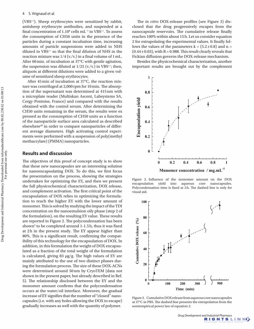
The objectives of this proof of concept study is to show that these new nanocapsules are an interesting solution for nanoencapsulating DOX. To do this, we first focus the presentation on the process, showing the strategies undertaken for optimizing the EY, and then we present the full physicochemical characterization, DOX release, and complement activation. The first critical point of the encapsulation of DOX relies in optimizing the formula-tion to reach the higher EY with the lower amount of monomer. This is solved by studying the impact of the TDI concentration on the nanoemulsion oily phase (step 2 of the formulation), on the resulting EY value. These results are reported in Figure 2. The polycondensation has been shown5 to be completed around 1-1.5 h, thus it was fixed at 2 h in the present study. The EY appear higher than 80%. This is a significant result, confirming the compat-ibility of this technology for the encapsulation of DOX. In addition, in this formulation the weight of DOX encapsu-lated as a fraction of the total weight of the formulation is calculated, giving 83 µg/g. The high values of EY are mainly attributed to the use of two distinct phases dur-ing the formulation process. The size of these DOX-ACNs were determined around 50 nm by CryoTEM (data not shown in the present paper, but already described in Ref. 5). The relationship disclosed between the EY and the monomer amount confirms that the polycondensation occurs at the water/oil interface. Moreover, the gradual increase of EY signifies that the number of “closed” nano-capsules (i.e. with any holes allowing the DOX to escape) gradually increases as well with the quantity of polymer.
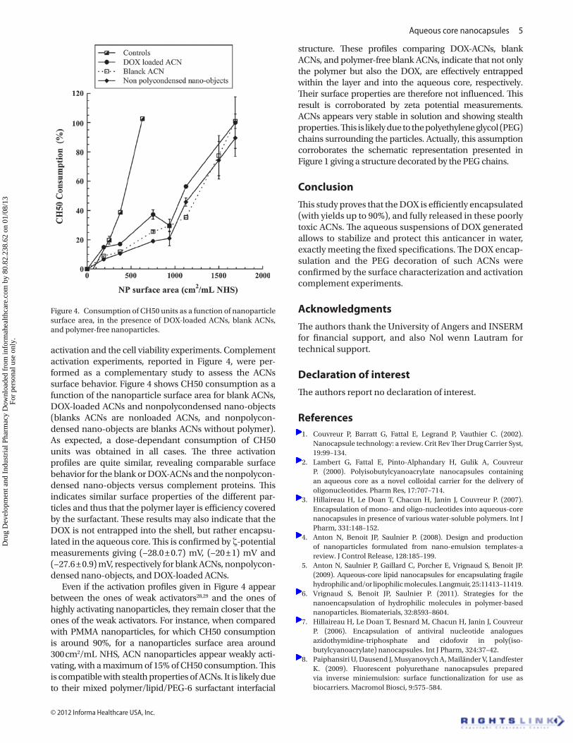
The in vitro DOX-release profiles (see Figure 3) dis-closed that the drug progressively escapes from the nanocapsule reservoirs. The cumulative release finally reaches 100% within about 15 h. Let us consider equation 2 for extrapolating the experimental values. It finally fol-lows the values of the parameters k = (5.2 ± 0.8) and n = (0.44 ± 0.03), with R = 0.988. This result clearly reveals that Fickian diffusion governs the DOX-release mechanism.
Besides the physicochemical characterization, another important results are brought out by the complement
Figure 2. Influence of the monomer amount on the DOX encapsulation yield into aqueous core nanocapsules. Polycondensation time is fixed at 2 h. The dashed line is only for visual aid.
Figure 3. Cumulative DOX release from aqueous core nanocapsules at 37°C in PBS. The dashed line presents the extrapolation from the semiempirical power law of equation 2.
Dru
g D
evel
opm
ent a
nd I
ndus
tria
l Pha
rmac
y D
ownl
oade
d fr
om in
form
ahea
lthca
re.c
om b
y 80
.82.
238.
62 o
n 01
/08/
13Fo
r pe
rson
al u
se o
nly.
Aqueous core nanocapsules 5
© 2012 Informa Healthcare USA, Inc.
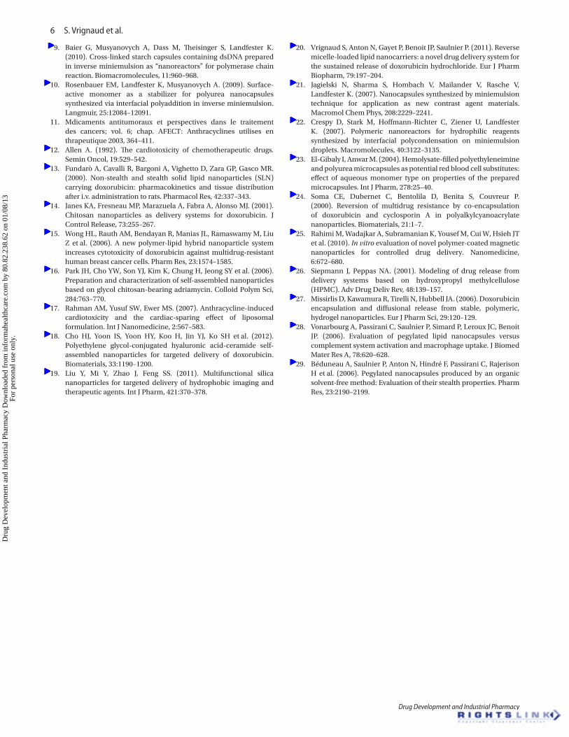
activation and the cell viability experiments. Complement activation experiments, reported in Figure 4, were per-formed as a complementary study to assess the ACNs surface behavior. Figure 4 shows CH50 consumption as a function of the nanoparticle surface area for blank ACNs, DOX-loaded ACNs and nonpolycondensed nano-objects (blanks ACNs are nonloaded ACNs, and nonpolycon-densed nano-objects are blanks ACNs without polymer). As expected, a dose-dependant consumption of CH50 units was obtained in all cases. The three activation profiles are quite similar, revealing comparable surface behavior for the blank or DOX-ACNs and the nonpolycon-densed nano-objects versus complement proteins. This indicates similar surface properties of the different par-ticles and thus that the polymer layer is efficiency covered by the surfactant. These results may also indicate that the DOX is not entrapped into the shell, but rather encapsu-lated in the aqueous core. This is confirmed by ζ-potential measurements giving (−28.0 ± 0.7) mV, (−20 ± 1) mV and (−27.6 ± 0.9) mV, respectively for blank ACNs, nonpolycon-densed nano-objects, and DOX-loaded ACNs.
Even if the activation profiles given in Figure 4 appear between the ones of weak activators28,29 and the ones of highly activating nanoparticles, they remain closer that the ones of the weak activators. For instance, when compared with PMMA nanoparticles, for which CH50 consumption is around 90%, for a nanoparticles surface area around 300 cm2/mL NHS, ACN nanoparticles appear weakly acti-vating, with a maximum of 15% of CH50 consumption. This is compatible with stealth properties of ACNs. It is likely due to their mixed polymer/lipid/PEG-6 surfactant interfacial
structure. These profiles comparing DOX-ACNs, blank ACNs, and polymer-free blank ACNs, indicate that not only the polymer but also the DOX, are effectively entrapped within the layer and into the aqueous core, respectively. Their surface properties are therefore not influenced. This result is corroborated by zeta potential measurements. ACNs appears very stable in solution and showing stealth properties. This is likely due to the polyethylene glycol (PEG) chains surrounding the particles. Actually, this assumption corroborates the schematic representation presented in Figure 1 giving a structure decorated by the PEG chains.
conclusion
This study proves that the DOX is efficiently encapsulated (with yields up to 90%), and fully released in these poorly toxic ACNs. The aqueous suspensions of DOX generated allows to stabilize and protect this anticancer in water, exactly meeting the fixed specifications. The DOX encap-sulation and the PEG decoration of such ACNs were confirmed by the surface characterization and activation complement experiments.
acknowledgments
The authors thank the University of Angers and INSERM for financial support, and also Nol wenn Lautram for technical support.
Declaration of interest
The authors report no declaration of interest.
References 1. Couvreur P, Barratt G, Fattal E, Legrand P, Vauthier C. (2002).
Nanocapsule technology: a review. Crit Rev Ther Drug Carrier Syst, 19:99–134.
2. Lambert G, Fattal E, Pinto-Alphandary H, Gulik A, Couvreur P. (2000). Polyisobutylcyanoacrylate nanocapsules containing an aqueous core as a novel colloidal carrier for the delivery of oligonucleotides. Pharm Res, 17:707–714.
3. Hillaireau H, Le Doan T, Chacun H, Janin J, Couvreur P. (2007). Encapsulation of mono- and oligo-nucleotides into aqueous-core nanocapsules in presence of various water-soluble polymers. Int J Pharm, 331:148–152.
4. Anton N, Benoit JP, Saulnier P. (2008). Design and production of nanoparticles formulated from nano-emulsion templates-a review. J Control Release, 128:185–199.
5. Anton N, Saulnier P, Gaillard C, Porcher E, Vrignaud S, Benoit JP. (2009). Aqueous-core lipid nanocapsules for encapsulating fragile hydrophilic and/or lipophilic molecules. Langmuir, 25:11413–11419.
6. Vrignaud S, Benoit JP, Saulnier P. (2011). Strategies for the nanoencapsulation of hydrophilic molecules in polymer-based nanoparticles. Biomaterials, 32:8593–8604.
7. Hillaireau H, Le Doan T, Besnard M, Chacun H, Janin J, Couvreur P. (2006). Encapsulation of antiviral nucleotide analogues azidothymidine-triphosphate and cidofovir in poly(iso-butylcyanoacrylate) nanocapsules. Int J Pharm, 324:37–42.
8. Paiphansiri U, Dausend J, Musyanovych A, Mailänder V, Landfester K. (2009). Fluorescent polyurethane nanocapsules prepared via inverse miniemulsion: surface functionalization for use as biocarriers. Macromol Biosci, 9:575–584.
Figure 4. Consumption of CH50 units as a function of nanoparticle surface area, in the presence of DOX-loaded ACNs, blank ACNs, and polymer-free nanoparticles.
Dru
g D
evel
opm
ent a
nd I
ndus
tria
l Pha
rmac
y D
ownl
oade
d fr
om in
form
ahea
lthca
re.c
om b
y 80
.82.
238.
62 o
n 01
/08/
13Fo
r pe
rson
al u
se o
nly.
6 S. Vrignaud et al.
Drug Development and Industrial Pharmacy
9. Baier G, Musyanovych A, Dass M, Theisinger S, Landfester K. (2010). Cross-linked starch capsules containing dsDNA prepared in inverse miniemulsion as “nanoreactors” for polymerase chain reaction. Biomacromolecules, 11:960–968.
10. Rosenbauer EM, Landfester K, Musyanovych A. (2009). Surface-active monomer as a stabilizer for polyurea nanocapsules synthesized via interfacial polyaddition in inverse miniemulsion. Langmuir, 25:12084–12091.
11. Mdicaments antitumoraux et perspectives dans le traitement des cancers; vol. 6; chap. AFECT: Anthracyclines utilises en thrapeutique 2003, 364–411.
12. Allen A. (1992). The cardiotoxicity of chemotherapeutic drugs. Semin Oncol, 19:529–542.
13. Fundarò A, Cavalli R, Bargoni A, Vighetto D, Zara GP, Gasco MR. (2000). Non-stealth and stealth solid lipid nanoparticles (SLN) carrying doxorubicin: pharmacokinetics and tissue distribution after i.v. administration to rats. Pharmacol Res, 42:337–343.
14. Janes KA, Fresneau MP, Marazuela A, Fabra A, Alonso MJ. (2001). Chitosan nanoparticles as delivery systems for doxorubicin. J Control Release, 73:255–267.
15. Wong HL, Rauth AM, Bendayan R, Manias JL, Ramaswamy M, Liu Z et al. (2006). A new polymer-lipid hybrid nanoparticle system increases cytotoxicity of doxorubicin against multidrug-resistant human breast cancer cells. Pharm Res, 23:1574–1585.
16. Park JH, Cho YW, Son YJ, Kim K, Chung H, Jeong SY et al. (2006). Preparation and characterization of self-assembled nanoparticles based on glycol chitosan-bearing adriamycin. Colloid Polym Sci, 284:763–770.
17. Rahman AM, Yusuf SW, Ewer MS. (2007). Anthracycline-induced cardiotoxicity and the cardiac-sparing effect of liposomal formulation. Int J Nanomedicine, 2:567–583.
18. Cho HJ, Yoon IS, Yoon HY, Koo H, Jin YJ, Ko SH et al. (2012). Polyethylene glycol-conjugated hyaluronic acid-ceramide self-assembled nanoparticles for targeted delivery of doxorubicin. Biomaterials, 33:1190–1200.
19. Liu Y, Mi Y, Zhao J, Feng SS. (2011). Multifunctional silica nanoparticles for targeted delivery of hydrophobic imaging and therapeutic agents. Int J Pharm, 421:370–378.
20. Vrignaud S, Anton N, Gayet P, Benoit JP, Saulnier P. (2011). Reverse micelle-loaded lipid nanocarriers: a novel drug delivery system for the sustained release of doxorubicin hydrochloride. Eur J Pharm Biopharm, 79:197–204.
21. Jagielski N, Sharma S, Hombach V, Mailander V, Rasche V, Landfester K. (2007). Nanocapsules synthesized by miniemulsion technique for application as new contrast agent materials. Macromol Chem Phys, 208:2229–2241.
22. Crespy D, Stark M, Hoffmann-Richter C, Ziener U, Landfester K. (2007). Polymeric nanoreactors for hydrophilic reagents synthesized by interfacial polycondensation on miniemulsion droplets. Macromolecules, 40:3122–3135.
23. El-Gibaly I, Anwar M. (2004). Hemolysate-filled polyethyleneimine and polyurea microcapsules as potential red blood cell substitutes: effect of aqueous monomer type on properties of the prepared microcapsules. Int J Pharm, 278:25–40.
24. Soma CE, Dubernet C, Bentolila D, Benita S, Couvreur P. (2000). Reversion of multidrug resistance by co-encapsulation of doxorubicin and cyclosporin A in polyalkylcyanoacrylate nanoparticles. Biomaterials, 21:1–7.
25. Rahimi M, Wadajkar A, Subramanian K, Yousef M, Cui W, Hsieh JT et al. (2010). In vitro evaluation of novel polymer-coated magnetic nanoparticles for controlled drug delivery. Nanomedicine, 6:672–680.
26. Siepmann J, Peppas NA. (2001). Modeling of drug release from delivery systems based on hydroxypropyl methylcellulose (HPMC). Adv Drug Deliv Rev, 48:139–157.
27. Missirlis D, Kawamura R, Tirelli N, Hubbell JA. (2006). Doxorubicin encapsulation and diffusional release from stable, polymeric, hydrogel nanoparticles. Eur J Pharm Sci, 29:120–129.
28. Vonarbourg A, Passirani C, Saulnier P, Simard P, Leroux JC, Benoit JP. (2006). Evaluation of pegylated lipid nanocapsules versus complement system activation and macrophage uptake. J Biomed Mater Res A, 78:620–628.
29. Béduneau A, Saulnier P, Anton N, Hindré F, Passirani C, Rajerison H et al. (2006). Pegylated nanocapsules produced by an organic solvent-free method: Evaluation of their stealth properties. Pharm Res, 23:2190–2199.
Dru
g D
evel
opm
ent a
nd I
ndus
tria
l Pha
rmac
y D
ownl
oade
d fr
om in
form
ahea
lthca
re.c
om b
y 80
.82.
238.
62 o
n 01
/08/
13Fo
r pe
rson
al u
se o
nly.