Embed Size (px)
Citation preview

© 2014 Frank et al. This work is published by Dove Medical Press Limited, and licensed under Creative Commons Attribution – Non Commercial (unported, v3.0) License. The full terms of the License are available at http://creativecommons.org/licenses/by-nc/3.0/. Non-commercial uses of the work are permitted without any further
permission from Dove Medical Press Limited, provided the work is properly attributed. Permissions beyond the scope of the License are administered by Dove Medical Press Limited. Information on how to request permission may be found at: http://www.dovepress.com/permissions.php
International Journal of Nanomedicine 2014:9 3151–3161
International Journal of Nanomedicine Dovepress
submit your manuscript | www.dovepress.com
Dovepress 3151
O r I g I N a l r e s e a r c h
open access to scientific and medical research
Open access Full Text article
http://dx.doi.org/10.2147/IJN.S62599
chitosan gel containing polymeric nanocapsules: a new formulation for vaginal drug delivery
correspondence: silvia s guterresFaculdade de Farmácia, Universidade Federal do rio grande do sul, av Ipiranga, 2752/405 ceP 90610-000, Porto alegre, rs, BrazilTel +55 51 3308 5500Fax +55 51 3308 5247email [email protected]
luiza a Frank1
giuseppina sandri2
Francesca D´autilia2
renata V contri1
Maria cristina Bonferoni2
carla caramella2
alejandro g Frank3
adriana r Pohlmann1
silvia s guterres1
1Pharmaceutical science graduate Program, Federal University of rio grande do sul, Porto alegre, rs, Brazil; 2Department of Drug sciences, University of Pavia, Pavia, PV, Italy; 3Department of Industrial engineering, Federal University of rio grande do sul, Porto alegre, rs, Brazil
Abstract: The vaginal route of administration is an alternative for several treatments for
either local or systemic pharmacological effects. However, the permanence of a drug in this
route represents a challenge for formulation development that can be overcome by using
nanoencapsulation and chitosan gel. Thus, this work aimed to evaluate the performance of
chitosan hydrogels containing cationic and anionic acrylic-based nanocapsules (Eudragit®
RS 100 and Eudragit® S 100, respectively) with Nile red as a model of lipophilic substance
in the vaginal route of administration, as measured by increases in the residence time and
the penetration of these formulations. Several formulations were prepared with increasing
chitosan concentrations, and were analyzed in terms of pH and rheological behavior so that
the most suitable formulation could be selected. The enhancement of the adhesion (tensile
stress test and washability profile) and penetration (confocal laser scanning microscopy and
extraction followed by quantification) properties of the formulations, when applied to porcine
vaginal mucosa, were evaluated. The nanocapsule suspensions produced presented adequate
properties: size of approximately 200 nm (polydispersity index of 0.2); zeta potential around
+10 mV for the cationic formulation and -10 mV for the anionic formulation; and pH
values of 6.1±0.1 (Eudragit RS 100), 5.3±0.2 (Eudragit S 100), 6.2±0.1 (Nile red loaded
Eudragit RS 100), and 5.1±0.1 (Nile red loaded Eudragit S 100). The chitosan formula-
tion presented suitable viscosity for vaginal application and acidic pH (approximately 4.5).
The tensile stress test showed that both formulations containing polymeric nanocapsules
presented higher mucoadhesion when compared with the formulation without nanocap-
sules. In the washability experiment, no significant differences were found between for-
mulations. Confocal microscopy and fluorescence quantification after extraction from the
mucosa showed higher penetration of Nile red when it was nanoencapsulated, particularly
in cationic nanocapsules. The formulations developed based on chitosan gel vehicle at 2.5%
weight/weight containing polymeric nanocapsules, especially the cationic nanocapsules, dem-
onstrated applicability for the vaginal delivery of hydrophobic substances.
Keywords: Eudragit® RS 100, Eudragit® S 100, vaginal route, nanotechnology
IntroductionVaginal delivery is an alternative for several pharmacological treatments for either
local or systemic effect.1,2 However, the local effect is targeted more often as a method
of administration of antibacterials, antifungals, antivirals, antiprotozoals, spermicidal
agents, and steroids.1,3 Using the vaginal mucosa as a drug delivery route has advan-
tages such as facility of drug application, high contact surface area, high blood sup-
ply, good permeability to several substances, and avoidance of first pass metabolism
and gastrointestinal side effects.2,4,5 However, the development of formulations for
application via the vaginal route presents some difficulties. One of these difficulties
is that the histology and physiology of the vagina vary with age, menstrual cycle, and
hormonal changes. The other difficulty is that the vagina secretes endometrial fluids.
Journal name: International Journal of NanomedicineJournal Designation: Original ResearchYear: 2014Volume: 9Running head verso: Frank et alRunning head recto: A new formulation for vaginal drug deliveryDOI: http://dx.doi.org/10.2147/IJN.S62599

International Journal of Nanomedicine 2014:9submit your manuscript | www.dovepress.com
Dovepress
Dovepress
3152
Frank et al
Such difficulties contribute to easy removal of the formulation,
which leads to shorter periods between applications and,
therefore, to a low compliance with the treatment.2
Several authors have demonstrated that the use of nano-
technology can reduce the above-mentioned disadvantages of
vaginal drug administration.6–10 This is because, when drugs are
encapsulated into nanostructures, their release is controlled, they
are protected against degradation, and the dose applied can be
eventually reduced.11,12 Additionally, it must be considered that
some drugs administered via the vaginal route can cause side
effects, such as erythema, irritation, ulceration, and pain, and
such effects could be reduced by using nanotechnology.5,13
Nanoparticles have strong tissue-adhesion properties,
which is helpful for therapy because it increases the residence
time of drugs. However, nanoparticles are frequently obtained
in aqueous suspensions, so application at a specific site is dif-
ficult. Therefore, incorporating nanoparticles into a viscous
system is an alternative that facilitates the correct delivery of
the substance. Moreover, the use of a bioadhesive vehicle, such
as chitosan, can prolong residence time on the mucosa, leading
to the expected action,2,14,15 improving treatment acceptability,
and increasing penetration and/or absorption.13,16
The use of semisolid formulations represents an alterna-
tive method to increase the period of contact between the
drug and the mucosa.2,3 Additionally, the use of bioadhesive
vehicles, such as gels, tablets, suppositories, and films, can
improve the action of several drugs.5 Different polymers
have been described in the literature for the delivery of sub-
stances on the vaginal mucosa, such as: polyethylene glycol,
cellulose-derivatives, and chitosan.2 Among these, chitosan is
the only polymer that presents positive charge, which leads
to a probable interaction with the vaginal mucosa, increasing
the residence time of drugs that contribute to increased muco-
sae penetration. It also presents other interesting properties,
such as biocompatibility, biodegradability, mucoadhesion,
antimicrobial activity, and film formation, and the ability to
heal injured tissues.
Chitosan, a bioadhesive polymer, has received a great deal
of attention in the last few years.3,4,17 This polymer is obtained
through the deacetylation of chitin, one of the most abun-
dant polysaccharides found in nature.3,18,19 The mentioned
polymer has been widely applied in the development of
different pharmaceutical dosage forms, such as hydrogels,3,4
films,13 microspheres,20 and nanoparticles.21 Chitosan hydrogels
can be produced by covalent interactions between chitosan
chains.22 Among the therapeutic applications of the chito-
san hydrogel, the controlled release of drugs is particularly
notable.22 Additionally, chitosan hydrogels affect the tight
junction between epithelial cells such that these formulations
may increase drug penetration.2
The use of chitosan vehicles and nanoparticles for vagi-
nal delivery have been reported by some scholars.3,4,23 For
instance, hydrogels formed by chitosan derivatives have
shown higher adhesion to pig vaginal mucosa than a com-
mercial gel.3 The citrate salt of chitosan has been proven
to be suitable for the preparation of gels incorporating
substances with low permeation potential for vaginal
administration.4 For example, nanoparticles prepared by ionic
gelation containing tenofovir, an antiviral agent, were applied
to pig vaginal mucosa. The chitosan formulation presented
a high potential for the release of antimicrobial agents due
to controlled release, bioadhesion, and safety.23
The incorporation of nanostructured systems, such as
nanocapsules, into chitosan gels represents the unification
of different systems with interesting properties, as previ-
ously mentioned. The incorporation of Eudragit® RS 100
nanocapsules (Evonik Industries AG, Essen, Germany) in
chitosan hydrogels and the controlled-release property of the
formulation has already been described.24 The formulation
has shown high skin adhesion17 and no skin irritation in
humans.13 In addition to cutaneous administration, use
of Eudragit RS 100 nanocapsules has been described for
ocular25 and vaginal26 routes of administration.
The aim of this work was to evaluate the performance of
chitosan hydrogels containing cationic and anionic acrylic-
based nanocapsules (Eudragit RS 100 and Eudragit® S 100
[Evonik Industries AG], respectively), with Nile red (NR)
as a model lipophilic substance, for vaginal administration,
as measured by increased residence time and penetration of
these formulations.
Materials and methodsMaterialsChitosan of medium molecular weight (1,136 kDa) and 92%
deacetylation degree, 90% lactic acid, NR, polysorbate 80,
sorbitan monostearate 60, and acetone (analytical grade)
were purchased from Sigma-Aldrich (St Louis, MO, USA).
Capric/caprylic triglycerides (Miglyol 812) and Eudragit
(S 100 and RS 100) were gifts from Sasol (Johannesburg, South
Africa) and Evonik Industries AG, respectively. Isopentyl ace-
tate was obtained from Carlo Erba Reagents (Milan, Italy).
Production of nanocapsulesNanocapsules were produced by the method described
by Fessi et al27 called interfacial deposition of preformed
polymers, using Eudragit RS 10024 or Eudragit S 10028 as

International Journal of Nanomedicine 2014:9 submit your manuscript | www.dovepress.com
Dovepress
Dovepress
3153
a new formulation for vaginal drug delivery
the polymeric shell (10 mg/mL), capric/caprylic triglycerides
as the oily core (33 µg/mL), polysorbate 80 as the stabilizer
(7.6 mg/mL), and NR (0.0825 mg/mL, previously mixed
with the capric/caprylic triglycerides) as a model of lipophilic
drug. Sorbitan monostearate (7.6 mg/mL) was used for the
production of the Eudragit S 100 formulation. The nano-
capsules produced with different polymers containing NR
were named NC-RS–NR (prepared with Eudragit RS 100)
and NC-S–NR (prepared with Eudragit S 100). Similarly,
nanocapsule suspensions without the fluorescent marker
were used as controls and named NC-RS (Eudragit RS 100)
and NC-S (Eudragit S 100).
characterization of nanocapsulesThe nanocapsules were characterized in terms of pH, size, and
zeta potential immediately after production. The pH analysis
was performed by direct measurement using potentiometry
(B474, Micronal, São Paulo, Brazil). The nanocapsule size
was measured by different techniques, including laser dif-
fraction (Mastersizer 2000, Nano ZS; Malvern Instruments,
Malvern, UK), dynamic light scattering (Zetasizer Nano
ZS; Malvern Instruments), and particle tracking (Nanosight
LM 10), by dilution of nanocapsules in bidistilled water.
For the determination of the zeta potential, the nanocapsules
were diluted in NaCl solution (10 mM) and electrophoretic
mobility was analyzed (Zetasizer, Nano ZS).
Production of chitosan gelsFirst, different formulations with increasing chitosan con-
centrations (1.5%, 2%, 2.5%, and 3% weight/weight [w/w])
were produced to determine the most suitable chitosan gel for
vaginal administration. The hydrogels were prepared by man-
ual mixing of chitosan, lactic acid (115 µL), and bidistilled
water or nanocapsule suspension without the fluorescent
marker (10 mL). The formulations were named CH-RS (chi-
tosan gel containing Eudragit RS 100 nanocapsules), CH-S
(chitosan gel containing Eudragit S 100 nanocapsules), and
CH (chitosan gel prepared with water).
After determining the most suitable gel, nanocapsules
containing NR were loaded in the selected chitosan gel and
the formulations were named CH-RS–NR (chitosan gel
containing Eudragit RS 100 nanocapsules) and CH-S–NR
(chitosan gel containing Eudragit S 100 nanocapsules).
For the gel obtained with water instead of nanocapsules
(CH–NR), 330 µL of the mixture of NR and capric/caprylic
triglycerides (2.5 mg/mL) was added during the production
of 10 g of the formulation. Therefore, at the final stage, all of
the gels had the same concentration of NR (0.0825 mg/g).
characterization of chitosan gelsGels at increasing concentrations of chitosan (from 1.5%
to 3%) were evaluated in terms of pH values and rheological
properties to identify the most suitable gel for vaginal appli-
cation. pH values were measured by potentiometry (B474,
Micronal) after dilution of the formulation in ultrapure water
(1:10 weight:volume [w:v]). Each formulation was subjected
to rheological characterization by rotational rheometry
(Rheostress 600; Haake Technik, Vreden, Germany) at 25°C
using a cone plate combination (C35/1: 35 mm diameter
and 1° angle) as the measuring system. The rheological
measurements were performed after 3 minutes of rest time.
The apparent viscosity was determined at 200 s-1. Dynamic oscil-
latory tests were carried out in the linear viscoelastic range at
25°C; a constant shear stress value (previously determined
in the linear viscoelastic region) was applied to the sample, and
the viscoelastic response of the sample was recorded and expr-
essed by the conservative or elastic modulus G′ and viscous or
dissipative G″ modulus. G′ and G″ were recorded at 10 Hz.
The loss tangent (tgδ) was calculated as the G″ to G′ ratio.
In vitro studies using vaginal mucosaAfter the most suitable chitosan concentration was selected,
based on the viscosity, elasticity and pH, gels containing
nanocapsules or water were analyzed in terms of in vitro
mucoadhesion, washability, and NR penetration into the
vaginal mucosa. For the mucoadhesion experiment, previously
frozen (-20°C) vaginal mucosa was used while, for the wash-
ability and penetration experiments, fresh mucosa was used to
maintain the properties and viability of the tissue. To correctly
simulate the vaginal environment, the washing liquid selected
was phosphate buffered saline pH 4.5 maintained at 37°C.
Mucoadhesion measurementsThe samples (CH, CH-RS, and CH-S) were subjected to
mucoadhesion measurements by means of a tensile stress
tester (TA.XTplus Texture Analyzer; Stable Microsystem,
Godalming, UK) equipped with a 1 kg load cell and a mea-
suring system. The measuring system consists of a probe and
a support, both made of Teflon. The support consists of two
cylinders; the lower cylinder serves as a base for the biologi-
cal substrate, while the upper cylinder has a circular hole in
the center (∅: 14 mm), which allows the probe to enter.
Each sample of mucosa was wet with 40 µL of acetate
buffer, pH 4.5, in order to simulate the vaginal fluid. Forty
microliters of each sample was layered on a filter paper disc
(∅: 10 mm) that was fixed to the cylindrical probe with bi-
adhesive tape. After a predetermined time (180 seconds),
International Journal of Nanomedicine 2014:9submit your manuscript | www.dovepress.com
Dovepress
Dovepress
3154
Frank et al
the cylindrical probe was put in contact with the sample
by applying a 2,500 mN preload for 3 minutes. The probe
was lifted at 2.5 mm/min until complete separation of the
mucoadhesion interface was achieved.
The experimental parameters were chosen to enable accu-
rate and reproducible results as well as the best differentiation
between the various samples. In particular, the choice was
made on the basis of preliminary tests at various detachment
rates and preload values. The force of detachment (mN) and
the force of detachment as a function of displacement (work of
adhesion [mN∙mm]) were determined for all formulations.
Washability testThe samples (CH–NR, CH-RS–NR, and CH-S–NR) were
subjected to washability measurements by means of a Franz
diffusion cell (Permeager; Hellertown, PA, USA) with a
modified donor chamber as previously described.29,30 Briefly,
in the donor chamber, a stream of buffer (pH 4.5) was fluxed
between two holes. The flux was set at 0.2 mL/min with a
high-performance liquid chromatography pump (HPLC pump
420; Kontron AG, Eching, Germany). The outgoing buffer was
collected in a beaker with continuous stirring. The pig vaginal
mucosa was layered on a Parafilm membrane (impermeable
to fluids) and divided between the donor and acceptor cham-
bers of a Franz cell. Distilled water was used as the receptor
phase to maintain the vaginal mucosa at 37°C. Each sample
(100 mg) was spread on the vaginal mucosa, and pH 4.5 buffer
was fluxed (37°C) over the sample to simulate the removal
action of vaginal fluid. The pH 4.5 buffer exiting the donor
chamber was collected in different beakers, filling one beaker
per hour up to 6 hours and obtaining six fractions. The beakers
were frozen and the content was lyophilized for 24 hours
(Heto Dryer; Analitical De-Mori, Milan, Italy). Then, each
pellet was reconstituted in 500 µL of acetone to solubilize
the Eudragit polymers. The samples were then diluted with
isoamyl acetate to a suitable concentration. Each sample was
assayed for NR using a spectrofluorimetric method. The exci-
tation (λex) and emission (λem) wavelengths applied to all
samples were 517 nm and 587 nm, respectively. The method
was linear in the range from 6.25 ng/mL to 100 ng/mL with
an R2 always higher than 0.9995.
After the washability experiments, the vaginal tissues
were rinsed twice in saline solution to eliminate sample
residues and were frozen in liquid nitrogen. Three samples
were processed to evaluate the amount of NR that pen-
etrated into the tissue as a function of the tissue depth, and
three samples were subjected to confocal laser scanning
microscopy (CLSM) analysis.
Nr penetration into porcine vaginal mucosaA cryostat (working temperature -20°C) (Leica CM1510;
Leica Microsystem, Wetzlar, Italy) was used to cut horizon-
tal slices 50 µm in thickness. Four slices were collected in
Eppendorf microtubes. The amount of NR that penetrated
into the different slices was extracted by adding 300 µL
of isoamyl acetate and 50 µL of acetone. The samples
were maintained while shaking for 15 minutes at room
temperature and subjected to centrifugation (5,000× g) to
separate the tissue residues from the supernatant.
A calibration curve was prepared by processing the
samples as described above. The method was linear in the
range from 6.25 ng/mL to 100 ng/mL with an R2 always
higher than 0.9995.
clsM analysis of Nr penetration into porcine vaginal mucosaVaginal mucosa tissues subjected to washability experi-
ments were rinsed twice with phosphate buffer, pH 7.4
(USP 35) and frozen in liquid nitrogen after inclusion
in Optimal Cutting Temperature compound (Leica
Microsystem). The frozen samples were stored at -80°C
until the analysis.
NR internalized in the vaginal tissues was analyzed
using CLSM. Each frozen vaginal sample was cut per-
pendicularly to the mucosa surface in 25 µm-thick slices
using a cryostat (working temperature -20°C). After the
deposition on a microscope slide, each slice was dehy-
drated for 12 hours and dipped in acetone for fixing.
The nuclei present in the tissue slices were stained using
4′,6-diamidino-2-phenylindole (DAPI; Sigma-Aldrich)
at 1:100,000. The tissue slices on microscope slides were
dipped in DAPI solution for 2 minutes and washed in phos-
phate buffer, pH 7.4, to eliminate the staining excess that
did not react with the DNA.
After drying the tissue slices at room temperature
for 12 hours, polyvinyl alcohol mounting medium with
DABCO antifading (BioChemika, Fluka, Italy) – a mixture
of tris (hydroxymethyl) aminomethane/tris (hydroxym-
ethyl) aminomethane hydrochloride, 22,000 Da polyvinyl
alcohol, anhydrous glycerol, and 1,4-diazabicyclo[2,2,2]
octane – was spread on a microscope slide and a cover
glass was applied.
The CLSM analysis was performed by using λex =549 nm
and λem =628 nm for the visualization of NR and
λex =340 nm and λem =488 nm for the visualization of
DAPI. The acquired images were processed by means of
specific software (Leica Microsystem).

International Journal of Nanomedicine 2014:9 submit your manuscript | www.dovepress.com
Dovepress
Dovepress
3155
a new formulation for vaginal drug delivery
statistical analysesSignificant differences were determined by means of analysis
of variance followed by Tukey’s post hoc test for multiple
comparisons. Differences between groups were considered
to be significant at P0.05. Analyses were performed
with SPSS Statistics 17.0® software (SPSS Inc., Chicago,
IL, USA).
Results and discussionProperties of nanocapsulesTable 1 shows the nanocapsule sizes measured by different
techniques. All of the particles had nanometer diameters
(approximately 200 nm) and adequate homogeneity of
size distribution (polydispersity index [PDI] 0.2). The
incorporation of NR and the type of polymer used did not
significantly change the size of the nanocapsules. Through
such analyses, it was also possible to determine the concen-
tration of nanoparticles in the aqueous suspension (particles
per mL). Similar values were observed for all of the formu-
lations (NC-RS =11.83±2.5×10¹², NC-S =7.66±0.67×10¹²,
NC-RS–NR =9.0±2.4×10¹², NC-S–NR =8.9±1.0×10¹²).
This is an important parameter for an accurate comparison
of formulations.
Regarding the zeta potential, the nanocapsules pro-
duced with Eudragit RS 100 presented positive values
(+13.6±0.8 mV for NC-RS and +10.45±0.9 mV for NC-
RS–NR), while nanocapsules produced with Eudragit
S 100 presented negative values (-12.93±0.6 mV for
NC-S and -10.78±1.15 mV for NC-S–NR), as previously
described.31 The positive values observed for formulations
NC-RS and NC-RS–NR are due to the trimethyl-ammonium-
methyl-methacrylate present in Eudragit RS 100.
The NC-S and NC-S–NR formulations had negative zeta
potentials due to the methacrylic acid present in Eudragit
S 100. Charge differences in the zeta potential of nanocapsules
were studied to determine the effect of a lipophilic substance
on vaginal application. The nanocapsule suspensions
presented pH values of 6.1±0.1 (NC-RS), 5.3±0.2 (NC-S),
6.2±0.1 (NC-RS–NR), and 5.1±0.1 (NC-S–NR).
selection of the most suitable hydrogel for vaginal applicationRheology and pH measurements were applied to select the
most suitable chitosan concentration for vaginal application.
The gels presented pH values varying from 3.8 (1.5% chito-
san) to 5.2 (3% chitosan) due to the presence of lactic acid, as
already described by Contri et al.24 Except for the formulation
produced with 3% chitosan, all of the gels were considered
suitable for vaginal administration because their pH values
were compatible with the pH at the site of application. The
pH of vaginal mucosa is between 3.5 and 4.5, maintained by
the lactobacilli that convert the glycogen of the epithelium
into lactic acid.2
Regarding the rheological properties, all chitosan gels
presented a decrease in viscosity with an increase in shear
rate, indicating that they are non-Newtonian fluids with
pseudoplastic behavior (Figure 1). Increases in the chito-
san concentrations followed by increases in the viscosity
were also observed visually, as shown by Perioli et al3 and
Contri et al.24
For a better comparison between formulations, the
rheological parameters were obtained at a shear rate
of 200 s-1 (viscosity) and a frequency of 10 Hz (oscil-
latory rheology data) at 25°C (Table 2). The following
two variables were studied: 1) addition of nanocapsules
(NC-RS or NC-S) to the chitosan gels; and 2) increase of
chitosan concentration (from 1.5% to 3%). Table 2 shows
that the addition of nanocapsules increased the viscosity of
gels at all studied concentrations (P0.001). Comparing
the two types of gels containing nanocapsules (CH-RS and
CH-S), the viscosities were similar except at a chitosan
concentration of 3%, in which case the gel containing
Table 1 size characterization of nanocapsule suspensions by different techniques
Nanocapsule suspension
Light diffraction* Dynamic light scattering** Particle tracking
d (0.5) (nm) D [4.3] (nm) span d-average (nm) PDI (nm)
Nc-rs 158.6±5.8 169.3±4.7 0.4±0.0 168.5±18.7 0.1±0.0 146±4.36Nc-s 171.8±16.5 286.3±17.5 0.8±0.1 204.9±6.0 0.2±0.0 206±16.2Nc-rs–Nr 122.0±37.0 185.3±33.3 0.9±0.1 196.5±21.0 0.1±0.0 180.7±18.5Nc-s–Nr 144.2±24.5 279.0±17.0 1.0±0.2 185.7±7.9 0.1±0.0 201.7±9.0
Notes: Values displayed as mean ± standard deviation; n=3. *Values were obtained by analysis of number. **Values were obtained by analysis of intensity.Abbreviations: Nc-rs, eudragit® rs 100 nanocapsules; Nc-s, eudragit® s 100 nanocapsules; Nc-rs-Nr, Nile red eudragit rs 100 nanocapsules; Nc-s-Nr, Nile red eudragit s 100 nanocapsules; d, diameter for light diffraction; D, average diameter for light diffraction; d-average, average diameter for dynamic light scattering; PDI, polydispersity index.

International Journal of Nanomedicine 2014:9submit your manuscript | www.dovepress.com
Dovepress
Dovepress
3156
Frank et al
02468
1012141618
0 100 200 300 400
Visc
osity
(Pa∙
s)
Shear rate (s−1)
CH 1.5%
CH 2.0%
CH 2.5%
CH 3.0%
A
0
2
4
6
8
10
12
14
0 100 200 300 400
Visc
osity
(Pa∙
s)
Shear rate (s−1)
CH-RS 1.5%
CH-RS 2.0%
CH-RS 2.5%
CH-RS 3.0%
B
02468
101214161820
0 100 200 300 400
Visc
osity
(Pa∙
s)
Shear rate (s−1)
CH-S 1.5%
CH-S 2.0%
CH-S 2.5%
CH-S 3.0%
C
Figure 1 Viscosity as a function of shear rate graphs.Notes: (A) chitosan hydrogels, (B) chitosan hydrogels with eudragit® rs 100 nanocapsules, and (C) chitosan hydrogels with eudragit® s 100 nanocapsules (evonik Industries ag, essen, germany). Mean ± standard deviation, n=3.Abbreviations: ch, chitosan hydrogel; ch-rs, chitosan hydrogel with eudragit rs 100 nanocapsules; ch-s, chitosan hydrogel with eudragit s 100 nanocapsules.
anionic nanocapsules presented higher viscosity. This result
might be related to a higher viscosity of the formulation
obtained with NC-S due to the use of sorbitan monostear-
ate or the polymer characteristics. This result could also
be related to the increased interaction between the anionic
particles and the chitosan polymeric chains. In terms of
the rigidity/elasticity of the gels, Table 2 shows that the
incorporation of nanocapsules did not clearly influence the
oscillatory parameters at the frequency studied. More rigid
structures were obtained when NC-S was incorporated into
the 3% chitosan gel (P0.001). The higher rigidity could
also be related to the interaction between chitosan chains
and anionic nanocapsules, as described for the viscosity. The
difference in measured rigidity was also observed visually;
the formulation composed of NC-S presented a glue-like
aspect of low spreadability.
For all of the formulations studied (different chitosan
concentrations), Table 2 shows that a decrease in tgδ values
occurred with increased chitosan concentrations. These find-
ings indicate that the higher the chitosan concentration, the
higher the rigidity of the gel. Such behavior is desirable to
retain the formulation at the site of application. This helps
to avoid the flow of product out of the desired area, leading
to a possible increase of the formulation efficacy.
Table 2 rheological parameters at 25°c of formulations at different chitosan concentrations with and without polymeric nanocapsules
Hydrogel Viscosity at 200 s-1 (Pa⋅s)
G’ (Pa) at 10 Hz
G’’ (Pa) at 10 Hz
tgδ at 10 Hz
ch 1.5% 0.35±0.01 15.14±2.46 22.79±3.69 1.56±0.54ch 2.0% 0.66±0.01 56.92±13.53 73.78±7.47 1.33±0.20ch 2.5% 1.14±0.01 106.91±13.21 113.06±7.15 1.06±0.07ch 3.0% 2.18±0.23 208.53±6.99 174.61±7.16 0.84±0.01ch-rs 1.5% 0.49±0.05 29.12±1.86 43.54±2.86 1.50±0.08ch-rs 2.0% 0.90±0.04 63.37±4.94 86.43±4.01 1.37±0.04ch-rs 2.5% 1.53±0.03 127.25±0.02 148.46±0.43 1.26±0.16ch-rs 3.0% 2.45±0.01 235.5±15.27 248.35±14.34 1.05±0.01ch-s 1.5% 0.46±0.01 31.15±2.14 42.70±3.26 1.30±0.13ch-s 2.0% 0.90±0.00 64.44±0.27 79.63±0.51 1.19±0.08ch-s 2.5% 1.46±0.08 137.3±13.01 141.09±8.73 0.98±0.09ch-s 3.0% 2.78±0.08 338.23±37.05 254.75±21.10 0.75±0.02
Notes: Values displayed as mean ± standard deviation; n=3. eudragit® rs 100 and eudragit® s 100 (evonik Industries ag, essen, germany).Abbreviations: ch, chitosan hydrogel; ch-rs, chitosan hydrogel with eudragit rs 100 nanocapsules; ch-s, chitosan hydrogel with eudragit s 100 nanocapsules; g’, storage modulus; g’’, loss modulus; tgδ, loss tangent parameter calculated from the ratio between g’’ and g’.

International Journal of Nanomedicine 2014:9 submit your manuscript | www.dovepress.com
Dovepress
Dovepress
3157
a new formulation for vaginal drug delivery
Given that it had the highest values of measured vis-
cosity and highest elasticity of gels (lower values of tgδ),
the 3.0% w/w chitosan gel was chosen as the most suitable
for vaginal delivery. However, this formulation presented
some disadvantages. The pH values were higher than 4.5,
which could disturb the microbial environment. Addition-
ally, the visual viscosity was inadequate (too high), and a
difficulty in application on the mucosa was observed during
pretests. Such properties could compromise the removal
from the applicator and the correct application to the tis-
sue. Therefore, the gels composed of 2.5% w/w chitosan
(CH, CH-RS, and CH-S), which presented a pH of 4.5 and
suitable rheology properties, were chosen for further experi-
ments using vaginal mucosa (mucoadhesion, washability,
and penetration).
In vitro adhesion to vaginal mucosaIn vitro adhesion of the formulation to the vaginal mucosa
was studied by means of mucoadhesion experiments using
a tensile stress tester and by analyzing the washability
profiles. Gels with 2.5% w/w chitosan with and without
nanocapsules were evaluated (CH–NR, CH-RS–NR, and
CH-S–NR).
For the tensile stress measurements, the force, displace-
ment distance, and work needed to detach the hydrogels
from the vaginal mucosa were determined, as shown
in Figure 2. This figure shows the average force neces-
sary to separate the formulation from the mucosa and
the displacement distance achieved for the formulation
to detach from the mucosa. The area under the curve
(Figure 2) represents the work (ie, the product of force and
distance) necessary to detach a formulation from the
mucosa. Such work values [W(CH); W(CH-RS); W(CH-S)]
are a representation of the mucoadhesive properties of
each formulation.1
Figure 2 shows that the CH formulation required a
higher average effort to achieve detachment, while it trav-
eled a shorter stretching distance to detach from the mucosa.
In contrast, the CH-S formulation required lower average effort
but reached higher stretching distances for the formulation to
detach from the tissue. Formulation CH-RS presented inter-
mediate behavior. Considering the absolute values of work
for each formulation (Figure 2), significant differences were
found between the samples (P=0.033). In this sense, hydro-
gels with nanocapsules (CH-RS and CH-S) showed higher
values of work for detachment compared with the hydrogels
without nanocapsules (CH), while no differences were found
due to the type of nanocapsule incorporated in the gel (CH-RS
and CH-S). Consequently, the gels with nanocapsules
(CH-RS and CH-S) have the potential to stay in contact with
the mucosa for a longer time. Such results might be related to
the higher viscosity due to the incorporation of nanocapsules
and to the structural changes that the nanocapsules induce
in the gel network.32
Looking at Figure 2, in terms of the material proper-
ties of the formulations, it can be concluded that hydrogels
containing nanocapsules (CH-RS and CH-S) presented a
ductile behavior because a lower force was necessary to
separate the formulation and because greater stretching
distances were observed. In contrast, chitosan gel without
nanocapsules appeared to be fragile; stronger forces were
applied and shorter stretching distances were traveled until
detachment.
Additionally, hydrogels with and without polymeric
nanocapsules and the fluorescent marker NR (CH-RS–NR,
CH-S–NR, CH–NR) were tested in the washability experi-
ments. The experiment was performed for 6 hours, which is
the average time that the formulations, when applied in the
vaginal route of administration, should be kept in contact
with the mucosa to present the desired effect. Figure 3 shows
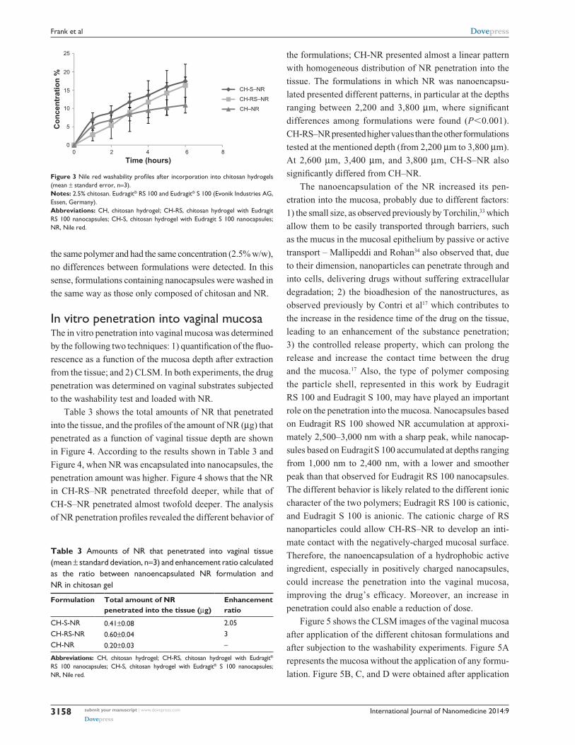
the washability profiles (percentage of NR washed away as a
function of time). No significant differences were observed
among formulations (analysis of variance, P=0.107) for all of
the studied times. One possible reason for this is that, when
time is considered in the analysis, the most relevant factor to
be considered is the type of vehicle that will be washed, ie, the
chitosan. Because the three formulations were derived from
00
50
100
150
200
250
300
350
0.5 1 1.5 2 2.5 3
Distance of stretching to detach from the mucosa (mm)
Forc
e (m
N)
W(CH) =371.19 mN∙mmW(CH-RS) =576.42 mN∙mm
W(CH-S) =579.92 mN∙mm
Figure 2 Mucoadhesion of chitosan hydrogels (2.5% chitosan) determined by the force of detachment and the distance of stretching to detach from the mucosa (mean, n=6).Notes: eudragit® rs 100 and eudragit® s 100 (evonik Industries ag, essen, germany). The area under the curve represents the work (ie, the product of force and distance) necessary to detach a formulation from the mucosa. such work values [W(ch); W(ch-rs); W(ch-s)] are a representation of the mucoadhesive properties of each formulation.Abbreviations: ch, chitosan hydrogel; ch-rs, chitosan hydrogel with eudragit rs 100 nanocapsules; ch-s, chitosan hydrogel with eudragit s 100 nanocapsules.
International Journal of Nanomedicine 2014:9submit your manuscript | www.dovepress.com
Dovepress
Dovepress
3158
Frank et al
the same polymer and had the same concentration (2.5% w/w),
no differences between formulations were detected. In this
sense, formulations containing nanocapsules were washed in
the same way as those only composed of chitosan and NR.
In vitro penetration into vaginal mucosaThe in vitro penetration into vaginal mucosa was determined
by the following two techniques: 1) quantification of the fluo-
rescence as a function of the mucosa depth after extraction
from the tissue; and 2) CLSM. In both experiments, the drug
penetration was determined on vaginal substrates subjected
to the washability test and loaded with NR.
Table 3 shows the total amounts of NR that penetrated
into the tissue, and the profiles of the amount of NR (µg) that
penetrated as a function of vaginal tissue depth are shown
in Figure 4. According to the results shown in Table 3 and
Figure 4, when NR was encapsulated into nanocapsules, the
penetration amount was higher. Figure 4 shows that the NR
in CH-RS–NR penetrated threefold deeper, while that of
CH-S–NR penetrated almost twofold deeper. The analysis
of NR penetration profiles revealed the different behavior of
the formulations; CH-NR presented almost a linear pattern
with homogeneous distribution of NR penetration into the
tissue. The formulations in which NR was nanoencapsu-
lated presented different patterns, in particular at the depths
ranging between 2,200 and 3,800 µm, where significant
differences among formulations were found (P0.001).
CH-RS–NR presented higher values than the other formulations
tested at the mentioned depth (from 2,200 µm to 3,800 µm).
At 2,600 µm, 3,400 µm, and 3,800 µm, CH-S–NR also
significantly differed from CH–NR.
The nanoencapsulation of the NR increased its pen-
etration into the mucosa, probably due to different factors:
1) the small size, as observed previously by Torchilin,33 which
allow them to be easily transported through barriers, such
as the mucus in the mucosal epithelium by passive or active
transport – Mallipeddi and Rohan34 also observed that, due
to their dimension, nanoparticles can penetrate through and
into cells, delivering drugs without suffering extracellular
degradation; 2) the bioadhesion of the nanostructures, as
observed previously by Contri et al17 which contributes to
the increase in the residence time of the drug on the tissue,
leading to an enhancement of the substance penetration;
3) the controlled release property, which can prolong the
release and increase the contact time between the drug
and the mucosa.17 Also, the type of polymer composing
the particle shell, represented in this work by Eudragit
RS 100 and Eudragit S 100, may have played an important
role on the penetration into the mucosa. Nanocapsules based
on Eudragit RS 100 showed NR accumulation at approxi-
mately 2,500–3,000 nm with a sharp peak, while nanocap-
sules based on Eudragit S 100 accumulated at depths ranging
from 1,000 nm to 2,400 nm, with a lower and smoother
peak than that observed for Eudragit RS 100 nanocapsules.
The different behavior is likely related to the different ionic
character of the two polymers; Eudragit RS 100 is cationic,
and Eudragit S 100 is anionic. The cationic charge of RS
nanoparticles could allow CH-RS–NR to develop an inti-
mate contact with the negatively-charged mucosal surface.
Therefore, the nanoencapsulation of a hydrophobic active
ingredient, especially in positively charged nanocapsules,
could increase the penetration into the vaginal mucosa,
improving the drug’s efficacy. Moreover, an increase in
penetration could also enable a reduction of dose.
Figure 5 shows the CLSM images of the vaginal mucosa
after application of the different chitosan formulations and
after subjection to the washability experiments. Figure 5A
represents the mucosa without the application of any formu-
lation. Figure 5B, C, and D were obtained after application
0
5
10
15
20
25
0 2 4 6 8
Con
cent
ratio
n %
Time (hours)
CH-S–NR
CH-RS–NR
CH–NR
Figure 3 Nile red washability profiles after incorporation into chitosan hydrogels (mean ± standard error, n=3).Notes: 2.5% chitosan. eudragit® rs 100 and eudragit® s 100 (evonik Industries ag, essen, germany).Abbreviations: ch, chitosan hydrogel; ch-rs, chitosan hydrogel with eudragit rs 100 nanocapsules; ch-s, chitosan hydrogel with eudragit s 100 nanocapsules; Nr, Nile red.
Table 3 amounts of Nr that penetrated into vaginal tissue (mean ± standard deviation, n=3) and enhancement ratio calculated as the ratio between nanoencapsulated Nr formulation and Nr in chitosan gel
Formulation Total amount of NR penetrated into the tissue (µg)
Enhancement ratio
ch-s-Nr 0.41±0.08 2.05ch-rs-Nr 0.60±0.04 3ch-Nr 0.20±0.03 –
Abbreviations: ch, chitosan hydrogel; ch-rs, chitosan hydrogel with eudragit® rs 100 nanocapsules; ch-s, chitosan hydrogel with eudragit® s 100 nanocapsules; Nr, Nile red.

International Journal of Nanomedicine 2014:9 submit your manuscript | www.dovepress.com
Dovepress
Dovepress
3159
a new formulation for vaginal drug delivery
A B
C D
20 μm20 μm
20 μm 20 μm
Figure 5 confocal laser scanning microscopy images of the vaginal mucosa after application of chitosan gels containing Nile red.Notes: 2.5% chitosan. The pictures are representatives of the measurement of three different batches: (A) blank; (B) ch–Nr; (C) ch-rs–Nr, (D) ch-s–Nr. eudragit® rs 100 and eudragit® s 100 (evonik Industries ag, essen, germany).Abbreviations: ch, chitosan hydrogel; ch-rs, chitosan hydrogel with eudragit rs 100 nanocapsules; ch-s, chitosan hydrogel with eudragit s 100 nanocapsules; Nr, Nile red.
0
0.02
0.04
0.06
0.08
0.1
0 1,000 2,000 3,000 4,000 5,000 6,000 7,000
Am
ount
pen
etra
ted
(mg)
Depth (mm)
* * *
CH-S–NRCH-RS–NRCH–NR
Figure 4 Quantification of Nile red accumulation into vaginal mucosa as a function of mucosa depth, after incorporation into chitosan hydrogels (mean ± standard error, n=3).Notes: 2.5% chitosan. *P0.001. eudragit® rs 100 and eudragit® s 100 (evonik Industries ag, essen, germany).Abbreviations: ch, chitosan hydrogel; ch-rs, chitosan hydrogel with eudragit rs 100 nanocapsules; ch-s, chitosan hydrogel with eudragit s 100 nanocapsules; Nr, Nile red.

International Journal of Nanomedicine 2014:9submit your manuscript | www.dovepress.com
Dovepress
Dovepress
3160
Frank et al
of CH–NR, CH-RS–NR, and CH-S–NR, respectively.
Figure 5B–D enables visual detection of fluorescence in
the mucosa slices. Figure 5B shows that the application of
the CH–NR formulation led to a lower amount of fluores-
cence in the mucosa (small red area of the picture) com-
pared with Figure 5C and D representing gels containing
nanoencapsulated NR. Higher fluorescence was visualized
in the CH-RS–NR formulation (Figure 5C) compared with
CH-S–NR formulation (Figure 5D). Results based on such
images are all in accordance with the penetration profile
obtained with extracted samples (Figure 4).
The comparison between the chitosan gels containing
cationic and anionic nanocapsules agrees with the tensile
stress measurement results for mucoadhesion, where the
formulations containing nanocapsules remained in contact
with the vaginal mucosa for a longer time. Thus, increased
contact time of these formulations containing nanocap-
sules leads to increased penetration of NR. Such results
show that nanoencapsulated drugs can easily penetrate
into the tissue.
ConclusionPolymeric nanocapsules with cationic and anionic surface
charges encapsulating NR as a lipophilic model substrate
demonstrated appropriate properties for vaginal applica-
tion. Chitosan gels were produced as vehicles for those
nanocapsules, with a chitosan concentration of 2.5% w/w
determined to be the most suitable for vaginal delivery. The
chitosan hydrogels exhibited higher adhesion to vaginal
mucosa, which is most likely related to the higher contribu-
tion of the viscous property over the elastic property of the
gels. Regarding the penetration of NR into the mucosa, the
results indicated that a greater amount of NR penetrated into
the mucosa when it was nanoencapsulated, especially when
loaded in positively charged nanocapsules, most likely due
to the electrostatic interaction with the negatively charged
tissue.
Considering these results, this work shows the applica-
bility of this innovative formulation composed of 2.5% w/w
chitosan gel, as the vehicle, and polymeric nanocapsules to
vaginal drug administration. The chitosan gel and polymeric
nanocapsules increase the mucoadhesion and the amount
of lipophilic drug that penetrates into the tissue and might
lead to a greater effect compared with the free drug. The
innovative formulation can be considered as an alternative
for treatments where the drug should be retained, for a long
period of time, in contact with the mucosa, and also for treat-
ments where the adverse effects of the drug, such as mucosa
irritation, should be decreased, which could be achieved by
the nanoencapsulation.
AcknowledgmentThe authors thank Capes, CNPq, and FAPERGS for the
financial support received.
DisclosureThe authors report no conflicts of interest in this work.
References 1. Sandri S, Rossi S, Ferrari F, Bonferoni MC, Muzzarelli C, Caramella C.
Assessment of chitosan derivatives as buccal and vaginal penetration enhancers. Eur J Pharm Sci. 2004;21:351–359.
2. Valenta C. The use of mucoadhesive polymers in vaginal delivery. Adv Drug Deliv Rev. 2005;57:1692–1712.
3. Perioli L, Ambrogi V, Venezia L, Pagano C, Ricci M, Rossi C. Chitosan and a modified chitosan as agents to improve performances of mucoad-hesive vaginal gels. Colloids Surf B Biointerfaces. 2008;66:141–145.
4. Bonferoni MC, Sandri G, Rossi S, Ferrari F, Gibin S, Caramella C. Chitosan citrate as multifunctional polymer for vaginal delivery Evalu-ation of penetration enhancement and peptidade inhibition properties. Eur J Pharm Sci. 2008;33:166–176.
5. Baloglu E, Senygit ZA, Karavana SY, Bernkop-Schnürch A. Strategies to prolong the intravaginal residence time of drug delivery systems. J Pharm Pharm Sci. 2009;12:312–336.
6. Bowman MC, Ballard TE, Ackerson CJ, Feldheim DL, Margolis DM, Melander C. Inhibition of HIV fusion with multivalent gold nanopar-ticles. J Am Chem Soc. 2008;130:6896–6897.
7. Lara HH, Ayala-Nuñez NV, Ixtepan-Turrent L, Rodriguez-Padilla C. Mode of antiviral action of silver nanoparticles against HIV-1. J Nanobiotechnology. 2010;8:1.
8. Alukda D, Sturgis T, Youan BB. Formulation of tenofovir-loaded functionalized solid lipid nanoparticles intended for HIV prevention. J Pharm Sci. 2011;100:3345–3356.
9. Patel GM, Patel PV. Novel vaginal anti-HIV drug delivery system of tenofovir disoproxil fumarate. Am J Pharm Tech Res. 2011;1:366–383.
10. Bachhav YG, Patravale VB. Microemulsion-based vaginal gel of clotrimazole: formulation, in vitro evaluation and stability studies. AAPS Pharm Sci Tech. 2009;10:476–481.
11. Schaffazick SR, Guterres SS. Caracterização e estabilidade físico-química de sistemas poliméricos nanoparticulados para administração de fármacos. [Physicochemical characterization and stability of the polymeric nanoparticle systems for drug administration]. Quim Nova. 2003;0:1–12. Portuguese.
12. Haas SE, Bettoni CC, de Oliveira LK, Guterres SS, Costa TD. Nanoen-capsulation increases quinine antimalarial efficacy against Plasmodium Berghei in vivo. Int J Antimicrob Agents. 2009;34:156–161.
13. Contri RV, Frank LA, Kaiser M, Pohlmann AR, Guterres SS. Decreased irritation of capsaicinoids to human skin by means of nanoencapsulation. Int J Nanomedicine. 2014;9:951.
14. Hussain A, Ahsan F. The vagina as a route for systemic drug delivery. J Control Release. 2005;103:301–313.
15. Illum L, Fischer AN, Jabbal-Gill I, Davis SS. Bioadhesive starch microspheres and absorpition enhancing agents act synergisti-cally to enhance the nasal absorption of polypeptides. Int J Pharm. 2001;222:109–119.
16. Valenta C, Auner BG. The use of polymers for dermal and transdermal delivery. Eur J Pharm Biopharm. 2204;58:279–289.
17. Contri RV, Katzer T, Ourique AF, et al. Combined effect of polymeric nanocapsules and chitosan hydrogel on the increase of capsicino-ids adhesion to the skin surface. J Biomed Nanotechnol. 2014;10: 820–830.

International Journal of Nanomedicine 2014:9 submit your manuscript | www.dovepress.com
Dovepress
Dovepress
International Journal of Nanomedicine
Publish your work in this journal
Submit your manuscript here: http://www.dovepress.com/international-journal-of-nanomedicine-journal
The International Journal of Nanomedicine is an international, peer-reviewed journal focusing on the application of nanotechnology in diagnostics, therapeutics, and drug delivery systems throughout the biomedical field. This journal is indexed on PubMed Central, MedLine, CAS, SciSearch®, Current Contents®/Clinical Medicine,
Journal Citation Reports/Science Edition, EMBase, Scopus and the Elsevier Bibliographic databases. The manuscript management system is completely online and includes a very quick and fair peer-review system, which is all easy to use. Visit http://www.dovepress.com/testimonials.php to read real quotes from published authors.
Dovepress
3161
a new formulation for vaginal drug delivery
18. Rinaudo M. Chitin and chitosan: properties and applications. Prog Polym Sci. 2006;31:603–632.
19. Sayin B, Somavarapu S, Li XW, Sesardic D, Senel S, Alpar OH. TMC-MCC (N-trimethyl chitosan-mono-N-carboxymethyl chitosan nanocomplexes for mucosal delivery of vaccines. Eur J Pharm Sci. 2009;38:362–369.
20. Wittaya-Areekul S, Kruenate J, Prahsarn C. Preparation and in vitro evaluation of mucoadhesive properties of alginate/chitosan microparticles containing prednisolone. Int J Pharm. 2006;312: 113–118.
21. Zhang S, Kawakami K. One-step preparation of chitosan solid nanoparticles by electrospray deposition. Int J Pharm. 2010;397: 211–217.
22. Berger J, Reist M, Mayer JM, Felt O, Peppas NA, Gurny R. Structure and interactions in covalently and ionically crosslinked chitosan hydro-gels for biomedical applications. Eur J Pharm Biopharm. 2004;57: 19–34.
23. Meng J, Sturgis TF, Youan BB. Engineering tenofovir loaded chitosan nanoparticles to maximize microbicide mucoadhesion. Eur J Pharm Sci. 2011;44:57–67.
24. Contri RV, Katzer T, Pohlmann AR, Guterres SS. Chitosan hydrogel containing capsaicinoids-loaded nanocapsules: an innovative formula-tion for topical delivery. Soft Matter. 2010;8:370–385.
25. Katzer T, Chaves P, Bernardi A, Pohlmann AR, Guterres SS, Beck RC. Castor oil and mineral oil nanoemulsion: development and com-patibility with a soft contact lens. Pharm Dev Technol. 2014;19: 232–237.
26. Santos SS, Lorenzoni A, Ferreira LM, et al. Clotrimazole-loaded Eudragit® RS 100 nanocapsules: preparation, characterization and in vitro evaluation of antifungal activity against Candida species. Mater Sci Eng C Mater Biol Appl. 2013;33:1389–1394.
27. Fessi H, Puisilux F, Devissaguet JP, Ammoury N, Benta S. Nanocapsule formation by interfacial polymer deposition following solvent displace-ment. Int J Pharm. 1989;55:R1–R4.
28. Schaffazick SR, Pohlmann AR, de Cordova CA, Creczynski-Pasa DB, Guterres SS. Protective properties of melatonin-loaded nanoparticles against lipid peroxidation. Int J Pharm. 2005;289:209–213.
29. Bonferoni MC, Rossi S, Ferrari F, Caramella C. A modified Franz dif-fusion cell for simultaneous assessment of drug release and washability of mucoadhesive gels. Pharm Dev Technol. 1999;4:45–53.
30. Rossi S, Bonferoni MC, Ferrari F, Caramella C. Drug release and wash-ability of mucoadhesive gels based on sodium carboxymethylcellulose and polyacrylic acid. Pharm Dev Technol. 1999;4:55–63.
31. Contri RV, Kaiser M, Poletto FS, Pohlmann AR, Guterres SS. Simultaneous control of capsaicinoids release from polymeric nano-capsules. J Nanosci Nanotechnol. 2011;11:2398–2406.
32. Contri RV, Soares RMD, da Silveira NP, Pohlmann AR, Guterres SS. Develoment of chitosan hydrogels containing nanocapsules and charac-terization by oscilatory rheology. Proceedings of the XII International Macromolecular Colloquium; September 7–10; 2010; Gramado.
33. Torchilin VP. Drug targeting. Eur J Pharm Sci. 2000;11 Suppl 2: S81–S91.
34. Mallipeddi R, Rohan LC. Nanoparticle-based vaginal drug delivery sys-tems for HIV prevention. Expert Opin Drug Deliv. 2010;7(1):37–48.