Embed Size (px)
Citation preview

1
A review of image enhanced endoscopy (IEE) in the evaluation of colonic polyps Dr. G Longcroft-Wheaton. MB, BS, MD, MRCP(UK) MRCP(gastro)
Consultant Gastroenterologist
Portsmouth Hospitals NHS Trust,
Queen Alexandra Hospital,
Cosham
Hamphire
PO6 3LY
Main author of article
Professor P Bhandari MD, FRCP
Portsmouth Hospitals NHS Trust,
Queen Alexandra Hospital,
Cosham
Hamphire
PO6 3LY
Senior author of article
Word count: 6789
Conflicts of interest: none

2
Summary
The practice of colonoscopy has changed considerably over the last decade.
The growth of image enhanced endoscopy (IEE) have altered our concepts of
how we perform colonoscopy. This article examines the evidence base behind
these techniques and looks at where future research needs to be directed.
Key words
Colonic polyp, NBI, FICE, i-scan, indigo carmine
Expert commentary
Paradigms in our understanding of how colonoscopy should be performed are
shifting. Image enhanced endoscopy using both dye based chromoendoscopy
and electronic imaging are providing us with methods of improving lesion
detection and characterization beyond what we previously thought possible.
Traditional views that the neoplastic potential of a lesion can only be
determined by the pathologist are being challenged, and it is likely that the era
of protocol guided mapping biopsies for surveillance of conditions such as
ulcerative colitis are nearing an end. However, these technologies are not
without limitations, and although they are all trying to achieve similar goals
there are significant differences between them. Perhaps the biggest challenge
that we now face will be to understand both how to train in these techniques
effectively, and how to translate the large body of published research into
routine clinical practice.

3
Five year view
There will be a growth in the publication of guidelines from the World
endoscopy societies setting standards for the use of image enhanced
endoscopy in place of conventional approaches of mapping biopsies and
histological examination. Training tools will emerge to enable structured
training programmes to be developed, as will strategies for auditing success
in application of these techniques to clinical practice. Initially the techniques
will be adopted by experts in tertiary referral centres, but dissemination to the
wider community will occur as the practical difficulties of adoption of these
approaches are solved.
Key issues
Indigo carmine or methylene blue chromoendoscopy increases
adenoma detection during routine colonoscopy
Indigo carmine or methylene blue Chromoendoscopy is the method of
choice for the surveillance of patients with ulcerative colitis
Indigo carmine or methylene blue Chromoendoscopy is an effective
tool for in-vivo histology prediction of colonic polyps
NBI and FICE have no role in polyp detection in a surveillance
population. The position with i-scan is less clear. There is very little
evidence in a high risk population, with a small selection of studies on
NBI and i-scan suggesting they may be of some benefit in increasing
the polyp detection rate

4
There is a lack of evidence to recommend NBI, FICE or i-scan for
surveillance of ulcerative colitis
There is good evidence for the use of NBI, FICE and i-scan for the in-
vivo histology prediction of colonic polyps
The adoption of a ‘resect and discard’ policy for colonic polyps may
well be a very cost effective measure with minimal clinical
consequences in expert hands

5
Introduction
There have been considerable advances in the endoscopic examination and
treatment of colonic neoplasia with the development of techniques for
performing image enhanced endoscopy (IEE), including chromoendoscopy
and electronic imaging. It is important to understand what can and cannot be
achieved with these emerging technologies. This article reviews the evidence
behind these new endoscopic enhancement techniques, and discusses where
this field is likely to be moving in the future
Background
A key role of colonoscopy is lesion detection and characterization. Colorectal
cancer accounts for an estimated 550,000 deaths worldwide [1], with poor
outcomes for advanced disease. Colorectal cancer develops from
adenomatous polyps through the adenoma-carcinoma sequence [2,3].
Therefore detection of adenomatous polyps before they turn into cancer and
polypectomy is important, and was shown to reduce colon cancer mortality.
However, small hyperplastic polyps, accounting for one third of all polyps,
have negligible malignant potential, especially if located in the left side of the
colon.
It has been traditionally felt that hyperplastic polyps cannot be separated
clinically from adenomas or polyp cancers. For this reason all polyps are
removed. However, polypectomy is associated with significant risks [4],

6
results in an immediate cost in processing the samples, increases the
workload for pathologists, and increases the procedure time. However, It is
becoming recognized that in-vivo characterization of lesions is possible, with
the ASGE recently proposing standards for in-vivo assessments [5].
Image enhanced endoscopy in the colon
IEE can help in two ways:
1) Improved lesion detection
2) Improved lesion characterization
There are many emerging technologies which can impact on both of these
areas. These include high resolution (HD) colonoscopy and electronic imaging
techniques including narrow band Imaging, FICE and i-scan,
chromoendoscopy and novel devices including cap assisted colonoscopy and
confocal endoscopy. This article will focus on chromoendoscopy and
electronic imaging. To understand how these technologies can impact on
lesion detection and characterization it is necessary to broadly understand
how they work and the principles behind their use.
Chromoendoscopy
Chromoendoscopy involves application of a dye to the gastrointestinal tract.
The techniques were pioneered in Japan where initial experience was in the
use of the vital stain crystal violet to characterize colonic neoplasia. Crystal

7
violet irreversibly binds to cellular structures, highlighting surface patterns in
great detail. It was with this dye that the first attempts were made at in-vivo
histology prediction for colonic polyps. It cannot be used for lesion detection
but is very effective for lesion characterization. However, it poses a number of
problems. Vital stains are inconvenient to use. They have to be dripped onto
the lesion surface and allowed to fix for several minutes, followed by washing
prior to evaluation. This is time consuming and subjective. For this reason it is
generally accepted that vital stains are not practical for daily use outside of a
research setting. This led to a search for alternative dyes.
Indigo carmine, generally used at a concentration of 0.2%, is a blue dye which
does not bond to or react with human tissue. It simply sits on the surface of
tissues, highlighting surface patterns. For this reason it is very safe.
Furthermore, it is easier to use than crystal violet as the results are instant. As
it does not bind to tissues, excess dye can be sucked away. Indigo carmine
can be used for two purposes; to find polyps or to characterize neoplasia.
Methylene blue is a similar blue dye. It differs however from indigo carmine in
that it binds to tissues and therefore carries a theoretical risk of DNA damage.
In practice it can be used in a very similar way to indigo carmine.
Pan-chromoendoscopy for lesion detection in a surveillance population
A Cochrane meta-analysis concluded that chromoendoscopy with indigo
carmine enhances the detection of neoplastic polyps [6]. The review
examined five randomized controlled trials, excluding polyposis or colitis

8
patients [7-11], representing 1059 patients. Chromoendoscopy significantly
increased both the number of patients with at least one polyp detected (OR
2.22) and the number of patients with at least one dysplastic lesion detected
(OR 1.67). The predominant increase was in the number of diminutive
adenomas detected. Four other randomized controlled trials were not included
in the meta-analysis [12-15]. All but one of these studies [14] demonstrated
improved lesion detection with indigo carmine. Methylene blue has shown
similar results [102].
Pan-chromoendoscopy for lesion detection in a high risk population
Chromoendoscopy is potentially of benefit in hereditary syndromes by
enhancing detection of subtle lesions. Indigo carmine chromoendoscopy has
been studied in Familial adenomatous polyposis (FAP), attenuated FAP, and
hereditary non-polyposis colorectal cancer (Lynch syndrome).
Chromoendoscopy may help in making a diagnosis by revealing additional
lesions required to meet a diagnostic criteria. A very small study has
suggested that indigo carmine can help distinguish between attenuated FAP
and classical FAP (>100 adenomas) [16]. Likewise, the diagnosis of
hyperplastic polyposis syndrome is dependent on identification of a specific
number of polyps, and chromoendoscopy may help in meeting the criteria
[17].
9
Chromoendoscopy may be beneficial in the surveillance of polyp syndromes.
back-to-back studies in Lynch syndrome suggest that polyp detection may be
improved [18,19,20]. Dye-spray increases polyp detection in FAP surveillance
[21]. Whether this is of any clinical value is unclear as most true FAP patients
are treated with colectomy rather than surveillance. There are no studies
published in hyperplastic polyposis syndrome or Peutz-Jeghers syndrome.
Pan-chromoendoscopy in inflammatory bowel disease
There is growing evidence that chromoendoscopy using indigo carmine is the
optimum method for performing colitis surveillance. There are two randomized
controlled trials and several large cohort studies comparing the technique to
conventional mapping biopsies [22-30]. All of these trials have demonstrated
improved neoplasia yields with pan-chromoendoscopy. A meta-analysis [31]
examining these studies showed a 44% increase in detection of neoplasia
with the majority of them being flat. The meta analysis also demonstrates a
dramatic reduction in number of biopsies taken per patient from 40 with the
conventional strategy to 11 with chromoendoscopy directed targeted biopsies.
Studies with methylene blue have yielded similar results [103]. Recent ECCO
guidelines recommends this as the strategy for ulcerative colitis surveillance.
It should be noted that pan-chromoendoscopy is only of value when the
patient is in remission. In the presence of active inflammation there is little to
be gained through the application of dye spray, as ulceration, mucous and
pus interferes with the assessment of surface patterns and makes such
evaluations unreliable.

10
There is good evidence that pan-chromoendoscopy improves lesion detection
in a routine surveillance population. However, pan-colonic dye spray, where
indigo-carmine is applied to the entire colon using a spray catheter, has not
become routine practice. There are a number of reasons for this. Dye
spraying is time consuming messy and inconvenient. The colon has to be
clean and free of debris and this remains a big challenge as bowel
preparation in western settings is not perfect in all patients. Practically most
western units would find the practice of pan-chromoendoscopy very
challenging. Furthermore, there is a lack of data to support whether this
increase in lesion detection results in a long term reduction in cancer risk.
Chromoendoscopy for lesion characterization
There has been considerable work evaluating the use of chromoendoscopy in
characterizing colonic lesions. The initial work with indigo carmine for in-vivo
diagnosis was conducted in Japan by Kato et al. who retrospectively analysed
4445 lesions using magnifying endoscopy with indigo carmine dye spray. All
of the lesions were less than 5mm in size and assessed by evaluating surface
patterns [32]. These patterns were originally described using vital stains
(crystal violet) by Professor Kudo and have formed the cornerstone of most of
the subsequent in-vivo diagnostic studies [33,34]. All of the lesions were
assessed in-vivo, with the predicted diagnosis correlated with the
histopathological diagnosis. The findings suggested that a sensitivity for
adenoma of 98% and specificity of 52% could be achieved. The excellent
sensitivity was achieved by compromising the specificity, resulting in a large
11
proportion of hyperplastic polyps being overcalled as adenomas. The data
was dependent on 100x magnification with a magnifying endoscope.
Further work was conducted in Japan [35] which investigated the differences
between indigo carmine with and without magnification in the examination of
small (<10mm) polyps. The results were encouraging, with a sensitivity for
neoplasia of 93.1% and specificity of 76.1% being achieved. However, there
was improvement with magnification. This suggested that in appropriately
skilled hands, in-vivo diagnosis was possible without the need for
magnification endoscopy or vital stains. There were further Japanese studies
looking at magnification endoscopy with indigo carmine for in-vivo histology
prediction which showed similar results [36-38]. See figure 1.
There has been work from outside of Japan using magnifying
chromoendoscopy with indigo carmine. Tischendorf et al in Germany
conducted a prospective cohort study of neoplastic vs non neoplastic polyps
using both narrow band imaging and indigo carmine with magnifying
endoscopy. A sensitivity of 91.7% and specificity of 90% was achieved for
indigo carmine using Kudo pit pattern analysis [39]. A large German study
compared indigo carmine and the electronic imaging modality FICE in the
assessment of polyps <10mm. The primary aims of this study were lesion
detection. However a sub-group of 280 lesions were assessed using indigo
carmine for histology prediction. A sensitivity for neoplasia of 87.6% and
specificity of 62.0% was achieved [40]. High definition endoscopes were used

12
without optical magnification. There was a further German study by a different
group examining indigo carmine with high resolution endoscopes. This study
investigated 273 lesions <5mm, with a sensitivity of 94%, specificity of 64%
and accuracy of 83% [41]. This study differed from the other studies described
in that it only examined rectosigmoid polyps. Further similar studies [42-46]
are summarized in tables 1 and 2.
It should be noted that methylene blue has also been studied for lesion
characterization. The results have been excellent, and it is widely accepted
that it can be used in the same way as indigo carmine [102].
A notable point observed in most of the published studies is the trade off
between adenoma sensitivity and specificity. Many of the studies with the
highest sensitivity have a low specificity, typically between 60-70%. Whilst this
is the safest approach to in-vivo diagnosis, it is not ideal. The ultimate goal for
diminuitive polyps <5mm in size would be to have the ability to confidently
leave small hyperplastic polyps, reducing the risks posed by polypectomy. To
achieve this, sensitivity and specificity both need to be very high.
High definition colonoscopes are becoming an industry standard and it is
important to know if they improve lesion characterization. A recent study
published from the United Kingdom has suggested that diagnostic accuracy,
sensitivity and specificity of assessment of colonic polyps <10mm in size was
not affected by the resolution of the colonoscope used [47]. This was a single
centre, single endoscopist study, but is the only study in this field. However, it
is encouraging as standard resolution endoscopes are still in widespread use

13
and still being marketed and sold by most of the major endoscope
manufacturers. HD endoscopes are more expensive and require updated
processor and display screen which all come at extra cost and do improve the
quality of image. However, clinicians can draw comfort from the above study
that if they are using indigo carmine for in-vivo assessments then even
standard definition endoscopes can produce comparable accuracy.
Because of growing interest in the use of in-vivo diagnosis The American
Society for gastrointestinal endoscopy (ASGE) has produced PIVI guidelines,
setting standards a technique or technology needs to achieved to be used for
in-vivo diagnosis [5]. This includes standards which need to be met for setting
rescope intervals and for leaving small rectosigmoid hyperplastic polyps in-
vivo. Most of the indigo carmine studies are from the pre-PIVI era so do not
report on PIVI standards but a recent study looked at the PIVI standards that
can be achieved with indigo carmine [48]. This study used indigo carmine
without optical magnification. The results were encouraging, with indigo
carmine meeting both the requirements for rescope intervals and for leaving
polyps in situ. It should be noted that the assessments were made after first
assessing using the electronic imaging modality FICE. Indigo carmine did
improve the negative predictive value of assessment, enough to meet the PIVI
standard for leaving small left sided hyperplastic polyps in situ, although the
change in accuracy, sensitivity and specificity was not statistically significant.
See table 2. We can conclude from this study that indigo carmine when used
after FICE will improve the negative predictive value to a standard that will let

14
us implement ‘do not resect’ policy for diminuitive rectosigmoid polyps <5mm
in size.
There has been some work examining magnifying chromoendoscopy in the
evaluation of sessile serrated polyps. A paper from Japan has suggested that,
using a modified form of the Kudo pit pattern classification system, it is
possible to identify sessile serrated adenomas with 83.7% sensitivity and
85.7% specificity [101]. More work is needed in this field.
Chromoendoscopy has been shown to be an effective tool in predicting depth
of sub-mucosal invasion of early cancers in the colon [96-97], with accuracy
between 71% and 91%. This requires magnifying endoscopes and highly
skilled endoscopists. Furthermore, whilst some of the published work has
used indigo carmine [97] the majority of assessments have used vital staining
with crystal violet. The data has come from specialist centres in Japan and it
is unclear whether such techniques could currently be used in a western
setting. However, with the growth of EMR and ESD as the standard for
removal of large benign colonic polyps it is possible that endoscopists will
become more confident in their diagnostic abilities and that skills in this area
will improve.
Electronic imaging
Some endoscopists have been critical of chromoendoscopy, claiming that it is
a messy time consuming process. Furthermore, it physically colours the
mucosa, requiring extensive washing if the endoscopist decides that he or she
wants an unstained view. This has led to the development of push button
15
‘virtual chromoendoscopy’ techniques. These will be referred to collectively as
‘electronic imaging’ for the purposes of this article.
Narrow band imaging
The first commercially available system came from Olympus, known as
narrow band imaging (NBI). The concept of NBI is to improve visualization of
mucosal vascular patterns. It is based on the principle of variable penetration
of light depending on its wavelength. Red light penetrates deep into the
submucosa but doesn’t help with surface pattern assessment. Blue and green
light at a wavelength range of 415-540nm does not penetrate deep but
enhances mucosal vessel patterns. Blue light displays superficial capillary
networks whilst green light highlights subepithelial vessels. The result is a
high contrast image which makes the interpretation of surface vascular
patterns possible. NBI uses a physical filter to block red light and to narrow
the bandwidth of the blue and green light, hence improving visualisation of
surface patterns.
Narrow band imaging for lesion detection
Early studies suggested that there was some improvement in lesion detection
using NBI [49-50]. This was not however repeated in later investigations
[51,52,53]. The overall conclusions were that the gains seen in the preliminary
studies were largely due to inexperienced endoscopists, still on a steep

16
section of the polyp detection learning curve. However, NBI did help improve
polyp detection skills. When used by experts, who already had high adenoma
detection rates, there was no gain [52] [54]. There has been a tandem
endoscopy published which suggested that the adenoma miss rate may be
lower in the proximal colon. Furthermore, a significantly higher number of
small lesions <5mm were found using NBI compared to white light imaging in
this study [94]. It is likely therefore that if there is any gain from NBI in lesion
detection that it is small.
Narrow band imaging in familial polyp syndromes
There is limited evidence for use of NBI in any of the polyp syndromes. Due to
profound differences between the syndromes it is difficult to consider them as
a single group. The greatest evidence base is in hyperplastic polyposis, where
a randomized controlled trial has been conducted [58]. This is perhaps
unsurprising, as the key clinical aim in this condition is to identify adenomas
amongst a sea of hyperplastic lesions, and there is evidence to suggest that
electronic imaging is effective in differentiating hyperplastic from
adenomatous polyps. There is some evidence in Lynch syndrome, but this is
from small cohort studies and it is questionable whether these were
adequately powered. Of the studies examining lynch syndrome it would
appear there may be benefit from NBI, but there is a greater gain from
chromoendoscopy.

17
There has been one cohort study examining NBI in the assessment of familial
adenomatous polyposis (FAP). This involved analysis of images captured at
live endoscopy [55]. In total thirteen patients with FAP were examined.
Colonoscopic images were obtained using white light colonoscopy,
autofluorescence imaging, NBI, and chromoendoscopy, with all images
captured at equivalent angles and distances from the colorectal mucosa.
Chromoendoscopy detected the greatest number of lesions at all sites. NBI
depicted more lesions than white light. Autofluorescence imaging appeared
superior in the rectum. The authors concluded that chromoendoscopy was the
optimum imaging modality, and superior to white light colonoscopy,
autofluorescence imaging, and NBI for detection of diminutive colorectal
lesions in adenomatous polyposis. However, NBI was better than white light
alone.
There have been three published studies examining narrow band imaging in
Lynch syndrome. A cohort study from the United Kingdom [56] examined 62
patients from Lynch syndrome families, all diagnosed using the Amsterdam II
or genetic criteria as part of a colonoscopic surveillance programme. All
patients were examined twice from caecum to sigmoid-descending junction,
first with high definition white light and then after a second pass with NBI in a
back-to-back fashion. Initial adenoma detection in the proximal colon with
white light was 17/62 (27%). 26/62 (42%) patients had at least one adenoma
detected after NBI, with an absolute difference of 15% (95% CI 4-25%),
p=0.004 versus white light alone. The authors commented that the proportion
of flat adenomas detected in the NBI pass (9/21 (45%)) was higher than in the

18
white light pass (3/25 (12%)) p=0.03, suggesting that the principal gain was in
the detection of flat adenomas in the proximal colon.
A cohort study from Germany [57] examined 109 patients with HNPCC. In 47
patients, standard colonoscopy was followed by chromoendoscopy with indigo
carmine, and In 62 patients NBI was performed first followed by
chromoendoscopy. 128 hyperplastic and 52 adenomatous lesions were
detected in total. 0.5 lesions/patient were identified by standard colonoscopy
and 1.5 lesions/patient by chromoendoscopy ( P < 0.001). 0.7 lesions/patient
were detected by NBI. At least one adenoma was detected in 15 % of patients
by both standard and NBI colonoscopy compared with 28 % of patients by
chromoendoscopy. The authors concluded that chromoendoscopy detected
significantly more adenomatous lesions than standard white light colonoscopy
or NBI.
There has been one randomized controlled trial [58] investigating NBI for the
assessment of patients with hyperplastic polyposis syndrome. In total 22
patients was identified. Patients underwent tandem colonoscopy with high
resolution white light and NBI, in randomized order with removal of all
detected polyps. 209 polyps were detected (27 with normal histology, 116
hyperplastic polyps, 42 sessile serrated adenomas, and 24 adenomas).
Among patients assigned to white light first (n = 11) a total of 78 polyps was
detected; subsequent NBI added 44 polyps. In patients examined with NBI
first, 78 polyps were detected and subsequent white light added 9. Polyp miss
rates of white light and NBI were 36% and 10% (OR 0.21; 0.09-0.45). Again,

19
in a similar fashion to the Lynch syndrome studies, flat polyp shape was
independently associated with increased miss rate. The authors concluded
that NBI significantly reduces polyp miss rates in hyperplastic polyposis
patients.
Narrow band imaging in inflammatory bowel disease
Because of the success of chromoendoscopy in colitis surveillance, the
question was asked whether narrow band imaging could achieve similar
results. Three randomized controlled trials have attempted to answer this
question [59-61], with all of these trials giving negative results.
Two randomized controlled trials compared NBI to conventional
chromoendoscopy. One of these trials [30] demonstrated a considerably
higher miss rate with NBI compared to pan-chromoendoscopy (31.8% vs
13.6%). The second trial [62] (which used methylene blue as the dye) showed
comparable neoplasia detection rates. The position is therefore uncertain
whether NBI can be used as an alternative to chromoendoscopy for colitis
surveillance. However, it cannot be recommended as a suitable technique at
present.
Narrow band imaging for lesion characterization
There have been numerous publications investigating the potential for In-vivo

20
histology prediction using NBI [63-70], with several classification systems
validated specifically for use with NBI [99,100]. Similar results have been
achieved to those seen with indigo carmine. Several publications have
concentrated on non-magnifying endoscopes, perhaps most notable being the
DISCARD study [68]. This was however a general study of in-vivo diagnosis,
and the use of indigo carmine was allowed. Whilst the authors argued that this
was only needed in a minority of cases, it is difficult to ascertain the efficacy of
NBI on its own. Another study compared the accuracy of NBI with and without
magnification [71]. This showed no statistically significant difference with or
without magnification. However, the polyps were not assessed in-vivo.
Pictures were taken and then reviewed after the procedure by two
endoscopists. Therefore the results have to be interpreted with caution.
NBI certainly appears very promising and provides accuracy close to
histology. With the new validated classification systems it is ready for use in
expert hands. However, acquiring competence may be challenging. Recent
data has shown that, despite training, NBI was not that good in the hands of
general endoscopists compared to the results obtained in expert centres,
failing to meet the key standards laid down in the ASGE PIVI [74]. Similar
issues have been raised in other, similar studies [93]. Whilst there are
published studies suggesting that NBI can be taught relatively easily [101],
this raises a concern about the widespread applicability of NBI / in-vivo
diagnostic techniques outside of expert centres. This is an area needing
further research. There has been a study published which has suggested that,

21
with appropriate training, high magnification NBI can increase the diagnostic
skill of less experienced endoscopists to that of highly experienced
endoscopists [95]. It may be that when learning to perform in-vivo
assessments the greater clarity of patterns seen with magnification is highly
beneficial, but that this becomes less important as experience increases. This
should form the basis for future studies.
Unlike the studies into indigo carmine dye spray, where Japanese research
predominates, the work into NBI have come from a larger range of countries.
This perhaps reflects the reluctance of western endoscopists to embrace dye
spray. It should be noted however that the largest NBI study (1473 polyps)
comes from Japan [69].
Of all vascular enhancement techniques the biggest evidence base exists for
NBI. It is a tool where classification systems and assessment techniques have
been developed specifically for use with it. Data has been produced from
more than one centre, suggesting the techniques are reproducible in expert
hands. Unfortunately all of the data has been produced using high definition
equipment and it is necessary to assume, at present, that this is a prerequisite
for in-vivo diagnosis. See table 3. Most studies have reported sensitivity
between 82-95% and specificity of 75-90%. See figure 2 and table 3.
There has been some work examining the use of narrow band imaging with
optical magnification in examining depth of invasion of early colorectal
cancers [98]. The early data was promising, suggesting that thick and
severely irregular microvessels were diagnostic of sub-mucosal invasion. This

22
needs further evaluation in larger studies before introduction to mainstream
practice.
Flexible spectral imaging color enhancement (FICE)
FICE is a post processor technology found on Fujifilm endoscopes. Unlike
NBI, which utilizes a physical filter, FICE uses the charged coupled device
(CCD) in the endoscope to capture spectral reflectance data. This is sent to a
spectral estimation matrix processing circuit contained in the video processor.
The reflectance spectra of corresponding pixels that make up the
conventional image are mathematically estimated. From these spectra, a
virtual image is reconstructed of a single wavelength. Three such single-
wavelength images can be selected and assigned to the red, green, and blue
monitor inputs to display a composite colour enhanced multi band image in
real time. This can be used like narrow band imaging to remove data from the
red part of the waveband and narrow the green and blue spectra. However,
the system is flexible. It has 10 pre set digital filter settings with the ability to
program more.
FICE is a technically more complex than NBI, and therefore potentially more
flexible. This can prove off putting to clinicians who can find the multitude of
settings confusing.
FICE for lesion detection

23
In a similar position to NBI, a multicenter randomized controlled trial
investigating FICE for lesion detection in a surveillance population has
concluded that FICE was no more effective than white light for lesion
detection [40]. These results have been repeated in several further studies
[75,76] with the same negative results. It would therefore appear that FICE
has no role in improving polyp detection rates in the colon. There is no
published data for FICE in high risk patient groups.
FICE for lesion characterization
Several studies into the in-vivo histology prediction of colonic polyps come
from Germany. A prospective study of 150 polyps <2cm was compared to
indigo carmine dye spray with low (50x) and high (100x) magnification using
high resolution endoscopes (650,000 pixel CCD). The study was performed
by taking static pictures of each polyp and reviewing them by 3 different
readers after the procedure [77]. An accuracy of 83% and 90%, sensitivity of
89.9% and 96.6% and specificity of 73.8% and 80.3% could be achieved with
low and high magnification respectively. The results were essentially the
same with Indigo carmine with no statistically significant difference between
the two modalities. There are some important criticisms to note about this
study. As it is based on static images it is unclear whether the results are
directly transferrable to in-vivo diagnosis. Furthermore, as lesions over 1cm
were allowed, it is unclear whether these results could be achieved with

24
smaller polyps which are arguably harder to assess. High definition (650,000
pixel CCD) endoscopes were used and the results cannot be applied to
standard definition equipment.
The same team went on to conduct a further prospective randomized study
with the primary aim to investigate the impact of FICE on adenoma detection
rates (ADR) [40]. In this study lesion characterization was a secondary end
point. It demonstrated that FICE was able to differentiate adenomas from
hyperplastic polyps<10mm in size with a sensitivity of 93% and specificity of
61.2%. Accuracy was 84.7%. This was comparable but not superior to that of
indigo carmine, with no statistically significant difference between the two
techniques (p=0.44). Specificity was sacrificed to achieve adequate
sensitivity. Whilst safe, this approach limits the cost benefit position of in-vivo
diagnosis. Lesions were assessed using Kudo’s pit patterns which are not
validated for FICE. Furthermore, the primary end point of the study was not
lesion differentiation, but lesion detection.
There has been a Japanese study looking at histology prediction using FICE.
This study was small, examining 107 polyps <5mm in size and utilizing optical
magnification with high definition scopes. With high magnification (100x) a
sensitivity of 93% specificity of 70% and accuracy of 87% was achieved.
There was a small drop in accuracy with low (50x) magnification (87%) [37].
Again Kudo’s patterns were used for the assessments performed by
Japanese experts.
25
Not all studies support these findings. There was a study in which five
endoscopists assessed 144 pictures of 19 polyps to establish the diagnostic
accuracy of WLI, FICE and indigo carmine in making a histology prediction for
polyps <10mm in size. The results were disappointing, with a mean diagnostic
accuracy for WLI of 57%, FICE without magnification of 58.9% and IC without
zoom of 70.5% [78]. The methodology of this study could be criticized in many
ways. The number of lesions was extremely small and it was picture based.
Furthermore, It is unclear how experienced the endoscopists were in making
an in-vivo diagnosis. They achieved similar (poor) results with indigo carmine
which is out of keeping with previous studies. The Sano classification was
used to assess the lesions with FICE. This is a system designed for Narrow
band imaging [72]. Practically, the appearances are different with FICE to
NBI, and the Sano classification has never been validated for use with FICE.
See figure 3.
A study from Brazil has described a surface pattern system which is not
dissimilar to Kudo pit pattern classification but describing the vascular
patterns seen with FICE [79]. The study enrolled 309 lesions ranging in size
from 1-50mm, with 242 lesions <5mm in size. Again only high definition
endoscopes were used and no attempt to examine without magnification was
made. The authors commented that they felt optical magnification was
essential for analysis of vascular patterns. An accuracy of 98.3% sensitivity of
99.2% and specificity of 94.9% was achieved. The advantage of including
larger lesions was that 22 cases of colorectal cancer could be examined,
enabling a classification system to be validated. However, it does mean that

26
the very high sensitivity and specificity cannot be directly compared to the
other studies looking at much smaller lesions. The authors did not attempt to
analyze accuracy on the basis of lesion size.
A further study has attempted to validate a more simple classification system
for use with FICE, known as N.A.C.[92]. This classification system makes use
of vascularity, vascular patterns and surface patterns. It is designed for use
without optical magnification and makes use of both the white light and FICE
enhanced images. The details of this system are shown in table 5.
A recent study has examined FICE in a U.K. bowel cancer screening
population, as has compared results to the ASGE PIVI standard [48]. The
study suggested that FICE meets the PIVI standards for adopting a resect
and discard strategy, but could not produce a negative predictive value
sufficient for leaving recto-sigmoid hyperplastic polyps in situ. The same team
went on to examine the impact of colonoscope resolution on diagnostic
accuracy [80]. This study demonstrated an improvement in lesion
characterization with a high resolution colonoscope, with greater accuracy for
setting rescope intervals using ASGE and British Society of Gastroenterology
(BSG) guidelines. It clearly demonstrated that if electronic imaging like FICE
is to be used for in-vivo diagnosis then HD scopes are a must as they improve
the accuracy to a level that all PIVI standards are comfortably met. This raises
an important concept; whereas indigo carmine based assessments appear to
be independent of colonoscope resolution, the same cannot be said for FICE.
Whilst it is not clear whether the same issues apply to NBI or i-scan it should

27
be noted that all of the research on these systems has been performed using
high resolution equipment. Therefore it is safest to assume that, until studies
have been performed to prove or refute the position, that all electronic
imaging assessments are best made using high resolution colonoscopes. See
table 4.
i-scan
The most recent introduction to vascular enhancement has come from
Pentax. In some ways i-scan is a similar technology to FICE. It is a post
processor reconstruction from spectral reflectance data. However, in addition
to vascular enhancement it can also enhance surface patterns, increasing the
contrast between edges without reducing the brightness of images. At present
high definition 1.3 million pixel CCD endoscopes are available which have
been marketed for use with this system. These are not equipped with optical
magnification.
An early study using i-scan has suggested that increased lesion detection can
be achieved using the surface pattern enhancement setting [81]. This is in
stark contrast to results seen with NBI and FICE. The argument has been
made that the surface pattern enhancement features are fundamentally
different from the vascular enhancement of NBI and FICE, which explains the
difference. However, these results were not replicated in a subsequent trial
[82] and this is an area requiring further research.

28
There has been a study examining i-scan in hereditary polyp syndromes (83).
This study used the tone enhancement (TE) capacity of i-scan in Lynch
syndrome. In total 49 patients underwent back-to-back colonoscopy with two
imaging modalities, randomized into 2 groups. Group 1 (25 patients)
underwent High definition white light (HDWL) first followed by i-scan, group 2
(24 patients) i-scan first followed by High definition white light. The lesion
detection rate was 0.73 for i-scan and 0.36 for HDWL (p=0.095). In group 1,
14 lesions were detected with HDWL first and 15 with subsequent i-scan. In
group 2, 21 lesions were detected with i-scan first and 4 with subsequent
HDWL. The miss rate for endoscopic lesions was 52% and 16% respectively
and was significantly different in favor of i-scan (p<0.01). The authors
concluded that In patients with Lynch syndrome the miss rate for polyps is
significantly reduced during colonoscopy performed with i-scan in comparison
to high definition white light.
i-scan for lesion characterization
The previously described study examining i-scan for lesion detection [81] also
examined the role of i-scan in lesion characterization. in-vivo histology
prediction was made on 145 polyps <10mm with a sensitivity of 98%,
specificity of 100% and accuracy of 98.6%. Kudo pit patterns were used for
assessment. The most recent study from the United Kingdom called the Hi-
scope trial suggested similar results for i-scan [85]. However, this study was
unique in comparing the high resolution white light diagnosis to the i-scan
diagnosis. The authors found no significant difference in accuracy rates
between high definition white light and i-scan. This is in sharp contrast with

29
other similar studies comparing white light to enhanced imaging such as NBI
or FICE. Of note the white light accuracy was excellent, with much better
results achieved than those seen in previous studies where white light was
used for histology prediction [48]. It should be noted that Pentax
colonoscopes have a higher resolution than the other endoscope
manufacturers. The authors argued that such high accuracy with high
definition white light could be due to high levels of expertise and high
definition colonoscopes. It is quite likely that in the past publications and
investigations could have been biased in favour of enhanced technologies like
NBI, etc. and has undervalued the role of high definition white light. It is also
possible that the endoscopists involved in the Hi-scope trial have improved
their in-vivo diagnostic skills over the years with the aid of enhanced imaging
techniques and chromoendoscopy and now they can make very accurate
diagnoses even with high definition white light. These results need to be
replicated by other investigators. See figure 4.
The cost effectiveness of in-vivo diagnosis
An important aspect of all in-vivo diagnostic techniques is what value they add
to clinical assessment. We feel such assessments have several functions. In
larger polyps it is essential to correctly identify potential areas of invasive
cancer, as it can affect whether to undertake endoscopic resection or refer for
surgery. Assessment can also help target the correct area for biopsy to
achieve accurate histological confirmation. In contrast, in small polyps the key

30
is distinguishing neoplastic from non-neoplastic polyps. This can allow the
endoscopist to practice strategies like ‘resect and discard’ and ‘do not resect’
small hyperplastic polyps in the rectosigmoid. This could reduce complications
associated with unnecessary polypectomy and result in a reduction in costs
and burden to histopathology departments. Finally, it can enable surveillance
intervals to be set accurately at the time of endoscopy.
The cost effectiveness of in-vivo diagnosis has been examined in 5 papers.
The first study from the United Kingdom [68] introduced the concept of resect
and discard and made cost calculations, suggesting savings of $169 per
patient undergoing colonoscopy. Rescope intervals could be set accurately
98% of the time using BSG guidelines.
A study based upon a Markov model examined potential cost and clinical
implications of applying resect and discard policy to the United States of
America screening population [87]. The authors concluded that an annual
undiscounted saving of $33 million /year could be made ($25/person) with a
negligible impact on rescope intervals or screening efficacy. A further cross
sectional analysis of American surveillance colonoscopy [88] examined data
from a single-institution tertiary referral centre. A Decision analysis model
examined the effects on surveillance intervals, costs and clinical outcomes of
two strategies for polyp assessment; conventional histological examination of
all polyps and in-vivo diagnosis of diminuitive lesions <5mm in size. It
calculated up-front cost savings which could be achieved from forgoing
conventional histological assessment and assessed the frequency of incorrect

31
surveillance intervals based on errors in both histological and endoscopic in-
vivo assessment. It also assessed the number needed to cause harm from
adoption of an in-vivo assessment strategy, based on published sensitivity
and specificity data of endoscopic assessments using NBI, FICE and i-scan.
The model predicted that pathology set surveillance intervals incorrectly in
1.9% of cases, and that this would increase to 11.8% of cases if an in-vivo
assessment was used instead. The annual up-front cost savings from in-vivo
diagnosis would exceed a billion dollars. Less than 10% of this would be
offset by downstream costs and consequences of forgoing pathology. The
number needed to harm would be over 11,000. This study includes data on all
of the main diagnostic systems but did not include any papers where
chromoendoscopy with indigo carmine was used, although such papers were
referenced. Given that most studies have suggested at least equal efficacy
from chromoendoscopy this paper would effectively be relevant to such
models for in-vivo diagnosis as well.
The fourth study came from the United Kingdom which looked at in-vivo
diagnosis in the UK Bowel Cancer Screening Programme (BCSP) [48]. This
single centre study included polyps less than 10mm in size. Calculations for
cost effectiveness were based on histology costs alone. It suggested a total
saving of £678,253 (€762,767) could be made per annum within the
programme (which was at its infancy), or £55 (€62) per patient undergoing
colonoscopy, with a negligible impact on rescope intervals. The UK BCSP
uses Faecal occult blood tests prior to colonoscopy, hence polyp detection
rates in this population were high, making the potential savings higher than in

32
the US screening population. The proposed cost savings from this should, if
applied to the current screening programme, result in a several fold higher
cost savings than initially proposed due to the increase in the number of
patients undergoing colonoscopy in the porgramme. The most recent work on
cost effectiveness is an American retrospective multicentre study [89]. The
authors suggest an even larger saving of $309 per patient screened could be
made, with assessments meeting the standards set in the ASGE PIVI.
A flaw with all of the cost effectiveness studies is the assumption that
endoscopists reach the required level of expertise quickly and easily. None of
the studies take into account the cost of training to achieve this, which may be
significant. This has not been studied and should form the focus for future
work.
Summary
There is a growing body of literature demonstrating what can and cannot be
achieved by image enhanced endoscopy. It would appear that the greatest
role lies in lesion characterization, where all of the available techniques have
demonstrated effectiveness in expert hands. It is an essential skill for all
endoscopists involved in resection of large and challenging polyps but is
becoming increasingly recognized as an effective technique for the
assessment of small polyps. Such techniques may one day be able to replace
conventional histology in selected patient groups. The role of lesion detection
is more controversial. Whilst indigo carmine is effective, challenges in its

33
usage have prevented its widespread adoption. Electronic imaging does not
seem adequate for this purpose, although the picture is less clear with i-scan
where more work is needed. The role in high risk patient groups is less clear,
with most of the studies small with varying objectives. Chromoendoscopy is
the technique of choice for surveillance of inflammatory bowel disease. Data
is lacking with electronic imaging for this purpose and large studies will be
required to answer this question. What has become very evident is that
advanced imaging techniques have challenged the way we detect and
evaluate polyps. Lesion identification has become a priority, and the
endoscopist is no longer perceived as simply a tissue retrieval technician but
an integral part of the diagnostic process. We feel that literature from expert
centres has proven the potential of these imaging technique but generalizing
of these techniques outside the expert centres still remains to be proven. This
is an area for future research and we remain optimistic.

34
References
1. Greenlee RT, Murray T, Bolden S, Wingo PA. Cancer statistics. CA Cancer J Clin. 2000 Jan-Feb; Vol. 50(1): 7-33.
2. Lieberman DA, Weiss DG, Harford WV, Ahnen DJ, Provenzale D, Sontaq SJ, Schnell TG, Cheifec G, Campbell DR, Kidao J, Bond JH, Nelson DB, Triadafilopoulos G, Ramirez FC, Collins JF, Johnson TK, McQuaid KR, Garewal H, Sampliner RE, Esquivel R, Robertson D. Five-year colon surveillance after screening colonoscopy. Gastroenterology. 2007 Oct;133(4): 1077-85.
3. Laiyemo AO, Murphy G, Sansbury LB, Wang Z, Albert PS, Marcus PM, Schoen RE, Cross AJ, Schatzkin A, Lanza E. Hyperplastic polyps and the risk of adenoma recurrence in the polyp prevention trial. Clin Gastroenterol Hepatol. 2009 Feb;7(2): 192-7.
4. Heldwein W, Dollhopf M, Rösch T, Meining A, Schmidtsdorff G, Hasford J, Hermanek P, Burlfinger R, Birkner B, Schmitt W. The Munich Polypectomy Study (MUPS): prospective analysis of complications and risk factors in 4000 colonic snare polypectomies. Endoscopy. 2005 Nov;37(11): 1116-22.
5. Rex DK, Kahi C, O'Brien M, Levin TR, Pohl H, Rastogi A, Burgart L, Imperiale T, Ladabaum U, Cohen J, Lieberman DA. The American Society for Gastrointestinal Endoscopy PIVI (Preservation and Incorporation of Valuable Endoscopic Innovations) on real-time endoscopic assessment of the histology of diminutive colorectal polyps. Gastrointest Endosc. 2011 Mar;73(3):419-22.
6. Brown SR, Baraza W. Chromoscopy versus conventional endoscopy for the detection of polyps in the colon and rectum. Cochrane Database Syst Rev. 2010 Oct 6;(10):CD006439.
7. Brooker JC, Saunders BP, Shah SG, Thapar CJ, Thomas HJ, Atkin WS, et al.
Total colonic dye-spray increases the detection of diminutive adenomas during routine colonoscopy: a randomized controlled trial. Gastrointest Endosc. 2002 Sep;56(3):333-8.
8. Hurlstone DP, Cross SS, Slater R, Sanders DS, Brown S. Detecting diminutive colorectal lesions at colonoscopy: a randomised controlled trial of pan-colonic versus targeted chromoscopy. Gut. 2004 Mar;53(3):376-80.
9. Lapalus MG, Helbert T, Napoleon B, Rey JF, Houcke P, Ponchon T. Does chromoendoscopy with structure enhancement improve the colonoscopic adenoma detection rate? Endoscopy. 2006 May;38(5):444-8.
10. Le Rhun M, Coron E, Parlier D, Nguyen JM, Canard JM, Alamdari A, et al. High resolution colonoscopy with chromoscopy versus standard colonoscopy for the detection of colonic neoplasia: a randomized study. Clin Gastroenterol Hepatol. 2006 Mar;4(3):349-54.
11. Stoffel EM, Turgeon DK, Stockwell DH, Normolle DP, Tuck MK, Marcon NE, et al. Chromoendoscopy detects more adenomas than colonoscopy using intensive inspection without dye spraying. Cancer prevention research. [Multicenter Study Randomized Controlled Trial; Research Support, N.I.H., Extramural]. 2008 Dec;1(7):507-13.
12. Pohl J, Schneider A, Vogell H, Mayer G, Kaiser G, Ell C. Pancolonic chromoendoscopy with indigo carmine versus standard colonoscopy for

35
detection of neoplastic lesions: a randomised two-centre trial. Gut. [Multicenter Study Randomized Controlled Trial; Research Support, Non-U.S. Gov't]. 2011 Apr;60(4):485-90.
13. Togashi K, Hewett DG, Radford-Smith GL, Francis L, Leggett BA, Appleyard MN. The use of indigocarmine spray increases the colonoscopic detection rate of adenomas. J Gastroenterol. 2009;44(8):826-33
14. Hashimoto K, Higaki S, Nishiahi M, Fujiwara K, Gondo T, Sakaida I. Does chromoendoscopy improve the colonoscopic adenoma detection rate? Hepato-gastroenterology. [Randomized Controlled Trial]. 2010 Nov-Dec;57(104):1399-404.
15. Kahi CJ, Anderson JC, Waxman I, Kessler WR, Imperiale TF, Li X, et al. High-definition chromocolonoscopy vs. high-definition white light colonoscopy for average-risk colorectal cancer screening. The American journal of gastroenterology. [Comparative Study Multicenter Study Randomized Controlled Trial Research Support, Non-U.S. Gov't]. 2010 Jun;105(6):1301-7.
16. Wallace MH, Frayling IM, Clark SK, Neale K, Phillips RK. Attenuated adenomatous polyposis coli: the role of ascertainment bias through failure to dye-spray at colonoscopy. Dis Colon Rectum. 1999 Aug;42(8):1078-80.
17. East JE, Saunders SP, Jass JR. Sporadic and Syndromic Hyperplastic Polyps and Serrated Adenomas of the Colon: Classification, Molecular Genetics, Natural History, and Clinical Management. GI Clin N Am 2008;37:25-46
18. Hurlstone DP et al. The Role of High-Magnification-Chromoscopic Colonoscopy in Hereditary Nonpolyposis Colorectal Cancer Screening: A Prospective “Back-to-Back” Endoscopic Study. Am J Gastroenterol 2005;100:2167–2173
19. Huneberg R et al. Chromocolonoscopy detects more adenomas than white light colonoscopy or narrow band imaging in hereditary nonpolyposis colorectal cancer screening. Endoscopy 2009;41:316-322
20. Stoffel EM et al. Missed adenomas during colonoscopic surveillance in individuals with lynch syndrome (Hereditary nonpolypoisis colorectal cancer) Cancer Prev Res 2008;1:470-475
21. Matsumoto T, Esaki M, Fujisawa, R et al. Chromoendoscopy, Narrow-Band Imaging Colonoscopy, and Autofluorescence Colonoscopy for Detection of Diminutive Colorectal Neoplasia in Familial Adenomatous Polyposis. Dis Colon Rectum 2009;52:1160-65
22. Kiesslich R, Fritsch J, Holtmann M, Koehler HH, Stolte M, Kanzler S, Nafe B, Jung M, Galle PR, Neurath MF. Methylene blue-aided chromoendoscopy for the detection of intraepithelial neoplasia and colon cancer in ulcerative colitis. Gastroenterology 2003; 124: 880–8.
23. Matsumoto T, Nakamura S, Jo Y, Yao T, Iida M. Chromoscopy might improve diagnostic accuracy in cancer surveillance for ulcerative colitis. Am J Gastroenterol. 2003;98(8):1827-33.
24. Rutter MD, Saunders BP, Schofield G, Forbes A, Price AB, Talbot IC. Pancolonic indigo carmine dye spraying for the detection of dysplasia in ulcerative colitis. Gut 2004 ;53(2):256-60.
25. Hurlstone DP, Sanders DS, Lobo AJ, McAlindon ME, Cross SS. Indigo carmine-assisted high-magnification chromoscopic colonoscopy for the detection and characterisation of intraepithelial neoplasia in ulcerative colitis: a prospective evaluation. Endoscopy. 2005;37(12):1186-92.
26. Kiesslich R, Goetz M, Lammersdorf K, Schneider C, Burg J, Stolte M, Vieth M, Nafe B, Galle PR, Neurath MF. Chromoscopy-guided endomicroscopy increases the diagnostic yield of intraepithelial neoplasia in ulcerative colitis. Gastroenterology. 2007;132(3):874-82.
27. Marion JF, Waye JD, Present DH, Israel Y, Bodian C, Harpaz N, Chapman M, Itzkowitz S, Steinlauf AF, Abreu MT, Ullman TA, Aisenberg J, Mayer L

36
Chromoendoscopy Study Group at Mount Sinai School of Medicine. Chromoendoscopy-targeted biopsies are superior to standard colonoscopic surveillance for detecting dysplasia in inflammatory bowel disease patients: a prospective endoscopic trial. Am J Gastroenterol. 2008;103(9):2342-9.
28. Hlavaty T, Huorka M, Koller T, Zita P, Kresanova E, Rychly B, Toth J. Colorectal cancer screening in patients with ulcerative and Crohn's colitis with use of colonoscopy, chromoendoscopy and confocal endomicroscopy. Eur J Gastroenterol Hepatol. 2011;23(8):680-9.
29. Günther U, Kusch D, Heller F, Bürgel N, Leonhardt S, Daum S, Siegmund B, Loddenkemper C, Grünbaum M, Buhr HJ, Schulzke JD, Zeitz M, Bojarski C. Surveillance colonoscopy in patients with inflammatory bowel disease: comparison of random biopsy vs. targeted biopsy protocols. Int J Colorectal Dis. 2011;26(5):667-72.
30. Pellisé M, López-Cerón M, Rodríguez de Miguel C, et al. Narrow-band imaging as an alternative to chromoendoscopy for the detection of dysplasia in long-standing inflammatory bowel disease: a prospective, randomized, crossover study. Gastrointest Endosc 2011;74(4):840-8.
31. Subramanian V, Mannath J, Ragunath K, et al. Meta-analysis: the diagnostic yield of chromoendoscopy for detecting dysplasia in patients with colonic inflammatory bowel disease. Aliment Pharmacol Ther 2011;33:304–12.
32. Kato S, Fujii T, Koba I,Sano Y,Fu KI, Parra-Blanco A, tajiri H, Yoshida S,
Rembacken B. Assessment of colorectal lesions using magnifing colonoscopy and mucosal dye spraying: can significant lesions be distinguished? Endoscopy. 2001 Apr;33(4): 306-310.
33. Kudo S, Hirota S, Nakajima T, Hosobe S, Kusaka H, Kobayashi T, Himori M, Yagyuu A. Colorectal tumours and pit pattern. J Clin Pathology. 1994 Oct;47(10): 880-885.
34. Kudo S, Tamura S, Nakajima T, Yamano H, Kusaka H, Watanabe H. Diagnosis of colorectal tumorous lesions by magnifying endoscopy. Gastrointestinal endoscopy. 1996 Jul;44(1): 8-14.
35. Fu KI, Sano Y, Kato S, Fujii T, Nagashima F. Yoshino T, Okuno T,Yoshida S,
Fujimori T. Chromoendoscopy Using Indigo Carmine Dye Spraying with Magnifying Observation Is the Most Reliable Method for Differential Diagnosis between Non-Neoplastic and Neoplastic Colorectal Lesions: A Prospective Study. Endoscopy. 2004 Dec;36(12): 1089-1093.
36. Tung SY, Wu CS, Su MY. Magnifying colonoscopy in differentiating neoplastic
from nonneoplastic colorectal lesions. Am J Gastroenterology. 2001 Sep;96(9): 2628-32.
37. Togashi K, Osawa H, Koinuma K, Hayashi Y, Miyata T,Sunada K, Nokubi M,
Horie H, Yamamoto H. A comparison of conventional endoscopy, chromoendoscopy, and the optimal-band imaging system for the differentiation of neoplastic and non-neoplastic colonic polyps. Gastrointestinal Endoscopy. 2009 Mar;69(3): 734-741.
38. Togashi K, Konishi F, Ishizuka T, Sato T, Senba S, Kanazawa K. Efficacy of
magnifying endoscopy in the differential diagnosis of neoplastic and non-neoplastic polyps of the large bowel. Dis Colon Rectum. 1999 Dec;42(12), 1602-8.

37
39. Tischendorf JJ, Wasmuth HE, Koch A, Hecker H, Trautwein C, Winograd R. Value of magnifying chromoendoscopy and narrow band imaging (NBI) in classifying colorectal polyps: a prospective controlled study. Endoscopy. 2007 Dec;39(12): 1092-6.
40. Pohl J, Lotterer E, C Balzer C, Sackmann M, Schmidt KD, Gossner L,Schaab
C,Frieling T, Medve M,Mayer G, Nguyen-Tat M, Ell C. Computed virtual chromoendoscopy versus standard colonoscopy with targeted indigocarmine chromoscopy: a randomised multicentre trial. Gut. 2009 Jan;58(1): 73-78.
41. Apel D, Jakobs R, Schilling D, Weickert U, Teichmann J, Bohrer MH,
Riemann JF. Accuracy of high-resolution chromoendoscopy in prediction of histologic findings in diminutive lesions of the rectosigmoid. Gastrointestinal endoscopy. 2006 May;63(6): 824-8.
42. Eisen GM, Kim CY, Fleischer DE, Kozarek RA, Carr-Locke DL, Li TC, Gostout
CJ, Heller SJ, Montgomery EA, Al-Kawas FH, Lewis JH, Benjamin SB. High-resolution chromoendoscopy for classifying colonic polyps: a multicenter study. Gastrointest Endosc. 2002 May;55(6): 687-94.
43. De Palma GD, Rega M, Masone S, Persico M, Siciliano S, Addeo P, Persico G.
Conventional colonoscopy and magnified chromoendoscopy for the endoscopic histological prediction of diminutive colorectal polyps: a single operator study. World Journal of Gastroenterology. 2006 Apr 21;12(15): 2402-5.
44. Hurlstone DP, Cross SS, Adam I, Shorthouse AJ, Brown S, Sanders DS, Lobo
AJ. Efficacy of high magnification chromoscopic colonoscopy for the diagnosis of neoplasia in flat and depressed lesions of the colorectum: a prospective analysis. Gut. 2004 Feb;53(2), 284-90.
45. Konishi K, Kaneko K, Kurahashi T, Yamamoto T, Kushima M, Kanda A,
Tajiri H, Mitamura K. A comparison of magnifying and nonmagnifying colonoscopy for diagnosis of colorectal polyps: A prospective study. Gastrointest Endosc. 2003 Jan;57(1):48-53.
46. Sonwalkar S, Rotimi O, Rembacken BJ. Characterization of colonic polyps at
conventional (nonmagnifying) colonoscopy after spraying with 0.2 % indigo carmine dye. Endoscopy. 2006 Dec; 38(12), 1218-23.
47. Longcroft-Wheaton G, Brown J, Cowlishaw D, Higgins B, Bhandari P, High-
definition vs. standard-definition endoscopy with indigo carmine for the in vivo diagnosis of colonic polyps UEG Journal 2013 Aug;30 e pub ahead of print doi:10.1177/2050640613502963
48. Longcroft-Wheaton GR, Higgins B, Bhandari P Flexible spectral imaging color
enhancement and indigo carmine in neoplasia diagnosis during colonoscopy: a large prospective UK series. Eur J Gastroenterol Hepatol. 2011 Oct;23(10):903-11.
49. Inoue T, Murano M, Murano N, Kuramoto T, Kawakami K, Abe Y, Morita E,
Toshina K, Hoshiro H, Egashira Y, Umegaki E, Higuchi K. Comparative study of conventional colonoscopy and pan-colonic narrow-band imaging system in the detection of neoplastic colonic polyps: a randomized, controlled trial. J gastroenterology. 2008;43(1): 45-50.

38
50. Rastogi A, Bansal A, Wani S, Callahan P, McGregor DH, Cherian R, Sharma P. Narrow-band imaging colonoscopy--a pilot feasibility study for the detection of polyps and correlation of surface patterns with polyp histologic diagnosis. Gastrointestinal Endoscopy. 2008 Feb;67(2): 280-6.
51. Kaltenbach T, Friedland S, Soetikno R. A randomised tandem colonoscopy
trial of narrow band imaging versus white light examination to compare neoplasia miss rates. Gut. 2008 Oct;57(10): 1406-12.
52. Adler A, Pohl H, Papanikolaou IS, Abourebyeh H, Schachschal G, Veltzke-
Schlieker W, Khalifa AC, setka E, Koch M, Wiednmann B, Rosch T. A prospective randomised study on narrow-band imaging versus conventional colonoscopy for adenoma detection: does narrow-band imaging induce a learning effect? Gut. 2008 Jan;57(1): 59-64.
53. Adler A, Aschenbeck J, Yenerim T, Mayr M, Aminalai A, Drossel R, Schroder
A, Scheel M, Weidenmann Bm Rosch T. Narrow-band versus white-light high definition television endoscopic imaging for screening colonoscopy: a prospective randomized trial. Gastroenterology. 2009 Feb;136(2): 410-6.
54. Machida H, Sano Y, Hamamoto Y, Muto M, Kozu T, Tajiri H, Yoshida S.
Narrow-band imaging in the diagnosis of colorectal mucosal lesions: a pilot study. Endoscopy. 2004 Dec;36(12): 1094-8.
55. Matsumoto T, Esaki M, Fujisawa R, Nakamura S, Yao T, Iida
M.Chromoendoscopy, narrow-band imaging colonoscopy, and autofluorescence colonoscopy for detection of diminutive colorectal neoplasia in familial adenomatous polyposis. Dis Colon Rectum. 2009 Jun;52(6):1160-5
56. East JE, Suzuki N, Stavrinidis M, Guenther T, Thomas HJ, Saunders
BP.Narrow band imaging for colonoscopic surveillance in hereditary non-polyposis colorectal cancer. Gut. 2008 Jan;57(1):65-70.
57. Hüneburg R, Lammert F, Rabe C, Rahner N, Kahl P, Büttner R, Propping P,
Sauerbruch T, Lamberti C. Chromocolonoscopy detects more adenomas than white light colonoscopy or narrow band imaging colonoscopy in hereditary nonpolyposis colorectal cancer screening. Endoscopy. 2009 Apr;41(4):316-22.
58. Boparai KS, van den Broek FJ, van Eeden S, Fockens P, Dekker E.Increased
polyp detection using narrow band imaging compared with high resolution endoscopy in patients with hyperplastic polyposis syndrome. Endoscopy. 2011 Aug;43(8):676-82
59. Ignjatovic A, East JE, Subramanian V, et al. Narrow band imaging for detection
of dysplasia in colitis: a randomized controlled trial. Am J Gastroenterol 2012;107(6):885-90.
60. Dekker E, van den Broek FJ, Reitsma JB, et al. Narrow-band imaging
compared with conventional colonoscopy for the detection of dysplasia in patients with longstanding ulcerative colitis. Endoscopy 2007;39(3):216-21.
61. van den Broek FJ, Fockens P, van Eeden S, et al. Endoscopic tri-modal imaging
for surveillance in ulcerative colitis: randomised comparison of high-resolution

39
endoscopy and autofluorescence imaging for neoplasia detection; and evaluation of narrow-band imaging for classification of lesions. Gut 2008;57(8):1083-9.
62. Bisschops R, Bessissow T, Baert FJ et al. Chromoendoscopy versus narrow
band imaging in ulcerative colitis: a prospective randomized controlled trial. Gastrointest Endosc 2012; 75:AB148
63. Machida H, Sano Y, Hamamoto Y, Muto M, Kozu T, Tajiri H, Yoshida S.
Narrow-band imaging in the diagnosis of colorectal mucosal lesions: a pilot study. Endoscopy. 2004 Dec;36(12): 1094-8.
64. Sikka S, Ringold DA, Jonnalagadda S, Banerjee B. Comparison of white light
and narrow band high definition images in predicting colon polyp histology, using standard colonoscopes without optical magnification. Endoscopy. 2008 Oct;40(10): 818-822.
65. Chiu HM, Chang CY, Chen CC, Lee YC, Wu MS, Lin JT, Shun CT, Wang HP. A
prospective comparative study of narrow-band imaging, chromoendoscopy, and conventional colonoscopy in the diagnosis of colorectal neoplasia. Gut. 2007 Mar;56(3): 373-9.
66. Rogart JN, Jain D, Siddiqui UD, Oren T, Lim J, Jamidar P, Aslanian H. Narrow-
band imaging without high magnification to differentiate polyps during real-time colonoscopy: improvement with experience. Gastrointestinal endoscopy. 2008 Dec; Vol. 68(6): 1136-45.
67. Rastogi A, Keighley J, Singh V, Callahan P, Bansal A, Wani S, Sharma P. High
accuracy of narrow band imaging without magnification for the real-time characterization of polyp histology and its comparison with high-definition white light colonoscopy: a prospective study. Am J Gastroenterology. 2009 Oct;104(10): 2422-30.
68. Ignjatovic A, East JE, Suzuki N, Vance M, Guenther T, Saunders BP. Optical
diagnosis of small colorectal polyps at routine colonoscopy (Detect InSpect ChAracterise Resect and Discard; DISCARD trial): a prospective cohort study. Lancet oncology. 2009;10(12): 1171-8.
69. Wada Y, Kashida H, Kudo SE, Misawa M, Ikehara N, Hamatani S. Diagnostic
accuracy of pit pattern and vascular pattern analyses in colorectal lesions. Dig Endosc. 2010 Jul;22(3):192-9.
70. Wada Y, Kudo SE, Kashida H, Ikehara N, Inoue H, Yamamura F, Ohtsuka K, Hamatani S. Diagnosis of colorectal lesions with the magnifying narrow-band imaging system. Gastrointestinal endoscopy. 2009 Sep;70(3): 522-31.
71. Tischendorf JJ, Schirin-Sokhan R, Streetz K, Gassler N, Hecker HE, Meyer M,
Tacke F, Wasmuth HE, Trautwein C, Winograd R. Value of magnifying endoscopy in classifying colorectal polyps based on vascular pattern. Endoscopy. 2010 Jan;42(1): 22-7.
40
72. Sano Y, Ikematsu H, Fu KI, Emura F, Katagiri A, Horimatsu T, Kaneko K, Soetikno R, Yoshida S. Meshed capillary vessels by use of narrow-band imaging for differential diagnosis of small colorectal polyps. Gastrointestinal Endoscopy. 2009 Feb;69(2): 278-83.
73. Henry ZH, Yeaton P, Shami VM, Kahaleh M, Patrie JT, Cox DG, Peura DA,
Emura F, Wang AY. Meshed capillary vessels found on narrow-band imaging without optical magnification effectively identifies colorectal neoplasia: a North American validation of the Japanese experience. Gastrointestinal endoscopy. 2010 Jul; 72(1): 118-26.
74. Ladabaum U, Fioritto A, Mitani A, Desai M, Kim JP, Rex DK, Imperiale T,
Gunaratnam N. Real-time optical biopsy of colon polyps with narrow band imaging in community practice does not yet meet key thresholds for clinical decisions Gastroenterology. 2013. Jan;144(1):88-91
75. Aminalai A, Rösch T, Aschenbeck J, Mayr M, Drossel R, Schröder A, Scheel
M, Treytnar D, Gauger U, Stange G, Simon F, Adler A. Live image processing does not increase adenoma detection rate during colonoscopy: a randomized comparison between FICE and conventional imaging (Berlin Colonoscopy Project 5, BECOP-5). Am J Gastroenterol. 2010 Nov;105(11):2383-8.
76. Chung SJ, Kim D, Song JH, Park MJ, Kim YS, Kim JS, Jung HC, Song IS. Efficacy
of computed virtual chromoendoscopy on colorectal cancer screening: a prospective, randomized, back-to-back trial of Fuji Intelligent Color Enhancement versus conventional colonoscopy to compare adenoma miss rates. Gastrointest Endosc. 2010 Jul;72(1):136-42.
77. Pohl J, Nguyen-Tat M, Pech O, May A, Rabenstein T, Ell C. Computed virtual
chromoendoscopy for classification of small colorectal lesions: a prospective comparative study. Am J Gastroenterol. . 2008 Mar; 103(3): 562-9.
78. Parra-Blanco A, Jiménez A, Rembacken B, González N, Nicolás-Pérez D, Gimeno-García AZ, Carrillo-Palau M, Matsuda T, Quintero E. Validation of Fujinon intelligent chromoendoscopy with high definition endoscopes in colonoscopy. World Journal of Gastroenterology. 2009 Nov 14; 15(42): 5266-73.
79. Teixeira CR, Torresini RS, Canali C Figueiredo LF, Mucenic M, Perira Lima JC, Carballo MT, Saul C, Toneloto EB. Endoscopic classification of the capillary-vessel pattern of colorectal lesions by spectral estimation technology and magnifying zoom imaging. Gastrointest Endosc. 2009 Mar;69(3): 750-755.
80. Longcroft-Wheaton G, Brown J, Cowlishaw D, Higgins B, Bhandari P. High-defiinition vs. standard-definition colonoscopy in the characterization of small colonic polyps: results from a randomized trial. Endoscopy. 2012 Oct;44(10):905-10
81. Hoffman A, Sar F, Goetz M, Tresch A, Mudter J, Biesterfeld S, Galle PR, Neurath MF, Kiesslich R. High definition colonoscopy combined with i-Scan is superior in the detection of colorectal neoplasias compared with standard video colonoscopy: a prospective randomized controlled trial. Endoscopy.2010 Oct;42(10):827-33
82. Hong SN, Choe WH, Lee JH, Kim SI, Kim JH, Lee TY, Kim JH, Lee SY, Cheon YK, Sung IK, Park HS, Shim CS. Prospective, randomized, back-to-back trial evaluating the usefulness of i-SCAN in screening colonoscopy. Gastrointest Endosc. 2012 May;75(5):1011-1021
83. Bisschops R Teipar S, Willekens H, De Hertogh G, Van Cutsem E. I-SCAN detects more polyps in Lynch syndrome (HNPCC) Patients: A prospective

41
controlled Randomized Back-to-Back Study. GIE Vol 75, Issue 4, Supplement 1, Page AB 330, April 2012 Su 1432
84. Hoffman A, Kagel C, Goetz M, Tresch A, Mudter J, Biesterfeld S, Galle PR, Neurath MF, Kiesslich R. Recognition and characterization of small colonic neoplasia with high-definition colonoscopy using i-Scan is as precise as chromoendoscopy. Dig Liver Dis. 2010 Jan;42(1): 45-50.
85. Basford PJ, Longcroft-Wheaton G, Higgins B, Bhandari P. High-definition endoscopy with i-Scan for evaluation of small colon polyps: the HiSCOPE study. Gastrointest Endosc. 2013 Jul 17. doi:pii: S0016-5107(13)02078-6. 10.1016/j.gie.2013.06.013. [Epub ahead of print]
86. Chan JL, Lin L, Feiler M, Wolf AI, Cardona DM, Gellad ZF. Comparative effectiveness of i-SCAN™ and high-definition white light characterizing small colonic polyps. World J Gastroenterol. 2012 Nov 7;18(41):5905-11.
87. Hassan C, Pickhardt PJ, Rex DK.A resect and discard strategy would improve cost-effectiveness of colorectal cancer screening. Clin Gastroenterol Hepatol. 2010 Oct;8(10):865-9
88. Kessler WR, Imperiale TF, Klein RW, Wielage RC, Rex DK. A quantitative assessment of the risks and cost savings of forgoing histologic examination of diminutive polyps. Endoscopy. 2011 Aug;43(8):683-91.
89. Gupta N, Bansal A, Rao D, Early DS, Jonnalagadda S, Edmundowicz SA, Sharma P, Rastogi A. Accuracy of in vivo optical diagnosis of colon polyp histology by narrow-band imaging in predicting colonoscopy surveillance intervals. Gastrointest Endosc. 2012 Mar;75(3):494-502
90. Su MY, Ho YP, Chen PC, Chiu CT, Wu CS, Hsu CM, Tung SY. Magnifying endoscopy with indigo carmine contrast for differential diagnosis of neoplastic and nonneoplastic colonic polyps. Dig Dis Sci. 2004 Aug;49(7-8):1123-7
91. van den Broek FJ, van Soest EJ, Naber AH, van Oijen AH, Mallant-Hent RCh, Böhmer CJ, Scholten P, Stokkers PC, Marsman WA, Mathus-Vliegen EM, Curvers WL, Bergman JJ, van Eeden S, Hardwick JC, Fockens P, Reitsma JB, Dekker E. Combining autofluorescence imaging and narrow-band imaging for the differentiation of adenomas from non-neoplastic colonic polyps among experienced and non-experienced endoscopists. Am J Gastroenterol. 2009 Jun;104(6):1498-507.
92. Longcroft-Wheaton G, Bhandari P Validation of a novel classification system (N.A.C.) for colonic polyps using FICE without optical magnification Endoscopy 2011; 43 (Suppl I) A213
93. Kuiper T, Marsman WA, Jansen JM, van Soest EJ, Haan YC, Bakker GJ, Fockens P, Dekker E. Accuracy for optical diagnosis of small colorectal polyps in nonacademic settings. Clin Gastroenterol Hepatol. 2012 Sep;10(9):1016-20
94. Ikematsu H, Saito Y, Tanaka S, Uraoka T, Sano Y, Horimatsu T, Matsuda T, Oka S, Higashi R, Ishikawa H, Kaneko K. The impact of narrow band imaging for colon polyp detection: a multicenter randomized controlled trial by tandem colonoscopy. J Gastroenterol. 2012 Oct;47(10):1099-107.)
95. Higashi R, Uraoka T, Kato J, Kuwaki K, Ishikawa S, Saito Y, Matsuda T, Ikematsu H, Sano Y, Suzuki S, Murakami Y, Yamamoto K. Diagnostic accuracy of narrow-band imaging and pit pattern analysis significantly improved for less-experienced endoscopists after an expanded training program. Gastrointest Endosc. 2010 Jul;72(1):127-35
96. Shimura T, Ebi M, Yamada T, Hirata Y, Nishiwaki H, Mizushima T, Asukai K, Togawa S, Takahashi S, Joh T. Magnifying Chromoendoscopy and Endoscopic Ultrasonography Measure Invasion Depth of Early-stage Colorectal Cancer With

42
Equal Accuracy on the Basis of a Prospective Trial. Clin Gastroenterol Hepatol. 2013 Jul 18. pii: S1542-3565(13)01034-3. doi: 10.1016/j.cgh.2013.06.022. [Epub ahead of print]
97. Saitoh Y, Obara T, Watari J, Nomura M, Taruishi M, Orii Y, Taniguchi M, Ayabe T, Ashida T, Kohgo Y. Invasion depth diagnosis of depressed type early colorectal cancers by combined use of videoendoscopy and chromoendoscopy. Gastrointest Endosc. 1998 Oct;48(4):362-70.
98. Hirata M, Tanaka S, Oka S, Kaneko I, Yoshida S, Yoshihara M, Chayama K. Evaluation of microvessels in colorectal tumors by narrow band imaging magnification. Gastrointest Endosc. 2007 Nov;66(5):945-52.
99. Sano Y, Ikematsu H, Fu KI, Emura F, Katagiri A, Horimatsu T, Kaneko K, Soetikno R, Yoshida S. Meshed capillary vessels by use of narrow-band imaging for differential diagnosis of small colorectal polyps. Gastrointestinal Endoscopy. 2009 Feb;69(2): 278-83.
100. East JE, Suzuki N, Bassett P, Stavrinidis M, Thomas HJ, Guenther T, Tekkis PP, Saunders BP. Narrow band imaging with magnification for the characterization of small and diminutive colonic polyps: pit pattern and vascular pattern intensity. Endoscopy. 2008 Oct;40(10): 811-17
101. Raghavendra M, Hewett D, Rex D. Differentiating adenomas from hyperplastic colorectal polyps: narrowband imaging can be learned in 20 minutes. Gastrointestinal Endoscopy. 2010 Sep;72(3): 572-576.
102. Hoffman A, Kagel C, Goetz M, Tresch A, Mudter J, Biesterfeld S, Galle PR, Neurath MF, Kiesslich R. Recognition and characterization of small colonic neoplasia with high-definition colonoscopy using i-Scan is as precise as chromoendoscopy. Dig Liver Dis. 2010 Jan;42(1): 45-50.
103. Kiesslich R, Fritsch J, Holtmann M, Koehler HH, Stolte M, Kanzler S, Nafe B, Jung M, Galle PR, Neurath MF. Methylene blue-aided chromoendoscopy for the detection of intraepithelial neoplasia and colon cancer in ulcerative colitis. Gastroenterology. 2003 Apr;124(4):880-8.

43
Figure 1: Adenomatous polyp after indigo carmine dye spray
Figure 2: Adenomatous polyp viewed with Narrow band imaging
Figure 3: An adenomatous polyp viewed with FICE on setting 4
Figure 4: Adenomatous polyp viewed with i-scan setting 3
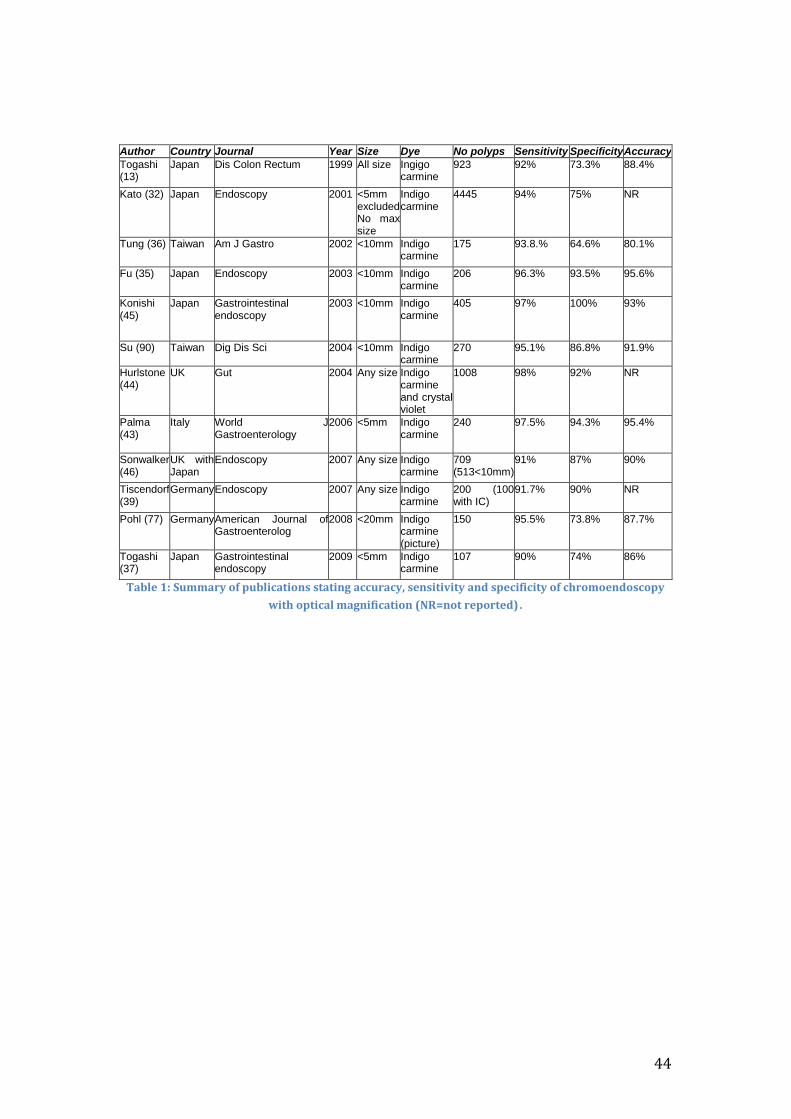
44
Author Country Journal Year Size Dye No polyps Sensitivity Specificity Accuracy
Togashi (13)
Japan Dis Colon Rectum 1999 All size Ingigo carmine
923 92% 73.3% 88.4%
Kato (32) Japan Endoscopy 2001 <5mm excluded No max size
Indigo carmine
4445 94% 75% NR
Tung (36) Taiwan Am J Gastro 2002 <10mm Indigo carmine
175 93.8.% 64.6% 80.1%
Fu (35) Japan Endoscopy 2003 <10mm Indigo carmine
206 96.3% 93.5% 95.6%
Konishi (45)
Japan Gastrointestinal endoscopy
2003 <10mm Indigo carmine
405 97% 100% 93%
Su (90) Taiwan Dig Dis Sci 2004 <10mm Indigo carmine
270 95.1% 86.8% 91.9%
Hurlstone (44)
UK Gut 2004 Any size Indigo carmine and crystal violet
1008 98% 92% NR
Palma (43)
Italy World J Gastroenterology
2006 <5mm Indigo carmine
240 97.5% 94.3% 95.4%
Sonwalker (46)
UK with Japan
Endoscopy
2007 Any size Indigo carmine
709 (513<10mm)
91% 87% 90%
Tiscendorf (39)
Germany Endoscopy 2007 Any size Indigo carmine
200 (100 with IC)
91.7% 90% NR
Pohl (77) Germany American Journal of Gastroenterolog
2008 <20mm Indigo carmine (picture)
150 95.5% 73.8% 87.7%
Togashi (37)
Japan Gastrointestinal endoscopy
2009 <5mm Indigo carmine
107 90% 74% 86%
Table 1: Summary of publications stating accuracy, sensitivity and specificity of chromoendoscopy
with optical magnification (NR=not reported).

45
Author Country Journal Year Size No polyps
Sensitivity Specificity Accuracy
Eisem (42)
USA Gastrointestinal Endoscopy
2002 <10mm 520 82% 82% 82%
Fu (35) Japan Endoscopy 2003 <10mm 206 96.3% 93.5% 95.6%
Konishi (45)
Japan Gastrointestinal endoscopy
2003 <10mm 405 97% 86%
100% 61%
68% 93%
Apel (41) Germany Gastrointestinal endoscopy
2006 <5mm 273 94% 64% 83%
Pohl (40) Germany Gut 2009 <10mm 280 87.6% 62.0% NR
Longcroft-Wheaton (47)
UK UEG journal 2013 <10mm 237 91% SD 96% HD
87% SD 84% HD
89% SD 92% HD
Longcroft-Wjeaton (48)
UK Eur J Gastrohep
2011 <10mm 232 94% 84% 91%
Table 2: Summary of publications stating accuracy, sensitivity and specificity of chromoendoscopy with indigo carmine on polyps<10mm without optical magnification (NR=not reported)

46
Author Country
Journal
Year Modality Endoscope
No Sensitivity Specificity Accuracy
Machida (54)
Japan Endosc 2004 NBI Zoom 43 100% 75% NR
Chiu (65)
Taiwan Gut 2007 NBI (pictures)
Zoom non zoom
180 82-86% 87-95%
59-83% 71-88%
81-82% 87-90%
Rastogi (50)
USA GIE 2008 NBI HD [1] 123 86-92% 86-92% NR
Rogart (66)
USA GIE 2008 NBI Zoom 265 80% 81% 80%
East (56)
UK Endosc 2008 NBI HD+zoom 116 88% 91% 89.6%
Ragstogi (67)
USA Am J Gastro
2009 NBI HD without magnification
236 all size
96% NR 93%
Ignjatovic (68)
UK Lancet Onc
2009 NBI with IC
HD 278 94% 89% 93%
Sano (72)
Japan GIE 2009 NBI HD+zoom 150 96.4% 92.3% 95.3%
Wada (70)
Japan GIE 2009 NBI (pictures)
HD+zoom 617 90.9% 97.1% NR
Henry (73)
USA GIE 2010 NBI (retrospective pictures)
Uncertain 126 93% 88% 91%
Wada (69)
Japan Dig Endosc
2010 NBI Uncertain 1473
88.9% 98.9% 98.2%
Tischendorf (71)
Germany
Endosc 2010 NBI (pictures)
HD with and without zoom
200 87.9% 92.1%
90.5% 89.2%
NR
Van Den Broek (91)
The Netherlands
Clinical Gastro and Hepat
2009 Trimodal imaging
HD + zoom 208 99% 35% 63%
Table 3: Accuracy, sensitivity and specificity for narrow band imaging (NBI), auto fluorescence imaging (AFI) and trimodal imaging. IC= indigo carmine, HD= high definition NR= not reported

47
Author Country Journal Year Modality Endoscope No Sensitivity Specificity Accuracy
Pohl (77)
German Am J Gastro
2008 FICE (picture)
HD low and high magnification<20mm
150 89.9% 96.6%
73.8% 80.3%
83% 90%
Pohl (12)
German Gut 2009 FICE (subgroup analysis)
HD non zoom<10mm
321 <10mm
93% 61.2% 84.7%
Togashi (37)
Japan GIE 2009 FICE HD low (50x) and high (100x) magnification<5mm
107 93% 70% 87%
Teixiera (79)
Brazil GIE 2009 FICE HD with zoom polyps up to 50mm
309 99.2% 94.9% 98.3%
Parra-Blanco (78)
Spain World Journal Gastro
2009 FICE (picture)
HD with and without zoom<5mm (picture)
19 NR NR 58.9% 70.5%
Longcroft-Wheaton (48)
UK Eur J. Gastro
2011 FICE Polyps <10mm resolution not stated
232 88% 82%
86%
Longcroft-Wheaton (80)
UK Endoscopy
2012 FICE HD and SD assessments
293 SD 83% HD 93%
SD 82% HD81%
SD 83% HD 89%
Hoffman (81)
Germany
endoscopy
2010 i-scan HD<10mm 145 98% 100% 98.6%
Hoffman (84)
Germany
Dig Liver Dis
2010 i-scan HD<5mm rectosigmoid
335 100% 100% 100%
Basford P (85)
UK GIE 2013 I scan HD<10mm 209 97% 90.7% 94.7%
Table 4: Summary of papers published using FICE and i-scan for in-vivo diagnosis. NR=not reported

48
Hyperplastic Adenomas Cancers
White light vascularity Pale Dark Dark
FICE vascularity Pale Dark Very dark
FICE vascular pattern Absent vascular
pattern or faint
vessels not
following crypts
Regular pericryptal
pattern
Dense irregular pattern
FICE surface pattern No surface pattern
or large non
compact crypt
pattern
Small compact regular
pattern
Disorganised irregular
pattern
Table 5: N.A.C. classification system