Embed Size (px)
Citation preview

Nicola Bizzocchi, DosimetristProton Therapy CenterTrento
A Planning Approach for Lens-Sparing Proton Cranio-Spinal Irradiation
in Pediatric Patients

Italian Proton CentersCatania: Laboratory AIFNequipped with one fixedprotons low energy line (60MV-cyclotron) for eye’stumors
Pavia: two fixed proton andcarbon ion lines (250 MeVfor protons and 4800 MeVfor carbon ions) for everytype of tumor

Trento Proton Therapy Center

From conformal-3D to 3D-dose modulation

Delivery with Pencil Beam Scanning
Energy from 70 to 226 MV (from 4 to 33 cm in W)
MU per spot from 0.02 to 12
Scanning grid dimension: 30x40cm
Spot σ 3 mm

Some important calendar dates
• October 2014: first treated patient – adult -parotid tumor
• June 2015: first pediatric patient – 9 years old– cranial chordoma tumor
• September 2015: first CSI pediatric patient

CSI: a technically-complex treatment
• Extensive cranio-caudal tumor volume →
• Necessity to have two or more isocenters →
• Necessity to have one or more zones of junction
Note: The conventional radiotherapy has resolved these problems withTomotherapy helical delivery

• Medulloblastoma is the most common malignant tumor in childhood. Being a tumor that primarily strikes in pediatric age, the patient usually needs to be put under anesthesia (cut off around 5\6 years of age)
• Total column irradiation to compensatebone-growth related dysfunction (cut-off 12/14 years of age)
• Cribriform plate under risk ofrecurrences
CSI: a clinically-complex treatment

Waiting for the first CSI patient(one year ago)
• Definition of the set-up position (prone vs. supine) and the positioning devices
• Study of a Planning strategy• Definition of the dosimetric patient QA
verification
Work teams
Study Phase

CharlieAnthropomorphous Rando phantom, which has the dimensions of an adult patient of around 180 cm in height (3
isocenters , 2 junctions)
FOCUS• individualize the more suitable
zones of junction and accordinglythe position of the isocenters
• to find the way to create robustjunctions
• To elaborate an effective beamarrangement
Study Phase

Simulation CT and contouring
• Phantom prone positioned (couch not yet characterized)
• Contouring of the CTV (from brain to the second sacral vertebra) then expanded to 4mm and divided into PTV Brain, PTVspineUP, PTVspineDOWN, PTVjunUP, PTVjunDown
• Contouring of the OAR: lens, eye and cochlea
Study Phase

Junctions: the ancillary beam techniqueStudy Phase

Beam arrangementEvidence Based Practice
Boston/Houston Philadelphia
PSI Switzerland
Study Phase

Study Phase conclusions
• First patient desired characteristics1) pediatric not in anesthesia
2) pediatric post-puberal• Beam arrangement:
Lat-Lat for the brain and direct posterior field for the spine

Case
• 6 year-old boy • Headache, vomiting and gait instability• MRI Brain: 5 cm posterior fossa mass with obstructive
hydrocephalus• MRI spine: leptomeningeal thickening abnormalities
from C7 to D3

• EVD placed to relieve hydrocephalus• Posterior fossa Craniectomy and Gross Tumor
Resection
– Path: Medulloblastoma, WHO grade IV (with anaplastic features)
– Post-op developed posterior fossa syndrome (Mutism and hemiparesis, truncal ataxia, VII CN L palsy)
Case

• Post-op brain MRI: No residual disease• Lumbar Puncture ten days after surgery: + for
malignant cells • Bone scan: no metastasis
Case

Management
• High Risk MBL
– Surgery GTR
– High dose CHT and Bone Marrow transplant
– MRI brain and spine evaluation pre_Proton
– CSI (36 Gy\20 fr) + Tumor bed boost (18.0 Gy\10 fr)

Simulation CT and Set-UpClinical Phase
CT scanning: 3 mm contiguous slice thickness from the top of the head to the lower sacrum
Patient position: supine under anesthesia

Clinical PhaseSet-up Devices
Anesthesia: Midazolam intravenous injection then maintenance with Propofol

Contouring
PTV Brain PTV
JunctUP
PTV Spine
UP
PTV JunctDown
PTV Spine Down
1 isocenter 2 isocenter 3 isocenter
Clinical Phase
Contouring: TPS Focal by ElektaPlanning: TPS XiO by Elekta
OAREyesLensesONCochleaLarynxEsophagusLungsHeartLiverKidneysBowel

PlanningClinical Phase
TPS XIO by Elekta
SFO technique: optimization field per field independently for reducing dose calculation uncertainties. The evolution of this technique is the IMPT but the risk related to the dose calculation uncertainties is too high without an algorithm like Monte Carlo that is nuclear and atomic interactions correlated

PlanningClinical Phase

Clinical Phase
Planning

PlanningClinical Phase
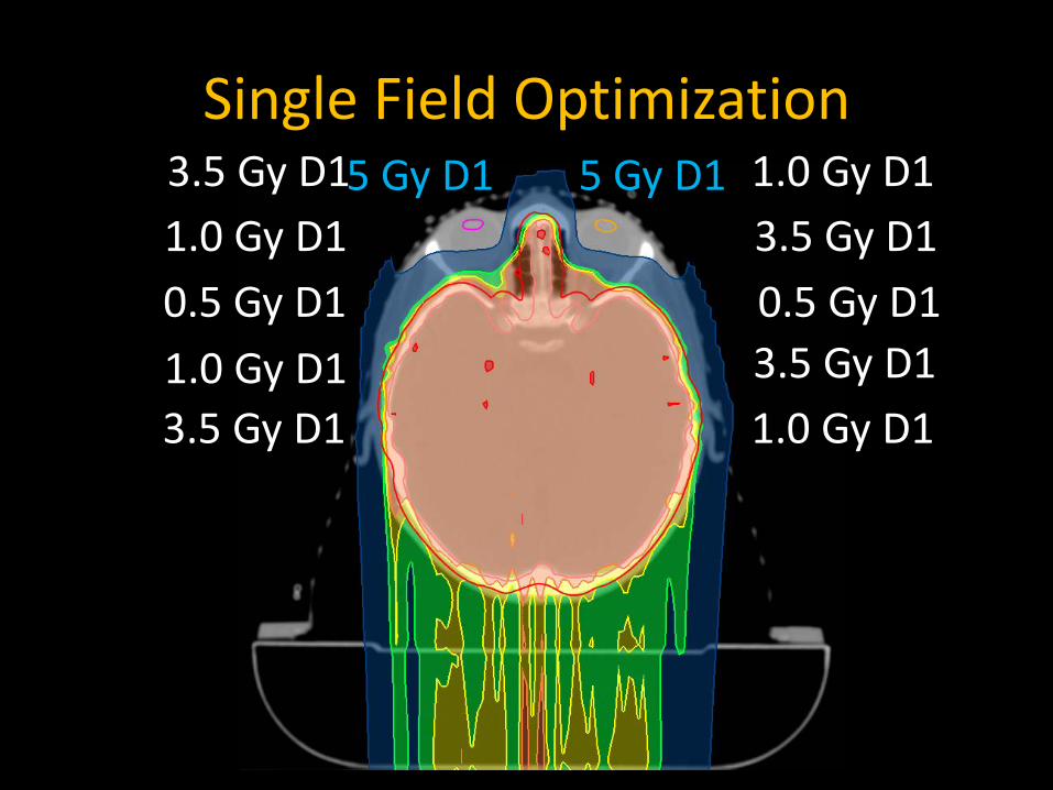
Single Field Optimization1.0 Gy D13.5 Gy D1
1.0 Gy D1 3.5 Gy D10.5 Gy D1 0.5 Gy D11.0 Gy D1 3.5 Gy D13.5 Gy D1 1.0 Gy D1
5 Gy D1 5 Gy D1

Evaluation98%
95%90%
14%
35,3Gy34,2Gy
32,4Gy
5,0Gy

Evaluation
PTVBrain
CochleaEyes
Lenses

Related Papers
Philadelphia
Boston
Houston

Results

3 months after PTComplete hair regrowth

After 6 months PT and one hair-cutDuring PT

Basal testing (before P-CSI)
• Neurocognitive testing• Neuro visual testing and ophthalmic exam• Neuroendocrine testing: N • ENT and hearing loss testing: mild
neurosensorial R hearing loss• PKT evaluations
Goldwein et al. IJROBP 1996, 34:899-90

Sequelae (6 months after P)
• Neurocognitive decline (negative, good math performance testing and memory tests, QI normal)
• Neuroendocrine deficits (negative)• Hearing loss testing (negative)• Neuro visual testing (visual-spatial setting improved,
no cataracts)• PKT evaluations improved globally: speech skills
improved, mild ataxia, some steps

After the first patient…High Risk CSI Protocol
Patients Age Lens dx D1 cGy Lens sx D1 cGy
Pz 1 6 439 533
Pz 2 14 442 250
Pz 3 8 460 470
Pz 4 4 447 468
Pz 5 7 716 709
Pz 6 5 794 790

Cribriform Plate Coverage

Boost on Posterior Fossa18 Gy\10 fr

Boost on Posterior Fossa18 Gy\10 fr

Boost on Posterior Fossa18 Gy\10 fr

Acknowledgments

GrazieThank you all