Embed Size (px)
Citation preview

ZNF366 is an estrogen receptor corepressorthat acts through CtBP and histone deacetylasesJorge Lopez-Garcia, Manikandan Periyasamy, Ross S. Thomas, Mark Christian1,
Maria Leao2, Parmjit Jat3, Karin B. Kindle4, David M. Heery4, Malcolm G. Parker1,
Lakjaya Buluwela, Tahereh Kamalati and Simak Ali*
Department of Oncology, Imperial College London, Du Cane Road, London W12 0NN, UK, 1Institute of Reproductiveand Developmental Biology, Imperial College London, Du Cane Road, London W12 0NN, UK, 2Ludwig Institute forCancer Research, University College London Branch, 91 Riding House Street, London W1W 7BS, UK, 3Department ofNeurodegenerative Disease, Institute of Neurology, University College London, Queen Square, London WC1N 3BG,UK and 4School of Pharmacy, University of Nottingham, University Park, Nottingham NG7 2RD, UK
Received August 4, 2006; Revised October 5, 2006; Accepted October 6, 2006
ABSTRACT
The regulation of gene expression by estrogenreceptor-a (ERa) requires the coordinated and tem-poral recruitment of diverse sets of transcriptionalco-regulator complexes, which mediate nucleosomeremodelling and histone modification. Using ERa asbait in a yeast two-hybrid screen, we have identified anovel ERa-interacting protein, ZNF366, which is apotent corepressor of ERa activity. The interactionbetween ZNF366 and ERa has been confirmedin vitro and in vivo, and is mediated by the zincfinger domains of the two proteins. Further, we showthat ZNF366 acts as a corepressor by interacting withother known ERa corepressors, namely RIP140 andCtBP, to inhibit expression of estrogen-responsivegenes in vivo. Together, our results indicate thatZNF366 may play an important role in regulating theexpression of genes in response to estrogen.
INTRODUCTION
Estrogens play diverse roles in the body, most notably in thedevelopment and maintenance of the female and the male,reproductive systems and secondary sexual characteristics(1). Estrogens also play a central role in promoting breastcancer growth (2), as well as being implicated in uterineand ovarian cancers (3,4), and are also implicated in thephysiology of the brain, bone and the cardiovascular system,as evidenced by the increased risk of cardiovascular diseaseand osteoporosis engendered by the decline in estrogen levelsduring menopause (1).
Estrogen action is mediated by two highly relatedestrogen receptors (ERa and ERb), which are members of
the ligand-activated nuclear receptor (NR) superfamily oftranscription factors (5,6). NRs are characterized by a DNAbinding domain (DBD), comprised of two zinc fingers,which mediate receptor dimerization and binding to specificresponse elements in the promoters of target genes. Bindingof the ligand to the ligand binding domain (LBD), locatedC-terminal to the DBD, results in a conformational changein the LBD and activation of the intrinsic transcription activa-tion function AF2, which facilitates the recruitment oftranscriptional coactivators (7,8). Transcription activationrequires cooperation of AF2 with a region N-terminal to theDBD that encodes transcription activation function AF1,which is often, as in the case of ERa and ERb, regulatedby phosphorylation at specific serine residues (9–11).
The liganded estrogen receptors regulate gene expressionby direct binding to DNA at estrogen response elements intarget genes, resulting in the recruitment of diverse transcrip-tional coregulators, including the SWI–SNF complexes thatremodel chromatin to alter nucleosomal organization in anATP-dependent manner (12), the p160 family of coactivators(SRC1/NCo-A1, TIF-2/GRIP1 and AIB1/pCIP/ACTR/RAC3/TRAM1) and TRAP/DRIP complexes. (13–15). Thep160 coactivators facilitate the recruitment of other proteins,including CBP, its homologue p300 and p/CAF, which pos-sess intrinsic histone acetyltransferase activity, as well ashistone methyltransferases CARM1 and PRMT1 that methy-late arginine residues in histone tails (16,17). The thyroidreceptor–associated protein (TRAP) complex, similar to oridentical with the vitamin D3 receptor–interacting protein(DRIP) complex, which is also similar in many respects tothe Mediator complex acts to bridge RNA polymerase IIwith basal transcription factors and transcription activators.These and other coregulators are recruited to gene promotersin a sequential/ordered manner, resulting in cycles ofchromatin remodelling and modification that facilitatetranscription (13,15,18).
*To whom correspondence should be addressed. Tel: +44 20 8383 3789; Fax: +44 20 8383 5830; Email: [email protected]
� 2006 The Author(s).This is an Open Access article distributed under the terms of the Creative Commons Attribution Non-Commercial License (http://creativecommons.org/licenses/by-nc/2.0/uk/) which permits unrestricted non-commercial use, distribution, and reproduction in any medium, provided the original work is properly cited.
6126–6136 Nucleic Acids Research, 2006, Vol. 34, No. 21 Published online 3 November 2006doi:10.1093/nar/gkl875
by guest on Novem
ber 9, 2015http://nar.oxfordjournals.org/
Dow
nloaded from

The ERa LBD is composed of 12 a-helices packed in threelayers, with a central hydrophobic pocket that accommodatesthe ligand (7). Helix 12, together with helices 3, 4 and 5, forma coactivator-binding groove. Most coactivators recruited byagonist-bound ERa contain LXXLL motifs, which form atwo-turn amphipathic a-helix that fits into the coactivator-binding groove in the ERa LBD (8,19–22). In addition tothe well-characterized recruitment of coactivators to theLBD/AF2, coactivator interaction with AF1 of estrogenreceptors has also been described, the interaction beinginfluenced by the phosphorylation status of the receptor insome cases (23–25).
When unliganded, some DNA-bound NR recruit the core-pressors NCoR and SMRT and associated protein complexesimplicated in transcriptional repression and histone deacety-lation, these complexes being dissociated upon ligand binding(13–15,26). NCoR/SMRT bind to NR through CoRNR boxes,LXXI/HIXXXL/I motifs, that form a more extended a-helixthan the LXXLL motifs, with helix 12 in the LBD beingdisplaced from the conformation it occupies in the agonist-bound LBD (27,28). NCoR/SMRT is also recruited byantagonist-bound ERa to inhibit gene expression (29–31).
In addition to stimulating gene expression, estrogen-boundER represses the expression of many genes. Indeed, gene pro-filing studies show that the down-regulation of gene expres-sion is a significant feature of the response to estrogen inthe ERa-positive, estrogen-responsive MCF7 breast cancercell line (32). This is likely to involve transcriptionalcorepressors, such as LCoR and RIP140, which can berecruited to the agonist-bound ERa via LXXLL motifs(20,33,34). Repression by LCoR and RIP140 occurs throughHDAC-dependent and -independent mechanisms and involvesthe recruitment of HDACs and the C-terminal binding protein(CtBP) corepressor (34–39). CtBP, originally identified basedon its interaction with the C-terminal end of adenovirus E1Avia the sequence PLDLS, is highly conserved in highereukaryotes and plays a critical role in development (40,41).Other transcription factors also interact with CtBP1, and thehighly related CtBP2, through PXDLS motifs. Although, themechanisms by which CtBP acts as a corepressor have notbeen fully defined, a recent study has identified CtBP com-plexes that contain HDACs and histone lysine methyltrans-ferases (42).
The ERa DBD participates in the recruitment of transcrip-tional co-regulator proteins. These include the coactivatorXBP-1 (43), which modulates ERa signalling both inthe absence and presence of estrogen, the signal transducerand activator of transcription-5 (STAT 5) (44) and thecorepressor TAF-Ib that has been shown to decrease ERaacetylation (45). Here, we report that ZNF366, which encodesan evolutionarily conserved zinc finger protein, interacts withthe ERa DBD. We also show that ZNF366 represses ERaactivity through association with RIP140, CtBP and histonedeacetylases.
MATERIALS AND METHODS
Plasmids
The mammalian expression plasmids and reporter geneshave previously been described (37,46–49). Site-directed
mutagenesis was used to introduce an EcoRI site 50 to theGAL4 translation initiation site in the pBridge yeast expres-sion plasmid (BD Biosciences, UK), enabling cDNAsequences encoding ERa and ERa�DLBD to be cloned atthis position following removal of the GAL4 sequencesencoded between the introduced EcoRI site and the multiplecloning site in pBridge. The pACTII-ZNF366 clone isolatedfrom the yeast 2-hybrid screening encodes sequences corre-sponding to 439–2761 bp of the ZNF366 mRNA sequencewith the accession number NM_152625 in the NCBI database(www.ncbi.nlm.nih.gov). The full-length ZNF366 open read-ing frame was reconstituted from the pACTII-ZNF366 cloneand IMAGE EST clone 5204702 (accession no. BI770486),to generate pCMVSPORT6-ZNF366 in which ZNF366 isC-terminally FLAG-tagged. ZNF366 deletion and pointmutants were generated by site-directed mutagenesis accord-ing to manufacturer’s protocols (Stratagene, UK).
Yeast 2-hybrid screening
PL1a (MATa ura3-D1 his3-D200 leu2-D1 trp1::ERE)1-URA3yeast strain (50) was transformed with pBridge(Mod)-ERa-DLBD, together with a human placental cDNA expres-sion library (BD Biosciences, UK), using the Alkali-Cationyeast transformation kit (BIO 101 systems, UK). Followingtransformation, the cells were plated on 15 cm trp� leu�
ura� plates. Positive clones arising from the screening of2 · 104 transformants were re-screened and plasmidDNAs were isolated using the lyticase method from BDBiosciences, UK. Plasmids from positive clones were re-transformed, together with pBridge(Mod)-ERa-DLBD orpBridge(Mod)-ERa and interactions confirmed by growthon trp� leu� ura� plates.
Northern blotting
Multiple tissue northern blots MTN I and MTN II (BDBiosciences, Europe) were probed following 32P-labellingof the ZNF366 cDNA isolated from the pACTII-ZNF366clone, as described (51).
Protein expression, purification and glutathioneS-transferase (GST)-based interaction assay
In vitro transcription/translations were performed using TNTrabbit reticulocyte lysates (Promega, UK), in the presence of[35S]-labelled methionine. GST proteins were induced andEscherichia coli lysates prepared as described previously(33). For pulldowns, GST fusion proteins were purified byaffinity chromatography on glutathione-agarose beads andretained as 50% slurry in 20 mM HEPES (pH 7.6),100 mM KCl, 1 mM EDTA, 1 mM DTT, 20% glycerol,supplemented with protease inhibitors. A total of 100 ml vol-umes of glutathione-agarose bead slurry loaded with 10 mg ofGST fusion proteins were then used directly in binding assayswith 10 ml of radiolabelled in vitro translation reactions and890 ml of low salt buffer [50 mM HEPES (pH 7.6),250 mM NaCl, 0.5% NP-40, 5 mM EDTA, 0.1% BSA,0.5 mM DTT, 0.005% SDS and protease inhibitors]. Follow-ing 1 h incubation at room temperature, the beads werewashed twice with low salt buffer and twice with high-saltbuffer (low salt buffer, but with 1 M NaCl). Samples were
Nucleic Acids Research, 2006, Vol. 34, No. 21 6127
by guest on Novem
ber 9, 2015http://nar.oxfordjournals.org/
Dow
nloaded from

boiled for 10 min in 80 ml of Laemmli buffer and fractionatedby SDS–PAGE. Gels were dried and autoradiographed.
Reporter gene assays
COS-1 cells were maintained in DMEM, supplemented with5% fetal calf serum (FCS). For transient transfection, cellswere seeded in 24-well plates in DMEM lacking phenol redand supplemented with 5% dextran-coated charcoal-strippedFCS (DSS). Following seeding for 24 h, the cells weretransfected using Fugene 6 (Roche Diagnostics, UK), with100 ng of luciferase reporter gene and amounts of expressionplasmids as indicated in the figure legends. E2 (10 nM),4-hydroxytamoxifen (OHT; 100 nM) or ICI 182, 780 (ICI;100 nM) were added as appropriate. Since the ligands wereprepared in ethanol, an equal volume of ethanol was addedto the no ligand controls. Luciferase activities were deter-mined using the Dual-Glo Luciferase Assay kit (Promega,UK). For the other reporter gene assays, cells weremaintained in DMEM, supplemented with 5% FCS andtransfections carried out as above.
Immunoprecipitations and immunoblotting
COS-1 cells were plated in 9 cm dishes in DMEM supple-mented with 5% FCS 16 to 24 h prior to transfection. Thecells were transfected with 5 mg of the ZNF366-FLAG andERa expression plasmids using Lipofectamine 2000 (Invitro-gen, UK). Following transfection for 48 h, the cells werelysed in RIPA buffer [150 mM NaCl, 1% NP-40, 0.5%deoxycholic acid, 0.1% SDS and 50 mM Tris–HCl(pH 7.5)] containing protease inhibitors. Lysates (2 mg)were immunoprecipitated (IP) using the M2 anti-FLAGmouse monoclonal antibody (Sigma–Aldrich, UK), or usingan anti-ERa antibody (6F11; Novocastra, UK). Control IPswas carried out using mouse IgG (Sigma–Aldrich, UK). IPswere resolved by SDS–PAGE and immunoblotted usinghorseradish peroxidase (HRP)-labelled HA antibody (Sigma–Aldrich, UK) or using an anti-ERa rabbit polyclonal antibodyHC20 (Santa Cruz, UK). Co-IP of ZNF366-FLAG with CtBPwas carried out as above, except that a mouse monoclonalCtBP antibody (sc-17759; Santa Cruz) was used for the IPsand a rabbit polyclonal CtBP antibody (sc-11390; SantaCruz) was used for immunoblotting.
MCF7 cells cultured for 3 days in DMEM lacking phenolred and supplemented with 5% DSS, were transfected with1 mg of ZNF366-FLAG or vector control, using Fugene 6.The media were replaced with media containing E2(10 nM) or vehicle, 24 h following transfection and thecells were harvested after a further 24 h. Immunoblottingwas performed using antibodies for cathepsin D (ab6313;Abcam, UK), progesterone receptor (SC538; Santa CruzBiotechnologies, UK), FLAG-M2 and b-actin (ab6276;Abcam, UK).
Immunofluorescence
COS-1 cells plated on glass coverslips placed in 24-wellplates in DMEM lacking phenol red and containing 5%DSS, were transiently transfected with 50 ng of ZNF366-FLAG and/or [ERa-DNLS (HE257G; (48)] using Fugene 6.Five hours following transfection, culture media werereplaced by fresh media containing E2 (100 nM), OHT
(1 mM) or ICI 182, 780 (100 nM), or an equal volume ofvehicle (ethanol), as appropriate. 24 h later, cells were fixedby the addition of 4% formaldehyde for 10 min at roomtemperature, washed with phosphate-buffered saline (PBS)and 0.1 M glycine was added for 10 min to neutralize theformaldehyde. Following further washing with PBS, thecells were permeabilized in 1% Triton/PBS for 5 min. Afterwashing with PBS, the cells were incubated at 37�C for 1 hwith the 6F11 ERa antibody (1:50 dilution) and rabbit poly-clonal FLAG antiserum (Santa Cruz Biotechnology, UK)(1:350 dilution). The cells were washed and incubatedfor 1 h at 37�C with Alexa Fluor 488 goat anti-mouseimmunoglobulins (green) and Alexa Fluor 594 goat anti-rabbit immunoglobulins (red) (1:3000 dilution). The cover-slips were mounted on microscope slides using mountantcontaining Dapi (Vector Laboratories, UK) and immunofluo-rescence observed using a Zeiss LSM510 confocalmicroscope.
Growth assays
MCF7 and MDA-MB-231 cells (2 · 105 cells per well) wereseeded in 6-well plates in DMEM lacking phenol red andcontaining 5% DSS. After 48 h the cells were transfectedwith 1 mg of ZNF366 or empty vector using Fugene 6. E2(10 nM) was added after 24 h and cell numbers determinedusing a haemocytometer after a further 48 h.
RNA interference and RT–PCR analysisof gene expression
PE04 cells (2.5 · 105 cells) seeded in 6-well plates in RPMIlacking phenol red and containing 5% DSS, were transfectedwith double-stranded RNA oligonucleotides for ZNF366,lamin A/C or a non-targeting siRNA (Ambion, UK), inserum-free DMEM lacking phenol red, using Oligofectamine(Invitrogen, UK), according to manufacturer’s protocols.After 4 h the medium was changed to DMEM lackingphenol red, supplemented with 5% DSS and containing E2(10 nM), as appropriate. RNA was prepared after a further24 h and RT–PCR carried out using primers with thesequences: 50-GGACCAGCTTCAGTCACCTTTCCAGT-GGTGGCC-30 and 50-GGGAGTAAAGCTGGTGCCTGG-GGCACAGGTCACG-30 (GREB1), 50-CCCCATCCAGT-ACAACTGCT-30 and 50-CTTCACGTCAGAGTGGACGA-30 (ZNF366), 50-GCGTACGGCTCTCATCAACT-30 and 50-GACACTGGAGGCAGAAGAGC-30 (Lamin A/C) and 50-TCCCATCACCATCTTCCA-30 and 50-CATCACGCCACA-GTTTCC-30 (GAPDH).
RESULTS
Identification of ZNF366
We utilized yeast strain PL1a, encoding an integratedestrogen-responsive URA3 gene (50), for screening of ahuman placental cDNA expression library for proteins thatinteract with an ERa deletion mutant lacking the LBD(ERa-DLBD). Screening of 2 · 104 transformants yielded24 positive clones. One of these encoded the C-terminalportion of ZNF366, described previously based on geneprediction of genomic DNA sequence of human chromosome
6128 Nucleic Acids Research, 2006, Vol. 34, No. 21
by guest on Novem
ber 9, 2015http://nar.oxfordjournals.org/
Dow
nloaded from

5q13.2. Human ESTs were identified using the NCBI site(http://www.ncbi.nlm.nih.gov/BLAST/) by carrying out aBLAST search against the database of human ESTs, usingsequences derived from the yeast 2-hybrid clone. The exactintron/exon structure of ZNF366 was established by completeDNA sequencing of overlapping EST IMAGE clones withGenBank accession nos BI523869, 5201353, BI770486 andBE552137 to generate an mRNA sequence encoding a pre-dicted polypeptide of 744 amino acids (Figure 1A), encodedwithin 5 exons. Based on the homology with the Fugu fZF1gene, this was subsequently confirmed by Gilligan et al. (52).
Homology searches identified genes in mouse, rat, dog,chicken and Xenopus tropicalis, having very high aminoacid sequence homology (93% or greater) in the zinc fingerregion and all of which encode 11 zinc fingers (Figure 1B).Regions N- and C-terminal to the zinc fingers were alsowell conserved, albeit less so than the zinc finger region.Together, the high degree of sequence conservation indicatesthat the identified vertebrate genes represent orthologues ofhuman ZNF366. Support for this idea is provided byadditional homology searches, which indicated that thehuman gene most closely related to ZNF366 is ZNF710,also encoding an 11 zinc finger protein, the zinc fingerregions of human ZNF366 and ZNF710 showing aminoacid sequence identity of 78%, considerably lower than theidentity between human and the putative ZNF366 homologuein chicken or Xenopus. Moreover, no significant homologywas detectable between human ZNF366 and ZNF710 outsidethe zinc finger region. All other human genes demonstratinghomology to human ZNF366 displayed amino acid sequenceidentities no better than 39%, when the zinc finger region ofZNF366 was used in the BLAST searching. Together with theprevious report demonstrating, based on very high (88%)amino acid sequence homology in the zinc finger region, aswell as conservation of the chromosome location of humanZNF366 and the Fugu fZF1 gene (52), these analyses indicatethat ZNF366 is highly conserved in vertebrate evolution.
Northern blotting of human tissue RNAs demonstrated thatZNF366 is broadly expressed at varying levels in humanadult tissues, with highest expression in heart, placenta,muscle and spleen, with possible alternative splice forms inthe liver and muscle (Figure 1C).
Interaction of ZNF366 with ER-a
To further confirm interaction between ERa and ZNF366,whole cell lysates were prepared from COS-1 cells transientlytransfected with ERa and FLAG-tagged ZNF366, in thepresence of E2. IP with FLAG antibody, followed byimmunoblotting using the HC20 rabbit polyclonal ERa anti-serum, showed that ERa interacts with ZNF366 (Figure 2A),whilst IP of ERa co-immunoprecipitated ZNF366-FLAG(Figure 2B), indicating that ERa and ZNF366 interactin vivo.
As expected, PL1a cells co-transformed withpBridge(Mod)-ERa-DLBD and pACTII-ZNF366 grew onminimal medium lacking lacking uracil (Figure 2C). How-ever, for full-length ERa, productive interaction, as assayedby growth in the absence of uracil, was ligand-dependent,requiring the addition of estrogen (17b-estradiol; E2) or ananti-estrogen 4-hydroxytamoxifen (OHT).
In order to confirm the ligand requirement for theinteraction between ERa and/or ZNF366 in mammaliancells, COS-1 cells were transfected with ERa�DNLS,which is excluded from the nucleus (48). ZNF366-FLAGwas exclusively localized to the nuclei (Figure 2D), whilst
Figure 1. The amino acid sequence and tissue distribution of human ZNF366.(A) The deduced amino acid sequence of human ZNF366 is shown in thesingle letter amino acid code, with the cysteine (C) and histidine (H) residuesof the 11 zinc fingers in ZNF366 highlighted, as are the conservedphenylalanine (F) and leucine (L) residues. Potential CtBP binding motifsand an LXXLL motif are underlined. (B) A schematic representation ofZNF366 from different species is shown, together with the position of thezinc fingers and the putative CtBP and LXXLL motifs, the sequences forthese motifs in human ZNF366 being shown. The numbers below therepresentation of ZNF366 from each species refer to percentage amino acidsequence homology relative to human ZNF366. The numbers to the rightrefer to the predicted number of amino acids encoded by the ZNF366 genesfrom different species. The region encoding 277–1053 amino acids was usedfor alignment in the case of the chicken ZNF366. ZNF366 sequences usedhere have GenBank accession nos NM_152625 (human), NM_001004149(mouse), XM_226715 (rat), XP_544370 (dog) and XP_429153 (chicken).The Xenopus tropicalis ZNF366 sequence was derived from the ENSEMBLdatabase, from sequences having the Gene ID ENSGALG00000015008.(C) Northern blot of 2 mg of poly(A)+ RNA from the tissues indicated, probedwith ZNF366 cDNA. Size markers are shown on the right.
Nucleic Acids Research, 2006, Vol. 34, No. 21 6129
by guest on Novem
ber 9, 2015http://nar.oxfordjournals.org/
Dow
nloaded from
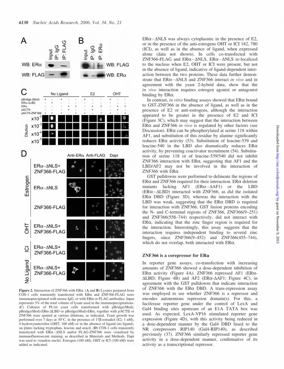
ERa�DNLS was always cytoplasmic in the presence of E2,or in the presence of the anti-estrogens OHT or ICI 182, 780(ICI), as well as in the absence of ligand, when expressedalone (data not shown). In cells co-transfected withZNF366-FLAG and ERa�DNLS, ERa�DNLS re-localizedto the nucleus when E2, OHT or ICI were present, but notin the absence of ligand, indicative of ligand-dependent inter-action between the two proteins. These data further demon-strate that ERa�DNLS and ZNF366 interact in vivo and inagreement with the yeast 2-hybrid data, show that thein vivo interaction requires estrogen agonist or antagonistbinding by ERa.
In contrast, in vitro binding assays showed that ERa boundto GST-ZNF366 in the absence of ligand, as well as in thepresence of E2 or anti-estrogens, although the interactionappeared to be greater in the presence of E2 and ICI(Figure 3C), which may suggest that the interaction betweenERa and ZNF366 in vivo is regulated by other factors (seeDiscussion). ERa can be phosphorylated at serine 118 withinAF1, and substitution of this residue by alanine significantlyreduces ERa activity (53). Substitution of leucine-539 andleucine-540 in the LBD also dramatically reduces ERaactivity, by preventing coactivator recruitment (54). Substitu-tion of serine 118 or of leucine-539/540 did not inhibitZNF366 interaction with ERa, suggesting that AF1 and theLBD/AF2 may not be involved in the interaction ofZNF366 with ERa.
GST pulldowns were performed to delineate the regions ofERa and ZNF366 required for their interaction. ERa deletionmutants lacking AF1 (ERa�DAF1) or the LBD(ERa�DLBD) interacted with ZNF366, as did the isolatedERa DBD (Figure 3D), whereas the interaction with theLBD was weak, suggesting that the ERa DBD is requiredfor interaction with ZNF366. GST fusion proteins encodingthe N- and C-terminal regions of ZNF366, ZNF366(9–251)and ZNF366(558–744) respectively, did not interact withERa, indicating that the zinc finger region is required forthe interaction. Interestingly, this assay suggests that theinteraction requires independent binding to several zincfingers, since ZNF366(9–452) and ZNF366(455–744),which do not overlap, both interacted with ERa.
ZNF366 is a corepressor for ERa
In reporter gene assays, co-transfection with increasingamounts of ZNF366 showed a dose-dependent inhibition ofERa activity (Figure 4A). ZNF366 repressed AF1 (ERa-DLBD; Figure 4B) and AF2 (ERa-DAF1; Figure 4C), inagreement with the GST pulldowns that indicate interactionof ZNF366 with the ERa DBD. A trans-repression assaywas employed to see whether ZNF366 is a repressor andencodes autonomous repression domain(s). For this, aluciferase reporter gene under the control of LexA andGal4 binding sites upstream of an E1A TATA box wasused. As expected, LexA-VP16 stimulated reporter geneexpression (Figure 4D), with this activity being reduced ina dose-dependent manner by the Gal4 DBD fused to theNR corepressors RIP140 (Gal4-RIP140), as describedpreviously (37). ZNF366 similarly repressed reporter geneactivity in a dose-dependent manner, confirmative of itsactivity as a transcriptional repressor.
Figure 2. Interaction of ZNF366 with ERa. (A and B) Lysates prepared fromCOS-1 cells transiently transfected with ERa and ZNF366-FLAG wereimmunoprecipitated with mouse IgG, or with ERa or FLAG antibodies. Inputrepresents 5% of the total volume of lysate used in the immunoprecipitations.(C) Cultures of PL1a yeast cells transformed with pBridge(Mod),pBridge(Mod)-ERa-DLBD or pBridge(Mod)-ERa, together with pACTII orZNF366 were spotted at various dilutions, as indicated. Yeast growth wasperformed over 7 days at 30�C, in the presence of 17b-estradiol (E2; 1 nM),4-hydroxytamoxifen (OHT; 100 nM) or in the absence of ligand (no ligand),on plates lacking tryptophan, leucine and uracil. (D) COS-1 cells transientlytransfected with ERa�DNLS and/or FLAG-ZNF366 were visualized byimmunofluoresecent staining, as described in Materials and Methods. Dapiwas used to visualize nuclei. Estrogen (100 nM), OHT or ICI (100 nM) wereadded as indicated.
6130 Nucleic Acids Research, 2006, Vol. 34, No. 21
by guest on Novem
ber 9, 2015http://nar.oxfordjournals.org/
Dow
nloaded from

In order to address the potential activity of ZNF366on ERa-regulated gene expression, we looked for humancell lines in which the genes were co-expressed (data notshown). The PE04 ovarian cancer cell line was foundto express both ERa (3) and ZNF366 (Figure 4E). Transfec-tion with siRNA for ZNF366 resulted in down-regulation ofZNF366 expression and concomitant increase in theexpression of the estrogen-responsive GREB1 and TERTgenes, indicating that ZNF366 is involved in the regulationof estrogen-responsive gene expression in vivo.
ZNF366 interacts with CtBP in vitro and in vivo
Since ZNF366 can repress ligand-stimulated ERa activity,we wondered whether it interacts with other corepressorsthat are known to associate with ERa in a ligand-dependentmanner. Amongst these is RIP140 (33), which acts by recruit-ing HDACs and C-terminal binding protein (CtBP) (37).In GST pulldown assays, ZNF366 interacted with RIP1401–415 and 753–1158 amino acids (Figure 5A), which encoderepression domains RD1 and RD4 (37). This interactionappeared to require the C-terminal-most zinc fingers 8–11of ZNF366 (Figure 5B).
Interestingly, ZNF366 also interacted with CtBP1(Figure 5A), the interaction apparently requiring sequencesC-terminal to the zinc fingers (Figure 5B). Many proteinsinteract with CtBP through sequence motifs having theconsensus sequence PXDLS, with a lysine residue twoamino acids C-terminal to the serine also often beingpresent (41). Two such motifs, 590-PFDLS(QK)-596 and645-PEDLS(TK)-651 (Figure 1) are located within the regionof ZNF366 required for interaction with CtBP. Mutation ofthe CtBP motifs by substituting the proline (P) and asparticacid (D) residues by alanines, prevented interaction betweenZNF366 and CtBP in GST pulldown assays (Figure 5C) andin a mammalian two-hybrid assay (Figure 5D). Mutationof M1 or the M2 motif reduced the interaction betweenZNF366 and CtBP1. In these assays, the N-terminal CtBPmotif (M1) appeared to be more important than theC-terminal motif (M2).
In the trans-repression assay, mutation of the N-terminal-most CtBP binding motif in ZNF366 (M1) partially relievedthe repression of LexA-VP16 (Figure 5E), whereas mutationof the second motif (M2) did not significantly relievethe repression and mutation of both motifs almost completelyabolished the repression by ZNF366. These findingsshow that the interaction between ZNF366 and CtBP isimportant for the repression activity of ZNF366, the interac-tion being mediated by two CtBP-interaction motifs, withboth motifs being required for the interaction with CtBP,although motif M1 may be more important than M2 for theinteraction.
Whole cell lysates prepared from COS-1 cells transientlytransfected with CtBP1 and FLAG-tagged ZNF366 immuno-precipitated using a CtBP antibody resulted in co-IP ofFLAG-ZNF366 (Figure 5F). In the reciprocal experiment,CtBP1 was co-immunoprecipitated with FLAG-ZNF366(Figure 5G). CtBP1 was not co-immunoprecipitated withZNF366 in which the CtBP binding motifs were mutated.Collectively these data demonstrate that ZNF366 interactsin vitro and in vivo with CtBP1, the interaction beingmediated by two CtBP binding motifs. In agreement withthese findings substitution of the CtBP motifs in ZNF366significantly reduced the repression of ERa activity byZNF366 (Figure 6A).
Gene repression by transcriptional corepressors, includingRIP140 and CtBP, frequently requires HDAC recruitmentand histone deacetylation. The HDAC inhibitor suberoy-lanilide hydroxamic acid (SAHA) relieved the repressionof ERa activity by ZNF366 (Figure 6B), whilst GST pull-downs showed that ZNF366 interacts with HDACs 1, 3 and6 (Figure 6C), indicating that transcriptional repression by
Figure 3. In vitro interaction between ER and ZNF366. (A and B) Schematicrepresentations of ERa, ZNF366 and deletion mutants are shown. AllN-terminal ZNF366 fusion proteins started at amino acid 9, thereby avoidingpotential internal translation start sites. (C and D) GST binding assays werecarried out by incubation of 35S-labelled ERa or mutants with GST orGST–ZNF366 fusion proteins, in the absence of ligand (NL) or in thepresence of 100 nM E2, OHT or ICI. E2 was present throughout in part (D).Input lanes represent 20% of the total volume of the in vitro translationreaction used in the binding assay.
Nucleic Acids Research, 2006, Vol. 34, No. 21 6131
by guest on Novem
ber 9, 2015http://nar.oxfordjournals.org/
Dow
nloaded from

ZNF366 is mediated, at least in part through histonedeacetylation.
ZNF366 represses the expression of estrogen-responsivegenes in breast cancer cells
The majority of breast cancers express ERa, and the growthof ERa-positive breast tumours is stimulated by estrogen,
as evidenced by the utility of anti-estrogens and inhibitorsof estrogen biosynthesis in breast cancer treatment (2). TheMCF7 breast cancer cell line expresses ERa and grows inresponse to estrogen, its growth being inhibited byanti-estrogens. Further, MCF7 cells demonstrate estrogen-stimulated expression of a number of well-characterizedestrogen-responsive genes, including cathepsin D and pS2.In order to evaluate the effect of ZNF366 on ERa-regulation
Figure 4. ZNF366 is a repressor of ERa activity. (A) COS-1 cells were transfected with ERE-3-TATA-luc (100 ng), ERa (100 ng), ZNF366 and the RLTKrenilla luciferase reporter (100 ng). Results represent the mean of three independent experiments individually corrected for transfection efficiency against renillaluciferase activity. Error bars represent the standard error of the mean. The activity for ERa in the presence of E2 and in the absence of ZNF366 was taken as100%. All other activities are shown relative to this. The amounts of ZNF366 transfected were 0 ng (lanes 1, 7, 13 and 19), 0.1 ng (lanes 2, 8, 14 and 20), 1 ng(lanes 3, 9, 15 and 21), 10 ng (lanes 4, 10, 16 and 22), 30 ng (lanes 5, 11, 17 and 23) or 100 ng (lanes 6, 12, 18 and 24). (B and C) Reporter gene assays wereperformed following transfection of 100 ng ERa�DLBD (B) or ERa�DAF1 (C), as for (A). (D) COS-1 cells were co-transfected with 100 ng Gal4 DBD,the Gal4 DBD fused to full-length RIP140 or ZNF366 together with LexA-VP16 and the Lex-Gal-luc reporter gene. Relative reporter gene activities from threeindependent experiments are shown. (E) Shown are RT–PCR carried out using PCR primers for GREB1, TERT, ZNF366, Lamin A/C and GAPDH, using totalRNA prepared from PE04 cells transfected with non-targeting control, Lamin A/C, or ZNF366 siRNA.
6132 Nucleic Acids Research, 2006, Vol. 34, No. 21
by guest on Novem
ber 9, 2015http://nar.oxfordjournals.org/
Dow
nloaded from

of these genomically encoded estrogen-responsive genes,MCF7 cells were transfected with ZNF366. This resultedin a marked reduction in expression of both cathepsinD and pS2 (Figure 7A). MCF7 cell growth was also reducedfollowing ZNF366 transfection (Figure 7B), whilst growth ofan ERa-negative breast cancer cell line that is not estrogen-responsive, was not inhibited by ZNF366 (Figure 7C).
DISCUSSION
Zinc finger proteins constitute a very large family of trans-criptional regulators and can be further subdivided intogroupings based on the type of zinc finger present, as well
as by the presence of additional motifs elsewhere in theprotein that mediate protein–protein interactions and tran-scriptional regulation. Sequence analysis of ZNF366 showsthat it encodes encodes a protein containing 11 Kruppel-type C2H2 zinc fingers, which is highly conserved in verte-brate evolution. However, ZNF366 does not belong to anyof the major subfamilies of the Kruppel zinc finger familyand shows most significant amino acid sequence similarityto one other Kruppel zinc finger protein, ZNF710, ofunknown function, where the homology is restricted to theKruppel zinc finger region. However, several other Kruppel-type zinc finger proteins act as transcriptional repressors,including ZNF217, a putative oncogene that is amplifiedand overexpressed in breast and other cancers (55,56), and
Figure 5. ZNF366 interacts with CtBP in vitro and in vivo. RIP140 fragments (A), ZNF366 fragments (B), CtBP1 (A and E) or ERa�DLBD (A) fused to GST,were immobilized on glutathione beads and incubated with 35S-labelled ZNF366 (A and C), CtBP1 (A, B) or RIP140 (B). Bound proteins were eluted, resolvedby 10% SDS–PAGE and exposed to autoradiography. (D) Mammalian 2-hybrid assay was performed by co-transfecting 100 ng Gal4 DBD or Gal4 fusions withZNF366 558–744 amino acids, and 100 ng of VP16 or VP16 fused to CtBP. (E) COS-1 cells were co-transfected with LexA-VP16 and the Lex-Gal-luc reporterplasmid, together with the Gal4 DBD or Gal4 fused to 558–744 amino acids of ZNF366 in which the N-terminal (M1), the C-terminal (M2) or both (M1/M2)CtBP binding motifs have been mutated. Relative reporter gene activities from three independent experiments are shown. (F and G) Whole cell lysates preparedfrom COS-1 cells transiently transfected with CtBP1 and FLAG-tagged ZNF366 or FLAG-tagged ZNF366 mutated in the CtBP binding sites wereimmunoprecipitated using antibodies to CtBP1, followed by western blotting for CtBP1 and FLAG (F) or using the FLAG antibody followed by western blottingfor FLAG and CtBP1 (G). In each case control immunoprecipitations were performed using mouse IgG.
Nucleic Acids Research, 2006, Vol. 34, No. 21 6133
by guest on Novem
ber 9, 2015http://nar.oxfordjournals.org/
Dow
nloaded from

copurifies with a CtBP corepressor complex (42). Further, theBcl11 Kruppel zinc finger gene, which is translocated inB-cell chronic lymphocytic leukemias, acts as a corepressorfor the COUP-TF NR (57).
Although we did not investigate the potential of ZNF366 tobind DNA, alter reporter gene activities in yeast or in mam-malian cells in the absence of ERa (see Results and Supple-mentary Figure 1A). Further, ZNF366 did not bind toestrogen response elements in gelshift assays (SupplementaryFigure 1B). Recruitment of ERa to the estrogen-responsivepS2 gene promoter was also not inhibited by ZNF366 expres-sion in MCF7 cells (Supplementary Figure 1C). Collectively,these findings indicate that the repression of estrogen-responsive reporter genes by ZNF366 does not involveinhibition of DNA binding by ERa.
ZNF366 appears to be recruited to estrogen-responsivegenes through interaction of the zinc finger region ofZNF366 with the zinc finger region (DBD) of ERa. Whilstthe zinc finger region was required for ZNF366 interactionwith ERa, the exact sequence requirements for the interactionwith ERa were not established, although non-overlappingregions of ZNF366, encoding zinc fingers 1–7 or 8–11 weresufficient for the interaction. Whilst the interaction ofZNF366 and ERa did not require ligand for in vitro assays,the interaction was apparently better in the presence of estro-gen. Further, in vivo assays demonstrated a requirement forestrogen or anti-estrogen binding for the interaction betweenZNF366 and ERa. The in vivo requirement for ligand bindingmay be influenced by post-translational modifications. Addi-tionally, steroid receptors, including ERa, are complexed in
the unliganded state, with the Hsp90 chaperone complex,required for appropriate folding of steroid receptors (58).The Hsp90-steroid receptor also likely interferes with steroidreceptor interaction with some proteins. Ligand bindingresults in a conformational change in steroid receptors andHsp90 dissociation. This could explain the estrogen andanti-estrogen regulation of ZNF366 recruitment by ERa.
In vitro, ZNF366 also interacted with other steroid recep-tors (ERb, androgen and glucocorticoid receptors), as wellas the non-steroid retinoic acid, retinoid X and peroxisomeproliferators-activated receptors (data not shown). In allcases the ZNF366 zinc finger region mediated the interaction,with no interaction being detected for the region C-terminalto the zinc finger region (558–744 amino acids), withthe exception of retinoid X receptor-a (RXRa). In thiscase, ligand-stimulated interaction of RXRa with 558–744amino acids was observed, indicating that the interactionbetween ZNF366 and RXRa is mechanistically distinctfrom the interaction of ZNF366 with other NRs, perhapsrequiring the potential LXXLL motif located near theC-terminus of ZNF366.
ZNF366 inhibited ligand-dependent transactivation byERa in a dose-dependent manner and functioned as a repres-sor when tethered to DNA by the GAL4 DBD. Its interactionwith the ERa DBD suggests that the observed repression isnot due simply to prevention of coactivator recruitment, nordoes ZNF366 inhibit DNA binding by ERa (data notshown). Rather, ZNF366 appears to recruit multiple factorsthat act to repress transcription. Hence, the HDAC inhibitorSAHA partially relieved the repression of ERa by ZNF366and in vitro binding assays showed that ZNF366 interactswith Class I HDACs 1 and 3, as well as the Class IIHDAC6, indicating that the corepressor activity of ZNF366is, at least in part, HDAC-dependent.
Figure 6. Repression of ERa activity by ZNF366 involves HDACs. (A) COS-1cells were transfected with ERE-3-TATA-luc (100 ng), ERa (100 ng) andZNF366 (10 ng) or ZNF366 in which the CtBP sites have been mutated(ZNF366-Mut). E2 (10 nM) was present throughout. Results represent themean of three independent experiments. (B) SAHA and E2 were added 24 hfollowing transfection with ERa and ZNF366 and cells processed as above.GST pulldowns were carried out by incubation of 35S-labelled HDAC 1, 3, 4 or6, with GST-ZNF366 fusion proteins. The input lanes represent 20% of thetotal volume of the in vitro translation reaction used in the binding assay.
Figure 7. ZNF366 acts as a corepressor for endogenous estrogen-responsivegenes. (A) MCF7 cells cultured in estrogen-free medium for 3 days weretransfected with ZNF366 or control vector. E2 (10 nM) was added 24 hfollowing transfection and lysates prepared after a further 24 h, wereimmunoblotted for cathepsin D (CTD), pS2, FLAG-ZNF366 and b-actin.(B and C) MCF7 and MDA-MB-231 cells were transfected as above and cellcounts obtained 72 h after the addition of E2. The means of three experimentsare shown, error bars representing the standard error of the mean.
6134 Nucleic Acids Research, 2006, Vol. 34, No. 21
by guest on Novem
ber 9, 2015http://nar.oxfordjournals.org/
Dow
nloaded from

Several different corepressor complexes that are associatedwith NRs have been identified, most notably N-CoR–SMRTcomplexes that include HDACs (59). These are usuallyrecruited to unliganded or antagonist-bound NRs, such asthe tamoxifen-bound ERa (60). RIP140 is unusual in beinga corepressor that is recruited by agonist-bound NRs. Therepressive activity of RIP140 is achieved by the recruitmentof class I HDACs and CtBP1 (35–39,61). CtBP1 and therelated protein CtBP2 are potent corepressors that are presentin protein complexes containing HDACs and histone lysinemethyltransferases (42), and its corepressor activity ismediated through HDAC-dependent and—independentmechanisms, the HDAC-independent mechanisms likelyinvolving PcG complexes (62). ZNF366 interacted withRIP140, the interaction requiring 455–558 amino acids ofZNF366, which encode zinc fingers 7–11. ZNF366 alsointeracted with CtBP1 in vitro and in vivo, the interactionbeing mediated by two PXDLS CtBP-interaction motifslocated C-terminal to the zinc fingers in ZNF366. Mutationof the CtBP-interacting motifs prevented the interaction ofZNF366 and CtBP1 and relieved repression of ERa activityby ZNF366, confirming the importance of CtBP recruitmentfor the corepressor activity of ZNF366. However, the mutantZNF366 still significantly repressed ERa activity, likely dueto the fact that it directly interacts with HDACs and withRIP140.
These studies suggest that ZNF366 acts as a corepressorfor ERa. In agreement with these findings, transfection ofZNF366 into the estrogen-responsive and ERa-positiveMCF7 breast cancer cell line, which does not expressZNF366 (data not shown), reduced expression of genomicallyencoded ERa-regulated genes. Moreover, expression ofZNF366 inhibited MCF7 cell growth in response to estrogen,whereas ZNF366 expression did not inhibit growth of theERa-negative, MDA-MB-231 breast cancer cell line that isnot estrogen-responsive. Finally, RNAi-mediated down-regulation of ZNF366 in the ERa-positive PE04 ovariancell line, stimulated expression of the estrogen-regulatedGREB1 and TERT genes, further evidence for the in vivorole of ZNF366 as a corepressor for ERa.
In summary, we have identified a novel ERa-interactingprotein ZNF366, which represses ligand-dependent ERatransactivation by recruitment of multiple factors, to regulatethe expression of estrogen-responsive genes. ZNF366 iswidely expressed in adult tissues and our preliminary findingsof interaction between ZNF366 and many other NRs suggestthat ZNF366 may have a widespread role as a NR corepres-sor, in addition to its action as an ERa corepressor, as definedin this study.
SUPPLEMENTARY DATA
Supplementary Data are available at NAR online.
ACKNOWLEDGEMENTS
The authors would like to thank Drs P. Chambon, J. H. White,C. Bevan, G. Williams. J. Brosens and B. Gellersen forgenerous gifts of plasmids. The authors are also extremelygrateful to Drs S. P. Langdon and A. Paige for the ovarian
cancer cell lines. This work has been made possible byfunding generously provided by Cancer Research UK.
Conflict of interest statement. None declared.
REFERENCES
1. Hewitt,S.C., Harrell,J.C. and Korach,K.S. (2005) Lessons in estrogenbiology from knockout and transgenic animals. Annu. Rev. Physiol., 67,285–308.
2. Ali,S. and Coombes,R.C. (2002) Endocrine-responsive breast cancerand strategies for combatting resistance. Nature Rev. Cancer, 2,101–112.
3. O’Donnell,A.J., Macleod,K.G., Burns,D.J., Smyth,J.F. andLangdon,S.P. (2005) Estrogen receptor-alpha mediates gene expressionchanges and growth response in ovarian cancer cells exposed toestrogen. Endocr. Relat. Cancer, 12, 851–866.
4. Shang,Y. (2006) Molecular mechanisms of oestrogen and SERMs inendometrial carcinogenesis. Nature Rev. Cancer, 6, 360–368.
5. Nilsson,S. and Gustafsson,J.A. (2002) Estrogen receptor action. Crit.Rev. Eukaryot. Gene Expr., 12, 237–257.
6. Mangelsdorf,D.J., Thummel,C., Beato,M., Herrlich,P., Schutz,G.,Umesono,K., Blumberg,B., Kastner,P., Mark,M., Chambon,P. et al.(1995) The nuclear receptor superfamily: the second decade. Cell, 83,835–839.
7. Brzozowski,A.M., Pike,A.C., Dauter,Z., Hubbard,R.E., Bonn,T.,Engstrom,O., Ohman,L., Greene,G.L., Gustafsson,J.A. andCarlquist,M. (1997) Molecular basis of agonism and antagonism in theoestrogen receptor. Nature, 389, 753–758.
8. Shiau,A.K., Barstad,D., Loria,P.M., Cheng,L., Kushner,P.J.,Agard,D.A. and Greene,G.L. (1998) The structural basis of estrogenreceptor/coactivator recognition and the antagonism of this interactionby tamoxifen. Cell, 95, 927–937.
9. Lannigan,D.A. (2003) Estrogen receptor phosphorylation. Steroids,68, 1–9.
10. Rochette-Egly,C. (2003) Nuclear receptors: integration of multiplesignalling pathways through phosphorylation. Cell Signal, 15, 355–366.
11. Moras,D. and Gronemeyer,H. (1998) The nuclear receptorligand-binding domain: structure and function. Curr. Opin. Cell. Biol.,10, 384–391.
12. Belandia,B., Orford,R.L., Hurst,H.C. and Parker,M.G. (2002) Targetingof SWI/SNF chromatin remodelling complexes to estrogen-responsivegenes. EMBO J., 21, 4094–4103.
13. McKenna,N.J. and O’Malley,B.W. (2002) Combinatorial control ofgene expression by nuclear receptors and coregulators. Cell, 108,465–474.
14. Rachez,C. and Freedman,L.P. (2001) Mediator complexes andtranscription. Curr. Opin. Cell. Biol., 13, 274–280.
15. Rosenfeld,M.G., Lunyak,V.V. and Glass,C.K. (2006) Sensors andsignals: a coactivator/corepressor/epigenetic code for integratingsignal-dependent programs of transcriptional response. Genes Dev., 20,1405–1428.
16. Chen,D., Ma,H., Hong,H., Koh,S.S., Huang,S.M., Schurter,B.T.,Aswad,D.W. and Stallcup,M.R. (1999) Regulation of transcription by aprotein methyltransferase. Science, 284, 2174–2177.
17. Koh,S.S., Chen,D., Lee,Y.H. and Stallcup,M.R. (2001) Synergisticenhancement of nuclear receptor function by p160 coactivators and twocoactivators with protein methyltransferase activities. J. Biol. Chem.,276, 1089–1098.
18. Metivier,R., Reid,G. and Gannon,F. (2006) Transcription in fourdimensions: nuclear receptor-directed initiation of gene expression.EMBO Rep., 7, 161–167.
19. Le Douarin,B., Nielsen,A.L., Garnier,J.M., Ichinose,H., Jeanmougin,F.,Losson,R. and Chambon,P. (1996) A possible involvement of TIF1alpha and TIF1 beta in the epigenetic control of transcription bynuclear receptors. EMBO J., 15, 6701–6715.
20. Heery,D.M., Kalkhoven,E., Hoare,S. and Parker,M.G. (1997) Asignature motif in transcriptional co-activators mediates binding tonuclear receptors. Nature, 387, 733–736.
21. Torchia,J., Rose,D.W., Inostroza,J., Kamei,Y., Westin,S., Glass,C.K.and Rosenfeld,M.G. (1997) The transcriptional co-activator p/CIPbinds CBP and mediates nuclear-receptor function. Nature, 387,677–684.
Nucleic Acids Research, 2006, Vol. 34, No. 21 6135
by guest on Novem
ber 9, 2015http://nar.oxfordjournals.org/
Dow
nloaded from

22. Nettles,K.W. and Greene,G.L. (2005) Ligand control of coregulatorrecruitment to nuclear receptors. Annu. Rev. Physiol., 67, 309–333.
23. Tremblay,A., Tremblay,G.B., Labrie,F. and Giguere,V. (1999)Ligand-independent recruitment of SRC-1 to estrogen receptor betathrough phosphorylation of activation function AF-1. Mol. Cell, 3,513–519.
24. Dutertre,M. and Smith,C.L. (2003) Ligand-independent interactions ofp160/steroid receptor coactivators and CREB-binding protein (CBP)with estrogen receptor-alpha: regulation by phosphorylation sites in theA/B region depends on other receptor domains. Mol. Endocrinol., 17,1296–1314.
25. Endoh,H., Maruyama,K., Masuhiro,Y., Kobayashi,Y., Goto,M., Tai,H.,Yanagisawa,J., Metzger,D., Hashimoto,S. and Kato,S. (1999)Purification and identification of p68 RNA helicase acting as atranscriptional coactivator specific for the activation function 1 ofhuman estrogen receptor alpha. Mol. Cell. Biol., 19, 5363–5372.
26. Goodson,M., Jonas,B.A. and Privalsky,M.A. (2005) Corepressors:custom tailoring and alterations while you wait. Nucleic Recept. Signal,3, e003.
27. Hu,X. and Lazar,M.A. (1999) The CoRNR motif controls therecruitment of corepressors by nuclear hormone receptors. Nature, 402,93–96.
28. Xu,H.E., Stanley,T.B., Montana,V.G., Lambert,M.H., Shearer,B.G.,Cobb,J.E., McKee,D.D., Galardi,C.M., Plunket,K.D., Nolte,R.T. et al.(2002) Structural basis for antagonist-mediated recruitment of nuclearco-repressors by PPARalpha. Nature, 415, 813–817.
29. Jackson,T.A., Richer,J.K., Bain,D.L., Takimoto,G.S., Tung,L. andHorwitz,K.B. (1997) The partial agonist activity of antagonist-occupiedsteroid receptors is controlled by a novel hinge domain-bindingcoactivator L7/SPA and the corepressors N-CoR or SMRT. Mol.Endocrinol., 11, 693–705.
30. Liu,X.F. and Bagchi,M.K. (2004) Recruitment of distinctchromatin-modifying complexes by tamoxifen-complexed estrogenreceptor at natural target gene promoters in vivo. J. Biol. Chem., 279,15050–15058.
31. Webb,P., Nguyen,P. and Kushner,P.J. (2003) Differential SERMeffects on corepressor binding dictate ERalpha activity in vivo. J. Biol.Chem., 278, 6912–6920.
32. Frasor,J., Danes,J.M., Komm,B., Chang,K.C., Lyttle,C.R. andKatzenellenbogen,B.S. (2003) Profiling of estrogen up- anddown-regulated gene expression in human breast cancer cells: insightsinto gene networks and pathways underlying estrogenic control ofproliferation and cell phenotype. Endocrinology, 144, 4562–4574.
33. Cavailles,V., Dauvois,S., L’Horset,F., Lopez,G., Hoare,S., Kushner,P.J.and Parker,M.G. (1995) Nuclear factor RIP140 modulatestranscriptional activation by the estrogen receptor. EMBO J., 14,3741–3751.
34. Fernandes,I., Bastien,Y., Wai,T., Nygard,K., Lin,R., Cormier,O.,Lee,H.S., Eng,F., Bertos,N.R., Pelletier,N. et al. (2003)Ligand-dependent nuclear receptor corepressor LCoR functions byhistone deacetylase-dependent and -independent mechanisms. Mol.Cell, 11, 139–150.
35. Wei,L.N., Hu,X., Chandra,D., Seto,E. and Farooqui,M. (2000)Receptor-interacting protein 140 directly recruits histone deacetylasesfor gene silencing. J. Biol. Chem., 275, 40782–40787.
36. Vo,N., Fjeld,C. and Goodman,R.H. (2001) Acetylation of nuclearhormone receptor-interacting protein RIP140 regulates binding of thetranscriptional corepressor CtBP. Mol. Cell. Biol., 21, 6181–6188.
37. Christian,M., Tullet,J.M. and Parker,M.G. (2004) Characterization offour autonomous repression domains in the corepressor receptorinteracting protein 140. J. Biol. Chem., 279, 15645–15651.
38. Tazawa,H., Osman,W., Shoji,Y., Treuter,E., Gustafsson,J.A. andZilliacus,J. (2003) Regulation of subnuclear localization is associatedwith a mechanism for nuclear receptor corepression by RIP140. Mol.Cell. Biol., 23, 4187–4198.
39. Lee,C.H. and Wei,L.N. (1999) Characterization of receptor-interactingprotein 140 in retinoid receptor activities. J. Biol. Chem., 274,31320–31326.
40. Boyd,J.M., Subramanian,T., Schaeper,U., La Regina,M., Bayley,S. andChinnadurai,G. (1993) A region in the C-terminus of adenovirus 2/5E1a protein is required for association with a cellular phosphoproteinand important for the negative modulation of T24-ras mediatedtransformation, tumorigenesis and metastasis. EMBO J., 12,469–478.
41. Chinnadurai,G. (2002) CtBP, an unconventional transcriptionalcorepressor in development and oncogenesis. Mol. Cell, 9, 213–224.
42. Shi,Y., Sawada,J., Sui,G., Affar el,B., Whetstine,J.R., Lan,F.,Ogawa,H., Luke,M.P., Nakatani,Y. and Shi,Y. (2003) Coordinatedhistone modifications mediated by a CtBP co-repressor complex.Nature, 422, 735–738.
43. Ding,L., Yan,J., Zhu,J., Zhong,H., Lu,Q., Wang,Z., Huang,C. andYe,Q. (2003) Ligand-independent activation of estrogen receptor alphaby XBP-1. Nucleic Acids Res., 31, 5266–5274.
44. Faulds,M.H., Pettersson,K., Gustafsson,J.A. and Haldosen,L.A. (2001)Cross-talk between ERs and signal transducer and activator oftranscription 5 is E2 dependent and involves two functionally separatemechanisms. Mol. Endocrinol., 15, 1929–1940.
45. Loven,M.A., Muster,N., Yates,J.R. and Nardulli,A.M. (2003) A novelestrogen receptor alpha-associated protein, template-activating factor Ibeta,inhibits acetylation and transactivation. Mol. Endocrinol., 17, 67–78.
46. Tora,L., White,J., Brou,C., Tasset,D., Webster,N., Scheer,E. andChambon,P. (1989) The human estrogen receptor has two independentnonacidic transcriptional activation functions. Cell, 59, 477–487.
47. Tora,L., Mullick,A., Metzger,D., Ponglikitmongkol,M., Park,I. andChambon,P. (1989) The cloned human oestrogen receptor contains amutation which alters its hormone binding properties. EMBO J., 8,1981–1986.
48. Ylikomi,T., Bocquel,M.T., Berry,M., Gronemeyer,H. and Chambon,P.(1992) Cooperation of proto-signals for nuclear accumulation ofestrogen and progesterone receptors. EMBO J., 11, 3681–3694.
49. Lucey,M.J., Chen,D., Lopez-Garcia,J., Hart,S.M., Phoenix,F.,Al-Jehani,R., Alao,J.P., White,R., Kindle,K.B., Losson,R. et al. (2005)T:G mismatch-specific thymine-DNA glycosylase (TDG) as acoregulator of transcription interacts with SRC1 family membersthrough a novel tyrosine repeat motif. Nucleic Acids Res., 33,6393–6404.
50. Pierrat,B., Heery,D.M., Lemoine,Y. and Losson,R. (1992) Functionalanalysis of the human estrogen receptor using a phenotypictransactivation assay in yeast. Gene, 119, 237–245.
51. Sambrook,J., Fritsch,E.F. and Maniatis,T. (1989) Molecular Cloning: ALaboratory Manual, 2nd edn. Cold Spring Harbor Laboratory Press,Cold Spring Harbor, NY.
52. Gilligan,P., Brenner,S. and Venkatesh,B. (2002) Fugu and humansequence comparison identifies novel human genes and conservednon-coding sequences. Gene, 294, 35–44.
53. Ali,S., Metzger,D., Bornert,J.M. and Chambon,P. (1993) Modulation oftranscriptional activation by ligand-dependent phosphorylation of thehuman oestrogen receptor A/B region. EMBO J., 12, 1153–1160.
54. Danielian,P.S., White,R., Lees,J.A. and Parker,M.G. (1992)Identification of a conserved region required for hormone dependenttranscriptional activation by steroid hormone receptors. EMBO J., 11,1025–1033.
55. Collins,C., Rommens,J.M., Kowbel,D., Godfrey,T., Tanner,M.,Hwang,S.I., Polikoff,D., Nonet,G., Cochran,J., Myambo,K. et al.(1998) Positional cloning of ZNF217 and NABC1: genes amplified at20q13.2 and overexpressed in breast carcinoma. Proc. Natl Acad. Sci.USA, 95, 8703–8708.
56. Rooney,P.H., Boonsong,A., McFadyen,M.C., McLeod,H.L., Cassidy,J.,Curran,S. and Murray,G.I. (2004) The candidate oncogene ZNF217 isfrequently amplified in colon cancer. J. Pathol., 204, 282–288.
57. Avram,D., Fields,A., Pretty On Top,K., Nevrivy,D.J., Ishmael,J.E. andLeid,M. (2000) Isolation of a novel family of C(2)H(2) zinc fingerproteins implicated in transcriptional repression mediated by chickenovalbumin upstream promoter transcription factor (COUP-TF) orphannuclear receptors. J. Biol. Chem., 275, 10315–10322.
58. Picard,D. (2006) Chaperoning steroid hormone action. TrendsEndocrinol. Metab., 7, 229–235.
59. Glass,C.K. and Rosenfeld,M.G. (2000) The coregulator exchange intranscriptional functions of nuclear receptors. Genes Dev., 14,121–141.
60. Shang,Y. and Brown,M. (2002) Molecular determinants for the tissuespecificity of SERMs. Science, 295, 2465–2468.
61. Treuter,E., Albrektsen,T., Johansson,L., Leers,J. and Gustafsson,J.A.(1998) A regulatory role for RIP140 in nuclear receptor activation.Mol. Endocrinol., 12, 864–881.
62. Dahiya,A., Wong,S., Gonzalo,S., Gavin,M. and Dean,D.C. (2001)Linking the Rb and polycomb pathways. Mol. Cell, 8,557–569.
6136 Nucleic Acids Research, 2006, Vol. 34, No. 21
by guest on Novem
ber 9, 2015http://nar.oxfordjournals.org/
Dow
nloaded from


