Embed Size (px)
Citation preview

Inhibition of NAD+-dependent histone deacetylases(sirtuins) causes growth arrest and activates bothapoptosis and autophagy in the pathogenic protozoanTrypanosoma cruzi
PHERCYLES VEIGA-SANTOS1,2, LISSA CATHERINE REIGNAULT1,2,KILIAN HUBER3,4, FRANZ BRACHER3, WANDERLEY DE SOUZA1,2,5
and TECIA MARIA ULISSES DE CARVALHO1,2*1Laboratório de Ultraestrutura Celular Hertha Meyer, CCS, Instituto de Biofísica Carlos Chagas Filho, UniversidadeFederal do Rio de Janeiro, Bloco G, Ilha do Fundão, Rio de Janeiro, CEP 21941-902, Brazil2Instituto Nacional de Ciência e Tecnologia em Biologia Estrutural e Bioimagens, Universidade Federal do Rio de Janeiro,Cidade Universitária, Ilha do Fundão, Rio de Janeiro, Brazil3Department of Pharmacy, Center for Drug Research, Ludwig-Maximilians-University of Munich, Butenandtstrasse 5-13,81377 Munich, Germany4CeMM Research Center for Molecular Medicine of the Austrian Academy of Sciences, Lazarettgasse 14, A-1090 Vienna,Austria5Instituto Nacional de Metrologia, Qualidade e Tecnologia – Inmetro, Duque de Caxias, Rio de Janeiro, Brazil
(Received 19 March 2013; revised 5 July, 9 August and 18 August 2013; accepted 21 August 2013)
SUMMARY
Chagas disease, which is caused by the parasite Trypanosoma cruzi, affects approximately 7–8 million people in LatinAmerica. The drugs available to treat this disease are ineffective against chronic phase disease and are associated with toxicside effects. Therefore, the development of new compounds that can kill T. cruzi at low concentrations is criticallyimportant. Herein, we report the effects of a novel 3-arylideneindolin-2-one that inhibits sirtuins, which are highlyconserved proteins that are involved in a variety of physiological processes. The compoundKH-TFMDIwas tested againstthe epimastigote, trypomastigote and amastigote forms ofT. cruzi, and its effects were evaluated using flow cytometry, lightand electron microscopy. KH-TFMDI inhibited the replication of T. cruzi intracellular amastigotes with an IC50 of0·5±0·2 μM, which is significantly lower than the IC50 of benznidazole. The compound also lysed the highly infectiousbloodstream trypomastigotes (BST) with LC50 values of 0·8±0·3 μM at 4 °C and 2·5±1·1 μM at 37 °C. KH-TFMDIinhibited cytokinesis and induced several morphological changes in the parasite, leading to its death by apoptosis andautophagy. This study highlights sirtuins as a potential new target for Chagas disease therapy.
Key words: Chagas disease, sirtuin inhibitors, KH-TFMDI, autophagy, apoptosis.
INTRODUCTION
Chagas disease is caused by Trypanosoma cruzi andaffects approximately 7–8 million people in LatinAmerica (WHO, 2013). The currently availablechemotherapeutic agents for Chagas disease arenifurtimox and benznidazole, both of which areeffective in the acute stage; however, their efficacy isvery limited during the later chronic stage. Inaddition, both drugs demonstrate poor activityagainst many T. cruzi isolates and exhibit consider-able side effects that can lead to the discontinuation ofthe therapy (Soeiro and Castro, 2011). In recentyears, several studies have been carried out to identify
selective and metabolic targets for Chagas diseasetherapy. Among these, sterol biosynthesis has beenstudied in some detail, and several inhibitors of thismetabolic pathway are considered the most advancedcandidate treatments, including posaconazole, whichinhibits the sterol C14-demethylase of the protozoan(Urbina, 2010).Class III histone deacetylases (HDACs), also
known as sirtuins, are highly conserved fromArchaea to higher eukaryotes. Todate, seven differenthuman sirtuins (SIRT1 to SIRT7) have beenreported. SIRT1 is involved in cell calorie restriction,fat mobilization and the regulation of autophagicdegradation and works together with SIRT3 topromote lifespan extension in various organisms(Salminen and Kaarniranta, 2009; Nakagawa andGuarente, 2011). SIRT2 is responsible for tubulindeacetylation, while SIRT3, SIRT4 and SIRT5 areassociated with stress responses and mitochondrialmetabolism. SIRT6 is involved in DNA repair
* Corresponding author: Laboratório de UltraestruturaCelular HerthaMeyer, Instituto de Biofísica Carlos ChagasFilho, Universidade Federal do Rio de Janeiro, Centro deCiências da Saúde, Bloco G, Cidade Universitária, Ilha doFundão, Rio de Janeiro, CEP 21949-900, Brazil. E-mail:[email protected]
1
Parasitology, Page 1 of 12. © Cambridge University Press 2013doi:10.1017/S0031182013001704

mechanisms, and SIRT7 activates RNA polymerase(NakagawaandGuarente, 2011). Several sirtuinshavebeen identified in the genomes of various protozoa.Indeed, T. cruzi possesses two sirtuin-like genes,TcSIR2rp1 and TcSIR2rp3. However, the functionsand localization of the products of these genes arecurrently unknown (Religa and Waters, 2012). InLeishmania, sirtuins are involved in the virulence andsurvival of the parasite and demonstrate differentialdistribution patterns during the cell cycle (Zemzoumiet al. 1998). In the bloodstream forms ofTrypanosomabrucei, SIRT2 localizes to the nucleus and themitochondria of the parasite and is involved in DNArepair (Alsford et al. 2007).
Recently, the sirtuin pathway has been studiedextensively, and the results of these studieshave indicated that sirtuin activator/inhibitordrugs have great potential in the treatment of severaldiseases, including those caused by protozoa (Bauret al. 2012; Zheng, 2013). Herein, we report theeffects of the novel sirtuin inhibitor KH-TFMDI,a 3-arylideneindolin-2-one, based on a 6,7-dichloro-2-oxindole scaffold, which showed in vitro activityagainst mammalian SIRT1-3 (Huber et al. 2010). Atvery low concentrations, this compound was ableto inhibit the replication of the epimastigote andamastigote forms. In addition, KH-TFDMI causedthe lysis of the highly infective bloodstream trypo-mastigote forms. Microscopic analysis of treatedparasites revealed significant morphological changesthat led to the interruption of cell division and theactivation of apoptosis and autophagy.
MATERIALS AND METHODS
Drug solutions
The sirtuin inhibitor KH-TFMDI (Fig. 1) wassynthesized by Huber and Bracher (Huber et al.2010) at the Department of Pharmacy, Center forDrug Research, Ludwig-Maximilians-University inMunich, Germany. A stock solution of the com-pound (10mM) was prepared in dimethyl sulfoxide(DMSO) (Merck, Darmstadt, Germany), and thefinal concentration of DMSO in the experimentsnever exceeded 0·05%.
Parasites and host cell culture
All of the assays were performed using T. cruziepimastigotes, trypomastigotes and intracellularamastigotes of the Y strain (TcII) (Zingales et al.2009). Trypomastigotes were obtained from thesupernatants of previously infected LLC-MK2 cellsat 7 days post-infection. Bloodstream trypomasti-gotes (BST) were obtained from infected Swiss CF1mice at the peak of parasitaemia by differentialcentrifugation (Meirelles et al. 1982). Epimastigoteswere cultivated in LIT medium supplemented with10% fetal bovine serum (FBS) (GIBCO, GrandIsland, NY) (Camargo, 1964). Resident peritonealmacrophages were obtained from Swiss CF1 miceand cultivated in RPMI medium (GIBCO, GrandIsland, NY) supplemented with 10% FBS at 37 °C ina 5% CO2 atmosphere.
Cytotoxicity in peritoneal macrophages
Peritoneal macrophages were obtained as previouslydescribed and cultured for 24 h. Thereafter, the cellswere treated with KH-TFMDI (10–100 μM) andincubated for 96 h at 37 °C. Fresh RPMI-1640medium containing only 10% FBS was added tountreated samples (control). To analyse the toxicityeffects, the MTS/PMS (3-(4,5-dimethyl-2-thiazolyl)-5-(3-carboxymethoxy-phenyl)-2-(4-sulfophenyl)-2H-tetrazolium inner salt/5-methylphenazinium methylsulfate) assay was performed as described byHenriques et al. (2011). The selectivity index ofKH-TFMDI was also determined based on itsactivity against the trypomastigote and intracellularamastigote forms of T. cruzi, as follows: (ratio: 50%cytotoxic concentration [CC50] mammalian cells/half maximal inhibitory concentration [IC50] proto-zoans). All experiments were performed in duplicate.The means were determined based on at least threeexperiments.
Antitrypanosomal activity
For the antiproliferative assay involving the epimas-tigote forms, 1×10⁶ parasites mL−1 were cultivatedin LIT medium supplemented with 10% FBS. After24 h of epimastigote growth, concentrations of 5 to80 μM of KH TFMDI were added to the culture andincubated for 96 h at 28 °C. Cells were collected every24 h for cell counting in a Neubauer chamber.Two controls consisted of LIT supplemented with10% FBS and LIT+0·05% DMSO. Then, the cellswere allowed to adhere to the coverslips with0·1 mgmL−1 poly-L-Lysine, fixed with Bouin’ssolution and stained with Giemsa for morphologicalanalysis under a Zeiss Axioplan 2 light microscope(Oberkochen, Germany) equipped with a Color ViewXS digital video camera.
To investigate the effect of KH-TFMDI onthe amastigote form, peritoneal macrophages were
Fig. 1. Chemical structure of KH-TFMDI.
2Phercyles Veiga-Santos and others

seeded as previously described and incubated withT. cruziBST at a ratio of 10 parasites to 1macrophagefor 2 h. Non-internalized parasites were removedby washing, and the host cells were incubated for24 h at 37 °C to allow the full internalization anddifferentiation of trypomastigotes to amastigotes.Fresh 10%FBS–RPMI-1640medium alone (control)or containing KH-TFMDI (0·5–10 μM) was addedto infected cells, and the cells were incubated for 96 hat 37 °C. Infected cultures were fixed in Bouin’ssolution and stained with Giemsa. Parasite infectionwas quantified under a Zeiss Axioplan light micro-scope (Jena, Germany) equipped with a 100× lens.The antiproliferative assay was performed as de-scribed by Veiga-Santos et al. (2012). Infectionindexes (i.e. the percentage of infected host cellsmultiplied by the average number of intracellularamastigotes per infected host cell) were determinedby counting a total of 500 host cells and then used asa parameter to calculate the intensity of infectionunder each condition tested in the study. Drugactivity was calculated using the SigmaPlot® (version10) program (Systat Software Inc., San Jose, CA,USA). The results are expressed as the mean of threeindependent experiments.For bloodstream trypomastigote assays,
5×10⁶ parasites mL−1 were incubated for 24 h at 4and 37 °C in RPMI-1640 medium supplementedwith 90% mouse blood in the presence or absenceof KH-TFMDI at concentrations ranging from 1to 5 μM (Silva et al. 2011). This experiment was alsoperformed with trypomastigotes obtained fromthe supernatant of a previously infected cell lineLLC-MK2. Between 5 to 7 days after infectionprotozoa were collected from the supernatants ofinfected LLC-MK2 cells and incubated with freshRPMI-1640 medium supplemented with 0·5% FBS,with or without KH-TFMDI (0·1 to 5 μM), for 24 hat 37 °C under a 5% CO2 atmosphere. The concen-tration of KH-TFMDI at which 50% of the parasiteswere lysed (LC50) was calculated by counting thecells in a Neubauer chamber. The experiment wasperformed in duplicate for each of the three differentexperiments.To compare the control and treated groups in all
assays, paired t-tests and a 95% confidence intervalwere applied (GraphPad Prism v.5.00 for Windows;GraphPad Software Inc., San Diego, CA).
Flow cytometry
For flow cytometry assays, epimastigotes were treatedwith 7 and 10 μM of KH-TFMDI and incubated for96 h at 28 °C. In addition, the trypomastigotesobtained from the supernatants of previously infectedLLC-MK2 cells were treated with 1·8 μM TFMDIand incubated for 24 h. After treatment, the parasiteswere washed, suspended in annexin V binding bufferand incubated at room temperature for 15min with
annexin V –Alexa 488 (Dead Cell Apoptosis Kit withAnnexin V Alexa Fluor® 488 & PI; MolecularProbes®). Propidium iodide from the same kit wasadded to the samples at the moment of acquisition.Data were collected on a BD FACSCalibur® instru-ment controlled by CellQuest Pro® software (BDBiosciences, San Jose, CA, USA) and analysed withSummit 4.3® (Dako Colorado, Inc., Fort Collins,CO,USA). Ten thousand gated events were analysedin each sample. The values were organized in columngraphs, and statistical analysis was performed usingStudent’s t-test. The values are presented as themean±S.E.M. The results were considered to besignificant when P<0·05.
Scanning electron microscopy (SEM)
Trypanosoma cruzi epimastigotes were incubatedwith KH-TFMDI (7 μM) for 96 h at 28 °C. Try-pomastigotes obtained from LLC-MK2 cells wereincubated with 1·8 μM of the drug for 24 h at 37 °C.Subsequently, the samples were fixed in 2·5%glutaraldehyde in 0·1 M phosphate buffer (pH 7·2),processed for SEM as previously described (Veiga-Santos et al. 2012) and observed under a QuantaX50 Scanning Electron Microscope. Images wereobtained and processed using XTm version4.1.1.1935 (FEI Company).
Transmission electron microscopy (TEM)
For TEM, epimastigotes were treated withKH-TFMDI (7 μM) and incubated for 120 h.Trypomastigotes were incubated with 1·8 μM of thedrug for 24 h. Macrophages were infected for 24 hand subsequently treated with 1 μM of KH-TFMDIfor 96 h. After the experimental procedure, thesamples were fixed and processed as previouslydescribed (Macedo-Silva et al. 2011). The sampleswere observed under a JEOL JEM-1200EX trans-mission electron microscope operating at 80 kV.
Fluorescence microscopy
For the fluorescence assays, epimastigotes weretreated with 10 μM KH-TFMDI and incubated for96 h at 28 °C; macrophages were infected for 24 h andthen treated with 1 μM of KH-TFMDI for 96 h. Thesamples were fixed in 4% formaldehyde, permeabi-lized with 0·1% saponin in 3% bovine serum albumin(BSA) in PBS (pH 8·0) for 30min at roomtemperature and washed with the BSA solutionwith saponin for 15min. For the autophagy assays,cells were incubated overnight with a 1:100 dilutionof rabbit anti-LC3B antibody (Sigma). To evaluatetubulin localization, cells were incubated for 2 h witha 1:200 dilution of mouse anti-α tubulin antibodies.Then, the samples were incubated with 1:400 Alexa
3Antitrypanosomal activity of KH-TFMDI
Fluor 546 (red)-labelled or 1:100 Alexa Fluor 488(green)-labelled secondary antibodies at room temp-erature for 1 h (Molecular Probes). After rinsing inPBS (pH 8·0) containing 3% BSA, the coverslipswere incubated with Hoechst dye for DNA staining(Molecular Probes). The cells were observed under aZeiss Axioplan microscope. As a positive control forthe induction of autophagy, the epimastigotes weretreated with 0·05mgmL−1 rapamycin for 2 h priorto the experiment.
Ethical guidelines
This study was approved by the Ethics Committee ofthe Carlos Chagas Filho Biophysics Institute in theHealth Sciences Center of Rio de Janeiro FederalUniversity (Licence number: IBCCF106). All ani-mals received humane care in compliance with thePrinciples of Laboratory Animal Care of theNationalSociety for Medical Research and the Guide for the
Care and Use of Laboratory Animals prepared by theUSA National Institutes of Health.
RESULTS
Inhibition of cell growth and the inductionof trypomastigote lysis
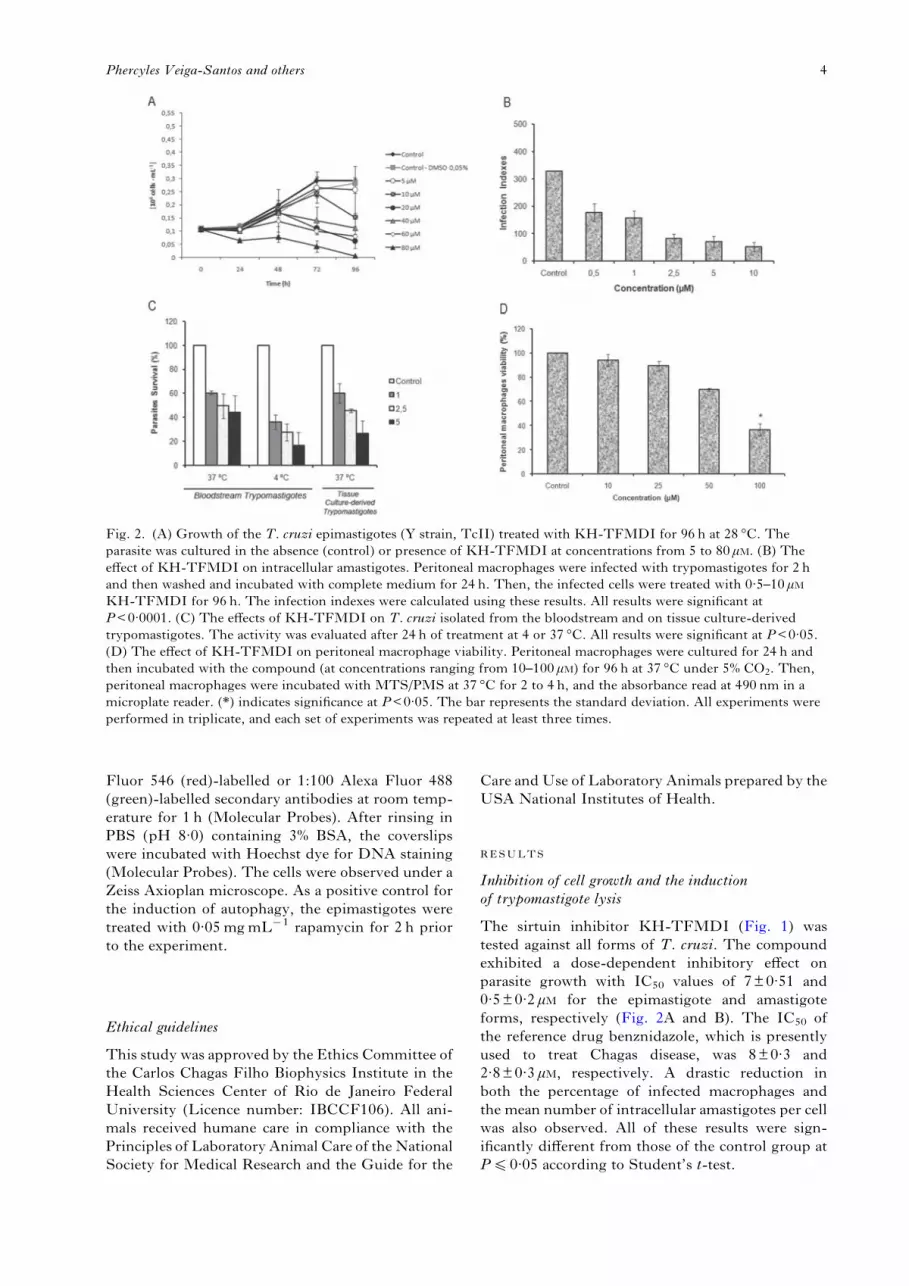
The sirtuin inhibitor KH-TFMDI (Fig. 1) wastested against all forms of T. cruzi. The compoundexhibited a dose-dependent inhibitory effect onparasite growth with IC50 values of 7±0·51 and0·5±0·2 μM for the epimastigote and amastigoteforms, respectively (Fig. 2A and B). The IC50 ofthe reference drug benznidazole, which is presentlyused to treat Chagas disease, was 8±0·3 and2·8±0·3 μM, respectively. A drastic reduction inboth the percentage of infected macrophages andthe mean number of intracellular amastigotes per cellwas also observed. All of these results were sign-ificantly different from those of the control group atP40·05 according to Student’s t-test.
Fig. 2. (A) Growth of the T. cruzi epimastigotes (Y strain, TcII) treated with KH-TFMDI for 96 h at 28 °C. Theparasite was cultured in the absence (control) or presence of KH-TFMDI at concentrations from 5 to 80 μM. (B) Theeffect of KH-TFMDI on intracellular amastigotes. Peritoneal macrophages were infected with trypomastigotes for 2 hand then washed and incubated with complete medium for 24 h. Then, the infected cells were treated with 0·5–10 μMKH-TFMDI for 96 h. The infection indexes were calculated using these results. All results were significant atP<0·0001. (C) The effects of KH-TFMDI on T. cruzi isolated from the bloodstream and on tissue culture-derivedtrypomastigotes. The activity was evaluated after 24 h of treatment at 4 or 37 °C. All results were significant at P<0·05.(D) The effect of KH-TFMDI on peritoneal macrophage viability. Peritoneal macrophages were cultured for 24 h andthen incubated with the compound (at concentrations ranging from 10–100 μM) for 96 h at 37 °C under 5% CO2. Then,peritoneal macrophages were incubated with MTS/PMS at 37 °C for 2 to 4 h, and the absorbance read at 490 nm in amicroplate reader. (*) indicates significance at P<0·05. The bar represents the standard deviation. All experiments wereperformed in triplicate, and each set of experiments was repeated at least three times.
4Phercyles Veiga-Santos and others
KH-TFMDI showed a strong lytic activity againsttissue culture-derived trypomastigotes (TCT) andBST; indeed, approximately 50% of TCTwere killedat a concentration of 1·6±0·18 μM at 37 °C. AgainstBSTs, an LC50 of 0·8±0·3 μM at 4 °C was observed.For comparison, crystal violet, the drug that iscurrently routinely used in blood banks to controlinfection by blood transfusion, has an LC50 of11·5 μM. When the incubation was carried out at37 °C, KH-TFMDI showed an LC50 of 2·5±1·1 μM(Fig. 2C), while benznidazole showed an LC50
greater than 400 μM against BSTs at 37 °C (data notshown).
Effects on host cells
The effects ofKH-TFMDI onmammalian cells wereevaluated in peritoneal macrophages after 96 h ofincubation at 37 °C via the MTS/PMS assay(Henriques et al. 2011). The 50% cytotoxic effects(CC50) value of KH-TFMDI in the macrophageswas 81±12 μM (Fig. 2D).
Effects on parasite structure
Epimastigotes that were incubated with 7 μM KH-TFMDI exhibited several changes that could beobserved using light microscopy. KH-TFMDItreatment for 96 h altered the kinetoplast shape in45% of the parasites, interfered with the process ofcytokinesis in approximately 37% of cells, and causedrounding of the parasite body in approximately 26%of the parasites. In addition, KH-TFMDI caused thedetachment of the flagellum from the cell body inapproximately 70% of the epimastigotes (Table 1).To better visualize this flagellum detachment, epi-mastigotes treated with 7 μM KH-TFMDI for 120 hwere analysed by fluorescence microscopy using ananti-α tubulin antibody andHoechst stain to label thenucleus (Fig. 3).We also used SEM and TEM to analyse the
effects of KH-TFMDI on the protozoan structure.Untreated T. cruzi epimastigotes exhibited atypical elongated shape with a smooth cell surface(Supplementary Figure S1A). In contrast, parasites
treated with 7 μM KH-TFMDI for 96 h revealedsevere morphological changes in the parasite body,such as the detachment of the parasite flagellumand an inhibition of cytokinesis leading to the failedseparation of the newly formed protozoans. We alsoobserved a depression and disruption of the plasmamembrane and an intense wrinkling of the parasitesurface (Figure S1 B–D).Figure 4A shows a section of an untreated
epimastigote highlighting different organelles, suchas the mitochondrion, nucleus, kinetoplast andflagellum, with normal ultrastructure. Epimastigotestreated with 7 μMKH-TFMDI for 12 h (Fig. 4B) and24 h (Fig. 4C and D) showed a progressive loss ofkDNA compaction. In addition, the drug alsoinduced other morphological changes, varying fromdiscrete alterations to a total destruction of theparasite, depending on the time of drug incubation.After 48 to 96 h of incubation, we observed four mainpoints of cell alterations: an increase in the number oflipid-storage bodies, an accumulation of medialcisternae of the Golgi complex, large structures re-sembling myelin-like figures localized near theflagellum, and the appearance of unpacked nuclearchromatin (Fig. 4E and G–I). In addition, well-developed endoplasmic reticulum (ER) profileswere observed surrounding reservosomes and lipid-storage bodies after 72 hof treatment (Fig. 4GandH).The treatment of intracellular amastigote forms
with KH-TFMDI also caused alterations in theparasite plasma membrane, such as a loss of cytoplas-mic contents, disruption of the plasma membrane,membrane shedding, nuclear membrane detachmentand membrane convolutions, thereby completelyaltering the parasite morphology (Fig. 5B–F).Trypomastigotes treatedwith 1·8 μMKH-TFMDI
for 24 h revealed alterations similar to those observedin epimastigotes (Supplementary Fig. S2). Note thatuntreated trypomastigotes exhibited characteristicmorphology, as evidenced by SEM (Fig. S2A), andTEM revealed that the organelles, including themitochondrion, nucleus, kinetoplast and flagellum,were also normal (Fig. S2D). KH-TFMDI causeddisorganization of the parasite plasma membrane,with bleb formation and intense wrinkling of thecell surface (Fig. S2B and C). The drug also alterednuclear chromatin and kDNA compaction (Fig. S2E).Lipid body accumulation was also observed in thetrypomastigote forms (Fig. S2F).
The mechanism of killing of the parasite
Two types of observed morphological changesprovide some insight into the mechanism of parasitedeath induced by KH-TFMDI. After incubationfor 12 or 24 h, the unpacking of nuclear chromatin,which is indicative of apoptosis, was observed (Fig. 4Cand D). After 24 h of treatment with 10 μM KH-TFMDI, approximately 92% of the epimastigotes
Table 1. Morphological alterations caused byKH-TFMDI treatment in T. cruzi epimastigotesafter 96 h of incubation at 28 °C
AlterationControl(%)a
7 μM KH-TFMDI(%)a
Kinetoplast rounding 4·4±3·11 45±6·06Flagellum detachment 0 70±10·21Cytokinesis inhibition 0 37±8·56Cell body rounding 2·5±0·7 26±12·26
a A total of 500 cells were evaluated in each group.
5Antitrypanosomal activity of KH-TFMDI

were labelled with annexin V (Fig. 6A), which bindsto phosphatidylserine (PS), a phospholipid relatedto apoptotic events (Lee et al. 2013). However,no disruption of the plasma membrane was observed;
the percentage of propidium iodide (PI)-positive cellswas lower than 1% (data not shown). After 48 and72 h of treatment, we observed a similar labellingpattern, with 87 and 88% of treated parasites positive
Fig. 3. Immunofluorescence microscopy demonstrating flagellum detachment in treated epimastigotes (7 μMKH-TFMDI for 120 h). The cells were labelled with anti-α-tubulin for 2 h and with an Alexa Fluor 488(green)-labelled secondary antibody for 1 h. Hoechst stain (5 μgmL−1) was used to label the DNA (blue).(A, B) In untreated epimastigotes, the flagellum is attached to the parasite body; (C–F) in epimastigotes treated withKH-TFMDI, the flagellum is detached from the parasite body; (A, C, E) phase contrast images; (B, D, F) mergedimages of Hoechst and α-tubulin labelling. Bars: 10 μm.
6Phercyles Veiga-Santos and others

for annexin V (Fig. 6A), and the proportion ofPI-positive parasites increased to almost 2 and 4%(data not shown). However, after 96 h of treatment,we observed a significant reduction in PS-positiveepimastigotes, such that only 50% of cells were PSpositive (Fig. 6A). In the case of trypomastigotes,after 24 h of treatment with 1·8 μM KH-TFMDI, weobserved that 40% of parasites were positive forAnnexin V (Fig. 6B), and half of these parasites (20%)were already positive for PI, indicating that apoptosishad occurred. However, only 3% of trypomastigoteswere positive only for PI, and less than 1% ofuntreated trypomastigotes were positive for PI (datanot shown). Unfortunately, all attempts to carry outsimilar experiments with intracellular amastigoteswere unsuccessful (data not shown).
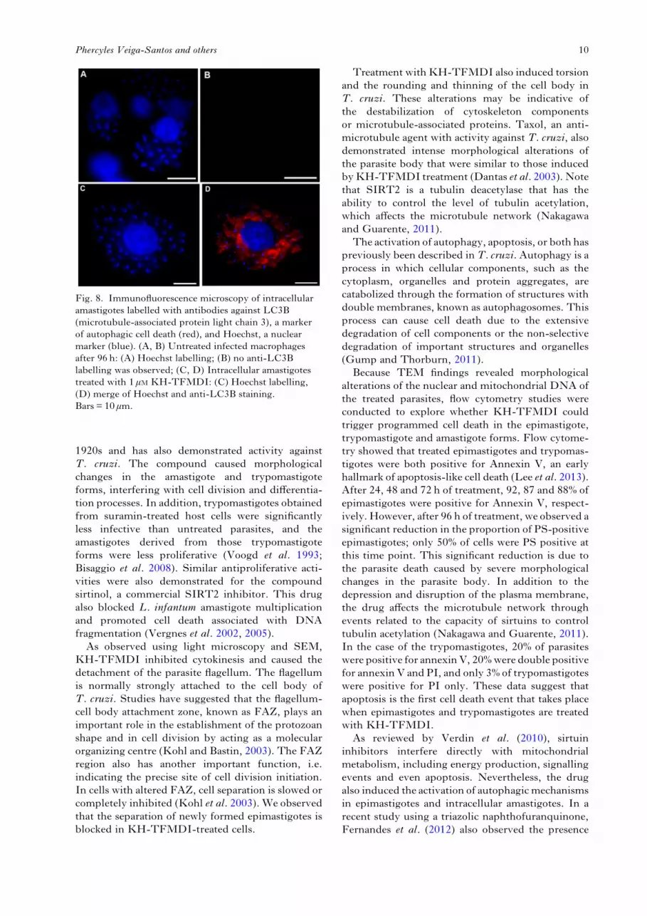
Analyses of epimastigotes treated for 72 and 96 hrevealed the presence of ER profiles surroundingstructures such as the reservosomes and lipid-storagebodies (Fig. 4G and H). The obtained images areindicative of autophagy. Autophagy was confirmedby labelling the drug-treated epimastigotes withan anti-LC3B antibody (i.e. to label microtubule-associated protein light chain 3) that is widelyemployed to identify autophagic structures byfluorescence microscopy (Fig. 7). Strong labellingof the structures was observed in treated parasites(Fig. 7F), whereas no labelling was observed inuntreated epimastigotes (Fig. 7C). Rapamycin, adrug that induces autophagy, was used as a positivecontrol at a concentration of 0·05 μgmL−1 (Fig. 7I),demonstrating the functionality of the assay. We also
Fig. 4. TEM images of T. cruzi epimastigotes treated with 7 or 10 μM KH-TFMDI. (A) Untreated epimastigotesexhibit organelles with normal morphology. Kinetoplast (k), nucleus (n), Golgi (CG) and flagellum (f) after 96 h ofincubation. Epimastigotes treated with 7 μM for 12 h (B) and 24 h (C, D) show an aberrant loss of kDNA compactioninside the mitochondrion (white arrow and arrowhead); (E, F) epimastigotes treated for 48 and 72 h contain largestructures similar to myelin-like figures (black arrow). After 48 to 96 h of treatment, lipid-storage bodies (whitearrowhead) (E–I) and the accumulation of cisternae in the medial region of the Golgi complex were observed (only in E);(G, H) in epimastigotes after 72 h of treatment, well-developed endoplasmic reticulum membranes surroundingreservosomes, lipid-storage bodies (arrow) and abnormal nucleus condensation (star) were observed; (I) after 120 hof treatment, autophagosome formation (large black arrow) and membranes surrounding the lipid-storage body(white arrowhead) were observed. Bar: (A–D) 0·5 μm; (E, F) 2 μm; (G, H) 5 μm; (I) 1 μm.
7Antitrypanosomal activity of KH-TFMDI

analysed the activation of autophagy in treatedintracellular amastigotes. Strong labelling of cyto-plasmatic structures was observed in parasites treatedwith KH-TFMDI for 96 h (Fig. 8D). No LC3Blabelling was observed in untreated amastigotes(Fig. 8B).
DISCUSSION
The observations reported here clearly show thatthe sirtuin inhibitor KH-TFMDI significantlyinterfered with all developmental stages of T. cruzi.Sirtuins in mammalian cells are associated withhistone deacetylation, causing heterochromatin
Fig. 5. TEM images of T. cruzi intracellular amastigotes treated with 1 μM KH-TFMDI for 96 h. (A) Untreatedintracellular amastigote: kinetoplast (k), mitochondrion (m), nucleus (n), Golgi (CG) and flagellum (f); (B) treatmentcaused plasma membrane disruption (arrow) and a loss of cytoplasmic content (stars); (C, D) plasma membraneshedding (arrow); (E) nuclear membrane detachment (arrowhead); and (F) severe alterations in the plasma membrane,leading to convolutions and the loss of parasite plasma membrane integrity (arrow).
Fig. 6. Column graphs representing flow cytometry experiments on T. cruzi epimastigotes (A) and trypomastigotes (B).Epimastigotes were treated with 10 μM of KH-TFMDI for 24, 48, 72 or 96 h, while trypomastigotes were treated with1·8 μM KH-TFMDI for 24 h. After incubation, treated or control parasites were washed, resuspended in Annexin VBinding Buffer, incubated with Annexin V/PI and analysed on a BD FACSCalibur®, as described in the Materials andMethods section. (A) After 24 h of treatment, 92% of epimastigotes were positive for Annexin V. After 96 h of treatment,only 40% of epimastigotes presented the same labelling pattern. (B) After 24 h of treatment, 40% of trypomastigotes werealready positive for Annexin V. Ten thousand gated events were counted from each sample. The statistical analysiswas carried out using Student’s t-test. Values are presented as the mean±S.E.M. (***) P<0·0001 (A and B) and(*) P<0·05 (A).
8Phercyles Veiga-Santos and others

formation and transcription repression (Nakagawaand Guarente, 2011). As previously discussed,T. cruzi encodes two sirtuin-like genes, TcSIR2rp1and TcSIR2rp3, but the localization and functionof the encoded proteins remain unclear (Religa andWaters, 2012).At very low concentrations, the sirtuin inhibitor
KH-TFMDI was able to inhibit the replication ofthe epimastigote forms, which proliferate in axeniccultures, and the amastigote form, which replicatesin the cytoplasm of host cells and is responsible forthe amplification of the infection. Interestingly, KH-TFMDI showed better activity against intracellularamastigotes than the reference drug benznidazole. Inaddition, KH-TFDMI caused the lysis of the highlyinfective bloodstream trypomastigote form and wasmore active than crystal violet. Moreover, treatmentwith crystal violet changes the colour of the blood toviolet, which is not well accepted by some patients.These observations have encouraged us to initiatefurther studies focusing on the use of KH-TFMDIin blood banks.One important criterion for potential anti-
trypanosomal drugs is a low toxicity against mam-malian cells. Here, KH-TFMDI was found to have
significant selectivity indexes; it was 50-fold moreselective against TCT and 162-fold more selectiveagainst intracellular amastigotes than peritonealmacrophages. Therefore, KH-TFMDI shows prom-ising mammalian cell tolerance indexes comparedwith other drugs currently being used in clinicaltherapy. The reference drug benznidazole, which isused to treat Chagas disease, presents a CC50 value of1218 μM in LLC-MK2 cells (Cogo et al. 2012).However, this drug still has considerable side effectsfor patients, including hypersensitivity reactions,bone marrow depletion and peripheral polyneuro-pathy (Soeiro and Castro, 2011).Recently, several molecules with the ability to
inhibit/activate the deacetylase activity of sirtuinshave shown anticancer, anti-HIV, anti-protozoan,metabolic and neurological activities (Baur et al.2012; Zheng, 2013). Significant activities werefound in assays testing this group of drugs againsttrypanosomatids. Bisnaphthalimidopropyl (BNIP),an anti-cancer drug, also showed strong and selectiveeffects against the SIRT1 of Leishmania infantum(Tavares et al. 2010). In addition, suramin, a selectiveSIRT1 inhibitor with anti-proliferative effects, hasbeen used to treat African trypanosomiasis since the
Fig. 7. Immunofluorescence microscopy demonstrating LCB3 labelling (i.e. a marker of autophagic cell death) ofepimastigotes treated with 10 μM of KH-TFMDI. (A, C) Untreated epimastigotes after 96 h of incubation show onlyHoechst labelling; epimastigotes treated with KH-TFMDI (D–F) and rapamycin (0·05mgmL−1) (G–I) exhibit intensered labelling (arrowhead). Bar: 5 μm.
9Antitrypanosomal activity of KH-TFMDI
1920s and has also demonstrated activity againstT. cruzi. The compound caused morphologicalchanges in the amastigote and trypomastigoteforms, interfering with cell division and differentia-tion processes. In addition, trypomastigotes obtainedfrom suramin-treated host cells were significantlyless infective than untreated parasites, and theamastigotes derived from those trypomastigoteforms were less proliferative (Voogd et al. 1993;Bisaggio et al. 2008). Similar antiproliferative acti-vities were also demonstrated for the compoundsirtinol, a commercial SIRT2 inhibitor. This drugalso blocked L. infantum amastigote multiplicationand promoted cell death associated with DNAfragmentation (Vergnes et al. 2002, 2005).
As observed using light microscopy and SEM,KH-TFMDI inhibited cytokinesis and caused thedetachment of the parasite flagellum. The flagellumis normally strongly attached to the cell body ofT. cruzi. Studies have suggested that the flagellum-cell body attachment zone, known as FAZ, plays animportant role in the establishment of the protozoanshape and in cell division by acting as a molecularorganizing centre (Kohl and Bastin, 2003). The FAZregion also has another important function, i.e.indicating the precise site of cell division initiation.In cells with altered FAZ, cell separation is slowed orcompletely inhibited (Kohl et al. 2003). We observedthat the separation of newly formed epimastigotes isblocked in KH-TFMDI-treated cells.
Treatment withKH-TFMDI also induced torsionand the rounding and thinning of the cell body inT. cruzi. These alterations may be indicative ofthe destabilization of cytoskeleton componentsor microtubule-associated proteins. Taxol, an anti-microtubule agent with activity against T. cruzi, alsodemonstrated intense morphological alterations ofthe parasite body that were similar to those inducedbyKH-TFMDI treatment (Dantas et al. 2003). Notethat SIRT2 is a tubulin deacetylase that has theability to control the level of tubulin acetylation,which affects the microtubule network (Nakagawaand Guarente, 2011).
The activation of autophagy, apoptosis, or both haspreviously been described inT. cruzi. Autophagy is aprocess in which cellular components, such as thecytoplasm, organelles and protein aggregates, arecatabolized through the formation of structures withdouble membranes, known as autophagosomes. Thisprocess can cause cell death due to the extensivedegradation of cell components or the non-selectivedegradation of important structures and organelles(Gump and Thorburn, 2011).
Because TEM findings revealed morphologicalalterations of the nuclear and mitochondrial DNA ofthe treated parasites, flow cytometry studies wereconducted to explore whether KH-TFMDI couldtrigger programmed cell death in the epimastigote,trypomastigote and amastigote forms. Flow cytome-try showed that treated epimastigotes and trypomas-tigotes were both positive for Annexin V, an earlyhallmark of apoptosis-like cell death (Lee et al. 2013).After 24, 48 and 72 h of treatment, 92, 87 and 88% ofepimastigotes were positive for Annexin V, respect-ively. However, after 96 h of treatment, we observed asignificant reduction in the proportion of PS-positiveepimastigotes; only 50% of cells were PS positive atthis time point. This significant reduction is due tothe parasite death caused by severe morphologicalchanges in the parasite body. In addition to thedepression and disruption of the plasma membrane,the drug affects the microtubule network throughevents related to the capacity of sirtuins to controltubulin acetylation (Nakagawa and Guarente, 2011).In the case of the trypomastigotes, 20% of parasiteswere positive for annexinV, 20%were double positivefor annexin V and PI, and only 3% of trypomastigoteswere positive for PI only. These data suggest thatapoptosis is the first cell death event that takes placewhen epimastigotes and trypomastigotes are treatedwith KH-TFMDI.
As reviewed by Verdin et al. (2010), sirtuininhibitors interfere directly with mitochondrialmetabolism, including energy production, signallingevents and even apoptosis. Nevertheless, the drugalso induced the activation of autophagicmechanismsin epimastigotes and intracellular amastigotes. In arecent study using a triazolic naphthofuranquinone,Fernandes et al. (2012) also observed the presence
Fig. 8. Immunofluorescence microscopy of intracellularamastigotes labelled with antibodies against LC3B(microtubule-associated protein light chain 3), a markerof autophagic cell death (red), and Hoechst, a nuclearmarker (blue). (A, B) Untreated infected macrophagesafter 96 h: (A) Hoechst labelling; (B) no anti-LC3Blabelling was observed; (C, D) Intracellular amastigotestreated with 1 μM KH-TFMDI: (C) Hoechst labelling,(D) merge of Hoechst and anti-LC3B staining.Bars = 10 μm.
10Phercyles Veiga-Santos and others

of endoplasmic ER surrounding reservosomes. Theseauthors also suggested that the presence of thismembrane network is associated with an increasein monodansylcadaverine labelling, which is anindicator of autophagy activation. We confirmed theactivation of autophagy by immunofluorescencemicroscopy.In conclusion, our results suggest that KH-
TFMDI impairs epimastigote and amastigoteproliferation by causing drastic alterations in theultrastructure of T. cruzi and induces the activationof apoptosis and autophagy mechanisms that lead tocell death. In addition, KH-TFMDI also showedimportant trypanosomicidal activity against BTs.Collectively, the results of the present study indicatethat sirtuin inhibitors are potential new compoundsfor the treatment of Chagas disease and blood controlin blood banks.
SUPPLEMENTARY MATERIAL
To view supplementary material for this article, please visithttp://dx.doi.org/S0031182013001704.
ACKNOWLEDGEMENTS
The authors thank Luzinete da Silva and Rachel Rachid fortheir technical assistance and CristinaHenriques and EmileBarrias for their help with the animals.
FINANCIAL SUPPORT
This work was supported by the Conselho Nacionalde Desenvolvimento Cientifico e Tecnológico (CNPq),Financiadora de Estudos e Projetos (FINEP), Fundação deAperfeiçoamento de Pessoal de Nível Superior (CAPES)and Fundação Carlos Chagas Filho de Amparo a Pesquisado Estado do Rio de Janeiro (FAPERJ).
REFERENCES
Alsford, S., Kawahara, T., Isamah, C. and Horn, D. (2007). A sirtuin inthe African trypanosome is involved in bothDNA repair and telomeric genesilencing but is not required for antigenic variation. Molecular Microbiology63, 724–736. doi: 10.1111/j.1365-2958.2006.05553.x.Baur, J. A., Ungvari, Z., Minor, R. K., Le Couteur, D. G. and Cabo, R.(2012). Are sirtuins viable targets for improving healthspan and lifespan?Nature Review Drug Discovery 11, 443–461. doi: 10.1038/nrd3738.Bisaggio, D. F. R., Adade, C.M. and Souto- Padrón, T. (2008). In vitroeffects of suramin on Trypanosoma cruzi. International Journal ofAntimicrobial Agents 31, 282–286. doi: 10.1016/j.ijantimicag.2007.11.001.Camargo, E. P. (1964). Growth and differentiation in Trypanosoma cruzi.Origin of metacyclic trypanosomes in liquid media. Revista do Instituto deMedicina tropical de São Paulo 6, 93–100.Cogo, J., Caleare, A. O., Ueda-Nakamura, T., Dias Filho, B. P.,Ferreira, I. C. P. and Nakamura, C. V. (2012). Trypanocidal activity ofguaianolide obtained from Tanacetum parthenium (L.) Schultz-Bip. and itscombinational effect with benznidazole. Phytomedicine 1, 59–66. doi:10.1016/j.phymed.2012.09.011.Dantas, A. P., Barbosa, H. S. and De Castro, S. L. (2003). Biologicaland ultrastructural effects of the anti-microtubule agent taxol againstTrypanosoma cruzi. Journal of Submicroscopic Cytology and Pathology 35,287–294.Fernandes, M. C., Da Silva, E. N., Jr., Pinto, A. V., De Castro, S. L.and Menna-Barreto, R. F. S. (2012). A novel triazolic naphthofuranqui-none induces autophagy in reservosomes and impairment of mitosis
in Trypanosoma cruzi. Parasitology 139, 26–36. doi: 10.1017/S0031182011001612.Gump, J.M. and Thorburn, A. (2011). Autophagy and apoptosis: whatis the connection? Trends in Cell Biology 21, 387–392. doi: 10.1016/j.tcb.2011.03.007.Henriques, C., Moreira, T. L. B., Maia-Brigagão, C., Henriques-Pons, A., Carvalho, T.M. U. and De Souza, W. (2011). Tetrazoliumsalts based methods for high-throughput evaluation of anti-parasitechemotherapy. Analytical Methods 3, 2148–2155.Huber, K., Schemies, J., Uciechowska, U., Wagner, J. M., Rumpf, T.,Lewrick, F., Süss, R., Sippl, W., Jung,M. and Bracher, F. (2010). Novel3-arylideneindolin-2-ones as inhibitors of NAD+-dependent histonedeacetylases (sirtuins). Journal of Medicinal Chemistry 53, 1383–1386. doi:10.1021/jm901055u.Kohl, L. and Bastin, P. (2003). The flagellum of trypanosomes.International Review of Cytology 244, 227–285.Kohl, L., Robinson, D. and Bastin, P. (2003). Novel roles for the flagellumin cell morphogenesis and cytokinesis of trypanosomes. EMBO Journal 22,5336–5346.Lee, S. H., Meng, X.W., Flatten, K. S., Loegering, D. A.and Kaufmann, S. H. (2013). Phosphatidylserine exposure duringapoptosis reflects bidirectional trafficking between plasma membrane andcytoplasm. Cell Death and Differentiation 20, 64–76. doi: 10.1038/cdd.2012.93.Macedo-Silva, S. T., Silva, T. L. A. O., Urbina, J. A., De Souza, W.and Rodrigues, J. C. F. (2011). Antiproliferative, ultrastructural, andphysiological effects of amiodarone on promastigote and amastigoteforms of Leishmania amazonensis. Molecular Biology International 876021.doi: 10.4061/2011/876021.Meirelles, M.N., De Araújo Jorge, T. C. and De Souza, W. (1982).Interaction of Trypanosoma cruzi with macrophages in vitro: dissociation ofthe attachment and internalization phases by low temperature andcytochalasin B. Zeitschrift für Parasitenkunde 68, 7–14.Nakagawa, T. and Guarente, L. (2011). Sirtuins at a glance. Journal ofCell Science 124, 833–838. doi: 10.1242/jcs.081067.Religa, A. A. and Waters, A. P. (2012). Sirtuins of parasitic protozoa: insearch of function(s). Molecular and Biochemical Parasitology 185, 71–88.doi: 10.1016/j.molbiopara.2012.08.003.Salminen, A. and Kaarniranta, K. (2009). SIRT1: regulation oflongevity via autophagy. Cellular Signalling 21, 1356–1360. doi: 10.1016/j.cellsig.2009.02.014.Soeiro, M.N. C. and Castro, S. L. (2011). Screening of potential anti-Trypanosoma cruzi candidates: in vitro and in vivo studies. Open MedicinalChemistry Journal 5, 21–30. doi: 10.2174/1874104501105010021.Silva, C. F., Daliry, A., Bernardino, P., Silva, P. B., Akay, S.,Banerjee, B., Farahat, A. A., Fisher, M., Hu, L., Kumar, A., Liu, Z.,Stephens, C. E., Boykin, D. and Soeiro, M.N. C. (2011). The efficacy ofnovel arylimidamides against Trypanosoma cruzi in vitro. Parasitology 138,1863–1869.Tavares, J., Ouaissi, A., Kong Thoo Lin, P., Loureireo, I., Kaur, S.,Roy, N. and Cordeiro-Da-Silva, A. (2010). Bisnaphthalimidopropylderivatives as inhibitors of Leishmania SIR2 related protein 1.ChemMedChem 5, 140–147.Urbina, J. A. (2010). New insights in Chagas’ disease treatment.Drugs of theFuture 35, 409–419. doi: 10.1358/dof.2010.35.5.1484391.Veiga-Santos, P., Barrias, E. S., Santos, J. F. C., Moreira, T. L. B.,Carvalho, T.M. U. and De Souza, W. (2012). Effects of amiodarone andposaconazole on the growth and ultrastructure of Trypanosoma cruzi.International Journal of Antimicrobial Agents 40, 61–71. doi: 10.1016/j.ijantimicag.2012.03.009.Verdin, E., Hirschey, M. D., Finley, L.W. S. and Haigis, M. C. (2010).Sirtuin regulation of mitochondria: energy production, apoptosis, andsignaling. Trends in Biochemical Sciences 35, 669–675. doi: 10.1016/j.tibs.2010.07.003.Vergnes, B., Sereno, D., Madjidian-Sereno, N., Lemesre, J. L. andOuaissi, A. (2002). Cytoplasmic SIR2 homologue overexpression promotessurvival of Leishmania parasites by preventing programmed cell death.Gene296, 139–150. doi: 10.1016/S0378-1119(02)00842-9.Vergnes, B., Vanhille, L., Ouaissi, A. and Sereno, D. (2005). Stage-specific antileishmanial activity of an inhibitor of SIR2 histone deacetylase.Acta Tropica 94, 107–115. doi: 10.1016/j.actatropica.2005.03.004.Voogd, T. E., Vansterkenburg, E. L.M., Wilting, J. andJanssen, L. H.M. (1993). Recent research on the biological activity ofsuramin. Pharmacological Reviews 46, 177–199.Zemzoumi, K., Sereno, D., Francois, C., Guilvard, E., Lemesre, J. L.and Ouaissi, A. (1998). Leishmania major: cell type dependent distributionof a 43 kDa antigen related to silent information regulatory-2 protein family.Biology of the Cell 90, 239–245.
11Antitrypanosomal activity of KH-TFMDI

Zheng, W. (2013). Sirtuins as emerging anti-parasitic targets. EuropeanJournal of Medicinal Chemistry 59, 132–140. doi: 10.1016/j.ejmech.2012.11.014.Zingales, B., Andrade, S. G., Briones, M. R. S., Campbell, D. A.,Chiari, E., Fernandes, O., Guhl, F., Lages-Silva, E., Macedo, A.M.,Machado, C. R., Miles, M. A., Romanha, A. J., Sturm, N. R.,Tibayrenc, M. and Schijman, A. G. (2009). A new consensus for
Trypanosoma cruzi intraspecific nomenclature: second revision meetingrecommends TcI to TcVI. Memórias do Instituto Oswaldo Cruz 104, 1051–1054.World Health Organization (2013). Control of Chagas Disease. Factsheet No 340 Updated March 2013. World Health Organization, Geneva,Switzerland.
12Phercyles Veiga-Santos and others


