Embed Size (px)
Citation preview
帯広畜産大学学術情報リポジトリOAK:Obihiro university Archives of Knowledge
' '
TitleMorphological study on the encystment of the
ciliated protozoan Colpoda cucullus
Author(s)Watoh, Tomoko, Sekida, Satoko, Yamamoto,
Keisuke, Kida, Akemi, Matsuoka, Tatsuomi
Citation The Journal of Protozoology Research, 15: 20-28
Issue Date 2005
URL http://ir.obihiro.ac.jp/dspace/handle/10322/148
Rights

J. Protozool. Res. 15, 20-28 (2005) Copyright©2005, National Research Center for Protozoan Diseases
20
Morphological study on the encystment of the ciliated protozoan Colpoda cucullus
Tomoko Watoh, Satoko Sekida, Keisuke Yamamoto, Akemi Kida and Tatsuomi Matsuoka*
Institute of Biological Science, Faculty of Science, Kochi University, Kochi 780-8520, Japan *Corresponding author: Tatsuomi Matsuoka, E-mail [email protected]
Key words: Colpoda; encystment; cyst wall; toluidine blue
ABSTRACT Morphological changes during resting cyst formation (encystment) of Colpoda cucullus were examined. At 1 hr after encystment induction, a number of vesicles containing the electron-dense and toluidine blue (TB) positive materials were fused with large vacuoles to excrete the contents, and the vacuoles opened to the extracellular space to excrete. In this stage, the rounded precystic cells were surrounded by the first-synthesized cyst wall layer (L1 layer), and the cilia were resorbed inside the L1 layer. Thereafter (1-2 hr), a secondary layer (L2 layer) was formed and subsequently a deeply TB stained substance was diffused into the space between the plasma membrane and L2 layer. At 3-6 hr, numerous endoplasmic reticula (ER) associated with ribosomes occupied the cytoplasm, indicating that cyst wall precursors such as glycoconjugates were being synthesized. By a week after encystment induction, the cyst wall composed of three distinct layers covered with a fibrous (mucous) coat was completed. The kinetosomes were still observed in 3-day old cysts. INTRODUCTION
The encystment of Colpoda cucullus is mediated by intracellular signaling chains activated by an increase in the concentration of external cations such as Ca2+ (Watoh et al. 2003; Yamaoka et al. 2004) that is suppressed by the addition of components released from bacteria (Watoh et al. 2003; Yamasaki et al. 2004) or components such as porphyrins contained in cereal infusion or artificial porphyrin analogue, chlorophyllin-Cu (Tsutsumi et al. 2004). However, the excystment of C. cucullus is elicited by the elimination of cations such as Ca2+ (our unpublished data) or by the addition of cereal infusion or chlorophyllin-Cu (Tsutsumi et al. 2004). Prior to starting the studies for analyzing molecular events such as signaling pathways including gene expression or silencing leading to cytodifferentiation during resting cyst formation, cytological events should be carefully observed stage by stage during encystment. Up to now, the morphological changes during the resting cyst formation of Colpoda have been extensively studied, and the results obtained supply important information as follows: (1) the cytoplasm of the Colpoda cyst is surrounded by a cyst wall that is composed of several layers (Kawakami & Yagiu 1963b; Tibbs 1968; Janisch 1980; Ruthmann & Kuck 1985; Foissner 1993; Martín-González et al. 1994; Chessa et al. 2002); (2) the presumed precursors contained in vesicles (or vacuoles) for the cyst walls of C. inflata are excreted in the precystic stages (Martín-González et al. 1994); (3) the cyst wall layers of C. steinii originate from membranes (Ruthmann and Kuck 1985); and (4) the kinetosomes are partially resorbed in the
Encystment in Colpoda
21
resting cysts of C. inflata (Martín-González et al. 1991a). In the resting cysts of C. steinii, the cilia are still retained (Tibbs 1968; Ruthmann & Kuck 1985); and (5) chromatin extrusion takes place in the encystment process of C. inflata (Martín-González et al. 1991a). Unfortunately, such information seems to be insufficient to understanding intracellular events during the cyst formation of Colpoda. For example, it is not possible to answer even the simplest questions, such as how many layers constitute the resting cyst wall of C. cucullus and when they are synthesized. Therefore, we aimed to observe intracellular events in the relatively early stage during the encystment of C. cucullus with special reference to cyst wall formation and cytoplasmic events, using an electron microscope and other cytological methods described as follows.
The studies of the chemical composition of the cyst wall indicate that it is mainly composed of glycoproteins (Izquierdo et al. 2000; Chessa et al. 2002), or of proteins and polysaccharides (Calvo et al. 1983; Gutiérrez et al. 1984; Delgado et al. 1987; Martín-González et al. 1991b; Calvo et al. 2003). Toluidine blue (TB) dye forms complexes with anionic polysaccharides such as glycosaminoglycans (Shepard & Mitchell 1976; Terry et al. 2000). This means that the TB is an effective probe for detecting the precursor materials of the cyst wall appearing in the cytoplasm. In the present study, we tried to observe the cyst wall and its precursors by using TB staining.
MATERIALS AND METHODS Culture and encystment induction
Colpoda cucullus was cultured in a 0.1% (w/v) infusion of dried cereal leaves (autoclaved) inoculated with bacteria (Enterobacter aerogenes) at 23 oC in the dark. The bacteria were cultured on agar plates containing 1.5% agar, 0.5% polypeptone, 1% meat extract and 0.5% NaCl. The cultured vegetative cells were collected by centrifugation (1,000 g, 1-2 min) and subsequently suspended in a standard saline solution containing 1 mM CaCl2, 1 mM KCl, 5 mM Tris-HCl (pH 7.2). The cells were rinsed 2-3 times by repeating the sedimentation and suspension in the standard saline solution, and finally suspended in the solution to induce encystment. Scanning electronmicroscopy
The cells were fixed with 2% OsO4 for 1 min and rinsed twice in distilled water. The suspension of fixed cells (20 µl) was placed on the SEMpore (JEOL), which had been coated with 0.1% poly-L-lysine hydrobromide (Sigma). The distilled water surrounding the cells was replaced by 100% t-butyl alcohol by dropping 100% t-butyl alcohol onto the sample and concomitantly sucking it out through the micropores of the SEMpore with a small syringe. The samples suspended in 100% t-butyl alcohol were quickly frozen in liquid nitrogen and subsequently freeze-dried with a freeze-drying device (JEOL, JFD-300). The dried samples were coated with gold using an ion sputtering device (JEOL, JFC-1500) and observed with a scanning electron microscope (JEOL, JSM-5300).
Cytological methods for light microscopy In order to visualize the infraciliature, silver carbonate impregnation was performed according to the
method reported by Foissner (1993). Prior to the silver impregnation, the cyst walls were peeled mechanically (gently homogenized) after the cysts were frozen (-80° C) and thawed.
Transmission electronmicroscopy

Encystment in Colpoda
22
One volume of the suspension of vegetative cells was mixed with 6 volumes of a GA fixative containing 6% glutaraldehyde, 1% OsO4, 100 mM cacodylate buffer (pH 7.2) and 4 mM sucrose, and incubated for 10 min at room temperature. After the prefixed samples were rinsed 5 times in 100 mM cacodylate buffer (pH 7.2), they were postfixed for 2 hr below 4° C in a fixative containing 1% OsO4, 100 mM cacodylate buffer (pH 7.2) and 2 mM sucrose. The cysts were prefixed with GA fixative without OsO4
for 6 hr and postfixed for 1 week, because the fixative hardly penetrated the cyst walls. The post-fixed samples were rinsed 5 times in distilled water, gradually dehydrated through an ethanol series (30, 40, 50, 60, 70, 80, 90 and 100% ethanol; 15 min each), and finally suspended in acetone. The dehydrated samples were embedded in Spurr’s resin. Ultrathin sections were stained with 3% uranyl acetate for 10 min, and then with lead citrate for 10 min. The sections were observed under a transmission electron microscope (JEOL, 1010T).
RESULTS Cyst wall of resting cysts
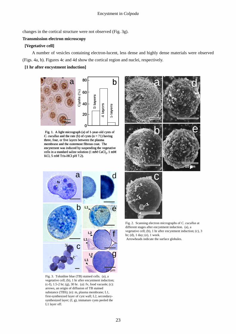
Figures 1a and 1b are the photomicrographs of the 1-year-old cysts of C. cucullus and the percentage of cysts having three, four or five layers between the plasma membrane and the outermost fibrous coat. As shown in Fig. 1b, the wall of the mature cyst was composed of 3-5 layers; in this case, two- or 6- layered cysts were not observed. In the present study, therefore, we defined the cysts having at least three layers as mature cysts. As has been reported (Foissner 1993), the interlayer spaces tended to be wide in older mature cysts, while in some of the younger mature cysts, the cyst wall layers were closely stacked (data not shown).
Scanning electron microscopy Figure 2 shows the morphological changes of the surface of the cells during encystment. At 1 hr after
encystment induction (Fig. 2b), most of the cells were rounded, and the cilia were gradually shortened. At 3 hr, small globules began to appear on the surface of the cell (Fig. 2c, arrowheads). The surface globules gradually increased (Fig. 2d) and the surface of the 1-week old cyst was entirely covered with an outermost fibrous layer associated with small globules (Fig. 2e). Toluidine blue (TB) staining
Figure 3 shows the toluidine blue (TB) stained cells at the early stage during cyst formation. In vegetative cells, only food vacuoles were stained with TB (Fig. 3a). In the precystic cells at 30–60 min after encystment induction, a number of vesicles (or granules) stained with TB appeared (Fig. 3b). Until the end of this stage, most of the cells were surrounded by a first-synthesized cyst wall layer (L1 layer), and continued to rotate inside the layer. At 1-2 hr, the rotating movement stopped and a secondary cyst wall layer (L2 layer) was formed (see Fig. 3d, left; Figs. 3e, f). Thereafter, a drastic event occurred; the substance deeply stained with TB appeared at the region near the cell surface, and diffused over the entire cell surface within a few minutes (Fig. 3c). The interlayer space between the L1 and L2 layers was not occupied by the TB stained substance (TBS) (Fig. 3d right), indicating that it probably diffuses beneath the L2 layer. In the partially disordered cells in which the L2 layer was detached from the plasma membrane by violent pipetting, the TBS was demonstrated to diffuse into the space between the L2 layer and plasma membrane (Fig. 3e). The L2 layer seemed to become thicker (Fig. 3f) after TBS was diffused, suggesting that TBS may be responsible for the development of the L2 layer. At 30 hr, the L1 layer seemed to become hard, although other remarkable
Encystment in Colpoda
23
changes in the cortical structure were not observed (Fig. 3g).
Transmission electron microscopy [Vegetative cell]
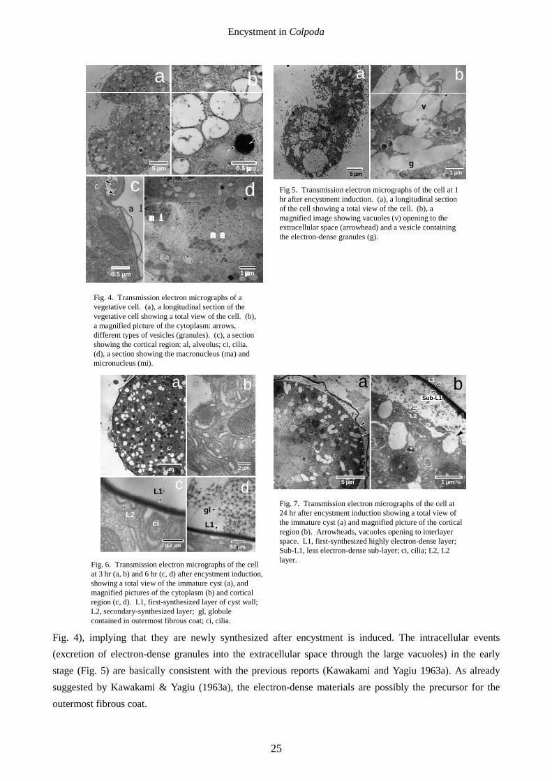
A number of vesicles containing electron-lucent, less dense and highly dense materials were observed (Figs. 4a, b). Figures 4c and 4d show the cortical region and nuclei, respectively.
[1 hr after encystment induction]
a
b e
10 µm
c
d
Fig. 2. Scanning electron micrographs of C. cucullus at different stages after encystment induction. (a), a vegetative cell; (b), 1 hr after encystment induction; (c), 3 hr; (d), 1 day; (e), 1 week. Arrowheads indicate the surface globules.
80
40
60
20
0
ba
20 µm
Fig. 1. A light micrograph (a) of 1-year-old cysts of C. cucullus and the rate (b) of cysts (n = 71) having three, four, or five layers between the plasma membrane and the outermost fibrous coat. Theencystment was induced by suspending the vegetative cells in a standard saline solution (1 mM CaCl2, 1 mM KCl, 5 mM Tris-HCl pH 7.2).
em
fv
a
b
c
d
L2L1
20 µm
f
g
L2
L1
L1L2
Fig. 3. Toluidine blue (TB) stained cells. (a), a vegetative cell; (b), 1 hr after encystment induction; (c-f), 1.5-2 hr; (g), 30 hr. (a): fv, food vacuole; (c): arrows, an origin of diffusion of TB stained substance (TBS); (e): m, plasma membrane; L1, first-synthesized layer of cyst wall; L2, secondary-synthesized layer; (f, g), immature cysts peeled the L1 layer off.
Encystment in Colpoda
24
A number of large vacuoles (Figs. 5a, b) were fused with the plasma membrane and opened to the extracellular space (Fig. 5b, arrowhead). Vesicles containing electron-dense materials (Fig. 5b, g) were also observed to fuse with the large vacuoles and electron-dense contents were excreted to the inside of the vacuoles (Fig. 5b). The electron-dense materials packed in the vesicles are probably excreted to the extracellular space through the large vacuoles.
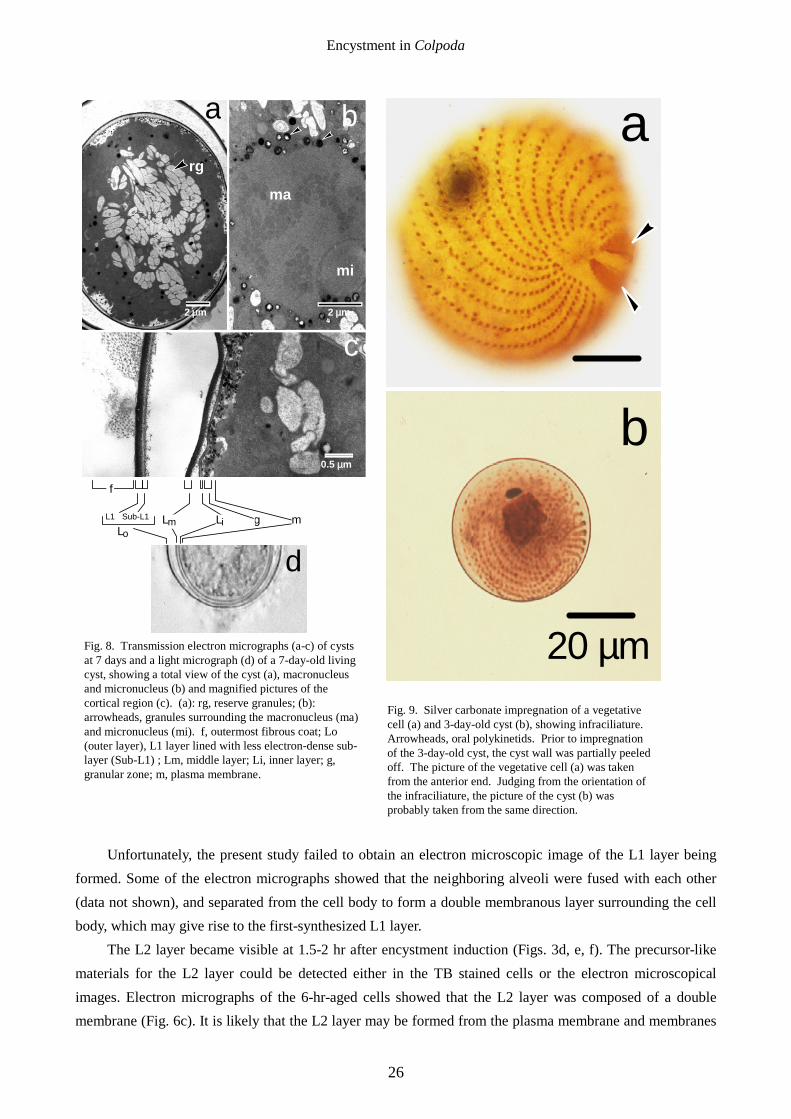
[3-6 hr after encystment induction] The L1 layer was seen (Fig. 6a), and numerous ribosome-bearing endoplasmic reticula occupied the
cytoplasm (Fig. 6b). The L2 layer seems to be a double membranous layer just covering the cytoplasm (Fig. 6c). Cilia were resorbed inside the L1 layer (Fig. 6c) but outside the L2 layer. As shown in Fig. 6d, the surface globules were constituted of a regularly arranged netlike structure. [24 hr after encystment induction]
The L1 layer had been lined with a less electron-dense sub-layer (Sub-L1 layer) to form a double layer (Fig. 7). Many vesicles were observed to fuse with the plasma membrane to open in the space between the L1 and L2 layers (Figs. 7a, b, arrowheads) and amorphous materials were observed in the interlayer space. The L2 layer became a more distinct double membranous layer separated from the cell surface (Fig. 7b).
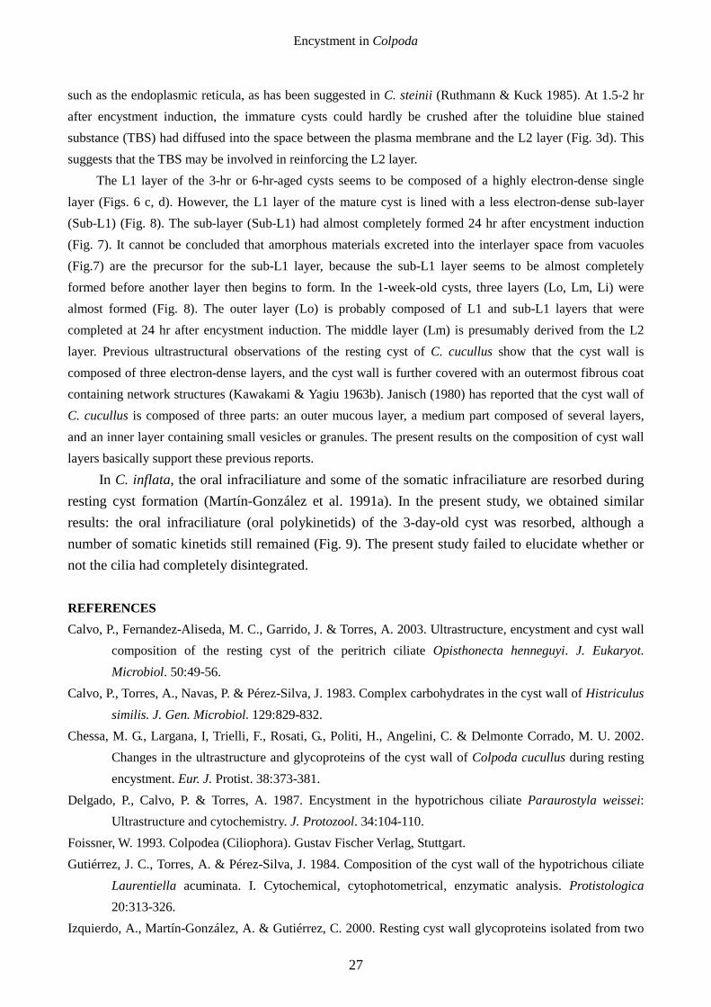
[7 days after encystment induction] Three electron-dense outer (Lo), middle (Lm) and inner layers (Li) were formed, and a number of
reserve grains (starch-like grains) accumulated in the cytoplasm (Figs. 8a, c). The macronucleus (Fig. 8b) was somewhat shrunk and was surrounded by a number of small electron-dense granules (Fig. 8b, arrowheads). Light microscopical observation (Fig. 8d) of a 7-day-old living cyst also revealed that the cyst wall was constituted of three distinct layers covered with an outermost fibrous coat, each of which corresponded to that observed in the ultrathin sections of the cyst. In the space between the inner layer (Li) and plasma membrane, a number of electron-dense granules were seen (Figs. 8a, c), and many of them seemed to be fused with the Li layer (Fig. 8a), implying that the granules might be precursors for the Li layer.
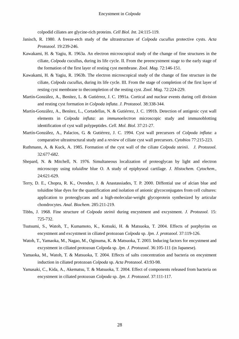
Silver impregnation In the electron-micrographs of 7-day-old cysts (Fig. 8), whether or not the cilia and
kinetosomes disappeared could not be concluded, because the intracellular structures of the mature cysts were not clear due to the electron-dense condensation of the cytoplasm. Silver impregnation revealed that oral polykinetids disappeared and the somatic infraciliature still remained in a 3-day-old cyst (Fig. 9). Unfortunately, we failed to obtain the silver-impregnated image of 1-week-old cysts because it was difficult to peel the cyst walls in this stage.
DISCUSSION It is known that toluidine blue (TB) dye forms complexes with polysaccharides such as
glycosaminoglycans (Shepard & Mitchell 1976; Terry et al. 2000). The fact that prior to formation of the cyst wall a number of TB stained granules or vesicles appear in the cytoplasm (Fig. 3b) suggests that glycoconjugates, which are probably the precursors for cyst wall components including the outermost fibrous coat, are being synthesized in this stage. The TB stained materials may be identical with the electron-dense granules shown in Fig. 5b. Such presumed precursor granules were not observed in vegetative cells (Fig. 3a,
Encystment in Colpoda
25
Fig. 4), implying that they are newly synthesized after encystment is induced. The intracellular events (excretion of electron-dense granules into the extracellular space through the large vacuoles) in the early stage (Fig. 5) are basically consistent with the previous reports (Kawakami and Yagiu 1963a). As already suggested by Kawakami & Yagiu (1963a), the electron-dense materials are possibly the precursor for the outermost fibrous coat.
5 µm
a
1 µm
d
ma
mi
b
0.5 µm
0.5 µm
cal
ci
Fig. 4. Transmission electron micrographs of a vegetative cell. (a), a longitudinal section of the vegetative cell showing a total view of the cell. (b), a magnified picture of the cytoplasm: arrows, different types of vesicles (granules). (c), a section showing the cortical region: al, alveolus; ci, cilia. (d), a section showing the macronucleus (ma) and micronucleus (mi).
5 µm
a b
1 µmg
v
Fig 5. Transmission electron micrographs of the cell at 1 hr after encystment induction. (a), a longitudinal section of the cell showing a total view of the cell. (b), a magnified image showing vacuoles (v) opening to theextracellular space (arrowhead) and a vesicle containing the electron-dense granules (g).
b
2 µm5 µm
a
0.2 µm
gl
L1
0.2 µm
dL1
ci
c
L2
Fig. 6. Transmission electron micrographs of the cell at 3 hr (a, b) and 6 hr (c, d) after encystment induction, showing a total view of the immature cyst (a), and magnified pictures of the cytoplasm (b) and cortical region (c, d). L1, first-synthesized layer of cyst wall; L2, secondary-synthesized layer; gl, globule contained in outermost fibrous coat; ci, cilia.
5 µm
a b
1 µm
L1
L2
Sub-L1
ci
Fig. 7. Transmission electron micrographs of the cell at 24 hr after encystment induction showing a total view of the immature cyst (a) and magnified picture of the cortical region (b). Arrowheads, vacuoles opening to interlayer space. L1, first-synthesized highly electron-dense layer; Sub-L1, less electron-dense sub-layer; ci, cilia; L2, L2 layer.
Encystment in Colpoda
26
Unfortunately, the present study failed to obtain an electron microscopic image of the L1 layer being formed. Some of the electron micrographs showed that the neighboring alveoli were fused with each other (data not shown), and separated from the cell body to form a double membranous layer surrounding the cell body, which may give rise to the first-synthesized L1 layer.
The L2 layer became visible at 1.5-2 hr after encystment induction (Figs. 3d, e, f). The precursor-like materials for the L2 layer could be detected either in the TB stained cells or the electron microscopical images. Electron micrographs of the 6-hr-aged cells showed that the L2 layer was composed of a double membrane (Fig. 6c). It is likely that the L2 layer may be formed from the plasma membrane and membranes
b
2 µm
ma
a
2 µm
rg
c
mi
0.5 µm
L1
f
dmgLiLm
Lo
Sub-L1
Fig. 8. Transmission electron micrographs (a-c) of cysts at 7 days and a light micrograph (d) of a 7-day-old living cyst, showing a total view of the cyst (a), macronucleus and micronucleus (b) and magnified pictures of the cortical region (c). (a): rg, reserve granules; (b): arrowheads, granules surrounding the macronucleus (ma) and micronucleus (mi). f, outermost fibrous coat; Lo (outer layer), L1 layer lined with less electron-dense sub-layer (Sub-L1) ; Lm, middle layer; Li, inner layer; g, granular zone; m, plasma membrane.
20 µm
b
a
Fig. 9. Silver carbonate impregnation of a vegetative cell (a) and 3-day-old cyst (b), showing infraciliature. Arrowheads, oral polykinetids. Prior to impregnation of the 3-day-old cyst, the cyst wall was partially peeled off. The picture of the vegetative cell (a) was taken from the anterior end. Judging from the orientation of the infraciliature, the picture of the cyst (b) was probably taken from the same direction.
Encystment in Colpoda
27
such as the endoplasmic reticula, as has been suggested in C. steinii (Ruthmann & Kuck 1985). At 1.5-2 hr after encystment induction, the immature cysts could hardly be crushed after the toluidine blue stained substance (TBS) had diffused into the space between the plasma membrane and the L2 layer (Fig. 3d). This suggests that the TBS may be involved in reinforcing the L2 layer.
The L1 layer of the 3-hr or 6-hr-aged cysts seems to be composed of a highly electron-dense single layer (Figs. 6 c, d). However, the L1 layer of the mature cyst is lined with a less electron-dense sub-layer (Sub-L1) (Fig. 8). The sub-layer (Sub-L1) had almost completely formed 24 hr after encystment induction (Fig. 7). It cannot be concluded that amorphous materials excreted into the interlayer space from vacuoles (Fig.7) are the precursor for the sub-L1 layer, because the sub-L1 layer seems to be almost completely formed before another layer then begins to form. In the 1-week-old cysts, three layers (Lo, Lm, Li) were almost formed (Fig. 8). The outer layer (Lo) is probably composed of L1 and sub-L1 layers that were completed at 24 hr after encystment induction. The middle layer (Lm) is presumably derived from the L2 layer. Previous ultrastructural observations of the resting cyst of C. cucullus show that the cyst wall is composed of three electron-dense layers, and the cyst wall is further covered with an outermost fibrous coat containing network structures (Kawakami & Yagiu 1963b). Janisch (1980) has reported that the cyst wall of C. cucullus is composed of three parts: an outer mucous layer, a medium part composed of several layers, and an inner layer containing small vesicles or granules. The present results on the composition of cyst wall layers basically support these previous reports.
In C. inflata, the oral infraciliature and some of the somatic infraciliature are resorbed during resting cyst formation (Martín-González et al. 1991a). In the present study, we obtained similar results: the oral infraciliature (oral polykinetids) of the 3-day-old cyst was resorbed, although a number of somatic kinetids still remained (Fig. 9). The present study failed to elucidate whether or not the cilia had completely disintegrated.
REFERENCES Calvo, P., Fernandez-Aliseda, M. C., Garrido, J. & Torres, A. 2003. Ultrastructure, encystment and cyst wall
composition of the resting cyst of the peritrich ciliate Opisthonecta henneguyi. J. Eukaryot. Microbiol. 50:49-56.
Calvo, P., Torres, A., Navas, P. & Pérez-Silva, J. 1983. Complex carbohydrates in the cyst wall of Histriculus similis. J. Gen. Microbiol. 129:829-832.
Chessa, M. G., Largana, I, Trielli, F., Rosati, G., Politi, H., Angelini, C. & Delmonte Corrado, M. U. 2002. Changes in the ultrastructure and glycoproteins of the cyst wall of Colpoda cucullus during resting encystment. Eur. J. Protist. 38:373-381.
Delgado, P., Calvo, P. & Torres, A. 1987. Encystment in the hypotrichous ciliate Paraurostyla weissei:Ultrastructure and cytochemistry. J. Protozool. 34:104-110.
Foissner, W. 1993. Colpodea (Ciliophora). Gustav Fischer Verlag, Stuttgart. Gutiérrez, J. C., Torres, A. & Pérez-Silva, J. 1984. Composition of the cyst wall of the hypotrichous ciliate
Laurentiella acuminata. I. Cytochemical, cytophotometrical, enzymatic analysis. Protistologica 20:313-326.
Izquierdo, A., Martín-González, A. & Gutiérrez, C. 2000. Resting cyst wall glycoproteins isolated from two
Encystment in Colpoda
28
colpodid ciliates are glycine-rich proteins. Cell Biol. Int. 24:115-119. Janisch, R. 1980. A freeze-etch study of the ultrastructure of Colpoda cucullus protective cysts. Acta
Protozool. 19:239-246. Kawakami, H. & Yagiu, R. 1963a. An electron microscopical study of the change of fine structures in the
ciliate, Colpoda cucullus, during its life cycle. II. From the preencystment stage to the early stage of the formation of the first layer of resting cyst membrane. Zool. Mag. 72:146-151.
Kawakami, H. & Yagiu, R. 1963b. The electron microscopical study of the change of fine structure in the ciliate, Colpoda cucullus, during its life cycle. III. From the stage of completion of the first layer of resting cyst membrane to thecompletion of the resting cyst. Zool. Mag. 72:224-229.
Martín-González, A., Benítez, L. & Gutiérrez, J. C. 1991a. Cortical and nuclear events during cell division and resting cyst formation in Colpoda inflata. J. Protozool. 38:338-344.
Martín-González, A., Benítez, L., Cortadellas, N. & Gutiérrez, J. C. 1991b. Detection of antigenic cyst wall elements in Colpoda inflata: an immunoelectron microscopic study and immunoblotting identification of cyst wall polypeptides. Cell. Mol. Biol. 37:21-27.
Martín-González, A., Palacios, G. & Gutiérrez, J. C. 1994. Cyst wall precursors of Colpoda inflata: acomparative ultrastructural study and a review of ciliate cyst wall precursors. Cytobios 77:215-223.
Ruthmann, A. & Kuck, A. 1985. Formation of the cyst wall of the ciliate Colpoda steinii. J. Protozool. 32:677-682.
Shepard, N. & Mitchell, N. 1976. Simultaneous localization of proteoglycan by light and electron microscopy using toluidine blue O. A study of epiphyseal cartilage. J. Histochem. Cytochem., 24:621-629.
Terry, D. E., Chopra, R. K., Ovenden, J. & Anastassiades, T. P. 2000. Diffential use of alcian blue and toluidine blue dyes for the quantification and isolation of anionic glycoconjugates from cell cultures: application to proteoglycans and a high-molecular-weight glycoprotein synthesized by articular chondrocytes. Anal. Biochem. 285:211-219.
Tibbs, J. 1968. Fine structure of Colpoda steinii during encystment and excystment. J. Protozool. 15: 725-732.
Tsutsumi, S., Watoh, T., Kumamoto, K., Kotsuki, H. & Matsuoka, T. 2004. Effects of porphyrins on encystment and excystment in ciliated protozoan Colpoda sp. Jpn. J. protozool. 37:119-126.
Watoh, T., Yamaoka, M., Nagao, M., Oginuma, K. & Matsuoka, T. 2003. Inducing factors for encystment and excystment in ciliated protozoan Colpoda sp. Jpn. J. Protozool. 36:105-111 (in Japanese).
Yamaoka, M., Watoh, T. & Matsuoka, T. 2004. Effects of salts concentration and bacteria on encystment induction in ciliated protozoan Colpoda sp. Acta Protozool. 43:93-98.
Yamasaki, C., Kida, A., Akematsu, T. & Matsuoka, T. 2004. Effect of components released from bacteria on encystment in ciliated protozoan Colpoda sp. Jpn. J. Protozool. 37:111-117.


![Морфологична типология [Morphological Typology]](https://img.dokumen.tips/doc/110x75/6349231a2cd4c1a3540d5c6a/morfologichna-tipologiya-morphological-typology.jpg)


