Embed Size (px)
Citation preview
plants
Review
Uncovering Research Trends of Phycobiliproteins UsingBibliometric Approach
Hui Teng Tan 1, Fatimah Md. Yusoff 2,3,* , Yam Sim Khaw 1, Siti Aqlima Ahmad 4 and Noor Azmi Shaharuddin 4
�����������������
Citation: Tan, H.T.; Yusoff, F.M.;
Khaw, Y.S.; Ahmad, S.A.;
Shaharuddin, N.A. Uncovering
Research Trends of Phycobiliproteins
Using Bibliometric Approach. Plants
2021, 10, 2358. https://doi.org/
10.3390/plants10112358
Academic Editors: Laura Arru,
Luca Forti and Moreno Bondi
Received: 11 August 2021
Accepted: 24 September 2021
Published: 1 Novemeber 2021
Publisher’s Note: MDPI stays neutral
with regard to jurisdictional claims in
published maps and institutional affil-
iations.
Copyright: © 2021 by the authors.
Licensee MDPI, Basel, Switzerland.
This article is an open access article
distributed under the terms and
conditions of the Creative Commons
Attribution (CC BY) license (https://
creativecommons.org/licenses/by/
4.0/).
1 Aquatic Animal Health and Therapeutics Laboratory, Institute of Bioscience, Universiti Putra Malaysia,Serdang 43400, Selangor, Malaysia; [email protected] (H.T.T.); [email protected] (Y.S.K.)
2 International Institute of Aquaculture and Aquatic Sciences, Universiti Putra Malaysia, PortDickson 71050, Negeri Sembilan, Malaysia
3 Department of Aquaculture, Faculty of Agriculture, Universiti Putra Malaysia,Serdang 43400, Selangor, Malaysia
4 Department of Biochemistry, Faculty of Biotechnology and Biomolecular Sciences, Universiti Putra Malaysia,Serdang 43400, Selangor, Malaysia; [email protected] (S.A.A.); [email protected] (N.A.S.)
* Correspondence: [email protected]
Abstract: Phycobiliproteins are gaining popularity as long-term, high-value natural products whichcan be alternatives to synthetic products. This study analyzed research trends of phycobiliproteinsfrom 1909 to 2020 using a bibliometric approach based on the Scopus database. The current findingsshowed that phycobiliprotein is a burgeoning field in terms of publications outputs with “biochem-istry, genetics, and molecular biology” as the most related and focused subject. The Journal of AppliedPhycology was the most productive journal in publishing articles on phycobiliproteins. Althoughthe United States of America (U.S.A.) contributed the most publications on phycobiliproteins, theChinese Academy of Sciences (China) is the institution with the largest number of publications. Themost productive author on phycobiliproteins was Glazer, Alexander N. (U.S.A.). The U.S.A. andGermany were at the forefront of international collaboration in this field. According to the keywordanalysis, the most explored theme was the optimization of microalgae culture parameters and phy-cobiliproteins extraction methods. The bioactivity properties and extraction of phycobiliproteinswere identified as future research priorities. Synechococcus and Arthrospira were the most cited genera.This study serves as an initial step in fortifying the phycobiliproteins market, which is expected toexponentially expand in the future. Moreover, further research and global collaboration are necessaryto commercialize phycobiliproteins and increase the consumer acceptability of the pigments andtheir products.
Keywords: microalgae; pigment; commercial; biotechnology; applications; bioproduction; Scopusdatabase; R studio; CiteSpace
1. Introduction
Phycobiliproteins are indispensable photosynthetic accessory pigments responsiblefor light-harvesting in blue-green algae, cyanelles, cryptomonads, and red algae [1]. Theyare water-soluble pigments made up of proteins and covalently bound phycobilins [2,3].Phycobiliproteins are primarily derived from blue-green algae and constitute up to 50% oftotal cellular proteins [4]. Phycobiliproteins are divided into three major types based ontheir absorption peak and structure: phycocyanin, allophycocyanin, and phycoerythrin [5].Phycocyanin is the major phycobiliprotein, followed by phycoerythrin and allophycocyaninthat were found in blue-green algae [5,6]. On the other hand, phycoerythrin is the dominantphycobiliprotein in most of the red algae, Rhodophyceae [7]. Besides, a few species ofcryptophytes contain phycobiliproteins, and each cryptophyte usually has only one type ofphycobiliproteins [8].
In recent decades, algae bioactive compounds or metabolites, including pigmentssuch as phycobiliproteins, have sparked increasing interest [1,9]. Phycobiliproteins have
Plants 2021, 10, 2358. https://doi.org/10.3390/plants10112358 https://www.mdpi.com/journal/plants
Plants 2021, 10, 2358 2 of 28
been widely utilized in various fields, owing to their biological and pharmacologicalcharacteristics [10–12]. For example, phycobiliproteins’ non-carcinogenic and non-toxicproperties have led to their usage in the feed, food, and cosmetics industries. Further-more, phycobiliproteins have been employed as a surrogate of synthetic colorants [13,14].Nowadays, phycocyanin is the primary market target. Its use has been expanded sinceits approval by the Food and Drug Administration (FDA) in the United States of Amer-ica (U.S.A.) (https://www.foodbusinessnews.net/articles/2782-f-d-a-approves-natural-source-as-blue-color-in-candy-gum (Assessed date: 30 May 2021)). According to FutureMarket Insights, the overall market of phycobiliproteins for 2018 is USD 112.3 million, andit is expected to double this number in 2028 [1]. In the aquaculture industry, phycocyaninhas been utilized as a feed supplement in shrimp, fish, or ornamental fish as it containshigh nutrients and enables to increase the skin color [15].
Alan Pritchard proposed bibliometrics in 1969, which has been utilized in severalsubsequent research throughout the world [16–19]. Nowadays, it has grown into an indis-pensable instrument to analyze the current trends in scientific publications. Furthermore, itcould serve as a possible guide for current and future research in certain fields or disciplines.VOSviewer, Gephi, CiteSpace, and Bibexcel are examples of frequently used bibliometrictools that construct keyword networks, complex social networks, knowledge mapping, andbibliometrics, respectively [19–23]. The ideal approach to measure and analyze worldwidescientific production and qualitative data may produce functional indices to recognize thepresent status and future potential within a specific research field or area [24].
Recently, bibliometric evaluations on microalgae research, microalgae biofuel, andeven microalgae-derived pigments have been published [9,17,25]. Interestingly, althoughthese publications addressed algae or metabolites produced from algae, there is no exhaus-tive bibliometric evaluation on phycobiliproteins [9,25]. Hence, this study aimed to providea comprehensive worldwide research status and emerging trends of phycobiliproteinsstudy via analyzing the global publishing landscape on phycobiliproteins research from1909 to 2020. Herein, the primary information and the specific performance of publicationsrelated to phycobiliproteins research were retrieved from the Scopus database and analyzedthoroughly. Furthermore, keyword analysis over different periods was performed to high-light the directions and trends of phycobiliproteins research, thus raising the awareness ofpotential gaps in scientific collaboration.
2. Bibliometric Analysis2.1. Methodology
The methods applied in this study were based on the work of Qi et al. [19] andLim et al. [20], with modifications. Structured literature reviews were completed in threestages: identifying appropriate search terms, retrieving the content, and performing theanalysis [26]. In this review, this three-step process was used for collecting data anddetailed assessment.
2.1.1. Data Collection
The information of scientific publications was collected from the Elsevier Scopusdatabase (retrieved on 12 April 2021). A comprehensive search was performed using“phycobiliprotein* or phycocyanin* or phycoerythrin* or allophycocyanin*” as the searchquery of (TITLE-ABS-KEY) from 1909 to 2020 to compile a bibliography of all publicationswhose topics are related to phycobiliprotein*, phycocyanin*, phycoerythrin*, and allophy-cocyanin*. The use of an asterisk at the end of a word ensures that any terms with the sameroot are included in the search result. Data from 2021 were not included in this analysis fordata consistency. Due to the limited number of documents that can be retrieved from theScopus database per single search (maximum: 2000 documents), the data were collectedover shorter periods (i.e., ten years) using the same search query. These retrieved data from1909 to 2020 were combined prior to the bibliometric analysis. This search yielded a total of

Plants 2021, 10, 2358 3 of 28
8296 global publications; it should be noted that the results might vary if different searchparameters are used.
2.1.2. Refinement of the Search Results
Refinement of search results was conducted to exclude irrelevant articles and enhancethe accuracy of bibliometric analysis for reliable review [18]. Hence, each publication fromthe search results was properly filtered by manual inspection while those publicationsunrelated to the phycobiliproteins topic were eliminated.
2.1.3. Bibliometric Analysis
The obtained data were analyzed by using the open-source RStudio software(www.rstudio.com), version 4.0.5 (accessed on 30 April 2021), along with the bibliometrixR-package [27]. The information related to journal impact factors (IFs) and the h-index ofeach journal were obtained from the Journal Citation Reports (JCR) in 2020. The number oftotal citations was extracted from Scopus. VOSviewer (Leiden University, Netherlands), asoftware created by van Eck and Waltman, was used to construct and visualize a knowl-edge map of the co-occurrence network [28]. The keyword co-occurrence was performedto find the most often used keywords that related to phycobiliproteins. The study pro-vided three options, namely “author keywords”, “index keywords”, and “all keywords”,with “all keywords” being selected; then, “full counting” was applied as the countingmethod. A minimum number of keyword occurrences was set to 20, and a manual in-spection was carried out to remove unrelated keywords [9]. CiteSpace.5.7.R5W was alsoapplied to analyze co-occurring keywords with different visualizations [29]. The researchhotspots and frontiers in phycobiliproteins were assessed using this software by the burstdetection analysis.
2.2. General Characteristics of Research Publications
A total of 8296 documents from 2214 different sources and 22,871 authors were listedduring 1909–2020 (Table 1). The authors of multi-authored documents were 22,540, whilethe authors of single-authored documents were 331. The average years from publicationson phycobiliproteins is around 16.7 per year, with each publication receiving an average of30.88 citations and 1.85 citations per year. The annual growth rate of scientific productionis 9.00%.
Table 1. Main information of the retrieved phycobiliproteins data.
Description Results
Period 1909–2020Sources (journals, books, etc.) 2214Documents 8296Average years from publication 16.7Average citations per documents 30.88Average citations per year per doc 1.85Annual growth rate of scientific production 9%Authors 22,871Authors of single-authored documents 331Authors of multi-authored documents 22,540
2.2.1. Publication Types and Languages
A total of 8296 publications were classified into 12 document types. The majority ofthe publications were identified as articles, forming 89.49% (7424 papers) of the total docu-ments (Figure 1). Review publications accounted for 3.88% (322 documents), followed byconference papers and book chapters, with each accounting for 3.36% (279 documents) and1.13% (94 documents), respectively. Other documents demonstrated a lower percentage ofoccurrence among the publications type. These included letters, with 0.80% (66 documents),notes, with 0.39% (32 documents), short surveys, with 0.36% (30 documents), erratum,
Plants 2021, 10, 2358 4 of 28
with 0.19% (16 documents), editorials, with 0.14% (12 documents), conference reviews,with 0.13% (11 documents), books, with 0.10% (8 documents), and reports, with 0.02%(2 documents).
Figure 1. Distribution of phycobiliprotein publication types.
The majority of the publications were in English (95.53%), while 2.24% of the publica-tions were printed in Chinese (Figure 2). Publications in other languages accounted for2.23%, consisting of Russian (0.74%), German (0.40%), Korean (0.28%), Spanish (0.27%),French (0.19%), Japanese (0.18), Italian (0.10%), and Polish (0.08%).
Figure 2. Distribution of languages used in phycobiliproteins publications.

Plants 2021, 10, 2358 5 of 28
2.2.2. Subject Categories of Publications
A total of 8296 publications from 1909 to 2020 covered 28 subject areas. Biochemistry,genetics, and molecular biology ranked the top, with 26.00%, among the other subjectareas (Table 2). This was followed by agricultural and biological sciences, with 14.60%,medicine, with 12.60%, immunology and microbiology, with 9.10%, chemistry, with 7.00%,environmental science, with 5.90%, and chemical engineering, with 4.00%. Engineering,pharmacology, toxicology and pharmaceutics, and physics and astronomy accounted for3.32%, 3.30%, and 2.80% among the other subject areas, respectively.
Table 2. The top 10 prolific subject categories in the study of phycobiliprotein from 1909–2020.
Subject Area Percentage (%) Rank
Biochemistry, Genetics, andMolecular Biology 26.00 1
Agricultural and BiologicalSciences 14.60 2
Medicine 12.60 3Immunology andMicrobiology 9.10 4
Chemistry 7.00 5Environmental Science 5.90 6Chemical Engineering 4.00 7Engineering 3.32 8Pharmacology, Toxicology,and Pharmaceutics 3.30 9
Physics and Astronomy 2.80 10
2.2.3. Sources of Publications
Phycobiliproteins publications were distributed in 2214 different sources. The top10 sources published 1083 articles, accounting for 13.07% of all publications (Table 3).Journal of Applied Phycology (publisher: Springer) ranked the top with 184 publications(2.22%), 4122 total citations, and an impact factor of 3.215. This was followed by the Journalof Biological Chemistry, which owned 137 publications (1.65%) with a 5.157 impact factorand the highest total citation (6672) among these top ten sources. Journal of Phycologywas rated third with 127 publications (1.53%) and an impact factor of 2.923. Proceedingsof the National Academy of Sciences of the U.S.A. possessed the highest impact factor(11.205) and h-index (699). Furthermore, 70.00% of the ten most prolific sources performedrelatively well on the impact factor, ranging from 3 to 10, while the h-index value of all topten sources was more than 90, except Cytometry. Based on the journal impact factor (JIF)ranking, all these top ten journals were above rank Q3, with 50% of the journals classifiedas rank Q1. Besides, Springer and Wiley were the most prolific publishers among the topten most productive sources.
The research output of the top five journals displayed a growing fluctuation trendfrom 1965 to 2020 (Figure 3). Among the five journals, the Journal of Phycology was thejournal that first published a document related to phycobiliprotein in 1965. The number ofpublications of the Cytometry has outperformed those in the other journals from 1996 to2002, but this journal was discontinued by 2002 and published as Cytometry Part A andCytometry Part B from 2003 onwards. The number of publications of Photochemistry andPhotobiology dropped after the peak in 1985 and 1986, followed by a slower-growing trendtowards 2020. The Journal of Applied Phycology was the fastest-growing journal between2015 and 2020, despite a minor decline in 2017. The other four journals displayed a slightlyslower trend in phycobiliproteins research when approaching 2020.

Plants 2021, 10, 2358 6 of 28
Table 3. The top 10 productive sources in the study of phycobiliprotein from 1909–2020.
Sources TP TP (%) Cumulative(%) TC IF 2020 Rank
by JIFh-
Index Publisher
Journal of Applied Phycology 184 2.22 2.22 4122 3.215 Q1 93 Springer
Journal of Biological Chemistry 137 1.65 3.87 6672 5.157 Q1 477American Society for
Biochemistry andMolecular Biology
Journal of Phycology 127 1.53 5.40 5584 2.923 Q1 114 WileyCytometry 121 1.46 6.86 5396 2.698 n/a 61 WileyPhotochemistry and Photobiology 96 1.16 8.02 1979 3.421 Q3 120 WileyPhotosynthesis Research 95 1.15 9.17 2173 3.573 Q1 99 Springer
Proceedings of the National Academy ofSciences of the United States of America 83 1.00 10.17 5923 11.205 Q1 699
United StatesNational Academy of
Sciences (U.S.A.)Biochimica et BiophysicaActa—Bioenergetics 82 0.99 11.16 2556 3.991 Q2 154 Elsevier
Archives of Microbiology 81 0.98 12.14 3654 2.552 Q3 94 Springer
Biochemistry 77 0.93 13.07 3144 3.162 Q3 253 American ChemicalSociety
TP: total publications; TC: total citations; IF: impact factor; JIF: journal impact factor; n/a: not available.
Figure 3. The trend of major five sources on phycobiliproteins publications from 1965 to 2020.
2.3. Specific Performance of Research Publications2.3.1. Annual Publication Trend
The first publication was in 1909, according to the Scopus database (Figure 4). Thenumber of publications per year remained below ten before 1964. The number of publi-cations reached 182 in 1993, and the total publications were 1775 documents. Althoughthere was a fluctuation in the trend of publications after 1977, a slightly growing trendwas observed until 2015. There were 328 publications produced in 2015, followed by aslight drop in 2016 (287 publications). At this time, a total of 6505 total publications wereaccumulated. After that, a conspicuous increase was identified from 2017 to 2020, with apeak of 590 publications in 2020.

Plants 2021, 10, 2358 7 of 28
Figure 4. The annual and cumulative numbers of research publications on phycobiliproteins from 1909 to 2020.
2.3.2. Analysis of Growth Trend
Between 1909 and 1960, the total number of publications was less than 100, the totalnumber of authors and references was less than 100, and the total number of citations(until 2020) was less than 1000 (Table 4). However, the total number of publications, theaverage number of authors, and references per publication have increased gradually inthe following years. From 1961 to 1970, the average number of authors per publicationdecreased from 1.39 to 1.21. Then, the number slightly increased between 1971 and 1980,and before reaching 3.99 between 2011 and 2020. The number of citations (until 2020) andthe average number of citations per publication declined from 81,576 to 57,738, and 42.73to 16.45, respectively, between 2011 and 2020.
Table 4. The characteristics of periodical publications of phycobiliprotein from 1909–2020.
PY TP TA TA/TP TR TR/TP TC TC/TP
≤1960 33 46 1.39 11 0.03 841 25.481961–1970 133 161 1.21 413 3.11 3736 28.091971–1980 337 422 1.25 5691 16.89 11,462 34.011981–1990 821 1547 1.88 15,380 18.73 30,248 36.841991–2000 1553 4175 2.69 35,553 22.89 70,619 45.472001–2010 1909 6410 3.36 58,387 30.59 81,576 42.732011–2020 3510 14,000 3.99 158,176 45.06 57,738 16.45
PY: year of publication; TP: total publications; TA: total authors; TA/TP: authors per publication; TR: total references; TR/TP: averagereferences per publication; TC: total citations (until 2020); TC/TP: average citations per publication.
2.3.3. Highly Cited Publications
Out of 8296 publications, 7020 publications received less than 50 citations, while811 publications received 51–100 citations. There were 446 publications with citationsranging from 101 to 500, and 13 publications with citations ranging from 501 to 1000.A total of six publications received between 1001 and 2000 citations, while only fourpublications received more than 2000 citations. The article published by Huang et al. [30]in the Journal of Agricultural and Food Chemistry garnered the most citations (4010),in which the authors reviewed the chemical principles of antioxidant capacity assays
Plants 2021, 10, 2358 8 of 28
(Table 5). Spolaore et al. [31] discussed the applications of microalgae and published in theJournal of Bioscience and Bioengineering. This article was ranked second with 2574 totalcitations. Cao et al. [32] discussed the antioxidant and prooxidant behavior of flavonoidsand published it in Free Radical Biology and Medicine. This article was rated third in thetop ten cited publications. Ou et al. [33] developed an improved method of oxygen radicalabsorbance capacity (ORAC) assay, which the article was cited for 2024. Jones et al. [34]explored the new approach to protein fold recognition in their article, which was rankedlast in the top ten cited publications, with 1035 total citations. The journals with themost cited publications (Table 5) were not found in the top ten productive sources ofphycobiliproteins publication.
Table 5. The top 10 most cited publications related to phycobiliproteins from 1909–2020.
Title of Publications Journal TC PD References
The Chemistry behind Antioxidant Capacity Assays Journal of Agricultural andFood Chemistry 4010 Feb 2005 [30]
Commercial Applications of Microalgae Journal of Bioscienceand Bioengineering 2574 Oct 2006 [31]
Antioxidant and Prooxidant Behavior of Flavonoids:Structure-Activity Relationships
Free Radical Biologyand Medicine 2038 June 1997 [32]
Development and Validation of an Improved OxygenRadical Absorbance Capacity Assay Using Fluorescein asthe Fluorescent Probe
Journal of Agricultural andFood Chemistry 2024 Jan 2001 [35]
HLA-E Binds to Natural Killer Cell ReceptorsCD94/NKG2A, B and C Nature 1592 Feb 1998 [36]
Determination of Lymphocyte Division byFlow Cytometry
Journal ofImmunological Method 1439 Feb 1994 [37]
Resolution and Characterization of Pro-B and Pre-Pro-BCell Stages in Normal MouseBone Marrow
Journal ofExperimental Medicine 1317 May 1991 [38]
Accessing Genetic Information with High-DensityDNA Arrays Science 1309 Oct 1996 [39]
Oxygen-Radical Absorbance Capacity Assayfor Antioxidants
Free Radical Biologyand Medicine 1256 Jan 1993 [40]
A New Approach to Protein Fold Recognition Nature 1035 July 1992 [34]
TC: total citation; PD: publication date.
2.3.4. Performance of Publications by Countries
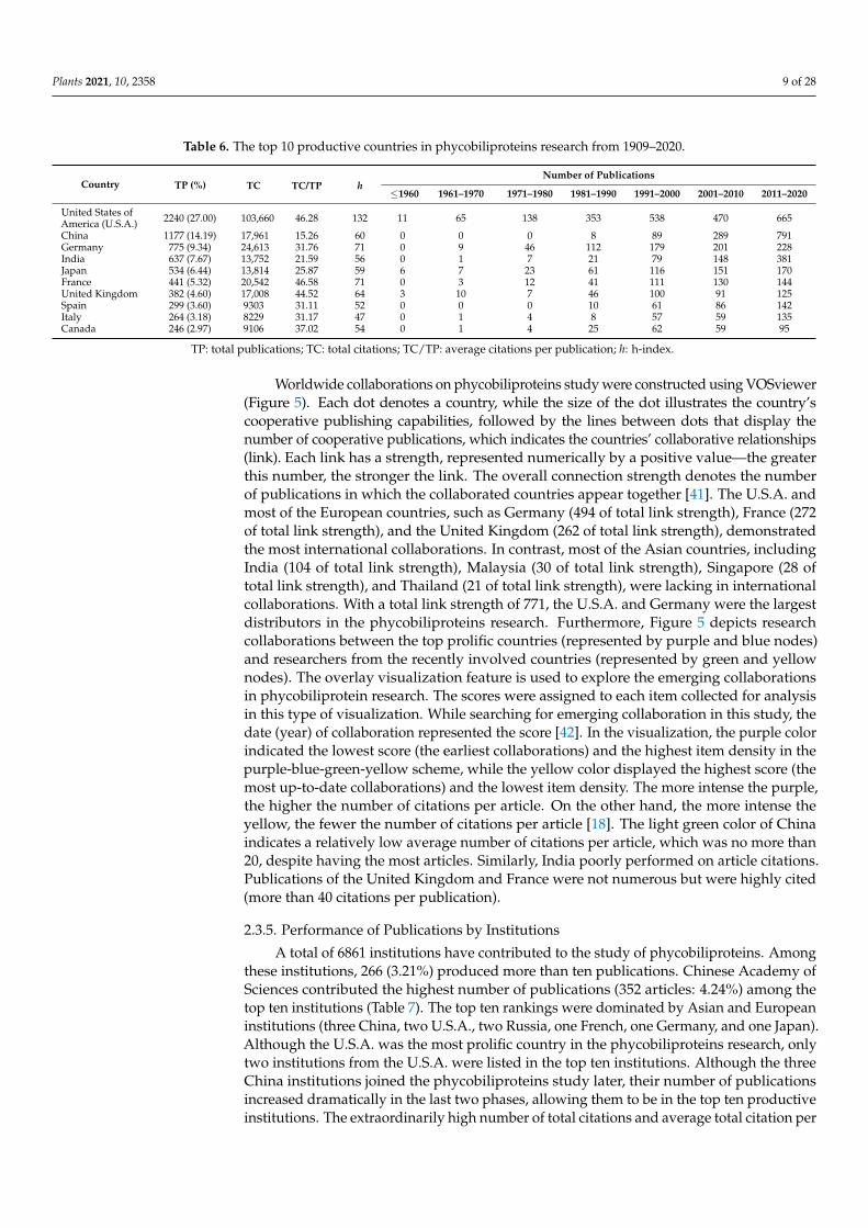
The U.S.A. led the total of publications in the study of phycobiliproteins, with 27.00%of publications output (Table 6). Each of the top prolific countries published at least200 publications. Before 1960, the U.S.A., Japan, and the United Kingdom contributedeleven, six, and three publications, respectively; this number progressively increased overthe following six stages with a slight drop between 2001 and 2010. The U.S.A. contributed538 and 470 publications during the fifth (1991–2000) and sixth period (2001–2010), makingit the most productive country. The number of publications produced by China increasedprofoundly from eight publications in the fourth period (1981–1990) to 791 publicationsin the last period (2011–2020), surpassing the U.S.A. Spain joined the phycobiliproteinfield later than the other countries. However, Spain contributed more publications inthe last stage compared to the United Kingdom, Italy, and Canada. Overall, the numberof publications of the top ten productive countries increased at different rates over theseven periods.
Plants 2021, 10, 2358 9 of 28
Table 6. The top 10 productive countries in phycobiliproteins research from 1909–2020.
Country TP (%) TC TC/TP hNumber of Publications
≤1960 1961–1970 1971–1980 1981–1990 1991–2000 2001–2010 2011–2020
United States ofAmerica (U.S.A.) 2240 (27.00) 103,660 46.28 132 11 65 138 353 538 470 665
China 1177 (14.19) 17,961 15.26 60 0 0 0 8 89 289 791Germany 775 (9.34) 24,613 31.76 71 0 9 46 112 179 201 228India 637 (7.67) 13,752 21.59 56 0 1 7 21 79 148 381Japan 534 (6.44) 13,814 25.87 59 6 7 23 61 116 151 170France 441 (5.32) 20,542 46.58 71 0 3 12 41 111 130 144United Kingdom 382 (4.60) 17,008 44.52 64 3 10 7 46 100 91 125Spain 299 (3.60) 9303 31.11 52 0 0 0 10 61 86 142Italy 264 (3.18) 8229 31.17 47 0 1 4 8 57 59 135Canada 246 (2.97) 9106 37.02 54 0 1 4 25 62 59 95
TP: total publications; TC: total citations; TC/TP: average citations per publication; h: h-index.
Worldwide collaborations on phycobiliproteins study were constructed using VOSviewer(Figure 5). Each dot denotes a country, while the size of the dot illustrates the country’scooperative publishing capabilities, followed by the lines between dots that display thenumber of cooperative publications, which indicates the countries’ collaborative relationships(link). Each link has a strength, represented numerically by a positive value—the greaterthis number, the stronger the link. The overall connection strength denotes the numberof publications in which the collaborated countries appear together [41]. The U.S.A. andmost of the European countries, such as Germany (494 of total link strength), France (272of total link strength), and the United Kingdom (262 of total link strength), demonstratedthe most international collaborations. In contrast, most of the Asian countries, includingIndia (104 of total link strength), Malaysia (30 of total link strength), Singapore (28 oftotal link strength), and Thailand (21 of total link strength), were lacking in internationalcollaborations. With a total link strength of 771, the U.S.A. and Germany were the largestdistributors in the phycobiliproteins research. Furthermore, Figure 5 depicts researchcollaborations between the top prolific countries (represented by purple and blue nodes)and researchers from the recently involved countries (represented by green and yellownodes). The overlay visualization feature is used to explore the emerging collaborationsin phycobiliprotein research. The scores were assigned to each item collected for analysisin this type of visualization. While searching for emerging collaboration in this study, thedate (year) of collaboration represented the score [42]. In the visualization, the purple colorindicated the lowest score (the earliest collaborations) and the highest item density in thepurple-blue-green-yellow scheme, while the yellow color displayed the highest score (themost up-to-date collaborations) and the lowest item density. The more intense the purple,the higher the number of citations per article. On the other hand, the more intense theyellow, the fewer the number of citations per article [18]. The light green color of Chinaindicates a relatively low average number of citations per article, which was no more than20, despite having the most articles. Similarly, India poorly performed on article citations.Publications of the United Kingdom and France were not numerous but were highly cited(more than 40 citations per publication).
2.3.5. Performance of Publications by Institutions
A total of 6861 institutions have contributed to the study of phycobiliproteins. Amongthese institutions, 266 (3.21%) produced more than ten publications. Chinese Academy ofSciences contributed the highest number of publications (352 articles: 4.24%) among thetop ten institutions (Table 7). The top ten rankings were dominated by Asian and Europeaninstitutions (three China, two U.S.A., two Russia, one French, one Germany, and one Japan).Although the U.S.A. was the most prolific country in the phycobiliproteins research, onlytwo institutions from the U.S.A. were listed in the top ten institutions. Although the threeChina institutions joined the phycobiliproteins study later, their number of publicationsincreased dramatically in the last two phases, allowing them to be in the top ten productiveinstitutions. The extraordinarily high number of total citations and average total citation per
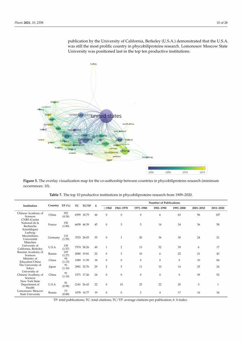
Plants 2021, 10, 2358 10 of 28
publication by the University of California, Berkeley (U.S.A.) demonstrated that the U.S.A.was still the most prolific country in phycobiliproteins research. Lomonosov Moscow StateUniversity was positioned last in the top ten productive institutions.
Figure 5. The overlay visualization map for the co-authorship between countries in phycobiliproteins research (minimumoccurrences: 10).
Table 7. The top 10 productive institutions in phycobiliproteins research from 1909–2020.
Institution Country TP (%) TC TC/TP hNumber of Publications
≤1960 1961–1970 1971–1980 1981–1990 1991–2000 2001–2010 2011–2020
Chinese Academy ofSciences China 352
(4.24) 6599 18.75 44 0 0 0 6 63 96 187
CNRS (CentreNational de la
RechercheScientifique)
France 150(1.80) 6658 44.39 45 0 3 5 14 34 36 58
Ludwig-Maximilians-UniversitätMünchen
Germany 132(1.59) 3703 28.05 35 0 1 20 36 30 24 21
University ofCalifornia, Berkeley U.S.A. 130
(1.57) 7574 58.26 49 1 2 13 52 39 6 17
Russian Academy ofSciences Russia 105
(1.27) 2080 19.81 22 0 3 10 6 22 21 43
Ministry ofEducation China China 94
(1.13) 1089 11.59 18 0 0 0 0 0 10 84
The University ofTokyo Japan 91
(1.10) 2981 32.76 29 2 5 11 10 14 25 24
University ofChinese Academy of
SciencesChina 91
(1.10) 1571 17.26 24 0 0 0 0 0 39 52
New York StateDepartment of
HealthU.S.A. 81
(0.98) 2141 26.43 22 0 10 25 22 20 3 1
Lomonosov MoscowState University Russia 73
(0.88) 1078 14.77 19 0 0 2 4 17 16 34
TP: total publications; TC: total citations; TC/TP: average citations per publication; h: h-index.
Plants 2021, 10, 2358 11 of 28
2.3.6. Performance of Publications by Authors
From 1909 to 2020, 22,871 authors contributed to the research on phycobiliproteins.The top 10 prolific authors were affiliated with the following countries: the U.S.A. (fourauthors), China (two authors), Germany (one author), Israel (one author), India (oneauthor), and Russia (one author). Glazer, Alexander N. from the University of California inthe U.S.A. led the top ten productive authors with 118 publications (1.42%; h-index: 65)since 1971 (Table 8). He had the most publications (57) during the period 1981–1990. Scheer,Hugo (Germany) came in second with 89 publications (1.07%; h-index: 49), while Bryant,Donald A. (U.S.A.) came in the third with 64 publications (0.77%; h-index: 72). Berns,Donald S. from the Israel Ministry of Health, Israel, was placed fifth with 59 publications(0.71%). MacColl, Robert from the Wadsworth Center for Laboratories and Research, U.S.A.,was the seventh among the top ten prolific authors with 52 publications (0.63%; h-index: 25).Madamwar, Datta B. from India and Bekasova, Olga D. from Russia were ranked as eighthand ninth productive authors with 45 (0.54%) and 40 (0.48%) publications, respectively.Two authors from China, Qin, Song and Zhao, Kaihong, were ranked as fourth and sixthamong the top ten authors with 62 (0.75%) and 55 (0.66%) publications, respectively.
2.4. Main Research Hotspot and Trends2.4.1. Keywords Analysis
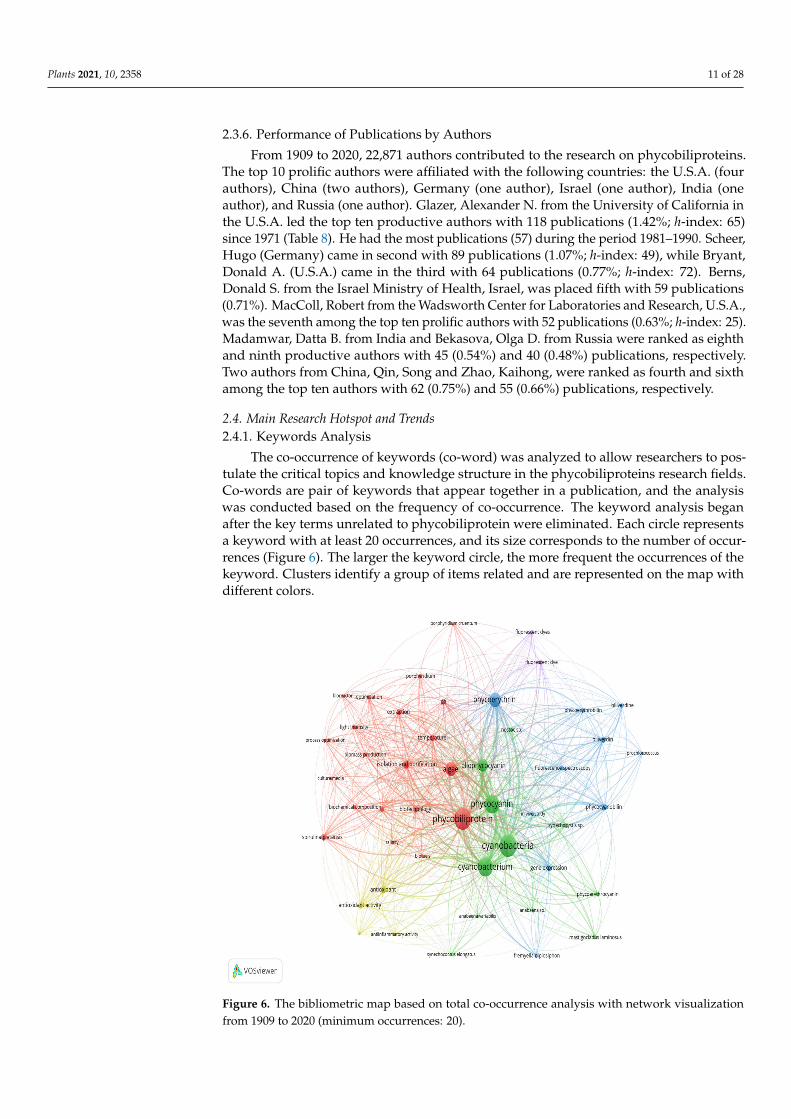
The co-occurrence of keywords (co-word) was analyzed to allow researchers to pos-tulate the critical topics and knowledge structure in the phycobiliproteins research fields.Co-words are pair of keywords that appear together in a publication, and the analysiswas conducted based on the frequency of co-occurrence. The keyword analysis beganafter the key terms unrelated to phycobiliprotein were eliminated. Each circle representsa keyword with at least 20 occurrences, and its size corresponds to the number of occur-rences (Figure 6). The larger the keyword circle, the more frequent the occurrences of thekeyword. Clusters identify a group of items related and are represented on the map withdifferent colors.
Figure 6. The bibliometric map based on total co-occurrence analysis with network visualizationfrom 1909 to 2020 (minimum occurrences: 20).

Plants 2021, 10, 2358 12 of 28
Table 8. The top 10 productive authors in phycobiliproteins research from 1909–2020.
Author Country Institutions TP (%) h EmailNumber of Publications
≤1960 1961–1970 1971–1980 1981–1990 1991–2000 2001–2010 2011–2020
Glazer,Alexander N. U.S.A. University of California 118 (1.42) 65 [email protected] 0 0 28 57 28 5 0
Scheer, Hugo GermanyLudwig-Maximilians-
UniversitätMünchen
89 (1.07) 49 [email protected] 0 0 6 23 26 16 18
Bryant,Donald A. U.S.A. Pennsylvania State
University 64 (0.77) 72 [email protected] 0 0 5 16 14 12 17
Qin, Song China
Yantai Institute ofCoastal Zone Research,Chinese Academy of
Sciences,
62 (0.75) 35 [email protected] 0 0 0 0 1 25 36
Berns,Donald S. Israel Israel Ministry of
Health 59 (0.71) 24 [email protected] 0 14 28 13 4 0 0
Zhao,Kaihong China Huazhong Agricultural
University 55 (0.66) 26 [email protected] 0 0 0 0 5 20 30
MacColl,Robert U.S.A.
Wadsworth Center forLaboratories and
Research52 (0.63) 25 [email protected] 0 0 14 15 19 3 1
Madamwar,Datta B. India Sardar Patel University 45 (0.54) 49 [email protected] 0 0 0 0 1 8 36
Bekasova,Olga D. Russia Bach Institute of
Biochemistry, 40 (0.48) 7 [email protected] 0 3 14 8 1 6 6
Gantt,Elisabeth U.S.A. University of Maryland 40 (0.48) 43 [email protected] 0 2 19 15 3 0 1
TP: total publications; h: h-index.

Plants 2021, 10, 2358 13 of 28
Although 879 terms met the predefined criteria in the phycobiliprotein research, only52 keywords were deemed relevant and analyzed in the keyword co-occurrence network.The results showed that the co-words were mapped into five major clusters, implying thatfive different research themes could be derived (Table 9).
Table 9. The clusters and themes derived from keywork co-occurrence network in the phycobiliproteins research.
Clusters Co-Words Themes
Cluster I (21 items)
Algae, Arthrospira platensis, biomass production, bioreactor,biotechnology, culture media, extraction, isolation andpurification, light intensity, light quality, optimization,oscillatoria, pH, photobioreactor, phycobiliprotein,Porphyridium, Porphyridium cruentum, process optimization,salinity, spirulina platensis and temperature
Optimization of cyanobacteria cultivationand phycobiliprotein harvesting process
Cluster II (12 items)
Allophycocyanin, Anabaena sp., Anabaena variabilis,cyanobacteria, cyanobacterium, Fremyella diplosiphon,Mastigocladus laminosu, nostoc sp., phycocyanin,phycoerythrocyanin, Synechococcus elongatus andSynechocystis sp.
Classification of phycobiliproteins fromdifferent cyanobacteria
Cluster III (11 items)Biliverdin, biliverdine, biosynthesis, ferredoxin, fremyelladiplosiphon, gene expression, phycobilins, phycocyanobilin,phycoerythrin, phycoerythrobilin and prochlorococcus
Gene expression of the phycobiliproteinbiosynthesis pathway
Cluster IV (4 items) Anti-inflammatory activity, antineoplastic agent,antioxidant, antioxidant activity Bioactivities of phycobiliprotein
Cluster V (4 items) Biofuels, fluorescent dye, fluorescent dyes, fluorescentspectroscopy Applications of phycobiliprotein
2.4.2. Analysis of Research Trend–Burst Detection Analysis
An apparent citation burst strength of the keywords in phycobiliproteins researchwas displayed from 1921 to 2020 (Figure 7). The citation burst began with biliproteinand cyanobacterium in 1971 with a burst strength of 12.62 and 10.04, respectively. Thiswas followed by energy transfer, which first appeared in 1976 with a burst strength of24.46. Cyanobacterium had the most extended burst, in which this keyword was citedfor 35 years. Flow cytometry had the greatest burst strength (45.15). The citation burstsof biliprotein, energy transfer, phycobilism, and immunofluorescence lasted for 25 years.The citation burst of each following keyword: photosynthesis, allophycocyanin, chromaticadaptation, phycobilisome, photosystem II, Mastigocladus laminosus, flow cytometry, pe(phycoerythrin), monoclonal antibody, phycoerythrin, and Microcystis aeruginosa were citedat least for 20 years. Based on the burst detection analysis, microalgae, antioxidant activity,Arthrospira platensis, and extraction emerged as new research areas in recent years.
Figure 7. The top 20 keywords with the strongest citation burst.

Plants 2021, 10, 2358 14 of 28
2.4.3. Analysis of Research Trend: Algae Genera
Thirty-eight algal genera appeared in the titles, abstracts, or keywords of the 8296 articles.Blue-green algae were accounted for 80% of the top ten listed genera, whereas red al-gae were responsible for the remaining 20% (Figure 8). Arthrospira genus appeared in553 articles, which was ranked first in the top ten genera. Synechococcus (488), Synechocystis(482), Nostoc (357), Microcystis (316), and Anabaena (212) were the species that were presentin at least 200 articles. Phormidium (131) and Gracillaria (143) were ranked underside inthis top ten genera, with approximately four times fewer appearances in publicationsthan Arthrospira.
Figure 8. The top ten genera of algae related to the phycobiliprotein study.
3. Overview of Previous Phycobiliprotein Research
To date, several researchers have published critical summaries and valuable insightson different aspects of phycobiliproteins study [1,5,43]. For example, Manirafasha et al. [5]outlined the properties of phycobiliproteins, the mechanism regulating phycobiliproteinsbiosynthesis, and the enhancement of phycobiliproteins production. Liu et al. [44] dis-cussed medical applications of phycocyanin extracted from Spirulina platensis. In addition,Li et al. [45] provided a critical review on the molecular structure, production, and ap-plications of phycobiliproteins. Ming et al. [43] reviewed the methods for enhancingphycocyanin and phycoerythrin production yield and chemical stability. Moreover, Kud-dus et al. [46] outlined the C-phycocyanin production and biotechnological applications.Sui [47] summarized the structure of phycobilisomes, while Silva et al. [48] reviewed themechanisms of action and multidrug resistance of phycocyanin in cancer. However, thereis still a paucity of information on the present, current, and future research trends of phyco-biliproteins. Following the rapid growth of scientific research, bibliometric analysis is nowfrequently applied to analyze the research dynamics in several fields. This type of studyfocuses on the quantitative assessment of publications as well as the related bibliographiccitations and proxies [49]. Garrido et al. [25] performed bibliometric analysis to analyze andprovide a global overview of microalgae research. Furthermore, Samara et al. [9] conducted

Plants 2021, 10, 2358 15 of 28
a ten-year bibliometric analysis on microalgae-derived pigments. Phycobiliproteins arewell-known natural pigments with several potential benefits and applications (Figure 9).These pigments have appeared as an emerging topic in various fields [3,5,50]. Therefore, abibliometric analysis is necessary to comprehend the current trend, identify the challenges,and discover future research opportunities.
Figure 9. The benefits and applications of phycobiliproteins in different industries (adapted from [1,50–53]).
3.1. General Characteristics of Research Publications in Phycobiliproteins Study
According to Scopus database, the first study on phycobiliproteins began in 1909 [54].According to Figure 1, phycobiliproteins research has been disseminated in 12 differenttypes of publications. This demonstrates that phycobiliproteins research embraces a varietyof thematics [9]. The highest percentage of documents published are journal articles.This revealed that numerous researchers contributed novel ideas to explore and studyphycobiliproteins in depth. Review occupies the second position in the publication typeas review underlying the essence development of a field [25]. The most widely appliedlanguage in publications was English (Figure 2). This is because articles that appeared inthe international journals are mainly in English [25]. Besides, it is essential to note that asingle publication may be published in more than one language.
Each publication indexed by Scopus was assigned with at least one subject area.Phycobiliproteins research could be categorized into 28 subject areas. Biochemistry, genet-ics, and molecular biology ranked first among the top ten most productive subject areas(Table 2). This indicates a strong scientific interest in studying the process or mechanismunderlying phycobiliproteins production at the molecular level, as well as their structures,properties, or gene expression of phycobiliproteins biosynthesis [55,56]. The growinginterest in phycobiliprotein production, purification, and uses led to the predominance ofthe agricultural and biological sciences subject area [57,58]. Phycobiliproteins’ bioactivitiesand applications of phycobiliproteins in the environment and medicine were the topics ofthe subject area of environmental science and medicine [1,5]. “Immunology, microbiologyand chemistry” were ranked high in the top ten due to the breakthrough of fluorescent
Plants 2021, 10, 2358 16 of 28
properties of phycobiliproteins and their potential application, such as labels in immunoas-says [59–61]. Indeed, phycobiliprotein study requires contributions from various researchareas, making it multidisciplinary. However, a modest number of publications (less than 20)were found in economics (16), business and management (13), and social sciences (12).This indicates that the current phycobiliprotein applied research is still immature, eventhough commercial applications for phycobiliproteins have been developed [62]. Hence,more research into the economic viability and consumers’ acceptance of phycobiliproteinsis needed.
The phycobiliprotein research was published in 2214 sources from 1909 to 2020. FiveQ1 journals, one Q2 journal, and three Q3 journals were among the top ten prolific sources(Table 3). The top ten productive sources accounted for 13.07% of all phycobiliproteinpublications. This signifies that one-fifth of the researchers selected high-impact or high-rankingjournals to publish their novel phycobiliproteins study. Springer and Wiley occupied threeplaces in the top ten journals, respectively. This could be due to the reputation of two publishersthat have been established for more than 140 years (https://en.wikipedia.org/wiki/Springer_Publishing) (Assesed date: 20 June 2021) (https://en.wikipedia.org/wiki/Wiley_(publisher))(Assesed date: 20 June 2021). Besides, they also provide scholars with access to millions ofpeer-reviewed, open-access scientific documents. The citation report of each journal wasgenerated based on the papers chosen for this study to compute the h-index [49]. The h-index was used to evaluate a country’s, institute’s, or researcher’s contribution. It is definedas the number of articles with citation numbers higher than or equal to h. In addition, theh-index does not only represent the actual production, but also the apparent influence of agroup’s or a scholar’s published work [18]. Previous research also found that the h-indexhas a higher predictive potential than the total number of articles published, total numberof citations, and average citations per publication [63]. The proceedings tend to have ahigh h-index. Furthermore, each journal’s impact factor value was determined using theJournal Citation Reports (JCR) in 2020. The JCR impact factor value and the h-index valueof a source could serve as good indicators in predicting the impact and number of citationsreceived by journals. The JCR impact factor value can be used as an index for researchers topostulate suitable journals when dealing with phycobiliproteins studies [25]. Both indexeshave the potential to impact certain authors’ judgments when it comes to select journalsthat are appropriate for their most novel and notable work [64]. Regarding the indexes inthis review, all the top five sources were ranked Q1 and Q3 based on the JCR impact factorranking except for the Cytometry. The impact factor of Cytometry was no longer availablein the JCR ranking, as the journal was published as Cytometry Part A and Cytometry PartB from 2003 onwards. For both indexes, Proceedings of the National Academy of Sciencesof the U.S.A. ranked the highest, implicating that it has the best quality source in thephycobiliprotein field. Although both indexes of the Journal of Applied Phycology wereslightly low, the increase in the number of publications and positive evolution in scientificproduction over the last five years has corroborated the Journal of Applied Phycology asone of the potential quality sources in the phycobiliprotein field.
3.2. Annual Publication Trend in Phycobiliproteins Research
In terms of annual publication trends, interest in phycobiliproteins research began in1909. Since then, annual publications have steadily increased (Figure 4). Dramatic elevationof publications was observed from 2017 to 2020, which could be attributed to a moreextensive involvement of China and India in phycobiliprotein research. The rising numberof publications suggests that there are still many undiscovered interesting topics relatedto phycobiliproteins. Therefore, it is predicted that the annual number of publicationswill continue to rise. Over the past 40 years, research on phycobiliproteins has continuedto expand, while its relation and collaboration have become increasingly active. Overall,the average number of authors, average number of references, and average number ofcitations per publication increased with the increase in the number of publications (Table 4).However, there were contrasts at particular periods. For example, the number of citations

Plants 2021, 10, 2358 17 of 28
until 2020 per publication decreased with the increasing number of publications between2001 and 2010. This is because the majority of these publications are not freely available andneed payment to access the information in them. If an article is published in an open-accesspublication, it will acquire more citations [64]. As of 2020, only 2614 (31.51%) articles havebeen published as open access.
Findings from highly cited publications are very impactful since they reflect scientificadvancement recognition, give novel and vital insights, and provide a historical perspectiveon scientific advancement [18]. The first- and second-ranked in the frequently cited articleswere reviews (Table 5). These reviews provide an overview of fundamental knowledge ofphycobiliproteins bioactivities and applications. Among them, the review that describedthe chemical principles of antioxidant capacity assays owned the most total citations [30].The second review summarized the commercial applications of microalgae [31]. This mightbe attributed to the growing market for phycobiliproteins and other pigments, whichpiques the interest of researchers in advancing the development for each application andcharacteristic of microalgae-derived pigments [1]. The third-ranked paper was publishedby Cao et al. [32], and studied the antioxidant and prooxidant behavior of flavonoidsas well as the related activity–structure relationships. The fluorescent characteristics ofphycobiliproteins aroused the interest of researchers, which led these fluorescent propertiesto be exploited into a variety of fields [59,65]. For example, Ou et al. [35] used fluorescentphycobiliproteins technology to develop an improved oxygen radical absorbance capacityassay (ORAC). Meanwhile, the fluorescent properties of phycobiliproteins, especially thoseexploited from the phycoerythrin, had been utilized in studies of Braud et al. [36], Lyonset al. [37], Chee et al. [39], and Cao et al. [40]. As a result, it is possible to infer that the topten most cited articles could demonstrate the progress and shift of researchers’ interesttoward the applications and beneficial properties of phycobiliproteins.
3.3. Country Involved in Phycobiliproteins Research
A total of 86 countries are involved in producing publications on phycobiliproteins;thus, phycobiliproteins have become a research focus worldwide. Each of the top tencountries contributed more than 200 publications, suggesting that these countries play asignificant role in phycobiliprotein research, and are constantly committing innovativeideas to that purpose (Table 6). The U.S.A. and China contributed around 41.19% of publi-cations. This indicated that these two countries were the key players in the advancementof phycobiliproteins research. During 2011–2020, the number of publications in Chinaoutnumbered the first-ranked (the U.S.A.). It is believed that China has strengthenedinternational collaboration with other countries (Figure 5). However, the U.S.A. continuedto have the most measurable impact on phycobiliproteins research, as evidenced by thehighest h-index, owing mostly to the earlier publications. Although China was rankedsecond in the article output, the impact of its publications was relatively low based on theaverage number of citations per article. Germany has fewer publications than China, but itstotal citations and h-index are higher. The same can be said for the fourth- to tenth-rankedcountries in the top ten most productive countries. Although the research by China hadimproved in terms of quantity, the quality of publications still requires polishing [19].
3.4. International Collaboration in Phycobiliproteins Field
International collaboration has emerged as an essential fraction of scientific study toimprove economic growth and advancement of society [66]. Around 46.20% of the coun-tries showed a total link strength greater than 50, indicating a strong collaboration amonginternational countries in studying phycobiliproteins. The U.S.A. and most Europeancountries have more international scientific collaboration than Asian countries. Thus, thisresulted in more European countries frequently occupying the top ten list of productivecountries. At the same time, there was a gap to fill in terms of exchanging of researchersand cooperation based on Figure 5. For example, the U.S.A. (the major international coop-erating country with the most publications) did not collaborate with Malaysia or Thailand

Plants 2021, 10, 2358 18 of 28
throughout the last ten decades. A large number of foreign postgraduates/visiting scholars,diversity of research partners, and robust research funding were all the likely variablesthat contributed to the dynamics of international collaboration. It is also crucial to have aflexible research policy to support the long-term viability of international cooperation [64].
The scientific research capabilities and exploratory atmosphere of the research institu-tions can be determined by analyzing the distribution of the research institutions wherethe authors work [19]. China, France, the U.S.A., Germany, Japan, and Russia had thetop ten most productive institutions (Table 7). China, the U.S.A., and Russia each hadat least two institutions on the list. This suggests that these countries have significantresearch capabilities and have invested more than other countries in phycobiliproteinsresearch. Both Russian institutions (Russian Academy of Sciences and Lomonosov MoscowState University) were listed in the top ten research institutions, even though Russia wasexcluded from the top ten most productive countries. Based on Figure 5, Russia wasdenoted with the light green color dot, indicating that Russian scientists are still novicesin international collaboration. Publications from Russian institutions began in 1964 andincreased over the last two decades (Table 7). The high number of publications led bothRussian institutions to be rated within the top ten prolific institutions. Most institutionsbegan to focus on phycobiliproteins research in the fifth stage (Table 7). In addition, fouruniversities from the top ten prolific institutions were listed among the world’s 100 bestuniversities in the World University Rankings 2020: Lomonosov Moscow State University(ranking 84th), Ludwig-Maximilians-Universität München (ranking 63rd), The Universityof Tokyo (ranking 22nd), and University of California, Berkeley (ranking 28th) (https://www.topuniversities.com/university-rankings/world-university-rankings/2020 As-sessed date: 25 June 2021)). This indicates that the world’s top institutions are interested inphycobiliproteins study.
Even though the U.S.A. led the world in the number of publications on phycobilipro-teins research (over 2000 publications), no institution ranks the first in the top ten prolificinstitutions. This could be explained by the fact that two American institutions producedat least 100 publications on phycobiliproteins. Still, only one French, one Chinese, or oneGerman institution could generate the same number of publications. The weight of researchinto phycobiliproteins study in the latter countries was primarily from a single institution—the Centre National de la Recherche Scientifique (CNRS) in France, the Chinese Academyof Sciences in China, and the Ludwig-Maximilians-Universität München in Germany. Inthe U.S.A., interest in phycobiliproteins is far more homogeneously distributed betweenresearch centers [25]. India was well ranked in terms of the number of publications, buttheir institution was not within the top ten prolific institutions. This condition is the sameas in the U.S.A. Although the number of publications produced by Centre National de laRecherche Scientifique (CNRS) and the University of California, Berkeley was lower thanthose of the Chinese Academy of Sciences, it should be recalled that the Chinese Academyof Sciences has 124 branches; hence, a direct comparison may be biased [64]. Althoughthe Chinese Academy of Sciences owned numerous subordinate research centers, such asthe Institute of Oceanology, the Institute of Chemistry, and the Institute of Microbiology,its h-index was only ranked third [19]. In contrast, the University of California, Berkeleyfrom the U.S.A. showed the highest average citations per publication and the highesth-index among the top ten institutions. This could imply that the University of Californiahas been focusing on the quality of each publication instead of quantity. The benefits ofinternational collaboration are also reflected in the top ten prolific author list (Table 8). Thetop ten productive authors were mostly from the U.S.A. and China, due to the strongerinternational collaboration of these countries with the other countries (Figure 5).
3.5. Research Trend in Phycobiliproteins Research
Keywords are the basis of bibliographic research of academic literature [67]. Keywordanalysis indicates researchers’ emphasis on a specific study topic, making it an importantcomponent of bibliometric analysis [68]. The visualization network map (Figure 6) was

Plants 2021, 10, 2358 19 of 28
created with VOSviewer to assess the occurrence relationships between the keywordscollected from phycobiliprotein research articles [49]. Five clusters with different colorswere determined in the keyword map, with a high degree of overlap (Table 9).
3.5.1. Optimization of Cyanobacteria Cultivation and PhycobiliproteinsHarvesting Process
The term “phycobiliprotein” was grouped with other 20 terms in cluster 1 (Figure 6, inred), which focuses primarily on process optimization of algae cultivation, algae biomassharvesting, and phycobiliprotein production. Successful cultivation technologies relyon algae species. In addition, phycobiliprotein production is also affected by culturalconditions and nutrition factors. These factors must be optimized to develop a feasible,sustainable, and economically viable culture system for algae [69,70]. The production ofhigh-purity phycobiliproteins comprised a series of concomitant steps that included twomain sequential processes, upstream and downstream processes, as shown in Figure 10.For high-quality phycobiliproteins, the optimum production conditions and parametersare required. Hence, it is critical to focus on each step of the phycobiliproteins productionprocess to improve the accumulation of high-quality phycobiliproteins from each species [1].Several studies have been carried out in depth for this purpose [1,2,71,72]. For example,Manirafasha et al. [5] reviewed the techniques to increase phycobiliprotein production, fromalgae strain selection to culture parameter optimization and phycobiliprotein extractionto phycobiliprotein purification. Furthermore, Begum et al. [73] discussed the effect ofdifferent drying methods on the production and purity of phycobiliproteins. In addition,Lo et al. [43] reviewed the procedure followed to increase phycocyanin and phycoerythrinproduction yield and stability.
Figure 10. The process involved in production of high-purity phycobiliproteins (adapted from [5,43,74,75]).
3.5.2. Classification with Structure
The term “cyanobacteria” was the central theme of the keyword map. It is part ofcluster 2 (Figure 6, in green), which included 11 additional keywords. The classification ofphycobiliproteins was the focus of this cluster. Phycobiliproteins are classified primarily bytheir absorbance spectrum properties: phycocyanin, PC (Amax = about 620 nm), phycoery-
Plants 2021, 10, 2358 20 of 28
thrin, PE (Amax = about 560 nm), and allophycocyanin, APC (Amax = about 650 nm) [1].Each phycobiliprotein comprises a different polypeptide subunit (α and ß), containingcovalently linked open-chain tetrapyrrole chromophores [3]. The chromophores, known asphycobilins, are covalently linked to the proteins via one or two thioether bonds to specificcysteine residues [76]. There are various structurally distinct phycobilin chromophores hav-ing distinctive spectroscopic characteristics that are also modulated by the phycobiliproteinquaternary structure [1]. The chromophores phycourobilin (PUB), phycobiliviolin (PXB),phycoerythrobilin (PEB), and phycocyanobilin (PCB) exhibit different absorbance maximaat around 498 nm, 568 nm, 535 to 567 nm, and 620 to 660 nm, respectively, when covalentlylinked to phycobiliproteins [76]. Furthermore, phycobiliprotein that exist as aggregates ofheterodimers of alpha and beta subunits have several highly conserved amino acid residuesnecessary for αß heterodimer formation and chromophore binding [77]. Hence, it shouldbe expected that some novel phycobiliproteins will be discovered throughout time. Forexample, Montgomery et al. [78] discovered a new and unique set of proteins that are mostclosely linked to allophycocyanin members of the phycobiliprotein superfamily. Each ofthese proteins are known as allophycocyanin-like (Apl) proteins. Novel chromophore typeshave been discovered in Cryptomonad phycobiliproteins [79,80]. The phycobiliproteins arefurther classified into many subtypes based on the chromophores’ number, combination,and position [79,80].
3.5.3. Gene Expression of the Phycobiliproteins Biosynthesis Pathway
The third cluster (Figure 6, in blue) focused on the gene expression of the phyco-biliproteins biosynthesis pathway. The biosynthesis of phycobiliproteins occurs via tran-scription, translation, and posttranslational pathways, which included the synthesis ofamino acids, proteins, and phycobilins, as well as the ligation of phycobilins formed toapoproteins during the posttranslational phase (Figure 11) [81,82]. Heme acts as a co-factor in various biological roles, including enzyme activity regulation, and representsthe appropriate metabolic path of all bilin biosynthesis pathways [83]. Biliverdin is acommon intermediate and precursor in the biosynthesis of bilins [84]. However, biliverdinsometimes will not be an intermediate if the reduction process happens before hemecleavages [84]. Manirafasha et al. [5] and Pagels et al. [1] summarized the mechanism ofphycobiliprotein biosynthesis.
3.5.4. Bioactivities and Applications of Phycobiliproteins
The theme of the fourth cluster (Figure 6, in yellow) was phycobiliprotein bioactivi-ties. Natural sources of bioactive compounds are gaining popularity, as they can benefithumans [50]. The rising demand for natural bioactive molecules extracted using a simpleapproach has sparked the attention of researchers to explore unusual sources, includingcyanobacteria [1]. Phycobiliproteins are highly valued natural products with various ap-plications, including medicinal, nutraceutical, food, feed, and cosmetics (Figure 9). Theresearch on phycobiliproteins bioactivities has grown in recent years, yet most of theresearch was focused on the bioactivities of phycocyanin [87–89]. For instance, Fernández-Rojas et al. [90] and Yu et al. [74] reviewed the bioactivities of phycocyanin. Furthermore,Pagels et al. [1] described the bioactivities from phycobiliproteins such as antioxidantcapacity, anticancer, anti-inflammatory, anti-diabetes, antibacterial, anti-obesity, and neu-roprotector agent. The fifth cluster (Figure 6, in purple) focused on the phycobiliproteinapplications especially as the fluorescent dye. As summarized in Section 3 (overview ofprevious phycobiliprotein research), phycobiliproteins have been widely utilized as highlyvaluable compounds or natural products in various industries (Figure 9) [7,31,44,45,52].

Plants 2021, 10, 2358 21 of 28
Figure 11. The biosynthesis pathway of phycobiliproteins (adapted from [85,86]).
3.6. Future Research Prospects in Phycobiliproteins Study
Research frontier refers to a growing trend in research theory and subject content thatmay be represented using burst keywords [91]. Kleinberg introduced the burst detectionapproach in 2002. Burst keywords are terms that suddenly increase within a short time [29].It is possible to reveal the information that does not meet the frequency criteria but has in-formatics importance in academic advancement using the burst detection approach. It maybe more practical and scientific to depict interaction and development trend of researchfrontiers by identifying hotspots change [19]. CiteSpace (a type of citation visualizationsoftware) was used to create the scientific knowledge mapping of burst detection to assessthe research hotspots of phycobiliproteins [92,93]. Over time, the research emphases andorientations can be more directly represented by analyzing the changes in the most usedauthor keywords in different periods. This study demonstrated that the earlier publica-tions related to phycobiliprotein were its role in photosynthesis and energy transfer tochlorophyll, resulting in a citation burst of “energy transfer” and “photosynthesis” from1976 (Figure 7) [62,74,94]. Phycoerythrin was widely explored due to its fluorescent proper-ties [59,65]. Furthermore, the application of phycoerythrin and allophycocyanin in flowcytometry has gained prominence since 1986 [95,96]. The majority of the phycobiliproteinswere identified in cyanobacteria, especially Arthrospira platensis. The bioactivity prop-erties of phycobiliproteins (specifically antioxidant activity) have piqued the interest ofresearchers since 2011 and have remained a research frontier until now [1,5]. Economicallysustainable and environmentally friendly phycobiliprotein extraction methods have alsogained popularity and have helped broaden the consumer acceptability of cheaper andsafer natural pigments [71,72,97,98].
Phycobiliproteins are mainly found in blue-green algae, yet could also be found in redalgae, cyanelles, and cryptomonads [1]. Most researchers used blue-green algae for theirphycobiliproteins research, whereas only two red algae genera out of ten were exploited(Figure 8). The recent studies on red algae investigated the structure of phycobilisomein Griffithsia pacifica and the structural basis of energy transfer in phycobilisome of Por-phyridium purpureum [99,100]. The Arhtrospira genus was the most used model organism.

Plants 2021, 10, 2358 22 of 28
The Arthrospira genus is broadly recognized for its high phycocyanin content [5]. In ad-dition, Arthrospira maxima has been commercially utilized as a food since 1521 [101]. Theoldest records of production of Arthrospira biomass for human consumption are from theAztecs [102]. Furthermore, Arthrospira has also been exploited as a protein supplement bythe Kanembu tribe of Africa near Lake Chad since 1940 [103]. Synechococcus is an unicellularand euryhaline cyanobacterium [104]. Synechococcus is the most plentiful (up to 105 mL−1)and widespread picophytoplankton genus in the open ocean [105]. It is commonly usedas a model organism to study cyanobacterial metabolism, particularly photosyntheticresearch, and has the potential for biotechnological uses [105,106]. It is also touted as aphycocyanin and phycoerythrin-rich genus [107]. Furthermore, it has a fast growth rateand an extraordinary resistance to high light irradiation [104]. Hence, these characteristicsof Synechococcus favored most researchers in selecting this cyanobacterium for their study.On the other hand, Synechocystis and Nostoc genus are the other blue-green algae frequentlyemployed in phycobiliproteins research due to their high nutritional values, and theyare widely commercialized. Synechocystis has received attention in modeling studies andbiotechnological applications due to a variety of characteristics including its fast growth,the potential to fix carbon dioxide into valuable products, and the relative simplicity ofgenetic modification [108]. Despite Synechocystis, the Nostoc genus is employed as a foodand feed supplement in Mongolia, China, and South America [103]. Nostoc commune haslong been recognized as a worldwide nutritious meal and traditional medicine [109]. Awide variety of notably pharmacological and protective physiological properties of theNostoc genus aroused the attention of researchers [109]. On the other hand, the number ofalgae commonly claimed as toxic genera (Microcystis, Anabaena, Phormidium, and Nostoc)was lower than the nontoxic algae genera (Porphyridium, Oscillatoria, Gracilaria, Synechocys-tis, Arthrospira, and Synechococcus) (Figure 8). This indicated that more studies were focusedon the benefits of cyanobacteria and their bioactivities. Microcystis and Anabaena are themost important toxic cyanobacteria bloom genera in terms of diversity, impact potential,and cascading ecological effects [110,111]. Although numerous microalgae species areavailable in various culture collections worldwide, only a minority have been thoroughlystudied [25]. Strains such as Haematococcus pluvialis (main source of astaxanthin), Dunaliellasalina (the major source of beta-carotene), and Spirulina platensis (prime source of phy-cocyanin), are the examples of microalgae that have finally reached commercial-scalesuccess [9,87,112]. Hundreds of many strains have been described in the literature assources of phycobiliproteins. However, the lack of strain robustness or low productivityunder outdoor environments has been typically cited as the cause of the failure of thesestrains in achieving commercial-scale production [25]. As a result, only selected strains cansurvive and perform well across a wide variety of culture conditions, including resistanceto unfavorable short-term conditions, which can be cultivated outdoors [5]. Further addi-tional research is needed to optimize the appropriate algal candidates to grow on a largescale and improve the productivity of valuable biomolecules.
3.7. Challenges and Approaches in the Phycobiliprotein Field
The corpus of phycobiliprotein studies has been steadily enhanced and deepeneddue to the passion and efforts of researchers in studying phycobiliproteins. It graduallyevolved from a fundamental and unitary topic to a multiperspective and sustainable devel-opment study field involving biology, chemistry, technology, and the environment. Themarket of phycobiliproteins will most likely continue to develop due to the rising naturalproduct demand, the discovery of novel phycobiliproteins, advancements in the upstreamand downstream processes, and expanding of the market potential [5,62]. The presentstudy postulates that phycobiliprotein research would continue to be active and expandin bioactivity properties and applications. To meet the demand of the market, severalstrategies should be adopted (Figure 12). First, worldwide collaboration should be priori-tized in order to conduct higher quality research. Second, most of the phycobiliproteinsresearch is performed on a laboratory scale currently. Until now, China has achieved
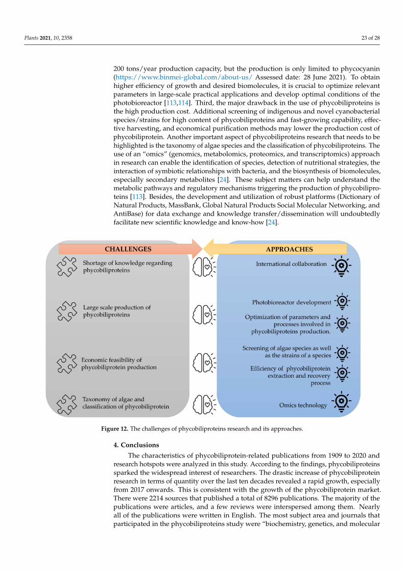
Plants 2021, 10, 2358 23 of 28
200 tons/year production capacity, but the production is only limited to phycocyanin(https://www.binmei-global.com/about-us/ Assessed date: 28 June 2021). To obtainhigher efficiency of growth and desired biomolecules, it is crucial to optimize relevantparameters in large-scale practical applications and develop optimal conditions of thephotobioreactor [113,114]. Third, the major drawback in the use of phycobiliproteins isthe high production cost. Additional screening of indigenous and novel cyanobacterialspecies/strains for high content of phycobiliproteins and fast-growing capability, effec-tive harvesting, and economical purification methods may lower the production cost ofphycobiliprotein. Another important aspect of phycobiliproteins research that needs to behighlighted is the taxonomy of algae species and the classification of phycobiliproteins. Theuse of an “omics” (genomics, metabolomics, proteomics, and transcriptomics) approachin research can enable the identification of species, detection of nutritional strategies, theinteraction of symbiotic relationships with bacteria, and the biosynthesis of biomolecules,especially secondary metabolites [24]. These subject matters can help understand themetabolic pathways and regulatory mechanisms triggering the production of phycobilipro-teins [113]. Besides, the development and utilization of robust platforms (Dictionary ofNatural Products, MassBank, Global Natural Products Social Molecular Networking, andAntiBase) for data exchange and knowledge transfer/dissemination will undoubtedlyfacilitate new scientific knowledge and know-how [24].
Figure 12. The challenges of phycobiliproteins research and its approaches.
4. Conclusions
The characteristics of phycobiliprotein-related publications from 1909 to 2020 andresearch hotspots were analyzed in this study. According to the findings, phycobiliproteinssparked the widespread interest of researchers. The drastic increase of phycobiliproteinresearch in terms of quantity over the last ten decades revealed a rapid growth, especiallyfrom 2017 onwards. This is consistent with the growth of the phycobiliprotein market.There were 2214 sources that published a total of 8296 publications. The majority of thepublications were articles, and a few reviews were interspersed among them. Nearlyall of the publications were written in English. The most subject area and journals thatparticipated in the phycobiliproteins study were “biochemistry, genetics, and molecular

Plants 2021, 10, 2358 24 of 28
biology” and “Journal of Applied Phycology”, respectively. The U.S.A. and China werethe major contributors in the study of phycobiliproteins, while the U.S.A. and Germanywere strongly interwoven in a worldwide research network. Glazer, Alexander N. from theUniversity of California, U.S.A. was the top prolific author in phycobiliprotein research.Overall, the worldwide collaboration network has benefited from the rapid growth of phy-cobiliprotein research. According to keyword analysis trends, the scope of phycobiliproteinresearch includes the optimization of algae culture techniques and phycobiliprotein extrac-tion processes, categorization of phycobiliproteins, phycobiliprotein biosynthesis pathway,phycobiliprotein bioactivities, and phycobiliprotein applications. The most commonlyutilized model organisms in the phycobiliprotein study were Arthrospira and Synechococcus.The study trend using keyword burst detection revealed a growing concern regarding theextraction of phycobiliproteins and their bioactivities, especially their antioxidant proper-ties. This is consistent with the keen interest in phycobiliproteins as bioactive compounds.Overall, this study provided essential information for researchers in seeking suitable insti-tutions, initiating institutional research, or establishing collaborations in phycobiliproteinresearch. The findings might also aid researchers in identifying the trends and resources toconstruct impactful investigations that broaden the scientific frontier.
Author Contributions: Conceptualization, H.T.T., Y.S.K. and S.A.A.; methodology, H.T.T.; software,H.T.T. and Y.S.K.; validation, Y.S.K., S.A.A., N.A.S. and F.M.Y.; formal analysis, H.T.T.; investigation,H.T.T.; resources, F.M.Y.; data curation, H.T.T.; writing—original draft preparation, H.T.T.; writing—review and editing, Y.S.K., S.A.A., N.A.S. and F.M.Y.; visualization, F.M.Y.; supervision, S.A.A., N.A.S.and F.M.Y.; project administration, F.M.Y.; funding acquisition, F.M.Y. All authors have read andagreed to the published version of the manuscript.
Funding: This research was supported by the Ministry of Higher Education Malaysia through theJapan–Malaysia Collaborative SATREPS-COSMOS (JPMJSA1509) project. The funders had no role inthe study design, data collection and analyses, decision to publish or prepare of the manuscript.
Institutional Review Board Statement: Not applicable.
Informed Consent Statement: Not applicable.
Data Availability Statement: Not applicable.
Acknowledgments: The first author is the recipient of the Excellent Students’ Program (PPC) Mas-ter’s program scholarship.
Conflicts of Interest: The authors declare no conflict of interest.
References1. Pagels, F.; Guedes, A.C.; Amaro, H.M.; Kijjoa, A.; Vasconcelos, V. Phycobiliproteins from Cyanobacteria: Chemistry and biotechno-
logical applications. Biotechnol. Adv. 2019, 37, 422–443. [CrossRef] [PubMed]2. Viskari, P.J.; Colyer, C.L. Rapid extraction of phycobiliproteins from cultured Cyanobacteria samples. Anal. Biochem. 2003, 319,
263–271. [CrossRef]3. Stadnichuk, I.N.; Tropin, I.V. Phycobiliproteins: Structure, functions and biotechnological applications. Appl. Biochem. Microbiol.
2017, 53, 1–10. [CrossRef]4. Bryant, D.A.; Guglielmi, G.; de Marsac, N.T.; Castets, A.M.; Cohen-Bazire, G. The structure of Cyanobacterial Phycobilisomes: A
model. Arch. Microbiol. 1979, 123, 113–127. [CrossRef]5. Manirafasha, E.; Ndikubwimana, T.; Zeng, X.; Lu, Y.; Jing, K. Phycobiliprotein: Potential microalgae derived pharmaceutical and
biological reagent. Biochem. Eng. J. 2016, 109, 282–296. [CrossRef]6. Wildman, R.B.; Bowen, C.C. Phycobilisomes in blue green algae. J. Bacteriol. 1974, 117, 866–881. [CrossRef]7. Fleurence, J. R-phycoerythrin from red macroalgae: Strategies for extraction and potential application in biotechnological area.
Appl. Biotechnol. Food Sci. Policy 2003, 1, 3–9.8. Van Der Weij-De Wit, C.D.; Doust, A.B.; Van Stokkum, I.H.M.; Dekker, J.P.; Wilk, K.E.; Curmi, P.M.G.; Van Grondelle, R.
Phycocyanin sensitizes both photosystem I and photosystem II in Cryptophyte Chroomonas CCMP270 Cells. Biophys. J. 2008, 94,2423–2433. [CrossRef]
9. Silva, S.C.; Ferreira, I.C.; Dias, M.M.; Barreiro, M.F. Microalgae-derived pigments: A 10-year bibliometric review and industryand market trend analysis. Molecules 2020, 25, 3406. [CrossRef]

Plants 2021, 10, 2358 25 of 28
10. Santiago-Santos, M.C.; Ponce-Noyola, T.; Olvera-Ramírez, R.; Ortega-López, J.; Cañizares-Villanueva, R.O. Extraction andpurification of phycocyanin from Calothrix sp. Process. Biochem. 2004, 39, 2047–2052. [CrossRef]
11. Koyande, A.K.; Chew, K.W.; Rambabu, K.; Tao, Y.; Chu, D.T.; Show, P.L. Microalgae: A potential alternative to health supplemen-tation for humans. Food Sci. Hum. Wellness 2019, 8, 16–24. [CrossRef]
12. Barkia, I.; Saari, N.; Manning, S.R. Microalgae for high-value products towards human health and nutrition. Mar. Drugs 2019, 17,304. [CrossRef] [PubMed]
13. Wu, H.L.; Wang, G.H.; Xiang, W.Z.; Li, T.; He, H. Stability and antioxidant activity of food-grade phycocyanin isolated fromSpirulina platensis. Int. J. Food Prop. 2016, 19, 2349–2362. [CrossRef]
14. Pradeep, H.N.; Nayak, C.A. Enhanced stability of C-phycocyanin colorant by extrusion encapsulation. J. Food Sci. Technol. 2019,56, 4526–4534. [CrossRef] [PubMed]
15. Yusoff, F.M.; Banerjee, S.; Nagao, N.; Imaizumi, Y.; Shariff, M.; Toda, T. Use of microalgae pigments in aquaculture. In Pigmentsfrom Microalgae Handbook; Jacob-Lopes, E., Queiroz, M.I., Zepka, L.Q., Eds.; Springer International Publishing: Cham, Switzerland,2020; pp. 471–513. [CrossRef]
16. Pritchard, A. Statistical bibliography or bibliometrics? J. Doc. 1962, 25, 348–349.17. Rumin, J.; Nicolau, E.; de Oliveira, R.G.; Fuentes-Grünewald, C.; Flynn, K.J.; Picot, L. A bibliometric analysis of microalgae
research in the world, Europe, and the European Atlantic area. Mar. Drugs 2020, 18, 79. [CrossRef]18. Tang, Y.; Xin, H.; Yang, F.; Long, X. A historical review and bibliometric analysis of nanoparticles toxicity on algae. J. Nanopart.
Res. 2018, 20, 92. [CrossRef]19. Qi, Y.; Chen, X.; Hu, Z.; Song, C.; Cui, Y. Bibliometric analysis of algal-bacterial symbiosis in wastewater treatment. Int. J. Environ.
Res. Public Health 2019, 16, 1077. [CrossRef]20. Lim, Z.S.; Wong, R.R.; Wong, C.Y.; Zulkharnain, A.; Shaharuddin, N.A.; Ahmad, S.A. Bibliometric analysis of research on diesel
pollution in Antarctica and a review on remediation techniques. Appl. Sci. 2021, 11, 1123. [CrossRef]21. Li, Z.; Ho, Y.S. Use of citation per publication as an indicator to evaluate contingent valuation research. Scientometrics 2008, 75,
97–110. [CrossRef]22. Gan, C.; Wang, W. Research Characteristics and Status on Social Media in China: A Bibliometric and Co-Word Analysis.
Scientometrics 2015, 105, 1167–1182. [CrossRef]23. Macías-Chapula, C.A.; Mijangos-Nolasco, A. Bibliometric analysis of AIDS literature in Central Africa. Scientometrics 2002, 54,
309–317. [CrossRef]24. Oliveira, C.Y.B.; Oliveira, C.D.L.; Müller, M.N.; Santos, E.P.; Dantas, D.M.M.; Gálvez, A.O. A scientometric overview of global
dinoflagellate research. Publications 2020, 8, 52. [CrossRef]25. Garrido-Cardenas, J.A.; Manzano-Agugliaro, F.; Acien-Fernandez, F.G.; Molina-Grima, E. Microalgae research worldwide. Algal
Res. 2018, 35, 50–60. [CrossRef]26. Rowley, J.; Slack, F. Conducting a literature review. Manag. Res. News 2004, 27, 31–39. [CrossRef]27. Aria, M.; Cuccurullo, C. Bibliometrix: An R-tool for comprehensive science mapping analysis. J. Informetr. 2017, 11, 959–975.
[CrossRef]28. Van Eck, N.J.; Waltman, L. Software survey: VOSviewer, a computer program for bibliometric mapping. Scientometrics 2010, 84,
523–538. [CrossRef] [PubMed]29. Chen, C. CiteSpace II: Detecting and visualizing emerging trends and transient patterns in scientific literature. J. Assoc. Inf. Sci.
Technol. 2006, 57, 359–377. [CrossRef]30. Huang, D.; Boxin, O.U.; Prior, R.L. The chemistry behind antioxidant capacity assays. J. Agric. Food Chem. 2005, 53, 1841–1856.
[CrossRef] [PubMed]31. Spolaore, P.; Joannis-Cassan, C.; Duran, E.; Isambert, A. Commercial applications of microalgae. J. Biosci. Bioeng. 2006, 101, 87–96.
[CrossRef]32. Cao, G.; Sofic, E.; Prior, R.L. Antioxidant and prooxidant behavior of flavonoids: Structure-activity relationships. Free Radic. Biol.
Med. 1997, 22, 749–760. [CrossRef]33. Croce, R.; Van Amerongen, H. Natural strategies for photosynthetic light harvesting. Nat. Chem. Biol. 2014, 10, 492–501. [CrossRef]
[PubMed]34. Jones, D.T.; Taylort, W.R.; Thornton, J.M. A new approach to protein fold recognition. Nature 1992, 358, 86–89. [CrossRef]35. Ou, B.; Hampsch-Woodill, M.; Prior, R.L. Development and validation of an improved oxygen radical absorbance capacity assay
using fluorescein as the fluorescent probe. J. Agric. Food Chem. 2001, 49, 4619–4626. [CrossRef]36. Braud, V.M.; Allan, D.S.J.; O’Callaghan, C.A.; Soderstrom, K.; D’Andrea, A.; Ogg, G.S.; Lazetic, S.; Young, N.T.; Bell, J.I.; Phillips,
J.H.; et al. HLA-E binds to natural killer cell receptors CD94/NKG2A, B and C. Nature 1998, 391, 795–799. [CrossRef]37. Lyons, A.B.; Parish, C.R. Determination of lymphocyte division by flow cytometry. J. Immunol. Methods 1994, 171, 131–137.
[CrossRef]38. Hardy, R.R.; Carmack, C.E.; Shinton, S.A.; Kemp, J.D.; Hayakawa, K. Resolution and characterization of pro-B and pre-pro-B cell
Stages in normal mouse bone marrow. J. Exp. Med. 1991, 173, 1213–1225. [CrossRef]39. Chee, M.; Yang, R.; Hubbell, E.; Berno, A.; Huang, X.C.; Stern, D.; Winkler, J.; Lockhart, D.J.; Morris, M.S.; Fodor, S.P.A. Accessing
genetic information with high-density DNA arrays. Science 1996, 274, 610–614. [CrossRef]

Plants 2021, 10, 2358 26 of 28
40. Cao, G.; Alessio, H.M.; Cutler, R.G. Oxygen-radical absorbance capacity assay for antioxidants. Free Radic. Biol. Med. 1993, 14,303–311. [CrossRef]
41. Guo, Y.-M.; Huang, Z.L.; Guo, J.; Li, H.; Guo, X.-R.; Nkeli, M.J. Bibliometric analysis on smart cities research. Sustainability 2019,11, 3606. [CrossRef]
42. Eck, N.J.V.; Waltman, L. VOSviewer Manual; University Leiden: Leiden, The Netherlands, 2018; Volume 1, pp. 1–51.43. Hsieh-Lo, M.; Castillo, G.; Ochoa-Becerra, M.A.; Mojica, L. Phycocyanin and phycoerythrin: Strategies to improve production
yield and chemical stability. Algal Res. 2019, 42, 101600. [CrossRef]44. Liu, Q.; Huang, Y.; Zhang, R.; Cai, T.; Cai, Y. Medical application of Spirulina Platensis derived C-phycocyanin. Evid. Based
Complement. Altern. Med. 2016, 2016, 7803846. [CrossRef]45. Li, W.; Su, H.N.; Pu, Y.; Chen, J.; Liu, L.N.; Liu, Q.; Qin, S. Phycobiliproteins: Molecular structure, production, applications, and
prospects. Biotechnol. Adv. 2019, 37, 340–353. [CrossRef]46. Kuddus, M.; Singh, P.; Thomas, G.; Al-Hazimi, A. Recent developments in production and biotechnological applications of
C-phycocyanin. Biomed. Res. Int. 2013, 2013, 1–9. [CrossRef]47. Sui, S.-F. Structure of phycobilisomes. Annu. Rev. Biophys. 2021, 50, 53–72. [CrossRef] [PubMed]48. Fernandes e Silva, E.; Figueira, F.d.S.; Lettnin, A.P.; Carrett-Dias, M.; Filgueira, D.d.M.V.B.; Kalil, S.; Trindade, G.S.; Votto, A.P.d.S.
C-phycocyanin: Cellular targets, mechanisms of action and multi drug resistance in cancer. Pharmacol. Rep. 2018, 70, 75–80.[CrossRef] [PubMed]
49. Wong, S.L.; Nyakuma, B.B.; Nordin, A.H.; Lee, C.T.; Ngadi, N.; Wong, K.Y.; Oladokun, O. Uncovering the dynamics in globalcarbon dioxide utilization research: A bibliometric analysis (1995–2019). Environ. Sci. Pollut. Res. 2021, 28, 13842–13860.[CrossRef] [PubMed]
50. Eriksen, N.T. Production of phycocyanin—A pigment with applications in biology, biotechnology, foods and medicine. Appl.Microbiol. Biotechnol. 2008, 80, 1–14. [CrossRef]
51. Manirafasha, E.; Guo, L.; Jing, K. Nutraceutical and Pharmaceutical Applications of Phycobiliproteins; Springer: Berlin/Heidelberg,Germany, 2021. [CrossRef]
52. Ma, J.; Chen, H.; Qin, S.; Lin, H. Applications of natural and artificial phycobiliproteins in solar cells. Curr. Biotechnol. 2015, 4,275–281. [CrossRef]
53. Wang, L.; Tian, L.; Deng, X.; Zhang, M.; Sun, S.; Zhang, W.; Zhao, L. Photosensitizers from Spirulina for solar cell. J. Chem. 2014,2014, 430806. [CrossRef]
54. Bogorad, L. Studies of phycobiliproteins. Rec. Chem. Prog. 1965, 26, 1–12. [PubMed]55. Grossman, K.E.A.; Grosman, A.R. Characterization and transcript analysis of the majar phycobiliprotein subunit genes from
Aglaothamnion Neglectum (Rhodophyta). Plant. Mol. Biol. 1993, 21, 27–38.56. Shen, G.; Schluchter, W.M.; Bryant, D.A. Biogenesis of phycobiliproteins: I. CpcS-I and CpcU mutants of the Cyanobacterium
Synechococcus Sp. PCC 7002 define a heterodimeric phyococyanobilin lyase specific for β-phycocyanin and allophycocyaninsubunits. J. Biol. Chem. 2008, 283, 7503–7512. [CrossRef] [PubMed]
57. Sonani, R.R. Recent advances in production, purification and applications of phycobiliproteins. World J. Biol. Chem. 2016, 7, 100.[CrossRef]
58. Maurya, S.S.; Maurya, J.N.; Pandey, V.D. Factors regulating phycobiliprotein production in cyanobacteria. Int. J. Curr. Microbiol.Appl. Sci. 2014, 3, 764–771.
59. Kronick, M.N.; Grossman, P.D. Immunoassay techniques with fluorescent phycobiliprotein conjugates. Clin. Chem. 1983, 29,1582–1586. [CrossRef] [PubMed]
60. Díaz Domínguez, G.; Marsán Suárez, V.; del Valle Pérez, L.O. Main immunomodulatory and anti-inflamatory properties ofphycobiliproteins C-phycocyanin. Rev. Cuba. Hematol. Inmunol. Hemoter. 2016, 32, 447–454.
61. Oi, V.T.; Glazer, A.N.; Stryer, L. Fluorescent phycobiliprotein conjugates for analyses of cells and molecules. J. Cell Biol. 1982, 93,981–986. [CrossRef]
62. Sekar, S.; Chandramohan, M. Phycobiliproteins as a commodity: Trends in applied research, patents and commercialization. J.Appl. Phycol. 2008, 20, 113–136. [CrossRef]
63. Wang, Y.; Hou, S.; Ke, F.; Gao, H. Bibliometric analysis of research on microcystins in China and worldwide from 1991 to 2011.Desalin. Water Treat. 2015, 53, 272–283. [CrossRef]
64. Md Khudzari, J.; Kurian, J.; Tartakovsky, B.; Raghavan, G.S.V. Bibliometric analysis of global research trends on microbial fuelcells using scopus database. Biochem. Eng. J. 2018, 136, 51–60. [CrossRef]
65. Sun, L.; Wang, S.; Chen, L.; Gong, X. Promising fluorescent probes from phycobiliproteins. IEEE J. Sel. Top. Quantum Electron.2003, 9, 177–188. [CrossRef]
66. Mindeli, L.E.; Markusova, V.A. Bibliometric studies of scientific collaboration: International trends. Autom. Doc. Math. Linguist.2015, 49, 59–64. [CrossRef]
67. Jones, K.S.; Jackson, D.M. The use of automatically-obtained keyword classifications for information retrieval. Inf. Storage Retr.1970, 5, 175–201. [CrossRef]
68. Wang, C.C.; Ho, Y.S. Research trend of metal–Organic frameworks: A bibliometric analysis. Scientometrics 2016, 109, 481–513.[CrossRef]

Plants 2021, 10, 2358 27 of 28
69. Mutanda, T.; Naidoo, D.; Bwapwa, J.K.; Anandraj, A. Biotechnological applications of microalgal oleaginous compounds: Currenttrends on microalgal bioprocessing of products. Front. Energy Res. 2020, 8, 598803. [CrossRef]
70. Khan, M.I.; Shin, J.H.; Kim, J.D. The promising future of microalgae: Current status, challenges, and optimization of a sustainableand renewable industry for biofuels, feed, and other products. Microb. Cell Fact. 2018, 17, 36. [CrossRef] [PubMed]
71. Tan, H.T.; Khong, N.M.H.; Khaw, Y.S.; Ahmad, S.A.; Yusoff, F.M. Optimization of the freezing-thawing method for extractingphycobiliproteins from Arthrospira sp. Molecules 2020, 25, 3894. [CrossRef]
72. Chamorro-Cevallos, G. Methods for extraction, isolation and purification of C-phycocyanin: 50 years of research in review. Int. J.Food Nutr. Sci. 2016, 3, 1–10. [CrossRef]
73. Begum, H.; Khatoon, H.; Yusoff, F.M.; Shariff, M. Production and purity of phycobiliproteins from selected marine and freshwatercyanobacteria subjected to different drying methods. Asian Fish. Sci. 2020, 33, 258–265. [CrossRef]
74. Yu, P.; Wu, Y.; Wang, G.; Jia, T.; Zhang, Y. Purification and bioactivities of phycocyanin. Crit. Rev. Food Sci. Nutr. 2017, 57,3840–3849. [CrossRef]
75. Hashimoto, E.; Yabuta, Y.; Watanabe, F.; Morimoto, M.; Yamaguchi, Y.; Takenaka, H. Purification and characterization ofphycobiliproteins from edible Cyanobacterium Nostochopsis sp. Food Sci. Technol. Res. 2012, 18, 485–490. [CrossRef]
76. Apt, K.E.; Collier, J.L.; Grossman, A.R. Evolution of the phycobiliproteins. J. Mol. Biol. 1995, 248, 79–96. [CrossRef]77. Scheer, H.; Zhao, K.H. Biliprotein maturation: The chromophore attachment. Mol. Microbiol. 2008, 68, 263–276. [CrossRef]78. Montgomery, B.L.; Casey, E.S.; Grossman, A.R.; Kehoe, D.M. AplA, a member of a new class of phycobiliproteins lacking a
traditional role in photosynthetic light harvesting. Society 2004, 186, 7420–7428. [CrossRef]79. Wedemayer, G.J.; Wemmer, D.E.; Glazer, A.N. Phycobilins of Cryptophycean algae: Structures of novel bilins with acryloyl
substituents from phycoerythrin 566. J. Biol. Chem. 1991, 266, 4731–4741. [CrossRef]80. Wedemayer, G.J.; Kidd, D.G.; Wemmer, D.E.; Glazer, A.N. Phycobilins of Cryptophycean algae. Occurrence of dihydrobiliverdin
and mesobiliverdin in cryptomonad biliproteins. J. Biol. Chem. 1992, 267, 7315–7331. [CrossRef]81. Schluchter, W.M.; Shen, G.; Alvey, R.M.; Biswas, A.; Saunée, N.A.; Williams, S.R.; Mille, C.A.; Bryant, D.A. Phycobiliprotein
Biosynthesis in Cyanobacteria: Structure and Function of Enzymes Involved in Post-Translational Modification; Springer: New York, NY,USA, 2010; Volume 675. [CrossRef]
82. Belford, H.S.; Offier, G.D.; Troxler, R.F. Phycobiliprotein synthesis in the unicellular rhodophyte, Cyanidiurn cazdurium. J. Biol.Chem. 1983, 258, 4503–4510. [CrossRef]
83. McErlean, M.; Liu, X.; Cui, Z.; Gust, B.; Van Lanen, S.G. Identification and characterization of enzymes involved in the biosynthesisof pyrimidine nucleoside antibiotics. Nat. Prod. Rep. 2021, 7. [CrossRef]
84. Brown, S.B.; Holroyd, J.A.; Vernon, D.I. Biosynthesis of phycobiliproteins. Biochem. J. 1984, 219, 905–909. [CrossRef]85. Kumar, V.; Maurya, P.K.; Mondal, S.; Sinha, R.P.; Singh, S.P. Photomorphogenesis in the Cyanobacterium Fremyella Diplosiphon Improves
Photosynthetic Efficiency; Elsevier Inc.: Amsterdam, The Netherlands, 2018. [CrossRef]86. Singh, N.K.; Sonani, R.R.; Prasad Rastogi, R.; Madamwar, D. The phycobilisomes: An early requisite for efficient photosynthesis
in Cyanobacteria. EXCLI J. 2015, 14, 268–289. [CrossRef] [PubMed]87. Romay, C.; Gonzalez, R.; Ledon, N.; Remirez, D.; Rimbau, V. C-phycocyanin: A biliprotein with antioxidant, anti-inflammatory
and neuroprotective effects. Curr. Protein Pept. Sci. 2003, 4, 207–216. [CrossRef] [PubMed]88. Abd El-Baky, H.H.; El-Baroty, G.S. Characterization and bioactivity of phycocyanin isolated from Spirulina Maxima grown under
salt stress. Food Funct. 2012, 3, 381–388. [CrossRef] [PubMed]89. Safari, R.; Amiri, R.Z.; Esmaeilzadeh Kenari, R. Antioxidant and antibacterial activities of C-Phycocyanin from common name
Spirulina platensis. Iran. J. Fish. Sci. 2020, 19, 1911–1927. [CrossRef]90. Fernández-Rojas, B.; Hernández-Juárez, J.; Pedraza-Chaverri, J. Nutraceutical properties of phycocyanin. J. Funct. Foods 2014, 11,
375–392. [CrossRef]91. Chen, C. Searching for intellectual turning points: Progressive knowledge domain visualization. Proc. Natl. Acad. Sci. USA 2004,
101, 5303–5310. [CrossRef]92. Jiang, M.; Qi, Y.; Liu, H.; Chen, Y. The role of nanomaterials and nanotechnologies in wastewater treatment: A bibliometric
analysis. Nanoscale Res. Lett. 2018, 13, 233. [CrossRef]93. Chen, C. CiteSpace: A Practical Guide for Mapping Scientific Literature; Nova Science Publishers: Hauppauge, NY, USA, 2016.94. Sundström, V.; van Grondelle, R. Dynamics of excitation energy transfer in photosynthetic bacteria. Chlorophylls 1991, 2137,
627–704.95. Jung, T.M.; Dailey, M.O. A novel and inexpensive source of allophycocyanin for multicolor flow cytometry. J. Immunol. Methods
1989, 121, 9–18. [CrossRef]96. Glazer, A.N. Phycobiliproteins—A family of valuable, widely used fluorophores. J. Appl. Phycol. 1994, 6, 105–112. [CrossRef]97. Tavanandi, H.A.; Mittal, R.; Chandrasekhar, J.; Raghavarao, K.S.M.S. Simple and efficient method for extraction of C-phycocyanin
from dry biomass of Arthospira platensis. Algal Res. 2018, 31, 239–251. [CrossRef]98. Ruiz-Domínguez, M.C.; Jáuregui, M.; Medina, E.; Jaime, C.; Cerezal, P. Rapid green extractions of C-phycocyanin from Arthrospira
Maxima for functional applications. Appl. Sci. 2019, 9, 1987. [CrossRef]99. Zhang, J.; Ma, J.; Liu, D.; Qin, S.; Sun, S.; Zhao, J.; Sui, S.F. Structure of phycobilisome from the red alga Griffithsia pacifica. Nature
2017, 551, 57–63. [CrossRef] [PubMed]

Plants 2021, 10, 2358 28 of 28
100. Ma, J.; You, X.; Sun, S.; Wang, X.; Qin, S.; Sui, S.F. Structural basis of energy transfer in Porphyridium Purpureum phycobilisome.Nature 2020, 579, 146–151. [CrossRef]
101. Farrar, W.V. Tecuitlatl: A Glimpse of Aztec food technology. Nature 1966, 211, 341–342. [CrossRef]102. Hamed, I. The evolution and versatility of microalgal biotechnology: A review. Compr. Rev. Food Sci. Food Saf. 2016, 15, 1104–1123.
[CrossRef]103. Abdulqader, G.; Barsanti, L.; Tredici, M.R. Harvest of Arthrospira Platensis from Lake Kossorom (Chad) and its household usage
among the Kanembu. J. Appl. Phycol. 2000, 12, 493–498. [CrossRef]104. Ludwig, M.; Bryant, D.A. Synechococcus Sp. Strain PCC 7002 transcriptome: Acclimation to temperature, salinity, oxidative stress,
and mixotrophic growth conditions. Front. Microbiol. 2012, 3, 354. [CrossRef]105. Coutinho, F.; Tschoeke, D.A.; Thompson, F.; Thompson, C. Comparative genomics of Synechococcus and proposal of the new
GENUS Parasynechococcus. PeerJ 2016, 2016, 1–18. [CrossRef] [PubMed]106. Kumar, J.; Singh, D.; Tyagi, M.B.; Kumar, A. Cyanobacteria: Applications in Biotechnology; Elsevier Inc.: Amsterdam, The Netherlands,
2019; Volume 7421. [CrossRef]107. Wang, K.; Wommack, K.E.; Chen, F. Abundance and distribution of Synechococcus spp. and cyanophages in the Chesapeake Bay.
Appl. Environ. Microbiol. 2011, 77, 7459–7468. [CrossRef] [PubMed]108. Cordara, A.; Re, A.; Pagliano, C.; Van Alphen, P.; Pirone, R.; Saracco, G.; dos Santos, F.B.; Hellingwerf, K.; Vasile, N. Analysis of
the light intensity dependence of the growth of Synechocystis and of the light distribution in a photobioreactor energized by 635Nm light. PeerJ 2018, 2018, e5256. [CrossRef]
109. Li, Z.; Guo, M. Healthy efficacy of Nostoc Commune vaucher. Oncotarget 2018, 9, 14669–14679. [CrossRef]110. Cheung, M.Y.; Liang, S.; Lee, J. Toxin-producing Cyanobacteria in freshwater: A review of the problems, impact on drinking water
safety, and efforts for protecting public health. J. Microbiol. 2013, 51, 1–10. [CrossRef] [PubMed]111. Saad, A.; Atia, A. Review on freshwater blue-green algae (Cyanobacteria): Occurrence, classification and toxicology. Biosci.
Biotechnol. Res. Asia 2014, 11, 1319–1325. [CrossRef]112. Xu, Y.; Ibrahim, I.M.; Wosu, C.I.; Ben-Amotz, A.; Harvey, P.J. Potential of new isolates of Dunaliella Salina for natural β-carotene
production. Biology 2018, 7, 14. [CrossRef] [PubMed]113. García, J.L.; de Vicente, M.; Galán, B. Microalgae, old sustainable food and fashion nutraceuticals. Microb. Biotechnol. 2017, 10,
1017–1024. [CrossRef]114. Yusoff, F.M.; Nagao, N.; Imaizumi, Y.; Toda, T. Bioreactor for microalgal cultivation systems: Strategy and development. In
Prospects of Renewable Bioprocessing in Future Energy Systems, Biofuel and Biorefinery Technologies; Springer: Berlin/Heidelberg,Germany, 2019; pp. 117–159. [CrossRef]


