Embed Size (px)
Citation preview
KJAJA2Ö*)O)Ó
Food handling and mastication in the carp (Cyprinus carpio L.)
« « U B * * * 1 *
WIT1 TUOSW«- « *
omslag tekening : Wim Valen
-2-
Promotor: dr. J.W.M. Osse, hoogleraar in de algemene dierkunde
^iJOttO^ 1o ID
Ferdinand A. Sibbing
FOOD HANDLING AND MASTICATION IN THE CARP (Cyprinus carpio L.)
Proefschr i f t
ter verkri jging van de graad van
doctor in de landbouwwetenschappen,
op gezag van de rector magnificus,
dr. C.C.Oosterlee,
in het openbaar te verdedigen
op dinsdag 11 december 1984
des namiddags te vier uur in de aula
van de Landbouwhogeschool te Wageningen.
l^V-, ^ v ^ b i S . o a
BIBLIOTHEEK
^LANDBOUWHOGESCHOOL WAGENINGEN
^/y/of^OÏ, fOtO
STELLINGEN
1. De taakverdeling tussen de kauwspieren van de karper is analoog aan die tussen
vliegspieren van insekten: grote lichaamsspieren leveren indirekt het vermogen,
te rw i j l d irekt aangehechte kleinere spieren de beweging vooral sturen. Deze
analogie komt voort uit architekturale en kinematische principes.
2. Naamgeving van spieren op grond van hun verwachte rol (b.v. levator, retractor)
zonder dat deze fe i te l i jk is onderzocht leidt tot lang doorwerkende misvattingen
over hun funkt ie en geeft b l i jk van een onderschatting van de p last ic i te i t waarmee
spieren worden ingezet. Een nomenclatuur die gebaseerd is op origo en insertie
van de spier verdient de voorkeur.
3. De uitstulpbaarheid van de gesloten bek bij veel cypriniden maakt een getrapte
zuivering van het voedsel mogelijk en speelt zo een wezenli jke rol in de selektie
van bodemvoedsel. Dit proefschrift.
4. Op grond van de vele funkties die aan sl i jm in biologische systemen worden
toegeschreven is meer onderzoek naar zi jn chemische en fysische eigenschappen
dringend gewenst. Dit proefschrift.
5. Het samenvallen van het moment van gereedkomen van het kauwapparaat bij
de karper (na drie weken; Geyer, 1937) met de overgang naar groter voedsel
(Uribe-Zamora, 1975) is begri jpeli jk vanuit de eisen die aan de groter wordende
vis worden gesteld.
6. De bouw en fysiologie van een soort stellen grenzen aan haar gedrag, en daarmee
aan haar positie in een oecosysteem. Deze grenzen vormen belangrijke oecolo-
gische parameters maar zi jn nauwelijks bekend.
7. Dat het afsterven van bossen pas een pr ikkel vormt die de aktiedrempel overschrijdt
geeft de inf lat ie in de waardering van mi l ieu-indikatoren schrijnend weer.
8. Optel len, af t rekken, vermenigvuldigen en wortel t rekken hebben we goed geleerd.
Voor de toekomst moeten we opnieuw leren delen.
BIBLIOTHEEK P'- ' i
LANDBOUW •' HOOL WAGKMJNGEN
9. Onderzoek zou moeten worden gestimuleerd naar optimale organisatievormen
om visteelt op ondergelopen landbouwgronden, in waterreservoirs en in i r r igat ie
kanalen een maximale bijdrage te laten leveren aan produkt-diversi f ikat ie en
aan de stabil isatie en verdeling van inkomen. D i t zou de plattelandsontwikkel ing,
met name in ontwikkelingslanden, sterk bevorderen.
10. Het opnieuw inbrengen van de wi ld-vorm verhoogt de smaak en v i ta l i te i t van
de karper (Balon, 1974). Er zi jn aanwijzingen dat d i t ook voor de kuituur van
de mens geldt.
11. De fraaie ui tvoering van postzegels ui t ontwikkelingslanden is vaak evenredig
met de armoede die er heerst en past binnen het verhullende beeld dat het
lokale regiem exporteert .
12. Het ontbreken van het jaar van u i tg i f te op de t i telpagina van een wetenschap
peli jk boek doet vermoeden dat de uitgever nauwelijks pr i jst stelt op de referent ie
ervan.
13. De voorgestelde taakverdeling voor UHD's en UD's in het nieuwe rangenstelsel
voor wetenschappelijk personeel betekent een achteruitgang voor de benutt ing,
ontplooiing en samenwerking van het personeel.
14. Aanwijzingen dat smaakknopjes van de karper gevoeliger zi jn voor menselijk
speeksel dan voor een standaardspektrum smaakstoffen (Konishi & Zot terman,
1963) werpt nieuw l icht op het met speeksel samenkneden van de deegpluim
door hengelaars.
Balon, E.K. (1974). Domestication of the carp Cyprinus carpio L.. Roy. Ontario Mus. L i fe Sei. Misc. Publ.: 1:35. Geyer, E. (1937). Der Zeitliche Ablauf der Bezahnung und des Zahnwechsels bei Cyprinus carpio L., unter besonderer
Berücksichtigung des einsömmrigen Karpfens. Morph. Jahrb. 80: 280-354. Konishi, J . & Zotterman, Y. (1963). Taste functions in fish. In: Olfaction and Taste: 215-233. Y. Zotterman (Ed).
New York: McMillan. Uribe-Zamora, M. (1975). Selection des proies par le f i l t re branchial de la carpe miroir (Cyprinus carpio L.). Thesis,
Univ. of Lyon.
Stellingen behorende bij het proefschri f t "Food handling and masticat ion in the
carp (Cyprinus carpio L.)" door F.A. Sibbing. Wageningen, 11 december 1984.
-5-
Voorwoord
Het verschijnen van dit proefschri f t biedt mij een welkome gelegenheid allen
te danken die aan zijn wording hebben bijgedragen.
Al lereerst w i l ik mi jn ouders bedanken voor de stimulerende vorming en de
waardevolle huiselijke sfeer waarin deze plaats vond. Geen inspanning was ju l l ie -
ooit te veel. Het baanbrekende werk van mijn oudere broers en zusters waardeer
ik zeer.
Jan Osse, de belangstelling voor dit vak moet zeker zi jn oorsprong hebben
in het niet aflatende enthousiasme waarmee je het vanaf mijn studententi jd en
ook nu nog steeds ui tdraagt. Onze diskussies over de diverse manuskripten hebben
in belangrijke mate tot verheldering, relat ivering en verscherping van hun inhoud
bijgedragen. Ik w i l je hier graag bedanken voor de stimulerende begeleiding als
promotor en voor het in i t iëren van dit onderzoek.
Ar ie Terlouw, jouw deskundige en plezierige ondersteuning bij de voorbereiding
en uitvoering van experimenten stel ik erg op pri js. Onze röntgenfilm-sessie in
Leiden heeft z'n vruchten afgeworpen. Ook de hulp van Ben van Schie en Albert
Ramakers wi l ik hier noemen. Rosario Ur ibe, dank voor de toewijding en kr i t ische
zin waarmee je veel histologisch werk hebt ingebracht.
Wim Valen, jouw vakmanschap is aan de i l lustrat ies in dit proefschri f t af
te lezen en onmisbaar bij het beschrijven van de morfologie. Bedankt voor je
inzet, je mee-denken en voor de plezierige samenwerking.
Als student hebben R ië t te van Beek, Tonnie te Brinke, Jeannet Ennik, Peter
Vossen, Paul van Zwieten en Peter Roessingh steeds een verfrissende en k r i t i
sche rol vervuld in het onderzoek, dat zich door ju l l ie ook tot andere karperachtige
vissen kon ui tstrekken. De samenwerking met studenten uit de sectie ethologie
(vakgroep Veehouderij), Hans Manni en Arno Brunink, heeft ook mi jn kijk op de
karper verbreed.
Mi jn kollega's van de sektie Funktionele Morfologie, Rie Akster, Mees Mul ler,
Johan van Leeuwen, Maarten Drost en Annet Kroon, dank ik voor de diskussies
over het onderzoek en voor de vriendschap waarin wordt samengewerkt. Ook de
andere medewerkers van de vakgroep Experimentele Diermorfologie en Celbiologie
moeten zich hierin aangesproken weten. Prof.dr. Lucy Timmermans w i l ik speciaal
bedanken voor de stimulerende kontakten en voor de behartiging van al ler lei perso
nele en materiële zaken waarmee zij de laatste jaren de voortgang van onder
zoek en onderwijs heeft gewaarborgd.
-6-
Sietze Leenstra, Piet van Kleef en Ar thur Rep ben ik erkentel i jk voor hun
zorg voor de proefdieren. De Organisatie ter Verbetering van de Binnenvisserij
dank ik voor de levering van de eerste karpers.
Lies van Beek, de inzet en kwal i te i t waarmee j i j de teksten verwerkt hebt
is me tot grote steun geweest. Ook Mw. E.A.Scheffers dank ik voor haar bijdragen.
Gerda van Malsen, jouw organisatie van al lerlei administratieve zaken heeft,
vaak onopgemerkt, de voortgang van mijn werk zeker bespoedigd. Hulp van Amy
Tiemessen en technische steun van Sytse van den Berg waren er a l t i jd wanneer
dat nodig was.
De afdeling scanning electronen microscopie van de TFDL wordt bedankt voor
hun bijdragen aan dit proefschr i f t . In het bijzonder wi l ik hier Felix Thiel noemen.
De kollega's van de sektie Morfologie van het Zoölogisch Laborator ium te
Leiden hebben ons gastvri j ontvangen voor het maken van de röntgenfi lms. Voor
het vr i jmaken van hun apparatuur en de technische ondersteuning dank ik hen
zeer. Wim Weijs stelde de radiopaque voedselpellets to t mi jn beschikking.
Gesprekken met de oecologen van het Limnologisch Inst i tuut te Oosterzee
(Fr.) hebben mijn blik op de rol van de vis in het aquatisch systeem verruimd.
Eddy Lammens en Koos Vi jverberg, ik hoop dat onze samenwerking in het brasem-
projekt een tussenfase naar verdere uitbouw is. Wim van Densen (vakgroep Visteelt
en Visserij), ik verwacht dat we de interakt ie tussen het onderzoek naar de voedsel-
opname en -verwerking van vissen en de visserijbiologie, nu dit proefschr i f t is
afgerond, verder kunnen ui twerken.
Cyprinus carpio, dank voor je sl i jmerige medewerking en de eetlust die je
ondanks enige prikakties a l t i jd weer snel aan de dag legde. Onze verschillen in
afkomst en mi l ieu hebben een goede samenwerking nooit in de weg gestaan.
Mieke, het is niet makkeli jk schijnbaar machteloos toe te zien wanneer ik
me wéér in m'n werk terugtrek. Weet dat de vr i jheid en rust thuis het to t stand
komen van dit boekje sterk heeft ver l icht . Bouke en Sjoerd, ju l l ie vraag naar
aandacht heeft voor de nodige ontspanning en relat iver ing gezorgd.
INHOUD / CONTENTS
Hoofdstuk I : Doel van het onderzoek en samenvatting van de resultaten
achtergrond van het onderzoek 9
karperachtige vissen 9
probleemstell ing en methoden 10
voedselopname en -verwerking door de karper 15
samenvatting en konklusies 26
Hoofdstuk I I : Food handling in the carp (Cyprinus carpio L.), its movement
patterns, mechanisms and l imi tat ions.
F.A.Sibbing, J.W.M.Osse and A.Terlouw
(to be submitted to the Journal of Zoology).
31
Hoofdstuk III : Regional specializations in the oro-pharyngeal wal l and food
processing in the carp {Cyprinus carpio L.).
F.A.Sibbing and R.Uribe
(to be submitted to the Journal of Zoology).
81
Hoofdstuk IV : Pharyngeal mastication and food transport in the carp
{Cyprinus carpio L.): a c ineradiography and electromyographic
study.
F.A.Sibbing.
Journal of Morphology (1982) 172: 223-258.
125
General summary 161
Curr iculum vitae 165
-9 -
HOOFDSTUK 1
DOEL VAN HET ONDERZOEK EN SAMENVATTING VAN DE RESULTATEN
Achtergrond van het onderzoek
Vissen vormen een belangrijke schakel op verschillende niveau's in het voedsel-
web, als prooi maar ook als konsument. De c irca 20.000 recente soorten (Nelson,
1976) vormen de grootste groep van de gewervelde dieren en het scala voedseltypen
dat zi j benutten is groter dan voor elke andere vertebraten groep (Nikolsky, 1963).
Maagonderzoek aan vissen, mits uitgevoerd over een groot deel van hun biotoop
en zich uitstrekkend over etmaal en seizoenen, geeft een beeld van het dieet van
de soort. Het dieet wordt bepaald door de selektieve opname uit een variabel voed
selaanbod, vermoedelijk op grond van de smaak en nutr iënten van een voedseltype,
de t i jd die haar verwerking vereist en de verhouding tussen bestede en verworven
energie. Verschil len in plaats, groot te, vorm, beweegli jkheid, chemische en mecha
nische samenstelling van het voedselaanbod doen ons specialisaties verwachten
in bouw, funkt ioneren en gedrag van de vis. Deze bl i jken mede uit het naast elkaar
aanwezig zi jn van vele vissoorten in één levensgemeenschap. Welke deze special i
saties z i jn , hoe z i j het dieet begrenzen en hoe zij de konkurrentieposit ie van de
vis bepalen is nauwelijks bekend. De gevolgen van opzettel i jke of toevall ige ecolo
gische veranderingen in het mi l ieu op de trof ische relaties en samenstelling van
de visfauna zi jn dan ook niet voorspelbaar. D i t b l i jk t ondermeer bij introdukt ie
van uitheemse vissoorten. Inzicht in de relaties tussen de vis en zi jn omgeving
vereist een gekombineerde aanpak van funktionele morfologie, ethologie en ecologie.
Kennis hiervan kan r icht ing geven aan de beheersing van natuurl i jke en kunstmatige
aquatische systemen.
Door de bovengenoemde specialisaties te onderzoeken wordt inzicht verkregen
in de grondslagen van bouw en werking van het sensorisch, het motorisch en het
regelapparaat voor de voedselopname. Deze dragen tesamen zorg voor zoeken,
detekt ie , opname en verwerking van het voedsel.
Karperachtiqe vissen
De karper (Cyprinus carpio L.) werd als onderzoeksobject gekozen op grond van
de volgende argumenten.
1) Karperachtigen (Cyprinidae) vormen met 1600 soorten de grootste fami l ie van
vissen (Nelson, 1976). Over hun voedselverwerkingsmechanisme is v r i jwel niets
bekend.
-10-
2) In Nederland is deze fami l ie in het zoete water het sterkst vertegenwoordigd
in aantal soorten (20) en biomassa.
3) Z i j tonen een grote var iat ie in ecologische en t rof ische typen. Zo worden voor
een maximale benutting van het voedselaanbod in v i jverkul turen zoöplanktivoren,
fy toplankt ivoren, macrofytofagen, Omnivoren en bodemvreters gemengd geteeld
(Bardach et a l . , 1972). De graskarper wordt ingezet bij de biologische kontrole
van plantengroei in watersystemen.
4) De karper wordt op grote schaal gekweekt en vormt enerzijds een gewaardeerd
voedsel (Oost-Europa, Azië), maar kan door explosieve aantalsvermeerdering
ook to t plagen leiden (N-Amer ika, Austral ië).
5) De karper is als proefdier de ' laborator ium-rat ' onder de vissen. Een synthese
van gegevens met die uit andere vakgebieden verdiept onze kennis over deze
zeer algemeen voorkomende vis. Gegevens over darminhoud zi jn in de l i teratuur
op ruime schaal voorhanden.
Cypriniden vormen ook uit morfologisch oogpunt een bijzondere fami l ie . Tanden
op de kaken en in de mondholte ontbreken; de laatste kieuwbogen zi jn to t sterk
ontwikkelde tandendragende keelkaken omgevormd die samen met een hoornplaat
in de schedelbasis een krachtig kauwsysteem vormen (Eig. 1, pag.129). De maag
ontbreekt bij alle soorten. De sterke ontwikkeling van het kauwapparaat in de
keel ontlast de mondkaken van een b i j t funkt ie en heeft mogelijk zo bijgedragen
to t de ontwikkeling van een uitstulpbaar mondapparaat, dat uniek is onder de lagere
beenvissen (slot hoofdstuk 2).
Samen met de vier andere suborden van de Ostariophysi (Chanoidei, Gonorynchoidei,
Characoidei, Si luroidei; Roberts, 1973; Fink and Fink, 1981) bezi t ten de Cyprinoidei
alarmcellen (p. 110) en het apparaat van Weber, een verbinding tussen zwemblaas
en gehoorstreek die voor geluidswaarneming dient (Alexander, 1967).
Probleemstell ing en methoden
De funktionele morfologie onderzoekt de samenhang tussen bouw en funkt ie
in een biologisch systeem, er van uitgaand dat hierop, op grond van fysische wet ten,
in de natuur wordt geselekteerd. De vorm-funkt ie re lat ie kan door vergeli jking
van bestaande strukturen en hun funkties worden bepaald ( induktie). Z i j kan ook
door deduktie van een theoretisch fysisch model, dat aan bestaande vormen wordt
getoetst, vanuit de funkt ie worden vastgesteld (Dul lemeijer, 1974). Vaak worden
beide en de experimentele methode (bi jv. aanbod van verschillende voedseltypen)
in kombinatie toegepast.
-11-
Ofschoon de vis als samenhangend geheel funkt ioneert , wordt hij bij vormanalyse
opgedeeld in strukturen van verschillend weefseltype (bv. beenelementen, spieren,
l igamenten). Struktuurkomplexen die een specifieke funkt ie uitoefenen noemen
we funktionele komponenten (van der Klaauw, 1945; Dul lemeijer, 1974). Deze kunnen
zich to t het hele organisme ui tstrekken (bv. voortbewegingsapparaat) maar men
kan dit begrip ook toepassen op niveau van een spiervezel (cf. Akster, 1981). Inzicht
in de vorm-funkt ie relat ies binnen een funktionele komponent is pas goed mogeli jk
wanneer ook de wederzijdse afhankeli jkheid van zulke komponenten in het onderzoek
worden betrokken. Zo maken strukturen van het voedselopname apparaat van de
karper (bv. de sternohyoideus spier) tevens deel uit van het ademhalingssysteem
en het kauwapparaat (p.149). Bij deze integrat ie stellen verschillende funkties eisen
aan eenzelfde struktuur. Integrat ie van struktuurkomplexen voor deelfunkties in
het geheel van de vis betekent dan ook vaak een inperking van de vr i jheid om deel
funkties opt imaal te realiseren (cf . Barel, 1983). Een holistische benadering vormt
daarom het uitgangspunt voor de analyse van het voedingssysteem en z i jn deel
funkt ies.
Opvatt ing van het begrip ' funkt ie ' als de 'biologische betekenis van een akt ie in
de natuurl i jke omgeving' is impl ic iet aan het onderzoeksthema van de sektie funk
tionele diermorfologie van de Landbouwhogeschool: 'Voedselopname en -verwerking
bij vissen: e co-morfologische aspekten'. D i t betekent voor de toekomst van het
onderzoek dat de wi jze waarop de vis het voedselaanbod in zi jn natuurl i jke omgeving
benut een noodzakelijk onderdeel van de studie moet z i jn, meer dan tot nu toe
gerealiseerd kon worden.
Vergeli jking van de vormenri jkdom in het voedselopname-apparaat der cypriniden
en hun leefwijze leidde to t gewaagde uitspraken over specialisaties waarbij de
funkt ie vooral u i t de bouw werd afgeleid (Suyehiro, 1942; Al-Hussaini, 1949; Matthes,
1963; Verighina, 1969; Kapoor et a l . , 1975).
Een gedetail leerd onderzoek van bouw èn funct ie van het voedselopname-apparaat
werd gestart bij vissen van het baars-type (Percoidei) (Osse, 1969; L iem, 1973).
Het zuigproces bi j de voedselopname van cypriniden werd voor het eerst via meting
van waterdruk in de mondholte door Alexander (1969) bestudeerd. Het gekompliceer-
de proces van voedselverwerking nâ opname vindt binnen de mond- en keelholte
plaats. Een analyse van de reeks van deelprocessen, die hierbi j optreedt werd niet
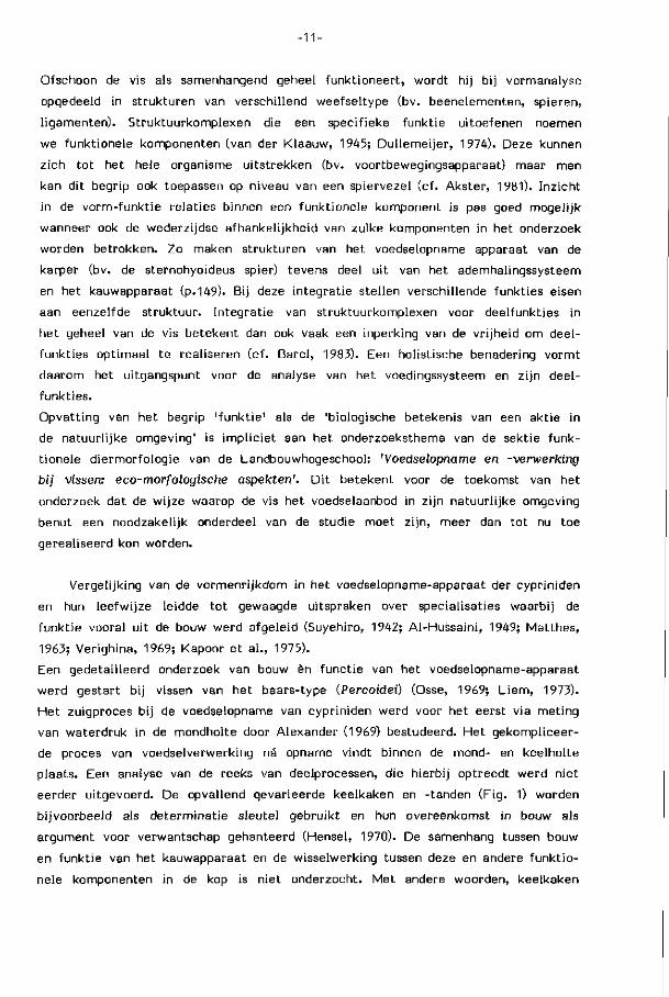
eerder ui tgevoerd. De opvallend gevarieerde keelkaken en -tanden (Fig. 1) worden
bi jvoorbeeld als determinat ie sleutel gebruikt en hun overeenkomst in bouw als
argument voor verwantschap gehanteerd (Hensel, 1970). De samenhang tussen bouw
en funkt ie van het kauwapparaat en de wisselwerking tussen deze en andere funkt io
nele komponenten in de kop is niet onderzocht. Met andere woorden, keelkaken
- 1 2 -
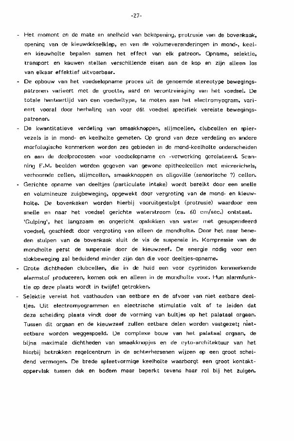
view A
Cyprinuscarpio Abramis brama Blicca bjoerkna Barbus barbus Carassius Chondrostoma Karper brasem kolblei barbeel carassius
kroeskarper nasus
sneep
view A to illustrate trje dental formula as a plan
Gobiogobio Leuciscus leuciscus Leuciscus cephalus Rutilus rutilus Scardinius grondel serpeling kopvoorn blankvoorn eiythrophthalmus
rietvoorn
Tinea tinea zeelt
naar Wheeler ( I978)
FIG. 1. Rechter-keelkaken met keeltanden van enkele der 20 karperachtige vissoorten, die in de Nederlandse binnenwateren voorkomen. Let op de verschillen in tandbouw en tandformule. De keelkaken werken als paar tegen een verhoornde kauwplaat (vgl. Plaat I) in het gehemelte van de vis. Zij zijn in grootte, stevigheid en ruimteli jke bouw (uitstekende armen) sterk gevarieerd.
-13-
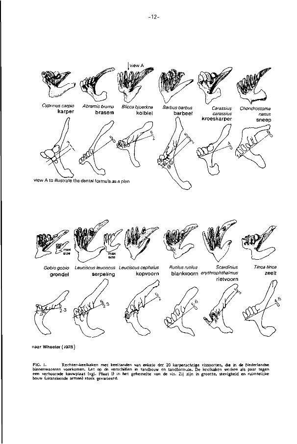
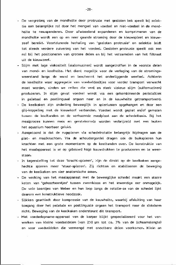
PLAAT I, SEM-beeld van de keelkaken van de rietvoorn (16.1 cm; boven) en van de keeltanden en verhoornde kauwplaat van de blankvoorn (21 cm; onder). De detaillering in de tandsculptuur verschilt vooral vooraan sterk en houdt verband met het uiteenlopende dieet van deze uitwendig op elkaar gelijkende vissen. Terwijl beiden ondermeer insektelarven en plantedelen eten, heeft de blankvoorn ook vooral slakken op het menu.
-14-
van cypriniden worden algemeen als kenmerk gebruikt maar hun invloed op het
bouwplan van de hele vis en hun specifieke ro l in de kompet i t ie tussen soorten
is onbekend.
De vraagstell ing die aan di t proefschr i f t ten gronde l ig t is v ier ledig:
1) Uit welke delen bestaat het proces van de voedselopname en -verwerking bij
de karper?
2) Welke strukturen zijn bij elk deelproces betrokken en hoe is de samenhang
tussen hun bouw en funktie?
3) Hoe beVnvloeden de deelprocessen elkaar en vindt afstemming van het totaal
proces plaats in relatie tot de aard van het voedsel?
4) Welke beperkingen in het gebruik van het voedselaanbod zijn het gevolg van
de 'omnivorie' van de karper en de daarbij horende aanpassingen in bouw en
funktie ?
De vraagstelling r icht zich op het niveau van het totale funktionerende dier.
Het onderzoek is er op gericht om de funkt ie uitoefening aan de zoveel mogelijk
ongestoorde vis te meten.
Gedetail leerde gegevens omtrent bouw en funktioneren werden verkregen met
de volgende technieken:
- macroscopische, microscopische en scanning-electronen-microscopische (SEM)
vormanalyse. U i t coupe-series werd het verspreidingspatroon van smaakknopjes,
s l i jm, spierweefsel en alarmcellen in de mond-keelholte gemeten. Het s l i jm werd
histochemisch onderzocht.
- af leiding van spieract iv i te i ten via e lectromyograf ie (9 kanalen), al dan niet in
kombinatie met gewone of röntgen- f i lm.
- meting van uitwendige en inwendige bewegingen van kopdelen bij aanbod van
verschillende voedseltypen: pellets, gerstkorrels, regenwormen, tubi fex, water
vlooien en bodem-tubifex mengsels. Door het aanbrengen van bariumsulfaat in
pellets en wormen kan de procesgang van di t voedsel in de röntgenf i lm worden
gevolgd.
Analyse heeft dus ook plaatsgevonden op het niveau van weefsel en cel alsmede
op het niveau van de motorische ak t i v i te i t binnen afzonderl i jke spieren. Anderzijds
vereist inzicht in de re lat ie tussen bouw van het organisme en z i jn funkt ioneren
in een natuurl i jke omgeving, zoals eerder aangegeven, diepgaande analyse van het
natuurl i jk gedrag en zi jn ecologische niche. N ie t elk van deze aspekten kon op
gewenste diepte worden onderzocht, vaak werd hiervoor naar l i teratuurgegevens
-15-
teruggegrepen.
De gegevens worden benut om ook de vraag naar de evolutie van de strukturele
kenmerken te behandelen.
De verkregen kennis geeft een samenhangend beeld van de zich voedende karper
en levert een nieuw vertrekpunt op voor gespecialiseerd onderzoek naar aanpassingen
in bv. zintuigen en s l i jm, naar de neurale regulatie van de voedselverwerking en
naar de ef f ic iency waarmee verschillende voedselbronnen in een natuurl i jke s i tuat ie
worden benut.
De resultaten van het onderzoek zi jn verwerkt in de hoofdstukken 2, 3 en 4, waarvan
de inhoud hieronder kort wordt genoemd.
In Hoofdstuk 2 wordt de totale procesgang en de afzonderli jke mechanismen
voor de opeenvolgende fasen van voedselverwerking tesamen met zi jn p last ic i te i t
en beperkingen besproken.
Hoofdstuk 3 geeft een macro- en microscopische analyse van de mond-keelholte
bekleding en re lateert deze aan deelfunkties van het voedingssysteem.
In Hoofdstuk 4 worden vorm-funkt ie relaties binnen het kauwsysteem van de
karper in detail u i tgewerkt . H ier in is ook een globaal beeld van het dieet van de
karper opgenomen (pag. 138).
Voedselopname en voedselverwerking door de karper
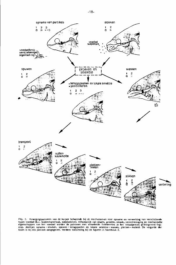
Het voedselopname en -verwerkingsproces valt uiteen in een aantal herkenbare
deelprocessen (Fig. 2) die, aangepast aan het type voedsel, in herkenbare volgorde
worden ingezet: zoeken, detekt ie, opname, selektie door proeven en reiniging,
t ransport, kauwen en slikken (hoofdstuk 2). Vertering valt buiten het bestek van
deze studie maar werd eerder voor cypriniden overzien en bij de graskarper bestu
deerd door Stroband (1980).
Wat eet de karper?
Resultaten voortkomend ui t darminhoud onderzoek worden met hoge f rekwent ie
gerapporteerd. Ur ibe-Zamora (1975) stelde hierin de grote l i jn vast (p.130- De volwas
sen karper b l i j k t een omnivore vis, die z ich, afhankelijk van het watersysteem
en seizoen, vooral met de volgende organismen voedt: 1) bodem-evertebraten (mugge-
larven, tubi fex, copepoden, mollusken, andere insektenlarven), 2) zoöplankton (de
grotere soorten watervlooien en copepoden), 3) l i t to ra le vegetatie (vooral zachtere
waterplanten als eendenkroos) en dieren die zich daartussen bevinden (slakken,
copepoden, kokerjuffers en wormen).
-16-
Hoe en wanneer zoekt de karper zijn voedsel?
Jönsson (1967) heeft in laboratorium-studies het voedselopname gedrag van
éénzomerige karpers uitvoerig onderzocht. Het door interne faktoren bepaalde
deel van de zoekakt iv i te i t heeft een hoge piek in de schemering en 's nachts. Kondi-
t ionering op uitwendige faktoren als voederti jd en -plaats v indt, ook overdag, gemak
keli jk plaats. Detect ie van voedsel gebeurt vooral met behulp van smaakzintuigen
op baarddraden en l ippen, die hij in kontakt met de bodem brengt. Geleiding naar
voedsel over langere afstand vindt waarschi jnl i jk via de reuk plaats. Het oog speelt
vooral bij bewegende prooi een ro l , voor zover de troebeling van het water d i t
toelaat. De ondergrens van de l icht intensi te i t voor ef fekt ieve lokalisatie van voedsel
l igt voor de meeste vissen bij 10 me (meter-candela), overeenkomend met late
schemering (Blaxter, 1970). Hele graankorrels, vaak gevoerd in de karperteel t ,
werken niet of nauwelijks st imulerend. Kondit ionering of bijmenging van stimulerend
voedsel verhoogt de opname. Zelfs van de karper b l i jken weinig gegevens bekend
over zi jn voedselopnamegedrag in het natuurl i jk mi l ieu. D i t hangt samen met proble
men van waarneming.
Baarddraden en lippen zi jn bezet met hoge dichtheden smaakknopjes (ca. 380
per mm 2 ; Plaat I, pag. 88 )• De hoge lichaamsbouw - die in snelstromend water
to t a fdr i jven zou af leiden - en de overheersing van rode, langzaam kontrahierende
maar niet snel vermoeibare spiervezels in de romp passen bij het langzaam zwem
mend zoeken, dat zich over lange periodes u i ts t rekt (stayer; Boddeke, Slijper en
van der Ste l t , 1959). Snelle prooien hebben hierdoor een re lat ief grote ontsnappings
kans.
Hoe vindt voedselopname plaats?
Afhankel i jk van het type voedsel treden verschillende mechanismen voor opname
in werking. A fb i j ten van voedselbrokken is met de tandeloze kaken vr i jwel onmoge
l i jk , de verhoornde kaakranden (Plaat III a, pag. 92) spelen een rol bij het manipule
ren van groter mater iaal (macrofyten, steentjes).
Opname van deeltjes (Part iculate feeding, F ig . 5, pag. 60). Moeil i jk bereikbaar
en zwaar voedsel, door de maten van de mondopening beperkt to t deeltjes met
een diameter kleiner dan ca. 9% van de standaardlengte van de vis, wordt opgeno
men door aanzienlijke expansie van de mond- en kieuwholte ( terminologie in F ig .
1, pag. 37 ) onder gel i jkt i jd ige protrusie (uitstulping) van de bovenkaak. D i t bewe
gingspatroon wekt een grote snelheid van het water op in de mondopening (ca.
60 cm /sec ) , sterk gericht op het voedsel. Modi f ikat ie van de protrusiericht ing
laat bi jregeling van de zuigricht ing t .o.v. het voedsel toe. Protrusie kan de mond
opening van een vr i jwel eindstandige in een onderstandige posit ie brengen, hetgeen
-17-
de vis in staat stel t de bodem al zwemmend af te zoeken, zonder dat een voort
durende standsverandering van het l ichaam nodig is.
Slokken (Gulping, F ig . 6, pag. 60). Gesuspendeerd materiaal wordt met kleine
slokjes over langere periodes herhaald opgenomen (vgl . Janssen, 1976). De expansie
van de kieuwholte speelt nu voor de opname zelf een ondergeschikte ro l , de suspen
sie wordt vooral door depressie van de mondholte langzaam opgenomen en kan
ook via de mondhoeken binnentreden. De karper stulpt zi jn bovenkaken pas laat
en naar beneden u i t , vormt daarmee een kap over de suspensie en sluit zo zi jn
bek. U i ts tot ing van deeltjes bi j de nu volgende kompressie van de mondholte wordt
zo voorkomen. Gulping wordt vooral bij het grazen van plankton toegepast maar
ook bij bodemvreten wanneer materiaal door spuwen in suspensie raakt. Grote
volumina water worden over lange periodes verwerkt zonder dat het water sterk
versneld wordt. De afzonderl i jke slok kost hoogstwaarschijnli jk minder energie
dan gerichte opname van part ikels, omdat de expansie in omvang en snelheid beperkt
is en de kieuwdekselklep vroeg opengaat.
Op grond van observaties van het opnamegedrag van zoöplankton, de detect ie-
mogelijkheden van de z intuigen, en berekeningen die aangeven dat de vis gedurende
24 uur 1.000.000 maal z i jn bek zou moeten openen en sluiten om in zi jn voedsel-
behoefte te voorzien, wordt het sterk betwi j fe ld of de karper bij opname van zoö
plankton gericht partikels ui tkiest (Jönsson, 1967). Opname via slokjes met een
grote part ikeldichtheid l i j k t , gezien de voor opname benodigde t i j d en energie,
voor de karper het meest e f f i c iënt . Ook bij andere vissen is het gedrag bij selektieve
opname van zoöplankton in re lat ie to t de energie opbrengst een belangrijk onder
zoeksthema (cf. Wright and O'Brien, 1984).
Retentie van partikels in de keel
Grote partikels worden bij opname gevangen in de nauwe doch brede keelspleet,
die in tegenstell ing t o t de s i tuatie bi j veel n iet-cypr in iden, slechts beperkte volume-
veranderingen ondergaat. F i jn verdeeld voedsel dr ingt verder in de keelspleet door
en verzamelt zich op de kieuwzeef (Plaat I, pag. 98 ), die part ikels kleiner dan 250
um (karpers 150-1500 gram; Ur ibe-Zamora, 1975) met het water naar buiten laat.
De werking en ef f ic iency van de kieuwzeef val t buiten het bestek van dit
proefschr i f t en wordt bi j de brasem onderzocht door A.G.Kroon via een BION/ZWO-
project in samenwerking met het Limnologisch Inst i tuut van de KNAW.
Hoe vindt scheiding van eetbare en niet-eetbare deeltjes plaats?
Bij het bodemvreten van de karper vindt waarschijnl i jk wel een grove keuze
van het substraat plaats, doch de fe i te l i jke selektie tussen voedsel en onbruikbaar
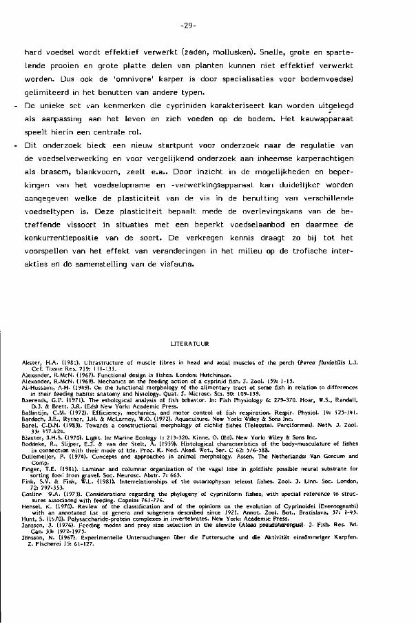
opname van partikels slokken
voedselbrok zand /steentjes' organisch vui)-^
transport
vertering
FIG. 2. Bewegingspatronen van de karper behorende bij de mechanismen voor opname en verwerking van verschillende typen voedsel (b.v. bodemmateriaal, zoöplankton). Afhankelijk van plaats, grootte, smaak, verontreiniging en mechanische eigenschappen van het voedsel worden de patronen met wisselende frekwenties in het totaalproces geïntegreerd (vgl. resp. deeltjes opname - slokken, spuwen - terugspoelen en lokale se lekt ie- wassen, p le t ten-malen) . De volgorde der fasen is bij elk patroon aangegeven. Verdere toelichting bij de figuren in hoofdstuk 2.
-19-
mater iaal t reedt né opname in de mondkeelholte op. Welke aspekten in bouw en
akt ie stellen de karper to t een zo gespecialiseerde voedselexploitatie in staat?
Het palataal orgaan van de karper, een omvangrijk en complex gebouwd spierkus-
sen in het gehemelte, is bezet met welhaast maximale dichtheden smaakknopjes
(820/mm2 ; hoofdstuk 3). D i t s t i jgt ver uit boven waarden bij andere vissen, voorzover
gemeten. Reeds in 1827 werd di t orgaan door Weber een rol bij selektie toegeschre
ven. Regionale analyse van bouw en ak t i v i te i t van het palataal orgaan en z i jn analo-
gon in het midden van de keelbodem, het postlinguale orgaan (Plaat II d, pag.90),
toont hun funkties in de voedselverwerking. Drie niveau's van beweging worden
voorgesteld (pag. 113): 1) beweging van het palataal orgaan als geheel als gevolg
van ak t iv i te i t in de dorsale kieuwboogspieren, 2) regionale zwell ingen en per istal t iek
en 3) zeer lokale beweging van gespierde papillen in het palatale oppervlak. D i t
leidt t o t het volgende beeld voor het selektiemechanisme tussen voedsel en n iet-
voedsel.
Zware anorganische part ikels als zand en grind zinken wel l icht voor het grootste
deel d irekt door de wijde eerste kieuwspleet, l ichte organische verontreinigingen
komen met het voedsel in de keel. De grote dichtheid aan smaakknopjes en spierve
zels, vooral in het palataal orgaan maar ook op de kieuwzeef, vormen de basis
voor een mechanisme waarbij voedsel waarschijnl i jk door lokale buitvorming tussen
keeldak en keelbodem kan worden vastgezet. Niet-eetbare part ikels worden niet
vastgezet en bij kompressie via de kieuwspleten met het water naar buiten gespoeld.
De nauwe keelspleet levert een groot selektie oppervlak. De grote dichtheid aan
smaakknopjes, het papi l len- prof ie l van zowel spierkussenoppervlak als kieuwzeef,
en de gemeten d i f ferent ia t ie in lokale ak t iv i te i t van het palataal orgaan duiden
op een groot scheidend vermogen. De laminaire cyto-archi tektuur van de vagus
lob in de nauw verwante goudvis suggereert bovendien palatotopie. Deze ru imte l i jke
afbeelding van de in format ie ui t het palatale orgaan in de hersenen is vermoedeli jk
een voorwaarde voor een zeer lokale bewegingssturing (cf. Finger, 1981). Dat d i t
mechanisme niet d irekt een volledige scheiding teweeg brengt b l i j k t u i t de herhaalde
terugspoelbewegingen naar de mondholte (back-washing), waarin resuspensie van
het materiaal voor een volgende selektiefase plaatsvindt, en u i t de geringe veront
reinigingen in de darm. Protrusiebewegingen met gesloten bek veroorzaken het
terugspoelen naar de mondholte en z i jn bij dit getrapte zuiveringsproces van essen
t ieel belang. Z i j maken de mondholte to t een spoelkamer (Fig. 8, pag.64).
Grove zuivering van grote voedselbrokken, die het kontakt tussen keelbodem en
keeldak beperken, kan ook door intensief wassen via pompbewegingen met een
van mond- naar kieuwholte gerichte waterstroom plaatsvinden (rinsing). Bij sterk
verontreinigd of moei l i jk hanteerbaar voedsel t reedt meestal resuspensie door spuwen
-20-
en her-opname op.
Uitspuwen van onsmakelijk of zelfs toxisch voedsel (bv. watermi j ten) is een
bekend fenomeen, ook bij vissen. Part ikels worden bij gesloten bek door expansie
van de mondholte naar voren gespoeld en daarna door adductie van de kieuwdeksels
via de mond uitgestoten (Fig. 9, pag.66).
Sl i jmkleuringen in de mond-keelholte tonen de aanwezigheid van minstens
twee verschillende typen sl i jm aan (hoofdstuk 3): sialomucines met re lat ief lage
viskositeit in de mond en voorste keelholte, sulfomucines met re lat ief hoge viskosi-
te i t (Hunt, 1970) achter in de keelholte. Het weinig viskeuze s l i jm langs de weg
van het water dient behalve als bescherming waarschijnl i jk ook om turbulenties
in de grenslaag langs de wand te verminderen en daarmee de weerstand voor water
transport te verkleinen (Rosen & Cornford, 1971). Sterk viskeus sl i jm achterin
de keel bevordert waarschijnl i jk het vangen van kleine partikels en hun samenklonte
r ing. De afwezigheid van dit s l i jmtype in het voorste deel van de mond-keelholte
laat ef fekt ieve resuspensie van het materiaal bij zuivering toe. Deze en andere
funkt ies van sl i jm (bv. smering bi j transport) komen ter diskussie in hoofdstuk 3.
Hoe wordt geselekteerd voedsel naar de kauwholte getransporteerd?
Voor zover het voedsel nog niet achter in de keelholte tussen palataal en post-
linguaal orgaan l igt wordt het door gesloten protrusiebewegingen teruggespoeld
en door gekoördineerde aktie van het palataal orgaan en de keelbodem in deze
transportposit ie gemanoeuvreerd (repositioning). Voedselpartikels uit de kieuwzeef
worden welhaast zeker op eenzelfde wijze door terugspoeling verzameld.
Tandendragende kieuwboogelementen in het keeldak zorgen voor transport bi j de
meeste niet-cypriniden (L iem, 1973; Lauder, 1983), maar ontbreken bij karper
achtige vissen. Gekoördineerde kontrakt ies in het achterste deel van palatale en
postlinguale orgaan dr i jven het voedsel langzaam (2-3 cm/sec.) met peristalt ische
golven to t in de kauwholte voort (Fig. 10, pag. 67). De rol van de sterk toegenomen
sl i jmproduktie in dit gebied (sulfomucines) werd reeds vermeld. De dichtheid aan
smaakknopjes neemt in deze zelfde r icht ing af (Fig. 7 , pag. 107).
Hoe vindt de mechanische verkleining van het voedsel plaats?
Het kauwsysteem dient er toe het voedsel zodanig te vervormen, dat het opper
vlak voor de inwerking van spijsverteringsenzymen wordt vergroot en om slecht
doordringbare kapsels te breken. Gezien het ontbreken van een maag is d i t van
groot belang bij karperachtigen.
In tegenstelling t o t zoogdierkaken hebben de keelkaken van de karper een
-21-
groot aantal vrijheidsgraden (4) in beweging. Ze zijn onderling beweeglijk en via
negen paar spieren aan schedel, schoudergordel en kieuwkorf opgehangen. Een glij-
koppeling tussen keelkaken en kieuwbogen laat rostro-caudale translatie toe. De
beweging van deze kaken tegen de kauwplaat van de schedel is verder samengesteld
uit rotaties om vier verschillende anatomische assen (hoofdstuk 4). Het kauwproces
verloopt bilateraal synchroon en is opgebouwd uit een of meer kauwtreinen, elk
samengesteld uit series kauwslagen. Op grond van bewegings- en aktiviteitspatroon
van de kauwspieren worden plet- en maalslagen (resp. crushing en grinding) van
elkaar onderscheiden.
De belasting van het voedsel (kompressie, rek, afschuiving, torsie en buiging)
hangt af van de profielen van de occlusievlakken (tanden en maalplaat) en de rich
ting waarin zij ten opzichte van elkaar bewegen. Het heterodonte kauwapparaat
van de karper (Fig. 2, pag.12^ is slecht uitgerust voor snijden, knippen en uiteentrek-
ken van voedsel maar biedt goede mogelijkheden tot pletten en malen (p.15^. Het
kauweffekt hangt af van de mechanische eigenschappen van het aangeboden voedsel.
Brosse en stijve materialen worden verbrijzeld, meer elastisch, taai en/of vezelig
voedsel wordt vooral geplet. De kauwholte kan slechts beperkt expanderen, delen
met een diameter groter dan ca. 3% van de lichaamslengte kunnen er niet in.
Het grote kauwoppervlak, de lengte van de kauwslag en ook de aanwezigheid van
zandkorrels in de tandgroeven bevorderen de vermaling van kleine partikels, kwantita
tief en kwalitatief.
Welke kauwdruk kan dit systeem produceren? Dit hangt in de eerste plaats
af van het kontaktoppervlak tussen occlusievlakken en voedsel en zal dus met
de vorm en grootte van het voedsel variëren. Ofschoon de positie van de kauwholte,
midden in de kop, kauwdrukmetingen in de weg staat, geeft vorm-funktie onderzoek
aan dat de kauwkracht groot is. Als argumenten hiervoor gelden ondermeer:
1) De direkt op de keelkaken aangehechte spieren stralen wijd uit naar omringende
beenelementen, maar hun werklijnen vormen gunstige rotatie-koppels rond de
onderhavige assen. Hierdoor worden de krachten van de afzonderlijke spieren
sterk gebundeld (vgl. Fig. 7 pag.134).
2) De keelkaken vormen lange uitsteeksels en vleugels die, behalve dat zij spieren
een groot oppervlak voor aanhechting bieden, grote momentarmen leveren. De
kauwvlakken liggen dicht bij de rotatie-assen van de keelkaken en brengen zo
vergrote krachten op het voedsel over. Alleen de transversale as, voorin door
de symfysis van de keelkaken, ligt ver van de tanden verwijderd en laat grotere
amplitudes van de keelkaken toe. Dit is van belang tijdens expansie van de kauw
holte bij het opladen van het voedsel.
3) De rug- en buikspieren van de karper zijn tijdens het kauwen actief en dragen
-22-
hun krachten op de kauwplaat en keelkaken over via resp. de schedel en de
schoudergordel en via kauwspieren met pezige komponenten. Vergeleken bij
de d irekte kauwspieren leveren zij een zeer groot vermogen (vgl . F ig . 20, pag.155).
4) De hoge schedel en lange schoudergordel vormen grote momentarmen voor deze
l ichaamsspieren. De ligging van de kauwplaat vlak onder het ro ta t iecentrum
van de schedel resulteert in een kleine doch zéér krachtige beweging van de
kauwplaat, tegengesteld aan die van de tanden. De kauwplaat werd in de l i te ra
tuur to t nu toe door haar f ixat ie in de schedelbasis zonder uitzondering van
de kauwbeweging uitgesloten.
De grote kauwkracht stelt eisen aan het kauwapparaat wat be t re f t s tur ing, stabil isa
t ie en absorbtie van reakt iekrachten. D i t is des te meer van belang daar de keelka
ken in spieren zi jn opgehangen en het hart van de vis d irekt tussen de keelkaken
l igt en door deze overdekt wordt (Plaat II d, pag.90 ). Sturing en stabil isatie wordt
behalve door l igamenten tussen en achter de keelkaken vooral ook bereikt door
de geprogrammeerde aktie van de kauwspieren, die elkaar door antagonistische
werking in e f fekt balanceren (F ig. 15, p. 147). De pharyngo-cleithral is externus spier
speelt hierbij een s leutelrol . Beenbalkjes die zowel vanuit de tanden over de keel
kaken als vanuit de kauwplaat-kom over de schedelbasis u i tstralen zorgen voor
een gel i jkmatige spreiding van reakt iekrachten en voorkomen zo te sterke belasting
van het eigen systeem.
De vereiste rotatieslag van de keelkaken is ondermeer mogeli jk dankzij de gl i jkoppe-
ling in de basis van de k ieuwkorf. De ak t iv i te i t van de spierketen in de bodem
van de kop draagt zowel bij in voor- en achterwaartse bewegingen van de schouder
gordel bij het kauwen, als to t het vergroten van de slagruimte van de keelkaken.
De sterke expansie van de kop en het open gaan van de bek bij het kauwen is
meer een gevolg van het kauwproces dan dat het to t voedselopname of respiratie
dient. Het kauw- en ademhalingsritme verschil len overigens niet belangri jk.
Het kauwapparaat beperkt zich dus niet t o t de keelkaken en de daarop aange
hechte spieren. Kauwen vereist bewegingen van de kop als geheel en van de rug
en buik van de vis en beperkt daarmee het ge l i jk t i jd ig ui tvoeren van andere funkties
(bv. respirat ie, opname, selektie). De ro tat ie van de schedel to t ca. 15° maakt
tevens de loop van de botjes van Weber, dicht langs het rotat iecentrum van de
schedel welhaast to t een strukturele noodzaak. De opbouw van deze verbinding
tussen middenoor en zwemblaas uit vele elementen l i j k t een kompromis, dat zowel
het overbrengen van t r i l l ingen als de beweging van de schedel bij het kauwen toe
laat. Het is te verwachten dat de f rekwent ie gevoeligheid van het gehoor en de
bij het kauwen geproduceerde t r i l l ingen weinig overlap hebben. Bij het breken van
maiskorrels z i jn de kauwslagen van de karper to t buiten het aquarium hoorbaar!
-23-
Het doorslikken van het voedsel
Transport van verkleind voedsel naar de darm geschiedt door middel van kom-
pressie van de kauwholte door de constr ictor pharyngis spier, ondersteund door
een kleine beweging van de keelkaken. Het palataal en postlinguaal orgaan sluiten
door opzwelling de toegang to t de kauwholte af en r ichten daarmee het transport
naar de slokdarm. Ak t i v i t e i t van de constr ictor pharyngis zet zich wel l icht per ista l
t isch in de slokdarm voort .
Hoe stemt de karper zijn gedrag af op de eigenschappen van het voedsel?
De procesgang bij verwerking van verschillende typen voedsel (pellets, gerst,
wo rm, tubi fex, watervlooien, bodem-tubifex mengsels) duidt er op dat bovengenoem
de deelprocessen een in beweging en ak t iv i te i t stereotyp karakter dragen ( ' f ixed
act ion patterns'?; Baerends, 1979). Z i j kunnen als herkenbare eenheden van gedrag
worden onderscheiden. Afhankeli jk van positie, groot te en aard van het voedsel
worden zij in herkenbare volgorde maar met wisselende f rekwent ie ingezet (bv.
part ikel opname-slokken, p letten-malen), hetgeen leidt to t variaties in het totale
verwerkingsproces (hoofdstuk 2, pag. 56 ). De voor de totale voedselverwerking bij
één opname benodigde t i jd (handling t ime) kan direkt uit de electromyogrammen
worden afgelezen en b l i jk t voor verschillende voedseltypen sterk te verschillen
(Fig. 3-4, p. 53 ). Z i j wordt vooral bepaald door herhaling van voor een voedseltype
specif iek benodigde deelprocessen. Zo is de 'handling t ime' voor grote part ikels
vooral door herhaald kauwen lang, te rwi j l bi j verontreinigd voedsel voortdurend
terugspoelen bij selektie veel t i jd vergt. Gesloten protrusie b l i jk t een kernpatroon,
dat aan de basis l igt van meerdere deelprocessen (positioneren, terugspoelen, verza
meling voedsel van de kieuwzeef), die op grond van hun ef fekt op het voedsel
worden onderscheiden. Spuwen en monsteren van bodemmateriaal (probing) leiden
niet to t opname in het darmkanaal en worden als aparte seguenties onderscheiden.
De mechanismen die deze gedragskomponenten opwekken, vasthouden en stoppen
z i jn onbekend, evenals de regulatiemechanismen die de deelpatronen to t een e f f i
ciënt voedingsproces samenvoegt. Afstemming van de kauwdruk op de momentane
eigenschappen van het voedsel en registrat ie van de kondit ie van het voedsel, zodat
sl ikken op het ju iste moment begint, z i jn bijvoorbeeld noodzakelijk voor een e f f ic iënt
verloop van het kauwproces. Of proprioceptoren in de spieren, dan wel zintuigen
in de weefselflappen die tussen tanden en kauwplaat uitsteken en hen waarschijnl i jk
schoonvegen, hierbij een rol spelen is onbekend. Smaakknopjes en ol igovil le cel len
hebben mogelijk mechanoreceptieve funkt ies en z i jn de enige sensoren die vooralsnog
in de mond-keelholte van de karper struktureel werden aangetoond (hoofdstuk 3).
-24-
Over funktionele verbindingen tussen proprioceptoren en het centrale zenuwstelsel
is alleen voor de ademhalingsregulatie een en ander bi j de karper bekend (Bal l in t i jn ,
1972; Lu i ten, 1977).
Welke mogelijkheden en beperkingen heeft de karper om de beschikbare voedselbron
nen te benutten?
Naar de aard van het voedsel worden algemeen carnivore, herbivore, det r i t ivore
en omnivore vissen onderscheiden (Nikolsky, 1963). Dat de vis zich niet in een
dergelijk schema laat ordenen wordt overtuigend geïl lustreerd aan de Afrikaanse
Haplochromis soorten (cichliden) waarin de trof ische d i f ferent ia t ie zich zo sterk
manifesteert , dat een verregaande opsplitsing van deze trof ische hoofdgroepen
noodzakelijk is. Ook sterk gevarieerde mengvormen komen voor. Barel (1983) laat
voor deze groep zien, dat binnen eenzelfde voedselkategorie de funktionele eisen
aan het voedingsapparaat zeer verschil lend kunnen zi jn (eieren zuigen van de bodem
- eieren zuigen uit een bek-broedend vrouwtje), t e rw i j l ook verschillende kategorieën
dezelfde eisen kunnen stel len (inwendig transport van zoöplankton en eieren). Het
zal duidelijk zi jn dat ook de mechanische eigenschappen van het voedsel niet eenvou
dig met de vermelde voedselkategorieën overeenkomen (vgl . s lak-worm; slak-planten-
zaden). Bovendien wordt de darminhoud bij voedselovervloed sterk bepaald door
het voedselaanbod. Ook het voederen met pellets in de visteelt maakt de vis nog
niet to t een gespecialiseerde pel letfeeder.
De ef f ic iency van de voedselopname wordt waarschi jnl i jk vooral bepaald door
de vereiste behandelingstijd ('handling t ime') en de verhouding tussen bestede en
verworven energie. In een si tuatie van voedselschaarste spelen specialisaties in
bouw en gedrag een belangrijke ro l . De soortspecifieke komponent in het dieet
van de vis wordt waarschijnl i jk bepaald door specialisaties voor dat voedsel waarmee
ze in ef f ic iency van opname en verwerking boven andere soorten u i ts t i jg t . Zo b l i jk t
de graskarper beter dan de andere vissen in staat planten als voedselbron te benut
ten. Well icht speelt de gespecialiseerde bouw van het kauwapparaat hierin een
beslissende ro l . Het is te verwachten dat specialisaties voor het ene deelproces
die voor andere beperkt. Zo vereist de selektie tussen voedsel en niet-voedsel bij
de karper een nauwe keelspleet en reduceert daarmee de rol van de keelholte bij
het zuigen.
De specialisaties voor deelprocessen in de karper en de beperkingen die hier
voor de benutting van het voedselaanbod u i t voortvloeien worden voor elk deelproces
zo goed mogelijk aangegeven in hoofdstuk 2. Optel l ing van deze restr ikt ies le idt
t o t het volgende beeld. Slechts langzame en kleine voedseldelen (< ca. 9% van
-25-
de l ichaamslengte van de karper (SL) in diameter) kunnen worden opgenomen en
deze hebben slechts gedeeltel i jk to t de kauwholte toegang (< ca. 3% SL). Fi jne
part ikels ( < 250 urn) gaan door de kieuwzeef verloren. Taaie en vezelige mater ialen
kunnen slecht worden gebroken, waardoor de e f f ic iënte vertering van plantaardig
voedsel sterk wordt beperkt. D i t vernauwt het e f f ic iënt te benutten voedselskala
van deze 'omnivore' vis goeddeels to t plantenzaden, macro- en micro-evertebraten
(250 ym - 3% SL).
Als voornaamste specialisaties gelden het benutten van harde, brosse en st i jve
voedselobjekten (plantenzaden, mollusken) en het exploiteren van voedsel dat met
bodemmateriaal is vermengd. U i t de l i teratuur b l i jk t opvallend genoeg dat de darm-
inhoud van deze euryfage vis soms meer (weliswaar zachte) macrofyten bevat
dan verwacht. Gebrek aan ander voedsel kan hier mede oorzaak van z i jn.
Deze konklusies strekken zich niet u i t to t jonge karpers. Over het verband
tussen hun specifieke dieet (vg l . Ur ibe-Zamora, 1975) en de relat ieve groei van
de verschillende kopelementen ti jdens de kr i t ieke juveniele fase is nog weinig be
kend. Het mechanisme van prooivangst in relat ie to t de groei wordt momenteel
aan juveniele karpers onderzocht (M.Drost).
Ontwikkel ing van het protrusie-systeem van de bovenkaak, het palatale en
postlinguale orgaan in de keel, en de kauwplaat in de schedelbasis hebben waarschijn
l i jk door het exploiteerbaar worden van de bodem belangrijk bijgedragen to t het
grote succes van de cypriniden in het zoete water. De set van unieke strukturele
kenmerken die de cypriniden en catostomiden karakteriseert en ver uiteen liggende
delen van de kop omvat wordt vanuit een sterke ontwikkeling van het keelmaalappa-
raat in een funktioneel verband geplaatst (p. 7 5 ) . In hoofdstuk 3 wordt het plausibel
gemaakt dat de karperachtigen zich door specialisatie op de bodem-voedselbronnen
u i t predatoire voorouders hebben ontwikkeld.
Een verdere ui twerking van ethologische en ekologische aspekten en vergeli jking
met andere soorten cypriniden zal t o t toetsing en verf i jning van gestelde hypotheses
en konklusies bi jdragen. D i t zal ook t o t meer inzicht leiden in de t rof ische segrega
t ie van verschillende soorten, die naast elkaar in één levensgemeenschap voorkomen.
•26-
Samenvattinq en konklusies
In dit proefschr i f t worden de onderdelen beschreven van het proces van voedsel-
opname en -verwerking bij de karper (Cyprinus carpio L.), de bouw van de l ichaams
delen die hierbij betrokken zi jn en de funkties van de afzonderli jke deelprocessen
en s t rukturen. Het doel is de samenhang tussen de architektuur van de karperkop
en zi jn funkt ies te bepalen en inzicht te geven in het naast elkaar bestaan van
verschillende vissoorten in een levensgemeenschap door naast de specialisaties
van deze vis voor bepaalde voedselsoorten de daaruit voortvloeiende beperkingen
voor andere vast te stel len.
- De cypriniden of karperachtige vissen bezi t ten to t keelkaken gemodificeerde
v i j fde kieuwbogen, die tegen een kauwplaat worden bewogen door oorspronkelijke
kieuwboogspieren. De bek draagt geen tanden. Een maag ontbreekt. De bovenkaak
van de karper is uitstulpbaar, waardoor een ronde zuigmond gevormd kan worden.
De mond- en kieuwholte kunnen sterk in volume veranderen. De keelholte wordt
v r i jwel geheel ingenomen door een dorsaal gespierd palataal orgaan. Ventraal
l ig t het postlinguaal orgaan en de kieuwzeef. He t oppervlak van keeldak en
bodem is bijna geheel bedekt met smaakknoppen ( to t 820/mm2) en s l i jmcel len.
De spleetvormige keelholte kan maar zeer beperkt in volume variëren, in tegen
stell ing t o t bi j de meeste andere vissen. Het bij dit pharyngeaal systeem behoren
de hersencentrum l igt in het verlengde merg en benadert in omvang de grote
hersenen.
- De ro l , die deze structuren bij de opname en verwerking van voedsel vervullen
is bij de karper onderzocht m.b.v. gewone- en röntgenfi lmopnamen van levende
vissen. Meting van ak t iv i te i t in de bi j d i t proces betrokken spieren werd, syn
chroon op negen kanalen, gekoppeld aan deze bewegingsstudies. Het voedsel
bestond uit visvoerpellets, graan, wormen, watervlooien, tubifex en bodemma
ter iaal vermengd met tubi fex. Door impregnatie van voedsel met BaSO^ werd
de weg van het voedsel op de röntgenf i lm zichtbaar gemaakt. Struktuur onderzoek
geschiedde op macroscopisch, l ichtmicroscopisch en scanning-electronenmicro-
scopisch niveau.
- Elk eetproces b l i jk t uit een wisselend aantal stereotype bewegingspatronen opge
bouwd: deeltjes opname (part iculate intake) of slokken (gulping), selektie tussen
eetbare en niet-eetbare delen (wassen, positioneren of terugspoelen), verzamelen
van het door de kieuwzeef gevangen mater iaal , transport van voedsel en het
vullen van de kauwholte, p le t - en/of maalbewegingen afgesloten door sl ikken.
Monsteren van de bodem en spuwen worden apart beschouwd.
-27-
Het moment en de mate en snelheid van bekopening, protrusie van de bovenkaak,
opening van de kieuwdekselklep, en van de volumeveranderingen in mond-, keel-
en kieuwholte bepalen samen het e f fect van elk patroon. Opname, selektie,
transport en kauwen stel len verschillende eisen aan de kop en zijn alleen los
van elkaar e f fekt ie f uitvoerbaar.
De opbouw van het voedselopname proces uit de genoemde stereotype bewegings
patronen varieert met de grootte, aard en verontreiniging van het voedsel. De
totale hanteert i jd van een voedseltype, te meten aan het e lectromyogram, var i
eert vooral door herhaling van voor dót voedsel specifiek vereiste bewegings
patronen.
De kwant i tat ieve verdeling van smaakknoppen, s l i jmcel len, clubcellen en spier
vezels is in mond- en keelholte gemeten. Op grond van deze verdeling en andere
morfologische kenmerken worden zes gebieden in de mond-keelholte onderscheiden
en aan de deelprocessen voor voedselopname en -verwerking gerelateerd. Scan
ning E.M. beelden worden gegeven van gewone epitheelcellen met microrichels,
verhoornde cel len, s l i jmcel len, smaakknoppen en ol igovi l le (sensorische ?) cel len.
Ger ichte opname van deeltjes (part iculate intake) wordt bereikt door een snelle
en volumineuze zuigbeweging, opgewekt door vergroting van de mond- en kieuw
holte. De bovenkaken worden hierbij vooruitgestulpt (protrusie) waardoor een
snelle en naar het voedsel gerichte waterstroom (ca. 60 cm/sec.) ontstaat.
'Gulping' , het langzaam en ongericht opslokken van water met gesuspendeerd
voedsel, geschiedt door vergroting van alleen de mondholte. Door het naar bene
den stulpen van de bovenkaak sluit de vis de suspensie in. Kompressie van de
mondholte perst de suspensie door de kieuwzeef. De energie nodig voor een
slokbeweging zal beduidend minder zi jn dan die voor deeltjes-opname.
Grote dichtheden clubcellen, die in de huid een voor cypriniden kenmerkende
alarmstof produceren, komen ook en alleen in de mondholte voor. Hun a larmfunk-
t ie op deze plaats wordt in tw i j fe l getrokken.
Selektie vereist het vasthouden van eetbare en de afvoer van niet eetbare deel
t jes. U i t e lectromyogrammen en electrische s t imulat ie val t af te leiden dat
deze scheiding plaats vindt door de vorming van bultjes op het palataal orgaan.
Tussen dit orgaan en de kieuwzeef zullen eetbare delen worden vastgezet; n ie t -
eetbare worden weggespoeld. De complexe bouw van het palataal orgaan, de
bijna maximale dichtheden van smaakknopjes en de cyto-archi tektuur van het
hierbij betrokken regelcentrum in de achterhersenen wi jzen op een groot schei
dend vermogen. De brede spleetvormige keelholte waarborgt een groot kontakt-
oppervlak tussen dak en bodem maar beperkt tevens haar ro l bij het zuigen.
-28-
De vergroting van de mondholte door protrusie met gesloten bek speelt bi j selek-
t ie een belangrijke rol door het mengsel van voedsel en niet-voedsel in de mond
holte te resuspenderen. Door afwisselend expanderen en komprimeren van de
mondholte wordt een op en neer gaande stroming door de kieuwspleet en kieuw-
zeef bereikt. Voortdurende herhaling van 'gesloten protrusie' en selektie leidt
to t steeds verdere zuivering van het voedsel. Gesloten protrusie speelt ook een
rol bij het positioneren van grotere delen en bij het verzamelen van het f i l t r aa t
u i t de kieuwzeef.
Sl i jm met lage viskositeit (sialomucinen) wordt aangetroffen in de voorste delen
van mond- en keelholte. Het dient mogelijk voor de verlaging van de stromings
weerstand langs de wand en beschermt het onderliggende weefsel. Achter in
de keelholte waar aggregatie van voedseldeeltjes voor verder transport verwacht
moet worden, vinden we cellen die veel en sterk viskeus sl i jm (sulfomucinen)
produceren. In s l i jm gevat voedsel wordt via een gekombineerde per istal t iek
in palataal en postlinguaal orgaan naar en in de kauwholte getransporteerd.
De keelkaken zijn onderling beweeglijk in spierlussen opgehangen en door een
gl i j-koppeling met de kieuwkorf verbonden. Voedsel wordt geplet en/of gemalen
tussen de keeltanden en de verhoornde maalplaat aan de schedelbasis. Bij het
maalproces kunnen mais en gerstekorrels worden verbri jzeld met een buiten
het aquarium hoorbaar geluid.
Aangetoond is dat de rugspieren via schedelrotatie belangrijk bijdragen aan de
p le t - en maalkrachten. Via de schoudergordel dragen ook de buikspieren hun
krachten met een grote momentarm op de keeltanden over. De konstruktie van
het maalapparaat is er op gebouwd hoge kauwdrukken te produceren en te weer
staan.
In tegenstell ing t o t deze 'kracht-spieren' , 'zijn de direkt op de keelkaken aange
hechte spieren meer 'stuur-spieren'. Z i j r ichten en stabiliseren de beweging
van de keelkaken om vier anatomische assen.
De werking van het maalapparaat met de beweeglijke schedel maakt een starre
keten van 'gehoorbeentjes' tussen zwemblaas en het inwendige oor onmogeli jk.
De vele beentjes van Weber en hun loop langs de rotat ie-as van de schedel l i j k t
daarom een konstruktieve noodzaak.
Slikken geschiedt door kompressie van de kauwholte, waarbij afslui t ing van haar
toegang door het palatale en postlinguale orgaan het transport naar de slokdarm
r ich t . Beweging van de keelkaken ondersteunt d i t t ransport.
Het voedselopname-apparaat van de karper b l i j k t gespecialiseerd voor het ver
werken van kleine voedseldelen (van 250 urn tot ca. 3% van de l ichaamslengte)
en voor voedseldelen die vermengd met oneetbare delen voorkomen. K le in en
-29-
hard voedsel wordt e f fekt ie f verwerkt (zaden, mollusken). Snelle, grote en sparte
lende prooien en grote p lat te delen van planten kunnen niet e f fekt ie f verwerkt
worden. Dus ook de 'omnivore' karper is door specialisaties voor bodemvoedsel
gel imi teerd in het benutten van andere typen.
De unieke set van kenmerken die cypriniden karakteriseert kan worden uitgelegd
als aanpassing aan het leven en zich voeden op de bodem. Het kauwapparaat
speelt hierin een centrale ro l .
D i t onderzoek biedt een nieuw startpunt voor onderzoek naar de regulatie van
de voedselverwerking en voor vergeli jkend onderzoek aan inheemse karperachtigen
als brasem, blankvoorn, zeelt e.a.. Door inzicht in de mogelijkheden en beper
kingen van het voedselopname en -verwerkingsapparaat kan duideli jker worden
aangegeven welke de p last ic i te i t van de vis in de benutting van verschillende
voedseltypen is. Deze p last ic i te i t bepaalt mede de overlevingskans van de be
tref fende vissoort in situaties met een beperkt voedselaanbod en daarmee de
konkurrentieposit ie van de soort. De verkregen kennis draagt zo bij t o t het
voorspellen van het e f fekt van veranderingen in het mi l ieu op de trof ische inter-
akties en de samenstelling van de visfauna.
LITERATUUR
Akster, H.A. (1981). Ultrastructure of muscle fibres in head and axial muscles of the perch (Perca fluviatilis L.). Cell Tissue Res. 219: 111-131.
Alexander, R.McN. (1967). Functional design in fishes. London: Hutchinson. Alexander, R.McN. (1969). Mechanics on the feeding action of a cyprinid fish. 3. Zool. 159: 1-15. Al-Hussaini, A.H. (1949). On the functional morphology of the alimentary t ract of some fish in relation to differences
in their feeding habits: anatomy and histology. Quat. 3. Microsc. Sei. 90: 109-139. Baerends, G.P. (1971). The ethological analysis of fish behavior. In: Fish Physiology 6: 279-370. Hoar, W.S., Randall,
D.3. & Brett , 3.R. (Eds) New York: Academic Press. Bal l int i jn, C M . (1972). Efficiency, mechanics, and motor control of fish respiration. Respir. Physiol. 14: 125-141. Bardach, 3.E., Ryther, 3.H. & McLarney, W.O. (1972). Aquaculture. New York: Wiley & Sons Inc. Barel, C.D.N. (1983). Towards a constructional morphology of cichlid fishes (Teleostei. Perciformes). Neth. 3. Zool.
33: 357-424. Blaxter, 3.H.S. (1970). Light. In: Marine Ecology 1: 213-320. Kinne, O. (Ed). New York: Wiley & Sons Inc. Boddeke, R., Slijper, E.3. & van der Stelt, A. (1959). Histological characteristics of the body-musculature of fishes
in connection with their mode of l i fe . Proc. K. Ned. Akad. Wet., Ser. C 62: 576-588. Dullemeijer, P. (1974). Concepts and approaches in animal morphology. Assen, The Netherlands: Van Gorcum and
Comp. Finger, T.E. (1981). Laminar and columnar organization of the vagal lobe in goldfish: possible neural substrate for
sorting foot' f rom gravel. Soc. Neurose. Abstr. 7: 665. Fink, S.V. & Fink, W.L. (1981). Interrelationships of the ostariophysan teleost fishes. Zool. 3. Linn. Soc. London,
72: 297-353. Goslinp 'V.A. (1973). Considerations regarding the phylogenyof cypriniform fishes, w i th special reference to struc
tures associated with feeding. Copeia: 761-776. Hensel, K. (1970). Review of the classification and of the opinions on the evolution of Cyprinoidei (Eventognathi)
w i th an annotated list of genera and subgenera described since 1921. Annot. Zool. Bot-, Bratislava, 57: 1-45. Hunt, S. (1V70). Polysaccharide-protein complexes in invertebrates. New York: Academic Press. 3anssen, 3. (1976). Feeding modes and prey size selection in the alewife (Alosa pseudoharengus). 3. Fish. Res. Bd.
Can. 33: 1972-1975. 3onsson, N. (1967). Experimentelle Untersuchungen über die Futtersuche und die Akt iv i tät einsömmriger Karpfen.
Z. Fischerei 15: 61-127.
-30-
Kapoor, B.G., Smit, H. Ôc Verighina, I.A. (1975). The alimentary canal and digestion in teleosts. Adv. Mar. Biol. 13: 109-239.
Klaauw, C.J. van der (1945). Cerebral skull and facial skull. Arch, néerl. Zool. 7: 16-37. Lauder, G.V. (1983). Functional design and evolution of the pharyngeal jaw apparatus in euteleostean fishes. Zool.
3. Linn. Soc. London, 77: 1-38. L iem, K.F. (1973). Evolutionary strategies and morphological innovations: Cichlid pharyngeal jaws, Syst. Zool. 22:
425-441. Luiten, P.G.M. (1977). Analysis of the proprioceptive reflex loop of respiratory muscles in the carp (Cyprinus carpio
L.). Thesis, University of Groningen, The Netherlands. Matthes, H. (1963). A comparative study of the feeding mechanisms of some Afr ican Cyprinidae (Pisces, Cyprini-
formes). Bijdr. Dierkunde, Amsterdam, 33:3-35. Nelson, J.S. (1976). Fishes of the world. New York: John Wiley and Sons. Nikolsky, G.V. (1963). The ecology of fishes. London: Academic Press. Osse, J.W.M. (1969). Functional morphology of the head of the perch (Perca fluviatilis L.): An electromyographic
study. Neth. J . Zool. 19: 289-392. Roberts, T.R. (1973). Interrelationships of Ostariophysans. In: Interrelationships of fishes: 373-396. Greenwood, P.H.,
Miles, R.S. & Patterson, C. (Eds). London: Academic Press. Rosen, M.W. & Cornford, N.E. (1971). Fluid f r ict ion of fish slimes. Nature 234: 49-51. Stroband, H.W.J. (1980). Structure and function of the digestive t ract of the grasscarp. Thesis, University of Wage
ningen, The Netherlands. Suyehiro, Y. (1942). A study on the digestive system and feeding habits of f ish. Jap. J . Zool. 10: 1-303. Uribe-Zamora, M. (1975)* Selection des proies par le f i l t re branchial de la carpe miroir (Cyprinus carpio L.). Thesis,
University of Lyon. Verigina, I.A. (1969). Ecological and morphological peculiarities of the alimentary system in some carps. In: Ichthyo
logy, 88-117. Poznanin, L.P. (Ed). New Delhi: Ameril led Publ. Comp. Wheeler, A. (1978). Key to the fishes of Northern Europe. London: Frederick Warne Publishers. Wright, D.I. <3c O'Brien, W.J. (1984). The development and f ie ld test of a tact ical model of the planktivorous feeding
of white crappie (Pomoxis annularis). Ec. Mon. 54: 65-98.
-31-
HOOFDSTUK 2
FOOD HANDLING IN THE CARP (Cyprinus carpio L.), ITS MOVEMENT PATTERNS,
MECHANISMS, AND LIMITATIONS.
Ferdinand A. Sibbing
Jan W.M. Osse and
Ar ie Terlouw
-32-
SUMMARY
The oro-pharyngeal feeding mechanism of the carp was analyzed as a case
study for cyprinids. L ight and X-ray cinematography combined w i th e lectromyo
graphy allowed a detailed analysis of the external and internal events during pro
cessing of the fol lowing food types: radiopague pellets, earthworms, barley, tub i -
f ic ids, cladoceran suspensions and food-soil mixtures. In feeding twelve funct ional
patterns of headmovements and act iv i t ies are d ist inct . Part iculate feeding and
gulping for intake; r insing, repositioning or back-washing and spit t ing for selection;
recol lect ion f rom the branchial sieve, t ransport, loading of the teeth, crushing,
grinding and deglut i t ion. The mechanism of each single pattern is discussed. The
expansion mechanism of the head is versati lely used in food handling by the ade
quate t iming of upper jaw protrusion, volume changes in the oral, buccal and oper
cular cavit ies and of the opening of the mouth and opercular valves. Muscular
cushions in the pharyngeal roof (palatal organ) and f loor (postlingual organ) play
a prominent role in internal selection between food and non-food and in transport.
They permit postcapture selection of food. Protrusion of the upper jaw is crucial
in food processing and serves d i f ferent functions in part iculate intake, gulping
and in selection between food and non-food part icles inside the pharynx.
The effects of the separate movement patterns on the food and the impl icated
restr ict ions for processing d i f ferent types of food are discussed. Tentat ive l imi ts
are set to the feeding on the available food types in the environment. The feeding
apparatus appears to be i l l constructed for exploit ing very small part icles (<250ym),
plant and other materials of f ibrous context. Only slow and immobile food part icles
w i th a diameter up to ca. 3% of the carps body length are e f fect ively processed.
The carp appears to be a generalist in its diet w i th specializations for the exploi
tat ion of food and non-food mixtures f rom the bot tom, even if the contained food
is of considerable density and hardness.
The distinct elements of feeding behaviour are considered to be stereotyped
act ion patterns. They are released and steered according to the actual size, d istr ibu
t ion , consistency and contamination of the food and integrated into varied feeding
sequences. D i f fe rent food types require d i f ferent 'handling t imes' due to the varied
repet i t ion frequency of specific patterns wi th in one feeding sequence, related to
the properties of the food. Probing and spit t ing do not lead to ingestion and are
employed in search and re ject ion. Protrusion w i th closed mouth appears to be a
core pat tern in food handling as i t is basic to repositioning and back-washing during
pur i f icat ion of food. I t w i l l also serve recol lect ion of retained food f rom the bran
chial sieve.
-33-
Protrusion and the palatal and postlingual organs in this lower teleost are
discussed in re lat ion to the hypertrophy of the pharyngeal masticatory apparatus,
the recru i tment of body power for masticat ion and to the evolutionary loss of
toothed upper pharyngeal transporting bones. Protrusion as well as these sensory
muscular organs are basic to the substrate feeding habits for many cyprinoids.
The significance of funct ional morphology, ethology and ecology for the study
of l imi tat ions in the exploi tat ion of available niches is emphasized.
Contents
summary 32
introduct ion 34
materials and methods 35
results 36
. subdivisions of the headgut 36
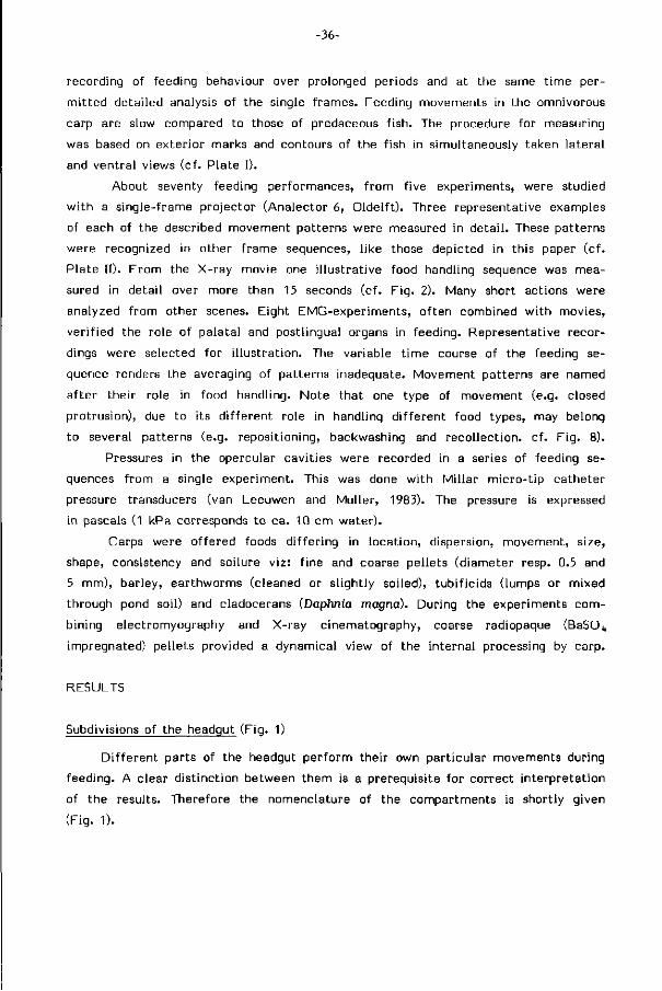
. twelve movement patterns compose the food processing sequences 37
. the role of the palatal and postlingual organ in food handling 50
. adjustment of the feeding sequences to the type of food 52
food handling mechanisms and their l imi tat ions 56
. search and detect ion of food, a survey 57
. food intake 58
. selection 63
. transport and loading 67
. mast icat ion 68
. deglut i t ion 71
. l imi tat ions of the adult carp in the exploi tat ion of food 71
behavioural adjustments in food processing and handling t ime 73
general discussion 75
. retracing the cyprinid feeding mechanism 75
. protrusion 77
references 78
-34-
INTRODUCTION
Abundant reports exist on feeding structures, pointing to their adaptive charac
ter. Paradoxically, few experimental evidence assess their role in feeding.
Over the last decades gradually more detailed analyses of funct ion have been per
formed in f ish to supply evidence for such feeding adaptations (Alexander, 1969;
Osse, 1969; L i em, 1973; Lauder, 1983). The specializations for food intake have
been focal points for functional anatomists but an overall theory providing testable
hypotheses was lacking. A recent model for suction-feeding (Muller, Osse and Ver
hagen, 1982; Muller and Osse, 1984; van Leeuwen and Muller, 1984) presents the
options which a f ish has to manipulate the f low of prey and water into the mouth.
However intake is only a f i rs t step in food processing, especially in benthic feeding
as employed by cyprinids. Enquiries into the funct ional aspects of dent i t ion and
food processing have been l im i ted in scope and are largely descriptive (Hyat t ,
1979). The need to develop precise, experimental ly based assessments of the mecha
nisms control l ing food exploi tat ion in fishes is apparent. The concealed character
of food processing, occurring inside the complex orobranchial cavi ty, largely obstruc
ted a detailed analysis of food handling once i t is entered the mouth.
This paper studies the external and internal food processing in the carp. X-ray
cinematography combined to electromyography recently allowed a detailed funct ional
analysis of pharyngeal mastication in the carp (Sibbing, 1982). Similar techniques
are applied here to analyse movement and act iv i t ies of the head parts, and the
path fol lowed by part icles f rom intake to deglut i t ion. We seek for dist inct move
ment patterns in feeding behaviour, to analyse their mechanism and to explain
their e f fect for food handling. This knowledge is used to describe optimizations
and restr ict ions of the feeding apparatus to exploit part icular food types in the
environment.
The fol lowing feeding elements are dist inct for the carp. Search and detection
of food, intake, gustation and selection between food and non-food, recol lect ion
of food f rom the branchial sieve, t ransport, mast icat ion, deglut i t ion and digestion.
Each single species w i l l have i ts specific set of structures and actions contr ibut ing
to the specif ic i ty of its feeding behaviour. Detai ls of the oro-pharyngeal l ining
and their role in food processing is presented in a concurrent paper (Sibbing et
a l . , 1984).
Selective pressure acts through the ef f ic iency of behaviour. Structures and
their actions allow as well as l im i t the exploi tat ion of the environment by the
f ish. The mechanisms of food processing in cyprinids should be understood to deter
mine their e f f ic iency. L im i ts thus set on the usable food are important to distinguish

-35-
the exploitable niches in the aquatic ecosystem.
Such knowledge is required when studying f ish-food and f ish-f ish interactions or
even exploit ing them in management and control of natural , or a r t i f i c i a l , f ish re
sources. Cyprinids have a high impact on aquatic systems and f isheries.
The Cyprinidae assemble the largest (freshwater) f ish family w i th a wide ecolo
gical and trophical d i f ferent ia t ion. Contrary to non-cyprinoid teleosts cyprinids
lack teeth on the jaws but instead have strong pharyngeal teeth impacting on a
corni f ied chewing pad (Sibbing, 1982). They include many bottomfeeders which
take portions of soiled food. The actual separation between food and non-food
proceeds in the orobranchial cavi ty.
St imulat ing studies on the buccopharyngeal feeding mechanism in cyprinids (Girgis,
1952; Matthes, 1963; Robotham, 1982) lack funct ional data to veri fy hypotheses.
For example, the size-selective ef fect of the branchial sieve w i th i ts g i l l rakers
is generally agreed upon (Zander, 1906; Iwai , 1964). The detailed mechanism of
size-selection and i ts ef f ic iency is however s t i l l unclear.
The omnivorous common carp (Cyprinus carpio L.) is a pract ical choice for this
study. Extensive data are available on its feeding habits and also f r om other d isci
plines, affording a wide p la t form of knowledge necessary for relat ing the s t ruc
tures of the feeding apparatus to the usable food in i ts habi tat .
MATERIALS AND METHODS
A l l mirror-carps (Cyprinus carpio L.) hatched in our laboratory culture and
were selected between 28 and 35 cm SL for the experiments. Records of the feeding
events were made at room temperature (+_ 2fJ°C). Care was taken to maintain the
proper pH and n i t r i te content of the water.
Detai ls of operation techniques, the experimental set-up, electromyography and
simultaneous X-ray cinematography have been described in a previous paper (Sibbing,
1982). Shortly summarized, electrodes and radiopaque markers were inserted in
the anaesthetized carp which had been trained to feed in a small cuvet w i th c i rcu la
t ing water. The cuvet keeps the head of the animal w i th in the X-ray beam and
paral lel to the image-intensif ier. I t fur thermore restr icts the volume of water
which would otherwise absorb most X-rays and thereby cause the image of the
carp to be vague or even absent. P lat inum markers allowed accurate measurements
of the movements of the jaws, the orobranchial roof and f loor and many other
s t ructural components of the head (cf. Plate I).
L ight-movies were taken f rom a large tank (80x50x40 cm) using a 16 mm Tele-
dyne DBM 54 camera at f i l m speeds between 24-60 frs/sec. This allowed sustained
-36-
recording of feeding behaviour over prolonged periods and at the same t ime per
mi t ted detailed analysis of the single frames. Feeding movements in the omnivorous
carp are slow compared to those of predaceous f ish. The procedure for measuring
was based on exter ior marks and contours of the fish in simultaneously taken lateral
and ventral views (cf. Plate I).
About seventy feeding performances, f rom f ive experiments, were studied
w i th a single-frame projector (Analector 6, Oldel f t ) . Three representative examples
of each of the described movement patterns were measured in deta i l . These patterns
were recognized in other f rame seguences, l ike those depicted in this paper (cf .
Plate II). F rom the X-ray movie one i l lustrat ive food handling sequence was mea
sured in detail over more than 15 seconds (cf. F ig . 2). Many short actions were
analyzed f rom other scenes. Eight EMG-experiments, of ten combined w i th movies,
ver i f ied the role of palatal and postlingual organs in feeding. Representative recor
dings were selected for i l lustrat ion. The variable t ime course of the feeding se
quence renders the averaging of patterns inadequate. Movement patterns are named
af ter their role in food handling. Note that one type of movement (e.g. closed
protrusion), due to its d i f ferent role in handling d i f ferent food types, may belong
to several patterns (e.g. reposit ioning, backwashing and recol lect ion, cf . F ig . 8).
Pressures in the opercular cavit ies were recorded in a series of feeding se
quences f rom a single experiment. This was done w i th Mi l lar micro- t ip catheter
pressure transducers (van Leeuwen and Muller, 1983). The pressure is expressed
in pascals (1 kPa corresponds to ca. 10 cm water).
Carps were of fered foods d i f fer ing in locat ion, dispersion, movement, size,
shape, consistency and soilure v iz: f ine and coarse pellets (diameter resp. 0.5 and
5 mm), barley, earthworms (cleaned or sl ightly soiled), tubi f ic ids (lumps or mixed
through pond soil) and cladocerans (Daphnia magna). During the experiments com
bining electromyography and X-ray cinematography, coarse radiopaque (BaSO,,
impregnated) pellets provided a dynamical view of the internal processing by carp.
RESULTS
Subdivisions of the headqut (Fig. 1)
D i f ferent parts of the headgut per form their own part icular movements during
feeding. A clear d ist inct ion between them is a prerequisite for correct in terpretat ion
of the results. Therefore the nomenclature of the compartments is shortly given
(Fig. 1).
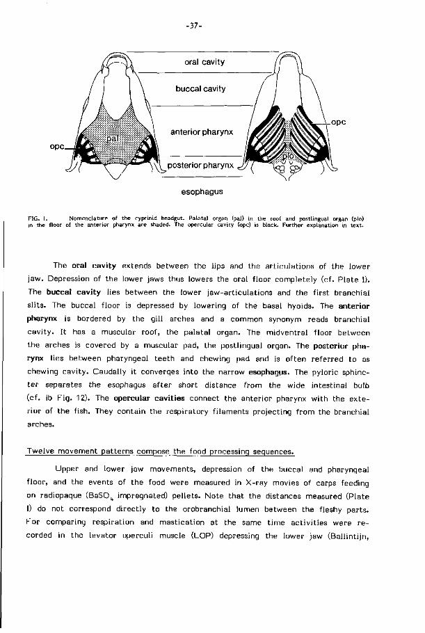
ope
-37-
oral cavity
buccal cavity
ope anterior pharynx
posterior pharynx
esophagus
FIG. 1. Nomenclature of the cyprinid headgut. Palatal organ (pal) in the roof and postlingual organ (plo) in the floor of the anterior pharynx are shaded. The opercular cavity (ope) is black. Further explanation in text .
The oral cavity extends between the lips and the art iculat ions of the lower
jaw. Depression of the lower jaws thus lowers the oral f loor completely (cf. Plate I).
The buccal cavity l ies between the lower jaw-art iculat ions and the f i rs t branchial
s l i ts. The buccal f loor is depressed by lowering of the basal hyoids. The anterior
pharynx is bordered by the g i l l arches and a common synonym reads branchial
cav i ty . I t has a muscular roof, the palatal organ. The midventral f loor between
the arches is covered by a muscular pad, the postlingual organ. The posterior pha
rynx l ies between pharyngeal teeth and chewing pad and is o f ten referred to as
chewing cavi ty. Caudally i t converges into the narrow esophagus. The pyloric sphinc
ter separates the esophagus af ter short distance f rom the wide intestinal bulb
(cf. ib F ig. 12). The opercular cavities connect the anterior pharynx w i th the exte
r ior of the f ish. They contain the respiratory f i laments projecting f rom the branchial
arches.
Twelve movement patterns compose the food processing sequences.
Upper and lower jaw movements, depression of the buccal and pharyngeal
f loor, and the events of the food were measured in X-ray movies of carps feeding
on radiopaque (BaSO^ impregnated) pel lets. Note that the distances measured (Plate
I) do not correspond d irect ly to the orobranchial lumen between the fleshy parts.
For comparing respiration and masticat ion at the same t ime act iv i t ies were r e
corded in the levator operculi muscle (LOP) depressing the lower jaw (Bal l in t i jn ,
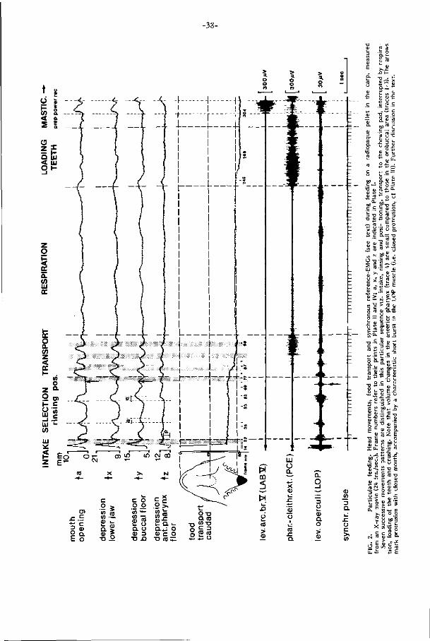
-38-
£ _ s.
i T j <y s >-
N" m
.Q Ó
> (D
u I
(0 JZ a
D O i_ V a o >
SU
a.
o c >» (0
2 s .:: o a. »-1) 03
>*.C
•° l _
t> - : if
£ ? £ 2 ,„ «
ïsi * 3=5
2 -CD U -Q j -
o .c <u^
i a> « £ ^
oc~ c -̂o c
• o a; +3 Q - 1_
~ « i § o ni "O O _
— tu a»
S -o -ira .Ï ro ^ •
I- i ai •
. v M «
" -S x Q. "S > • £ 0
• N f0
J=. (J c
TJ C <o
«-a.
</> c (0 J3
• o O
" H
-J2 c 0)
E (L> O E
•a (0
at
to
a ç
c
CL
"<u .c
o
ID
a> *" at
£
0) E tO
I L L
ci c a>
D
nj CL
(N
'ù V
-+H .3-CN
a» '> o £
(0
X c (0
E o
Z3 ' r; n - ai ÛJ
(/> JO
"3 u
'£. a.
JZ • * - '
c
X> at
-C
'3 00
c
"•6
0) (0
(7
<D
fO Q .
C at E V
o E <D
• a» u u a
c at > at
t / i
c (0 at -C
c
a> 00 c tO
J=
a> E
_2 "o > (0
at
o z 00 ç
U
T3 c ta .c
a> 01
at
^ -O
+;
o -C
u
0) u (0
td
.c o tO
• o at
"c tO Q. £ o u u (0
-C
o E •o at
_o u
JZ
ï c
o _o
11 O Q.
c _o
. J£
Î3 E
-39-
^ PCE ,
orale. \ buccale. \ ant.phar. \p.phar.\
PLATE I. X-ray picture of the common carp. Subdivisions of the oro-pharynx correspond to Fie. 1. Note the metal markers inserted into the skull, jaws, buccal f loor, anterior pharyngeal floor, pectoral girdle, opercula and pharyngeal laws to allow detailed analysis of movements. The pharyngeal jaws and teeth as well as the opposed projection from the skull (solid arrows) accomodating the chewing pad are conspicuous in front of the swimming-bladder (sb). Open arrows indicate the action lines of two pharyngeal jaw muscles connecting them to the skull (LAB V) and pectoral girdle (PCE); a, x, y and z indicate the distances measured in the X-ray frames (cf. Fig. 2).
Parameters measured from the l ight-movies (cf. Plate II) correspond to the distances between the surface-mark on the frontal skull and the t ip of the upper jaw (protrusion), between the tips of lower and upper jaw (mouth opening), between the frontal skull and the lower jaw t ip (depression lower jaw), between the sharp circumorbital edge and the external skin mark below the hyoid (depression buccal floor) and, in ventral view, between the caudal edees of left and right opercula (opercular abduction).
1969) and in two muscles characterist ic for the t ime course and e f fo r t during mast i
cat ion (Sibbing, 1982). These were the levator arcus branchialis V muscle (LAB
V) e f fect ive in crushing and grinding during the power stroke, and the pharyngo-
clei thral is externus muscle (PCE) re f lect ing the preparatory and recovery stroke
(Plate I). Similar and opercular movements were measured f rom the l ight movies
in lateral as wel l as in ventral view.
Combination of data f r om the inter ior and exter ior of the f ish and EMG results
show twelve dist inct movement patterns in feeding, applied in dependence of the
type and condit ion of the food, a) Food intake proceeds by particulate feeding
or gulping, b) Selection is e f fected by d i f ferent patterns, meanwhile tasting the
part ic les. I t proceeds by rinsing when a f i xed part ic le is flushed w i th a rostro-caudad
f low of water. Closed protrusion may result in repositioning, back-washing or re
collection of part ic les. Unwanted part icles may be ejected by spitting, c) Transport
indicates movement of selected food f rom the anterior pharynx to the chewing
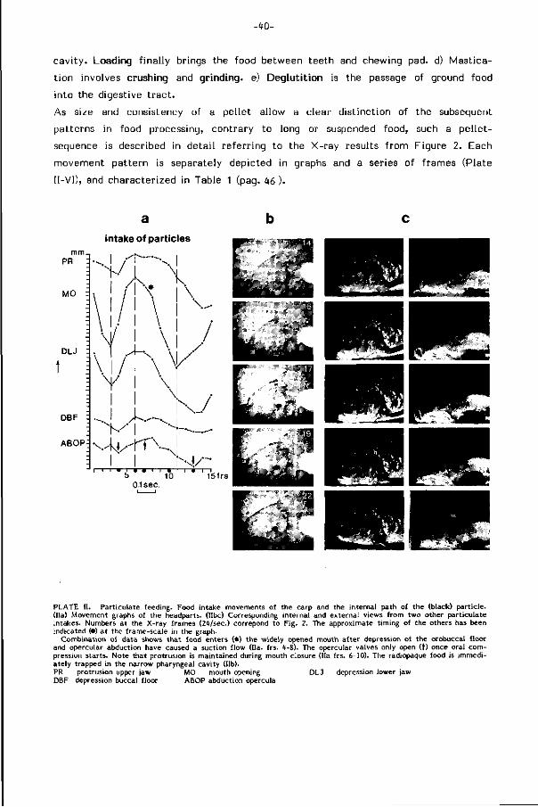
-w-
cavi ty . Loading f inal ly brings the food between teeth and chewing pad. d) Mast ica
t ion involves crushing and grinding, e) Deglutition is the passage of ground food
into the digestive t rac t .
As size and consistency of a pellet allow a clear d ist inct ion of the subsequent
patterns in food processing, contrary to long or suspended food, such a pel let-
sequence is described in detai l referr ing to the X-ray results f rom Figure 2. Each
movement pattern is separately depicted in graphs and a series of frames (Plate
II-VI), and characterized in Table 1 (pag. 46 ).
intake of particles
PR
MO
DLJ
K l /
DBF :
ABOP
/ \ \
V \
\ \ \l
^.y A-—--v
sb-rt v 5 10
Olsec.
\
/ . /
/
A/' 15frs
PLATE I I . Particulate feeding. Food intake movements of the carp and the internal path of the (black) particle. (IIa) Movement graphs of the headparts. (Hbc) Corresponding internal and external views from two other particulate intakes. Numbers at the X-ray frames (24/sec.) correpond to Fig. 2. The approximate t iming of the others has been indicated (•) at the frame-scale in the graph.
Combination of data shows that food enters (*) the widely opened mouth after depression of the orobuccal f loor and opercular abduction have caused a suction flow (Ha. frs. 4-8). The opercular valves only open (f) once oral compression starts. Note that protrusion is maintained during mouth closure (IIa frs. 6-10). The radiopaque food is immediately trapped in the narrow pharyngeal cavity (lib). PR protrusion upper jaw MO mouth opening DLJ depression lower jaw DBF depression buccal floor ABOP abduction opercula
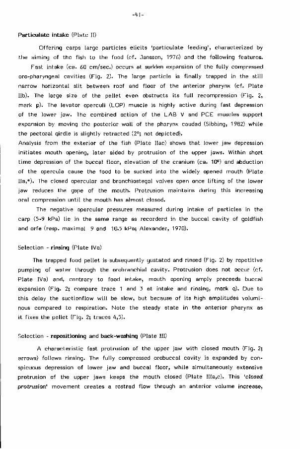
- 41 -
Particulate intake (Plate II)
Offer ing carps large part icles e l ic i ts 'part iculate feeding', characterized by
the aiming of the fish to the food (cf. Janssen, 1976) and the fol lowing features.
Fast intake (ca. 60 cm/sec.) occurs at sudden expansion of the fu l ly compressed
oro-pharyngeal cavit ies (F ig. 2). The large part ic le is f inal ly trapped in the s t i l l
narrow horizontal sl it between roof and f loor of the anterior pharynx (cf. Plate
l ib). The large size of the pellet even obstructs its fu l l recompression (Fig. 2,
mark p). The levator operculi (LOP) muscle is highly active during fast depression
of the lower jaw. The combined action of the LAB V and PCE muscles support
expansion by moving the posterior wal l of the pharynx caudad (Sibbing, 1982) while
the pectoral girdle is sl ightly ret racted (2°; not depicted).
Analysis f rom the exterior of the f ish (Plate Ilac) shows that lower jaw depression
in i t iates mouth opening, later aided by protrusion of the upper jaws. Within short
t ime depression of the buccal f loor, elevation of the cranium (ca. 10°) and abduction
of the opercula cause the food to be sucked into the widely opened mouth (Plate
Ha,*). The closed opercular and branchiostegal valves open once l i f t ing of the lower
jaw reduces the gape of the mouth. Protrusion maintains during this increasing
oral compression unt i l the mouth has almost closed.
The negative opercular pressures measured during intake of particles in the
carp (5-9 kPa) l ie in the same range as recorderd in the buccal cavi ty of goldfish
and orfe (resp. maximal 9 and 10.5 kPa; Alexander, 1970).
Selection - rinsing (Plate IVa)
The trapped food pellet is subsequently gustated and rinsed (Fig. 2) by repet i t ive
pumping of water through the orobranchial cavi ty. Protrusion does not occur (cf.
Plate IVa) and, contrary to food intake, mouth opening amply preceeds buccal
expansion (Fig. 2; compare trace 1 and 3 at intake and r insing, mark q). Due to
this delay the suctionflow w i l l be slow, but because of i ts high amplitudes vo lumi
nous compared to respirat ion. Note the steady state in the anterior pharynx as
i t f ixes the pel let (Fig. 2; traces 4,5).
Selection - repositioning and back-washing (Plate III)
A character ist ic fast protrusion of the upper jaw wi th closed mouth (Fig. 2;
arrows) follows r insing. The fu l ly compressed orobuccal cavity is expanded by con
spicuous depression of lower jaw and buccal f loor, while simultaneously extensive
protrusion of the upper jaws keeps the mouth closed (Plate IIIa,c). This 'closed
protrusion' movement creates a rostrad f low through an anterior volume increase,
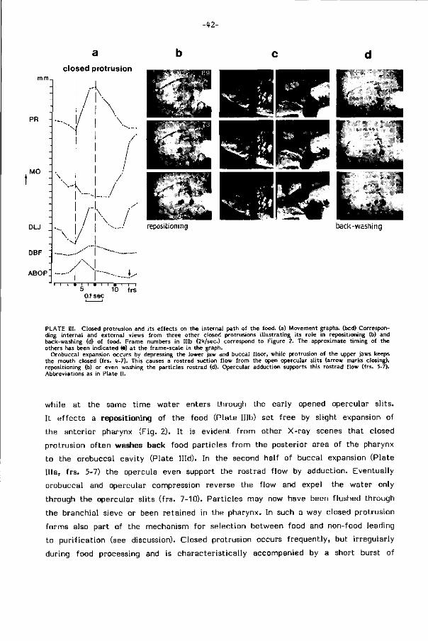
- f2-
closed protrusion mm_,
1 l i
PR -
MO -
DLJ "_
DBF "
ABOP]
i/N !! I
x ! > - ! . - /
. / / /
~iv-.^ ~1 I • I I • I—I » I—I—I
5 10 frs 0.1 sec
PLATE HI. Closed protrusion and its effects on the internal path of the food, (a) Movement graphs, (bed) Corresponding internal and external views from three other closed protrusions i l lustrating its role in repositioning (b) and back-washing (d) of food. Frame numbers in IHb (2<t/sec.) correspond to Figure 2. The approximate t iming of the others has been indicated (•) at the frame-scale in the graph.
Orobuccal expansion occurs by depressing the lower jaw and buccal floor, while protrusion of the upper jaws keeps the mouth closed (frs. 4-7). This causes a rostrad suction flow from the open opercular slits (arrow marks closing), repositioning (b) or even washing the particles rostrad (d). Opercular adduction supports this rostrad f low (frs. 5-7). Abbreviations as in Plate I I .
while at the same t ime water enters through the early opened opercular sl i ts.
I t e f fects a repositioning of the food (Plate IHb) set f ree by slight expansion of
the anterior pharynx (Fig. 2). I t is evident f rom other X-ray scenes that closed
protrusion of ten washes back food part icles f rom the posterior area of the pharynx
to the orobuccal cavity (Plate Hid). In the second half of buccal expansion (Plate
I l ia , f rs . 5-7) the opercula even support the rostrad f low by adduction. Eventually
orobuccal and opercular compression reverse the f low and expel the water only
through the opercular sl its ( f rs. 7-10). Part icles may now have been flushed through
the branchial sieve or been retained in the pharynx. In such a way closed protrusion
forms also part of the mechanism for selection between food and non-food leading
to pur i f icat ion (see discussion). Closed protrusion occurs f reguent ly, but i rregularly
during food processing and is character ist ical ly accompanied by a short burst of
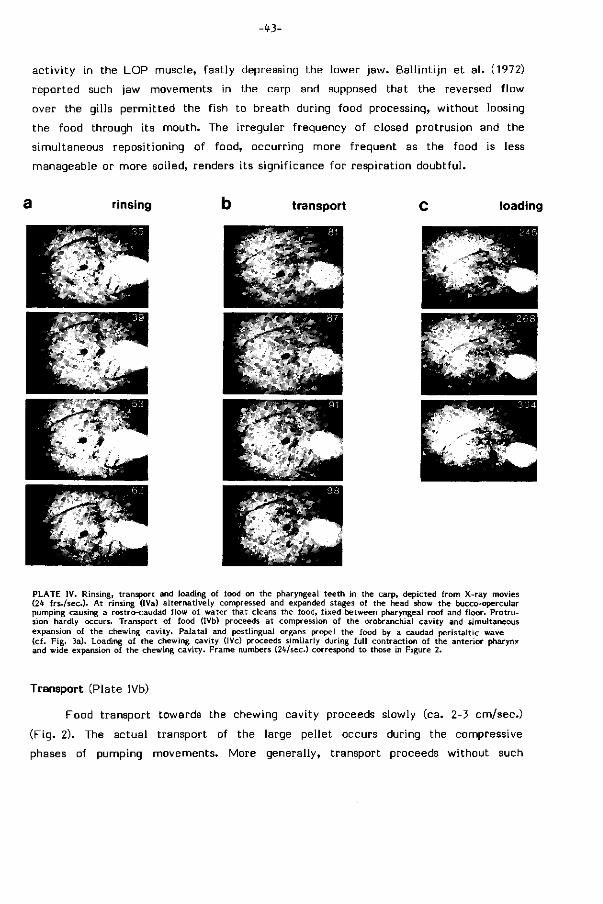
-in
act iv i ty in the LOP muscle, fast ly depressing the lower jaw. Bal l int i jn et a l . (1972)
reported such jaw movements in the carp and supposed that the reversed f low
over the gills permi t ted the fish to breath during food processing, wi thout loosing
the food through its mouth. The irregular frequency of closed protrusion and the
simultaneous repositioning of food, occurring more frequent as the food is less
manageable or more soiled, renders i ts significance for respirat ion doubtful .
a rinsing transport loading
PLATE IV. Rinsing, transport and loading of food on the pharyngeal teeth in the carp, depicted from X-ray movies (2^ f rs. /seo). At rinsing (IVa) alternatively compressed and expanded stages of the head show the bucco-opercular pumping causing a rostro-caudad f low of water that cleans the food, f ixed between pharyngeal roof and f loor. Protrusion hardly occurs. Transport of food (IVb) proceeds at compression of the orobranchial cavity and simultaneous expansion of the chewing cavity. Palatal and postlingual organs propel the food by a caudad peristaltic wave (cf. Fig. 3a). Loading of the chewing cavity (lVc) proceeds similarly during ful l contraction of the anterior pharynx and wide expansion of the chewing cavity. Frame numbers (24/sec.) correspond to those in Figure 2.
Transport (Plate IVb)
Food transport towards the chewing cavity proceeds slowly (ca. 2-3 cm/sec.)
(Fig. 2). The actual transport of the large pellet occurs during the compressive
phases of pumping movements. More generally, transport proceeds wi thout such
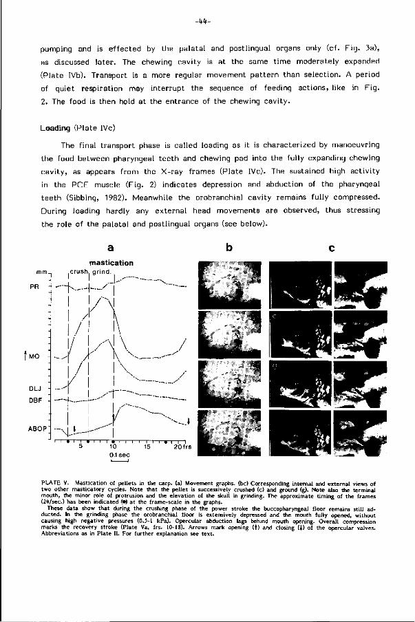
-kh-
pumping and is e f fected by the palatal and postlingual organs only (cf. F ig . 3a),
as discussed later. The chewing cavity is at the same t ime moderately expanded
(Plate IVb). Transport is a more regular movement pat tern than selection. A period
of guiet respiration may interrupt the seguence of feeding actions, l ike in F ig .
2. The food is then hold at the entrance of the chewing cavi ty.
Loading (Plate IVc)
The f inal transport phase is called loading as i t is characterized by manoeuvring
the food between pharyngeal teeth and chewing pad into the ful ly expanding chewing
cavi ty, as appears f rom the X-ray frames (Plate IVc). The sustained high act iv i ty
in the PCE muscle (Fig. 2) indicates depression and abduction of the pharyngeal
teeth (Sibbing, 1982). Meanwhile the orobranchial cavity remains fu l ly compressed.
During loading hardly any external head movements are observed, thus stressing
the role of the palatal and postlingual organs (see below).
mm.
PR
f MO
DLJ
DBF
ABOP
mastication crush, grind.
PLATE V. Mastication of pellets in the carp, (a) Movement graphs, (be) Corresponding internal and external views of two other masticatory cycles. Note that the pellet is successively crushed (c) and ground (g). Note also the terminal mouth, the minor role of protrusion and the elevation of the skuli in grinding. The approximate t iming of the frames (24/sec.) has been indicated (•) at the frame-scale in the graphs.
These data show that during the crushing phase of the power stroke the buccopharyngeal floor remains st i l l ad-ducted. In the grinding phase the orobranchial floor is extensively depressed and the mouth fully opened, without causing high negative pressures (0.5-1 kPa). Opercular abduction lags behind mouth opening. Overall compression marks the recovery stroke (Plate Va, frs. 10-18). Arrows mark opening ( t ) and closing (J) of the opercular valves. Abbreviations as in Plate I I . For further explanation see text.
-45-
Masticat ion: crushing and grinding (Plate V)
Loading of the teeth is immediately fol lowed by a series of masticatory cycles,
which are best characterized by the repet i t ive bursts of ac t iv i ty in the LAB V
muscle ef fect ing the power stroke (Fig. 2). In the crushing phase of the power
stroke (Plate V, f rs . 3-6) the buccal cavity is fu l ly compressed, whereas grinding
( f rs . 6-10) requires extensive expansion of the head (Sibbing, 1982). The restr ict ions
which such vigorous movements impose on other functions are discussed later.
During mastication of a large pellet the mouth opens widely w i th hardly or no
protrusion. A corresponding act iv i ty in the LOP muscle is evident (Fig. 2). The
extensive expansion during grinding resembles the events at food intake. Its e f fect
on f low is l imi ted as i t proceeds much slower. This was conf irmed by pressure
measurements in the opercular cavit ies, which gave a negative water pressure
of 0.5-1 kPa during mastication instead of 5-9 kPa during intake. The t iming of
opercular abduction rather varies and may considerably lag behind mouth opening
(Plate IV a). L i f t i ng the cranium and re t ract ion of the pectoral girdle mediate
the transmission of forces f rom the bodymuscles to the masticatory surfaces (Sib
bing, 1982). These movements are readily observed in a chewing carp.
Deglutition (Plate VI)
fe,' - ^
lx*
The masticatory t ra in is completed
by a deglut i t ion stroke, which is not
recognized f rom the exterior of the
f ish. Its EMG and movement pat tern
resembles a masticatory cycle of low
amplitude (Sibbing, 1982). During deglut i
t ion the ground food is transported
f rom the posterior pharynx into the
esophagus (Plate VI , f rs . 1-2) and f inal ly
passed through the pyloric sphincter
into the intestinal bulb ( f rs . 2-5).
PLATE VI. X-ray frames (24/sec.) visualize deglutition in the carp. Ground food is seen moving from the chewing cavity into the esophagus towards the intestinal bulb (arrows). Smaii pharyngeal jaw movements play a role in deglut i t ion. Frame numbers indicate t ime sequence.
-RO
TABLE 1 CHARACTERISTIC FEATURES BELONGING TO THE
Function of behavioural elements
Movement pattern
Effects
Reference EMG
Anterior palatal organ
Posterior palatal and postlingual organ
Particulate intake
-mouth opens by lower jaw and early protrusion
-orobuccal and opercular cavities expand
-opercular valves open late
-skull is lifted -lower jaw closes
protruded mouth and initiates compression
-fast suction flow directed to the particle
-food trapped into the pharyngeal slit
-long LOP burst -low PCE and
LAB V activity
-narrows the pharyngeal lumen
-close off posterior pharynx and esophagus
Gulping
-mouth opens without protrusion
-oral depression mainly
-opercular valves open early in abduction
-late protrusion closes depressed lower jaw
-bite of suspension enters the mouth slowly, suction even from the jaw angles
-maximal suspension enclosed prior to oral compression
-long LOP burst -low PCE and
LAB V activity
-narrows the pharyngeal lumen
-close off posterior pharynx and esophagus
Selection Rinsing
-mouth opens without protrusion
-orobuccal and subsequently opercular expansion
-pumping of water along the fixed particle
-washing waste through the opercular slits
long LOP burst
-fixes the particle
-
Repo s i t ioning Back-washing
-orobuccal depression
-protrusion keeps the mouth closed
-late opercular adduction
-compression orobuccal cavity
-inflow of water through the branchial slits a)merely
repositioning the particle
b)washing particles back into the orobuccal cav.
-expulsion of waste through opercular slits
-local retention of food in the pharyngeal slit
-short LOP burst
-manipulates and retains food particles locally
-
-47-
DISTINCT BEHAVIOURAL ELEMENTS IN FOOD HANDLING
Spitting Transport Loading Mastication Deglutition
-orobuccal expansion few protrusion
-mouth opens hardly protrusion
-opercular and buccal compression
-posterior pharynx gradually expands
-posterior pharynx spaciously expands
-overall expansion of oropharyngeal and opercular cavities
-elevation of the skull
-retraction pectoral girdle
-mouth widely open without protrusion in part, feeding
-mouth closed by protrusion in suspensionfeeding
-compression of the posterior pharynx
-small movement resembling mastication
-particles sucked into the orobuccal cavity and rapidly spat through the mouth
-food is propelled to the chewing cavity
-food is loaded into the chewing cavity
-the fixed food is crushed and ground
-particles are prevented from escape to the anterior pharynx
-anteriorly stored suspensions are prevented from escape through the mouth
-ground food is directed into the esophagus
-high LOP activity
-increasing -high PCE -high LAB V activity -low LAB V PCE activity activity -high PCE activity activity
-high LOP activity open mouth: long closed mouth: short
-occasionally active
-peristaltic waves propell the food caudally
-manoeuvre food between teeth and chewing pad
-fix the particle to be masticated
-close off the entrance of the chewing cavity
-close off the entrance of the chewing cavity
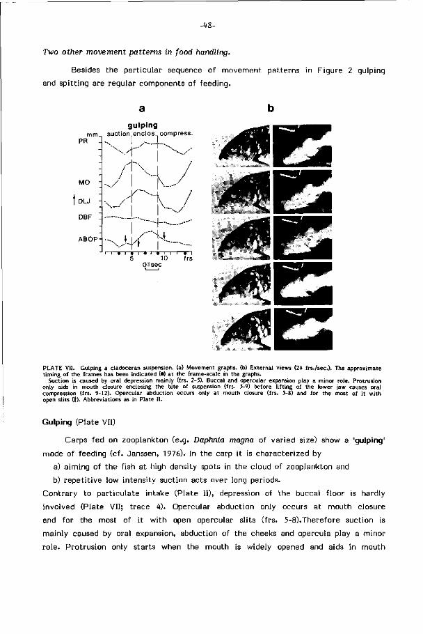
-48-
Two other movement patterns in food handling.
Besides the part icular sequence of movement patterns in Figure 2 gulping
and spi t t ing are regular components of feeding.
mm PR -
MO -
DLJ -
DBF
ABOP-
gulpmg suction enclos compress.
\<-~T\/ /N /
J 1 KS ^ /
— N . /
-,j\/~f~-^. 5 10 frs
01 sec
PLATE VII. Gulping a cladoceran suspension, (a) Movement graphs, (b) External views (2^ f rs. /seo). The approximate t iming of the frames has been indicated (•) at the frame-scale in the graphs.
Suction is caused by oral depression mainly (frs. 2-5). Buccal and opercular expansion play a minor role. Protrusion only aids in mouth closure enclosing the bite of suspension (frs. 5-9) before l i f t ing of the lower jaw causes oral compression (frs. 9-12). Opercular abduction occurs only at mouth closure (frs. 5-8) and for the most of i t w i th open slits (t). Abbreviations as in Plate I I .
Gulping (Plate VII)
Carps fed on zooplankton (e.g. Daphnia magna of varied size) show a 'gulping'
mode of feeding (cf. Janssen, 1976). In the carp i t is characterized by
a) aiming of the f ish at high density spots in the cloud of zooplankton and
b) repet i t ive low intensity suction acts over long periods.
Contrary to part iculate intake (Plate I I), depression of the buccal f loor is hardly
involved (Plate V I I ; t race 4). Opercular abduction only occurs at mouth closure
and for the most of i t w i th open opercular sl its ( f rs. 5-8).Therefore suction is
mainly caused by oral expansion, abduction of the cheeks and opercula play a minor
ro le. Protrusion only starts when the mouth is widely opened and aids in mouth
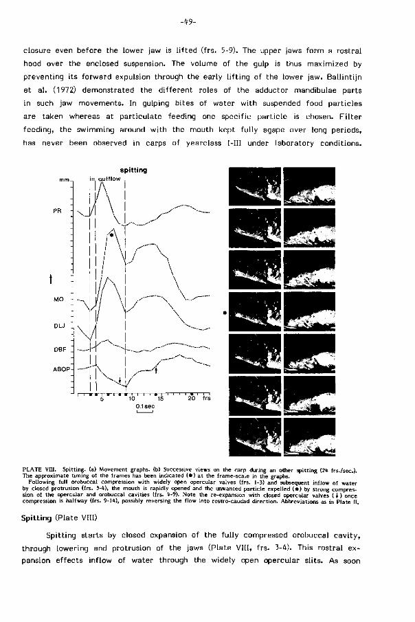
-t9-
closure even before the lower jaw is l i f ted ( frs. 5-9). The upper jaws form a rostral
hood over the enclosed suspension. The volume of the gulp is thus maximized by
preventing its forward expulsion through the early l i f t ing of the lower jaw. Bal l int i jn
et a l . (1972) demonstrated the d i f ferent roles of the adductor mandibulae parts
in such jaw movements. In gulping bites of water w i th suspended food part ic les
are taken whereas at part iculate feeding one specific part ic le is chosen. F i l te r
feeding, the swimming around w i th the mouth kept fu l ly agape over long periods,
has never been observed in carps of yearclass I—III under laboratory conditions.
spitting
PR
MO -
DLJ
DBF
ABOP
in, au t f low
20 frs
0.1sec
PLATE VIII. Spitting, (a) Movement graphs, (b) Successive views on the carp during an other spitting (2* f rs. /sec). The approximate t iming of the frames has been indicated ( • ) at the frame-scale in the graphs.
Following full orobuccal compression with widely open opercular valves (frs. 1-3) and subsequent inflow of water by closed protrusion (frs. 3-*), the mouth is rapidly opened and the unwanted particle expelled ( * ) by strong compression of the opercular and orobuccal cavities (frs. <t-9). Note the re-expansion with closed opercular valves ( > ) once compression is halfway (frs. 9-1*), possibly reversing the f low into rostro-caudad direction. Abbreviations as in Plate I I .
Spitting (Plate VIII)
Spitt ing starts by closed expansion of the fu l ly compressed orobuccal cav i ty ,
through lowering and protrusion of the jaws (Plate VII I , f rs . 3-4). This rostral ex
pansion effects inf low of water through the widely open opercular sl i ts. As soon
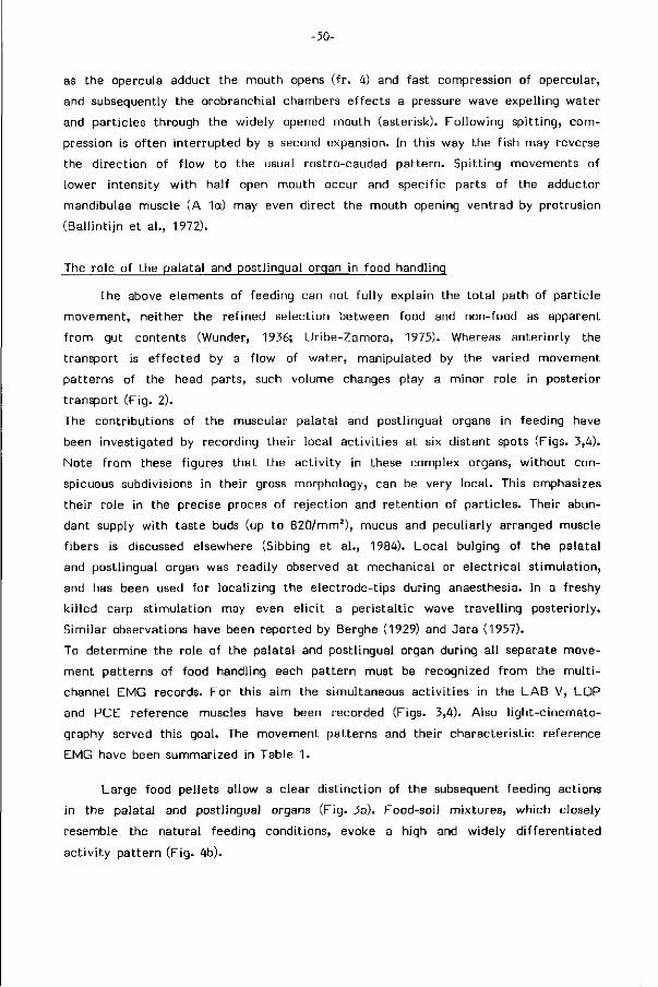
-50-
as the opercula adduct the mouth opens ( f r . 4) and fast compression of opercular,
and subsequently the orobranchial chambers ef fects a pressure wave expelling water
and part icles through the widely opened mouth (asterisk). Following spi t t ing, com
pression is of ten interrupted by a second expansion. In this way the f ish may reverse
the d i rect ion of f low to the usual rostro-caudad pat tern. Spitt ing movements of
lower intensity w i th half open mouth occur and specific parts of the adductor
mandibulae muscle (A 1a) may even direct the mouth opening ventrad by protrusion
(Bal l int i jn et a l . , 1972).
The role of the palatal and postlingual organ in food handling
The above elements of feeding can not fu l ly explain the to ta l path of par t ic le
movement, neither the ref ined selection between food and non-food as apparent
f rom gut contents (Wunder, 1936; Ur ibe-Zamora, 1975). Whereas anteriorly the
transport is e f fected by a f low of water, manipulated by the varied movement
patterns of the head parts, such volume changes play a minor role in posterior
transport (Fig. 2).
The contributions of the muscular palatal and postlingual organs in feeding have
been investigated by recording their local act iv i t ies at six distant spots (Figs. 3,4).
Note f rom these f igures that the act iv i ty in these complex organs, wi thout con
spicuous subdivisions in their gross morphology, can be very local . This emphasizes
their role in the precise proces of re ject ion and retent ion of part ic les. Their abun
dant supply w i th taste buds (up to 820/mm2), mucus and peculiarly arranged muscle
f ibers is discussed elsewhere (Sibbing et a l . , 1984). Local bulging of the palatal
and postlingual organ was readily observed at mechanical or e lectr ical s t imulat ion,
and has been used for localizing the electrode-tips during anaesthesia. In a freshy
k i l led carp st imulat ion may even e l ic i t a per istal t ic wave t ravel l ing posteriorly.
Similar observations have been reported by Berghe (1929) and Jara (1957).
To determine the role of the palatal and postlingual organ during all separate move
ment patterns of food handling each pattern must be recognized f rom the mu l t i
channel EMG records. For this aim the simultaneous act iv i t ies in the LAB V, LOP
and PCE reference muscles have been recorded (Figs. 3,4). Also l ight-c inemato
graphy served this goal. The movement patterns and their characterist ic reference
EMG have been summarized in Table 1.
Large food pellets allow a clear d ist inct ion of the subsequent feeding actions
in the palatal and postlingual organs (F ig. 3a). Food-soil mixtures, which closely
resemble the natural feeding conditions, evoke a high and widely d i f ferent iated
ac t iv i ty pattern (F ig. 4b).
-51-
Intake
The act iv i ty in the palatal and postlingual organs at intake (F ig. 3a) may close
of f the narrow entrance to the posterior pharynx and prevents that the high suction
force is also applied to the contents of chewing cavity and esophagus. They thus
aid in directing the suction f low.
Selection
Once the pellet has been trapped in the narrow pharyngeal lumen, act iv i ty
in the rostral and lateral parts of the palatal organ (traces 1-4) indicates gustation
and internal selection. Synchronous bursts in the levator operculi muscle indicate
lower jaw depression. Long LOP bursts point to rinsing whereas short LOP bursts
character ist ical ly belong to closed protrusion movements (cf. F ig . 2). As the internal
e f fects of closed protrusion on the food are invisible i t is only indicated by the
general term positioning. L ikewise has been done for the other EMG records (Figs.
3,4). The act iv i ty of the palatal organ may also serve for a repeated test of the
palatabi l i ty of the par t ic le .
Transport and loading
Progression of ac t iv i ty f rom the anterior to the posterior parts of the palatal
and postlingual organ (traces 4-6) corresponds to propulsion of the pellet to the
chewing cavity in the X-ray movies. Most of ten propulsive waves rapidly succeed
(F ig. 3a, arrows). They become gradually accompanied by act iv i ty in the PCE muscle
( lower trace) in i t ia t ing expansion of the chewing cavi ty.
Note that the postero-lateral part of the palatal organ (Fig. 3a, t race 4) acts in
selection as wel l as in transport and mast icat ion, indicating i ts boundary-position
between the funct ional ly d ist inct antero- lateral and posterior areas.
The end of anterior palatal ac t iv i ty announces loading of the pellet on the
pharyngeal teeth (Fig. 3a, Id.). The PCE muscles spaceously expand the chewing
cavity while the posterior parts of the palatal and postlingual organs continue
their propulsion of food on to the now depressed teeth.
Mast icat ion and deglut i t ion
The posterior parts of the palatal and postlingual organs are highly act ive,
f ix ing the pellet to be masticated (cf. Plate Vb) during the successive power strokes.
They w i l l also obstruct the escape of ground food back to the anterior pharynx.
Similar act iv i ty is observed at deglut i t ion (Fig. 3a, dgl.) whereby the constr ictor
pharyngis muscle aids in compressing the chewing cavity (Sibbing, 1982). Their
-52-
combined actions direct and propel the ground food into the esophagus. Ac t iv i ty
in the expansion muscles of the chewing cavity (PCE) is now conspicuously lacking.
Reloading
Prior to mastication of a second b i te, the pel let is repositioned by palatal-
postlingual act iv i ty and closed protrusion (short LOP bursts) (Fig. 3a, reloading).
When food part icles are washed back into the orobranchial cavi ty, as of ten seen
in the X-ray movies (cf. Plate Hid), fur ther masticat ion requires transport and
loading. The reloading pattern then even more resembles the events after intake.
The palatal and postlingual organs display an apparent regional d i f ferent ia t ion
in funct ioning. The anterior and lateral parts are active in t rapping, gustation,
selection and positioning, whereas the central and posterior parts are involved
in t ransport, loading, masticat ion and deglut i t ion. Contrary to Jara's hypothesis
(1957) the palatal or postlingual act iv i ty was never measured during respirat ion,
even if i t was forced to be intensive.
Adjustment of the feeding sequences to the type of food
To determine the adjustments of the movement patterns and the palatal-post-
lingual act iv i t ies to the type of food, d i f ferent types have been fed.
Movie and EMG-records showed conspicuous differences in the presence and
repet i t ion of the dist inct food handling patterns. The feeding sequences are analyzed
below and characterized in Table 2. Only the major differences w i th the discussed
pellet sequence are reported. They occur mainly in intake, selection and mast icat ion.
Barley
Grains of barley generally demand frequent repositioning to manoeuvre the
small dense part ic le f rom the branchial to the medial area, providing an appropriate
grip for transport. Back-washing movements prevent loss of the heavy part ic le
through i ts sinking f rom the lateral depressions in the buccal cavity into the large
f i rs t branchial s l i ts. Rinsing is never observed. Mastication may last for several
minutes and requires crushing pr ior to grinding (cf. Sibbing, 1982, f i g . 15).
Earthworm (Fig. 3b)
The earthworm-sequence shows that the palatal ac t iv i ty locally changes (F ig.
3b; arrows) and is even maintained in the anterior parts during masticat ion. This

-53-
a. PELLET
positions within the palatal organ
INTAKE I ' SELECTION
pos. i rinsing i pos.
postlingual organ
m. levator operculi LOP
m.lev.arc.branch.'Z LAB3C
m.pharyngo-cleithr.ext.PCE „ • -ÉH+%4
# — I I ' M » it » fr Mai h
1sec. , 300uv] lOOOuv]*
b . EARTHWORM
positions within the palatal organ
postlingual organ
m. levator operculi LOP
m.lev.arc branchX LABS
, SELECTION
positioning
« *)•<
«-•«••il w i m
HH
n.pharyngo-cleithr.ext. PCE t. » f i
ä «HI-^MMf
• 4 - » * -
-~J
M AST I C A T I O N
• H ^ H O * 1 »' '<• » "
mast. I mast. I mast. I mast.
pos.tr. pos.tr. pos.tr. pos.tr
.—..... ». ».i . ». ».I 4 éit H
'4 i *"• If" <N" *' fr
H r -
I . M . » . M * » ' »"i<l" « ' I * l«»|l -4
M** f »—v » ' if—>-tt*—» Hi 4» # 4 ' #•#•-*»• /
>.»>»•>» *• «M» « t ^ ' l l t f 'H » 4 - ^ K
» » i » » M * » ' ' > M -f-
1 M M M M M il I i-4 #n»H«>Hi'H«' I**'" N <Nfr#
FIG. 3a. Palatal and postlingual act ivi ty during pellet feeding. Act iv i ty pattern of the muscular palatal and postlingual organs at six indicated places during eventually succesful intake (large arrow) and internal processing of a pellet. The three muscles below monitor jaw movements (LOP) and mastication (LAB V, PCE).
The anterior half of the palatal organ (traces 1-3) is active during intake and selection and involved in gustation, rinsing and positioning (pos). Then the act ivi ty passes away from the anterior to the posterior parts of these organs (traces t -6) which serve transport (tr), loading (Id), mastication and deglutition (dgl). The postero-lateral area (trace U) overlaps most of these actions. The repetitive arrows mark the peristaltic waves in transport. The patterns are discussed in the text.
FIG. 3b. Act iv i ty pattern in earthworm feeding (same experiment). Note the overall similarity in the pattern, compared to Fig. 3a. Act iv i ty in the anterior palatal organ (traces 1-3) is however maintained during mastication. Palatal and postlingual act ivi ty vary locally (arrows), reflecting the local manipulation of the sprawling worm during mastication. In a later stage of processing masticatory cycles (LAB V act ivi ty) alternate with positioning-transport actions (short LOP bursts and overall palatal activity) suggesting an intermittant transport of the masticated worm through the chewing cavity.
-54-
ref lects more the length of the sprawling worm, being masticated at one end and
immobi l ized by the palatal organ at the other, than an essentially d i f ferent type
of processing. Due to the length of the worm feeding actions overlap. No rinsing
occurs. Closed protrusions aid in positioning to improve the grip on the long prey.
Transport of the worm through the chewing cavity may al ternate w i th masticat ion
(Fig. 3b). Such an i n te rmi t tan t transport was actually seen in an X-ray scene,
a f ter the worm had been injected w i th barium sulphate in its coelomic compart
ments. Masticat ion proceeds merely by grinding (Sibbing, 1982; F ig . 15).
A soft lump of tubi f ic ids was taken by forcefu l suction. The carp apparently
could not achieve a proper grip on the lenient mass and t r ied to disperse i t in
smaller clusters. This is concluded f rom the abundant repet i t ion of closed protrusion
movements, positioning the food and washing i t through the orobranchial cav i ty .
Even spit t ing and re- intake are applied for renewing the gr ip. During grinding p ro t ru
sion of the upper jaws kept the mouth closed, preventing tubi f ic ids f rom being
expelled through the mouth.
Zooplankton (Fig. 4a)
Whereas the above larger food i tems e l ic i ted 'par t iculate feeding' sequences,
cladocerans were taken by repet i t ive 'gulping' (cf. Plate VI I ; F ig . 4a, long LOP
bursts wi thout high LAB V ac t iv i ty ) . While suspending ground pellets into the tank
an eager carp attained frequencies of even eight gulps w i th in a second. This accumu
lates part icles f rom small volumes of suspension into the pharynx. Selection typical ly
proceeds by palatal act iv i ty and closed protrusion (short LOP burst wi thout LAB
V ac t iv i ty ) . This movement w i l l however also serve to wash part icles f rom all
over the branchial sieve back to the medial area between palatal and postlingual
organ. In this way closed protrusion serves recollection of the f i l t r a te for t ransport.
Also here protrusion of the upper jaws generally keeps the mouth closed during
mast icat ion, preventing the escape of f ree f loat ing part icles through the mouth.
Thus the external prof i le of the masticat ing f ish w i l l markedly d i f fer at part iculate
feeding and gulping. The separate gulping sequences are of short durat ion.
Tubifex-soil mixtures (Fig. 4b)
Substrate feeding was analyzed by offer ing a peaty pond soil intermingled
w i th tubi f ic ids. Gulping as wel l as part iculate intake was observed.
Feeding on food-soil mixtures asks increased act iv i ty in the palatal organ (Fig.
4b, traces 1-3) overlapping most other feeding actions. Closed protrusions (short
LOP bursts) in combination w i th anterior palatal ac t iv i ty are conspicuously frequent
-55-
â. CLADOCERANS
positions within the palatal organ
postlingual organ
- — M - 4 - — ~ * ( K
m. levator operculi LOP
m.lev.arc.branche LAB3E _
m.pharyngo-cleithrext. PCE _
fl OH
• > «••»>'
-Hi
r?
•*»
>#MW»
*#H
H #
•HM-4
«Ml» " • »
Ml
+••
V
""WlHW(r
- f .
h*-
<M-fH * - * - *
I (i i » i i - H -
i l l l i l >m m i « i il m
'M i l l—»H—~
I * • • » <
+4-4 — H i — « » .
• " » . . , t i » U | l h * * -H • — * - *
1sec. , 30Oiiv] IOC»*«]*
b. TUBIFEX -SOIL MIXTURE
GULPING
positions within the palatal organ
• I"" III
~rti~H I
»frmi'i'i
postlingual organ
m. levator operculi LOP
rn.lev.arcbranch.3Z! L A B 2
m.pharyngo-cleithr.ext. PCE
+ 1 n i | — » —
4+fH
positioning |lr MASTICATION
Hf
i ii »mil»
•*• 2 //////
n» . n . i U - w t - t ^
* ! « • i^ l^ l lH^nt ' i l i ' lh UnMl+H NlilHnHiH
«'M "f>»
#
(nit «»I
M M i l l
M * M •Ht« <"••»"•*«
pos. + tr.+ mast.
fa*!'Ü^
" • *
•t- t-r
..t>4 i»4> " t
i i i i *
Hr-
^ i » '» . 'M I ^ I »Jii
• « H * *
> • " » ' t"1
f+++
MASTICATION
H-H y r v
* — * —
jm i i . i f ^ i » » »> y i *
FIG. 4a. Act iv i ty pattern in zooplankton feeding (cf Fig. 3a). Intake of the cladoceran suspension proceeds by repetit ive gulping (merely long LOP bursts). Subsequent anterior palatal act ivi ty (traces 1-3) and closed protrusions (arrow) wi l l wash the cladocerans back from the branchial sieve recollecting them for transport (traces 4-6) and mastication (LAB V act iv i ty). Such short sequences are repeated over long periods. Further discussion in text .
FIG. 4b. Substrate feeding. Act iv i ty pattern during processing of a peaty pond soil containing tubificids (same experiment as Fig. 4a). The anterior palatal organ (traces 1-3) has a prominent role in selection between food and non-food particles, frequently accompanied by closed protrusion (arrows). Selection overlaps mastication (LAB V) and transport and takes much handling t ime. Further discussion in text.
-56-
in selection (Fig. 4b; arrows), gradually puri fying the mixture (see discussion) and
recol lect ing small food part icles f rom the branchial sieve. Note that processing
of such mixtures requires a considerable handling-t ime (cf. F ig . 4b). Spitt ing is
generally employed for coarse selection, allowing choice at re- intake, often by
gulping the suspended part ic les. A s imilar behaviour is observed at searching when
the carp keeps sampling the substrate by a short sequence of intake, gustation
and spi t t ing i.e. probing.
FOOD
pellets
barley
earthworm
lump of tul
cladoceran
>if icids
suspension
tubifex-soil mixture
P
P
P
P
P
E or G
UNITS
RI RP
|RP
RP
|RP
OF
-BW
-Bw|
-BW
-Bwj-
BW-
E-
BEHAVIOUR
RC
RC
RC
T
T
T
T
T
T
L
L
L
L
L
L
c [GR]
0 S
c
c
[GR]
GR
GR
D
D
D
D
D
D
TABLE 2. Composition of feeding sequences according to the type of food. Conspicuously repeated patterns are indicated by a square. Probing and spitting do not lead to food consumption and are considered separately from the feeding sequence (see text). P particulate intake RP repositioning T transport GR grinding G gulping BW back-washing L loading D deglutition RI rinsing RC recollection C crushing
FOOD HANDLING MECHANISMS AND THEIR LIMITATIONS
Postcapture feeding behaviour, divided into buccal manipulat ion, pharyngeal
manipulation and pharyngeal transport in generalized euteleosts (Lauder, 1983)
is at least in cyprinids split into a chain of d ist inct funct ional patterns, wherin
the buccal, pharyngeal and opercular mechanisms closely in teract . (Table 1). The
t iming and sequence of protrusion, orobuccal and opercular expansion and the mo
ment of opening of the mouth and opercular-branchiostegal valves allows a high
p last ic i ty in movements.
F rom its feeding ecology and diet the adult carp appears to be an omnivorous
f ish, feeding at d i f ferent levels of the watercolumn and the food chain, whenever
and wherever food is available (Uribe-Zamora, 1975; in Sibbing, 1982). In Table
4 a l ist of generally available food types is given. From these bot tom invertebrates
(chironomids and other dipteran larvae, tubi f ic ids, copepods), zooplankton (preferably
large cladocerans and copepods) and soft l i t t o ra l vegetation (Lemna minor, Glyceria
fluitans), but especially i ts fauna (gasteropods, oligochaets, t r ichopter larvae, cope
pods), according to season and avai labi l i ty , are exploited. Even aerial insects are
-57-
engulfed f rom the watersurface.
The omnivorous feeding habits of the carp raise the fol lowing questions. Does
the feeding ef f ic iency, taken as t ime expenditure to obtain and handle a food type,
or taken as the energy costs-gain rat io in feeding, widely d i f fer for d i f ferent types
of food? Do specializations for one type necessarily lower the feeding ef f ic iency
for others?
These questions demand a quant i tat ive approach and much more knowledge than
now is available. However the present qual i tat ive data about the type and sequence
of movement patterns employed in food processing allow an analysis of their e f fec t
on the food. Which optimizations in s t ructural tools and their application character
ize the separate feeding elements and which restr ict ions fol low for others? In
such a way the relations between s t ructure-funct ion components and their impact
on the available food and ecology of a f ish type can be deduced. Comparisons
between closely a l l ied species may then provide insight in the extent of their niches.
As the ef f ic iency and l imi tat ions of feeding is bound to its component parts the
dist inct feeding elements are discussed, start ing at the search for food.
Search and detection of food, a survey
Carps in search for food are typical stayers (Boddeke, Slijper et a l . , 1959)
feeding continuously on pelagic or benthic organisms, repet i t ively probing and scan
ning the substrate. Wunder (1927) investigated a variety of teleosts, including the
carp, to determine which of the diversif ied sense organs are involved in food detec
t ion . Jönsson (1967) elaborated Wunders experiments on one year old carps and
included an analysis of their daily rhy thm. Carps have a high endogenous ac t iv i ty
level at dusk and night. They are, however, readily t rained to feed at any t ime
of the day. Other major conclusions f rom this author are incorporated below.
Abiot ic conditions such as l ight and turbidi ty of the water as wel l as the st imul i
produced by the food determine which sense organs are involved in detect ion.
A t pelagic feeding the carp reacts on and is guided to its food by vision. Size,
shape, contrast, colour and motion of the prey w i l l fur ther d i f ferent ia te visual
d iscr iminat ion and the reactive distance. A l ight intensity of 10 mc (meter-
candle), corresponding to late dusk, marks the lower threshold for e f fect ive visual
locat ion of food by most fishes (Blaxter, 1970), w i th some notable exceptions.
The lateral l ine organ senses act ively moving preys in laboratory tanks but i ts
capacity to d iscriminate prey over long distances f rom other moving s t imul i , as
in a natural s i tuat ion, is doubtful . In the pike the lateral l ine is e f fect ive in detec
t ing water turbulence at distances less than 10 cm (Wunder, 1936). When vision
-58-
is poor or the food inactive smell guides the carp by t r ia l and error to the food.
O l factory organs generally have a high sensitivity and are long-range chemorecep-
tors. In f ish they are reported to be more of ten associated w i th social contacts
than w i th feeding (Hara, 1971). When carps feed on cereals or other encapsulated
seeds, of ten offered in f ish farms, smell s t imul i w i l l be low.
A t bottom feeding taste is the main detector in search for food. The fish
is alert on prey even at some distance f rom the mouth as taste buds are exposed
on barbels and f ins, and are spread even over the head and general body surface
(Herr ick, 1904). Contrary to reports of Jönsson (1967) the expected role of touch
in detecting of food has not been demonstrated in the carp , neither by experimental
nor by s tructural data.
Within the oropharyngeal cavity taste buds fur ther serve control and selection
of part icles to be swallowed (cf. Sibbing et a l . , 1984). The decision for processing
requires this internal evaluation. To distinguish between the external and internal
moments of choice the terms 'choice' and 'select ion' are respectively used.
The probing mode of searching and the abundant supply of external and internal
taste buds allows intensive substrate feeding. This extends the exploi tat ion of
the available area w i th its depth. Experiments f rom Suietov (1939) show carps
to penetrate more than 12 cm into a si l ty bo t tom. No general ecological areas
(bot tom, open water, surface, l i t tora l ) seem to be excluded f rom exploitat ion by
the carp due to its searching behaviour, except the open water at dark.
The sensory abi l i t ies seem hardly to exclude major categories of food f rom detection
by the carp (cf. Table 4). Detai led knowledge of d iscr iminat ion by the individual
senses of the carp is scarce in l i terature.
Food intake behaviour
The dominant mode of prey capture in teleosts consists of a combination of
suction (by rapid expansion and compression of orobuccal and opercular cavities)
and forward motion (by forward suction of the f ish i tself , protrusion and swimming)
(Muller and Osse, 1984). They distinguish a series of feeding types, based on the
re lat ive contributions of these components w i th the moment of opercular valve
opening as an important character ist ic. Optimizations of prey capture techniques
are discussed by van Leeuwen and Muller (1984), focussing on predator-prey distance
and prey velocit ies. These studies provide a f rame for analysis of food intake in
the carp.
In the carp swimming hardly contributes to food intake.This is readily observed
f rom its deep body shape and the slow approach to the food. At bot tom feeding
-59-
the substrate even prohibits overswimming the food. Swimming is not an essential
component in feeding on stationary preys. Drenner et a l . (1978) has however demon
strated that copepods are far more able in escaping prédation than cladocerans.
Grasping large part icles w i th the lips was occasionally observed and served
to remove gravel f rom underlying tubi f ic ids. It may wel l be applied in a natural
s i tuat ion e.g. for taking snails f rom macrophytes. No bit ing occurs in the carp,
as the only teeth of cyprinids lie posteriorly in the pharynx.
Two dist inct patterns of food intake are evident in the carp. Their mechanisms
and ef fects are compared in Table 3.
Particulate intake (F ig. 5)
During expansion of the slow swimming fish the late opening of the opercular
and branchiostegal valves w i l l prevent inf low through these valves and thus increase
the water velocity in the mouth. Expansion is however slow (ca. 160 msec.) com
pared to predacious f ish as a pike (ca. 40 msec, van Leeuwen and Muller, 1984).
Water velocity in the mouth opening is fur ther increased by protrusion of the upper
jaw creating a small rounded aperture of the mouth. Besides protrusion brings
the mouth closer to the food wi thout cost for adding impuls to the water or the
f ish and thereby causes a directed f low w i th respect to the mouth aperture. Combi
nation of these characters results in a fast and directed suction f low applied close
to the par t ic le. Such f low velocit ies allow intake of part icles w i th higher densities
than the water and increase the distance f rom which can be sucked. Forward suction
of the fish may wel l contr ibute to penetration into the bot tom substrate.
Sustained opercular abduction and the impuls of the water maintain the f low caudad
during oral compression. The part ic le w i l l be trapped in the narrow slit between
pharyngeal roof and f loor whereas the water is expelled through the branchial
and opercular sl i ts. I t is supposed that the main f low passes through the f i rs t bran
chial s l i t , serving as a shunt (hyoid-shunt cf . van Leeuwen, 1984). The gi l l f i laments
may be adducted to reduce the g i l l resistance in the suction f low, as during strong
vent i lat ion (Bal l int i jn et a l . , 1972).
Protrusion also permits the carp to apply i ts mouth ventrad and closely to
the bot tom without need for vert ical position of the body axis at intake. In the
spindle-shaped gudgeon this wi l l serve to maintain i ts position in fast f lowing
streams (Alexander, 1970). In the deep bodied carp, l iving in stagnant waters, i t
allows a continuous probing of the bot tom by slow swimming. Besides i t permits
a rapid escape f rom a predator during bot tom feeding. Protrusion is thus highly
funct ional . The mechanism of protrusion in the carp was investigated by Bal l in t i jn
et a l . (1972). D i f ferent parts of the adductor mandibulae muscle e f fec t forward
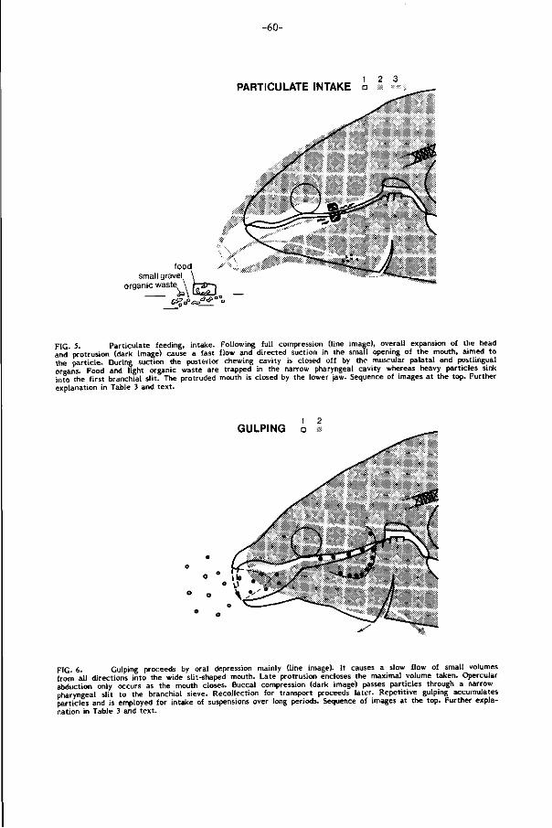
-60-
PARTICULATE INTAKE a
food small gravel,
organic waste
<?#"
PIC 5. Particulate ieeding, intake. Following ful l compression (line image), overall expansion of the head and'protrusion (dark image) cause a fast flow and directed suction in the small opening of the mouth, aimed to the part icle. During suction the posterior chewing cavity is closed off by the muscular palatal and postlingual organs. Food and light organic waste are trapped in the narrow pharyngeal cavity whereas heavy particles sink into the f i rst branchial sl i t. The protruded mouth is closed by the lower jaw. Sequence of images at the top. Further explanation in Table 3 and text .
GULPING a i
FIG. 6. Gulping proceeds by oral depression mainly (line image). It causes a slow flow of small volumes from all directions into the wide slit-shaped mouth. Late protrusion encloses the maximal volume taken. Opercular abduction only occurs as the mouth closes. Buccal compression (dark image) passes particles through a narrow pharyngeal slit to the branchial sieve. Recollection for transport proceeds later. Repetitive gulping accumulates particles and is employed for intake of suspensions over long periods. Sequence of images at the top. Further explanation in Table 3 and text.
-61-
PARTICULATE INTAKE GULPING
protrusion renders mouth cavity tube-like, directing the flow to a particular particle
protrusion allows ventrad aiming of the mouth to the particle
fast and voluminous flow by protrusion and extensive depression of oral and buccal floor; closed opercular abduction important
particle sucked into the anterior pharynx
mouth closure by lifting the lower jaw, compressing the open oral cavity
water expelled caudally; particle retained in the central pharynx
single actions at high energy-costs
suited for suction of dense or less accessible particles
mouth cavity with open jaw angles; causing inflow from all directions
mouth opening generally terminal; initially hardly protrusion
slow flow of small volumes by depression oral floor; buccal and opercular expansion play a minor role; valves open early
particles sucked into the oral cavity
mouth closure by late protrusion; oral cavity not compressed
maximal volume of water with particles is enclosed by a rostral hood and pressed slowly to the branchial sieve by oral compression
repeated low energy-costs actions over long periods
suited for gulping of suspensions
TABLE 3. Intake patterns and their effect on the food, explaining the differences between particulate intake and gulping.
protrusion (A 1 R ) , downward protrusion (A 1 ) and l i f t ing of the lower jaw ( A , and
A „ ) . The variable coordination of their actions allows a versatile aiming of p ro t ru
sion to the food. The mechanics of head expansion for creating a suction f low
has been studied f i rs t ly by L iem (1967) using cinematography and Osse (1969) adding
electromyography. Such studies are s t i l l lacking for the carp. The mechanical coup
lings in its head have however been studied (Bal l in t i jn , 1969).
Gulping (F ig. 6)
Low intensity suction and the early opening of the opercular valves reduce
volume as wel l as velocity of the water and suspended part icles at each gulp and
lowers its energy costs. The in i t ia l absence of protrusion causes a less directed
f low even f rom the jaw angles. As suspended part icles are of about the same density
-62-
as the water, have a l imi ted movement and are widely dispersed neither high velo
ci ty nor directed f low are demanded. Therefore gulping is appropriate to take
suspensions over long periods. Small volumes at each gulp may be demanded for
f i l te r ing the suspension ef fect ively over the branchial sieve maintaining l imi ted
and about equal branchial s l i ts. Food-soil mixtures and part iculate intake probably
require a large f i rst branchial sl it for shunting large volumes of water and debris.
Movement analysis of the branchial arches could falsify such hypotheses.
Late but extensive protrusion encloses the ingested suspension by forming a charac
ter ist ic rostral hood prior to oral compression. The maximal retainable suspension
is pressed caudad through the narrow buccopharyngeal s l i t , which improves par t ic le-
wal l contact for gustation and enhances the even spreading of the suspension over
the branchial sieve.
Limitations (cf . Table 4). Size, density, veloci ty, distance and location of the food
in the habitat impose d i f ferent demands to the f ish and are d irect ive characters
in the opt imizat ion of food capture techniques.
Large preys are excluded f rom intake by a carp (SL 22 cm) due to its short
jaws and the small gape of the mouth at protrusion. The maximal diameter of
the protruded circular mouth is about 9% of the standard body length (SL) for
carps ranging between 10-25 cm SL. The values for larger carps decrease to about
7% SL. The f ish is also unable to take bites f rom larger food objects l ike f ish
and macrophytes as oral teeth are absent. This also fac i l i ta tes the escape of s t rug
gling prey.
Fast preys easily escape. The deep body shape as wel l as the predominance
of red muscle f ibers in the trunk (Boddeke, Slijper and Van der Stel t , 1959) and
headmuscles (Akster, 1981; Akster and Sibbing, 1982), al lowing prolonged ac t iv i ty
but contract ing slowly, render the carp a slow swimming and slow feeding stayer.
Though its maximal burst speed is around ten body lengths per second (Blaxter,
1969), no reports of such velocit ies in feeding have been found. The endurance
or cruising speed of the carp is around 1.5 body lengths per second. These features
exclude hunting a f ter fast preys, even in open water. The small aperture of the
mouth makes f i l ter - feeding by overswimming the water inef f ic ient .
Foods of high density, such as some plantseeds, l ie w i th in reach for intake
due to the large velocity of f low. Part icles of low density may be taken f rom more
distant, less accessible locations l ike crevices due to the highly directed and fast
suction f low during protrusion.
-63-
Selection (Figs. 7,8)
Size selection between particles should be distinguished f rom selection between
food and non-food, as they are achieved by d i f ferent mechanisms.
The minimal mesh-width of the carps branchial sieve lies between 400-500 urn,
minimized to 250 vim if only smaller plankton is abundant (Uribe-Zamora, 1975).
Only the width of the plankton determines its retent ion, thus irrespective of i ts
length. The actual mechanism of the branchial sieve is not known which l imi ts
the insight in selective prédation on zooplankton (cf. Wright et a l . , 1983). I t is
important for many other planktivorous fish species (e.g. s i lvercarp, big head,
herr ing, menhaden).
A t bot tom and substrate feeding, mixtures of food, anorganic sediment and organic
debris impose high demands on the selection mechanism prior to fur ther processing.
Fine anorganic sediment ( < 2 mm) contains mainly part icles below 250 y m which
are passively lost through the branchial sieve (Uribe-Zamora, 1975). Though she
reports larger inorganic part icles to be not ingested, due to their size and density
(2.6), this is certainly not true at bot tom feeding. Ingested heavy medium sized
part icles such as sand grains and small gravel w i l l readily sink into the large f i rs t
branchial slits by gravi tat ional deposition (Fig. 5). Larger part icles are expelled
by spit t ing (see below). Act ive selection thus appears mainly to involve the separa
t ion of small to medium sized organic waste (250 y m - 9% SL) w i th densities
comparable to water. Though the separation of food and non-food is not complete
(Wunder, 1938; Ur ibe-Zamora, 1975) i t is s t i l l highly e f fect ive. The la t ter author
found only 3% of the f ine sediment to be retained w i th the food.
How is active selection internally achieved? It is commonly accepted that selection
between food and non-food proceeds w i th in the buccopharynx. Abundant taste buds
l ine this cavity (up to 820/mm2 c f . Sibbing et a l . , 1984) and allow gustation as
long as part icles ro l l along its walls. Matthes (1963) supposes cyprinids to select
by spi t t ing, retaining food part icles by entangling them in mucus. Spitt ing is however
a coarse mode of selection. Robotham (1982) presumed also buccal mucus to e f fect
selection at detr i tus intake of the spiny loach. More dense part icles would escape
mucous entrapment as they are given higher velocit ies (in our view impulse). Our
results show that the palatal organ is hardly involved in spit t ing and that palatal
ac t iv i ty as wel l as closed protrusion appear basic in separation and refined selection
between organic waste and food. This has been overlooked completely.
According to the size of the soiled food and the amount of organic waste
d i f ferent mechanisms of separation and selection are employed by the carp. These
are discussed below and i l lustrated in the Figures 5-9.
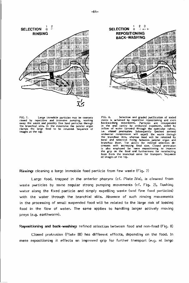
-64-
SELECTION D RINSING
2 3 SELECTION
REPOSITIONING BACK-WASHING
FIG. 7. Large immobile particles may be coarsely rinsed by repetit ive and intensive pumping, washing away the waste and possibly fine food particles through the branchial slits. In the meantime the palatal organ clamps the large food to be retained. Sequence of images at the top.
FIG. 8. Selection and graded purif ication of soiled items is achieved by repetitive repositioning and even backwashing movements. Particles are resuspended in the oral cavity by orobuccai expansion, aided by inflow of water (arrows) through the opercular valves, i.e. closed protrusion. Subsequently (pecked arrows) orobuccal compression wi l l expell the waste through the branchial slits, whereas food wi l l be retained by local and selective f ixing between palatal organ and branchial f loor. The ability for refined selection decreases with increasing food size. Closed protrusion is also employed for mere repositioning to improve the grip on the food and furthermore for recollecting food from the branchial sieve for transport. Sequence of images at the top.
Rinsing: cleaning a large immobile food part ic le f rom few waste (Fig. 7)
Large food, trapped in the anterior pharynx (cf. Plate IVa), is cleaned f r om
waste particles by some regular strong pumping movements (cf. F ig . 2), f lushing
water along the f ixed part ic le and simply expelling waste (and f ine food particles)
w i th the water through the branchial s l i ts. Absence of such rinsing movements
in the processing of small suspended food w i l l be related to the large risk of loosing
food in the f low of water. The same applies to handling larger act ively moving
preys (e.g. earthworm).
Repositioning and back-washing: ref ined selection between food and non-food (Fig. 8)
Closed protrusion (Plate III) has d i f ferent e f fects, depending on the food. In
mere repositioning i t e f fects an improved grip for fur ther transport (e.g. at large
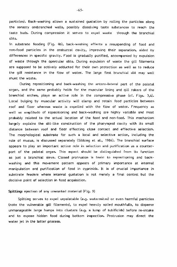
-65-
particles). Back-washing allows a sustained gustation by rol l ing the particles along
the sensory orobranchial walls, possibly dissolving taste substances to reach the
taste buds. During compression it serves to expel waste through the branchial
s l i ts.
In substrate feeding (Fig. 4b), back-washing effects a resuspending of food and
non-food part icles in the orobuccal cavi ty, improving their separation, aided by
differences in specific gravi ty. Food is gradually pur i f ied, accompanied by expulsion
of waste through the opercular sl i ts. During expulsion of waste the g i l l f i laments
are supposed to be actively adducted for their own protect ion as wel l as to reduce
the g i l l resistance in the f low of water. The large f i rst branchial sl it may wel l
shunt the waste.
During repositioning and back-washing the antero- lateral part of the palatal
organ, and the same probably holds for the muscular l ining and g i l l rakers of the
branchial arches, plays an active role in the compressive phase (cf. Figs. 3,4).
Local bulging by muscular act iv i ty w i l l clamp and retain food part icles between
roof and f loor whereas waste is expelled w i th the f low of water. Frequency as
wel l as amplitude of repositioning and back-washing are highly variable and most
probably related to the actual location of the food and non-food. This mechanism
largely explains the s l i t - l ike construction of the pharyngeal cavity w i th i ts small
distance between roof and f loor ef fect ing close contact and e f fect ive selection.
The morphological substrate for such a local and selective act ion, including the
role of mucus, is discussed separately (Sibbing et a l . , 1984). The branchial surface
appears to play an important active role in selection and pur i f icat ion as a counter
part of the palatal organ. This aspect should be distinguished f rom its funct ion
as just a branchial sieve. Closed protrusion is basic to repositioning and back-
washing and this movement pattern appears of pr imary importance at internal
manipulation and pur i f icat ion of food in cyprinids. I t is of crucial importance in
substrate feeders where internal gustation is not merely a f inal control but the
decisive point of selection in food acquisit ion.
Spitting: e ject ion of any unwanted mater ia l (F ig. 9)
Spitt ing serves to expel unpalatable (e.g. watermites) or even harmful part icles
(note the vulnerable g i l l f i laments), to expel heavily soiled mouthful ls, to disperse
unmanageable large humps into clusters (e.g. a lump of tubif icids) before re- intake
and to expose hidden food during bot tom inspection. Protrusion may direct the
water je t in the la t ter process.
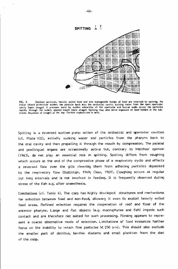
SPITTING a
FIG. 9. Disliked particles, heavily soiled food and less manageable humps of food are rejected by spitting. An in i t ia l closed protrusion washes the particle back into the orobuccal cavity, sucking water from the open opercular cavity (open image). A pressure wave by sudden adduction of the opercular and buccal walls drives the particles rapidly through the widely opened mouth (dark image). Spitting may also serve exposure of food hidden in the substrate. Sequence of images at the top. Further explanation in text.
Spitt ing is a reversed suction pump action of the orobuccal and opercular cavit ies
(cf. Plate VIII), in i t ia l ly sucking water and particles f rom the pharynx back to
the oral cavity and then propelling i t through the mouth by compression. The palatal
and postlingual organs are occasionally active but, contrary to Matthes' opinion
(1963), do not play an essential role in spi t t ing. Spitt ing di f fers f rom coughing
which occurs at the end of the compressive phase of a respiratory cycle and ef fects
a reversed f low over the gills cleaning them f rom adhering part icles deposited
by the respiratory f low (Bal l int i jn, 1969; Osse, 1969). Coughing occurs at regular
but long intervals and is not involved in feeding. I t is f requently observed during
stress of the f ish e.g. a f ter anaesthesia.
Limitations (cf . Table 4). The carp has highly developed structures and mechanisms
for selection between food and non-food, allowing i t even to exploit heavily soiled
food areas. Refined selection requires the cooperation of roof and f loor of the
anterior pharynx. Large and f la t objects (e.g. macrophytes and fish) impede such
contact and are therefore not suited for such processing. Rinsing appears to repre
sent a coarse a l ternat ive mode of selection. L imi ta t ions of food resources fur ther
focus on the inabil i ty to retain f ine particles (< 250 y m ) . This should also exclude
the smaller part of detr i tus, benthic diatoms and small plankton f rom the diet
of the carp.
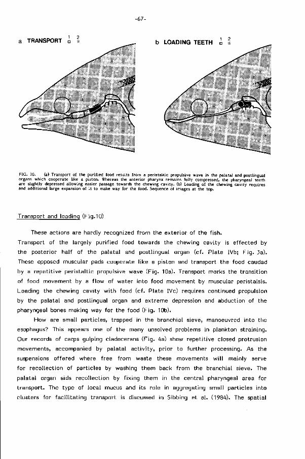
-67-
a TRANSPORT • 1 b LOADING TEETH a %
FIG. 10. (a) Transport of the purified food results from a peristaltic propulsive wave in the palatal and postlingual organs which cooperate like a piston. Whereas the anterior pharynx remains fully compressed, the pharyngeal teeth are slightly depressed allowing easier passage towards the chewing cavity, (b) Loading of the chewing cavity requires and additional large expansion of i t to make way for the food. Sequence of images at the top.
Transport and loading (Fig.10)
These actions are hardly recognized f rom the exter ior of the f ish.
Transport of the largely pur i f ied food towards the chewing cavity is e f fected by
the posterior half of the palatal and postlingual organ (cf. Plate IVb; F ig . 3a).
These opposed muscular pads cooperate l ike a piston and transport the food caudad
by a repet i t ive per istal t ic propulsive wave (Fig. 10a). Transport marks the t ransit ion
of food movement by a f low of water into food movement by muscular peristalsis.
Loading the chewing cavity w i th food (cf. Plate IVc) requires continued propulsion
by the palatal and postlingual organ and extreme depression and abduction of the
pharyngeal bones making way for the food (Fig. 10b).
How are small part icles, trapped in the branchial sieve, manoeuvred into the
esophagus? This appears one of the many unsolved problems in plankton straining.
Our records of carps gulping cladocerans (Fig. 4a) show repet i t ive closed protrusion
movements, accompanied by palatal ac t iv i ty , pr ior to fur ther processing. As the
suspensions of fered where free f rom waste these movements w i l l mainly serve
for recol lect ion of part icles by washing them back f rom the branchial sieve. The
palatal organ aids recol lect ion by f ix ing them in the central pharyngeal area for
t ransport. The type of local mucus and i ts role in aggregating small part icles into
clusters for fac i l i ta t ing transport is discussed in Sibbing et a l . (1984). The spatial
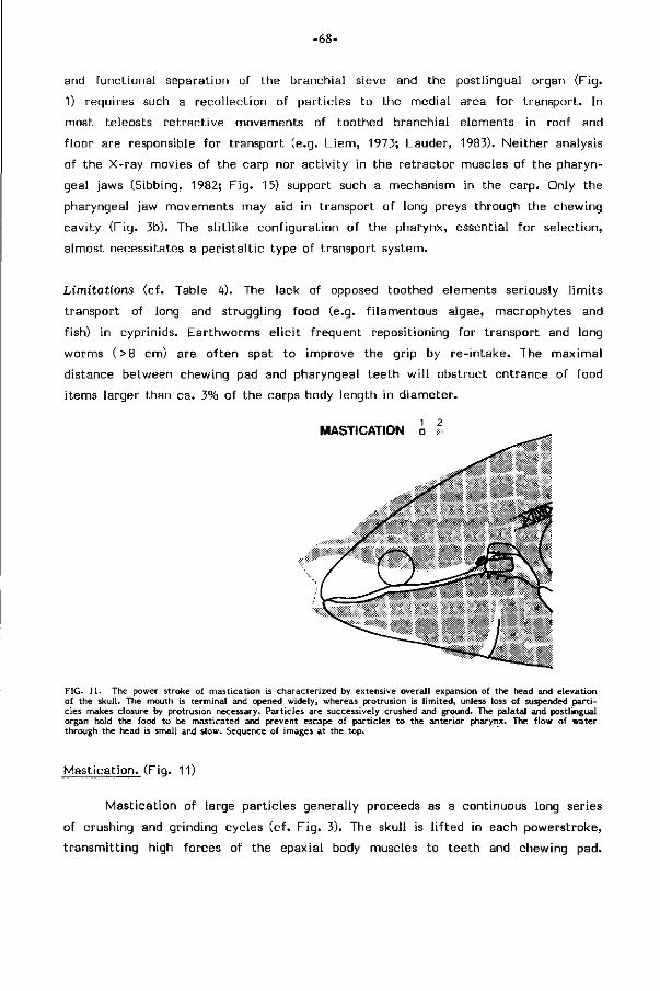
-68-
and funct ional separation of the branchial sieve and the postlingual organ (Fig.
1) requires such a recol lect ion of part icles to the medial area for t ransport. In
most teleosts re t ract ive movements of toothed branchial elements in roof and
f loor are responsible for transport (e.g. L i em, 1973; Lauder, 1983). Neither analysis
of the X-ray movies of the carp nor act iv i ty in the re t ractor muscles of the pharyn
geal jaws (Sibbing, 1982; F ig . 15) support such a mechanism in the carp. Only the
pharyngeal jaw movements may aid in transport of long preys through the chewing
cavity (F ig. 3b). The s l i t l ike configurat ion of the pharynx, essential for selection,
almost necessitates a per istal t ic type of transport system.
Limitations (cf . Table 4). The lack of opposed toothed elements seriously l imi ts
transport of long and struggling food (e.g. f i lamentous algae, macrophytes and
fish) in cyprinids. Earthworms e l ic i t f requent repositioning for transport and long
worms (>8 cm) are of ten spat to improve the grip by re- intake. The maximal
distance between chewing pad and pharyngeal teeth w i l l obstruct entrance of food
items larger than ca. 3% of the carps body length in diameter.
MASTICATION i l
FIG. 11. The power stroke of mastication is characterized by extensive overall expansion of the head and elevation of the skull. The mouth is terminal and opened widely, whereas protrusion is l imited, unless loss of suspended part i cles makes closure by protrusion necessary. Particles are successively crushed and ground. The palatal and postlingual organ hold the food to be masticated and prevent escape of particles to the anterior pharynx. The flow of water through the head is small and slow. Sequence of images at the top.
Masticat ion. (Fig. 11)
Masticat ion of large part icles generally proceeds as a continuous long series
of crushing and grinding cycles (cf. F ig . 3). The skull is l i f ted in each powerstroke,
t ransmit t ing high forces of the epaxial body muscles to teeth and chewing pad.
-69-
Retract ion of the pectoral girdle by the hypaxial bodymuscles plays a similar im
portant role. D i rect masticatory muscles that suspend and connect the pharyn
geal bones steer and stabil ize the masticatory movements (Sibbing, 1982). The
posterior parts of palatal and postlingual organ clamp the food to be masticated
in f ront of the chewing pad (Fig. 3; Plate Vb) and obstruct the passage of ground
food to the anterior pharynx.
The extensive expansion of the orobuccal cavity which is essential for the grinding
stroke ef fects the opening of the mouth as a consequence. Due to the slow expan
sion (compare Plates II and V) and the variable opercular abduction, of ten only
increasing at mouth closure and w i th open valves, this movement pattern does
not e f fect e f f ic ient suction (0.5-1 kPa). When suspended food (e.g. zooplankton
or bot tom material) is stored in the orobranchial cavity during mastication the
lower jaw is less depressed (short LOP bursts) and the mouth act ively closed in
the powerstroke by protrusion of the upper jaw (Fig. 4). This prevents the loss
of suspended food. Thus, whereas mastication after feeding large part icles is readily
recognized by the repet i t ive opening of the terminal mouth at regular intervals,
mast icat ion of small part icles proceeds w i th the mouth closed and may wel l be
confused wi th closed protrusion in selection. In selection i t serves a d i f ferent goal,
occurs more i r regular ly, usually faster and involves less skull and pectoral girdle
movements.
The consequences of the powerful and extensive pharyngeal jaw and skull
movements for respiration and for the functioning of the heart and Weberian ossicles
have been discussed in Sibbing (1982). The movement pat tern of the head during
masticat ion excludes e f f ic ient food intake or selection at the same t ime.
Limitations (cf . Table 4). Mastication serves comminut ion, rupturing and puncturing
food to fac i l i ta te the access of digestive enzyms. It is of major importance in
cyprinids, which character ist ical ly lack a stomach and oral teeth. The previous
study on mastication of the carp (Sibbing, 1982) showed that the profi les and
movements of the horny chewing pad and the opposite teeth allow powerful crushing
and grinding, whereas there is no abi l i ty for cut t ing and shearing. The masticatory
apparatus is bui l t for t ransferr ing high forces to the occlusal surfaces. This allows
e f fect ive comminution of s t i f f and b r i t t l e hard materials and even soft i tems of
f ibrous context, whereas tough and elastic food (e.g. macrophytes, worms) is mainly
f la t tened. Arthopod skeletons w i l l part ly be crushed, ruptured and punctured (cf.
also Klust , 1940). I t should be noted that comminution is not a prerequisite for
e f fect ive digestion of fleshy prey, as long as enzymes can easily penetrate. Fur ther
more masticat ion w i l l enhance autolysis. Macrophytes (e.g. common grass) are
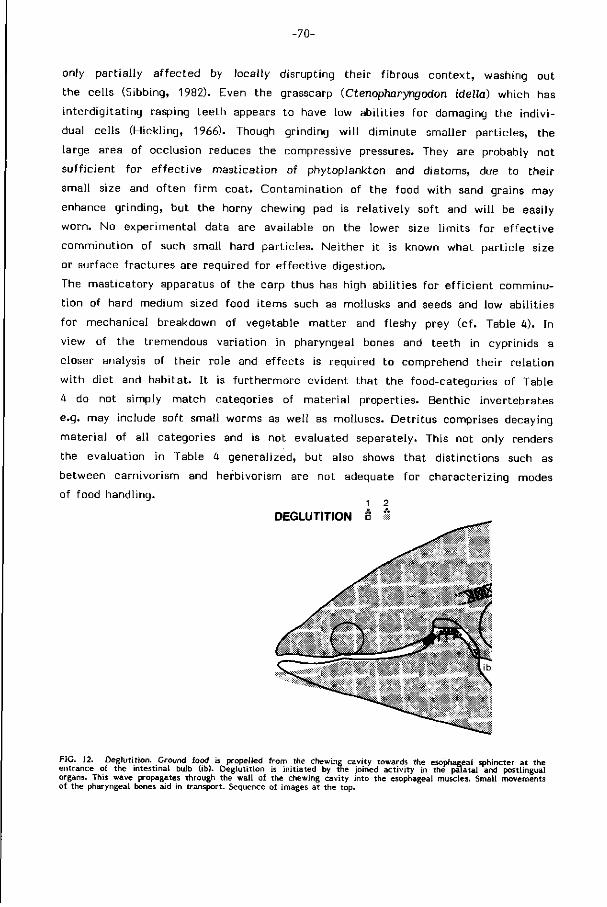
-70-
only part ia l ly a f fected by locally disrupting their f ibrous context , washing out
the cells (Sibbing, 1982). Even the grasscarp (Ctenopharyngodon idella) which has
interdigi tat ing rasping teeth appears to have low abi l i t ies for damaging the indiv i
dual cells (Hickl ing, 1966). Though grinding w i l l d iminute smaller part icles, the
large area of occlusion reduces the compressive pressures. They are probably not
suf f ic ient for e f fect ive mast icat ion of phytoplankton and diatoms, due to their
small size and of ten f i r m coat. Contaminat ion of the food w i th sand grains may
enhance grinding, but the horny chewing pad is re lat ively soft and w i l l be easily
worn. No experimental data are available on the lower size l imi ts for e f fect ive
comminut ion of such small hard part icles. Neither i t is known what part ic le size
or surface f ractures are required for e f fect ive digestion.
The masticatory apparatus of the carp thus has high abi l i t ies for e f f ic ient comminu
t ion of hard medium sized food i tems such as mollusks and seeds and low abi l i t ies
for mechanical breakdown of vegetable mat ter and fleshy prey (cf. Table 4). In
view of the tremendous var iat ion in pharyngeal bones and teeth in cyprinids a
closer analysis of their role and ef fects is required to comprehend their re lat ion
w i th diet and habitat. It is fur thermore evident that the food-categories of Table
4 do not simply match categories of mater ia l properties. Benthic invertebrates
e.g. may include soft small worms as wel l as molluscs. Detr i tus comprises decaying
mater ia l of al l categories and is not evaluated separately. This not only renders
the evaluation in Table 4 generalized, but also shows that dist inctions such as
between carnivorism and herbivorism are not adequate for characterizing modes
of food handling. 1 2
DEGLUTITION â *
FIG. 12. Deglutit ion. Ground food is propelled from the chewing cavity towards the esophageal sphincter at the entrance of the intestinal bulb (ib). Deglutit ion is init iated by the joined act ivi ty in the palatal and postlingual organs. This wave propagates through the wall of the chewing cavity into the esophageal muscles. Small movements of the pharyngeal bones aid in transport. Sequence of images at the top.
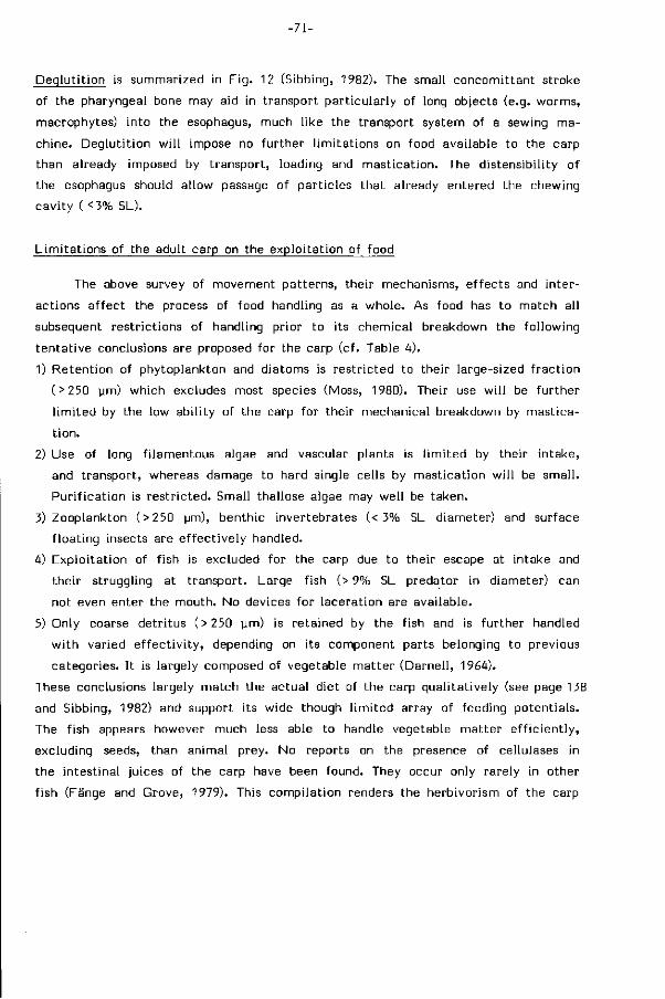
- 71 -
Deqlut i t ion is summarized in F ig . 12 (Sibbing, 1982). The small concomit tant stroke
of the pharyngeal bone may aid in transport part icular ly of long objects (e.g. worms,
macrophytes) into the esophagus, much l ike the transport system of a sewing ma
chine. Deglut i t ion w i l l impose no fur ther l imi tat ions on food available to the carp
than already imposed by t ransport, loading and mast icat ion. The distensibi l i ty of
the esophagus should allow passage of part icles that already entered the chewing
cavi ty ( <3% 5L).
L imi ta t ions of the adult carp on the exploi tat ion of food
The above survey of movement patterns, their mechanisms, e f fects and inter
actions a f fect the process of food handling as a whole. As food has to match all
subsequent restr ict ions of handling prior to its chemical breakdown the fol lowing
tentat ive conclusions are proposed for the carp (cf. Table 4).
1) Retent ion of phytoplankton and diatoms is restr ic ted to their large-sized f rac t ion
(>250 pm) which excludes most species (Moss, 1980). Their use w i l l be fur ther
l im i ted by the low abi l i ty of the carp for their mechanical breakdown by mastica
t ion.
2) Use of long f i lamentous algae and vascular plants is l im i ted by their intake,
and transport, whereas damage to hard single cells by masticat ion wi l l be smal l .
Pur i f icat ion is rest r ic ted. Small thallose algae may wel l be taken.
3) Zooplankton (>250 ym), benthic invertebrates (< 3% SL diameter) and surface
f loat ing insects are e f fect ive ly handled.
4) Exploi tat ion of f ish is excluded for the carp due to their escape at intake and
their struggling at t ransport. Large fish (> 9% SL predator in diameter) can
not even enter the mouth. No devices for laceration are available.
5) Only coarse detr i tus (>250 ym) is retained by the fish and is further handled
w i th varied e f fec t i v i t y , depending on its component parts belonging to previous
categories. I t is largely composed of vegetable mat ter (Darnel l , 1964).
These conclusions largely match the actual diet of the carp qual i tat ively (see page 138
and Sibbing, 1982) and support its wide though l im i ted array of feeding potentials.
The f ish appears however much less able to handle vegetable matter e f f ic ient ly ,
excluding seeds, than animal prey. No reports on the presence of cellulases in
the intestinal juices of the carp have been found. They occur only rarely in other
f ish (Fänge and Grove, 1979). This compi lat ion renders the herbivorism of the carp
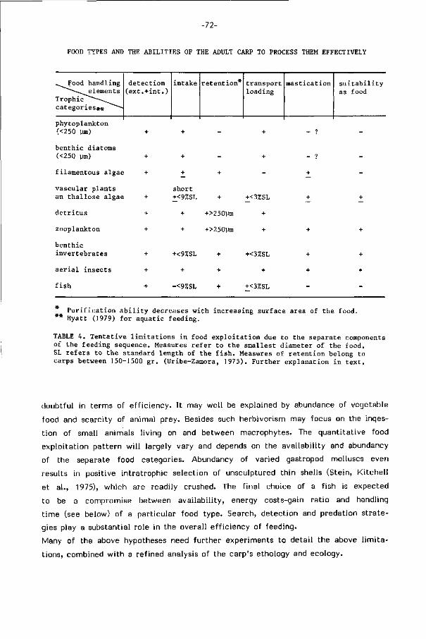
-72-
FOOD TYPES AND THE ABILITIES OF THE ADULT CARP TO PROCESS THEM EFFECTIVELY
..Food handling —^^elements
Trophic"""—«^.^ categories*« ̂""~-~
detection (ext.+int.)
intake retention* transport loading
mastication suitability as food
phytoplankton (<250 ym)
benthic diatoms (<250 um)
filamentous algae
vascular plants an thallose algae
detritus
Zooplankton
benthic invertebrates
aerial insects
fish
short +<9%SL
+
+
+<9%SL
+
-<9%SL
+
+>250ym
+>250Um
+
+
+
+<3ZSL
+
+
+<3%SL
+
+<3%SL
Purification ability decreases with increasing surface area of the food. ** Hyatt (1979) for aquatic feeding.
TABLE 4. Tentative limitations in food exploitation due to the separate components of the feeding sequence. Measures refer to the smallest diameter of the food. SL refers to the standard length of the fish. Measures of retention belong to carps between 150-1500 gr. (Uribe-Zamora, 1975). Further explanation in text.
doubtful in terms of e f f ic iency. I t may wel l be explained by abundance of vegetable
food and scarcity of animal prey. Besides such herbivorism may focus on the inges
t ion of small animals l iving on and between macrophytes. The quant i tat ive food
exploi tat ion pat tern w i l l largely vary and depends on the avai labi l i ty and abundancy
of the separate food categories. Abundancy of varied gastropod molluscs even
results in positive intratrophic selection of unsculptured thin shells (Stein, K i tche l l
et a l . , 1975), which are readily crushed. The f inal choice of a f ish is expected
to be a compromise between avai labi l i ty, energy costs-gain rat io and handling
t ime (see below) of a part icular food type. Search, detect ion and prédation s t rate
gies play a substantial role in the overall e f f ic iency of feeding.
Many of the above hypotheses need fur ther experiments to detai l the above l im i t a
tions, combined w i th a refined analysis of the carp's ethology and ecology.
-73-
BEHAVIOURAL ADJUSTMENTS IN FOOD PROCESSING AND HANDLING TIME.
Feeding sequences d i f fer according to the type and puri ty of the food (Figs.
3,4; Table 2). They are composed of d ist inct and of ten al ternative elements, in
corporated in a programmed order (Fig. 13). The composition of the feeding se
quence wi l l re f lect i ts adjustment to changing external (environmental, oropharyn
geal) and internal s t imul i (drive or mot ivat ion).
The dist inct elements of feeding behaviour may wel l be considered as f ixed
action patterns i.e. stereotyped and readily recognized units of behaviour (Baerends,
1971). L ike in non-behavioural characters there is some var iabi l i ty . The stereo
typed component of the pat tern (e.g. part iculate intake) is t r iggered by external
s t imul i (e.g. s t imul i of the food), the or ientation component (cf . Baerends, 1971)
steers the ac t iv i ty according to external cues (e.g. d i rect ion of protrusion). Reposi
t ioning, back-washing and recol lect ion f rom the branchial sieve have been d ist in
guished by their e f fect on the food. These elements are however expressions of
the same action pattern i.e. closed protrusion. In the electromyograms they were
all consequently referred to as posit ioning, because the events of the food were
not always recorded. Closed protrusion seems to be a core motor pat tern (cf . core
funct ion in Barel, 1983) common to a more complex set of functions in feeding.
Internal factors determine the drive or mot ivat ion for feeding and cause var iat ion
in the release threshold for the patterns. Thus an increased drive for feeding due
to persistent lack of animal prey may lower the threshold for feeding on vegetable
mat ter and increase the tendency for the consuming act. Combination of external
and internal factors determine if and what food is chosen and how i t is fur ther
handled.
When feeding is interrupted by spit t ing and immediately fol lowed by re-intake
of other part icles, which of ten occurs at bot tom inspection, this is considered
a searching sequence extended into the oropharyngeal cavity and referred to as
'probing' (F ig. 13), an appetit ive behaviour largely driven by internal factors. Spit
t ing also occurs as a u l t imate reject ion pat tern involved in search for food. I t
may even be used quite d i f ferent ly by exposure of hidden food through a je t of
water at searching.
The releasing, maintenance and stopping of the component action patterns,
their adjustment to the momentaneous si tuation of part icles and their integrat ion
into an e f fect ive feeding sequence requires ref ined sensory and regulatory mecha
nisms and probably involves positive and negative feedback loops. The enormous
differences in handling d i f ferent types of food (Figs. 3,4) show the uni formi ty of
the const i tut ing movement patterns but also the considerable d i f ferent ia t ion at
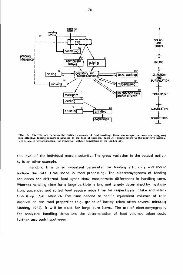
-74-
probing sequence
I rinsing —-Î gustatory and •— — . £actMescanningj-*-
spitting
BW
— | back-washing |
— | grinding
* . p. I deglutition I
SEARCH AND
CHOICE
I INTAKE +
SELECTION AND
PURIFICATION + TRANSPORT
I MASTICATION
4-DEGLUTITION
FIG. 13. Interrelations between the distinct elements of food handling. These stereotyped patterns are integrated into effective feeding sequences adjusted to the type of food (cf. Table 2). Probing refers to the repetit ive particulate intake of bottom-material for inspection without completion of the feeding act.
the level of the individual muscle ac t iv i ty . The great variat ion in the palatal ac t i v i
ty is an other example.
Handling t ime is an important parameter for feeding ef f ic iency and should
include the to ta l t ime spent in food processing. The electromyograms of feeding
sequences for d i f ferent food types show considerable differences in handling t ime .
Whereas handling t ime for a large part ic le is long and largely determined by mastica
t ion, suspended and soiled food require more t ime for respectively intake and selec
t ion (Figs. 3,4; Table 2). The t ime needed to handle equivalent volumes of food
depends on the food properties (e.g. grains of barley takes of ten several minutes;
Sibbing, 1982). I t w i l l be short for large pure i tems. The use of electromyography
for analyzing handling t imes and the determination of food volumes taken could
fur ther test such hypotheses.
-75-
GENERAL DISCUSSION
Retracing the cyprinid feeding mechanism
Detai led analysis of food handling revealed ten stereotyped act ion patterns
and involves a considerably higher d i f ferent ia t ion than already demonstrated for
euteleosts (cf. in i t ia l s t r ike, buccal and pharyngeal manipulation, and pharyngeal
t ransport (Lauder, 1983)). Though this analysis applies only to the carp, i t never
theless raises the question how such a complex biological design could have evolved
among the lower teleosts. No knowledge on fossils of the Cyprini formes exists
(Roberts, 1973). Eink and Eink (1981) recently provided a cladogram of the groups
of Ostariophysean fishes about the in terrelat ion between cyprinoids and the other
suborders. H is tor ica l data about the origin of their feeding mechanisms are lacking.
The present morphological and funct ional knowledge can be used to reconstruct
a possible evolutionary scheme.
The cyprinoids (Cyprinidae and Catostomidae) share, among others, the fol lowing
unique characters (Roberts, 1973).
1) A protrusi le mouth, comparably specialized as in Acanthopterygi i .
2) Toothless jaws.
3) Tootless palate
4) No upper pharyngeal elements of the f i f t h branchial arch.
5) Enlarged f i f t h ceratobranchials.
6) Basioccipital bony processes unit ing below the aorta.
7) Exceptionally deep subtemporal fossae.
The palatal organ appears s imi lar ly shared by cyprinoids and few other fishes,
the al l ied Cobit idae and some Salmonidae) (Kapoor et a l . , 1975).
The general p r imi t ive euteleostean pattern f rom which the Cyprini formes
deviated shows a grasping type of dent i t ion on small tooth plates fused to the
oral jaws and upper as wel l as lower branchial elements (Nelson, 1969). These bran
chial elements commonly funct ion in seizing and swallowing prey as mentioned
above (Lauder, 1983) and belong mostly to predaceous f ish w i th a wel l developed
prehensile and a simple selective apparatus. A plausible tentat ive scheme for the
development of the unique character-set of cyprinids could be visualized as fol lows.
Cyprinid ancestors could have chosen the bot tom areas, using the h i therto
rather unexploited vegetable mat ter as food. Recent cyprinids comprise a large
proport ion of herbivores compared to other teleost famil ies (Howes, 1978). The
hard and f ibrous consistency of plant mater ia l requires higher masticatory pressures
for r each jng the cel l contents than fleshy prey does. This could have induced
-76-
a hypertrophy of the f i f t h branchial arches and their associated muscles. A specia
l isation at this locat ion would l i t t l e in ter fer w i th respiratory functions of the bran
chial basket. Their posterior position fur thermore allowed the unique insertion
on these pharyngeal jaws of a sphincter oesophagi derivate, the ret ractor dorsalis
muscle (Holstvoogd, 1965), thus fur ther increasing masticatory abi l i t ies (cf. Sibbing,
1982). Another drastic change in this region of the skull is the vast subtemporal
fossa containing the massive levator muscle of the lower pharyngeal jaw. The reduc
t ion of movable upper pharyngeal elements may wel be explained by their l im i ted
abi l i ty to resist the high forces thus produced. Corni f icat ion of the pharyngeal
epithel ium and the underlying occipi tal region of the skull now serve in absorbing
the high masticatory forces. The functioning of the skull base as occlusal surface
in mastication forms part of this key-adaptation in the chewing apparatus, and
even permits epaxial body muscles to t ransmit their large forces ef fect ively to
the chewing pad.
The role of the pharyngeal masticatory apparatus may wel l have released the oral
jaws f rom their b i t ing funct ion. Reduction of the associated need for a f i r m upper
jaw abutment could wel l have allowed the development of the unique kinetics
in the cyprinid mouth apparatus (cf. Gosline, 1973) for bot tom feeding. The lack
of oral grasping teeth might be related to the feeding on immobile soiled prey
and probably required space for taste buds on the lips (ca. 380/mm2 in the carp,
c f . Sibbing et a l . , 1984), appropriate for bot tom inspection. One of the consequences
of such structural t ransformation is the exclusion of p iscivory, as fast and struggling
large prey can not e f fect ively be taken nor transported, due to the reduction of
posterior upper pharyngeal toothed elements. Predators are rarely met among cypr i -
nids. Hypertrophy of the pharyngeal wall into the palatal and postlingual organs
provides a substitute for the transport funct ion, lost w i th the upper pharyngeals.
Their sensory and e f fector abi l i t ies (cf . Sibbing et a l . , 1984) and the closed pro
trusion pattern of movement are crucial in selection between food and non-food
and character ist ic for cyprinids as wel l as catostomids. Due to protrusion volume
changes in the oral cavity increases in the carp for manipulating the f low of pa r t i
cles whereas volume changes in the pharynx are reduced in the carp for maintaining
a large contact area at selection. Even if such a scheme for the origin of the
cyprinoid feeding mechanism proves to be incorrect i t s t i l l provides a f rame for
understanding the interrelat ions of their unique set of characters. These structures
are in t imate ly funct ional ly l inked and together provide the complex requirements
for the subsequent movement patterns in feeding actions. In such a view the p isci
vorous cyprinids are not the p r imi t ive members of the fami ly but are secondarily
derived f rom bottomfeeding species because the above sequence of events explains
-77-
the early loss of characters appropriate for piscivory.
Protrusion
The important roles of premaxil lary protrusion in food handling of the carp
may be summarized shortly. 1) Protrusion directs the suction f low to a small area
in f ront of the mouth opening. I t increases the velocity of f low through the reduced
aperture of the mouth, allowing dense, distant and less accessible food to be taken.
2) By extending the length of the mouth, protrusion simply reduces f ish-food dis
tance wi thout adding impulse to the water or the f ish. 3) Ventrad protrusion main
tains a wider f ie ld of vision at bot tom feeding and fac i l i ta tes rapid escape f rom
predators. 4) Protrusion allows the aiming of the f ish at the food wi thout the need
for a sudden change of the body. This permits prey suction under variable small
angles w i th the body axis. 5) This fur thermore allows continuous probing of the
bot tom substrate while extending the search area by swimming. 6) Protrusion ef fects
a rostral hood at mouth closure during gulping thereby maximizing the volume
taken and preventing the loss of suspension. 7) Closed protrusion aids in resuspension
of food part icles in the orobuccal cavi ty, essential for the internal selection and
pur i f icat ion of food. 8) Closed protrusion allows part icles to be washed back for
repositioning and recol lect ion f rom the branchial sieve.
Whereas the f i rs t four advantages of protrusion for part iculate feeding in the carp
have been noted previously for other teleosts (cf. Alexander, 1970; van Leeuwen
and Muller, 1984; see Mot ta , 1984), the la t ter roles have been unknown but are
of crucial importance in bot tom feeding on mixtures of food and non-food. Thus
the contr ibut ion of protrusion to mechanisms for internal selection may have added
to the accessibil ity of the bot tom substrate as a possible food source in cyprinoids.
The comparison of s tructure and functioning wi th in several trophic types of
cyprinids (and other cypriniforms) is important to provide more data about the
above tentat ive scheme. The elegant comparison between trophic specialists in
cichlids by Barel (1983), though by its wide comparison necessary l imi ted in i ts
analysis of functions, provides a promising example of the impact that feeding
habits may have on the constructional morphology of f ish.
Detai led and quanti f ied knowledge of the feeding mechanisms and structures
combined w i th the ecological and ethological details of sympatric species w i l l
aid in predicting the probable e f fect of habitat changes for the natural f ish fauna.
-78-
Acknowledgements. The authors wish to thank the Zoological Laboratory of the Leiden University for the faci l i ties and assistance in making the X-ray movies. Peter Vossen analyzed the sequence of movements in Fig. 2. Mr. W. Valen applied his expertise in preparing the figures. Thanks are also due to Mrs. L. van Beek and Mrs. E.A. Scheffers for their endurance in typing the manuscripts.
REFERENCES
Akster, H.A. (1981). Ultrastructure of muscle fibres in head and axial muscles of the perch (Perca fluviatilis L.). Cell Tissue Res. 219: 111-131.
Akster, H.A. & Sibbing F.A. (1982). Fibre types in axial muscles, the m.hyohyoideus and masticatory muscles of the carp (Cyprinus carpio L.). 3. Muscle Res. Cell Mot. 3: 124.
Alexander, R.McN. (1969). Mechanics of the feeding action of a cyprinid fish. 3.Zool., 159: 1-15. Alexander, R.McN. (1970). Mechanics of the feeding action of various teleost fishes. 3. Zool., 162: 145-156. Baerends, G.P. (1971). The ethological analysis of fish behavior. In: Fish Physiology 6: 279-370. Hoar, W.S., Randall,
D J . & Brett, 3.R. (Eds). New York: Academic Press. Ball inti jn, C M . (1969). Functional anatomy and movement co-ordination of the respiratory pump of the carp [Cyprinus
carpio L.). 3.Exp. Biol. 50: 547-567. Ball int i jn, C M . , van den Burg, A. & Egberink, B.P. (1972). An electromyographic study of the adductor mandibulae
complex of a free-swimming carp (Cyprinus carpio L.) during feeding. 3. Exp. Biol. 57: 261-283. Barel, C.D.N. (1983). Towards a constructional morphology of cichlid fishes (Teleostei. Perciformes). Neth. 3. Zool.
33: 357-424. Berghe, L.van den (1929). Note sur ta déglutition chez les cyprinides. Bull. Acad. Roy. Belg. c l . Sei. 15: 867-876. Blaxter, 3.H.S. (1969). Swimming speeds of fish. FAO Fish. Rep. Rome, 2 (62): 69-100. Blaxter, 3.H.S. (1970). Light. In: Marine Ecology. 1: 213-320. Kinne, O. (Ed). New York: Wiley & Sons Inc. Boddeke, R., Slijper, E.3. & van der Stelt, A. (1959). Histological characteristics of the body-musculature of fishes
in connection with their mode of l i fe. Proc. K. Ned. Akad. Wet., Ser. C 62: 576-588. Darnell, R.M. (1964). Organic detritus in relation to secondary production in aquatic communities. Verh. Int. Ver.
Limnol. 15: 462-470. Drenner, R.W., Strickler, 3.R. & O'Brien, W.3. (1978). Capture probability: the role of zooplankter escape in the
selective feeding of planktivorous fish. 3. Fish. Res. Bd. Can. 35: 1370-1373. Fänge, R. & Grove, D. (1979). Digestion. In: Fish Physiology 8: 162-260. Hoar, W.S., Randall, D.3. & Brett , 3.R.
(Eds). New York: Academic Press. Fink, S.V. & Fink, W.L. (1981). Interrelationships of the ostariophysan teleost fishes. Zool. 3. Linn. Soc. London,
72: 297-353. Girgis, S. (1952). The bucco-pharyngeal feeding mechanism in an herbivorous bottom-feeding cyprinoid, Labeo horie
(Cuvier). 3. Morphol. 90: 281-315. Gosline, W.A. (1973). Considerations regarding the phylogeny of cypriniform fishes, wi th special reference to struc
tures associated with feeding. Copeia: 761-776. Hara, T.3. (1971). Chemoreception. In: Fish Physiology 5: 79-120. Hoar, W.S., Randall, D.3. & Brett , 3.R. (Eds).
New York: Academic Press. Herrick, C.3. (1904). The organ and sense of taste in fishes. Bull. U.S. Fish. Comm. 22: 239-272. Hickling, C F . (1966). On the feeding process in the White Amur, Ctenopharyngodon idella. 3. Zool. 148: 408-419. Holstvoogd, C. (1965). The pharyngeal bones and muscles in Teleostei, a taxonomie study. Proc. Kon. Ned. Akad.
Wet. 68: 209-218. Howes, G. (1981). Anatomy and phylogeny of the Chinese major carps Ctenopharyngodon Steind., 1866 and Hypoph-
thalmichthys BIKr., 1860. Bull. Br. Mus. Nat. Hist. (Zool.) 41 : 1-52. Hyatt, K.D. (1979). Feeding strategy. In: Fish Physiology 8: 71-120. Hoar, W.S., Randall, D.3. óc Brett , 3.R. (Eds).
New York: Academic Press. Iwai, T. (1964). A comparative study of the taste buds in g i l l rakers and gi l l arches of teleostean fishes. Bull. Misaki
Mar. Biol. Inst. Kyoto Univ. 7: 19-34. 3anssen, 3. (1976). Feeding modes and prey size selection in the alewife (Alosa pseudoharengus ). 3. Fish. Res. Bd.
Can. 33: 1972-1975. 3ara, Z. (1957). On the morphology and function of the so-called palatal organ in the carp (Cyprinus carpio L.).
Przeglad Zoologiczny 1: 110-112. 3onsson, N. (1967). Experimentelle Untersuchungen über die Futtersuche und die Akt iv i tät einsömmriger Karpfen.
Z. Fischerei 15: 61-127. Kapoor, B.C., Smit, H. 4: Verighina, I.A. (1975). The alimentary canal and digestion in teleosts. Adv. Mar. Biol.
13: 109-239. Klust, G. (1940). Über Entwicklung, Bau und Funktion des Darmes beim Karpfen (Cyprinus carpio L.). Int. Rev. Ges.
Hydrobiol. Hydrogr. 40: 88-173. Lauder, G.V. (1983). Functional design and evolution of the pharyngeal jaw apparatus in euteleostean fishes. Zool.
3. Linn. Soc. London, 77: 1-38. Leeuwen, 3. van (1984). A quantitative study of flow in prey capture by rainbow t rout, w i th general consideration
of the actinopterygian feeding mechanism. Trans. Zool. Soc. London, 37: 171-227. Leeuwen, 3.L. van óc Muller, M. (1983). The recording and interpretation of pressures in prey-sucking fish. Neth.
3. Zool. 33: 425-475. Leeuwen, 3.L. van & Muller, M. (1984). Optimum sucking techniques for predatory f ish. Trans. Zool. Soc. London,
37: 137-169.
-79-
Liem, K.F, (1967). Functional morphology of the head of the anabantoid teleost f ish, Helostoma temmincki. 3. Mor-phol., 121: 135-158.
Liem, K.F. (1973). Evolutionary strategies and morphological innovations: Cichlid pharyngeal jaws. Syst. Zool. 22: 425-441.
Liem, K.F. & Greenwood, P.H. (1981). A functional approach to the phylogeny of the pharyngognath teleosts. Am. Zool. 21: 83-101.
Matthes, H. (1963). A comparative study of the feeding mechanisms of some African Cyprinidae (Pisces, Cyprini-formes). Bijdr. Dierkunde, Amsterdam. 33: 3-35.
Moss, B. (1980). Ecology of freshwaters. London: Blackwell Scientific Publications. Motta, P.3. (1984). Mechanics and functions of jaw protrusion in teleost fishes: A review. Copeia: 1-18. Muller, M. & Osse, 3.W.M. (1984). Hydrodynamics of suction feeding in f ish. Trans. Zool. Soc. London, 37: 51-135. Muller, M., Osse, 3.W.M. & Verhagen, 3.H.C. (1982). A quantitative hydrodynamical model of suction feeding in
fish. 3. Theor. Biol. 95: 49-79. Nelson, G.3. (1969). Gil l arches and the phylogeny of fishes, with notes on the classification of vertebrates. Bull.
Am. Mus. Nat. Hist. 141: 475-552. Nelson, 3.S. (1976). Fishes of the world. New York: 3ohn Wiley and Sons. Nikolsky, G.V. (1963). The ecology of fishes. London: Academic Press. Osse, 3.W.M. (1969). Functional morphology of the head of the perch (Perca fluviatilis L.): An electromyographic
study. Neth. 3. Zool. 19: 289-392. Roberts, T-R. (1973). Interrelationships of Ostariophysans. In: Interrelationships of fishes: 373-396. Greenwood, P.H.,
Miles, R.S- & Patterson, C. (Eds). London: Academic Press. Robotham, P.W.3. (1982). An analysis of a specialized feeding mechanism of the spined loach, Cobitis taenia (L.),
and a description of the related structures. 3. Fish Biol. 20: 173-181. Sibbing, F.A. (1982). Pharyngeal mastication and food transport in the carp (Cyprinus carpio L.): A cineradiographic
and electromyographic study. 3. Morph. 172: 223-258. Sibbing, F.A. & Uribe, R. (1984). Regional specializations of the oro-pharyngeal wall and food processing in the
carp (Cyprinus carpio L.). In this thesis. Stein, R.A., Ki tchel l , 3.F. & Knezevic, B. (1975). Selective prédation by carp (Cyprinus carpio L.) on benthic molluscs
in Skadar Lake, Yugoslavia. 3. Fish Biol.: 391-399. Suietov, S.V. (1939). Contribution to the fish productivity of fresh waters. VIII. The thickness of bottom deposits
and its role in the uti l ization of natural food by fishes. Proc. Kossino Limnol. Station 22: 241-249. Uribe-Zamora, M. (1975). Selection des proies par le f i l t re branchial de la carpe miroir (Cyprinus carpio L.). Thesis,
University of Lyon. Wright, D.I., O'Brien, W.3. & Luecke, C. (1983). A new estimate of zooplankton retention by gi l l rakers and
its ecological significance. Transact. Am. Fish. Soc. 112: 638-646. Wunder, W. (1927). Sinnesphysiologische Untersuchungen: über die Nahrungsaufnahme bei verschiedenen Knochen
fischarten. Z. Vergl. Physiol. 6: 67-98. Wunder, W. (1936). Physiologie der Süsswasserfische. Handb. Binnenfisch. Mitteleur. 26: 1-340. Zander, E. (1906). Das Kiemenfilter der Teleosteer- Z. Wiss. Zool. 84: 619-713.

HOOFDSTUK 3
REGIONAL SPECIALIZATIONS IN THE ORO-PHARYNGEAL WALL AND FOOD
PROCESSING IN THE CARP {Cyprinus carpio L.).
F.A. Sibbing and R. Uribe
-82-
SUMMARY
The structure of the oro-pharyngeal wall of the bottomfeeding common carp
is investigated using l ight-and scanning electron microscopy. Densities of taste
buds, mucus, club cells and the thickness of muscular layers are measured. D is t r i
bution patterns of these elements over the oro-pharyngeal surface are reconstructed
f rom local counts. Six areas characterized by a specific combination of morpholo
gical features are distinguished and related to twelve movement patterns com
posing the process of food intake and handling in the carp. These areas are the
lips (detection and oral manipulation of food), the orobuccal cavity (suction and
resuspension chamber of ingested food and non-food particles), the most anterior
pharynx (coarse selection of large food particles), the lateral pharynx (selection
of small food particles), the posterior part of the anterior pharynx ( formation of
boluses, transport and loading of food into the chewing cavity) and the posterior
pharynx (mastication and deglutit ion), The conical shape of the orobuccal cavity
and the slit-shaped anterior pharynx ref lect two d i f ferent mechanisms for part ic le
handling v iz. by suction and by muscular bulging respectively. The opercular cavities
serve large volume changes for suction feeding.
Protruding types of taste buds and ol igivi l lous epithel ial cells may well serve
mechanoreceptive functions required for steering the process. Otherwise, specialized
mechanoreceptors have not been recognized. Mucous cells producing low-viscosity
sialomucines occur in the anterior part of the oro-pharynx. They wi l l serve main
taining a laminar f low during suction and lubricat ion of part icle handling in the
pharynx. Epithel ial microridges may aid in holding the mucus. High-viscosity sulfo-
mucines only appear in the posterior part of the pharynx and wi l l aid in trapping
small part icles and st icking them into boluses. The commonly accepted alarming
funct ion of club cells and their meachanism for release is questioned in view of
their abundancy in the orobuccal cavi ty.
The structure of the muscular palatal organ is discussed wi th respect to its
role in selection between food and non-food part icles. The available information
on the af ferent, e f ferent and central neural pathways of this system is br ief ly
reviewed. Three levels of movement, related to the part icle size to be handled,
are proposed. Movement of the palatal organ as a whole, local outbulging of its
surface into the pharyngeal sl it and a possible very local movement of the muscular
papillae in its anterior part. These hypotheses are based on the almost maximal
taste bud densities (820/mm2) in the palatal organ, the known complex laminated
-83-
cyto-archi tecture of the enormous vagal lobes processing its input and suggestive
of a palatotopic mapping, and on the complex muscle f iber systems in this organ.
The movable gi l l rakers of the branchial sieve, each supplied wi th a muscular pad
and numerous taste buds (325-625/mm2), suggest their additional active role in
selection.
Muscle f iber systems in the posterior part of the palatal as wel l as in the closely
appressed postlingual organ serve a peristalsis-l ike transport to the chewing cavity.
Both are copiously supplied wi th sulfomucines f rom their deep crypts.
Together these morphological and physiological features allow the carp a bot tom
feeding behaviour requiring the e f fect ive separation of food f rom soiled mixtures.
Contents
summary 82
introduction 84
materials and methods 85
results 89
. gross anatomy 89
. l ight- and scanning electron microscopy of the oro-pharyngeal wall 95
. specializations of the oro-pharyngeal epithelium 99
e f fector specializations 99
sensory specializations 101
. quant i tat ive distr ibution patterns of taste buds, mucus, club cells 103
and muscle f ibers
discussion 108
. the role of mucus, microridges, club cells and sensory elements in the 108
oro-pharynx
. the pharyngeal wall and its mechanisms for selection between food 113
and non-food
. pathways connecting oro-pharyngeal sensors and effectors 115
. six specialized areas in the oro-pharynx related to food handling in the carp 116
abbreviations 122
references 123
-84-
INTRODUCTION
The trophic diversity among cyprinids st imulated many studies on the adaptive
characters in the al imentary t ract of this largest teleost fami ly . Suyehiro (1942)
and Kapoor et a l . (1975) have presented reviews on this subject for teleosts in
general. Verigina (1969) has done so for cyprinids. Structural data on the carp
are reported by Curry (1939) and Al-Hussaini (1949). In the last decades new tech
niques revealed additional types of specialized sensory and secretory structures
in f ish epidermis (cf. Whitear, 1971; Fox et a l . , 1980; M i t ta l et a l . , 1980).
Bottomfeeding cyprinids suck mixtures of food, detri tus and inorganic materials
w i th their toothless protrusile mouth. Af terwards they select between food and
non-food wi th in their oro-pharyngeal cavi ty. The muscular palatal organ in the
pharyngeal roof, most developed among cyprinids in the carp (Weber, 1827; Herr ick,
1904; Dorier and Bellon, 1952) and even larger in catostomids (Weisel, 1962; East
man, 1977), is a conspicuous cyprinoid feeding s tructure. Its size, complicated
structure and abundant taste buds has led to a variety of hypotheses about its
role in food selection and respiration (cf. Kapoor et a l . , 1975). Its innervation by
a huge vagal lobe in the brain fur thermore suggests an important role in feeding
(Evans, 1952).
The lack of a functional analysis of the actual events during feeding l imi ted
the explanation of the varied structural components as adaptive characters. There
fore previous papers on the common carp present an analysis of the feeding me
chanism using electromyography, l ight and X-ray cinematography (Sibbing, 1976,
1979, 1982; Sibbing et a l . , 1984). The feeding sequence of this species appears
to be composed of twelve stereotyped movement patterns serving intake, selection,
t ransport, mastication and deglut i t ion. The feeding sequences are adjusted to the
locat ion, size, movement, contamination and consistency of the food by the appro
pr iate employment and repet i t ion of these specific movement patterns.
The aim of the present study is to analyze the sensory and e f fector structures
in the oro-pharyngeal wal l which determine the ef f ic iency of internal food pro
cessing. To understand the s t ructure-funct ion relationships the macroscopical and
(electron) microscopical morphology of the oro-pharyngeal wal l are related to the
actual events during feeding. Detai ls of taste buds, oligovillous cells, mucus, micro-
ridges and muscle f iber arrangment can now be f i t t ed in one comprehensive view
of food handling and food processing. As the funct ional d i f ferent ia t ion is now known
into great detai l also quant i tat ive measurements of the above elements and their
reconstruct ion into distr ibution patterns are required to establish form-funct ion
relationships. For example, disagreement between Curry (1939) and Al-Hussaini
-85-
(1949) on the presence and number of part icular cell types and taste buds in the
carp is largely due to their inadequate descriptive terms.
The present data and a c r i t ica l review of carp feeding show a considerable
gap in our knowledge. This concerns mainly the lack of information on the presence
and properties of the involved sensors and the way the fish manages to integrate
and use their enormous input to regulate its feeding behaviour. We hope this study
to be a s t imulat ing factor for such research. Another aim of this study is to provide
a general f rame for comparing feeding in cyprinid species. This w i l l improve our
insight into the trophic segregation of cyprinid species l iving together in one com
munity.
MATERIALS AND METHODS
A l l mirror-carps (Cyprinus carpio L.) were reared in our laboratory and fed
on small Trouvit grains (Trouw and Co., Put ten, The Netherlands).
Histology
Eor histological examination descaled carps of 58 mm SL were f ixed in Bouin's
f lu id (Romeis, 1968) and decalcif ied over two weeks in a mixture of 200 ml formic
acid, 200 ml ethanol (70%) and 40 gr. sodiumcitrate, renewed every three days.
A f te r dehydration over a graded ethanol series the specimens were embedded over
amylacetate in paraf f in. Serial sections of the head (7 pm) were stained after
Crossmon (Romeis, 1968) allowing a clear dist inct ion between connective tissue,
muscle f ibers, mucous cells, club cells and taste buds.
Neutral as well as acid mucus is stained wi th Periodic Acid Schiffs reagens
(PAS; McManus et a l . , 1963). Acid mucopolysaccharides and glycoproteins have
been localized by positively staining w i th Alcian Blue (AB) at pH 2.5 (McManus
et a l . , 1963). A reaction w i th High Iron Diamin (HID; Spicer, 1965) fur ther d ist in
guished acid sulfomucines (pos. staining) f rom acid sialomucines (neg. staining).
Morphometry
Four parameters were measured at 980 ym or smaller intervals through the
length of the oro-pharyngeal cavi ty (ca. 15 mm).
The density of taste buds was measured by counting in the 7 y m transverse
sections each taste bud intersection that was clearly dist inct f rom the common
epithel ial cells. Because the mean diameter at the centre of the taste bud, measured
f rom di f ferent regions, was 32.5 _+ 5.1 ym (n=60), the actual number of taste buds
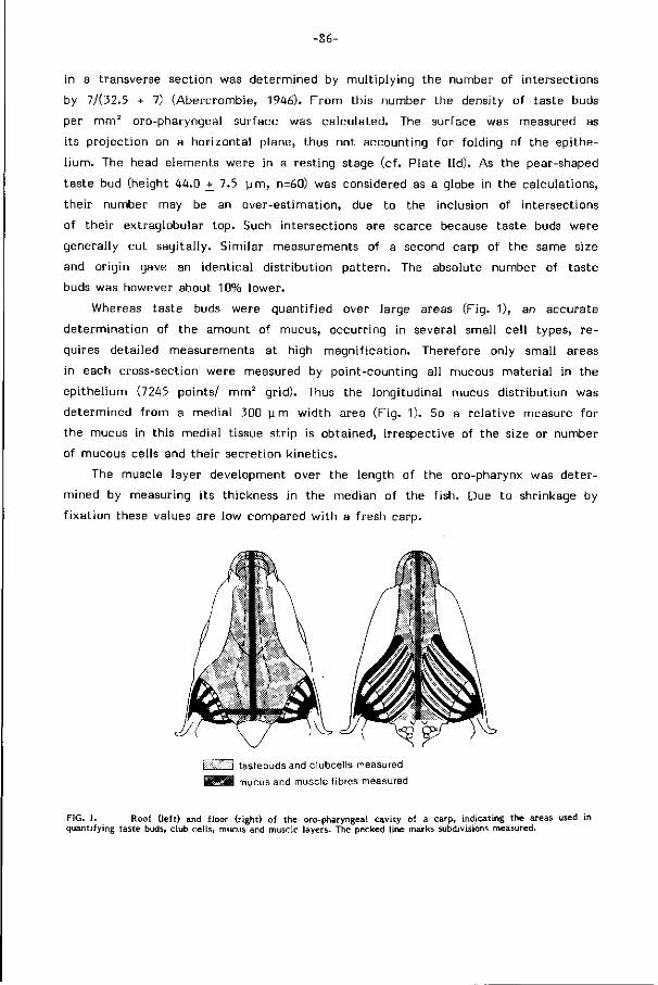
-86-
in a transverse section was determined by mult iply ing the number of intersections
by 7/(32.5 + 7) (Abercrombie, 1946). F rom this number the density of taste buds
per mm 2 oro-pharyngeal surface was calculated. The surface was measured as
its project ion on a horizontal plane, thus not accounting for folding of the epithe
l ium. The head elements were in a resting stage (cf. Plate l id). As the pear-shaped
taste bud (height 44.0 +_ 7.5 u rn , n=60) was considered as a globe in the calculations,
their number may be an over-est imation, due to the inclusion of intersections
of their extraglobular top. Such intersections are scarce because taste buds were
generally cut sagital ly. Similar measurements of a second carp of the same size
and origin gave an identical d istr ibut ion pat tern. The absolute number of taste
buds was however about 10% lower.
Whereas taste buds were quanti f ied over large areas (Fig. 1), an accurate
determinat ion of the amount of mucus, occurring in several small cel l types, re
quires detailed measurements at high magnif icat ion. Therefore only small areas
in each cross-section were measured by point-counting all mucous mater ial in the
epithel ium (7245 points/ mm2 gr id). Thus the longitudinal mucus distr ibut ion was
determined f rom a medial 300 y m width area (Fig. 1). So a relat ive measure for
the mucus in this medial tissue strip is obtained, i rrespective of the size or number
of mucous cells and their secretion k inetics.
The muscle layer development over the length of the oro-pharynx was deter
mined by measuring i ts thickness in the median of the f ish. Due to shrinkage by
f i xa t ion these values are low compared w i th a fresh carp.
tastebuds and clubcells measured mucus and muscle fibres measured
FIG. 1. Roof ( left) and floor (right) of the oro-pharyngeal cavity of a carp, indicating the areas used in quantifying taste buds, club cells, mucus and muscle layers. The pecked line marks subdivisions measured.
-87-
Th e density of club cells was determined by counting the number of their
intersections over large areas (Fig. 1). These are expressed per horizontal m i l l imeter
transverse section. Only those intersections of club cells were counted which had
the unmistakable central nucleus as a character ist ic. So the numbers in the graphs
provide a re lat ive measure of their intersections, the actual number being higher.
Detai led measurements of taste buds, mucus and muscle layer over the fu l l
w idth of the pharyngeal roof have been made in the posterior part of the palatal
organ (Fig. 1) where a varied transverse distr ibut ion was most conspicuous. Further
more, transverse differences in taste bud densities at other sites have been inferred
f rom comparison between counts of the to ta l and the medial areas (Fig. 1). As
at the level of the branchial arches and the chewing cavity an accurate measure
ment of the surface is impossible, data on these areas have been estimated f rom
the SEM photographs and f rom the microscopic slides respectively (dotted lines
in the f igures).
Semi-quanti tat ive measurements by distinguishing four density levels of taste
buds, mucous cells and club cells in an older carp (SL 20 cm), prior to accurate
counting in sections of small carps (SL 58 mm), provided a corresponding p icture
of their d istr ibut ion.
Scanning electron microscopy
For scanning electron microscopy (SEM) carps of similar size (53-58 mm SL)
were decapitated after anesthesia by an overdose of 2Ü0 mgr MS 222/l i ter (Sandoz,
Basel). The specimens were careful ly washed in buffered physiological saline to
remove mucus adhering to the surface and f ixed for eight days at 5 C in 3% glutar-
aldehyde, buffered at pH 7.2 in 0.1 M sodiumcacodylate containing 2.5% glucose
(Reutter et a l . , 1974). Subsequently the heads were cut horizontal ly in a dorsal
and ventral half, each reduced to a slice of few mi l l imeters to f i t in the micro
scope. This material was washed repeatedly in a buffered saline solution prior
to f inal f ixat ion for six hours in 1% osmiumtetroxide, buffered in 0.1 M sodium
cacodylate. A f te r repet i t ive washing in the buffer, dehydration in a graded series
of ethanol (10-100%) and c r i t i ca l point drying, the specimens were mounted on
a stub and coated wi th a f i lm of gold. For examination a JEOL JSM-35C scanning
electron microscope operating at 25 KV and magnifications of 10 - 10.000 t imes
was used. The large specimens could be stored for three weeks over a si l ica gel
wi thout damage.
Though SEM photographs may seem to provide an excellent base for fastly
measuring the taste buds densities (they give closely similar results for the buccal
roof, ca. 40-50/mm2) these SEM measurements become increasingly inaccurate
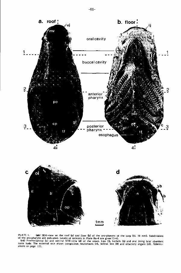
a. roof ! b. floor !
1mm
PLATE I. (ab) SEM-view on the roof (a) and floor (b) of the oro-pharynx of the carp (SL 58 mm). Subdivisions of the oro-pharynx are indicated. Levels of sections in Plate Ila-d are given ( l - ' i ) .
(cd) Fronto-lateral (c) and ventral SEM-view (d) of the snout. Lips (1), barbels (b) and oral lining bear abundant taste buds. The external skin shows conspicious neuromasts (n), lateral line (11) and olfactory organs (ol). Abbreviations on page 122.
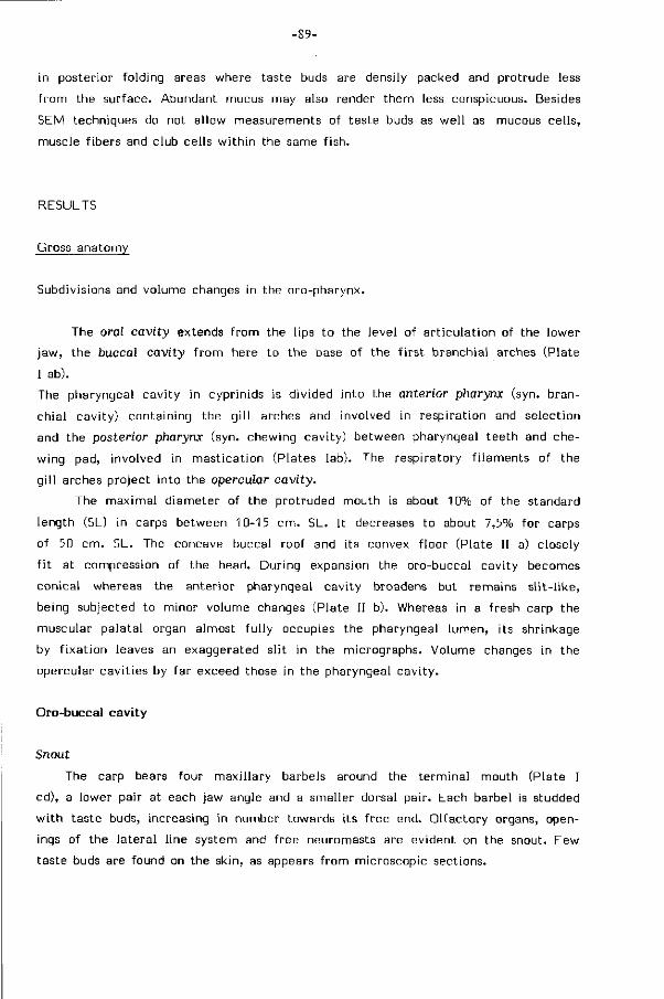
-89-
in posterior folding areas where taste buds are densily packed and protrude less
f rom the surface. Abundant mucus may also render them less conspicuous. Besides
SEM techniques do not allow measurements of taste buds as wel l as mucous cel ls,
muscle f ibers and club cells w i th in the same f ish.
RESULTS
Gross anatomy
Subdivisions and volume changes in the oro-pharynx.
The oral cavity extends f rom the lips to the level of a r t icu lat ion of the lower
jaw, the buccal cavity f r om here to the base of the f i rs t branchial arches (Plate
I ab).
The pharyngeal cavity in cyprinids is divided into the anterior pharynx (syn. bran
chial cavity) containing the g i l l arches and involved in respiration and selection
and the posterior pharynx (syn. chewing cavity) between pharyngeal teeth and che
wing pad, involved in masticat ion (Plates lab). The respiratory f i laments of the
g i l l arches project into the opercular cavity.
The maximal diameter of the protruded mouth is about 10% of the standard
length (SL) in carps between 10-15 cm. SL. It decreases to about 7,5% for carps
of 50 cm. SL. The concave buccal roof and its convex f loor (Plate II a) closely
f i t at compression of the head. During expansion the oro-buccal cavity becomes
conical whereas the anterior pharyngeal cavity broadens but remains s l i t - l i ke ,
being subjected to minor volume changes (Plate II b). Whereas in a fresh carp the
muscular palatal organ almost fu l ly occupies the pharyngeal lumen, its shrinkage
by f ixat ion leaves an exaggerated sl i t in the micrographs. Volume changes in the
opercular cavit ies by far exceed those in the pharyngeal cavi ty.
Oro-buccal cavity
Snout
The carp bears four maxi l lary barbels around the terminal mouth (Plate I
cd), a lower pair at each jaw angle and a smaller dorsal pair. Each barbel is studded
w i th taste buds, increasing in number towards i ts f ree end. Ol factory organs, open
ings of the lateral l ine system and free neuromasts are evident on the snout. Eew
taste buds are found on the skin, as appears f rom microscopic sections.
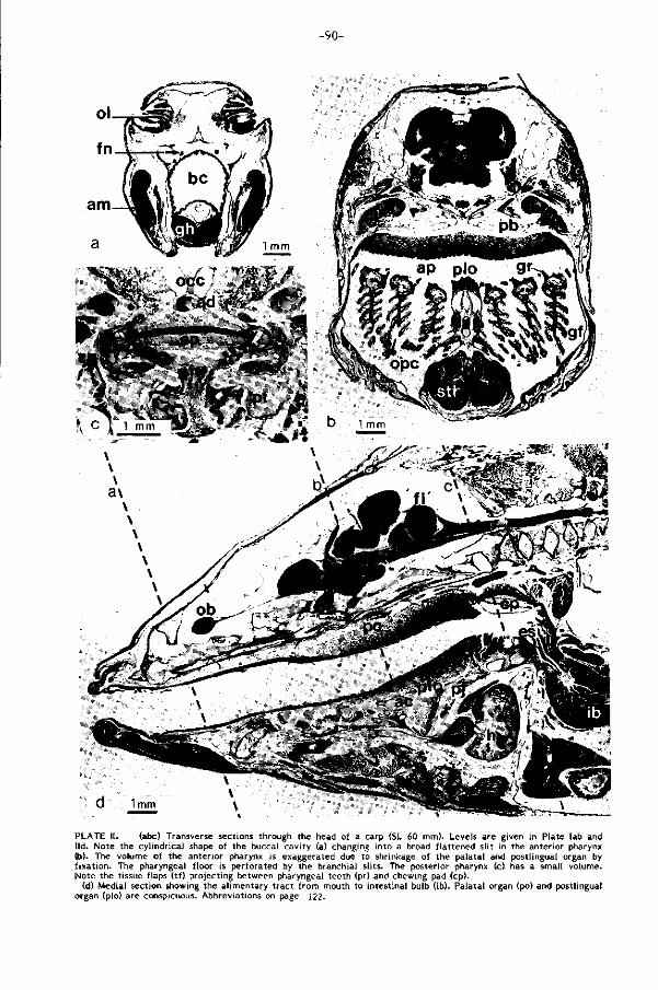
-90-
•T
: C t ' mm ±]J-^-''S' . * \ b Innm
\ \ a\
PLATE II. (abc) Transverse sections through the head of a carp (SL 60 mm). Levels are given in Plate lab and lid. Note the cylindrical shape of the buccal cavity (a) changing into a broad flattened slit in the anterior pharynx (b). The volume of the anterior pharynx is exaggerated due to shrinkage of the palatal and postlingual organ by fixation. The pharyngeal floor is perforated by the branchial slits. The posterior pharynx (c) has a small volume. Note the tissue flaps (tf) projecting between pharyngeal teeth (pt) and chewing pad (cp).
(d) Medial section showing the alimentary tract from mouth to intestinal bulb (ib). Palatal organ (po) and postlingual organ (plo) are conspicuous. Abbreviations on page 122.
-91-
The protrusile toothless mouth is bordered by thick lips bearing rows of longitu
dinally arranged taste buds (ca. 360/mm2 , Plate Il ia) on the f la t tops of closely
packed epithelial folds. Taste buds are absent at the mouth angles.
Inwards f rom the lips the taste bud area is interrupted by a small crescent shaped
cel lular area of cornif ied epithel ium, the area of contact between upper and lower
jaw. These horny single cells (Plate Il ia) may be similar to the 'uncul i ' described
for loricarids by Roberts (1982). The corni f ied area is devoid of taste buds.
Oro-buccal roof
A crescent shaped respiratory valve projects f rom the oral roof at the inner
horny edge of the upper jaw (Plate lac). Wide papillae (ca. 150 x 200 urn) w i th
abundant taste buds (ca. 250/mm2) are found in its basal part , its distal half is
smooth and taste buds are almost lacking. The roof of the oral cavi ty is p l icated.
Anter ior ly deep papil lated folds run transversad and follow the contours of the
protrusi le upper jaw (Plate la). Behind the jaw art iculat ion buccal plicae run in
creasingly longitudinally and bear few small papillae (30x40/70 p m , Plate I l lb).
Papillae and taste buds (ca. 50/mm2) merely occur on top of the folds.
Oro-buccal floor
Papillae similar to those on the base of the maxi l lary valve (ca. 250 tb /mm 2 )
extend inwards f rom the horny area of the lower jaw (Plates I b, III a). A mandibular
valve is absent. Whereas the transverse folds along the lower jaw are less pro
nounced, the lower jaw being non-protrusile, longitudinal folds at the level of the
cheeks are deep and run caudo-laterad towards the f i rs t branchial sl it (Plate lb).
These folds allow considerable extension of the buccal walls. Few taste buds l ie
on their tops (ca. 40/mm2) . The central convex area of the buccal f loor, of ten
referred to as tongue, is supported by the mobile glossohyal, connected to the
ventral elements of the hyoid arch. A war t - l ike protuberance marks the posterior
end of the glossohyal. Medium-sized papillae (ca. 100x160 urn) w i th taste buds
(ca. 75/mm2) are conspicuous over al l this area.
Pharyngeal cavity
Anterior pharynx
The roof of the anterior pharynx is formed by a thick muscular cushion, the
palatal organ, covering the base of the skul l , the epibranchials and the pharyngo-
branchials (Plate II b). The surface of the slightly bilobed palatal organ closely
f i t s the pharyngeal surface and curvature of the gi l l arches. Thus the V-shaped
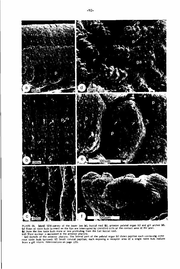
-92-
m
PLATE HI. (abed) SEM-survey of the lower jaw (a), buccal roof (b), anterior palatal organ (c) and gi l l arches (d). (a) Rows of taste buds (arrows) on the lips are interrupted by cornified cells at the contact area of the jaws. (b) Note the few taste buds more or less protruding from the f lat buccal roof, (cd) Their number is increased in the anterior pharynx. ' (ef) Details of the anterior pharynx. The lateral part of the palatal organ (e) shows papillae each containing numerous taste buds (arrows), (f) Small conical papillae, each exposing a receptor area of a single taste bud, radiate f rom a gi l l rakers. Abbreviations on page 122.
-93-
rostral border of the organ follows the at tachment of the g i l l arches (cf. Plates
Ia,b) and the organ bends postero-lateral ly dorsad along w i th the epibranchials.
The sl it between roof and f loor is broad but narrow and even vanishes close to
the chewing cavity in a fresh carp.
The surface of the palatal organ is i rregularly but heavily papil lated (Plates la ,
l ib , I l lce, IVb) and densily packed w i th taste buds on the f la t tops, even more dense
in the la teral areas (average up to 670 tb/mm2 ) than in the medial region (av.
up to 400 tb /mm 2 ) . Papillae in the lateral areas are smaller (ca. 90 pm diameter)
than in the medial region (ca. 140x150/550 pirn). In the posterior part of the palatal
organ, the tr iangular area enclosed between the last g i l l arches and the chewing
pad, the papillae lengthen into deep longitudinal folds (Plate la, IVc) w i th a markedly
reduced number of taste buds (av. up to 200/mm2).
The floor of the anterior pharynx (Plates lb, l ib) is largely composed of the
g i l l arches and their rakers. From these plates i t is clear that the g i l l arches and
their double rows of rakers fo rm a perforated plane that can be closely applied
to the palatal organ.
The pharyngeal surfaces of the g i l l rakers bear numerous small radiating conical
papillae (ca. 30-50 pm diameter), each w i th a taste bud (av. up to 480/mm2 in
the horizontal part of the g i l l arches; Plates III d f) . The tapering gi l lrakers f rom
subsequent arches in terdig i tate in a fresh carp and play a role in food selection
(Zander, 1906). Between the two rows of gi l l rakers the pharyngeal surface of
each g i l l arch (Plates l ib , Hid) is covered by a thin muscular str ip of tissue w i th
few low papillae and taste buds (ca. 100/mm2).
The midventral f loor of the branchial basket, widening caudally into a t r iangular
area, is covered by the "postlingual organ" (a f ter Dorier and Bellon, 1952; P late
lb, l ib). This f leshly muscular organ is supported by the anterior cart i lage (fused
basibranchials I I I and IV), and the pharyngeal jaws (cf . P late l id). The surface arch i
tecture of the postlingual cushion parallels that of the palatal organ in this area
(cf. Plate Vllcd). Posteriorly deep longitudinal folds run towards the teeth. Taste
buds density in the postlingual organ f luctuates around an intermediate level (ca.
200/mm2).
The posterior pharynx
The morphology and functioning of the tr iangular horny chewing pad in the
roof and the heterodont pharyngeal teeth in the f loor of the posterior pharynx
have been subject of a previous paper (Sibbing, 1982). Between the teeth of l e f t
and r ight pharyngeal jaw the pharyngeal mucosa is shaped into protruding foliaceous
papil lae w i th few taste buds near their tops (Plates lb,He). Two lateral ly f ixed
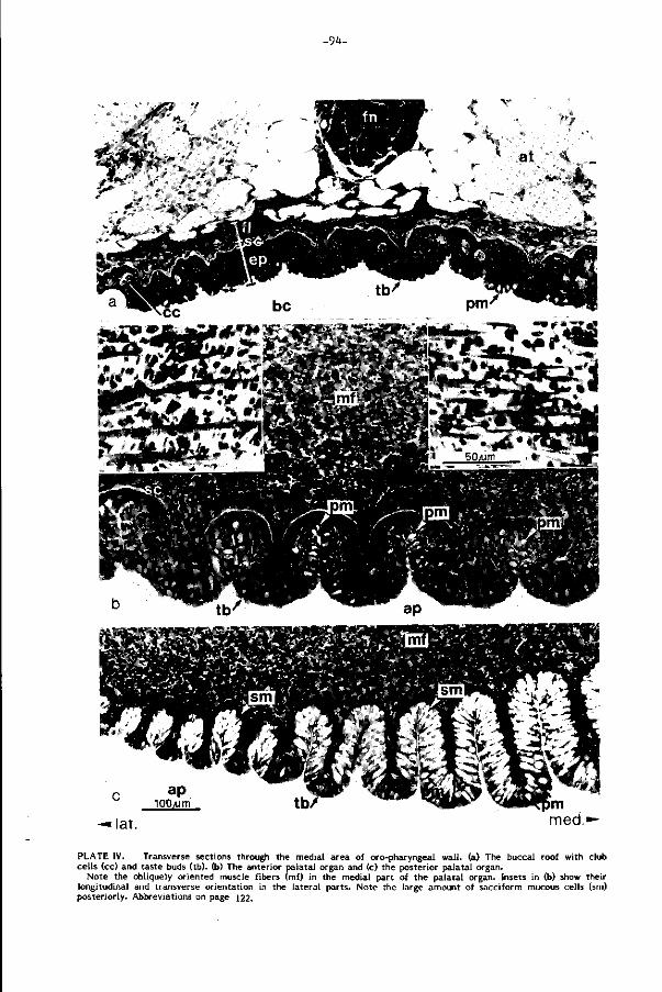
-9<f-
ap lOOyum
;ja^4P — lat.
PLATE IV. Transverse sections through the medial area of oro-pharyngeal wal l , (a) The buccal roof with club cells (cc) and taste buds (tb). (b) The anterior palatal organ and (c) the posterior palatal organ.
Note the obliquely oriented muscle fibers (mf) in the medial part of the palatal organ. Insets in (b) show their longitudinal and transverse orientation in the lateral parts. Note the large amount of sacciform mucous cells (sm) posteriorly. Abbreviations on page 122.
-95-
tissue flaps extend mediad between teeth and chewing pad.
Caudally f rom the teeth the posterior pharynx tapers ventrad into the, almost
ver t ica l , short esophagus, separated f rom the intestinal bulb by a pyloric sphincter
(Plate l id). Cyprinids lack a stomach. Rare taste buds do occur in the f i rs t few
sections of the pl icated esophageal wa l l .
L igh t - and scanning electron microscopy of the oro-pharynqeal wal l
Tissue layers
The general composition of the oro-buccal wall (Plate IVa), being a derivate
of the stomodaeum, closely resembles that of the scaleless skin. I t is covered by
a s t ra t i f ied epithelium (50-120 y m thick). The underlying connective tissue stroma
consists of 1) stratum compactum, a compact coat of collagen f ibers (5-7 ym),
2) a less dense fibrous layer (10-30 ym) w i th interposed nuclei of f ibroblasts, 3)
a loose areolar connective tissue w i th blood vessels and nerves. A d ist inct muscular
coat as reported by Curry (1939) has not been found in the buccal cav i ty .
The wal l of the pharynx (Plate IV be) mainly d i f fers by the gradually vanishing
of the s t ratum compactum and the f ibrous layer in la teral and posterior d i rect ion.
A thick muscular layer is now interposed between the f ibrous and the areolar con
nective tissue.
Labels such as lamina propria and submucosa are confusingly used (Curry,
1939; Al-Hussaini, 1949; Eastman, 1970). They have been avoided because they
are derived f rom intestinal nomenclature, which is inappropriate to describe the
area between skin and esophagus. A muscularis mucosae, separating the lamina
propria and the submucosa in the mammal intestine, is absent in fishes (cf. McVay
and Kaan, 1940).
The palatal organ
The pharyngeal walls show conspicuous specializations. The palatal organ has
a highly p l icated mucous epithel ium (Plate IVbc). Taste buds abound on the f la t
papil lär tops, mucous cells dominate along the narrow crypts. The underlying f ibers
f o rm muscular cores for the papillae and p lat forms for taste buds. The thick muscu
lar layer backing the papillae is composed'of roughly longitudinally and transversely
or iented str iated f ibers. In the medial part of the palatal organ however muscle
f ibers run obliquely to the body axis (Plate IV b). The scattered fascicul i l ie inter
mingled and gradually merge w i th the deep layer of areolar tissue. The palatal
organ covers the upper branchial arch elements and is at i ts edges attached to

-96-
cSsiF:
r PP "W^%';-d » IQo'/um"
»SfcÜta PLATE V. (a) Transverse section through the postero-lateral part of the palatal organ. I t is hardly plicated and contains an almost maximal density of taste buds (arrows). Some pyriform mucous cells are conspicuous (pm). (b) Transverse section through a gi l l raker bearing taste buds (arrows) onamuscular pad (mp). It is supported by a bony rod (br). (c) Survey of the medial part of the postlingual organ with the epithelium (ep) at the top, separated by a stratum compactum (sc) f rom longitudinal and more ventrally transverse muscle fibers. (d) Transverse section through the dorso-lateral wall of the chewing cavity, showing chewing pad (cp), foliaceous papillae and the constrictor pharyngis muscle fibers (cph) radiating into the lateral tissue flap (tf) (cf PI. l ie). Some masticatory muscles are at the top of the plate. Abbreviations on page 122.
-97-
bony elements and connective tissue plates (Plate l lb,d).
Outwards f rom the median the papillae (up to 150 y m high) become gradually
lower (Plate lib) and their supporting connective tissue layers decreases f rom 12 urn
to 2 y m . In the posterior medial part of the palatal organ, on the contrary, the
papillae lengthen and form high folds (up to 300 u rn high) w i th few connective
tissue (5 p m layer). Their small mainly longitudinally arranged muscular core is
bordered by abundant mucous cells (Plate IVc).
The postlingual organ and the branchial arches
The medial postlingual organ and the pharyngeal l ining of the branchial arches
(omit ted in the studies of Curry (1939) and Al-Hussaini (1949)), show a similar
regional d i f ferent iat ion as the palatal organ and may even be considered to fo rm
a tubular ent i ty wi th i t . However, the muscle layer f ibers over the branchial arches
is thin (cf. Plate lib) and runs along the length of the arches, oblique to the body
axis, whereas it is voluminous in the postlingual organ and runs mainly paral lel
to the body axis (Plate Vc). Thus the arrangement of muscle f ibers in the pharyngeal
f loor remains oblique w i th respect to opposing fibers in the pharyngeal roof, despite
the regional differences. The functional implications of such a peculiar arrangement
is discussed later. The core of the gi l l raker is supported by a basal bony rod and
density packed wi th muscle f ibers running in various directions, suggesting movabi l i ty
(Plate Vb). The bony rod i tself has a f ibrous at tachment to the branchial arch.
The chewing cavity
The foliaceous mucous l ining of the posterior pharynx forms two large lateral
flaps projecting between teeth and chewing pad and high branching papillae between
and around the teeth (Plates lab, l ie , Vd). The underlying s tr iated muscles are
continuous wi th those of the esophagus and distinct f rom the palatal and postlingual
organ. They are composed of criss-cross running fascicul i , radiating into the above
flaps and papillae, and a peripheral sphincter oesophagi, connecting pharyngeal
jaws and basioccipital process.
The roof of the posterior pharynx is formed by the corni f ied chewing pad (Plates
la, l ied, Vd) which is continuously renewed and rests on a th ick intruding layer
of connective tissue. Its histology has been described by Curry (1939) and Al -Hus
saini (1949). Sporadic muscle f ibers occur between the chewing pad and the basi
occip i ta l process.
The l ining of the esophagus closely resembles that of the posterior pharynx, except
for the area close to the pyloric sphincter (cf. Curry , 1939; Al-Hussaini, 1949).
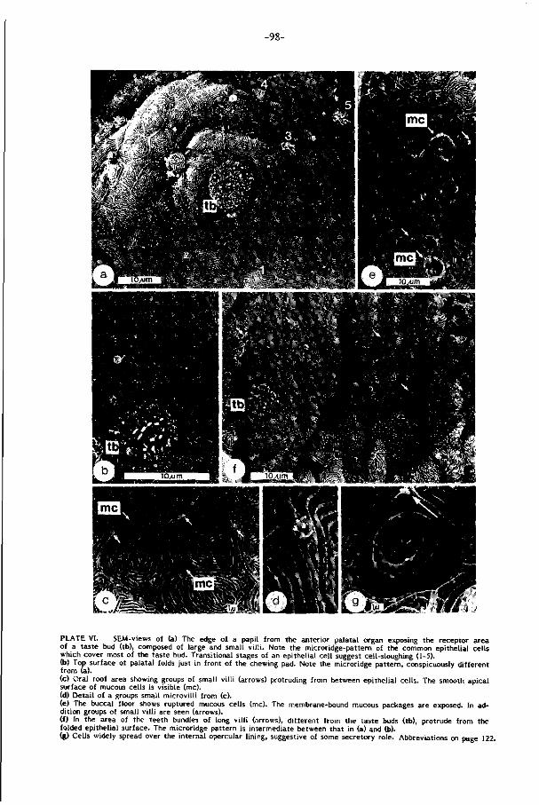
-98-
PLATE VI. SEM-views of (a) The edge of a papil from the anterior palatal organ exposing the receptor area of a taste bud (tb), composed of large and small v i l l i . Note the microridge-pattern of the common epithelial cells which cover most of the taste bud. Transitional stages of an epithelial cell suggest cell-sloughing (1-5). (b) Top surface of palatal folds just in front of the chewing pad. Note the microridge pattern, conspicuously different f rom (a). (c) Oral roof area showing groups of small v i l l i (arrows) protruding from between epithelial cells. The smooth apical surface of mucous cells is visible (mc). (d) Detai l of a groups small microvi l l i from (c). (e) The buccal floor shows ruptured mucous cells (mc). The membrane-bound mucous packages are exposed. In addition groups of small v i l l i are seen (arrows). (f) In the area of the teeth bundles of long v i l l i (arrows), different from the taste buds (tb), protrude from the folded epithelial surface. The microridge pattern is intermediate between that in (a) and (b). (g) Cells widely spread over the internal opercular l ining, suggestive of some secretory role. Abbreviations on page 122.
-99-
Connective tissue layers are thicker than in the chewing cavi ty, where they are
generally hardly recognizable in the papil lae.
Specializations of the oropharyngeal epi thel ium
The s t ra t i f ied oro-pharyngeal epithel ium contains specialized cells l ike horny
cells, mucous cells, club cel ls, chloride cells, sensory cells and 'round cel ls' as
in f ish epidermis (Whitear, 1971; M i t ta l et a l . , 1980). Their abundancy varies locally
and they serve a mult i tude of functions.
The surface of the common epithelial cells (5-10 urn, diameter) is common
ly in t r icate ly sculptured by microridges (Reutter, 1973, 1974; Hawkes, 1974; Hughes,
1979; M i t ta l and Banerjee, 1980; Ono, 1980). In the carp the microridges (width
0,17 - 0,23 urn) generally have a f ingerpr int pat tern (Plate VI). The bordering
microridges of adjacent cells run parallel and leave a narrow space between (ca.
0,05 urn). I t is suggestive to interprète the cells 1-5 in Plate Via as successive
stages in the cast off of an old ce l l .
In l i fe the whole epithel ial surface is covered by a slimy and f ibrous layer,
a cut ic le (about 1 pm th ick), which is secreted f rom the underlying common epithe
l ia l cells as has been demonstrated by Whitear (1970) in Phoxinus and some other
teleost fishes. This external coat continues over the aperture of taste buds and
mucous cells and is most probably composed of glycoproteins. I t was e l iminated
during preparation of the SEM sample by washing the specimen w i th buffered saline,
and i t is f requently lost during preparation of histological sections. The mucus
of the mucous cells forms a layer on top of this cut ic le (Whitear, 1970).
Local corni f icat ion is common among terrestr ia l vertebrates, not in f ish (M i t ta l
et a l . , 1980). Horny cells are found in the carp at the r ims of the upper and lower
jaw (Plate Il ia) and in the chewing pad (Plate Vd).
Effector specializations
Mucous cells
Mucous cells, usually called goblet cells, are the most common unicellular
glands of f ish epidermis. They have their secretory product packed in membrane-
bound vesicles, which displace the nucleus to the base of the cel l (cf. Plates IV,
V). During maturat ion they force the surrounding cells apart (Henrikson and Mato l t -
sy, 1968) and eventually expose their smooth top (diameter 3-7 pm) at the surface
(cf. Plate Vice). A t rupture of the cel l the membranes break down and the mucus
is released. Plate Vie shows most probably the surface morphology of part ly ruptured
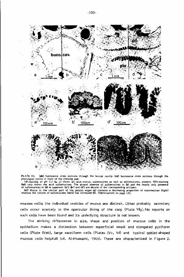
-100-
v—-s^| ,
* t ^ * % , , %
a ' 100/um
%..*f **<
PLATE VII. (ab) Successive cross sections through the buccal cavity, (cd) Successive cross sections through the pharyngeal cavity in front of the chewing pad.
AB-staining at pH 2.5 (a, c) shows al l acid mucus, sialomucines as well as sulfomucines, present. HID-staining (bd) only shows the acid sulfomucines. The almost absence of sulfomucines in (b) and the nearly only presence of sulfomucines in (d) is apparent, (a'), (b') and (d') are details of the corresponding sections.
(ef) Mucus in the central part of the palatal organ (e) contains a decreasing proportion of sialomucines (light) whereas the volume of sulfomucines (dark) has increased ( i). Abbreviations on page 122.
mucous cells; the individual vesicles of mucus are d ist inct . Other probably secretory
cells occur scarcely in the opercular l ining of the carp (Plate VIg). No reports on
such cells have been found and i ts underlying structure is not known.
The str iking differences in size, shape and position of mucous cells in the
epi thel ium makes a d ist inct ion between superf icial small and elongated pyr i fo rm
cells (Plate IVab), large sacciform cells (Plates IVc, Vd) and typical goblet-shaped
mucous cells helpful l (cf . Al-Hussaini, 1945). These are characterized in Figure 2.
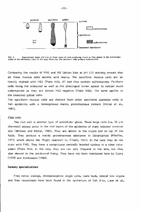
-101-
pyriform sacciform goblet
l^m
surface
l-'s ••>'l sialomucines
> epithelium
BSSKÜ sulfomucines
basement membrane
FIG. 2. Approximate shape and size of three types of cells producing mucus as they appear in the microscopic slides of the alimentary tract in the carp. Note that the sacciform cells produce sulfomucines.
C o m p a r i n g t h e r esu l t s o f PAS and A B ( a l c i a n b lue a t p H 2.5) s t a i n i n g r evea ls t h a t
a l l t hese mucous ce l l s s e c r e t e ac i d mucus . The s a c c i f o r m mucous ce l l s a re a l l
h e a v i l y s t a i ned w i t h H I D ( P l a t e V l l d , d') and thus c o n t a i n s u l f o m u c i n e s . P y r i f o r m
ce l l s l i n i ng t h e o r o b u c c a l as w e l l as t h e p h a r y n g e a l l u m e n appear t o c o n t a i n m u c h
s i a l o m u c i n e s as t hey a re a l m o s t H I D n e g a t i v e ( P l a t e V l l b ) . The same app l ies t o
t he i n t e s t i n a l g o b l e t c e l l s .
The s a c c i f o r m mucous ce l l s a re d i s t i n c t f r o m o t h e r s a c c i f o r m g l andu la r ce l l s in
f i s h e p i d e r m i s w i t h a homogeneous m a i n l y p r o t e i n a c e o u s c o n t e n t ( M i t t a l e t a l . ,
1981).
Club cells
The c l ub c e l l is a n o t h e r t ype of u n i c e l l u l a r g l a n d . These l a rge ce l l s ( ca . 18 u m
d i a m e t e r ) a lways o c c u r in t he m i d l aye rs of t h e e p i d e r m i s of m a n y t e l e o s t s ' e x t e r n a l
s k in ( W h i t e a r and M i t t a l , 1983). They a re absent in t he c r y p t s and on top o f t h e
f o l d s . They p roduce a m a i n l y p r o t e i n a c e o u s subs tance in O s t a r i o p h y s i ( P f e i f f e r ,
1971) w h i c h e l i c i t s t h e ' f r i g h t r e a c t i o n ' ( v . F r i s c h , 1941). In t h e ca rp t hey do no t
s t a i n w i t h P A S . They have a consp icuous c e n t r a l l y l o c a t e d nuc leus in a c l e a r c y t o
p l a s m ( P l a t e IVa ) . In t he ca rp t hey a re no t on ly f r e q u e n t in t he s k i n , b u t t h e y
also abound in t h e o r o - b u c c a l l i n i n g . They have no t been m e n t i o n e d he re by C u r r y
(1939) and A l - H u s s a i n i (1949) .
Sensory special izat ions
F r e e ne rve end ings , c h e m o r e c e p t i v e s ing le c e l l s , t a s t e buds, l a t e r a l l i ne o rgans
and f r e e neu romas ts have been f o u n d i n t he e p i t h e l i u m o f f i sh ( F o x , L a n e e t a l . ,
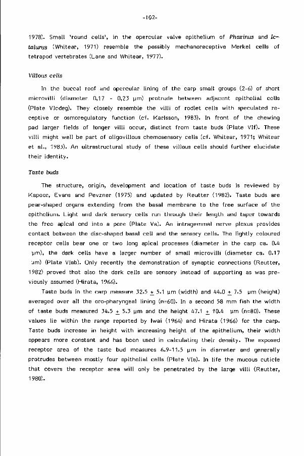
-102-
1978). Small 'round cel ls ' , in the opercular valve epithel ium of Phoxinus and Ic-
talurus (Whitear, 1971) resemble the possibly mechanoreceptive Merkel cells of
tetrapod vertebrates (Lane and Whitear, 1977).
Villous cells
In the buccal roof and opercular l ining of the carp small groups (2-6) of short
m icrov i l l i (diameter 0,17 - 0,23 urn) protrude between adjacent epithel ial cells
(Plate VIcdeg). They closely resemble the v i l l i of rodlet cells w i th speculated re
ceptive or osmoregulatory funct ion (cf. Karlsson, 1983). In f ront of the chewing
pad larger f ields of longer v i l l i occur, d ist inct f rom taste buds (Plate V l f ) . These
v i l l i might wel l be part of ol igovil lous chemosensory cells (cf. Whitear, 1971; Whitear
et a l . , 1983). An u l t rast ructura l study of these villous cells should fur ther elucidate
their ident i ty .
Taste buds
The structure, or igin, development and location of taste buds is reviewed by
Kapoor, Evans and Pevzner (1975) and updated by Reutter (1982). Taste buds are
pear-shaped organs extending f rom the basal membrane to the f ree surface of the
epi thel ium. L ight and dark sensory cells run through their length and taper towards
the f ree apical end into a pore (Plate Va). An intragemmal nerve plexus provides
contact between the disc-shaped basal cel l and the sensory cells. The l ight ly coloured
receptor cells bear one or two long apical processes (diameter in the carp ca. 0.4
(im), the dark cells have a larger number of small m icrov i l l i (diameter ca. 0.17
pm) (Plate Vlab). Only recently the demonstration of synaptic connections (Reutter,
1982) proved that also the dark cells are sensory instead of supporting as was pre
viously assumed (Hi rata, 1966).
Taste buds in the carp measure 32.5 +_ 5.1 urn (width) and 44.0 +_ 7.5 pm (height)
averaged over all the oro-pharyngeal l ining (n=60). In a second 58 mm fish the width
of taste buds measured 34.5 +_ 5.3 pm and the height 47.1 +_ 10.4 pm (n=80). These
values l ie w i th in the range reported by Iwai (1964) and H i rata (1966) for the carp.
Taste buds increase in height w i th increasing height of the epi thel ium, their w idth
appears more constant and has been used in calculating their density. The exposed
receptor area of the taste bud measures 6.9-11.5 pm in diameter and generally
protrudes between mostly four epithel ial cells (Plate Via). In l i fe the mucous cut ic le
that covers the receptor area w i l l only be penetrated by the large v i l l i (Reutter,
1980).
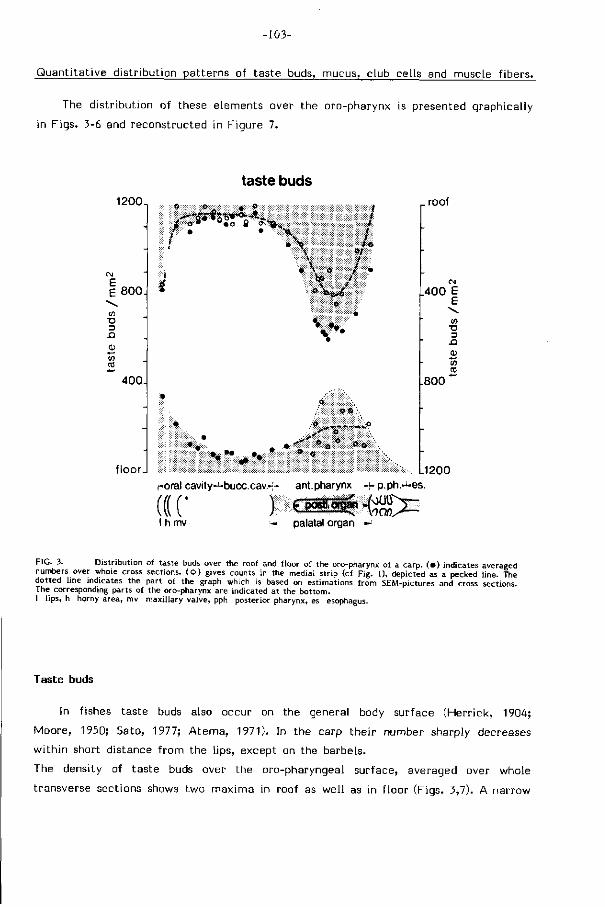
-103-
Quant i tat ive distr ibution patterns of taste buds, mucus, club cells and muscle f ibers.
The distr ibution of these elements over the oro-pharynx is presented graphically
in Figs. 3-6 and reconstructed in Figure 7.
120CL
1 800
-Q
400
floor.
taste buds
o • • o 8 o * ^
\ V \ • \
t f I te
*t t
>* » o ^
o o
_roof
400 E e \ co
T3 D
-O
£
800*"
L1200 r-oral cavity—bucc cav.~ ant.pharynx «p.ph.J-es.
(CC ) QMä»-(S> I h mv f- palatal organ
FIG. 3. Distribution of taste buds over the roof and floor of the oro-pharynx of a carp. ( • ) indicates averaged numbers over whole cross sections, ( o ) gives counts in the medial strip (cf Fig. 1), depicted as a pecked line. The dotted line indicates the part of the graph which is based on estimations from SEM-pictures and cross sections. The corresponding parts of the oro-pharynx are indicated at the bottom. I l ips, h horny area, mv maxillary valve, pph posterior pharynx, es esophagus.
Taste buds
In fishes taste buds also occur on the general body surface (Herr ick, 1904;
Moore, 1950; Sato, 1977; A tema, 1971). In the carp their number sharply decreases
wi th in short distance f rom the lips, except on the barbels.
The density of taste buds over the oro-pharyngeal surface, averaged over whole
transverse sections shows two maxima in roof as wel l as in f loor (Figs. 3,7). A narrow
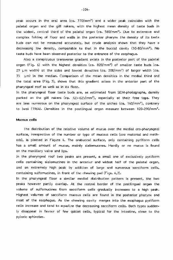
-10t-
peak occurs in the oral area (ca. 370/mm2) and a wider peak coincides w i th the
palatal organ and the g i l l rakers, w i th the highest mean density of taste buds in
the widest, central th i rd of the palatal organ (ca. 580/mm2). Due to extensive and
complex folding of f loor and walls in the posterior pharynx the density of its taste
buds can not be measured accurately, but crude analysis shows that they have a
decreasing low density, comparable to that in the buccal cavity (50-80/mm2). No
taste buds have been observed posterior to the entrance of the esophagus.
Also a conspicuous transverse gradient exists in the posterior part of the palatal
organ (Fig. 6) w i th the highest densities (ca. 820/mm2) of smaller taste buds (ca.
29 urn width) at the sides and lowest densities (ca. 200/mm2) of larger width (ca.
35 pm) in the median. Comparison of the mean densities in the medial th i rd and
the to ta l area (Fig. 3), shows that this gradient arises in the anterior part of the
pharyngeal roof as wel l as in its f loor.
In the pharyngeal f loor taste buds are, as estimated f rom SEM-photographs, densily
packed on the g i l l rakers (ca. 325-625/mm2), especially at their f ree tops. They
are less numerous on the pharyngeal surface of the arches (ca. 140/mm2), contrary
to Iwai (1964). Densities in the postlingual organ measure between 120-290/mm2 .
Mucous cells
The distr ibut ion of the re lat ive volume of mucus over the medial oro-pharyngeal
surface, i rrespective of the number or type of mucous cells (see mater ia l and meth
ods), is p lot ted in Figure 4. The orobuccal surface, only containing pyr i form cells
has a small amount of mucus, mainly sialomucines. Hardly or no mucus is found
on the maxi l lary valve and l ips.
In the pharyngeal roof two peaks are present, a small one of exclusively pyr i form
cells containing sialomucines in the anterior and widest half of the palatal organ,
and an extremely high peak by addit ion of large and numerous sacciform cells,
containing sulfomucines, in f ront of the chewing pad (Figs. 4,7).
In the pharyngeal f loor a s imilar medial d istr ibut ion pat tern is present, the two
peaks however part ly overlap. A t the rostral border of the postlingual organ the
volume of sulfomucines f rom sacciform cells gradually increases to a high peak.
Highest volumes of sacciform mucous cells are found in the posterior pharynx and
most of the esophagus. As the chewing cavity merges into the esophagus pyr i form
cells increase and tend to equalize the decreasing sacciform cells. Both types sudden
ly disappear in favour of few goblet cells, typical for the intestine, close to the
pyloric sphincter.
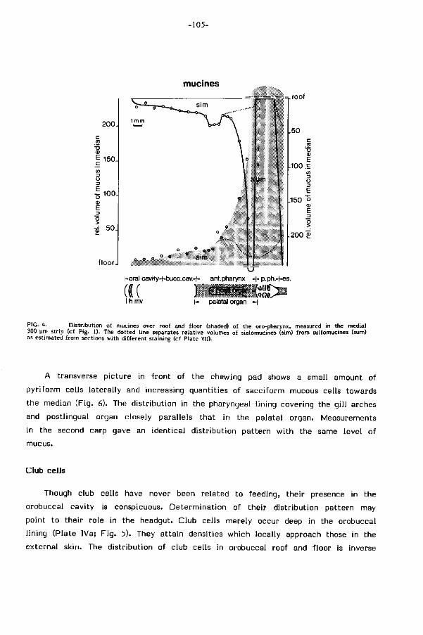
-105-
mucmes
200J 1 ™
E 150.
100.
floor.
..•> n
9 o
=-.=.roof
sum
.50 c a
<u E
100 S w
.150 o
200 £
horal cavity+bucc.cav.H- ant.pharynx •+• p.ph.-t-es.
I h mv h- palatal organ H
FIG. lt. Distribution of mucines over roof and floor (shaded) of the oro-pharynx, measured in the medial 300 urn strip (cf Fig. 1). The dotted line separates relative volumes of sialomucines (sim) from sulfomucines (sum) as estimated from sections with dif ferent staining (cf Plate VII).
A transverse picture in f ront of the chewing pad shows a small amount of
py r i fo rm cells lateral ly and increasing quantit ies of sacciform mucous cells towards
the median (Fig. 6). The distr ibut ion in the pharyngeal l ining covering the g i l l arches
and postlingual organ closely parallels that in the palatal organ. Measurements
in the second carp gave an identical d istr ibut ion pat tern w i th the same level of
mucus.
Club cells
Though club cells have never been related to feeding, their presence in the
orobuccal cavity is conspicuous. Determinat ion of their d istr ibut ion pattern may
point to their role in the headgut. Club cells merely occur deep in the orobuccal
l ining (Plate IVa; F ig . 5). They at ta in densities which locally approach those in the
external skin. The distr ibut ion of club cells in orobuccal roof and f loor is inverse
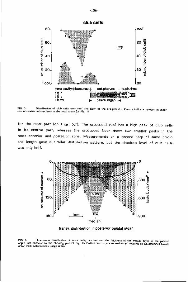
-106-
club cells 80-,
a> 60 -JQ _2 o o 40 k_ 0)
JD E | 204
floor.
1mm
•o
o f t «
,-roof
m I-20 ö>
o n o
40 o i _ 0) n E
1-60 |
o ' « o L80
Horal cavity+bucc.cav.H- ant.pharynx H-p.ph.-f-es.
(((( ) Gané-tigx I h mv I-. palatal organ H
FIG. 5. Distribution of club cells over roof and floor of the oro-pharynx. Counts indicate number of inner-sections (with cell-nucleus) in the to ta l areas (cf Fig. 1).
for the most part (cf. Figs. 5,7). The orobuccal roof has a high peak of club cells
in its central part , whereas the orobuccal f loor shows two smaller peaks in the
most anterior and posterior zone. Measurements on a second carp of same origin
and length gave a s imilar d istr ibut ion pat tern, but the absolute level of club cells
was only half .
0 ,
60 . O 3 E o O) E R 120 o >
180
«es'lWlVuscl© *•'*' 1*1 ' I ' I ' i ' i ' i ' i ' I ' I ' I ' I ' I '
^'lll'lil'lll'l'l'l'l'!'!' " l ' l ' l l'l I I I I I I I l
sum
1mm <* ö ' I
median
<;i,i;iji]iji t i i 1 1 1 1 1
' i l l1! ' !1!1!1!1
'«!
.300
to •a n <o
600 m to
900
transv. distr ibution in posterior palatal organ
FIG. 6. Transverse distribution of taste buds, mucines and the thickness of the muscle layer in the palatal organ just anterior to the chewing pad (cf Fig. 1). Dotted line separates estimated volumes of sialomucines (small area) from sulfomucines (large area).
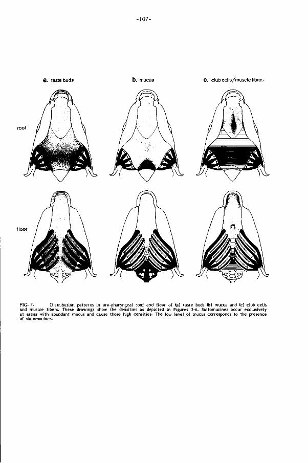
-107-
3. taste buds C. club cells/muscle fibres
roof
floor
FIG. 7. Distribution patterns in oro-pharyngeal roof and floor of (a) taste buds (b) mucus and (c) club cells and muslce fibers. These drawings show the densities as depicted in Figures 3-6. Sulfomucines occur exclusively at areas with abundant mucus and cause those high densities. The low level of mucus corresponds to the presence of sialomucines.
-108-
Muscle fibers
The thickness of the shrinked muscular layers in the median is readily observed
f rom Plate l id and measures up to 850 ym in such a small carp. It is only sl ightly
thinner in the lateral area of the palatal organ (Fig. 6). In the postlingual organ
the muscular layer only gradually increases towards the chewing cavi ty, contrary
to the palatal organ. The gi l l arches at both sides of the postlingual organ have
a thin muscular layer (cf. Plate l ib)
The density of muscle f ibers contained in these layers increases posteriorly (cf.
Plate lid) and renders thickness a rough measure. The complex structure of the
wal l of the posterior pharynx prevented measurement of its muscular content.
This part is only roughly indicated in F ig. 7.
The distr ibution pattern of taste buds, mucous cells, muscle fibers and club
cells allows a d ist inct ion of the oro-pharyngeal wal l in six zones (Fig. 9; Table 1),
each characterized by a specific combination of specialized features.
Their re lat ion wi th the separate feeding actions which compose food handling and
food processing in the carp are discussed later.
DISCUSSION
The role of mucus, microridges, club cells and sensory elements in the oro-pharynx
Mucus
The chemical properties of mucus were reviewed by Hunt (1970). Two main
components are d ist inct , glycoproteins and mucopolysaccharides, organized into
an entangled network. Glycoproteins are complexes of proteins covalently l inked
w i th varied low molecular weight saccharides. Protein properties are dominant.
Sialic acid is a character ist ic saccharid. Mucopolysaccharides are similar conjugates.
Except chit ine and few other s tructural compounds they are characterized by highly
charged, high molecular weight polysaccharides of less varied nature. Polysaccharide
properties dominate those of the proteins. Sialic acid is lacking, whereas sulpho-
groups abound. Mucus may comprise s t i l l other components (e.g. elastine). Also
do glycoproteins and mucopolysaccharides occur in other, mainly supporting, tissues
(cf. Plate VIIb,d). Mucus w i th mainly glycoproteins are referred to as sialomucines,
mucus wi th mainly mucopolysaccharides as sulfomucines.
Physically, mucus is a weak and reversible gel (Denny and Gosline, 1980; Silber-
-109-
berg and Meyer, 1982), best characterized by its viscosity and e last ic i ty. Though
sialic acid makes sialomucines rather viscous (Hunt, 1970), the highly acid sulfo-
mucines confer mucus a high viscosity (Hunt, 1973). The carbohydrate component
largely determines the Theological properties of mucus. Denny and Gosline (1980)
demonstrated that large strains reduce the viscosity of pedal mucus in gastropods.
The mucus recovered its solidity if al lowed to heal for some period.
Mucus may perform a mult i tude of roles in a wide array of biological systems
(Whitear, 1970; M i t ta l & Banerjee, 1980; Cook & Shirbate, 1983) e.g. 1) forming
a mechanical barrier to foreign bodies and pathogens (it has no ant ibiot ic e f fect
in the carp (Hatt ingh and van Warmelo, 1975)); 2) forming a chemical barrier sup
port ing osmoregulation and preventing f lux of mater ia l over steep chemical or elec
t r ica l gradients; 3) reducing of f r i c t ion ; 4) providing a mechanical buffer which
protects the epithelium f rom damage and abrasion; 5) aiding in precipi tat ion of
suspended mud by entrapment and cleaning of the epithelium (of ten by c i l ia); 6)
st icking together part iculate food; 7) preventing of desiccation by binding of water;
8) enhancing adhesion in sluggish locomotion; 9) serving means of communication
and navigation (e.g. molluscs); 10) feeding and attachment of young; 11) format ion
of cocoons and other envelopes for temporary shelter.
The orobuccal wall produces mainly sialomucines (cf. F ig. 4). This type of mucus
is common to the respiratory and al imentary t ract of vertebrates, probably serving
protect ion and lubrication (Hunt, 1970). Besides soluble fish slimes on the outer
skin e f fect ively minimize the boundary layer and f r i c t ion in swimming (Rosen and
Cornford, 1971). Sialomucines may thus well aid in orobuccal water transport, es
pecially at high suction velocit ies, when turbulent f low brings small amounts of
mucus in solution.
Many sulfomucines are added posteriorly in the anterior pharynx (Figs. 4,7). This
high-viscosity mucus is almost certainly related to the pure muscularly processing
of food serving lubricat ion and reduction of the high mechanical stresses on the
epi thel ium. They wi l l also aid in recol lect ion and aggregation of part icles into bo
luses, during transport as wel l as during masticat ion and deglut i t ion. Movement
of the lateral tissue flaps in the chewing cavi ty, secreting a large volume of sulfo
mucines, might serve cleaning the occlusal surfaces. A background volume of low-
viscosity sialomucines wi l l pr imari ly serve lubricat ion and i t increases indeed in
the esophagus.
As compared to sulfomucines, the sialomucines in the orobuccal cavity and antero
lateral part of the pharynx are expected to have a reduced tendency to bind par t i
cles, as this area is largely concerned wi th selection between food and non-food
part icles by repeated resuspension (Sibbing et a l . , 1984).
-110-
An analogous combination of similar secretory cells cleans the mantle cavity f rom
suspended d ir t in snails (Hunt, 1973). Interactions between the two secretory products
confers the mixture even a higher visco-elasticity and structural integri ty than
its component parts.
No tests have been performed yet to ascertain the physical properties and
secretion kinetics of oro-pharyngeal mucus in f ish. I t is expected that the release
of mucus is somehow regulated in accordance w i th the feeding act iv i t ies of the
f ish.
Microridges
Mucus secreting epithelia in f ish are commonly provided w i th microridges ar
ranged in varied patterns. Their possible functions (Hawkes, 1974) are 1) a mechanical
defense to t rauma, 2) an increase of the surface area for absorbtion and gas ex
change, 3) an aid in holding mucus to the cel l surface. Sperry and Wassersug (1976)
concluded microridges to guide the even spreading of mucus through their channels.
Few of these physiological and mechanical functions have been investigated experi
mental ly.
The differences in microridge pat tern at areas of d i f ferent mechanical stresses
(cf. Plate VIa,b) suggest an additional mechanical funct ion in the carp. On the l ips,
devoid of any specialized mucous cells, as wel l as in the area of the chewing cavi ty
more microridges per unit of cell surface are found than elsewhere. Both on the
lips and in this chewing area their principal role may be similar to that of the corni -
f ied cells in absorbing high forces and spreading them over the cell surface.
Club cells
These conspicuous cells never reach the surface of the epi thel ium, lack pores
and do not contact blood vessels. They are supposed to be holocrine glands producing
the alarm substance in ostariophysans and releasing this stored pheromone by damage
(Pfe i f fer , 1960). A l ternat ive club cel l functions which are suggested in the l i terature
are antipathogenous act ion, vehicles for antibodies, toxic cells, mechanical protec
t ion and lubricat ion (Whitear and M i t t a l , 1983). Club cells do also occur in some
non-ostariophysan teleosts (cf. Whitear, 1981) e.g. the eel where they are said to
be involved in f ibrous mucus production (Reid, 1894).
In the carp, club cells extend f rom the outer skin into the orobuccal cavity
(Fig. 7). Their mid-epithel ial locat ion neither at the top of folds nor in the crypts
-111-
where taste buds and mucous cells respectively abound, points to a rather indi f ferent
role of their posit ion. I f these club cells have alarming functions they should be
selectively emptied due to st imuli by harmful objects, not by those of the food
to prevent false a larm. Large damage w i l l impede the functions of the orobuccal
epithel ium in feeding. These arguments makes their alarming funct ion in this area
doubt fu l , even though club cel l densities are highest at areas of most probable
mechanical impact e.g. inside the lower jaw (Fig. 7). They may wel l serve other
unknown functions, possibly released by neural or hormonal mechanisms. Their ab
sence f rom the pharyngeal l ining suggests a direct relat ion between club cells and
the f low of water. Their sl ightly positive reaction on HID staining (Plate Vllb')
indicates the presence of some sulfomucines in their cytoplasm in addition to the
commonly known proteins.
Sensory structures
Mechano- and chemoreceptors are required to sense the qualit ies of the ingested
mater ia l during the subsequent actions in food processing. Besides the fish needs
constant monitoring of the quality and velocity of the water during respiration
and feeding.
Mechanoreception
Apart f rom the lateral l ine system and free neuromasts (cf. Plate led) few
evidence on mechanoreceptive organs in teleost skin is available (Fox et a l . , 1980).
Many types of nerve plexuses in or below the epithel ium are a t t r ibuted mechano
receptive functions (Grzycki , 1954; Whitear, 1971; Ono, 1979). Specialized epidermal
cells resembling Merkel cells of higher vertebrates have been described (Lane and
Whitear, 1977) but nothing is known of their physiology. As these cells are charac
ter ized by few apical f inger- l ike v i l l i , the presence of small groups of short v i l l i
in the buccal and opercular l ining (Plate VI) may well suggest here mechanoreception
in the carp.
Reut ter (1974) investigated previous hypotheses (Kolmer, 1927; H i ra ta , 1966) that
taste buds could also serve mechanoreceptive functions. He distinguished three
types of taste buds in the headgut of the swordtail based on their position w i th
respect to the surrounding epi thel ium. This dist inct ion was supported by neurohisto-
chemical characters (Reutter, 1973). Accepting Reutter 's theory most protruding
taste buds (type I), which would be most e f fect ive in mechanoreception, in the
carp occupy the gi l l rakers (Plate III). Less protruding buds (type II) l ine the buccal
-112-
folds. The other taste buds never rise above the normal level of the epithel ium
and their mechanoreceptive role is improbable. McGlone (1977) reports the palatal
organ to respond to tact i le st imul i at all stages of denervation w i th local bulging,
possibly caused by proprioceptive reflex loops. Konishi and Zot terman (1963) found
some glossopharyngeal nerve f ibers in the palatal organ of the carp reacting ex
clusively to mechanical s t imulat ion. Whether these f ibers originate f rom taste buds
w i th mechano- as well as chemoreceptive properties, or f rom separate mechano-
receptors is unclear. Processes l ike the adjusting of force, d i rect ion and amplitude
of masticatory movements to the mechanical properties and momentaneous size
of the food as well as decisions whether the main f rac t ion of part icles is suited
for deglut i t ion, require detailed information and feed back regulation. I t is supposed
that proprioceptors in the masticatory muscles and in the tissue flaps projecting
between teeth and chewing pad are the main sensors in masticat ion. Proprioceptors
in the gills of teleosts appear to respond to displacement of the gi l l rakers and
gi l l f i laments (Sutterl in and Saunders, 1969). Bal l int i jn demonstrated that tension
and length receptors in respiratory muscles play a role in steering the respiratory
movements in f ish (Bal l in t i jn , 1972; Bal l in t i jn and Bamford, 1975; Ba l l in t i jn , 1982).
The few unexpected muscle f ibers between the chewing pad and its bony socle may
wel l play a sensory role.
Che more ception
The chemoreceptive properties of taste buds are evident. They have varied
sensivity spectra. Konishi and Zot terman (1963) recorded responses f rom the glosso
pharyngeal nerve f ibers in the carp and subdivided them into seven groups according
to their response on a wide array of sapid solutions including human saliva, enabling
the f ish a high level of gustatory d iscr iminat ion. Some appeared to have a wide
gustatory spectrum, others a narrow one.
The important role of such sensors in a bot tom feeder l ike the carp is evident,
but their d istr ibut ion pat tern remains to be explained. In fact a density of 820 taste
buds/mm2 , as found in the lateral part of the palatal organ, has not been reported
previously. Given the mean width of a taste bud, about 1200/mm2 is the maximal
number (cf . Plate Va). Iwai (1964) reports a maximum of 285/mm2 on the branchial
arches of the cyprinid Biwa zezera.
Besides taste buds the peculiar vil lous cells in the oro-pharyngeal l ining of the carp
(Plate VI) might be involved in chemoreception.
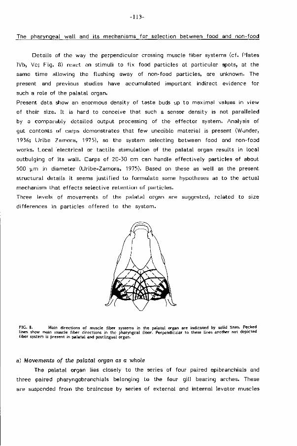
-113-
The pharyngeal wal l and i ts mechanisms for selection between food and non-food
Details of the way the perpendicular crossing muscle f iber systems (cf. Plates
IVb, Vc; F ig . 8) react on st imul i to f ix food particles at part icular spots, at the
same t ime allowing the flushing away of non-food particles, are unknown. The
present and previous studies have accumulated important indirect evidence for
such a role of the palatal organ.
Present data show an enormous density of taste buds up to maximal values in view
of their size. It is hard to conceive that such a sensor density is not paralleled
by a comparably detailed output processing of the ef fector system. Analysis of
gut contents of carps demonstrates that few unedible material is present (Wunder,
1936; Uribe Zamora, 1975), so the system selecting between food and non-food
works. Local e lectr ical or tact i le s t imulat ion of the palatal organ results in local
outbulging of its wa l l . Carps of 20-30 cm can handle e f fect ively particles of about
500 y m in diameter (Uribe-Zamora, 1975). Based on these as well as the present
s t ructural details it seems just i f ied to formulate some hypotheses as to the actual
mechanism that ef fects selective retent ion of part icles.
Three levels of movements of the palatal organ are suggested, related to size
differences in particles of fered to the system.
FIG. 8. Main directions of muscle fiber systems in the palatal organ are indicated by solid lines. Pecked lines show main muscle fiber directions in the pharyngeal f loor. Perpendicular to these lines another not depicted fiber system is present in palatal and postlingual organ.
a) Movements of the palatal organ as a whole
The palatal organ lies closely to the series of four paired epibranchials and
three paired pharyngobranchials belonging to the four gi l l bearing arches. These
are suspended f rom the braincase by series of external and internal levator muscles
-Ut
as well as interconnected by oblique muscles. During respiration part of these
dorsal gi l l arch muscles are active (Bal l int i jn et a l . , 1984). The combined act iv i ty
of the gil l arch muscles wi l l be able to expand, depress and l i f t the palatal organ
as a whole or locally. The expected palatal movements w i l l be minute in ampl i
tude. Slight antero-ventral movements seem possible. Through these movements
large areas of the palatal organ could be closely approached to parts of the bran
chial basket w i th its numerous gi l l rakers. This hypothesis w i l l be ver i f ied by elec
t r ica l st imulations of the involved muscles and by electromyography.
b) Local movements in the palatal organ, areas of several square mm.
Such local bulgings of the palatal organ do occur (Jara, 1957; McGlone, 1978).
Simultaneous contractions in the posterior area of the palatal and postlingual organ
even propel the food by peristalsis-l ike movements in transport (Sibbing et a l . ,
1984). Contract ion of the grid of muscle f ibers in a part icular area wi l l exert
compression on the local in terst i t ia l f luid and so cause bulging of the epithelium
into the pharyngeal s l i t . Whether or not the papillae are involved, is not known.
The differences in main directions of the muscle f ibers in pharyngeal f loor and
roof, except in the posterior transport area (Fig. 8), might even lead to the local
production of ridges which cross at roughly 45° angles in roof and f loor. Here pa r t i
cles could be f ixed. This hypothesis assumes equal contractions of the perpendicular
crossing muscle f ibers for bulging and unequal contractions for r idging.
c) Minute local movements of areas below one square mm.
Especially anteriorly the palatal folds fo rm papillae w i th a conspicuous muscular
core. In areas w i th an increased pressure on the in terst i t ia l f luid the local e f fect
of the pressure on shape and size of these minute projections depends upon the
ac t iv i ty of the muscle f ibres in their core and on the arrangement of the connective
tissue f ibers below the papillär epi thel ium. In these circumstances f lu id pressure
and muscle f ibre force act antagonistically and can so determine shape and size
of projections. The size of such papillae varies f rom ca. 150 x 550 urn anteriorly
to a c ircular form w i th a 90 urn diameter more lateral ly. Posteriorly, at the lateral
edges of the palatal organ and branchial sieve, papillae are v ir tual ly absent and
the connective tissue coat vanishes (Plate Va).
The la t ter hypotheses can be tested wi th small canulae and pressure transducers
combined wi th photographic data. The muscles of the branchial basket, the muscular
strips over the gil l arches, the muscular pads on the gi l l rakers and their movable
connection w i th the gi l l arches may all contribute or even solely be responsible
-115-
for local retent ion of part icles. The papillae can also be explained as a surface
increase for mucus supply. The huge vagal lobes and their complex cyto-archi tecture
(see below) seem to be in accordance w i th the detailed movements suggested above.
The bot tom feeding habits of carps have profound influences on structures and
functions in the oro-pharynx.
Pathways connecting oro-pharynqeal sensors and ef fectors
To obtain a more complete picture of food processing some important facts
f rom the l i terature on this subject have been compiled in the fol lowing lines.
Peripheral pathways
Herr ick (1899, 1901, 1906) concluded f rom his extensive studies on the senses
of touch and taste in fishes that the nervus tr igeminus (V) is the main pathway
for touch impulses f rom the orobuccal skin, whereas the facial nerve (VII) mainly
carries the sensory input f rom taste buds f rom the same area. The posterior buccal
taste buds send their information through the glossopharyngeal nerve ( IX), the
pharyngeal taste buds through the large vagal nerve (X). Nerves VII , IX and X pro
ject on distinct regions in the brain, respectively the fac ia l , glossopharyngeal and
the vagal lobes. L i t t l e detailed knowledge exists on the origin and terminat ion
of their motor components.
Evans (1952) suggested that feeding behaviour is expressed in the morphology of
the brain. When taste buds abound on the external skin, barbels and lips such as
in skin tasters l ike catfishes and minnows, the facial lobe is conspicuous. In mouth
tasters such as the carp and goldfish the vagal lobe is greatly enlarged. Sightfeeders
have large mesencephalic optic lobes. Evidence now exists f rom the catf ish that
the facial gustatory system is involved in locating and sensing an external food
source while the vagal gustatory system determines the palatabi l i ty of the food
(Atema, 1971)
Contrary to the facial lobe, the vagal lobe in cyprinids has a highly organized
laminar structure (Herr ick, 1906; Ozawa, 1951; I to, 1971). The ascending projections
were found to be topographically arranged (Mori ta et a l . , 1983). McGlone's physio
logical studies on the goldfish suggest some degree of somatotopy wi th respect
to the palatal organ. Appl icat ion of the horseradish peroxidase technigue (Lui ten,
1975b) by Finger (1981b) labeled only restr icted small areas in the palatal organ
and in the vagal lobe, supporting the palatotopic mapping in the vagal lobe. This
suggests a gustatory reflex mechanism regulating local palatal muscular contrac
tions. As the palatal organ performs its functions in close coordination w i th the
-116-
postlingual organ in the carp (Sibbing et a l . , 1984) we hypothesize that the post-
lingual organ is s imilarly mapped in the vagal lobe.
Generally the pharyngeal taste buds of teleosts are innervated according to
the fol lowing pat tern. The glossopharyngeal nerve (IX), after supplying the anterior
branchial levator muscles, sends a ramus into the most anterior part of the palatal
organ. More ventrally it divides into a sensory pretrematic branch to the hyoid
area and a posttrematic branch to the f i rst hemibranch, supplying mucous epi
thel ium, taste buds, g i l l rakers and the gil l arch musculature. This suggest that
opposed areas of the pharyngeal roof and f loor are locally innervated by the same
branch. In a similar way four rami of the huge vagal nerve (X) innervate corres
ponding areas around each of the subsequent four g i l l s l i ts. Posterior vagal rami
innervate the sensory and ef fector components of the pharyngeal masticatory ap
paratus and one runs caudad into the esophageal wal l .
The scattered chemosensory single cells in the buccal and opercular l ining
may well belong to the general cutaneous chemosensory component (Whitear, 1971).
No evidence exists on their innervation and specific role. The common chemical
sense in the skin generally detects the qualit ies of the water (avoiding reactions)
and is only exceptionally (e.g. in Triglids) used in food-seeking (Scharrer, 1963).
Central pathways
Steering the whole concert of movements in food uptake, selection, t ransport,
mast icat ion and deglut i t ion for e f f ic ient food processing as a whole, tuned to the
type of food (Sibbing et a l . , 1984) requires integrat ion of enormous sensory inputs
on a higher level in the brain. Dist inct central connections exist between projections
of nerves V, VII , IX and X in the carp (Lui ten, 1975a). These areas process not
only taste informat ion but also signals f rom other sources e.g. touch, proprioception
f rom the gills and jaw muscles. The central gustatory paths in the closely al l ied
crucian carp {Carassius carassius) (Mori ta et a l . , 1980) also suggest complex integra
t ion mechanisms between sensory input and motor output regulating cyprinid feeding.
Six specialized areas in the oro-pharynx related to food handling in the carp
This summarizing section relates distinct areas in the oro-pharynx which are
characterized by a part icular combination of s t ructural specializations (Figs. 9;
Table 1) to their sensory and ef fector roles in feeding. The mechanisms and stereo
typed movements of twelve dist inct actions composing a feeding sequence have
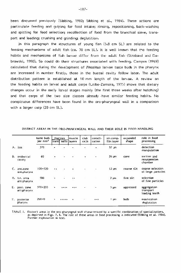
-117-
been discussed previously (Sibbing, 1982; Sibbing et a l . , 1984). These actions are
part iculate feeding and gulping for food intake; r insing, repositioning, back-washing
and spitt ing for food selection; recol lect ion of food f rom the branchial sieve, trans
port and loading; crushing and grinding; deglut i t ion.
In this paragraph the structures of young fish (5.8 cm SL) are related to the
feeding mechanisms of adult f ish (ca. 30 cm SL). It is well known that the feeding
habits and mechanisms of f ish larvae d i f fer f rom the adult f ish (Stroband and Da-
browski, 1980). So could do their structures associated w i th feeding. Campos (1969)
calculated that during the development of Phoxinus larvae taste buds in the pharynx
are increased in number f i rs t ly , those in the buccal cavity fol low later. The adult
d istr ibut ion pattern is established at 10 mm length of the larvae. A review on
the feeding habits on larval and adult carps (Uribe-Zamora, 1975) shows that dietary
changes occur in the early larval stages mainly (the f i rs t three weeks after hatching)
and that carps of the two size classes already have similar feeding habits. No
conspicuous differences have been found in the oro-pharyngeal wall in a comparison
w i th a larger carp (20 cm SL).
DISTINCT AREAS IN THE O R O - P H A R Y N G E A L WALL A N D THEIR ROLE IN FOOD H A N D L I N G
taste buds per m m 1
mucines sialo sul fo
muscle layers
c lub ce l ls
c o r n i f i -ca t ion
s t r .comp. f ib r . layer
expanded shape
ro le in food processing
lips 370
B.
C.
D.
E.
F.
orobuccal cav i t y
ant.zone ant .pharynx
l a t . area ant .pharynx
post, zone ant .pharynx
poster ior pharynx
60
100+520
580
570*200
200-0
37 pm
26 um
12 um
2 um
5 um
1 pm
coarse slit
fine slit
appressed
bulb
de tec t ion manipulat ion
suct ion and resuspension chamber
coarse select ion of large par t ic les
se lect ion o f f ine par t ic les
aggregat ion t ranspor t loading tee th
mas t ica t ion deg lu t i t i on
TABLE 1. D is t inc t areas in the oro-pharyngeal wa l l charac ter ized by a speci f ic combinat ion of specia l izat ions, as depicted in Figs. 7, S. The role of these areas in food processing is i nd icated (Sibbing e t a l . 19S<t). Fur ther exp lanat ion in t e x t .
-118-
Detection and oral manipulation (area A)
The taste buds on barbels and lips (380/mm2) are the primary sensors detecting
food at bot tom feeding. They repet i t ively contact and scan the substrate at probing.
Taste informat ion is passed through the facial nerve (VII) to its large lobe in the
brain. Free neuromasts possibly aid in detection of moving food objects. No mechano-
receptors for recording the velocity of f low or for touch could be recognized.
Though carps are pr imari ly night and bottomfeeders additional visual cues alarm
and guide the fish to pelagic and surface dwelling prey at daytime.
Pressures on the lips w i l l be considerable and taste buds lay sunken in a thick
epithel ium wi th dense microridges and without mucous cells, backed by a f i r m
coat of collagen. At the contact area of the jaws the epithel ium is corni f ied and
devoid of taste buds, resisting abrasion during manipulation of gravel, bot tom sub
strate and macrophytes.
Ventrad protrusibi l i ty of the upper jaws allows the fish to exploit the bottom sub
strate in depth while it at the same t ime can extend its search area by swimming
w i th an oblique angle of the body axis. Soiled food is taken, requiring internal
selection between food and non-food particles. As the protruded mouth is small
and cyl indr ical a high and directed suction force can be achieved to increase the
water velocity for ingestion of large (up to 9% of i ts bodylength), heavy, deeply
hidden or otherwise less accessible part icles. The small suction area and the type
of swimming almost excludes capture of large and rapid prey. The lack of oral
teeth excludes bit ing large objects (e.g. macrophytes). The d irect ion of protrusion
is readily adapted to the specific location of the prey. Proprioceptors w i th in the
jaw muscles are known to assist in steering the jaw movements.
Flow and resuspension of water and particles (area B)
Expansion of the orobuccal cavity occurs during food uptake. Unfolding by
protrusion enlarges the anterior part of the oral cavity considerably. Suction of
part icles is however mainly caused by voluminous opercular expansion, pharyngeal
widening plays a minor role. The posterior part of the buccal cavity gradually
widens and f lat tens, thus spreading and retarding the f low and retaining large ob
jects f rom the water to be expelled. Pyr i fo rm mucous cells produce low-viscosity
sialomucines for physiological and mechanical protect ion, and ef fect a considerable
reduction of f r i c t ion at large velocit ies of suction by maintaining a laminar f low
along the buccal wal l . Widely spaced microridges on its surface may assist in holding
the mucus. The orobuccal cavity fur thermore functions as a chamber for gustation
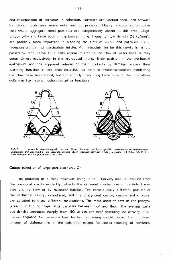
-119-
and resuspension of particles in selection. Part icles are washed back- and forward
by closed protrusion movements and compression. Highly viscous sulfomucines
that would aggregate small particles are conspicuously absent in this area. Oligo-
villous cells and taste buds in the buccal l ining, though of low density (50 tb /mm 2 ) ,
are probably more important in scanning the f low of water and particles during
resuspension, than at part iculate intake. At part iculate intake this cavity is rapidly
passed by food i tems. Club cells appear related to the f low of water because they
occur almost exclusively in the oro-buccal l ining. Their position in the oro-buccal
epithel ium and the supposed release of their contents by damage renders their
alarming funct ion in this area doubtful. No definite mechanoreceptors monitoring
the f low have been found, but the slightly protruding taste buds or the oligovillous
cells may have some mechanoreceptive functions.
FIG. 9. Areas in oro-pharyngeal roof and f loor, characterized by a specific combination of morphological characters and employed in the separate actions which together compose feeding sequences (cf Table 1.). Pecked lines indicate less sharply demarcated areas.
Coarse selection of large particles (area C)
The presence of a thick muscular l ining in the pharynx, and its absence f rom
the orobuccal cavity evidently ref lects the d i f ferent mechanisms of part icle trans
port v iz. by f low or by muscular bulging. The conspicuously d i f ferent profi les of
the orobuccal cavity, cy l indr ical , and the pharyngeal cavi ty, narrow and s l i t - l ike,
are adjusted to these d i f ferent mechanisms. The most anterior part of the pharynx
(area C in F ig . 9) traps large particles between roof and f loor. The average taste
bud density increases sharply f rom 100 to 520 per mm2 providing the sensory infor
mation required for decisions how further processing should occur. The increased
amount of sialomucines in the epithel ial crypts faci l i tates handling of part icles.
•120-
Water and small particles are further spread between palatal organ and branchial
sieve. This area is controled by the glossopharyngeal nerve IX and the anterior
rami of the vagus (X).
Selection of small particles (area D)
A t bottom feeding food must be selected f rom non-food. The opt imizat ion
of the abi l i ty to use tiny dispersed food particles present among non-food particles
is decisive in the exploi tat ion of this habitat. Repet i t ive back-washing movements
resuspend ingested particles and suggest a graded pur i f icat ion in the pharynx (Sibbing
et a l . , 1984). The lateral areas in the anterior pharynx are almost maximally packed
w i th taste buds (up to 820/mm2) and indicate a high resolving power in the gustatory
system. Local bulging of the palatal organ, possibly combined w i th act iv i ty of
the movable gil l rakers and their muscular pads, most probably clamp food particles
between roof and f loor, while waste is drained wi th the water. Few pyr i form mucous
cells (sialomucines wi th low viscosity) w i l l aid in part ic le handling. Locally varied
ac t iv i ty in the palatal organ has been recorded (Sibbing et a l . , 1984) and the palatal
bulging can readily be s t imulated. The arrangement of f ibers in the pharyngeal
roof and f loor as well as the interspersed connective tissue and f lu id might wel l
allow quite local f ixat ion of part icles. Taste information is carried to vagal lobes
of enormous size. Their laminated organization suggest a palatotopic mapping of
the pharyngeal wa l l . The muscular papillae covering the anterior surface of the
palatal organ serve very local retent ion of particles or merely increase the sialo
mucines supply. They are larger in the area for trapping and selection of large
part icles (area C).
The protruding position of taste buds on the gi l l rakers might confer them mechano-
receptive functions. The importance of f low regulation during size-selection, clea
ning of the branchial sieve and recol lect ion of food part icles for transport seems
without doubt. The papillae provide the gi l l rakers w i th profi les most probably
a f fect ing the mesh-width of the branchial sieve in selection between size-classes
of food part icles. Proprioceptors are known to occur in gi l l f i laments and rakers.
The s l i t - l ike prof i le of the anterior pharynx is a s t ructural necessity for the present
selection process and is probably influenced by palatal movements as a whole and
assures a large contact area. Large food objects can not pass.
-121-
Transport of food and loading of the chewing cavity (area E).
The tr iangular postlingual area in f ront of the chewing cavity is excluded
f rom the lateral paths of f low. The massive muscular roof and f loor f i l l the pharyn
geal s l i t . The arrangement of the muscle fibers and the act iv i t ies in this area,
coordinated in roof and f loor, take care of food transport (Sibbing et a l . , 1984)
and its propulsion into the chewing cavity. They also prevent loss of food f rom
the chewing cavity during mastication and deglut i t ion. High longitudinal folds direct
the movement of the food, lubricated by a copious mucus supply. This mucus is
largely composed of highly viscous sulfomucines, which now also wi l l aid in aggre
gation of food particles st icking together into boluses for further transport. Though
taste buds are s t i l l numerous (ca. 200/mm2) their density as well as the muscular
content of the folds is less than in the anterior areas (C, D). Taste buds do not
protrude f rom the epi thel ium. Mechanoreceptors steering transport have not been
recognized.
Mastication and deglutition (area F)
Movement of the pharyngeal teeth w i th respect to the cornif ied chewing pad
reguires extensibi l i ty of the posterior pharyngeal wa l l . Part icles larger than ca.
3% of the body length of the carp can not enter the chewing cavi ty. The composi
t ion of the epithelium is however closely similar to that of the postlingual area
(E). Foliaceous papillae crowded wi th cells producing sulfomucines lubricate mastica
t ion and aggregate ground particles into boluses. Lateral muscular tissue flaps
may clean the occlusal surfaces f rom part icles, acting l ike a tongue. Muscle f ibers
are part of the sphincter oesophagi. This muscle supports masticatory movements
and is the prime ef fector in deglut i t ion, propelling the food into the esophagus
(Sibbing, 1982).
Taste buds control the contents of the masticated food and their anterior density
(200/mm2) decreases sharply in the direct ion of t ransport. They vanish on the folds
converging into the esophagus. Proprioceptors in the pharyngeal jaw muscles are
supposed to play a prominent role in steering masticatory movements. Mechano
receptors have not been recognized.
The complex central pathways in the posterior brain are supposed to f u l f i l l
the integrat ion of the subseguent actions into an e f f ic ient feeding sequence, adjusted
to the type of food.
-122-
The combined mechanisms for uptake, selection and mastication although specia
l ized for bottom feeding allow also other food sources to be exploited (e.g. pelagic).
L imi tat ions on these have been outl ined previously. That carps may even be con
sidered as pests is most probably also due to the features discussed in this paper.
ABBREVIATIONS
ac ad am ap at b be c cc cp ep es f fl fn ga
gf gh
anterior cartilage aorta dorsalis adductor mandibulae anterior pharynx adipose tissue barbel buccal cavity-cornified area club cell chewing pad epithelium esophagus fibrous layer facial lobe facial nerve gill arch gill filament geniohyoid muscle
g r
h ib 1 lj 11 mf mv n ob oc occ ol op ope p
gill raker hart intestinal bulb lip lower jaw lateral line system muscle fibers maxillary valve free neuromast olfactory bulb oral cavity occipital skull area olfactory organ oral papillae opercular cavity papillae
pb Pi plo pm po PP Pt ropi rops sc sm st h tb tf uj V
pharyngobranchials pharyngeal jaw postlingual organ pyr i form mucous cell palatal organ posterior pharynx pharyngeal teeth inf. retractor phar. jaw sup. retractor phar. jaw stratum compactum sacciform mucous cel l sternohyoid muscle taste bud tissue f lap upper jaw vertebrae
Acknowledgements. Special thanks are due to Prof. dr. 3.W.M. Osse for the numerous discussions and his criticism on the manuscripts and to Mr. W. Valen for his skill in preparing the drawings and figures. The cooperation of the SEM-unit of the Institute of Agricultural Engineering (TFDL, Wageningen) is gratefully acknowledged. Mrs. L. van Beek was most helpful in typing the manuscripts. Thanks are also due to Mr. S. Leenstra and Mr. P. van Kleef for breeding and raising the carps.
-123-
REFERENCES
Abercrombie, M. (1946). Estimation of nuclear population from microtome sections. Anat. Rec. 94: 239-247. Al-Hussaini, A.H. (1945). The anatomy and histology of the alimentary tract of the coral feeding fish Scarus sordidus
(Klunz.). Bull. Inst. Egypte 27: 349-377. Al-Hussaini, A.H. (1949). On the functional morphology of the alimentary tract of some fish in relation to differences
in their feeding habits: anatomy and histology. Quat. J. Microsc. Sei. 90: 109-139. Atema, 3. (1971). Structure and function of the sense of taste in the catfish (Ictalarus natalis). Brain Behav. Evol.
4: 273-294. Bal l int i jn, C M . (1972). Efficiency, mechanics, and motor control of fish respiration. Respir. Physiol. 14: 125-141. Bal l int i jn, C.M. (1982). Neural control of respiration in fishes and mammals. In: Exogenous and endogenous influences
in metabolic and neural control of respiration, feeding : 127-140. Addink, A.D.F. & Spronk, N. (Eds.). Oxford: Pergamon Press.
Bal l int i jn, C.M. & Punt, G.J. (1984). Gi l l arch movements and the function of the dorsal g i l l arch muscles in the carp. J. Resp. Physiol, (submitted).
Bal l int i jn, C.M. & Bamford, O.S. (1975). Proprioceptive motor control in fish respiration. 3. Exp. Biol. 62: 99-114. Campos, H. (1969). Die Geschmacksknospen in Vorderdarm von Süsswasserfischen, Zahl, Verteilung und Entwicklung.
Z. wiss. Zool. 179: 253-299. Cook, A. & Shirbate, R. (1983). The mucus producing glands and the distribution of the ci l ia of the pulmonate slug
Limax pseudoflavus. 3. Zool. 201: 97-116. Curry, E. (1939). The histology of the digestive tube of the carp (Cyprinus carpio communis). 3. Morph. 65: 53-78. Denny, M.W. & Gosline, 3.M. (1980). The physical properties of the pedal mucus of the terrestrial slug, Ariolimax
columbianus. 1. exp. Biol. 88: 375-393. Dorier, A. & Belion, G. (1952). Sur l'organe palatin des cyprinides. Trav. Lab. Hydrob. Pisc. Grenoble 44: 47-60. Eastman, 3.T. (1970). The pharyngeal bones and teeth of Minnesota cyprinid and catastomid fishes: Functional mor
phology, variation and taxonomie significance. Thesis. University of Minnesota. Eastman, 3.T. (1977). The pharyngeal bones and teeth of catastomid fishes. Am. Midi. Nat. 97: 68-88. Evans, H.E. (1952). The correlation of brain pattern and feeding habits in four species of cyprinid fishes. 3. Comp.
Neur. 97: 133:142. Finger, T.E. (1981a). Enkephalin-like immunoreactivity in the gustatory lobes and visceral nuclei in the brains of
goldfish and catfish. Neuroscience 6: 2747-2758. Finger, T.E. (1981b). Laminar and columnar organization of the vagal lobe in goldfish: possible neural substrate
for sorting food from gravel. Soc. Neurose, Abstr. 7: 665. Fox, H., Lane, E.B. & Whitear, M. (1980). Sensory nerve endings and receptors in fish and amphibians. In: The skin
of vertebrates. Spearman, R.I.C. & Riley, P.A. (Eds). Symp. Linn. Soc. 9: 271-281. London: Academic Press. Frisch, K.v. (1941). über einen Schreckstoff der Fischhaut und seine biologische Bedeutung. Z. vergl. Physiol. 29:
46-145. Grzycki, S. (1954). Untersuchungen über die Nervenendigungen in die Bartel- und Lippen-epidermis der Cyprinus
carpio L. Ann. Univ. Mariae Curie Sklodowska Lublin, Polonia 9: 141-146. Hattingh, 3. & van Warmelo, K.T. (1975). The cuticular layer of the skin of certain Cyprinidae. Zool. Africana 10:
102-103. Hawkes, 3.W. (1974). The structure of fish skin. 1. General organization. Cell Tiss. Res. 149: 147-158. Henrikson, R.C. & Matoltsy, A.G. (1968). The fine structure of teleost epidermis. I. Introduction and fi lament contain
ing cells. 3. Ultrastruct. Res. 21: 194-212. Herrick, C.J. (1899). The cranial and f i rst spinal nerves of Menidia. 3. Comp. Neur. 9. Herrick, C.3. (1901). The cranial nerves and cutaneous sense organs of the North American siiuroid fishes. 3. Comp.
Neurol. 11: 177-249. Herrick, C.3. (1904). The organ and sense of taste in fishes. Bull. U.S. Fish Comm. (for 1902) 22: 237-272. Herrick, C.3. (1906). On the centers for taste and touch in the medulla oblongata of fishes. 3. Comp. Neur. 16:
403-421. Hirata, Y. (1966). Fine structure of the terminal buds on the barbels of some fishes. Arch. hist. jap. 26: 507-523. Hughes, G.M. (1979). Scanning electron microscopy of the respiratory surfaces of trout gills. 3. Zool. 187: 443-453. Hunt, S. (1970). Polysaccharide-protein complexes in invertebrates. New York: Academic Press. Hunt, S. (1973). Fine structure of the secretory epithelium in the hypobranchial gland of the prosobranch gastropod
mollusc Buccinum undatum L.. 3. mar. biol. Ass. U.K. 53: 59-71. I to, H. (1971). Fine structure of the teleostean vagal lobe, Z. Mikrosk Anat. Forsch. 83: 65-89. Iwai, T. (1964). A comparative study of the taste buds in gi l l rakers and gi l l arches of teleostean fishes. Bull. Misaki
Mar. Biol. Inst. Kyoto Univ. 7: 19-34. 3ara, Z. (1957). On the morphology and function of the so-called palatal organ in the carp (Cyprinus carpio L.).
Przeglad Zoologiczny 1: 110-112. Kapoor, B.G., Evans, H.E. & Pevzner, R.A. (1975). The gustatory system in fish. Adv. mar. biol. 13: 53-108. Kapoor, B.G., Smit, H. óc Verighina, I.A. (1975). The alimentary canal and digestion in teleosts. Adv. mar. biol.
13: 109-239. Karlsson, L. (1983). Gil l morphology in the zebrafish, Brachydanio rerio (Hamilton-Buchanan). 3. Fish Biol. 23: 511-524. Kolmer, W. (1927). Geschmacksorgan. In: Handbuch der mikroskopischen Anatomie des Menschen. 3. Haut und Sinnes
organe: 154-191. Möllendorff, W. (Ed). Berlin: Springer. Konishi, 3. & Zotterman, Y. (1963). Taste functions in fish. In: Olfaction and Taste, 215-233. Zotterman, Y. (Ed).
New York: MacMillan. Lane, E.B. & Whitear, M. (1977). On the occurence of Merkel cells in the epidermis of teleost fishes. Cell Tiss.
Res. 182: 235-246. Luiten, P.G.M. (1975a). The central projections of the tr igeminal, facial and anterior lateral line nerves in the carp
(Cyprinus carpio L.). 3. Comp. Neur. 160: 399-418. Luiten, P.G.M. (1975b). The horseradish peroxidase technique applied to the teleostean nervous system. Brain Res.
89: 181-186. McGlone, F.P. (1978). The gustatory system of the goldfish Carassius auratus. Thesis, Univ. Sussex School Biol.
Sei., Brighton, Sussex, U.K. McManus, 3.F.A. & Mowry, R.W. (1963). Staining methods. Histologic and histochemical. New York: Hoeber. McVay, 3.A. & Kaan, H.W. (1940). The digestive t ract of Carassius auratus. Biol. Bull. 78: 53-67.
-124-
Mittal , A.K. & Banerjee, T.K. (1980). Kerat inizat ion versus mucus secretion in fish epidermis. In: The Skin of Vertebra tes : 1-12. Spearman, R.I.C. & Riley, P.A. (Eds). Linn. Soc. Symp. Ser. 9, London: Academic Press.
Mittal , A.K., Whitear, M. & Bullock, A.M. (1981). Sacciform cells in the skin of t e leost fish. 2 . mikrosk.-anat . Forsch., Leipzig. 95: 559-585.
Moore, G.A. (1950). The cutaneous sense organs of barbeled minnows adapted to life in the muddy waters of the Grea t Plains region. Trans. Am. Micr. Soc- 69: 69-94.
Morita, Y., I to , H. óc Masai, H. (1980). Centra l gustatory paths in the crucian carp Carassius carassius. 3 . comp. Neur. 191: 119-132.
Morita, Y-, Murakami, T. & Ito, H. (1983). Cy toarch i t ec tu re and topographic projections of t h e gusta tory cen te r s in a te leost , Carassius carassius. J . Comp. Neur. 218: 378-394.
Ono, R.D. (1979). Sensory nerve endings of highly mobile s t ruc tures in two marine te leost fishes. Zoomorph. 92: 107-114.
Ono, R.D. (1980). Fine s t ruc ture and distribution of epidermal projections associated with t a s t e buds on the oral papil lae in some loricariid ca t f ishes (Siluroidei: Loricariidae). 3 . Morph. 164: 139-159.
Ozawa, N. (1951). Über den Lobus Vagi und Lobus Glossopharyngei der Fische. Folia Psychiatr . Neurol. Jpn. 5: 1-32. Pfeiffer, W. (1960). übe r die Schreckreaktion bei Fischen und die Herkunft des Schreckstoffes. Z. Vergl. Physiol.
43: 578-614. Pfeiffer, W., Sasse, D. & Arnold, M. (1971). Die Schreckstoffzellen von Phoxinus phoxinus und Morulius chrysophake-
dion (Cyprinidae, Ostariophysi, Pisces). Histochemische und elektronenmikroskopische Untersuchungen. 2 . Zellforsch. 118: 203-213.
Reid, E.W. (1894). The process of secret ion in the skin of the common eel . Phil. Trans. Roy. S o c B. 185: 319-354. Reu t t e r , K. (1973). Typisierung der Geschmacksknospen von Fischen. 1. Morphologische und neurohistochemische
Untersuchungen an Xiphophorus hellen Heckel (Poeciliidae, Cyprinodontiformes, Teleostei). 2 . 2elIforsch. 143: 409-423.
Reu t t e r , K., Breipohl, W. & Bijvank, G.3. (1974). Taste buds types in fishes. II. Scanning e lect ron microscopical investigations on Xiphophorus helleri Heckel (Poeciliidae, Cyprinodontiformes, Teleostei). Cell Tiss. Res. 153: 151-165.
Rober ts , T.R. (1982). Unculi (horny projections arising from single cells), an adapt ive f ea ture of t h e epidermis of ostariophysan fishes. 2ool. Scripta 11: 55-76.
Romeis, B. (1968). Mikroskopische Technik. München-Wien: Oldenbourg. Rosen, M.W. & Cornford, N.E. (1971). Fluid friction of fish s l imes. Nature 234: 49-51. Sato, M. (1977). Histology of barbels of Blepsias cirrhosus draciscus (Cott idae) . J ap . 3. Ichthyol. 23: 220-224. Scharrer , E. (1935). Die Empfindlichkeit der freien Flossenstrahlen des Knurrhahns (Trigla) für chemische Reize .
2 . vergl. Physiol. 22 : 145-154. Sibbing, F.A. (1976). Pharyngeal mast ica t ion in Cyprinus carpio (L.). Rev. Trav. Inst. Peches Marit. 40: 744-745. Sibbing, F.A. (1979). Functional analysis of mast icat ion in the ca rp . 3. Anat. 129: 209-210. Sibbing, F.A. (1982). Pharyngeal mast icat ion and food t ransport in the carp ( Cyprinus carpio L.): A c ineradiographic
and e lectromyographic study. 3. Morph. 172: 223-258. Sibbing, F.A., Osse, 3.W.M. & Terlouw, A. (1984). Food handling in the carp (Cyprinus carpio L.), i ts movement
pa t t e rns , mechanisms and l imitat ions. In this thesis . Silberberg, A. & Meyer, F.A. (1982). S t ruc ture and function of mucus. In: Mucus in health and disease II: 53-74.
Chant ier , E.N., Elder, 3.B. & Elstein, M. (Eds). New York: Plenum Press. Sperry, D.G. & Wassersug, R.3. (1976). A proposed function for microridges on epithelial cells. Anat. Rec . 185:
253-258. Spicer, S.S. (1965). Diamine methods for differentiating mucosubstances histochemically. J. Histochem. Cytochem.
13: 211-234. Stroband, H.W.3. & Dabrowski, K.R. ( 1979). Morphological and physiological aspects of t he digestive system and
feeding in f resh-water fish larvae, in: Nutrition des poissons, 355-376. Fontaine M. (Ed). Paris : Cen t re National de la Recherche Scientifique.
Sutterlin, A.M. & Saunders, R.L. (1969). Proprioceptors in the gills of t e leos ts . Can. 3. 2ool. 47: 1209-1212. Suyehiro, Y. (1942). A study on the digestive system and feeding habits of fish. 3ap. 3. 2ool. 10: 1-303. Uribe-Zamora, M. (1975). Selection des proies par le f i ltre branchial de la carpe miroir (Cyprinus carpio L-). Thesis,
University of Lyon. Verigina, I.A. (1969). Ecological and morphological peculiarit ies of the a l imentary system in some carps. In: Ich
thyology. Progress in science. 2oology: 88-117. Poznanin, L.P. (Ed). New Delhi: Amerilled Publ. Weber, E.H. (1827). Ueber das Geschmacksorgan der Karpfen und den Ursprung seiner Nerven. Arch. Anat . Physiol.
2: 309-315. Weisel, G.F. (1962). Comparat ive study of the digestive t r a c t of a sucker, Catastomus catastomus, and a predaceous
minnow, Ptychocheilus oregonense. Am. Midi. Nat . 68: 334-346. Whitear, M. (1970). The skin surface of bony fishes. 3. 2ool. 160: 437-454. Whitear, M. (1971). Cell specialization and sensory function in fish epidermis. 3. Zool. 163: 237-264. Whitear, M. (1981). Secretion in the epidermis of polypteriform fish. Z. mikrosk.-anat. Forsch. Leipzig. 95: 531-543. Whitear, M. & Lane, E.B. (1983). Oiigovillous cells of the epidermis: sensory e lements of lamprey skin. 3. Zool.
199: 359-384. Whitear, M. & Mittal , A.K. (1983). Fine s t ruc ture of the club cells in the skin of ostariophysan fish. Z. mikrosk.-anat.
Forsch. Leipzig, 97: 141-157. Wunder, W. (1936). Physiologie der Süsswasserfische. Handb. Binnenfisch. Mitteleur. 26: 1-340. Zander, E. (1906). Das Kiemenfilter der Teleosteer . Z. Wiss. Zool. 84: 619-713.
125
Pharyngeal Mastication and Food Transport in the Carp (Cyprinus carpio L): A Cineradiographic and Electromyographic Study
FERDINAND A. SIBBING Experimental Animal Morphology and Cell Biology, Agricultural University, Marijkeweg 40, 6709 PG Wageningen, The Netherlands
ABSTRACT Cyprinids constitute the largest fish family and are characterized by their pharyngeal teeth. The masticatory mechanism is still poorly understood. The complex of structures that determine the movements of pharyngeal teeth and chewing pad in the carp (Cyprinus carpio L.) is analyzed. Activities in 16 head muscles of a free-swimming carp were recorded. X-ray cinerecordings, synchronized with electromyograms, were made of the intake, transport, mastication, and deglutition of radiopaque food pellets. Metal markers allowed a detailed movement analysis.
Masticatory cycles are bilaterally synchronous and show distinct crushing and grinding patterns. Direct masticatory muscles that suspend and connect the pharyngeal bones steer and stabilize the masticatory movements. Baudelot's ligament, between skull and pectoral girdle, is applied as fulcrum, effects a crucial shift of the rotation axis of the pharyngeal jaw, and transforms crushing into grinding; simultaneous abduction lengthens the grinding stroke. Body muscles supply indirectly the power for mastication; they also appear to be regulated more distantly. The epaxial muscles lift the skull and thereby the levators of the pharyngeal bones, thus t ransmitting high forces to the teeth. They also stretch the levator of the bone as soon as occlusion is reached and thus optimize its production of forces during grinding. The hypaxial muscles retract the pharyngeal bones indirectly during grinding and power the teeth in sliding. The chewing pad, previously assumed to be motionless, rotates rostroventrad with the skull and intensifies grinding.
Respiration and mastication are mutually related. The extensive movements of the pharyngeal bones are permitted only by the simultaneous expansion of the buccopharynx and a slide-coupling in the branchial floor. Muscular pads that line the pharynx are shown to transport food toward the teeth. The constrictor pharyn-gis effects deglutition.
Natural food, intestinal contents, and feces of the carp were analyzed with respect to the capacity for distinct masticatory operations. During the experiments pellets, barley, and worms were fed. The carp is specialized for polyphagy and this appears to be based on the profiles of the heterodont teeth rather than on drastic changes in the two preprogrammed activity patterns. Comparison of the pharyngeal jaw system in the carp and higher teleosts emphasizes the structural design for the application of large forces in this cyprinid.
The Cyprinidae, with 275 genera and about cyprinids (Hensel, 70). The mechanism of 1,600 species, is by far the greatest family of mastication in carps and its bearing on other teleost fishes, characterized, among other fea- components in the head have never been fully tures, by pharyngeal teeth in one to three rows elucidated. with never more than eight teeth in a single The morphology of pharyngeal bones and row (Nelson, 76). Pharyngeal teeth and bones teeth in the cyprinids is tremendously varied are important characters in phyletic studies on (Heincke, 1892; Chu, '35). Teeth are large or
126
small, and arranged widely spread or closely together in multiple short or in single long rows. In the grasscarp (Ctenopharyngodon) they interdigitate from left and right sides, in the carp they do not. This variety is assumed to reflect an adaptation of cyprinid species to diversified trophic conditions.
There are scattered descriptions of the lower pharyngeal bones and teeth (Jurine, 1821; Owen, 1840-1845; Heincke, 1892; Shepherd, '12; Stoss, '21; Chu, '35; Vasnecov, '39; Rutte, '62), the horny chewing pad opposite the teeth and fixed at the posterior base of the skull (Gratzianow, 1900; Haempel, '07), and of the pharyngeal bone musculature (Haempel, '09; Takahasi, '25; Girgis, '52; Eastman, '71) of different cyprinids. For a review of literature on pharyngeal masticatory elements in the carp, the reader is referred to Eastman ('70). The studies mentioned treat only disjoined elements, are mainly comparative, and do not evaluate the whole set of masticatory elements, let alone their interrelations. Such a holistic approach is essential in explaining the relationships between structural elements of the feeding mechanism and the ecological conditions of some African cyprinids. In addition to the pharyngeal masticatory apparatus, external features and prehensile, selective, and digestive s t ructures were studied also. Matthes ('63) distinguishes three basic types of African cyprinids: a Labeo (microphytopha-gous), a Barbus (omnivorous), and a Barilius (carnivorous) type. Eastman ('70) treated the pharyngeal masticatory apparatus in the carp and in the River Redhorse (Moxostoma carina-tum), made some comparison with other Minnesota cyprinids and catostomids, and used pharyngeal bones and teeth to establish taxonomie keys for these fishes. Until now no functional study of mastication in cyprinids has been performed and the function and interaction of the separate and varied elements, such as teeth, chewing pad, bones, muscles, and ligaments, are still unknown. The present study demonstrates that, besides these directly involved elements, skull and pectoral girdle also participate in mastication.
The aim of this study is to determine which elements of the head are involved in pharyngeal mastication, their actual role, the relevance of topography, form, and structure, and how the various actions are integrated into effective masticatory movements. Owing to the integration of a multiplicity of functional components in the head, attention should be paid to competitive demands of other than mastica
tory functions on the pharyngeal structures (Dullemeijer, '58, '74). The present analysis comprises form as well as action. I t includes the recording of activities in the small and less accessible pharyngeal muscles in free-swimming and feeding carps (Cyprinus carpio L.), with simultaneous X-ray cinematography of pharyngeal bone movements.
A study of the relation between form and function of the masticatory apparatus should also involve an analysis of the natural food and a differentiation according to its shape, size, and mechanical properties. Cyprinids are highly suitable for studying the relation between mastication and diet in fishes, because the absence of any other teeth and the lack of a stomach might well increase the demands on the pharyngeal masticatory construction. The presence of a flat and fixed chewing pad in cyprinids instead of complex and movable suprapharyngeal teeth, as in most other tele-osts, simplifies the mechanical events between the diminutive surfaces and facilitates the search for relations between properties of the food and the morphology of the teeth.
The common carp {Cyprinus carpio L.) was selected because it is readily available and withstands experimental manipulation. I ts respiratory pump was studied by Ballintijn ('69 a,b); thus, there is a solid basis for evaluating the anticipated relations between mastication and respiration. Because the carp is extensively cultured, knowledge of its feeding mechanism might prove valuable for pisciculture.
The present study provides a standard of comparison to be used in future comparative studies within this family.
MATERIALS AND TECHNIQUES
Dissection and experiments were carried out with 3-year-old mirror-carps (Cyprinus carpio L.), standard length 28-38 cm. obtained from the OVB (Organisatie ter Verbetering van de Binnenvisserij, Utrecht, the Netherlands). Some were dissected fresh; others were fixed in formalin or Vin d'Alsace (Zweers, '74).
Prior to inserting electrodes and platinum markers, the animals were superficially anesthetized with a solution of 125 mg/liter MS 222 (Sandoz, Basel); the lower jaw kept slightly moving. Electromyograms were recorded as soon as the actively swimming fish started to feed, in most cases half a day after being returned to the experimental tank (80 X 50 X 40 cm), at about 20°C. Damage to the fish was only slight and restricted to the last gill arch;
> structure of the parts (Dullemeijer, '58) and was made by Matthes ('63) who analyzed the
127
within a week the injury disappeared. Nevertheless, no fish was used more than twice for experiments.
The food usually consisted of industrial t rout pellets (Trouvit 4, diameter 5 mm; Trouw and Co., N.V., Putten, the Netherlands), and occasionally of worms and barley.
Elec tromyograp hy
In order to measure the activity of the muscles involved in respiration, food transport and mastication in a single experiment, up to nine pairs of insulated copper wire electrodes (Povin D, 100-ftm diameter; Pope, Venlo, the Netherlands) were inserted with a modified fork-tipped hypodermic needle (Osse et al., '72). The distance between the two bared electrode tips was about 4 mm in each muscle; in very small muscles the electrodes were inserted simultaneously with one needle. The electrode positions were checked in lateral and dorso-ventral X-ray photographs and in later experiments even during the operation by short stimulation (square waves, 50 Hz, 2-10 V). Three experiments included bilateral measurements.
The bundle of electrode wires was pulled through a flexible rubber tube (2-mm inner diameter) that was closed at both ends with nail-polish. The tube protects the leads, largely prevents contact with water, prevents the fish from being entangled in the wires (the air-filled tube floats), and transmits the movements of the fish better to the collector. The cable was fixed to the first spine of the dorsal fin by means of a small clamping screw.
The instruments for amplification and registration, including an ink-jet recorder (Siemens; type S), are similar to those used by Osse ('69), who described details of the measuring procedure. Filters of low and high cutoff were set at 0.08 and 10 kHz; amplification varied from 103
to 10". An instrumentation tape recorder (Bell and Howell; type CPR 4010) was added. The connection between fish and apparatus was modified by applying a 14-pole miniature slip ring and brush collector (I.D.M. Electronics Ltd., Reading, England). This plug-in collector improves the transmission of the signals through its stable gold contacts, and rotates easier with the movements of the fish.
X-ray cinematography
Lateral and dorsoventral X-ray images (Siemens Gigantos X-ray apparatus; 100 kV lateral/75 kV dorsoventral, 125 m A, and 2 msec)
of the moving elements of the head were intensified (Siemens 50-kV image intensifier) and recorded with an Arriflex 35-mm motion picture camera (Agfa-Gevaert Copex Pan film) at 26 frames per second. The distances between X-ray tube and fish and between the fish and the image intensifier were 195 cm (in dorsoventral projection, 135 cm) and 5 cm, respectively.
In order to penetrate water with X-rays a high X-ray tube voltage is required, but a high voltage gives low contrast between the bony elements and the water. In order to reduce the water layer around the fish as much as possible, the fish was trained to feed in a narrow, 2.5-mm perspex cuvet (32 X 9 X 27 cm), equipped with a movable curtain behind the tail. This design also keeps the head of the fish above the image intensifier and tends to maintain the fish parallel to the plane of the film. In lateral view the fish was 4 cm thick at the pectoral girdle, and the water layer (progressively replaced by tempex) 2.5 cm; in a dorsoventral view these values were 6 and 1.5 cm, respectively. The necessary use of an image intensifier limits the definition of the images. To improve contrast and to allow accurate-measurements, pieces of 0.5-mm platinum wire were inserted on selected places (Figs. 8, 17b). Markers on the skull facilitated the elimination of distorted projections. X-ray movies were evaluated and the appropriate masticatory scenes selected on a Vanguard (type 16C) motion analyzer. Measurements were made in three-times enlarged individual frames, with a Leitz Makro Promar projector provided with a film-transport adaptor. With this procedure the required accuracy was obtained for measuring the minute changes in position between successive frames.
Simultaneously with X-ray cinematography, the activity of selected muscles was recorded. X-ray frames and electromyograms were synchronized by pulse trains produced at random and registered both on the film (by flashes of a diode bulb built into the camera body) and on the EMGs. Movements were analyzed of two trained carps; from the large quantity of material about 60 of the most stably projected chewing cycles of eight masticatory sequences were selected. Measurements corresponded very well for the two fishes. At least 30 EMG experiments supplied the data for the activity pattern of the muscles during mastication and respiration. The processing of movement analysis and EMG data will be discussed later, because the necessary anatomical data have not yet been provided.
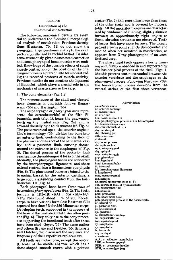
128
RESULTS
Description of the anatomical construction
The following anatomical details are essential to understand the functional-morphological analysis of mastication. Previous descriptions (Eastman, '70, '71) do not show the elements in their positions relative to the skull, pectoral girdle, and branchial basket. Besides, some erroneously given names need correction and some pharyngeal bone muscles were omitted. Knowledge of the possible effects of single muscle contraction on the position of the pharyngeal bones is a prerequisite for understanding the recorded patterns of muscle activity. Previous studies do not mention the ligament of Baudelot, which plays a crucial role in the mechanics of mastication in the carp.
1. The bony elements (Fig. 1,2)
The nomenclature of the skull and visceral bony elements in cyprinids follows Ramas-wami ('55) and Harrington ('55).
The os pharyngea or pharyngeal bone represents the ceratobranchial of the fifth (V) branchial arch (Fig. 1), bears the pharyngeal teeth on the medial side (Fig. 2a), and is covered dorsally by a thick mucosa (cf. Fig. 4). The posteroventral apex, the anterior angle in Chu's terminology ('35), divides the bone into an anterior limb, contributing to the floor of the pharynx and the roof of the pericardial cavity, and a posterior limb, curving dorsad around the entrance to the esophagus (cf. Fig. 5). The dorsal process of the posterior limb reaches into the subtemporal fossa of the skull. Medially, the pharyngeal bones are connected by the interpharyngeal ligaments, and these extend rostrad into a ligamentous symphysis (Fig. 6). The pharyngeal bones are joined to the branchial basket by the anterior cartilage, a large copula extending caudad from the basi-branchial I II (Fig. 5).
Each pharyngeal bone bears three rows of heterodontpharyngeal teeth (Fig. 2). The tooth formula is 1(C)-1(B)-3(A) : 3(A)-1(B)-1(C). Yang ('64) found about 11% of 260 Korean carps to have variant formulas; Eastman ('70) reported less than 6% for 380 Minnesota carps. Developing teeth, embedded in the mucosa at the base of the functional teeth, are often present (Fig. 6). They ankylose to the bony processes supporting the functional teeth after these have been shed (Geyer, '37). The same author and others (Evans and Deubler, '55; Schwartz and Dutcher, '62) discussed the sequence and frequency of their repetitive replacement.
All teeth are molariform, except the rostral (I) tooth of the medial (A) row, which has a dome-shaped smooth crown with a pointed
center (Fig. 2); this crown lies lower than those of the other teeth and is covered by mucosal folds. All flat molariform crowns are characterized by caudomediad running, slightly sinuous furrows; at approximately right angles to these, abrasion scratches are observed. Teeth of larger fish have more furrows. The closely packed crowns point slightly dorsocaudad and mediad when not involved in mastication, as appears from X-ray photographs of an anesthetized carp.
The pharyngeal teeth oppose a horny chewing pad, firmly embedded in and supported by the basioccipital process of the skull (Figs. 1, 2b); this process continues caudad between the anterior vertebrae and the esophagus as the pharyngeal process. Following Nusbaum ('08), the basioccipital process develops from the ventral arches of the first three vertebrae,
Abbreviations aa, anterior angle ac, anterior cartilage al, anterior limb ar, articular bbr, basibranchial I I I bocc pr, pharyngeal process of the basioccipital brr, branchiostegal rays cbr, ceratobranchial I - IV chy, ceratohyal chp, chewing pad clthr, cleithrum d. dental dp, dorsal process ebr, epibranchial ecpt, ectopterygoid ehy, epihyal enpt, entopterygoid ghy, glossohyal hhy, hypohyal hmd, hyomandibular ih, interhyal ipl, interpharyngeal ligaments k, kinethmoid mpt, metapterygoid mx, maxilla ns, neural spines vertebrae I I - IV opj, opercular joint of hyomandibular osu, os Suspensorium pal, palatine pmx, premaxilla phb, pharyngeal bone ppb, pharyngeal process of the basioccipital pr, pleural rib pi, posterior limb pt, posttemporal qu, quadrate sc, submaxillar cartilage scl, supracleithrum soc, supraoccipital sy, symphysis sy, symplectic tr, trabecula uhy, urohyal v, vertebra A2A3, m. adductor mandibulae LOP, m. levator operculi PRH, m. protractor hyoidei STH, m. sternohyoideus
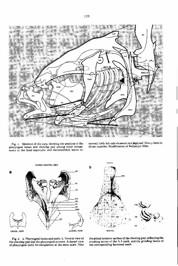
129
Fig. 1. Skeleton of the carp, showing the position of the pharyngeal bones and chewing pad among other components in the head (opercular and circumorbital bones re
moved). Only left-side elements are depicted. Heavy lines indicate muscles. Modification of Ballintijn ('69a|.
DORSO- ROSTRAL VIEW
CAUDAL VIEW LATERAL VIEW
/
• > >
Fig. 2. a. Pharyngeal bones and teeth, b. Ventral view of the chewing pad and the pharyngeal process. A dorsal view of pharyngeal teeth for comparison at the same scale. Note
*>s/
the pitted anterior surface of the chewing pad, reflecting the crushing action of the A I teeth, and the grinding facets of the corresponding furrowed teeth.
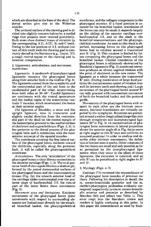
130
which are absorbed in the base of the skull. The dorsal arches give rise to the Weberian ossicles.
The occlusal surface of the chewing pad is divided into slightly concave halves by a medial ridge that projects most ventrad posteriorly. Both areas show distinct traces of abrasion by the corresponding A II, A I I I , B, and C teeth. Owing to the low position of A I, occlusal contact of this tooth with the chewing pad is commonly denied in the literature (e.g., Geyer, '37); a rough rostral region on the chewing pad is, however, conspicuous.
2. Ligaments, articulations, and movement restrictions
Ligaments. A meshwork of interpharyngeal ligaments connects the pharyngeal bones along their anterior limb in the midline (Fig. 6). The ligaments extend from the symphysis and the rostromedial part of the one bone to the caudomedial part of the other, intercrossing from both sides at 30-40°. Caudal ligaments are continuous with the rostral aponeurosis and the medial raphe of the transversus ven-tralis V muscles, which interconnect the bones at their anterior angles.
The ligament of Baudelot, a stout and flat upright ligament, runs in transverse and slightly caudal direction from the ventromedial part of the skull (at the rostral margin of the basioccipital process) to the medial surface of cleithrum and supracleithrum (Figs. 3, 5). It lies posterior to the dorsal process of the pharyngeal bone and is continuous with the most anterior myosept of the epaxial muscles.
The membrane covering the free lateral surface of the pharyngeal bones thickens toward the cleithrum, especially along the posterior limb. I t will be called the pharyngocleithral membrane.
Articulations. The only "articulation" of the pharyngeal bones is their fibrous connection to the anterior cartilage (Figs. 1, 5). The oval posterior head of this copula fits into a shallow pit, formed by the joined dorsomedial surfaces of the pharyngeal bones and the interconnecting tissues (Fig. 2a); the smooth anterior head of the cartilage slides rostrocaudad over the posterior slope of basibranchial I I I ; the rostral part of the latter limits these movements (Fig. 5).
Movement axes and limitations. Extrinsic movements of the pharyngeal bones (joint movements with respect to surrounding elements) are limited most directly by the attached branchial basket, the pharyngocleithral
membrane, and the collagen components in the pharyngeal muscles. If a fixed position is assumed for the branchial basket, translation of the bones can only be small; it depends largely on the sliding of the anterior cartilage over basibranchial I I I , and on the slack in the branchial interconnections and the pharyngocleithral membrane. As soon as this slack is absorbed, increasing forces on the pharyngeal bones lead to rotation around a transversal axis R1 (Fig. 3). This rotation is limited by tissue connecting the pharyngeal bones and the branchial basket. Caudad translation of the pharyngeal bones is ultimately obstructed by Baudelot 's ligaments (Fig. 3); continued retraction produces an almost sagittal rotation with the point of abutment as the new center. The ligament as a whole becomes the transversal R4 axis. (During mastication of large food particles the instantaneous center of rotation may well lie between teeth and chewing pad.) Large excursions of the pharyngeal bones around R4
can only proceed with concomitant movement of both the branchial basket and the pectoral girdle.
Movements of the pharyngeal bones with respect to each other are the intrinsic movements. Intrinsic translations are virtually impeded by the symphysis. Rotations are permitted around a rather longitudinal hinge-like axis through symphysis and interpharyngeal ligaments (R2 in Fig. 3). (A reconstruction of pharyngeal bone movements in lateral projection shows the anterior angle (B in Fig. 8a) to move at right angles to this R2 axis and confirms its assumed position.) In order to analyze and describe other intrinsic movements, the definition of further axes is useful. Other rotations of the two bones are small and only possible as far as permitted by the interpharyngeal ligaments; when they occur in the plane of these ligaments, the excursion is maximal, and an axis R3 can be postulated at right angles to R2
and R1.
3. Myology (Figs. 4-7)
Eastman ('71) reviewed the nomenclature of the pharyngeal bone muscles of previous authors. Following his description in detail, the m. coraco-branchialis posterior and the m. cleithro-pharyngeus profundus evidently correspond, respectively, to the m. coraco-branchialis anterior and posterior as described by Matthes ('63) and others (see Table 1). This error crept into the literature review and renders it highly confusing at this point. In this paper the nomenclature as used by East-
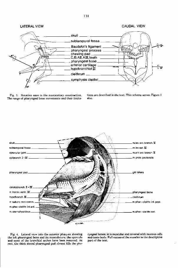
131
LATERAL VIEW CAUDAL VIEW
skull
subtemporal fossa _
Baudelot's ligament pharyngeal process chewing pad C,B, AH, AH , teeth pharyngeal bone anterior cartilage hypobranchial HI
cleithrum
symphysis cleithri
J R1
Fig. 3. Rotation axes in the masticatory construction. The range of pharyngeal bone movements and their limita
tions are described in the text. This scheme serves Figure 7 also.
skull
subtemporal fossa
opercular joint
epibranch. I-EZ
pharyngeal pad
ceratobranch. I -ÏÏ
m.transv. venir. EZ
hypobranch. lu
m.subarc. rect.comm.
m.phar.-cleithr. infant,
m.sternohyoideus
m.lev. arc. branch. 3£
m.lev.ext.nr
m.attr. arc. branch.3Z
m.protr. pectoralis
gill rakers
pharyngeal bone
cleithrum
m.phar.-cleithr. int. post.
m.phar.-cleithr.ext.
Fig. 4. Lateral view into the anterior pharynx showing the left pharyngeal bone and its musculature; the opercula and most of the branchial arches have been removed. At rest, the thick dorsal pharyngeal pad almost fills the pha
ryngeal lumen; it is muscular and covered with mucous cells and tas te buds. Full names of the muscles in the descriptive part of the text.
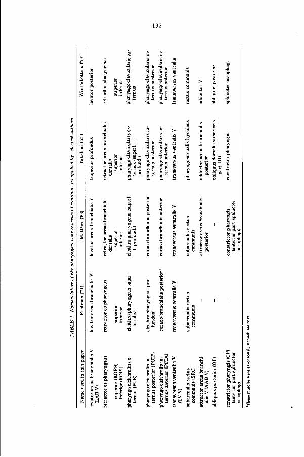
132
5. s c 2 g .S >>E
-S Ü
v v 5 bo m
ss
bß co
>> Ë
a« a
c.
.2 +
.Ht C<3 Û .
o — -a bc co C
b E o Safe.
•a s en HH
•5 S, 3 O.
'e
° "3 e.,» 6 £ u £ 3 c S= o.
13 13 -ffi + o *J
3 P
•s s
ttl
«3
S-3 2 -
5 ;s 5
H 3
3 I
73 — .2
£ « .-e
S3> b « 3 <
O-A
w IJ ft. 3 Ç
.So »Si 1.2
x& C 3 >, g
. < S ft. 'S >" S.S
S* bc » C 3 >>̂
CO
CO >
35 £3 Sfc | 8
o . • . _ o 2 ft. <-> « CO C
2 -al 5 ü
•a 3
i l
tß
s .2!
u t, Sc o g « «•E-S
•C 3 o
«Is c 3. o o
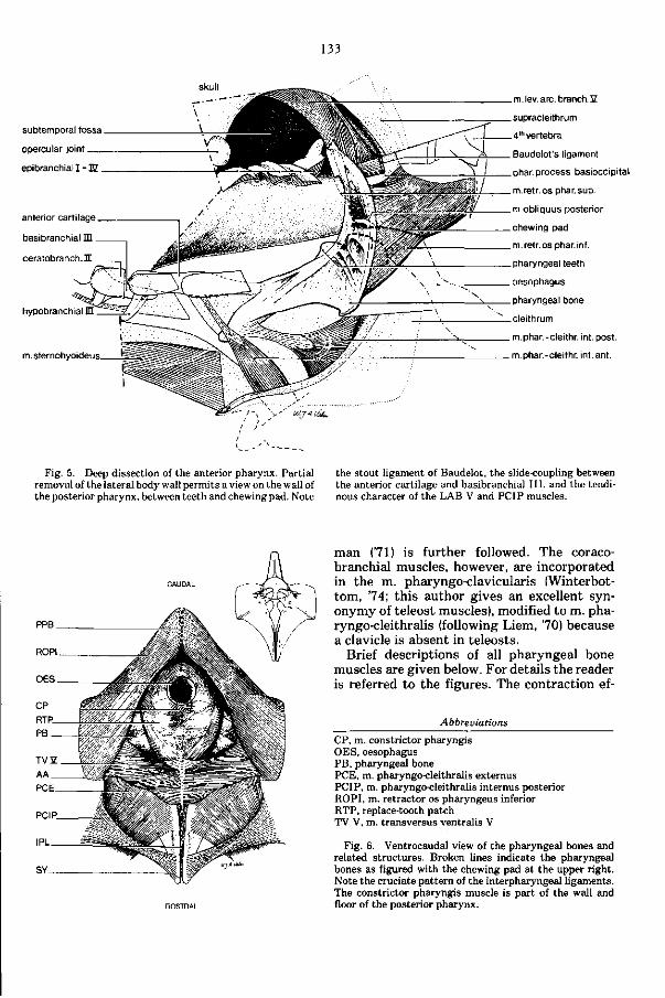
133
skull
subtemporal fossa
opercular joint
epibranchial I - E
anterior cartilage
basibranchial m
ceratobranch.I
hypobranchialH
m.sternohyoideus.
m. lev. arc. branch. 3Z
supracleithrum
4th vertebra
Baudelot's ligament
phar. process basioccipital
m.retr. os phar. sup.
m.obliquus posterior
chewing pad
m.retr.os phar.inf.
pharyngeal teeth
oesophagus
pharyngeal bone
cleithrum
m.phar. -cleithr. int. post.
m.phar.-cleithr. int. ant.
Fig. 5. Deep dissection of the anterior pharynx. Partial removal of the lateral body wall permits a view on the wall of the posterior pharynx, between teeth and chewing pad. Note
the stout ligament of Baudelot, the slide-coupling between the anterior cartilage and basibranchial III , and the tendinous character of the LAB V and PCIP muscles.
man (71) is further followed. The coracob r a ch i a l muscles, however, are incorporated in the m. pharyngo-clavicularis (Winterbot-tom, '74; this author gives an excellent synonymy of teleost muscles), modified to m. pha-ryngo-cleithralis (following Liem, 70) because a clavicle is absent in teleosts.
Brief descriptions of all pharyngeal bone muscles are given below. For details the reader is referred to the figures. The contraction ef-
Abbreuiations
CP, m. constrictor pharyngis OES, oesophagus PB, pharyngeal bone PCE, m. pharyngo-cleithralis externus PCIP, m. pharyngo-cleithralis internus posterior ROPI, m. retractor os pharyngeus inferior RTP, replace-tooth patch TV V, m. transversus ventralis V
Fig. 6. Ventrocaudal view of the pharyngeal bones and related structures. Broken lines indicate the pharyngeal bones as figured with the chewing pad at the upper right. Note the cruciate pattern of the interpharyngeal ligaments. The constrictor pharyngis muscle is part of the wall and floor of the posterior pharynx.
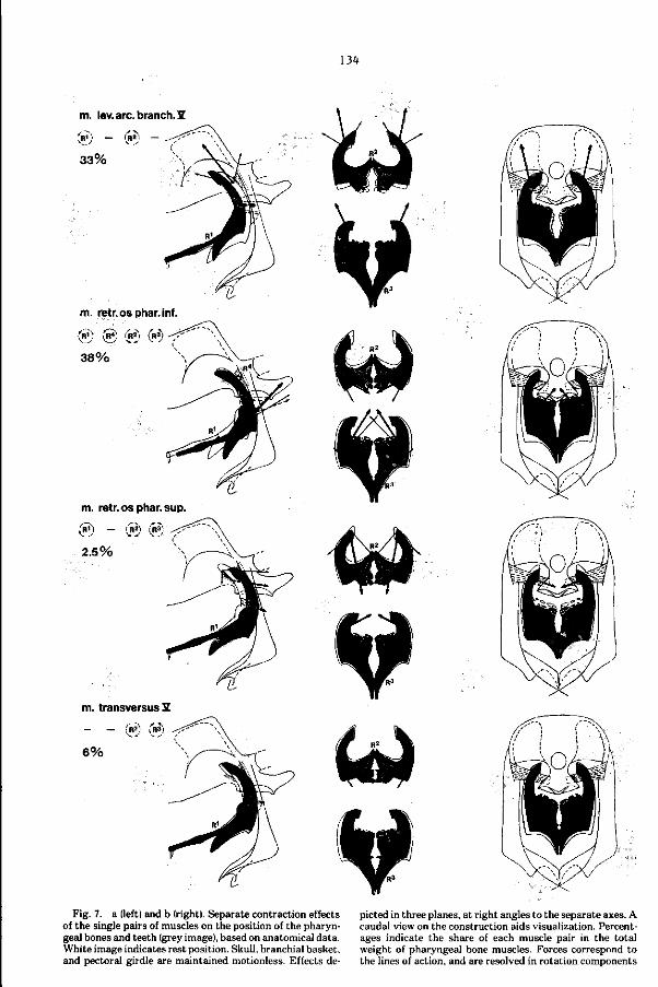
134
m. lev. arc. branch. S
(w) — (R») •
33%
m. retr.osphar.inf.
(iiî) ® (R^I (R3)
3 8 %
m. retr. os phar. sup.
® - ® c» 2.5%
m. transversus 2
!R2,: I R > )
6 %
Fig. 7. a (left) and b (right). Separate contraction effects of the single pairs of muscles on the position of the pharyngeal bones and teeth (grey image), based on anatomical data. White image indicates rest position. Skull, branchial basket, and pectoral girdle are maintained motionless. Effects de
picted in three planes, at right angles to the separate axes. A caudal view on the construction aids visualization. Percentages indicate the share of each muscle pair in the total weight of pharyngeal bone muscles. Forces correspond to the lines of action, and are resolved in rotation components
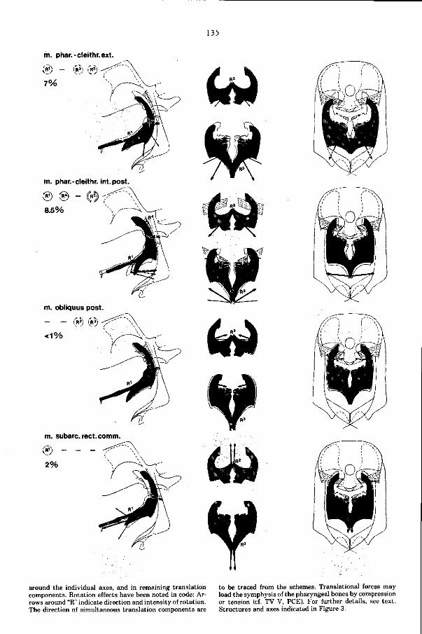
135
m. phar.-cleithr. ext.
7%
m. phar.-cleithr. int. post.
® © — (i'Ra)) 'r ' 8.5%
m. subarc. rect.comm.
'nO, _ _ _
2%
around the individual axes, and in remaining translation to be traced from the schemes. Translational forces may components. Rotation effects have been noted in code: Ar- load the symphysis of the pharyngeal bones by compression rows around "R" indicate direction and intensity of rotation. or tension (cf. TV V, PCE). For further details, see text. The direction of simultaneous translation components are Structures and axes indicated in Figure 3.
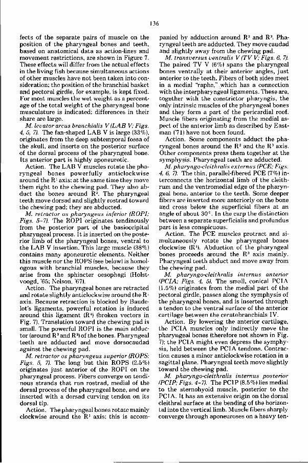
136
fects of the separate pairs of muscle on the position of the pharyngeal bones and teeth, based on anatomical data as action-lines and movement restrictions, are shown in Figure 7. These effects will differ from the actual effects in the living fish because simultaneous actions of other muscles have not been taken into consideration; the position of the branchial basket and pectoral girdle, for example, is kept fixed. For most muscles the wet weight as a percentage of the total weight of the pharyngeal bone musculature is indicated; differences in their share are large.
M. levator arcus branchialis V (LAB V; Figs. 4, 5, 7). The fan-shaped LAB V is large (33%), originates from the deep subtemporal fossa of the skull, and inserts on the posterior surface of the dorsal process of the pharyngeal bone. I ts anterior part is highly aponeurotic.
Action. The LAB V muscles rotate the pharyngeal bones powerfully anticlockwise around the R1 axis; at the same time they move them right to the chewing pad. They also abduct the bones around Rz. The pharyngeal teeth move dorsad and slightly rostrad toward the chewing pad; they are abducted.
M. retractor os pharyngeus inferior (ROPI; Figs. 5-7). The ROPI originates tendinously from the posterior part of the basioccipital pharyngeal process. I t is inserted on the posterior limb of the pharyngeal bones, ventral to the LAB V insertion. This large muscle (38%) contains many aponeurotic elements. Neither this muscle nor the ROPS (see below) is homologous with branchial muscles, because they arise from the sphincter oesophagi (Holst-voogd, '65; Nelson, '67).
Action. The pharyngeal bones are retracted and rotate slightly anticlockwise around the R1
axis. Because retraction is blocked by Baude-lot's ligaments, powerful rotation is induced around this ligament (R4) (broken vectors in Fig. 7). Translation toward the chewing pad is small. The powerful ROPI is the main adductor (around R3 and R2) of the bones. Pharyngeal teeth are adducted and move dorsocaudad against the chewing pad.
M. retractor os pharyngeus superior (ROPS; Figs. 5, 7). The long but thin ROPS (2.5%) originates just anterior of the ROPI on the pharyngeal process. Fibers converge on tendinous strands that run rostrad, medial of the dorsal process of the pharyngeal bone, and are inserted with a dorsad curving tendon on its dorsal tip.
Action. The pharyngeal bones rotate mainly clockwise around the R1 axis; this is accom
panied by adduction around R2 and R3. Pharyngeal teeth are adducted. They move caudad and slightly away from the chewing pad.
M. transversus ventralis V (TV V; Figs. 6, 7). The paired TV V (6%) spans the pharyngeal bones ventrally at their anterior angles, jus t anterior to the teeth. Fibers of both sides meet in a medial "raphe," which has a connection with the interpharyngeal ligaments. These are, together with the constrictor pharyngis, the only intrinsic muscles of the pharyngeal bones and they form a part of the pericardial roof. Muscle fibers originating from the medial aspect of the anterior limb as described by Eastman ('71) have not been found.
Action. Some components adduct the pharyngeal bones around the R2 and the R3 axis. Other components press them together at the symphysis. Pharyngeal teeth are adducted.
M. pharyngo-cleithralis externus (PCE; Figs. 4, 6, 7). The thin, parallel-fibered PCE (7%) interconnects the horizontal limb of the cleith-rum and the ventromedial edge of the pharyngeal bone, anterior to the teeth. Some deeper fibers are inserted more anteriorly on the bone and cross below the superficial fibers at an angle of about 30°. In the carp the distinction between a separate superficialis and profundus part is less conspicuous.
Action. The PCE muscles protract and simultaneously rotate the pharyngeal bones clockwise (R1). Abduction of the pharyngeal bones proceeds around the R3 axis mainly. Pharyngeal teeth abduct and move away from the chewing pad.
M. pharyngo-cleithralis internus anterior (PCIA; Figs. 4, 5). The small, conical PCIA (1.5%) originates from the medial part of the pectoral girdle, passes along the symphysis of the pharyngeal bones, and is inserted through a tendon to the ventral surface of the anterior cartilage between the ceratobranchials IV.
Action. By lowering the anterior cartilage, the PCIA muscles only indirectly move the pharyngeal bones (therefore not shown in Fig. 7); the PCIA might even depress the symphysis, held between the PCIA tendons. Contraction causes a minor anticlockwise rotation in a sagittal plane. Pharyngeal teeth move slightly toward the chewing pad.
M. pharyngo-cleithralis internus posterior (PCIP; Figs. 4-7). The PCIP (8.5%) lies medial to the sternohyoid muscle, posterior to the PCIA. It has an extensive origin on the dorsal cleithral surface at the bending of the horizontal into the vertical limb. Muscle fibers sharply converge through aponeuroses on a heavy ten-
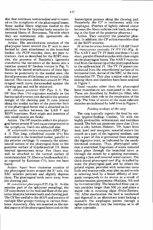
137
don that continues rostromediad and is inserted on the symphysis of the pharyngeal bones. Some medial fibers originate medial to the cleithrum from the hypaxial body muscles (intercostal fibers; cf. Brousseau, 76) with which they are continuous; only aponeurotic elements are interposed.
Action. The anticlockwise rotation of the pharyngeal bones around the R1 axis is soon limited by their attachment to the branchial basket; the PCIP muscles, then, are pure retractors. Jus t as described for the ROPI muscles, the presence of Baudelot's ligaments transforms the retraction of the bones into a rotation around R4 (broken vectors in Fig. 7). Because the lateral attachments of these ligaments lie posteriorly to the medial ones, the dorsal processes of the bones are forced to slide laterad. This causes abduction around R3. Pharyngeal teeth move dorsocaudad against the chewing pad and will be abducted.
M. obliquus posterior (OP; Figs. 5, 7). The OP (less than 1%) originates medially on the basioccipital process just dorsal to the anterior margin of the chewing pad. It runs laterad along the medial surface of the posterior limb of the pharyngeal bones and is attached on its posterior surface between the LAB V and ROPI insertions. Both origin and insertion of this small muscle are fleshy.
Action. The OP muscles adduct the pharyngeal bones around R3 and cause compression in the symphysis. Teeth are adducted also.
M. subarcualis rectus communis (SRC; Figs. 4, 7). This long, cylindrical muscle (2%) lies midventral in the branchial basket, parallel to the anterior cartilage. It connects the anterolateral surface of the pharyngeal bone to the posterior surface of hypobranchial I I I . Some internal aponeuroses occur. Few fibers may well be attached to the ventral surface of ceratobranchial IV; fibers to basibranchial I I I , as reported by Eastman (71), have not been found.
Action. Apart from clockwise rotation of the pharyngeal bones around the R1 axis, the SRC muscles protract and slightly depress them. The pharyngeal teeth move away from the chewing pad.
M. constrictorpharyngis (CP; Fig. 6). As the anterior part of the sphincter oesophagi, the CP contributes to the wall and floor of the posterior pharynx between teeth and chewing pad. The CP lies medial to the ROPI and consists of multiple fiber groups running in various directions. Anteriorly, they are inserted on the medial aspects of the pharyngeal bones and on the
basioccipital process along the chewing pad. Posteriorly the CP is continuous with the esophagus. (Patches of lightly colored tissue between the fibers indicate new teeth, developing in the floor of the posterior pharynx.)
Action. They constrict the posterior pharynx. In addition, the CP action resembles that of the ROPI muscles.
M. attractorarcus branchialis V(AAB Vjand M. transversus ventralis IV (TV IV) (Fig. 4). The AAB V and TV IV (both less than 1%) are short, parallel-fibered muscles with their origin on the pharyngeal bones. The AAB V runs ros-trad from the dorsal tip of the pharyngeal bone to the epibranchial IV. The TV IV passes ros-trodorsad from the anterolateral surface of its horizontal limb, dorsal of the SRC, to the ceratobranchial IV. They play a minor role in positioning them and therefore have been omitted in Figure 7.
Head muscles other than the pharyngeal bone musculature are mentioned in the text. These are described by Ballintijn ('69a), who determined their role in the respiration of the carp. The lines of action of the most relevant muscles are indicated by bold lines in Figure 1.
Feeding ecology of the carp
The carp typically ingests the food by suction (pipette-feeding; Gosline, 73) with the highly protrusible, subterminal, and toothless mouth. The fish can penetrate more than 12 cm into a silty bottom (Suietov, '39). Apart from food, inert and anorganic material enters the mouth as a part of the ingested medium, and only a part of this is prevented from entering the digestive tract, as indicated by the sandy intestinal contents. Thus, pharyngeal selection is restricted. Expulsion of waste material takes place through the branchial sieve or through the mouth by a spitting movement, causing a fast and reversed water current. The thick dorsal pharyngeal roof (Fig. 4) (called by some the pharyngeal pad, and by others the palatal organ), provided with numerous taste buds and mucous cells, may play an active role in selecting food by a lesser affinity of inorganic compounds to its mucus as a result of surface-tension phenomena (Matthes, '63) and by its contractility. The branchial filter can retain particles larger than 500 /un and plays a major role in straining algae (Uribe-Zamora, 75). After mastication the food is swallowed and digested in an alimentary tract lacking a stomach; the esophagus passes through a sphincter directly into the intestine, as in all other cyprinids.
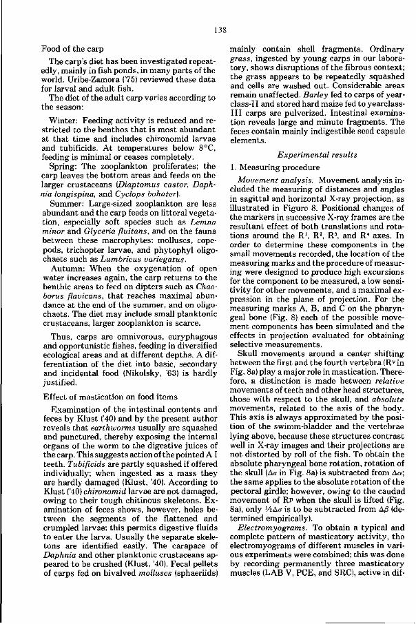
138
Food of the carp
The carp's diet has been investigated repeatedly, mainly in fish ponds, in many parts of the world. Uribe-Zamora ('75) reviewed these data for larval and adult fish.
The diet of the adult carp varies according to the season:
Winter: Feeding activity is reduced and restricted to the benthos that is most abundant at that time and includes chironomid larvae and tubificids. At temperatures below 8°C, feeding is minimal or ceases completely.
Spring: The zooplankton proliferates; the carp leaves the bottom areas and feeds on the larger crustaceans (Diaptomus castor, Daph-nia longispina, and Cyclops bohater).
Summer: Large-sized zooplankton are less abundant and the carp feeds on littoral vegetation, especially soft species such as Lemna minor and Glyceria fluitans, and on the fauna between these macrophytes: molluscs, cope-pods, trichopter larvae, and phytophyl oligo-chaets such as Lumbricus variegatus.
Autumn: When the oxygenation of open water increases again, the carp returns to the benthic areas to feed on dipters such as Chao-borus flavicans, t hat reaches maximal abundance at the end of the summer, and on oligo-chaets. The diet may include small planktonic crustaceans, larger zooplankton is scarce.
Thus, carps are omnivorous, euryphagous and opportunistic fishes, feeding in diversified ecological areas and at different depths. A differentiation of the diet into basic, secondary and incidental food (Nikolsky, '63) is hardly justified.
Effect of mastication on food items
Examination of the intestinal contents and feces by Klust ('40) and by the present author reveals that earthworms usually are squashed and punctured, thereby exposing the internal organs of the worm to the digestive juices of the carp. This suggests action of the pointed A I teeth. Tubificids are partly squashed if offered individually; when ingested as a mass they are hardly damaged (Klust, '40). According to Klust ('40) chironomid larvae are not damaged, owing to their tough chitinous skeletons. Examination of feces shows, however, holes between the segments of the flattened and crumpled larvae; this permits digestive fluids to enter the larva. Usually the separate skeletons are identified easily. The carapace of Daphnia and other planktonic crustaceans appeared to be crushed (Klust, '40). Fecal pellets of carps fed on bivalved molluscs (sphaeriids)
mainly contain shell fragments. Ordinary grass, ingested by young carps in our laboratory, shows disruptions of the fibrous context; the grass appears to be repeatedly squashed and cells are washed out. Considerable areas remain unaffected. Barley fed to carps of year-class-II and stored hard maize fed to yearclass-III carps are pulverized. Intestinal examination reveals large and minute fragments. The feces contain mainly indigestible seed capsule elements.
Experimental results
1. Measuring procedure
Movement analysis. Movement analysis included the measuring of distances and angles in sagittal and horizontal X-ray projection, as illustrated in Figure 8. Positional changes of the markers in successive X-ray frames are the resultant effect of both translations and rotations around the R1, R2, R3, and R4 axes. In order to determine these components in the small movements recorded, the location of the measuring marks and the procedure of measuring were designed to produce high excursions for the component to be measured, a low sensitivity for other movements, and a maximal expression in the plane of projection. For the measuring marks A, B, and C on the pharyngeal bone (Fig. 8) each of the possible movement components has been simulated and the effects in projection evaluated for obtaining selective measurements.
Skull movements around a center shifting between the first and the fourth vertebra (Rv in Fig. 8a) play a major role in mastication. Therefore, a distinction is made between relative movements of teeth and other head structures, those with respect to the skull, and absolute movements, related to the axis of the body. This axis is always approximated by the position of the swimm-bladder and the vertebrae lying above, because these structures contrast well in X-ray images and their projections are not distorted by roll of the fish. To obtain the absolute pharyngeal bone rotation, rotation of the skull (ACT in Fig. 8a) is subtracted from Aa; the same applies to the absolute rotation of the pectoral girdle; however, owing to the caudad movement of RP when the skull is lifted (Fig. 8a), only V2A0 is to be subtracted from A/3 (determined empirically).
Electromyograms. To obtain a typical and complete pattern of masticatory activity, the electromyograms of different muscles in various experiments were combined; this was done by recording permanently three masticatory muscles (LAB V, PCE, and SRC), active in dif-
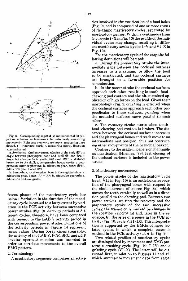
139
filmplane
Fig. 8. Corresponding sagittal (a) and horizontal (b) projection schemes as framework for selectively measuring movements. Reference elements are heavy; measuring lines dotted. ( - , reference mark; •, measuring mark). Rotation axes indicated.
a. Symbols:0. skull movement relative to the body (Rv); a, angle between pharyngeal bone and skull (Rl and R4); (3, angle between pectoral girdle and skull (RP); x, distance lower jaw to the skull; y, compression buccal cavity; z, compression anterior pharynx; h, adduction phar. bones (R2); 1, adduction phar. bones (R3).
b. Symbols: 7, rotation phar. bone in the sagittal plane; a, adduction phar. bones (R2 + R3): b, adduction opercula: c, adduction pectoral girdle.
ferent phases of the masticatory cycle (see below). Variation in the duration of the masticatory cycle is caused to a large extent by variation in the PCE activity between successive power strokes (Fig. 9). Activity periods of different cycles, therefore, have been compared with respect to the LAB V activity period in the corresponding power stroke. Durations of the activity periods in Figure 14 represent mean values. During X-ray cinematography, the activity of the LAB V, PCE, SRC, and LOP (levator operculi) muscles was recorded in order to correlate movements to the overall EMG pattern.
2. Terminology A masticatory sequence comprises all activi
ties involved in the mastication of a food bolus (Fig. 9), and is composed of one or more trains of rhythmic masticatory cycles, separated by masticatory pauses. Within a continuous train (e.g., cycle I -X in Fig. 10) the profile of the individual cycles may change, resulting in different masticatory series (cycles I-V and VI-X in Fig. 10).
For the masticatory cycle of the carp the following definitions will be used:
a. During the preparatory stroke the intermediate gape between the occlusal surfaces increases to a maximum to allow the food to be masticated, and the occlusal surfaces are brought in a favorable position for comminution.
b. In the power stroke the occlusal surfaces approach each other, resulting in tooth-food-chewing pad contact and the oft-sustained application of high forces on the food. Given their morphology (Fig. 2) crushing is effected when the occlusal surfaces approach each other perpendicular to these surfaces, grinding when the occluded surfaces move parallel to each other.
c. The recovery stroke s tarts when tooth-food-chewing pad contact is broken. The distance between the occlusal surfaces increases and the pharyngeal bones and teeth move to an intermediate rest position, thus not obstructing other movements of the branchial basket.
Contrary to the usage in papers on mammalian mastication (Hiiemae, 78), fast closing of the occlusal surfaces is included in the power stroke.
3. Masticatory movements
The power stroke of the masticatory cycle (cycle VII in Fig. 10) is an anticlockwise rotation of the pharyngeal bones with respect to the skull (increase of a; see Fig. 8a), which moves the teeth vertically as well as in a direction parallel to the chewing pad. Between two power strokes, we find the recovery and the preparatory stroke of the two successive cycles; the transition is marked by changes in the rotation velocity (a) and, later in the sequence, by the arise of a pause in the PCE activity (Fig. 10; cycle VII I - IX) . This interpretation is supported by the EMGs of more isolated cycles, in which a complete pause is noticed in the PCE activity (D, • in Fig. 9).
Two related profiles of masticatory cycles are distinguished by movement and EMG pattern; a crushing cycle (Fig. 10; I-IV) and a grinding cycle (VI-X). The latter will be discussed first, in relation to Figures 11 and 12, which summarize movement data from sagit-
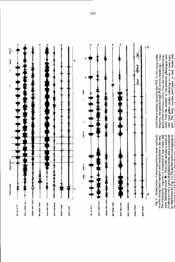
140
•
>
t - - » .
\ i r
' -r
f ir
!
r . 1 •
l •• 1
ir
«
n
" •
-3
i *
o'5 ° a - c co — O
8 S "Si cu _
G * .
.> J3 O _
°- s • CD ® * J
5 w ° S'a. »
CU T3 ^g
r»1*" • - .!
3 . S - a l
. 3 co --H •* CU - ^ dj !
O a 0)
"is
br>
CC
T3
CC
'G
3
J
*
u o > cB
Ë Sr g •2-S » CU . -C C o *->
- • s i = c — g • cd co
ftJ !
S u « -
> a o •! ,'S « o. s
1 O) ^ « £
! S a s •= : CD g - •
B..:
b ffi-S fc
eu C3 t*-C ü o
-° » TS _ CB ^
1 1 1
ni i o
cC -w w
- e b «
c f S 2 i 3 " _ C u -a eu C3 O co
'S 1 2 >>•
-S'g s . CO
CT> S ce • JH s
&0 o c
1 ^ - E V
S
>> 3
T I
> -o _B
a «
O t - t
•? M> U f v „
CO M- <
- 0 - Ü -5 e«-« O c _ H £ T M 3 .S
.=•"„!; - C CU c
? >>?
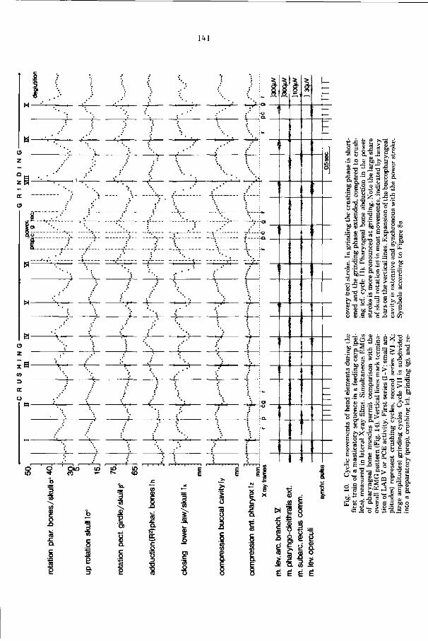
141
; s - — 7 er
_ j j y l
z
co
D
oc
ü
*
I t
'
1
•
i
•
i
•
••
.
•
i
'
^'
'
'
•
•
1
-' •
•
-
!
:
;
;
-
=
'i 8
—
o in
o •<* o
Î co CO CU
(5 a c o '̂ CO
e
OIO CO
lO
3 CO
1 I 2 a 3
SP
CO
0) 'S Ol
I c g
to ?
m CO
co
5 CO
a çc o •6 3 TJ
T8
3
CO
Î I o> c CO o o
W S
V 'S ó s ra c
& Si
CO CO
0) m
«
CJ
O 4 - i
• a ÛJ
ai 5b >> b L
" ÎS -a -§, S
| Z '
s -g M .5 i"S *
CD
1 s . .s ^
' & a C CD O
S -P ©
£ Q ,_C cy
RS _ ^ E
3 o .£ c o
EbS
5 3 o
P a g "
3 s « n P SP
o ca
>-> ca i
CD - o " •ï Ji » .S c ) £ M h > s.
ai ,
• " a c ° a
•Ë S3 3 U "° M m B C ^ E ai
es CD
» c • O — PS CD CD U
-C C u- « to a i
co ai
' S " ,w 5 5 * 3 £
3 l l >
I S> « • E :
ä S » i = - o 3 Q, ai œ s S P c .S o
r/i o _ ai ai w u " 3 œ tn
a i
-13 M
™ - ^ — CD 3 bC
' C OT — CD to bß f " —• C
s '•&
^^ o -
. bc
CD >>
s s ai ü
o o
e | . >> «
o
2-1 CL, to
£ •"3 '-E m m " _ E " •= S
i_ ai ^r . « bc
2 EÔH b-g s, 2 g gÉtU fer
C —
^ 01
0> E 2 r> -^ -̂ aï ' " - " M O »
J _ D . ^ 01 ^
Ü ^ ? O s ; ü O 0 3 D . J
Ê É Ë É
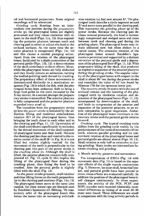
142
tal and horizontal projections. Some original recordings will be referenced.
Grinding cycle. Starting from an intermediate rest position during the preparatory stroke (p), the pharyngeal bones are slightly protracted and they rotate clockwise with respect to the skull (Figs. 11a, 12), thus expanding the posterior pharynx between teeth and chewing pad to admit or adjust the food to the occlusal surfaces. At the same time the anterior pharynx is compressed (Figs, l i e , 12) and this causes a caudad pumping action. Apart from the rotation of the pharyngeal bones, facilitated by a slight protraction of the pectoral girdle (Figs, l i b , 12), a down-rotation of the skull contributes to these effects. Meanwhile the pharyngeal bones are fully abducted and they finally initiate an adduction, turning the mediad-pointing teeth dorsad for crushing. The preparatory effect of these movements is demonstrated distinctly in a masticatory sequence (Fig. 13) in which the fish, with the pharyngeal bones kept abducted, fails to bring a large food pellet on the teeth (revealed by the X-ray movie). At a second at tempt the preparatory stroke is intensified: The anterior pharynx is fully compressed and the posterior pharynx expanded (very small a).
The transition from the preparatory stroke (p) into the power stroke is initiated by the adduction, lifting, and moderate anticlockwise rotation (R1) of the pharyngeal bones, thus bringing the teeth closer to each other and to the chewing pad (Figs. 11, 12). Up-rotation of the skull contributes significantly to occlusion by the dorsad movement of the skull-suspended pharyngeal bones and their teeth. Because the chewing pad lies close and ventral to the rotation center of the skull (Rv in Fig. 8), it is not lifted but rotates slightly rostrad. As the movement of the teeth is perpendicular to the chewing pad, this part of the power stroke is the crushing phase (c). Although the skull is lifted, the anterior pharynx remains fully compressed (cf. Fig. 13, cycle I); this implies the lifting of the pharyngeal floor during the crushing phase, thus fixing the food to be crushed. Also the pectoral girdle is slightly lifted with the skull (Fig. 12).
As the power stroke proceeds, skull-rotation and thus lifting forces on the teeth perpendicular to the chewing pad continue (Figs. 11a, 12). The pharyngeal bones move at the same time caudad, but their dorsal tips are blocked soon by Baudelot's ligaments (cf. Sibbing, '76; masticatory orbit of the pharyngeal bone); this forces the bones into an increasing anticlock
wise rotation (a), but now around R4. The pharyngeal teeth describe a circle segment around R4 and move more parallel to the chewing pad. This represents the grinding phase (g) of the power stroke. Because the chewing pad declines ventrad posteriorly, the food is increasingly compressed and wedged more and more between teeth and chewing pad during the grinding phase. The pharyngeal bones may remain adducted now, but often abduct to a varied extent. The extensive rotation of the pharyngeal bones around Baudelot's ligaments is permitted and supported by a distinct retraction of the pectoral girdle and a depression of the pharyngeal floor (Figs. 11,12). With progressive lifting of the skull, this produces an explosive expansion of the anterior pharynx during the grinding stroke. The angular velocity of the pharyngeal bones with respect to the skull is about 60°/sec during the power stroke and resembles the value of 64°/sec found by Hiiemae (76) for the opossum lower jaw.
The recovery stroke (r) s tarts with the loss of occlusal contact and the lowering of the pharyngeal teeth by clockwise rotation and depression of the pharyngeal bones (Figs. 11, 12), accompanied by down-rotation of the skull, and leads to compression of the anterior and expansion of the posterior pharynx, to an extent comparable to quiet respiration. Abduction of the pharyngeal bones continues in the recovery stroke and the pectoral girdle returns forward.
Crushing cycle. The typical crushing cycle differs from the grinding cycle mainly by the predominance of the vertical movements of the teeth, whereas parallel grinding and its concomitant rotation of the pharyngeal bones and pectoral girdle are almost absent (Figs. 10, 11). Skull rotation is only half as extensive as during grinding. Many cycles are intermediate between crushing and grinding.
4. Muscle activity patterns and movements
The interpretation of EMGs (Fig. 14) with movement data (Fig. 11) is based on the separate effects of the single muscle contractions (Fig. 7). However, because skull, branchial basket, and pectoral girdle have been proved to move, these effects are evaluated carefully. In addition the position of the pharyngeal bone itself changes continuously. In three experiments, the activity of the PCE, PCIP, and ROPI muscles were recorded bilaterally; occasional differences in timing of at most 20-30 msec were found. These differences are small in comparison with the long activity periods in
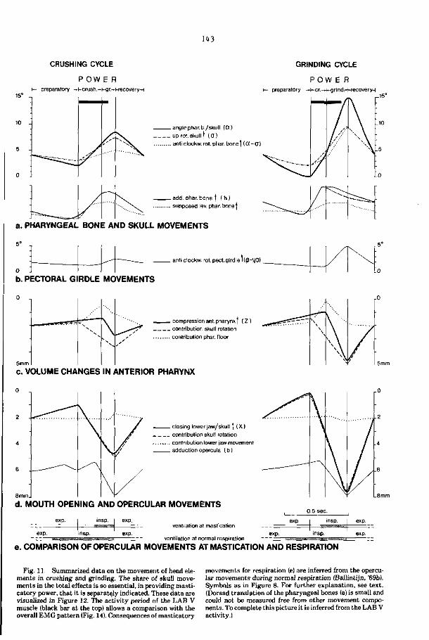
143
CRUSHING CYCLE GRINDING CYCLE
preparatory
P O W E R -kcrush.-kg r.-i-recoveryn preparatory
P O W E R -t-cr.^-grindr-HecoveryH
. anglephar.b./skult (CO
. up rot.skullt (O) . anti clockw. rot. phar. bone| (Oi-O)
. add. phar.bone ( ( h )
. supposed lev. phar. bone |
a. PHARYNGEAL BONE AND SKULL MOVEMENTS
5° -,
_^~ -___ anti clockw. rot. pect.girdle I iß-'iO)
b. PECTORAL GIRDLE MOVEMENTS
- compression ant.pharynx | ( Z ) _ contribution skull rotation . contribution phar. floor
c. VOLUME CHANGES IN ANTERIOR PHARYNX
. closing lower jaw/skull | (X)
. contribution skull rotation
. contribution lower jaw movement
. adduction opercula ( b )
d. MOUTH OPENING AND OPERCULAR MOVEMENTS
ventilation at mastication
ventilation at normal respiration
exp
0 5 sec.
exp.
insp.
insp.
J exp.
exp.
e. COMPARISON OF OPERCULAR MOVEMENTS AT MASTICATION AND RESPIRATION
Fig. 11. Summarized data on the movement of head elements in crushing and grinding. The share of skull movements in the total effects is so essential, in providing masticatory power, that it is separately indicated. These data are visualized in Figure 12. The activity period of the LAB V muscle (black bar at the top) allows a comparison with the overall EMG pattern (Fig. 14). Consequences of masticatory
movements for respiration (e) are inferred from the opercular movements during normal respiration (Ballintijn, '69b). Symbols as in Figure 8. For further explanation, see text. (Dorsad translation of the pharyngeal bones (a) is small and could not be measured free from other movement components. To complete this picture it is inferred from the LAB V activity.)
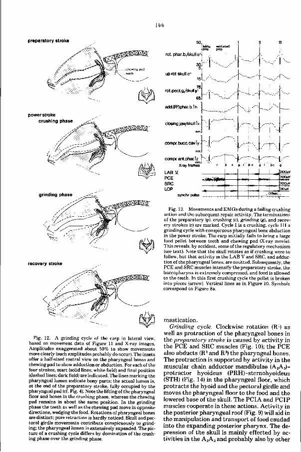
144
preparatory stroke
power stroke crushing phase
grinding phase
recovery stroke
Fig. 12. A grinding cycle of the carp in lateral view, based on movement data of Figure 11 and X-ray images. Amplitudes exaggerated about 50% to show movements more clearly (such amplitudes probably do occur). The insets offer a half-sized rostral view on the pharyngeal bones and chewing pad to show adduction or abduction. For each of the four strokes, s tar t (solid lines; white field) and final position (dashed lines; dark field) are indicated. The lines marking the pharyngeal lumen indicate bony parts; the actual lumen is, at the end of the preparatory stroke, fully occupied by the pharyngeal pad (cf. Fig. 4). Note the lifting of the pharyngeal floor and bones in the crushing phase, whereas the chewing pad remains in about the same position. In the grinding phase the teeth as well as the chewing pad move in opposite directions, wedging the food. Rotations of pharyngeal bones are distinct; pure retraction is hardly noticed. Skull and pectoral girdle movements contribute conspicuously to grinding; the pharyngeal lumen is extensively expanded. The picture of a crushing cycle differs by domination of the crushing phase over the grinding phase.
50 I failing reinforced
•prep prep
rot. phar.tys.kulla°-
up rot. skull o° 15J..,
rot. pect.g/skull p°
6!
add.(R2)phar.b.Ih
closing jaw/skull îx -
compr.bucccav.ly
compnant.phar.tz mm.
X ray frames L A B Ï PCE SRC LOP
r p c g r pc g
—\—J— - J 3 0 0 * r p c g r p c g r p c g
!300uV
synchr. pulse
]1O0uV
Fig. 13. Movements and EMGs during a failing crushing action and the subsequent repair activity. The terminations of the preparatory (p), crushing (ch grinding (g), and recovery strokes (r) are marked. Cycle I is a crushing, cycle III a grinding cycle with conspicuous pharyngeal bone abduction in the power stroke. The carp initially fails to bring a large food pellet between teeth and chewing pad (X-ray movie). This reveals, by accident, some of the regulatory mechanism (see text). Note that the skull rotates as if crushing were to follow, but that activity in the LAB V and SRC, and adduction of the pharyngeal bones, are omitted. Subsequently, the PCE and SRC muscles intensify the preparatory stroke, the buccopharynx is extremely compressed, and food is allowed to the teeth. In this first crushing cycle the pellet is broken into pieces (arrow). Vertical lines as in Figure 10. Symbols correspond to Figure 8a.
mastication. Grinding cycle. Clockwise rotation (R1) as
well as protraction of the pharyngeal bones in the preparatory stroke is caused by activity in the PCE and SRC muscles (Fig, 10); the PCE also abducts (R2 and R3) the pharyngeal bones. The protraction is supported by activity in the muscular chain adductor mandibulae (A2A3)-protractor hyoideus (PRH)-sternohyoideus (STH) (Fig. 14) in the pharyngeal floor, which protracts the hyoid and the pectoral girdle and moves the pharyngeal floor to the food and the lowered base of the skull. The PCI A and PCIP muscles cooperate in these actions. Activity in the posterior pharyngeal roof (Fig. 9) will aid in the manipulation and transport of food caudad into the expanding posterior pharynx. The depression of the skull is mainly effected by activities in the A2A3 and probably also by other
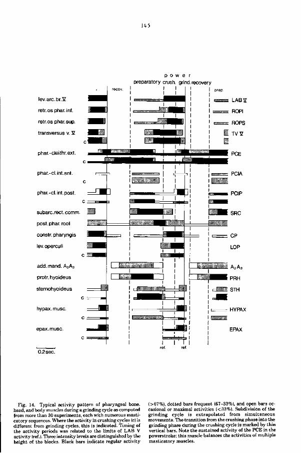
U 5
p o w e r preparatory crush, grind, recovery
lev. arc. br.îZ
retr. os phar. inf.
retr.os phar. sup.
transversus v. 2
phar.-cleithr.ext.
phar.-cl.int.ant.
phar.-cl.int.post.
subarc.rect.comm.
post. phar. roof
constr.pharyngis
lev.operculi
add.mand. A2A3
protr. hyoideus
sternohyoideus
hypax. muse.
epax.musc.
0.2 sec.
HYPAX
EPAX
Fig. 14. Typical activity pattern of pharyngeal bone, head, and body muscles during a grinding cycle as computed from more than 30 experiments, each with numerous masticatory sequences. Where the activity in crushing cycles (c) is different from grinding cycles, this is indicated. Timing of the activity periods was related to the limits of LAB V activity (réf.). Three intensity levels are distinguished by the height of the blocks. Black bars indicate regular activity
(>67%), dotted bars frequent (67-33%), and open bars occasional or maximal activities (<33%). Subdivision of the grinding cycle is extrapolated from simultaneous movements. The transition from the crushing phase into the grinding phase during the crushing cycle is marked by thin vertical bars. Note the sustained activity of the PCE in the powerstroke; this muscle balances the activities of multiple masticatory muscles.
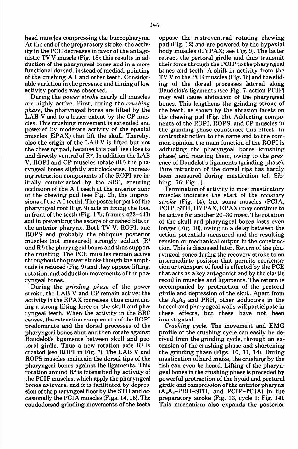
146
head muscles compressing the buccopharynx. At the end of the preparatory stroke, the activity in the PCE decreases in favor of the antagonistic TV V muscle (Fig. 18); this results in adduction of the pharyngeal bones and in a more functional dorsad, instead of mediad, pointing of the crushing A I and other teeth. Considerable variation in the presence and timing of low activity periods was observed.
During the power stroke nearly all muscles are highly active. First, during the crushing phase, the pharyngeal bones are lifted by the LAB V and to a lesser extent by the CP muscles. This crushing movement is extended and powered by moderate activity of the epaxial muscles (EPAX) that lift the skull. Thereby, also the origin of the LAB V is lifted but not the chewing pad, because this pad lies close to and directly ventral of Rv. In addition the LAB V, ROPI and CP muscles rotate (R1) the pharyngeal bones slightly anticlockwise. Increasing retraction components of the ROPI are initially counteracted by the SRC, ensuring occlusion of the A I teeth at the anterior zone of the chewing pad (see Fig. 2b, the impressions of the A I teeth). The posterior part of the pharyngeal roof (Fig. 9) acts in fixing the food in front of the teeth (Fig. 17b; frames 422-441) and in preventing the escape of crushed bits to the anterior pharynx. Both TV V, ROPI, and ROPS and probably the obliquus posterior muscles (not measured) strongly adduct (R2
and R3) the pharyngeal bones and thus support the crushing. The PCE muscles remain active throughout the power stroke though the amplitude is reduced (Fig. 9) and they oppose lifting, rotation, and adduction movements of the pharyngeal bones.
During the grinding phase of the power stroke, the LAB V and CP remain active; the activity in the EPAX increases, thus maintaining a strong lifting force on the skull and pharyngeal teeth. When the activity in the SRC ceases, the retraction components of the ROPI predominate and the dorsal processes of the pharyngeal bones abut and then rotate against Baudelot's ligaments between skull and pectoral girdle. Thus a new rotation axis R4 is created (see ROPI in Fig. 7). The LAB V and ROPS muscles maintain the dorsal tips of the pharyngeal bones against the ligaments. This rotation around R4 is intensified by activity of the PCIP muscles, which apply the pharyngeal bones as levers, and it is facilitated by depression of the pharyngeal floor by the STH and occasionally the PCIA muscles (Figs. 14,15). The caudodorsad grinding movements of the teeth
oppose the rostroventrad rotating chewing pad (Fig. 12) and are powered by the hypaxial body muscles (HYPAX; see Fig. 9). The latter retract the pectoral girdle and thus transmit their force through the PCIP to the pharyngeal bones and teeth. A shift in activity from the TV V to the PCE muscles (Fig. 18) and the sliding of the dorsal processes laterad along Baudelot's ligaments (see Fig. 7, action PCIP) may well cause abduction of the pharyngeal bones. This lengthens the grinding stroke of the teeth, as shown by the abrasion facets on the chewing pad (Fig. 2b). Adducting components of the ROPI, ROPS, and CP muscles in the grinding phase counteract this effect. In contradistinction to the name and to the common opinion, the main function of the ROPI is adducting the pharyngeal bones (crushing phase) and rotating them, owing to the presence of Baudelot's ligaments (grinding phase). Pure retraction of the dorsal tips has hardly been measured during mastication (cf. Sib-bing, '76: Fig. 1).
Termination of activity in most masticatory muscles indicates the s tart of the recovery stroke (Fig. 14), but some muscles (PCIA, PCIP, STH, HYPAX, EPAX) may continue to be active for another 20-30 msec. The rotation of the skull and pharyngeal bones lasts even longer (Fig. 10), owing to a delay between the action potentials measured and the resulting tension or mechanical output in the construction. This is discussed later. Return of the pharyngeal bones during the recovery stroke to an intermediate position that permits reorientation or transport of food is effected by the PCE that acts as a key antagonist and by the elastic recoil in muscles and ligaments. The return is accompanied by protraction of the pectoral girdle and depression of the skull. Apart from the A2A3 and PRH, other adductors in the buccal and pharyngeal walls will participate in these effects, but these have not been investigated.
Crushing cycle. The movement and EMG profile of the crushing cycle can easily be derived from the grinding cycle, through an extension of the crushing phase and shortening the grinding phase (Figs. 10, 11, 14). During mastication of hard maize, the crushing by the fish can even be heard. Lifting of the pharyngeal bones in the crushing phase is preceded by powerful protraction of the hyoid and pectoral girdle and compression of the anterior pharynx (A2A3-PRH-STH, and PCIP-PCIA) in the preparatory stroke (Fig. 13, cycle I; Fig. 14). This mechanism also expands the posterior
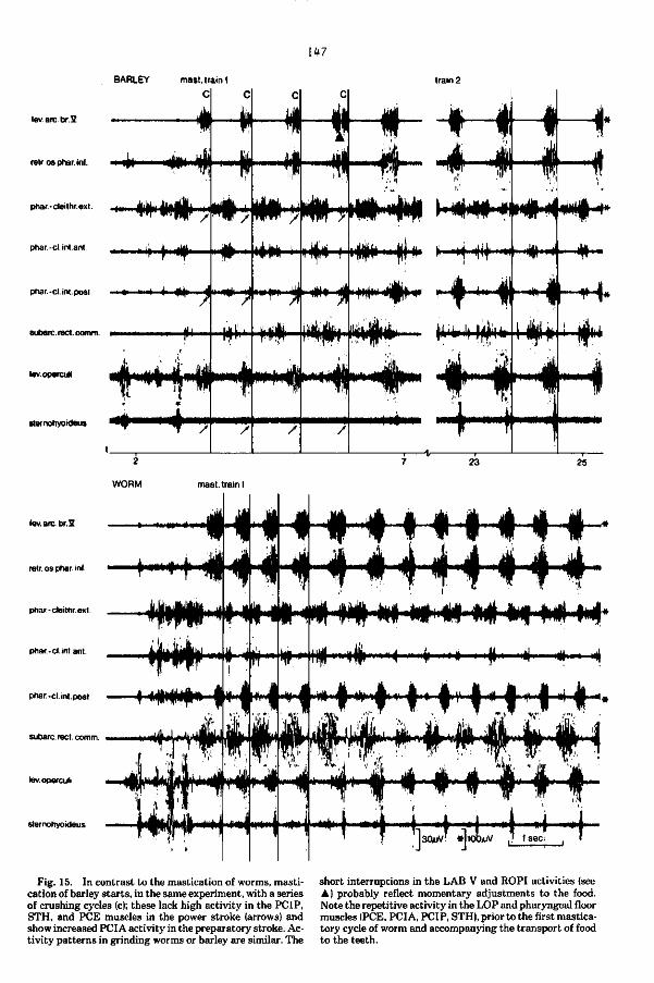
w BARLEY train 2
lev.arc.br.lZ
retr.osphar. - in » 4|—1»—jf—if
phar.-cleithr.ext.
phar.-cl.int.ant.
phar.-cl int post. « i il j Haf
subarc. reet. comm.
lev.operculi
sternohyoideus
retr. os phar inf.
phar.-cleithr.ext.
phar.-cl.int.ant.
phar-cl. int. post
subarc. reet. comm.
lev.operculi
sternohyoideus
Fig. 15. In contrast to the mastication of worms, mastication of barley starts, in the same experiment, with a series of crushing cycles (c); these lack high activity in the PCIP, STH, and PCE muscles in the power stroke (arrows) and show increased PCIA activity in the preparatory stroke. Activity patterns in grinding worms or barley are similar. The
short interruptions in the LAB V and ROPI activities (see • ) probably reflect momentary adjustments to the food. Note the repetitive activity in the LOP and pharyngeal floor muscles (PCE, PCIA, PCIP, STH), prior to the first masticatory cycle of worm and accompanying the transport of food to the teeth.
U8
pharynx widely and thus allows the passage of large particles between teeth and chewing pad. Adduction of the pharyngeal bones (R2 and R3) is as extensive as during grinding, but abduction scarcely occurs in the power stroke; this explains the sustained TV V activity and the lack of high activity in the PCE and PCIP in the power stroke (Figs. 14, 15). During the grinding phase of the crushing cycle, rotation of the pharyngeal bones around Baudelot's ligaments (R4) hardly occurs; this agrees with the absence of high PCIP activity. In addition, the retraction of the pectoral girdle (HYPAX), depression of the pharyngeal floor (STH and PCI A), lifting of the skull (EPAX), and consequently expansion of the anterior pharynx are only small in the crushing cycle (Fig. 11).
During the preparatory stroke of the crushing cycle, the STH and PCIP muscles can act as protractors of the pectoral girdle, because the jaws are adducted and the hyoid arch and pharyngeal bones are protracted. In the power stroke of the grinding cycle, these muscles act as retractors of the hyoids and depressors of the pharyngeal floor, as the pectoral girdle is retracted.
A time lag occurs between the end of electrical activity and the peak of mechanical events (e.g., pharyngeal bone/skull rotation in Figs. 10, 11). Miller ('66), and Ahlgren and Öwall (70) reported 100 msec for such a lag during mastication in man. Desmedt ('58) observed that muscular tonus lasts 150 msec longer than electrical activity. Inman et al. ('52) found a value of 80 ± 20 msec, and Hannam et al. (75) 73 ± 12 msec for the delay in man between the peak of electrical activity and the peak of muscular tension. The value measured for mastication in the carp, a time lag of 40-80 msec, fits these data. Because masticatory cycles commonly last between 600 and 900 msec, this delay does not affect the interpretation of the EMGs by more than other factors, such as elastic recoil.
5. EMG variations in the course of mastication
As the particle size and consistency of the food change continuously during mastication, adjustments in the processing of food are expected.
Crushing precedes grinding in a masticatory sequence (Figs. 9, 10, 13, 15) as well as within the individual cycle. Many cycles with an intermediate profile occur. Crushing is far less frequent and even absent during mastication of soft food (Fig. 15), whereas grinding is common.
During the early part of a masticatory train, much power is needed. Masticatory cycles are short and muscles are highly active (Fig. 9). Muscles that contract in the preparatory stroke may already be active in the recovery stroke. In the course of the masticatory sequence the activity diminishes to an intermediate level (shown in Fig. 14) characterized by continuous activity in the PCE only. Finally the activity of the PCE between successive power strokes breaks up and a masticatory pause is interposed (Fig. 9). Overall reduction of the activity level is seen in this final part of the masticatory sequence, accompanied by the more frequent occurrence of isolated cycles and masticatory pauses. Only periods of activity with high amplitudes in the early phase of mastication are still observed. This probably reflects the diminished force in the individual cycle and the smaller velocities of movement, as the desired particle size is attained.
6. The influence of the type of food on mastication
Feeding various foods in the same experiment produced different EMGs (Fig. 15), and it further justified the distinction of crushing and grinding cycles. Mastication of soft worms requires only grinding cycles with activity of the PCIP, PCIA, STH, and HYPAX muscles in the power stroke. If hard and compact barley is fed, grinding cycles are preceded by crushing cycles, without the activity of these specific muscles. The grinding cycles after feeding of worms and barley are similar and correspond to the typical pattern. Fewer cycles are observed in mastication of soft worms, compared with hard barley, which may cause mastication to last for minutes. No detailed knowledge exists about the sensory system t h a t d e t e c t s t he differences in food consistency.
7. Mastication and respiration
Mastication incorporates the respiratory movements. This is concluded from a comparison of the opening of the mouth, the volume changes in the buccopharyngeal cavities, the abduction of the opercula, and the activities in the LOP and pharyngeal floor muscles during mastication and normal respiration (Figs. 11, 16). Expiration coincides with the recovery and preparatory strokes and even continues in the crushing phase of the power stroke; this is inferred from the pharyngeal compression and opercular adduction (Fig. l lc,d,e) (Ballintijn, '69a). Inspiration coincides with the remainder of the power stroke. The duration of the masti-
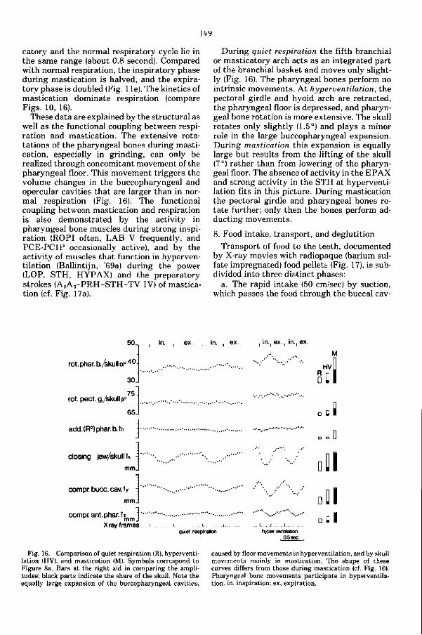
149
catory and the normal respiratory cycle lie in the same range (about 0.8 second). Compared with normal respiration, the inspiratory phase during mastication is halved, and the expiratory phase is doubled (Fig. 1 le). The kinetics of mastication dominate respiration (compare Figs. 10, 16).
These data are explained by the structural as well as the functional coupling between respiration and mastication. The extensive rota-tations of the pharyngeal bones during mastication, especially in grinding, can only be realized through concomitant movement of the pharyngeal floor. This movement triggers the volume changes in the buccopharyngeal and opercular cavities that are larger than in normal respiration (Fig. 16). The functional coupling between mastication and respiration is also demonstrated by the activity in pharyngeal bone muscles during strong inspiration (ROPI often, LAB V frequently, and PCE-PCIP occasionally active), and by the activity of muscles that function in hyperventilation (Ballintijn, '69a) during the power (LOP, STH, HYPAX) and the preparatory strokes (A 2A 3 -PRH-STH-TV IV) of mastication (cf. Fig. 17a).
During quiet respiration the fifth branchial or masticatory arch acts as an integrated part of the branchial basket and moves only slightly (Fig. 16). The pharyngeal bones perform no intrinsic movements. At hyperventilation, the pectoral girdle and hyoid arch are retracted, the pharyngeal floor is depressed, and pharyngeal bone rotation is more extensive. The skull rotates only slightly (1.5°) and plays a minor role in the large buccopharyngeal expansion. During mastication this expansion is equally large but results from the lifting of the skull (7°) rather than from lowering of the pharyngeal floor. The absence of activity in the E PAX and strong activity in the STH at hyperventilation fits in this picture. During mastication the pectoral girdle and pharyngeal bones rotate further; only then the bones perform ad-ducting movements.
8. Food intake, transport, and deglutition
Transport of food to the teeth, documented by X-ray movies with radiopaque (barium sulfate impregnated) food pellets (Fig. 17), is subdivided into three distinct phases:
a. The rapid intake (50 cm/sec) by suction, which passes the food through the buccal cav-
50.
rot. phar. b./skull a ° 4 0
rot. pect, g./skull p'
30
75.
65.
add. (R2) phar. b.th
closing jaw/skull tx -i
compr. bucc. cav. ty -
mm.
compr. ant. phar. tz mm
X ray frames
in. . ex. , in. , ex. in., ex.. in., ex.
HV
So
. I quiet respiration hyperventilation
, 0.5 sec.
Fig. 16. Comparison of quiet respiration (R), hyperventilation (HV), and mastication (M). Symbols correspond to Figure 8a. Bars at the right aid in comparing the amplitudes; black parts indicate the share of the skull. Note the equally large expansion of the buccopharyngeal cavities,
caused by floor movements in hyperventilation, and by skull movements mainly in mastication. The shape of these curves differs from those during mastication (cf. Fig. 10). Pharyngeal bone movements participate in hyperventilation, in, inspiration; ex, expiration.
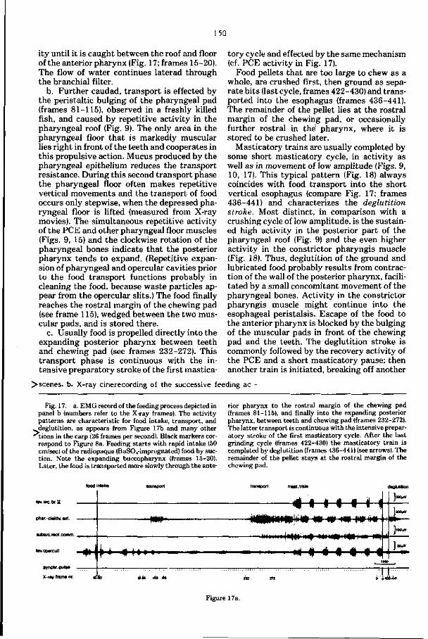
150
ity until it is caught between the roof and floor of the anterior pharynx (Fig. 17; frames 15-20). The flow of water continues laterad through the branchial filter.
b. Further caudad, t ransport is effected by the peristaltic bulging of the pharyngeal pad (frames 81-115), observed in a freshly killed fish, and caused by repetitive activity in the pharyngeal roof (Fig. 9). The only area in the pharyngeal floor that is markedly muscular lies right in front of the teeth and cooperates in this propulsive action. Mucus produced by the pharyngeal epithelium reduces the transport resistance. During this second transport phase the pharyngeal floor often makes repetitive vertical movements and the transport of food occurs only stepwise, when the depressed pharyngeal floor is lifted (measured from X-ray movies). The simultaneous repetitive activity of the PCE and other pharyngeal floor muscles (Figs. 9, 15) and the clockwise rotation of the pharyngeal bones indicate that the posterior pharynx tends to expand. (Repetitive expansion of pharyngeal and opercular cavities prior to the food transport functions probably in cleaning the food, because waste particles appear from the opercular slits.) The food finally reaches the rostral margin of the chewing pad (see frame 115), wedged between the two muscular pads, and is stored there.
c. Usually food is propelled directly into the expanding posterior pharynx between teeth and chewing pad (see frames 232-272). This transport phase is continuous with the intensive preparatory stroke of the first mastica
tory cycle and effected by the same mechanism (cf. PCE activity in Fig. 17).
Food pellets that are too large to chew as a whole, are crushed first, then ground as separate bits (last cycle, frames 422-430) and transported into the esophagus (frames 436-441). The remainder of the pellet lies at the rostral margin of the chewing pad, or occasionally further rostral in the pharynx, where it is stored to be crushed later.
Masticatory trains are usually completed by some short masticatory cycle, in activity as well as in movement of low amplitude (Figs. 9, 10, 17). This typical pattern (Fig. 18) always coincides with food transport into the short vertical esophagus (compare Fig. 17; frames 436-441) and characterizes the deglutition stroke. Most distinct, in comparison with a crushing cycle of low amplitude, is the sustained high activity in the posterior part of the pharyngeal roof (Fig. 9) and the even higher activity in the constrictor pharyngis muscle (Fig. 18). Thus, deglutition of the ground and lubricated food probably results from contraction of the wall of the posterior pharynx, facilitated by a small concomitant movement of the pharyngeal bones. Activity in the constrictor pharyngis muscle might continue into the esophageal peristalsis. Escape of the food to the anterior pharynx is blocked by the bulging of the muscular pads in front of the chewing pad and the teeth. The deglutition stroke is commonly followed by the recovery activity of the PCE and a short masticatory pause; then another train is initiated, breaking off another
> scenes, b. X-ray cinerecording of the successive feeding ac
Pig. 17. a. EMG record of the feeding process depicted in panel b (numbers refer to the X-ray frames). The activity pat terns are characteristic for food intake, transport, and
. j ieglutition, as appears from Figure 17b and many other MJons in the carp (26 frames per second). Black markers cor
respond to Figure 8a. Feeding s tar ts with rapid intake (50 cm/sec) of the radiopaque (BaSO(-impregnated) food by suction. Note the expanding buccopharynx (frames 15-20). Later, the food is transported more slowly through the ante
rior pharynx to the rostral margin of the chewing pad (frames 81-115), and finally into the expanding posterior pharynx, between teeth and chewing pad (frames 232-272). The latter t ransport is continuous with the intensive preparatory stroke of the first masticatory cycle. After the last grinding cycle (frames 422-430) the masticatory train is completed by deglutition (frames 436-441) (see arrows). The remainder of the pellet s tays at the rostral margin of the chewing pad.
Iev.arc.br.2
phar.-cleithr.ext.
subaic.rect.comm. ,
tevoperculi
synchr. pulse
X.ray frame nr.
M¥
HWk»*
deglutition
jsCKW
i«»i|l»)it>iir>) M<I»»I<<W » m
* • « > » » » ' x M t M «
Figure 17 a.
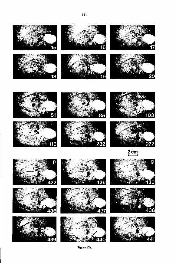
151
2 cm
Figure 17b.
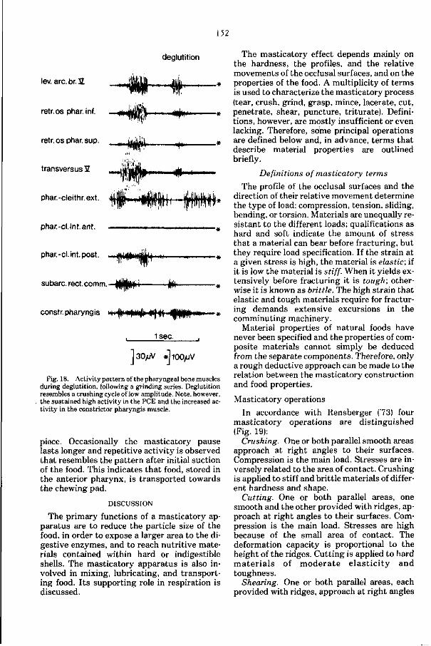
152
deglutition
-Hflfr
lev. arc.br. 2
retr.os phar. inf.
retr.osphar.sup.
transversus 3Z
phar.-cleithr.ext.
phar.-cl.int.ant.
phar.-cl. int.post.
subarc. reet. comm. ijÉllfi 1
+-H.
constr.pharyngis m^ imfM^»)^ Hpffni» »
1sec.
]30yuV »llOOyuV
Fig. 18. Activity pattern of the pharyngeal bone muscles during deglutition, following a grinding series. Deglutition resembles a crushing cycle of low amplitude. Note, however, the sustained high activity in the PCE and the increased activity in the constrictor pharyngis muscle.
piece. Occasionally the masticatory pause lasts longer and repetitive activity is observed that resembles the pattern after initial suction of the food. This indicates that food, stored in the anterior pharynx, is transported towards the chewing pad.
DISCUSSION
The primary functions of a masticatory apparatus are to reduce the particle size of the food, in order to expose a larger area to the digestive enzymes, and to reach nutritive materials contained within hard or indigestible shells. The masticatory apparatus is also involved in mixing, lubricating, and transporting food. I ts supporting role in respiration is discussed.
The masticatory effect depends mainly on the hardness, the profiles, and the relative movements of the occlusal surfaces, and on the properties of the food. A multiplicity of terms is used to characterize the masticatory process (tear, crush, grind, grasp, mince, lacerate, cut, penetrate, shear, puncture, triturate). Definitions, however, are mostly insufficient or even lacking. Therefore, some principal operations are defined below and, in advance, terms that describe material properties are outlined briefly.
Definitions of masticatory terms
The profile of the occlusal surfaces and the direction of their relative movement determine the type of load: compression, tension, sliding, bending, or torsion. Materials are unequally resistant to the different loads; qualifications as hard and soft indicate the amount of stress that a material can bear before fracturing, but they require load specification. If the strain at a given stress is high, the material is elastic; if it is low the material is stiff. When it yields extensively before fracturing it is tough; otherwise it is known as brittle. The high strain that elastic and tough materials require for fracturing demands extensive excursions in the comminuting machinery.
Material properties of natural foods have never been specified and the properties of composite materials cannot simply be deduced from the separate components. Therefore, only a rough deductive approach can be made to the relation between the masticatory construction and food properties.
Masticatory operations
In accordance with Rensberger ('73) four masticatory operations are distinguished (Fig. 19):
Crushing. One or both parallel smooth areas approach at right angles to their surfaces. Compression is the main load. Stresses are inversely related to the area of contact. Crushing is applied to stiff and brittle materials of different hardness and shape.
Cutting. One or both parallel areas, one smooth and the other provided with ridges, approach at right angles to their surfaces. Compression is the main load. Stresses are high because of the small area of contact. The deformation capacity is proportional to the height of the ridges. Cutting is applied to hard ma te r i a l s of mode ra t e e l a s t i c i ty and toughness.
Shearing. One or both parallel areas, each provided with ridges, approach at right angles
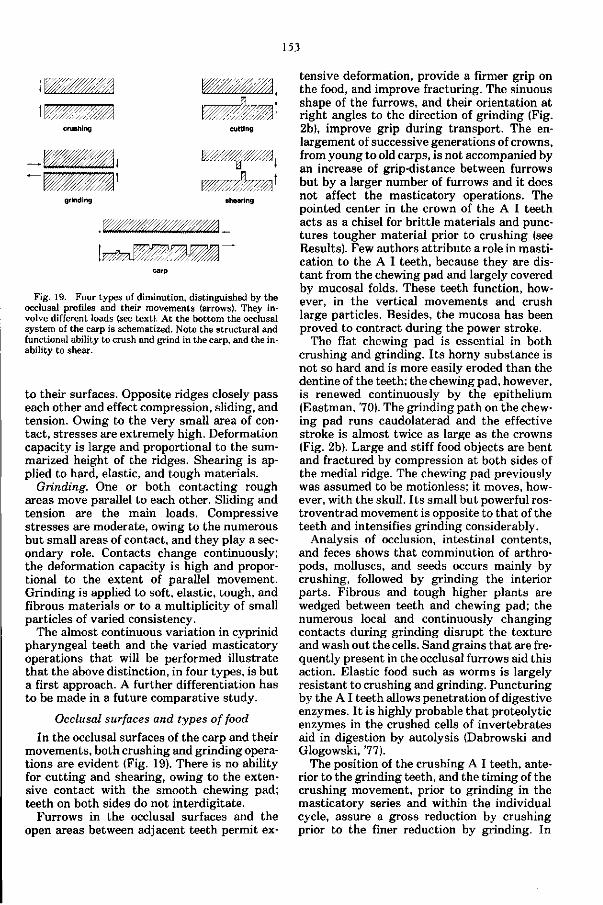
153
\WMmm
crushing
mémÊ-cutting
•WL l W/////////W/M,
-mm grinding shearing
Fig. 19. Four types of diminution, distinguished by the occlusal profiles and their movements (arrows). They involve different loads (see text). At the bottom the occlusal system of the carp is schematized. Note the structural and functional ability to crush and grind in the carp, and the inability to shear.
to their surfaces. Opposite ridges closely pass each other and effect compression, sliding, and tension. Owing to the very small area of contact, stresses are extremely high. Deformation capacity is large and proportional to the summarized height of the ridges. Shearing is applied to hard, elastic, and tough materials.
Grinding. One or both contacting rough areas move parallel to each other. Sliding and tension are the main loads. Compressive stresses are moderate, owing to the numerous but small areas of contact, and they play a secondary role. Contacts change continuously; the deformation capacity is high and proportional to the extent of parallel movement. Grinding is applied to soft, elastic, tough, and fibrous materials or to a multiplicity of small particles of varied consistency.
The almost continuous variation in cyprinid pharyngeal teeth and the varied masticatory operations that will be performed illustrate that the above distinction, in four types, is but a first approach. A further differentiation has to be made in a future comparative study.
Occlusal surfaces and types of food
In the occlusal surfaces of the carp and their movements, both crushing and grinding operations are evident (Fig. 19). There is no ability for cutting and shearing, owing to the extensive contact with the smooth chewing pad; teeth on both sides do not interdigitate.
Furrows in the occlusal surfaces and the open areas between adjacent teeth permit ex
tensive deformation, provide a firmer grip on the food, and improve fracturing. The sinuous shape of the furrows, and their orientation at right angles to the direction of grinding (Fig. 2b), improve grip during transport. The enlargement of successive generations of crowns, from young to old carps, is not accompanied by an increase of grip-distance between furrows but by a larger number of furrows and it does not affect the masticatory operations. The pointed center in the crown of the A I teeth acts as a chisel for brittle materials and punctures tougher material prior to crushing (see Results). Few authors attribute a role in mastication to the A I teeth, because they are distant from the chewing pad and largely covered by mucosal folds. These teeth function, however, in the vertical movements and crush large particles. Besides, the mucosa has been proved to contract during the power stroke.
The flat chewing pad is essential in both crushing and grinding. I ts horny substance is not so hard and is more easily eroded than the dentine of the teeth; the chewing pad, however, is renewed continuously by the epithelium (Eastman, '70). The grinding path on the chewing pad runs caudolaterad and the effective stroke is almost twice as large as the crowns (Fig. 2b). Large and stiff food objects are bent and fractured by compression at both sides of the medial ridge. The chewing pad previously was assumed to be motionless; it moves, however, with the skull. I ts small but powerful ros-troventrad movement is opposite to that of the teeth and intensifies grinding considerably.
Analysis of occlusion, intestinal contents, and feces shows that comminution of arthropods, molluscs, and seeds occurs mainly by crushing, followed by grinding the interior parts. Fibrous and tough higher plants are wedged between teeth and chewing pad; the numerous local and continuously changing contacts during grinding disrupt the texture and wash out the cells. Sand grains that are frequently present in the occlusal furrows aid this action. Elastic food such as worms is largely resistant to crushing and grinding. Puncturing by the A I teeth allows penetration of digestive enzymes. I t is highly probable that proteolytic enzymes in the crushed cells of invertebrates aid in digestion by autolysis (Dabrowski and Glogowski, 77).
The position of the crushing A I teeth, anterior to the grinding teeth, and the timing of the crushing movement, prior to grinding in the masticatory series and within the individual cycle, assure a gross reduction by crushing prior to the finer reduction by grinding. In
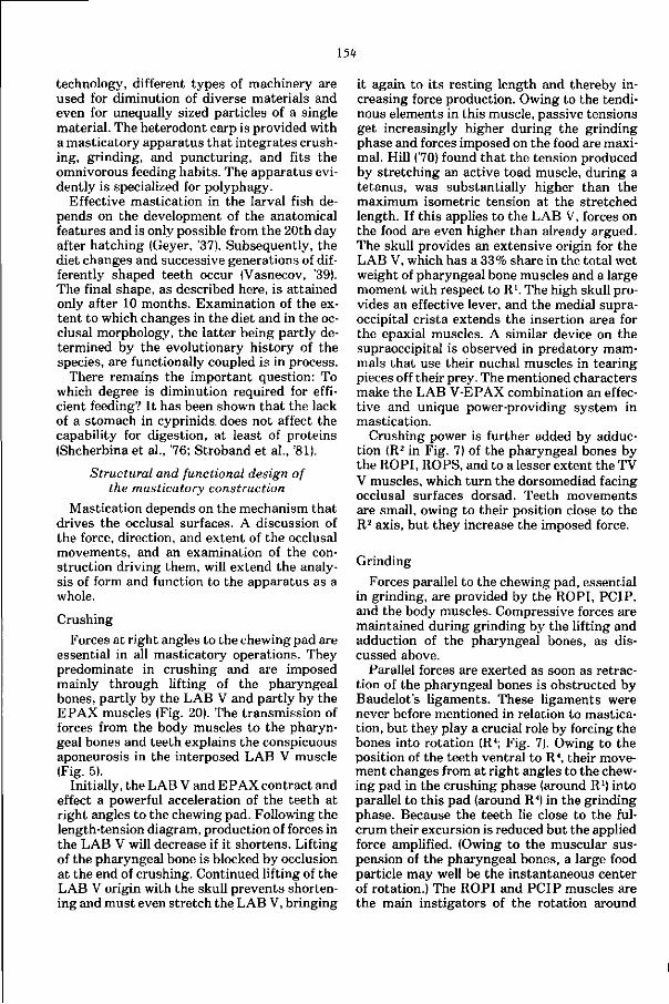
154
technology, different types of machinery are used for diminution of diverse materials and even for unequally sized particles of a single material. The heterodont carp is provided with a masticatory apparatus that integrates crushing, grinding, and puncturing, and fits the omnivorous feeding habits. The apparatus evidently is specialized for polyphagy.
Effective mastication in the larval fish depends on the development of the anatomical features and is only possible from the 20th day after hatching (Geyer, '37). Subsequently, the diet changes and successive generations of differently shaped teeth occur (Vasnecov, '39). The final shape, as described here, is attained only after 10 months. Examination of the extent to which changes in the diet and in the occlusal morphology, the latter being partly determined by the evolutionary history of the species, are functionally coupled is in process.
There remains the important question: To which degree is diminution required for efficient feeding? It has been shown that the lack of a stomach in cyprinids. does not affect the capability for digestion, at least of proteins (Shcherbina et al., '76; Stroband et al., '81).
Structural and functional design of the masticatory construction
Mastication depends on the mechanism that drives the occlusal surfaces. A discussion of the force, direction, and extent of the occlusal movements, and an examination of the construction driving them, will extend the analysis of form and function to the apparatus as a whole.
Crushing
Forces at right angles to the chewing pad are essential in all masticatory operations. They predominate in crushing and are imposed mainly through lifting of the pharyngeal bones, partly by the LAB V and partly by the EPAX muscles (Fig. 20). The transmission of forces from the body muscles to the pharyngeal bones and teeth explains the conspicuous aponeurosis in the interposed LAB V muscle (Fig. 5).
Initially, the LAB V and EPAX contract and effect a powerful acceleration of the teeth at right angles to the chewing pad. Following the length-tension diagram, production of forces in the LAB V will decrease if it shortens. Lifting of the pharyngeal bone is blocked by occlusion at the end of crushing. Continued lifting of the LAB V origin with the skull prevents shortening and must even stretch the LAB V, bringing
it again to i ts resting length and thereby increasing force production. Owing to the tendinous elements in this muscle, passive tensions get increasingly higher during the grinding phase and forces imposed on the food are maximal. Hill ('70) found that the tension produced by stretching an active toad muscle, during a tetanus, was substantially higher than the maximum isometric tension at the stretched length. If this applies to the LAB V, forces on the food are even higher than already argued. The skull provides an extensive origin for the LAB V, which has a 33% share in the total wet weight of pharyngeal bone muscles and a large moment with respect to R1. The high skull provides an effective lever, and the medial supra-occipital crista extends the insertion area for the epaxial muscles. A similar device on the supraoccipital is observed in predatory mammals that use their nuchal muscles in tearing pieces off their prey. The mentioned characters make the LAB V-EPAX combination an effective and unique power-providing system in mastication.
Crushing power is further added by adduction (R2 in Fig. 7) of the pharyngeal bones by the ROPI, ROPS, and to a lesser extent the TV V muscles, which turn the dorsomediad facing occlusal surfaces dorsad. Teeth movements are small, owing to their position close to the R2 axis, but they increase the imposed force.
Grinding
Forces parallel to the chewing pad, essential in grinding, are provided by the ROPI, PCIP, and the body muscles. Compressive forces are maintained during grinding by the lifting and adduction of the pharyngeal bones, as discussed above.
Parallel forces are exerted as soon as retraction of the pharyngeal bones is obstructed by Baudelot's ligaments. These ligaments were never before mentioned in relation to mastication, but they play a crucial role by forcing the bones into rotation (R4; Fig. 7). Owing to the position of the teeth ventral to R4, their movement changes from at right angles to the chewing pad in the crushing phase (around R1) into parallel to this pad (around R") in the grinding phase. Because the teeth lie close to the fulcrum their excursion is reduced but the applied force amplified. (Owing to the muscular suspension of the pharyngeal bones, a large food particle may well be the instantaneous center of rotation.) The ROPI and PCIP muscles are the main instigators of the rotation around
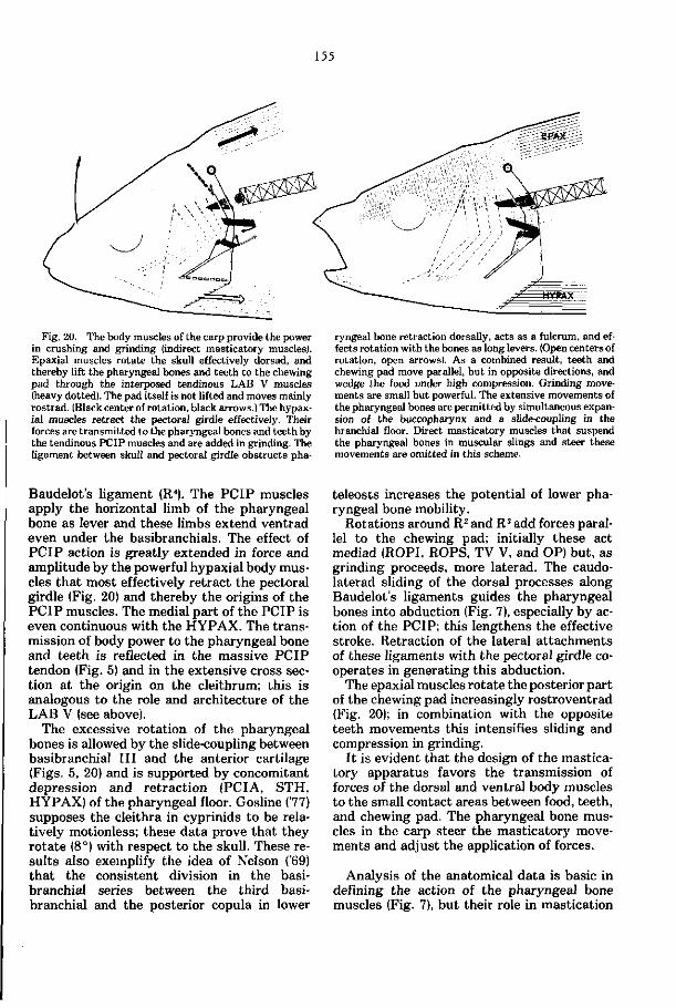
155
Fig. 20. The body muscles of the carp provide the power in crushing and grinding (indirect masticatory musclesl. Epaxial muscles rotate the skull effectively dorsad, and thereby lift the pharyngeal bones and teeth to the chewing pad through the interposed tendinous LAB V muscles (heavy dotted). The pad itself is not lifted and moves mainly rostrad. (Black center of rotation, black arrows.) The hypax-ial muscles retract the pectoral girdle effectively. Their forces are transmitted to the pharyngeal bones and teeth by the tendinous PCIP muscles and are added in grinding. The ligament between skull and pectoral girdle obstructs pha
ryngeal bone retraction dorsally, acts as a fulcrum, and effects rotation with the bones as long levers. (Open centers of rotation, open arrows). As a combined result, teeth and chewing pad move parallel, but in opposite directions, and wedge the food under high compression. Grinding movements are small but powerful. The extensive movements of the pharyngeal bones are permitted by simultaneous expansion of the buccopharynx and a slide-coupling in the branchial floor. Direct masticatory muscles that suspend the pharyngeal bones in muscular slings and steer these movements are omitted in this scheme.
Baudelot's ligament (R4). The PCIP muscles apply the horizontal limb of the pharyngeal bone as lever and these limbs extend ventrad even under the basibranchials. The effect of PCIP action is greatly extended in force and amplitude by the powerful hypaxial body muscles that most effectively retract the pectoral girdle (Fig. 20) and thereby the origins of the PCIP muscles. The medial part of the PCIP is even continuous with the HYPAX. The transmission of body power to the pharyngeal bone and teeth is reflected in the massive PCIP tendon (Fig. 5) and in the extensive cross section at the origin on the cleithrum; this is analogous to the role and architecture of the LAB V (see above).
The excessive rotation of the pharyngeal bones is allowed by the slide-coupling between basibranchial I I I and the anterior cartilage (Figs. 5, 20) and is supported by concomitant depression and retraction (PCIA, STH, HYPAX) of the pharyngeal floor. Gosline (77) supposes the cleithra in cyprinids to be relatively motionless; these data prove that they rotate (8°) with respect to the skull. These results also exemplify the idea of Nelson ('69) that the consistent division in the basibranchial series between the third basibranchial and the posterior copula in lower
teleosts increases the potential of lower pharyngeal bone mobility.
Rotations around R2 and R3 add forces parallel to the chewing pad; initially these act mediad (ROPI, ROPS, TV V, and OP) but, as grinding proceeds, more laterad. The caudo-laterad sliding of the dorsal processes along Baudelot's ligaments guides the pharyngeal bones into abduction (Fig. 7), especially by action of the PCIP; this lengthens the effective stroke. Retraction of the lateral at tachments of these ligaments with the pectoral girdle cooperates in generating this abduction.
The epaxial muscles rotate the posterior part of the chewing pad increasingly rostroventrad (Fig. 20); in combination with the opposite teeth movements this intensifies sliding and compression in grinding.
I t is evident tha t the design of the masticatory apparatus favors the transmission of forces of the dorsal and ventral body muscles to the small contact areas between food, teeth, and chewing pad. The pharyngeal bone muscles in the carp steer the masticatory movements and adjust the application of forces.
Analysis of the anatomical data is basic in defining the action of the pharyngeal bone muscles (Fig. 7), but their role in mastication
156
appears only from a functional analysis. The concert of pharyngeal bone, skull, and pectoral girdle movements affects the lines of action and the range of movements. Besides, axes of rotation may shift. Moments of the PCIP and TV V muscles, for example, increase during the motion they aid to perform. The STH muscles act as protractors in the preparatory stroke and as retractors during the grinding stroke. Furthermore, the ROPI muscles rather rotate and adduct the pharyngeal bones, more than their name would suggest. The PCIP appears to be one of the major, though indirect, abductors. The ROPS and TV V muscles play a concealed role in stabilizing the masticatory movements.
Suspension and structure of the pharyngeal bone
The pharyngeal bones are suspended from the skull in muscular slings (Kallen and Gans, 72; Liem, 78) held in constant tension by some of the muscles (e.g., the PCE in Fig. 9). Combined with a movable symphysis and a slide-coupling in the pharyngeal floor, this suspension permits highly versatile and complex movements. The LAB V, ROPI, PCIP, PCE, and SRC muscles control the pharyngeal bone movements in the sagittal plane, whereas the LAB V, ROPI, TV V, PCE, and OP muscles cooperate in the transversal plane. The essential role of the ROPI in each of these systems may account for the unique presence of such retrac-tores in cyprinids among lower teleosts (Holst-voogd, '65; Rosen, 73).
In the grinding phase Baudelot's ligament acts as a fulcrum and the pharyngeal bone is applied as lever. The pharyngeal bones provide areas of insertion and rotation axes at such sites that muscles form effective rotation couples. This renders the shape of the pharyngeal bone suited to accumulate power in mastication. Thus the LAB V, ROPI, and PCIP muscles, for example, although having completely different positions and lines of action, all contribute to rotation (R4) of the bones in the grinding stroke. Because teeth are close to the R2 and R4 axes during grinding their movement is small but forceful.
The direction of the trabeculae in bony elements will reflect the direction of loading (Kummer, '62). The orientation of the distinct trabeculae in the basioccipital process suggests that they spread forces, imposed on the chewing pad, over the base of the skull and the exoccipitals and thus reduce excessive local stresses. In a similar way the trabeculae radi
ating from the toothed center of the pharyngeal bone (Fig. 2a), as well as the anterior limb extending rostroventrad from the A I teeth, spread reaction forces on the occlusal surfaces of the teeth over the bone. The angle of about 65 ° between the anterior limb and the occlusal surfaces of the teeth may well be a compromise in absorbing loads varying between perpendicular and parallel to the occlusal surfaces.
The free suspension of the pharyngeal bones raises the need for stabilizing the powerful movements. Muscles act bilaterally and antagonistic muscles are synchronously active. A disturbed balance in the movement and load of left and right pharyngeal bone, induced by irregularities of the food, will be corrected by the adjustments in the muscular sling (e.g., ROPI and LAB V against PCE in Fig. 15). Such an unbalance loads the symphysis mainly by sliding. Because the interpharyngeal ligaments lie almost parallel to the longitudinal axis (Fig. 6, max. 30°), they provide more resistance to such sliding load than to tensile forces bringing the pharyngeal bones apart. Rotation around the R2 and R3 axes (Fig. 3) causes compression and tension in the symphysis; these stresses will be absorbed by the symphyseal tissue. Owing to the movable symphysis, torsion of the bones will hardly occur. Excessive rotations around R2 and R3 are prevented by the interpharyngeal ligaments, by TV V activity, and by the closely joined, flat medial sides of the symphysis, which abut at large rotations. Some other muscles may aid in stabilizing by effecting compression or tension in the symphysis (see A2 and A3 axes in Fig. 7). The role of the TV V in the carp appears to be analogous to that of the transversus man-dibularis muscle in the rat (Beecher, 79) in reducing the loading of the symphysis. The long symphysis in the carp, in comparison with most other cyprinids, may emphasize the large forces applied in this fish.
Differentiation and regulation of masticatory activities
Direct and indirect masticatory muscles
The different roles of the masticatory muscles are analogous to those of the direct and indirect flight muscles of insects: Muscles directly attached to the bony (chitinous) elements steer the movement, and the power is supplied indirectly by body muscles. Among steering muscles a further differentiation is made. The LAB V and ROPI still add significant power, as appears from their weight. In addition, the
157
LAB V and PCIP have a conspicuous role in t ransmitting forces from the body muscles to the pharyngeal bones. Other direct masticatory muscles, and especially the ROPS, rather stabilize and steer. Free recovery movements of the unloaded pharyngeal bone are achieved by the PCE; this muscle counteracts a multiplicity of movement components and functions as a prime antagonist in pharyngeal mastication. Masticatory movements are guided passively also by Baudelot's ligaments. A comparison of the fiber types in the pharyngeal bone, head, and body muscles in relation to their different roles in mastication is made (Akster and Sibbing, in press).
The time sequence in recruitment of the masticatory muscles in crushing and grinding shows a stable pattern and is independent of the particle size and the type of food (Figs. 9, 15). The duration of the cycle is negatively related, and the forces generated (as judged from the EMG amplitude) are positively related, to the intensity of crushing and grinding. The ability to chew various foods is determined by the multifunctional structure of the occlusal surfaces (Fig. 19) and by the two preprogrammed activity patterns rather than by drastic changes in these patterns. The experiments do provide examples of detailed neural regulation within a masticatory cycle. The balancing of activities between antagonistic muscles functions to improve the masticatory efficiency by maintaining a critical pressure that needs not be higher than required by the materials and must be instantly reduced when fracturing is reached. Transition from crushing into grinding, balancing of antagonistic activities, and instantaneous adjustments to the food (Fig. 15) thus are the active modulations in the stereotyped pattern.
The intensity and duration of the masticatory sequence should be regulated by sensory information; mastication of worms lasts only seconds, whereas chewing of barley lasts minutes. Regulation is mediated by the nervus vagus innervating all pharyngeal bone muscles. Proprioceptive reflex chains, similar to those demonstrated for respiration in the carp by Ballintijn ('72), may include pressure receptors; these are expected to be present, for example, in the connective tissue between chewing pad and the basioccipital process. Sensors in the ROPI muscles or in Baudelot's ligaments might trigger the initiation of the grinding pattern.
Experiments in which the fish accidently fails to bring a large food pellet between teeth
and chewing pad reveal some of the normal regulatory mechanism. The LAB V and SRC muscles are not activated (Fig. 13), as would be expected in a normal cycle, and the activity in the PCE muscles is prolonged; adduction of the pharyngeal bones does not start. Apparently, proprioceptive information prevents a normal continuation of the cycle. Lifting of the skull takes place, however, as if crushing were to follow. The powerful indirect masticatory muscles, innervated by spinal nerves, appear more distantly involved and continue their preprogrammed activity once this has been initiated.
Interaction of mastication with food transport, respiration, and other
functions in the head
It has been postulated that "the pharyngeal mechanism moves independently of the surrounding cavity" (Gosline, 77:334) and that mastication "need not interrupt respiration" (Alexander, '67:108), since the pharyngeal teeth in the carp lie posterior to the gills. However, it is clear that mastication and respiration are intimately coupled; they permit and even support each other. The kinetics of mastication dominate respiration (Figs. 10, l i e , 16).
During grinding, the vertical buccopharyngeal expansion largely results from the lifting of the skull; during hyperventilation the expansion is equally large but is mainly caused by depression of the pharyngeal floor. The latter consists of multiple components and permits a more accurately adjusted flow of water along the buccopharynx than movements of the rigid skull could provide. Large volume changes are considered to be more a consequence than an aim in mastication.
The frequency of masticatory movements in the carp is about equal to the frequency of respiration and probably is triggered by a similar central generating center. The functional coupling between these two processes in the carp may have been facilitated by the common origin of respiratory and masticatory elements in the branchial arches.
Transport of food to the pharyngeal teeth and esophagus is effected by different mechanisms (see Results, Fig. 17). Food is sucked in with water and trapped between the pharyngeal pads, which act like a muscular tongue and propel it caudad to the teeth. No teeth occur in the anterior buccopharynx. The repetitive movements of the pharyngeal floor probably act in gustation, selecting and cleaning the food. The transport of food from the teeth to
158
the esophagus, deglutition, is effected by the constrictor pharyngis aided by the pharyngeal bone muscles; this was not previously recorded and depicted in fishes.
Cardiac function and pharyngeal bone movements may well be coupled, as the horizontal limbs form the pericardial roof. Furthermore, the proximity of the Weberian ossicles "impose even greater than usual restriction on the flexibility between head and anterior vertebrae" (Gosline, 77:332). The Weberian ossicles, however, almost coincide with the rotation center of the skull and this minimizes their movement. The chain-like arrangement of the ossicles will be a necessity allowing skull movements during mastication and yet maintaining a connection between perilymph? and swim-bladder. The position of the os Suspensorium (Fig. 1) and the attached swimbladder prevents a further caudad extension of the basioc-cipital pharyngeal process and thus restricts the area of attachment for the ROPI muscles. The laterad movements of the molars against the chewing pad might be seen as a lengthening of the effective grinding stroke, given this restriction in space of the pharyngeal apparatus.
Thus the localization of the masticatory apparatus among those for respiration, hearing, and blood transport in the structural pattern of the head necessitates their functional and structural coordination during evolution.
Pharyngeal mastication in cyprinids and higher teleosts
It is assumed that the buccopharyngeal cavity was primitively covered by numerous small tooth plates that functioned in seizing and swallowing large prey. Fusion subsequently led to enlarged tooth plates in areas of particular functional significance such as jaws, basi-branchials, and the posterior gill arches (Nelson, '69). In lower teleosts a grasping type of dentition that includes separate upper pharyngeal tooth plates 4 and 5 seems to be normal. Subsequently, in higher teleosts, tooth plates fused to the gill arch elements and the dentition specialized (Nelson, '69). This trend resulted in a single pair of toothed areas, mainly the infrapharyngobranchials 3, which were equipped with retractores arcuum branchial-ium muscles, typical for all higher teleosts (Neoteleosts; Rosen, 73).
However, some Ostariophysi deviated from the general lower teleostean pattern (Nelson, '69). The pharyngeal jaw system in cyprinids, for example, is highly advanced and toothed upper pharyngeals are completely lacking. Retractor muscles, commonly absent in lower teleosts, insert on the lower pharyngeals.
These muscles are not homologous to the retractors found in higher teleosts (Holstvoogd, '65; Rosen, 73).
Liem and Greenwood ('81) recently treated pharyngeal jaws in several acanthopterygian assemblages. The pharyngeal jaw system in the carp, a lower teleost, is based on a structural and mechanical pattern that differs widely from systems found in cichlids, embi-otocids, labrids, odacids, and scarids, and is at least as specialized. This is concluded from the following comparison:
1. The shift of the insertion of the major levator components from the upper to the lower pharyngeal jaw is considered a key adaptation in the higher teleosts mentioned above (Liem, 73 ; Liem and Greenwood, '81). In the carp the huge subtemporal fossae contain bulky levator muscles that attach directly to the lower pharyngeal jaw. In addition, these muscles transmit forces of the epaxial body muscles to the lower pharyngeal jaw. Retractor muscles are inserted on the lower pharyngeals also.
2. The absence of upper pharyngeal jaws in cyprinids and the presence, instead, of a fixed chewing pad supported by the skull allows the recruitment of power from epaxial and hypax-ial body muscles for pharyngeal mastication. Summation of these indirect forces with those of the direct pharyngeal bone muscles effects a large load increase on the occlusal surfaces.
3. The muscular sling characterizes the suspension of the pharyngeal jaws in Embiotoci-dae and Cichlidae, whereas the lower pharyngeal j aw has been transformed into a lever that articulates with the cleithrum in Labridae (Liem, 78 ; Liem and Greenwood, '81). In the carp both characters are found. The muscular sling is kept in constant tension during crushing, and in the grinding phase of the power stroke muscles rotate the pharyngeal bone as a lever with the ligament of Baudelot as the fulcrum. In this way crushing is transformed into grinding. Thus, fine control, versatility, force, and stabilization are combined effectively.
4. The lack of upper pharyngeal jaws, which perform intrinsic movements and function in mastication as well as food transport in higher teleosts, seems to be compensated by the movable symphysis and intrinsic musculature between the lower pharyngeals in the carp and by the thick pharyngeal pad, which acts as a muscular tongue. These features do not occur in the higher teleosts mentioned.
These characters render the pharyngeal jaw apparatus of the carp both versatile and extremely powerful and, owing to the heterodont dentition, suited for comminution of a variety of food items. The retention of three rows of
159
pharyngeal teeth, considered a primitive character in cyprinids (Chu, '35; Vasnecov, '39; Hensel, '70), and the occlusion exclusively with the chewing pad, enlarge the grinding area in this specialized cyprinid.
From a functional point of view there appears to be a close association between the specialized pipette system of feeding, the development of the pharyngeal jaws, the loss of teeth in the jaws, and the development of pre-maxillary protrusion in cyprinids (Gosline, 73). The unique combination of these specializations may well have contributed to the development of the cyprinids as the largest family of freshwater fishes.
Catastomids resemble cyprinids to a great extent. The palatal organ is even more developed in catastomids; this is related to its prominent role in gustation, selection, and separation of organic and anorganic food items in these bottom-feeders (Eastman, 77). They, however, lack a chewing pad, except for two mollusc-crushing species, and effective mastication of food against the soft palatal organ will hardly be possible. Pharyngeal teeth are supposed to manipulate, strain, and masticate the food (Eastman, 77).
Among cyprinids a broad spectrum of anatomical features and dietary characteristics is found. Howes ('81) defines a squaliobarbine group of cyprinids involving apomorph characters that lie for the most part in the occipital region of the skull, and he relates them to the increased evolutionary development of the levator of the lower pharyngeal j aw. Following this author, these specializations could be associated with the essentially phytophagous habits of the squaliobarbine taxa. A functional analysis is in progress of pharyngeal mastication in the grasscarp, Ctenopharyngodon, a member of this squaliobarbine group that has teeth that, unlike the carp, interdigitate from left and right. Such analyses improve the definition of character complexes belonging to the masticatory system. They also provide a better view on parallel developments in its evolution. This will enhance the tracing of interrelationships between the diversified cyprinid taxa. The present paper demonstrates that the complex of structures involved in mastication extends, at least in carp, far beyond the pharyngeal bones and teeth.
ACKNOWLEDGMENTS
I am very much indebted to Dr. J.W.M. Osse for suggesting this study, for his advice and stimulating interest, and for the criticism and valuable discussions during preparation of the
manuscript. Thanks are due to Dr. P. Dulle-meijer (Department of Zoology, University of Leiden) for the use of the X-ray apparatus, to Arie Terlouw for technical assistance, to Wim Valen for his expertise in preparing the drawings and photographs, and to Sytze Leenstra for taking care of the carps. Dr. W.A. Weijs kindly supplied the radiopaque food pellets. The cooperation of Dr. L. Boomgaart, who corrected linguistic errors, and of Mrs. W.M. Laoh-Gieskes, who typed the manuscript, is gratefully acknowledged.
LITERATURE CITED
Ahlgren, J., and B. Owall (1970) Muscular activity and chewing force. A polygraphic study of human mandibular movements. Arch. Oral Biol. 15:211-280.
Akster, H.A., and F.A. Sibbing (in press) Fibre types in axial muscles, the m. hyohyoideus and masticatory muscles of the carp {Cyprinus carpio L.). J . Muscle Res. Cell Motil.
Alexander, R.McN. (1967) Functional Design in Fishes. London: Hutchinson University Library.
Ballintijn. C M . (1969a) Functional anatomy and movement co-ordination of the respiratory pump of the carp {Cyprinus carpio L.). J . Exp. Biol. 50:547-567.
Ballintijn, C M . (1969b) Muscle co-ordination of the respiratory pump of the carp (Cyprinus carpio L.). J . Exp. Biol. 50:569-591.
Ballintijn, C M . (1972) Efficiency, mechanics and motor control of fish respiration. Respir. Physiol. 24:125-141.
Beecher, R.M. (1979) Functional significance of the mandibular symphysis. J . Morphol. 259:117-130.
Brousseau, R.A. (1976) The pectoral anatomy of selected ostariophysi. II . The cypriniformes and siluriformes. J. Morphol. 750:79-116.
Chu, Y.T. (1935) Comparative studies on the scales and on the pharyngeals and their teeth in Chinese cyprinids, with particular reference to taxonomy and evolution. Biol. Bull. St. John's Univ.. Shanghai 2:1-225.
Dabrowski, K., and J. Glogowski (1977) Studies on the proteolytic enzymes of invertebrates constituting fish food. Hydrobiologia 52:171-174.
Desmedt, J .E. (1958) Methodes d'étude de la fonction neuromusculaire chez l'homme. Acta Neurol. Belg. 58:977-1017.
Dullemeijer, P. ( 1958) The mutual structural influence of the elements in a pattern. Arch. Néerl. Zool. 73(Suppl. 1):74-88.
Dullemeijer, P. (1974) Concepts and Approaches in Animal Morphology. Assen, The Netherlands: Van Gorcum and Comp.
Eastman, J.T. (1970) The pharyngeal bones and teeth of Minnesota cyprinid and catastomid fishes: Functional morphology, variation and taxonomie significance. Doctoral Thesis, University of Minnesota.
Eastman, J .T. (1971) The pharyngeal bone musculature of the carp, Cyprinus carpio. J . Morphol. 734:131-140.
Eastman, J.T. (1977) The pharyngeal bones and teeth of catastomid fishes. Am. Midi. Nat. 97:68-88.
Evans, H.E., and E.E. Deubler J r . (1955) Pharyngeal tooth replacement in Semotilus atromaculatus and Clinostomus elongatus, two species of cyprinid fishes. Copeia, 7955(1): 31-41.
Geyer, E. (1937) Der Zeitliche Ablauf der Bezahnung und des Zahnwechsels bei Cyprinus carpio L., unter besonderer Berücksichtigung des einsömmrigen Karpfens. Morphol. Jahrb. 80:280-354.
Girgis, S. (1952) The bueco-pharyngeal feeding mechanism in an herbivorous bottom-feeding cyprinoid, Labeo horie (Cuvier). J . Morphol. 90:281-315.
160
Gosline, W.A. (1973) Considerations regarding the phytogeny of cypriniform fishes, with special reference to structures associated with feeding. Copeia 7973:761-776.
Gosline W.A. (1977) The structure and function of the dermal pectoral girdle in bony fishes with particular reference to ostariophysines. J. Zool. 783:329-338.
Gratzianow, V. (1900) Über die sog. "Kauplatte" der Cyprinoiden. Zool. Anz. 23:66-73.
Haempel, O. (1907) Über die sogenannte Kauplatte der Cyprinoiden. Ber. K. Bayer. Biol. Versuchsst. München 7: 1-21.
Haempel, O. (1909) Die Schlundknochenmuskulatur der Cyprinoiden und ihre Funktion. Zool. Jahrb. (Anat.) 27: 95-101.
Hannam, A.G., W.C. Inkster, and J.D. Scott (1975) Peak electromyographic activity and jaw-closing force in man. J. Dent. Res. 54:694.
Harrington, R.W. (1955) The osteocranium of the American cyprinid fish, Notropsis bifrenatus, with an annotated synonymy of teleost skull bones. Copeia 7955(4):267-290.
Heincke, F. (1892) Variabilität und Bastardbildung bei Cyprinoiden. In Festschrift zum siebenzigsten Geburtstage Rudolf Leuckarts. Leipzig: Wilhelm Engelmann, pp. 65-73.
Hensel, K. (1970) Review of the classification and of the opinions on the evolution of Cyprinoidei (Eventognathi) with an annotated list of genera and subgenera described since 1921. Annot. Zool. Bot., Bratislava .57:1-45.
Hiiemae, K.M. (1976) Masticatory movements in primitive mammals. In D.J. Anderson and B. Matthews (eds): Mastication. Bristol: Wright and Sons, pp. 105-118.
Hiiemae, K.M. (1978) Mammalian mastication; a review of the activity of the jaw muscles and the movements they produce in chewing. In P.M. Butler and K.A. Joysey (eds): Development, Function and Evolution of Teeth. New York: Academic Press, pp. 359-398.
Hill, A.V. (1970) First and Last Experiments in Muscle Mechanics. Cambridge: Cambridge University Press.
Holstvoogd, C. (1965) The pharyngeal bones and muscles in Teleostei, a taxonomie study. Proc. Kon. Ned. Akad. Wet. 68:209-218.
Howes, G. (1981) Anatomy and phylogeny of the Chinese major carps Ctenopharyngodon Steind., 1866 and Hy-pophthalmichthys BIKr., 1860. Bull. Br. Mus. Nat. Hist. (Zool.) 47:1-52.
Inman, V.T., H.J. Ralston, J .B. de CM. Saunders, B. Fein-stein, and E.W. Wright, Jr . (1952) Relation of human elec-tromyogram to muscular tension. Electroenceph. Clin. Neurophysiol. 4:187-194.
Jurine, L. (1821) Sur les dents et la mastication des poissons appelés Cyprins. Mém. Soc. Phys. d'Hist. Nat. Genève, 7: 19-24.
Kallen, F.C., and C. Gans (1972) Mastication in the little brown bat, Myotis lucifugus. J . Morphol. 736:385-420.
Klust, G. (1940) Über Entwicklung, Bau und Funktion des Darmes beim Karpfen [Cyprinus carpio L.). Int. Rev. Ges. Hydrobiol. Hydrogr. 40:88-173.
Kummer, B. (1962) Funktionelle Anpassung und Präadaptation. Zool. Anz. 769:50-67.
Liem, K.F. (1970) Comparative functional anatomy of the Nandidae (Pisces: Teleostei). Fieldiana Zool. 56:1-166.
Liem, K.F. (1973) Evolutionary strategies and morphological innovations: Cichlid pharyngeal jaws, Syst. Zool. 22: 425-441.
Liem, K.F. (1978) Modulatory multiplicity in the functional repertoire of the feeding mechanism in cichlid fishes. I. Piscivores. J. Morphol. 758:323-360.
Liem, K.F., and P H . Greenwood (1981) A functional approach to the phylogeny of the pharyngognath teleosts. Am. Zool. 27:83-101.
Matthes, H. (1963) A comparative study of the feeding mechanisms of some African Cyprinidae (Pisces, Cyprini-formes). Bijdr. Dierkunde. 33:3-35.
Miller, E. (1966) The chewing apparatus. An electromyographic study of the action of the muscles of mastication and its correlation with facial morphology. Acta Physiol.
Scand. (Suppl. 280) 69:1-229. Nelson, G.J. (1967) Branchial muscles in some generalized
teleostean fishes. Acta Zool. 48:277-288. Nelson, G.J. (1969) Gill arches and the phylogeny of fishes,
with notes on the classification of vertebrates. Bull. Am. Mus. Nat. Hist. 747:475-552.
Nelson, J .S. (1976) Fishes of the World. New York: John Wiley and Sons.
Nikolsky, G.V. (1963) The Ecology of Fishes. London: Academic Press.
Nusbaum, J. (1908) Entwickelungsgeschichte und morphologische Beurteilung der Occipitalregion des Schädels und der Weberschen Knöchelchen bei den Knochenfischen {Cyprinus carpio L.). Anat. Anz. 32:513-532.
Osse, J.W.M. (1969) Functional morphology of the head of the perch (Perca fluviatilis L.): An electromyographic study. Neth. J. Zool. 79:289-392.
Osse, J.W.M., M. Oldenhave, and B. van Schie (1972) A new method for insertion of wire electrodes in electromyography. Electromyography 72:59-62.
Owen, R. (1840-1845) Odontography. London: Hippolyte Bailliere.
Ramaswami, L.S. (1955) Skeleton of cyprinoid fishes in relation to phylogenetic studies: 7. The skull and Weberian apparatus of Cyprininae (Cyprinidae). Acta Zool. 36:199-242.
Rensberger, J.M. (1973) An occlusion model for mastication and dental wear in herbivorous mammals. J. Paleontol. 47:515-528.
Rosen, D.E. (1973) Interrelationships of higher euteleostean fishes. In P.H. Greenwood, R.S. Miles, and C. Patterson (eds): Interrelationships of Fishes. London and New York: Academic Press, pp. 397-513.
Rutte, E. (1962) Schlundzähne von Susswasserfischen. Palaeontograph. Abt. A 720:165-212.
Schwartz, F.J., and B.W. Dutcher (1962) Tooth replacement and food of the cyprinid Notropsis cerasinus from the Roanoke River, Virginia. Am. Midi. Nat. 68:369-375.
Shcherbina, M.A., L.N. Trofimova, and O. Kazlauskene (1976) The activity of protease and the resorption intensity of protein with the introduction of different quantities of fat into the food of the carp, Cyprinus carpio. J. Ichthyol. 76:632-636.
Shepherd, C E . (1912) The pharyngeal teeth of fishes. Cyprininae, the carps. Zoologist, Ser. 4 76:201-209.
Sibbing, F.A. (1976) Pharyngeal mastication in Cyprinus carpio (L.). Rev. Trav. Inst. Pêches Marit. 40:744-745.
Stoss, H. (1921) Die Schlundzähne des Karpfens. Zool. Jahrb. 42:411-434.
Stroband, H.W.J., and F.H. van der Veen (1981) The localization of protein absorption during the t ransport of food in the intestine of the grasscarp, Ctenopharyngodon idella (Val.). J . Exp. Zool. 278:149-156.
Suietov, S.V. (1939) Contribution to the fish productivity of fresh waters. VIII . The thickness of bottom deposits and its role in the utilization of natural food by fishes. Proc. Kossino Limnol. Station 22:241-249.
Takahasi, N. (1925) On the homology of the cranial muscles of the cypriniform fishes. J. Morphol. Physiol. 40:1-109.
Uribe-Zamora, M. (1975) Selection des proies par le filtre branchial de la carpe miroir {Cyprinus carpio L.). Thèse, University of Lyon.
Vasnecov, V. (1939) Evolution of pharyngeal teeth in Cyprinidae. In: A la Mémoire de A.N. Sewertzoff. Moscow: Acad. Sei. USSR, pp. 439-491.
Winterbottom, R. (1974) A descriptive synonymy of the striated muscles of the Teleostei. Proc. Acad. Nat. Sei. Philadelphia 725:225-317.
Yang, H.J. (1964) On the pharyngeal bones and their teeth in Korean cyprinid fishes. II. Variation ratio of pharyngeal teeth of carp {Cyprinus carpio). Korean J. Zool. 7:6-8.
Zweers, G.A. (1974) Structure, movement and myography of the feeding apparatus of the mallard {Anas platyrhynchos L.). A study in functional anatomy. Neth. J. Zool. 24:323-467.
-161-
General summary
The process of food handling in the common carp (Cyprinus carpio L.) and
i ts structures associated w i th feeding are analyzed. The aim of this study is to
explain the relat ion between the the archi tecture of the head and its functions
in food processing and to determine the specializations for some food types and
the consequent restr ict ions for others. Such in format ion improves our understanding
of the trophic interrelat ions between d i f ferent f ish species l iving together in one
community.
- Cyprinids possess pharyngeal jaws, which are modif ied f i f t h branchial arches.
These are moved by the modif ied branchial arch muscles against a horny chewing
pad in the skul l . Oral teeth and a stomach are absent. The upper jaws are pro-
trusi le and so aid in the format ion of a round suction mouth. The oral and oper
cular cavity are highly variable in volume. The pharynx is almost fu l ly occupied
by the dorsal palatal organ. The pharyngeal f loor is composed of the postlingual
organ and the branchial sieve. Fibre systems of s t r iated muscles form the bulk
of these organs. The branchial arches bear numerous gi l l rakers. The surface
of the pharyngeal roof and f loor is almost fu l ly covered by taste buds (up to
820/mm2) and mucous cells. The medullar nervous centre of these organs is
of about equal size as the forebrain. The narrow s l i t - l ike space between both
organs has a restr ic ted capacity for volume change, contrary to the s i tuation
in most other fishes.
- The role which these structures per form during food uptake and processing in
the carp was investigated using c ine- and X-ray f i lming techniques and by syn
chronously recording the electromyograms of the involved muscles (9 channels).
The morphology was studied on the macroscopical and on l ight- and e lectron-
microscopical level. Food types included commercial f ish pellets, barley, ear th
worms, tubi fex, cladocerans and tubifex-soi l mixtures. BaSC^ - impregnated food
was used to fo l low i ts path in the X-ray movies.
- Each feeding process is composed of a variable number of stereotyped movement
patterns, v iz: part iculate intake and gulping, selection between food and non-food
mater ia l (through r insing, repositioning and backwashing), recol lect ion f rom the
branchial sieve fol lowed by food transport and f i l l ing of the chewing cavi ty,
crushing, grinding and deglut i t ion. Probing of the soil and spit t ing are considered
separately.
- The t iming, amplitude and velocity of mouth opening, protrusion of the upper
jaws, opening of the opercular valve and of the volume changes in the oral ,

-162-
buccal, pharyngeal and opercular cavit ies determine the ef fects of each single
pat tern. Food intake, selection, transport and masticat ion impose d i f ferent de
mands on the head and can not be combined e f fec t ive ly .
D i f fe rent food types are processed in sequences of movement patterns varying
in frequency and type according to the specific size, consistency and soilure
of the food. Handling t imes are read f rom the electromyograms and may d i f fer
widely.
The quanti tat ive distr ibution pat tern of taste buds, mucous cells, club cells
and muscle f ibers over the oro-pharyngeal surface is measured. Based on these
patterns and on other s t ructural characters six areas are distinguished in the
oro-pharynx and related w i th the functions of the above movement patterns
for food intake and processing. Scanning E.M. pictures are presented of the
common epithel ial cells w i th microridges, corni f ied cells, mucous cells, taste
buds and sensory (?) ol igovil lous cells.
Par t iculate intake is accomplished by fast and voluminous suction, caused by
expansion of the orobuccal and opercular cavit ies. The upper jaws are protruded
to produce a fast suction f low (>60 cm/sec), aimed to the par t ic le .
Gulping, the slow and less aimed uptake of a mouthful of water w i th suspended
foodpart icles is accomplished by size increase of the oral cavity mainly. The
carp f inal ly encloses the suspension by protruding its upper jaws downward.
Oral compression drives the water and food part icles over the branchial sieve.
The energy required for each gulp w i l l most probably be considerably less than
that needed for part iculate intake.
High densities of club cells, which produce the cyprinid alarming substance in
the skin, also occur in the orobuccal l ining. Their alarming funct ion in this area
is doubted.
Selection between food and non-food requires the retent ion of edible part icles
and the expulsion of waste. The electromyograms and e lectr ical st imulations
indicate that this separation is achieved by momentary bulgings on the palatal
organ, f ix ing edible part icles between pharyngeal roof and f loor. Waste part icles
are flushed through the branchial s l i ts. The complex structure of the palatal
organ, the almost maximal densities of taste buds and the cyto-archi tecture
of its regulatory centre in the hindbrain suggest a high level of d iscr iminat ion
in this selection process. The slit-shaped pharynx guarantees a large contact
area for selection, but l imi ts its role in suction.
Protrusion of the upper jaws w i th the mouth closed plays a crucial role in selec
t ion by resuspending food and non-food in the expanding oral cav i ty . A l ternat ive
expansion and compression of the oral cavity creates a fo r - and backward f low
-163-
through the pharyngeal sl it and the branchial sieve. Repet i t ion of such 'closed
protrusions' and selection effects are graded increasing pur i f icat ion. Closed
protrusion movements also serve for merely repositioning of large part icles
and for recol lect ion of the f i l t ra te f rom the branchial sieve.
Cells producing low-viscosity mucus (sialomucines) are found rostral ly in the
oro-pharynx. I t probably serves in lowering the resistance of the wal l for the
f low of water and in protect ion of the underlying tissue.
Cells producing large guantit ies of highly viscous mucus (sulfomucines) are found
in regions were aggregation and clustering of food particles prior to transport
is expected.
Transport of food enveloped in mucus is e f fected by a per istal t ic type of move
ment in the palatal and postlingual organs. These also propel the food into the
chewing cavity being enlarged by depression of the pharyngeal jaws.
The pharyngeal jaws are suspended in muscular slings f rom the caudal part of
the skull and pectoral g irdle. Except an antero-ventral gliding jo int w i th the
branchial basket no art iculat ions are present. The symphysis of the jaws allows
intr insic movements. Food is crushed and ground between the pharyngeal teeth
and a corni f ied chewing pad, f ixed to the base of the skull . Masticat ion of grains
of maize produces dist inct sounds, even dist inct close to the experimental tank.
The epaxial muscles of the carp contr ibute through rotat ion of the skull high
forces to crushing and grinding. The hypaxial muscles transfer their forces to
the pharyngeal teeth by re t ract ion of the pectoral g i rdle, which provides a large
moment-arm. The pharyngeal masticatory apparatus is bui l t for producing and
resisting high forces.
Contrary to these 'power muscles', the hypertrophied pharyngeal jaw muscles
act more l ike 'steering muscles'. They direct and stabil ize the pharyngeal jaw
movements around four anatomical rotat ional axes.
The chewing construction w i th the rotat ing skull renders a single Weberian ossicle
connecting the sound receiving swimming bladder and the internal ear inside
the skull almost impossible. The chain of Weberian ossicles running close to
the rotat ion centre of the skull seems to be a constructive necessity.
Deglut i t ion is accomplished by compression of the chewing cavi ty. Bulging of
the palatal and postlingual organs closes the entrance and thus d irect the trans
port to the esophagus. Movements of the pharyngeal jaws support t ransport.
The apparatus for food uptake and food processing of the carp appears to be
specialized to deal w i th medium-sized and hard food part icles (e.g. seeds and
shelled mollusks), f rom 250 ym to about 3% of i ts standard body length, but
also for food items mixed w i th unedible mater ia l . These specializations for
-164-
bottomfeeding are most l ikely basic to the present wide d istr ibut ion of the com
mon carp and fac i l i ta te f ish farming.
Large, fast and struggling preys as wel l as large and f la t plant material can
hardly be ut i l ized by the carp. Thus, also this 'omnivorous' f ish is l imi ted by
i ts specializations in the u t i l izat ion of the available food items in its environment.
A tentat ive scheme relates characters f rom d i f ferent parts of the head and
unique to the cyprinid fami ly in a funct ional and s t ructural context. The develop
ment of the masticatory apparatus may wel l have been a key adaptation in
the origin of the cyprinid feeding mechanism.
The present research of the carp provides a new and detai led start ingpoint for
investigation of the regulatory mechanisms in feeding and for comparisons w i th
native cyprinids l ike bream roach, tench etc. Knowledge of abi l i t ies and restr ic
tions of their s t ructural specializations associated w i th feeding eludidates which
p last ic i ty the f ish has to u t i l ize d i f ferent types of food. This p last ic i ty is a
crucial factor for the survival of the species in conditions of food scarcity and
co-determines its position in compet i t ion. The obtained knowledge thus aids
in predicting the ef fects of environmental changes on the trophic interactions
and composition of the f ish fauna.
-165-
Curriculum vitae.
Nand Sibbing werd op 16 december 1946 te 's-Gravenhage geboren. Verr i jk t
met een solide onderbouw op de kleuterschool, werd de St.Vincentius school door
lopen, daarmee een aaneensluitende periode van 25 jaar lager onderwijs aan ons
gezin volmakend. De brede en stimulerende vorming op het Aloysius college werd
in 1966 met het diploma gymnasium 3 afgesloten. De c i j fers voor de klassieke
talen op mijn examenlijst doen niet vermoeden hoezeer zi j mi j soms dwarslagen.
Het kandidaatsexamen biologie (B3; hoofdvak biofysika) werd in 1969 aan de
r i jksuniversi tei t te Leiden behaald. De Australische regering stelde mij daarna
met enkele andere studenten in staat drie maanden stage te lopen in de immunologie
(Walter & Eliza Hal l Inst i tute, Melbourne) en zo het land (en mijn broers en zuster
daar) beter te leren kennen. Afgewisseld met praktikum-assistentschappen werden
drie onderwerpen in mijn doktoraalstudie bewerkt:
1) Funktionele morfologie van het kauwapparaat bi j de karper (dr. J.W.M. Osse,
Zool. Lab. Leiden). D i t onderwerp wormde de eerste aanzet to t d i t proefschr i f t .
2) Cytochrome b-6 reacties in Porphyridium aerugineum (dr. J . Amesz, Lab.
voor Biofysika, Leiden).
3) Funktionele verschil len tussen het oog van de gele aal en de z i lveraal (Aguilla
anguüla L.) en de betekenis van deze verschillen voor de migrat ie van de
z i lveraal . (dr. F.J. Verheijen, Lab. Verg. Fysiologie, U t recht ) .
Het doctoraalexamen biologie werd in januari 1973 (cum laude) behaald. De onder
wijsbevoegdheid aantekening bleef steken op de hospiteerstages, die door mijn
tewerkstel l ing aan de Landbouwhogeschool te Wageningen niet meer konden worden
vervuld.
Sinds maart 1973 ben ik als wetenschappelijk medewerker aangesteld bij de
Vakgroep Experimentele Diermorfologie en Celbiologie (toen nog Dierkunde geheten).
De eerste drie jaren werden geïnvesteerd in de organisatie en opbouw van het
biologie onderwijs. Het onderzoek kreeg hierna geleideli jk meer ru imte en leidde
to t de bewerking van dit proefschr i f t . Taken in onderwijs, organisatie en beheer
vormen nog steeds een belangrijk bestanddeel.