Embed Size (px)
Citation preview

Downloaded from www.microbiologyresearch.org by
IP: 54.145.26.59
On: Sun, 27 Mar 2016 19:00:55
Transcriptional profiling of Helicobacter pyloriFur- and iron-regulated gene expression
Florian D. Ernst,1,2,3 Stefan Bereswill,23 Barbara Waidner,2 Jeroen Stoof,1
Ulrike Mader,34 Johannes G. Kusters,1 Ernst J. Kuipers,1 Manfred Kist,2
Arnoud H. M. van Vliet1 and Georg Homuth34
Correspondence
Arnoud H. M. van Vliet
1Department of Gastroenterology and Hepatology, Erasmus MC – University Medical CenterRotterdam, Dijkzigt L-455, Dr. Molewaterplein 40, 3015 GD Rotterdam, The Netherlands
2Department of Microbiology and Hygiene, Institute of Medical Microbiology and Hygiene,University Hospital of Freiburg, D-79140 Freiburg, Germany
3Institut fur Mikrobiologie und Molekularbiologie, Ernst-Moritz-Arndt-Universitat Greifswald,D-17487 Greifswald, Germany
Received 15 June 2004
Revised 25 October 2004
Accepted 2 November 2004
Intracellular iron homeostasis is a necessity for almost all living organisms, since both iron
restriction and iron overload can result in cell death. The ferric uptake regulator protein, Fur,
controls iron homeostasis in most Gram-negative bacteria. In the human gastric pathogen
Helicobacter pylori, Fur is thought to have acquired extra functions to compensate for the relative
paucity of regulatory genes. To identify H. pylori genes regulated by iron and Fur, we used DNA
array-based transcriptional profiling with RNA isolated from H. pylori 26695 wild-type and fur
mutant cells grown in iron-restricted and iron-replete conditions. Sixteen genes encoding proteins
involved in metal metabolism, nitrogen metabolism, motility, cell wall synthesis and cofactor
synthesis displayed iron-dependent Fur-repressed expression. Conversely, 16 genes encoding
proteins involved in iron storage, respiration, energy metabolism, chemotaxis, and oxygen
scavenging displayed iron-induced Fur-dependent expression. Several Fur-regulated genes
have been previously shown to be essential for acid resistance or gastric colonization in animal
models, such as those encoding the hydrogenase and superoxide dismutase enzymes. Overall,
there was a partial overlap between the sets of genes regulated by Fur and those previously
identified as growth-phase, iron or acid regulated. Regulatory patterns were confirmed for five
selected genes using Northern hybridization. In conclusion, H. pylori Fur is a versatile regulator
involved in many pathways essential for gastric colonization. These findings further delineate the
central role of Fur in regulating the unique capacity of H. pylori to colonize the human stomach.
INTRODUCTION
Infection with the human pathogen Helicobacter pyloriresults in persistent gastritis which can develop into pepticulcer disease and adenocarcinoma of the distal stomach(Blaser & Berg, 2001). Approximately half of the world’shuman population is colonized by H. pylori, leading tosignificant morbidity and mortality. For these reasons, theinfection is considered an important public health problem
with serious economic consequences. The only knownhabitat of H. pylori is the mucus layer overlaying theepithelial cells in the human stomach. Colonization of thisacidic and variable environmental niche has necessitated thedevelopment of adaptive stress responses by H. pylori.
In the gastric environment, changes in iron availabilityrepresent one of the important environmental stimuli forH. pylori. Iron is an essential element for almost all livingorganisms, as it is a cofactor of many enzymes and acts as acatalyst in electron transport processes. However, in thepresence of oxygen, iron potentiates the formation of toxicoxygen radicals. Therefore regulation of intracellular ironhomeostasis, as mediated by the ferric uptake regulator(Fur) protein, is of critical importance (Andrews et al.,2003). Regulation of gene expression via Fur has beenextensively investigated in several Gram-negative and Gram-positive bacteria, where it is involved in the regulation of
3Present address: Institut fur Mikrobiologie und Hygiene, CampusCharite Mitte, Berlin, Germany.
4Present address: School of Cell and Molecular Biosciences,Newcastle University Medical School, Newcastle-upon-Tyne, UK.
The complete MIAME information for the identification of Fur- and iron-regulated gene expression in the Helicobacter pylori dataset is shown inSupplementary Table S1, available online as supplementary data withthe online version of this paper at http://mic.sgmjournals.org.
0002-7404 G 2005 SGM Printed in Great Britain 533
Microbiology (2005), 151, 533–546 DOI 10.1099/mic.0.27404-0
Downloaded from www.microbiologyresearch.org by
IP: 54.145.26.59
On: Sun, 27 Mar 2016 19:00:55
many cellular processes, including iron metabolism, oxida-tive stress defence and central intermediary metabolism(Andrews et al., 2003; Hantke, 2001). However, while theabsence of Fur affects many cellular processes, several of theregulatory phenomena described mostly in E. coli are onlyindirectly affected by Fur (Masse & Gottesman, 2002).
Fur is a transcriptional repressor protein, which displaysiron-dependent binding to conserved DNA sequences (Furboxes) located in the promoters of iron-regulated genes(Hantke, 2001). In most bacteria, including H. pylori, theiron-complexed form of Fur binds to promoters of iron-uptake genes, thus repressing iron uptake in iron-repleteconditions (Delany et al., 2001b; van Vliet et al., 2002a).However, H. pylori Fur has acquired the thus far uniqueability also to bind the pfr promoter in its iron-free form,thus repressing expression of iron-storage proteins in iron-restricted conditions (Bereswill et al., 2000; Delany et al.,2001a; Waidner et al., 2002).
The relative paucity of transcriptional regulators in H.pylori, combined with the necessity to respond to environ-mental stresses, may have resulted in H. pylori Fur beinginvolved in the regulation of other adaptive responses.Other than regulation of iron metabolism, H. pylori Fur hasalso been implicated in the regulation of acid resistance(Bijlsma et al., 2002; Bury-Mone et al., 2004; van Vliet et al.,2004), nitrogen metabolism (van Vliet et al., 2001, 2003) andoxidative stress resistance (Barnard et al., 2004; Cooksleyet al., 2003; Harris et al., 2002). Fur-mediated regulationis also required for gastric colonization by H. pylori, asdemonstrated in a mouse model of infection (Bury-Moneet al., 2004).
DNA array technology has proved to be a powerfultechnique for the study of global gene regulation in manyorganisms (Conway & Schoolnik, 2003), and has also beensuccessfully applied to study alterations in H. pylori gene
expression (Bury-Mone et al., 2004; Forsyth et al., 2002;Kim et al., 2004; Merrell et al., 2003a, 2003b; Thompsonet al., 2003; Wen et al., 2003) and genetic variation betweenisolates (Israel et al., 2001a; Salama et al., 2000). In thisstudy, we have applied DNA array technology to growthexperiments with the well-characterized H. pylori referencestrain 26695, both to define the H. pylori responses tovariation in iron availability and to identify new members ofthe H. pylori Fur regulon.
METHODS
Bacterial strains, plasmids, media and growth conditions.The H. pylori strains used in this study were reference strain 26695(Tomb et al., 1997) and its isogenic fur mutant (Bijlsma et al., 2002;van Vliet et al., 2002a). H. pylori strains were routinely cultured onDent agar at 37 uC under microaerophilic conditions (10 % CO2,5 % O2 and 85 % N2). Broth cultures were grown in brucella broth(Difco) supplemented with 3 % newborn calf serum (Gibco) (BBN).Broth cultures were continuously shaken at 40 r.p.m. under micro-aerophilic conditions. Iron restriction was achieved by supple-menting BBN with desferal (deferoxamine mesylate, Sigma) to afinal concentration of 20 mM. Iron-replete conditions were achievedby supplementing desferal-treated BBN with ferric chloride (Sigma)to a final concentration of 100 mM (van Vliet et al., 2002a).
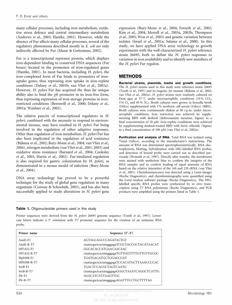
Purification and analysis of RNA. Total RNA was isolated usingTrizol (Gibco), according to the manufacturer’s instructions. Theamount of RNA was determined spectrophotometrically. RNA elec-trophoresis, blotting, hybridization with DIG-labelled RNA probes,and detection of bound probe were carried out as described pre-viously (Homuth et al., 1997). Directly after transfer, the membraneswere stained with methylene blue to confirm the integrity of theRNA samples and to confirm loading of equal amounts of RNAbased on the relative intensities of the 16S and 23S rRNA (van Vlietet al., 2001). Chemiluminescence was detected using a Lumi-Imager(Roche Diagnostics), and chemiluminographs were quantified usingthe Lumi-Analyse software package (Roche Diagnostics). The DIG-labelled specific RNA probes were synthesized by in vitro trans-cription using T7 RNA polymerase (Roche Diagnostics), and PCRproducts were amplified using the primers listed in Table 1.
Table 1. Oligonucleotide primers used in this study
Primer sequences were derived from the H. pylori 26695 genome sequence (Tomb et al., 1997). Lower-
case letters indicate a 59 extension with T7 promoter sequence for the creation of an antisense RNA
probe.
Primer name Sequence (5§–3§)
AmiE-F1 AGTAGCAGCCCAGATACTGT
AmiE-R-T7 ctaatacgactcactatagggagaTCGCTACCGCTACATAACAT
HP1432-F1 GGCACACCATGAACAACAAC
HP1432-R-T7 ctaatacgactcactatagggagaTGTTGGTTTTGTTGTTGCGC
Hp0388-F1 TGATGACATGCTGGAGCGAT
HP0388-R-T7 ctaatacgactcactatagggagaTCCACATGCTTAAACCCCAC
SerB-F1 TGACTCCACGCTAGTCAATG
SerB-R-T7 ctaatacgactcactatagggagaGGGCTAAATCAGGCTCATTG
Pfr-F1 AGACATCATTAAGTTGC
Pfr-R-T7 ctaatacgactcactatagggagaAGATTTCCTGCTTTTAG
534 Microbiology 151
F. D. Ernst and others

Downloaded from www.microbiologyresearch.org by
IP: 54.145.26.59
On: Sun, 27 Mar 2016 19:00:55
Synthesis of labelled cDNA for transcriptome analysis.H. pylori strain 26695 and its isogenic fur mutant were grown iniron-restricted and iron-replete conditions (van Vliet et al., 2002a),and total RNA was isolated from cells grown for 20 h and checkedby Northern hybridization using an amiE-specific probe (Fig. 1).For annealing of the specific oligonucleotide primers complementaryto the mRNAs specified by all H. pylori genes, 1 mg total RNA (con-centration determined photometrically) was hybridized to 4 mlcDNA primer mix (0?05 pmol ml21) (Eurogentec) in hybridizationbuffer (10 mM Tris/HCl, pH 7?9, 1 mM EDTA, 250 mM KCl) in atotal volume of 30 ml (1 h, 42 uC). After annealing, 30 ml of reversetranscription premix [12 ml 56 First Strand Buffer (Gibco-BRL),6 ml 0?1 mM DTT (Gibco-BRL), 2 ml 10 mM dATP, 2 ml 10 mMdGTP, 2 ml 10 mM dTTP, 4?5 ml [a-33P]dCTP (10 mCi ml21,Amersham Pharmacia), 1?5 ml reverse transcriptase (Superscript II;Gibco-BRL)] was added, and reverse transcription was carried outfor 1?5 h at 42 uC. Next, 2 ml 0?5 M EDTA was added to stop allreactions. Alkaline hydrolysis of the RNA was performed by additionof 6 ml 3?0 M NaOH and incubation of the solution for 30 min at65 uC, followed by 15 min at room temperature. The solution wasneutralized with 20 ml 1 M Tris/HCl, pH 8?0, and 6 ml 2N HCl.Finally, the cDNA was precipitated overnight at 220 uC after theaddition of 10 ml 3 M sodium acetate, pH 5?2, and 400 ml ethanol.The cDNA was pelleted by centrifugation at 17 600 g for 15 min at4 uC, washed with 70 % (v/v) ethanol, dried, and resuspended in100 ml sterile water. Labelling efficiency was determined by liquidscintillation measurement.
Hybridization of labelled cDNA to DNA macroarrays. H. pyloriarrays (Eurogentec): nylon membranes carrying PCR productswhich represented 97 % of all H. pylori 26695 and J99 protein-encoding genes (n=1578), were incubated for 10 min in 50 mlsaline sodium phosphate EDTA (SSPE) buffer (0?18 M NaCl,10 mM sodium phosphate, pH 7?7, 1 mM EDTA). Prehybridizationwas carried out in 10 ml hybridization solution [56 Denhardt
solution, 56 SSC, where 16 SSC is 0?15 M NaCl plus 0?015 Msodium citrate, 0?5 % SDS, 100 mg denatured salmon sperm DNA(Sigma) ml21] for 2 h at 65 uC. Subsequently, hybridization was per-formed for 20 h at 65 uC in 5 ml hybridization solution containingthe labelled cDNA probe which had been boiled for 5 min andrapidly cooled on ice before hybridization. Arrays were washed twicewith 200 ml 26 SSC and 0?1 % SDS (5 min at room temperatureand 20 min at 65 uC) and once with 200 ml 0?26 SSC and 0?1 %SDS. Finally, arrays were air-dried for 2 min, sealed in plastic bags,and exposed to PhosphorImager screens. The transcriptome analysiswas carried out twice, using two independently isolated sets of RNApreparations and two different array batches. Exposed Phosphor-Imager screens were scanned with a Storm 860 PhosphorImager(Molecular Dynamics) at a resolution of 50 mm and a colour depthof 16 bit. To remove the labelled cDNA from the arrays prior tosubsequent hybridizations, the membranes were incubated threetimes (2, 5 and 60 min) in 300 ml boiling stripping buffer (10 mMTris/HCl, pH 8?0; 1 mM EDTA; 1 % SDS). Exposure of the arraysafter stripping revealed that the complete activity was successfullyremoved from the membrane. Using this method, it was possible touse the macroarrays five times without significant loss in quality.
Analysis of transcriptome data. For quantification of the hybri-dization signals and background values, the ArrayVision software(Imaging Research) was used (Eymann et al., 2002). Subsequently, aquality score was calculated for each spot reflecting the ratiobetween the signal intensity and the background intensity. Thisquality score was utilized to identify hybridization signals close tothe detection limit. Data normalization and data anlysis were donewith the GeneSpring software (Silicon Genetics). After backgroundsubtraction, normalized intensity values of the individual spots werecalculated (median normalization). Only genes specifying signalswhich significantly exceeded the background signal level (determinedby the quality scores) under at least one condition were included infurther data analysis. The average of the normalized intensity values
Fig. 1. Selection of the growth phase ofcultures of H. pylori 26695 wild-type iso-genic fur mutant for isolation of RNA andsubsequent transcriptome analysis. (a) Growthcurves of H. pylori 26695 wild-type (circles)and fur mutant (triangles) grown in iron-restricted (open symbols) and iron-repleteconditions (closed symbols). Growth isexpressed as OD600. (b) Verification of iron-and Fur-responsive regulation at 8, 20 and30 h of growth in iron-restricted (”Fe) andiron-replete (+Fe) conditions using the iron-and Fur-repressed amiE gene. Northernhybridization of RNA isolated from H. pylori
26695 (WT) and its isogenic fur mutant (fur)with a probe specific for the amiE gene.The position of the two relevant RNA markersizes (in kb) is indicated on the left.
http://mic.sgmjournals.org 535
Transcriptional profiling of H. pylori Fur regulon

Downloaded from www.microbiologyresearch.org by
IP: 54.145.26.59
On: Sun, 27 Mar 2016 19:00:55
of the duplicate spots of each gene was used to calculate the expres-sion level ratios. Induction or repression ratios ¢2 in both experi-ments were considered as significant and used in subsequent analysis(Eymann et al., 2002).
Final evaluation of the macroarray data included the consideration ofputative operon structures derived from the genome sequence as wellas previously known operons. Genes exhibiting significant expressionratios were analysed for their transcriptional organization using thePyloriGene database (http://genolist.pasteur.fr/PyloriGene/) (Bonecaet al., 2003). The complete dataset is shown in SupplementaryTable S1, which is available online as supplementary data with theonline version of this paper at http://mic.sgmjournals.org.
Furbox Analyses. Sequences of Fur-regulated genes (Table 2)were obtained from the H. pylori 26695 genome sequence usingthe PyloriGene database (http://genolist.pasteur.fr/PyloriGene/).Sequences included the intergenic regions upstream of the regulatedgene when applicable. Genes were designated as being located down-stream of co-transcribed genes (fliP, murB, ispE, pdxJ and hp0241)when there was less than 10 bp between the stop codon of the pre-ceding gene and the start codon of the following gene. All genesincluded in this analyses had putative ribosome-binding sites (RBS)located upstream of the translational start codon.
To find putative binding sequences for iron-cofactored Fur, promotersequences were analysed for the presence of consecutive NAT triplets(van Vliet et al., 2002b) using the GeneRunner program (http://www.generunner.com). Putative binding sequences for apo-Fur wereidentified by aligning the promoter sequences with the Pfr boxes I andII (Delany et al., 2001a) using the Clone Manager 7 Suite (Scientificand Educational Software).
RESULTS
Identification of iron- and Fur-regulated H. pylorigenes by transcriptional profiling
For characterizing Fur- and iron-regulated gene expression,H. pylori strain 26695 was selected, as this allowed directcomparison with the available genome sequence (Tombet al., 1997). In addition, H. pylori 26695 has been exten-sively characterized for the role of Fur and iron in theregulation of genes putatively involved in iron transport(van Vliet et al., 2002a). The H. pylori 26695 isogenic furmutant used in this study contains the Campylobacter colichloramphenicol resistance cassette in the unique BclIrestriction inside the fur coding region, and was charac-terized previously with regard to acid resistance (Bijlsmaet al., 2002), iron uptake (van Vliet et al., 2002a) and iron-and acid-responsive regulation (Bury-Mone et al., 2004;van Vliet et al., 2004).
We selected a single time-point (20 h) to compare geneexpression, as this is when H. pylori 26695 reaches thelate exponential phase (Fig. 1a). To confirm that the 20 htime-point was representative for identification of iron-and Fur-regulated genes, RNA samples isolated at 8, 20and 30 h post-inoculation were hybridized with a probespecific for the amiE gene (Fig. 1b). The amiE gene waspreviously demonstrated to be iron- and Fur-repressed(van Vliet et al., 2003), and this regulation is apparent ateach of the three time-points (Fig. 1b). The amiE mRNA,
with a size of ~1 kb, was detected in the wild-type strainonly in iron-restricted conditions, but was constitutivelyexpressed in the fur mutant (Fig. 1b). Although the amiEgene has also been reported to be growth-phase regulated(Merrell et al., 2003b; Thompson et al., 2003), this was notapparent in the conditions used in this study.
RNA for array testing was isolated from two independentcultures of H. pylori 26695 and its isogenic fur mutant,grown in iron-restricted and iron-replete conditions. Sub-sequently the RNA samples were used for transcriptionalprofiling using the Eurogentec H. pylori DNA array, whichcontains 97 % of all ORFs of H. pylori strain 26695. Toexclude potential artefacts, only genes with a signal to noiseratio >3 were included in the subsequent data analysis.In total, 1248 out of 1551 genes (80?5 %) fullfilled thesecriteria for at least one of the conditions in both arrayexperiments, and this percentage of genes exhibiting signi-ficant expression signals is relatively high compared to thatreported in previous studies (~50 %) (Ang et al., 2001;Merrell et al., 2003a; Thompson et al., 2003). In total, datafor iron regulation were available for 1241 genes in the wild-type strain, and for 964 genes in the fur mutant. Data for Furregulation were available for 994 genes in iron-restrictedconditions, and 909 genes in iron-replete conditions.
For each of the 1248 genes, iron regulation was assessedby calculating the ratio between expression levels in iron-restricted conditions (2Fe) and the expression levels iniron-replete conditions (+Fe). To assess Fur regulation,the ratio between expression levels in the wild-type strain(WT) was compared with the expression levels in the furmutant strain (fur/WT ratio). Since H. pylori Fur affectstranscription in both iron-replete and iron-restricted con-ditions, the fur/WT ratio was calculated for both 2Fe and+Fe conditions. Genes were considered to be regulated byeither iron or Fur when the repression or induction ratiowas >2 in both independent RNA preparations. Genesregulated by Fur, together with the different ratios, arepresented in Table 2.
In total, 61 genes (4?9 %) displayed iron-repressed expres-sion in the wild-type strain, whereas 36 genes (2?9 %) dis-played iron-induced expression. Of these 97 iron-regulatedgenes, only 10 still displayed iron-dependent regulation inthe fur mutant, with data for 22 genes not being availablein the fur mutant. This underlines the central role of Fur iniron-regulated gene expression in H. pylori. Sixteen genesdisplayed derepressed expression in the fur mutant iniron-replete conditions, and thus these genes probably areregulated by the iron-complexed form of Fur. Conversely,16 genes displayed derepressed expression in iron-restrictedconditions, possibly representing repression by the iron-free form of Fur.
Fur-repressed genes
In most Gram-negative bacteria, Fur binds its target pro-moters in an iron-dependent fashion, in other words, only
536 Microbiology 151
F. D. Ernst and others

Downloaded from www.microbiologyresearch.org by
IP: 54.145.26.59
On: Sun, 27 Mar 2016 19:00:55
Table 2. Fur-regulated genes of Helicobacter pylori strain 26695
The Gene number shown is from the complete genome sequences of H. pylori strain 26695 (Tomb et al., 1997) and strain J99 (Alm et al.,
1999). The Predicted function column shows the functions and functional categories as defined on the PyloriGene database (Boneca et al.,
2003). Values in the Ratio columns show the ratio of expression levels in H. pylori wild-type (WT) or fur mutant (fur) strain, in iron-
restricted (2Fe) or iron-replete (+Fe) conditions. The value shown is the average ratio of two independent array experiments. Values in
italic type indicate significant down-regulation of expression; values in bold type indicate significant upregulation of expression. Significant
regulation was defined as at least twofold changes in the mRNA levels in both independent array experiments. ND, not detectable: the signal
on the array was below the detection threshold in both array experiments.
Gene number for strains: Predicted function Ratio
26695 J99 WT”Fe/
WT+Fe
fur”Fe/
fur+Fe
fur”Fe/
WT”Fe
fur+Fe/
WT+Fe
Repressed by iron-bound Fur
Biosynthesis of cofactors, prosthetic groups and carriers
HP1406 JHP1298 Biotin synthetase (bioB) 2?6 0?6 1?4 6?7
HP1443 JHP1336 4-disphosphocytidyl-2-C-methyl-D-erythritol kinase (ispE) 3?7 0?6 1?5 9?2
HP1583 JHP1490 Pyridoxal phosphate biosynthetic protein A (pdxA) 9?5 2?3 0?9 4?0
Cell envelope and surface structures
HP0685 JHP0625 Flagellar biosynthetic protein (fliP) 2?4 0?8 1?3 4?2
HP1418 JHP1313 UDP-N-acetylenolpyruvoylglucosamine reductase (murB) 2?8 0?6 1?6 7?8
Cellular processes
HP0115 JHP0107 Flagellin B (flaB) ND 0?4 ND 3?0
HP0870 JHP0804 Flagellar hook (flgE) ND 0?4 ND 4?2
Energy metabolism
HP0294 JHP0279 Aliphatic amidase (amiE) 2?2 0?9 0?8 2?1
HP1238 JHP1159 Formamidase (amiF) 0?9 0?5 1?7 3?5
Hypothetical proteins
HP0906 JHP0842 H. pylori predicted coding region HP0906 0?8 0?3 1?4 3?0
Protein synthesis
HP1431 JHP1322 16S rRNA (adenosine-N6,N6-)-dimethyltransferase (ksgA) 3?5 1?4 1?4 3?7
Transport and binding proteins
HP0686 JHP0626 Iron(III) dicitrate transport protein (fecA1) 8?5 1?2 0?6 4?1
HP0807 JHP0743 Iron(III) dicitrate transport protein (fecA2) 30?2 1?7 0?5 8?3
HP0876 JHP0810 Iron-regulated outer-membrane protein (frpB1) 5?5 0?7 0?9 6?8
HP1339 JHP1258 Biopolymer transport protein (exbB2) 1?6 1?4 2?1 2?2
HP1432 JHP1321 Histidine- and glutamine-rich protein 4?0 0?7 1?8 9?5
Repressed by iron-free Fur
Amino acid biosynthesis
HP0652 JHP0597 Phosphoserine phosphatase (serB) 0?7 2?3 8?6 2?9
Cellular processes
HP0389 JHP0992 Superoxide dismutase (sodB) 0?2 0?6 8?7 1?9
HP0616 JHP0559 Chemotaxis protein (cheV2) 0?2 1?8 14?1 1?9
HP0922 JHP0856 Toxin-like outer-membrane protein/VacA paralogue 1?1 2?3 2?4 1?3
Energy metabolism
HP0631 JHP0574 Quinone-reactive Ni/Fe hydrogenase, small subunit (hydA) 0?4 1?2 3?4 1?2
HP0632 JHP0575 Quinone-reactive Ni/Fe hydrogenase, large subunit (hydB) 0?3 0?9 3?2 1?2
HP0633 JHP0576 Quinone-reactive Ni/Fe hydrogenase, cytochrome b subunit
(hydC)
0?3 1?0 4?2 1?2
HP1227 JHP1148 Cytochrome c553 0?4 1?0 4?3 1?5
Hypothetical proteins/unknown function
HP0241 JHP0226 Predicted coding region HP0241 0?3 1?1 2?6 0?7
HP0388 JHP0993 Conserved hypothetical protein 0?3 0?9 4?9 1?4
HP0629 JHP0572 Predicted coding region HP0629 0?8 1?3 2?6 1?7
HP1094 None* Predicted coding region HP1094 ND 2?2 3?2
HP1502 JHP1395 Predicted coding region HP1502 0?9 1?7 3?6 1?8
HP1524 JHP1413 Predicted coding region HP1524 0?4 0?9 2?3 0?8
http://mic.sgmjournals.org 537
Transcriptional profiling of H. pylori Fur regulon

Downloaded from www.microbiologyresearch.org by
IP: 54.145.26.59
On: Sun, 27 Mar 2016 19:00:55
in iron-replete conditions. Uniquely, the iron-free formof H. pylori Fur is also capable of repressing transcription,thus allowing differential expression of genes dependingon iron availability in the cytoplasm (Delany et al., 2001a).Surprisingly, not all Fur-regulated genes identified in thisstudy displayed iron-responsive expression (Table 2).
(i) Genes repressed by the iron-complexed form ofFur (Fe-Fur). Of the 16 genes demonstrating derepressedexpression under iron-replete conditions in the furmutant, 10 also demonstrated iron-repressed expressionin the wild-type strain (Table 2), with four displayingiron-independent expression; for two genes, data were notavailable. As predicted in previous studies (Delany et al.,2001a, 2001b; Fassbinder et al., 2000; van Vliet et al.,2002a), several of these genes (fecA1, fecA2, frpB1 andexbB2) encode homologues of iron transport and bindingproteins, and probably play a role in the uptake andtransport of iron to the cytoplasm. In addition, thehp1432 gene, encoding a nickel-binding histidine- and
glutamine-rich protein (Gilbert et al., 1995), is also regu-lated by Fur, and this regulation was confirmed byNorthern hybridization (Fig. 2) and RNA slotblot hybridi-zation (data not shown). This gene has also been classifiedas nickel- and NikR-activated (Contreras et al., 2003), andacid-induced (Merrell et al., 2003a).
The fur mutation also influenced several other classes ofgenes (Table 2). These included genes putatively involvedin: i) biosynthesis of cofactors and prosthetic groups,including biotin (bioB), isoprenoid (ispE) and pyridoxalphosphate (pdxA); ii) production of cell envelope andsurface structures, such as the murB peptidoglycan syn-thesis gene and the flaB and fliP flagellar biosynthesisgenes (Josenhans et al., 2000); iii) energy metabolism, withboth the paralogous amidases amiE and amiF (Skouloubriset al., 2001; van Vliet et al., 2003); iv) protein synthesis, inwhich the 16S rRNA dimethyltransferase gene ksgA isputatively involved. Finally, expression of the hypotheticalprotein HP906 was repressed by Fe-Fur. For two of the
Table 2. cont.
Gene number for strains: Predicted function Ratio
26695 J99 WT”Fe/
WT+Fe
fur”Fe/
fur+Fe
fur”Fe/
WT”Fe
fur+Fe/
WT+Fe
Protein synthesis
HP1253 JHP1174 Tryptophanyl-tRNA synthetase (trpS) ND 1?2 3?0 1?0
Transport and binding protein
HP0653 JHP0598 Non-heme iron-containing ferritin (pfr) 0?2 2?5 53?1 2?3
Fur-induced
Cell envelope and surface structures
HP0638 JHP0581 Outer-membrane protein (omp13; oipA) 1?0 ND 0?2 0?1
HP0855 None* Alginate O-acetylation protein (algI) 5?8 ND 0?3 ND
HP1494 JHP1387 UDP-MurNac-tripeptide synthetase (murE) 4?3 2?1 0?5 0?9
DNA metabolism, restriction and modification
HP0260 JHP0244 Adenine-specific DNA methyltransferase (mod) 1?8 1?0 0?4 0?9
HP1323 JHP1243 Ribonuclease HII (rnhB) 4?4 1?6 0?4 0?9
Hypothetical proteins/unknown function
HP0207 JHP0193 ATP-binding protein (mpr) 4?0 2?6 0?4 0?5
HP0236 JHP0221 Predicted coding region HP0236 1?4 0?8 0?4 0?8
HP0248 JHP0233 Conserved hypothetical protein 2?6 0?9 0?4 1?1
HP0322 JHP0305 Poly-E-rich protein 0?3 0?8 0?9 0?4
HP0424 None* Predicted coding region HP0424 2?0 ND 0?4 ND
HP0773 JHP0710 Predicted coding region HP0773 2?6 1?8 0?5 0?7
HP1117 JHP1045 Cysteine-rich protein X (hcpX) 1?8 1?5 0?9 0?4
HP1142 JHP1070 Predicted coding region HP1142 0?5 ND ND 0?4
Protein synthesis
HP1141 JHP1069 Methionyl-tRNA formyltransferase (fmt) 2?3 1?3 0?4 0?7
HP1160 JHP1087 tRNA (5-methylaminomethyl-2-thiouridylate)-methyltransferase 2?6 1?2 0?4 0?8
Transport and binding proteins
HP1172 JHP1099 Glutamine ABC transporter, periplasmic glutamine-binding
protein (glnH)
3?2 1?5 0?4 0?7
*This gene is absent in the H. pylori J99 complete genome sequence.
538 Microbiology 151
F. D. Ernst and others

Downloaded from www.microbiologyresearch.org by
IP: 54.145.26.59
On: Sun, 27 Mar 2016 19:00:55
iron-repressed Fur-regulated genes (amiE and hp1432),the transcriptional pattern was confirmed using Northernhybridization (Fig. 2).
(ii) Genes repressed by the iron-free form of Fur.Sixteen genes demonstrated increased expression in thefur mutant, when compared to the wild-type strain grown
in iron-restricted conditions (Table 2). This unique formof regulation has so far only been reported for the pfrgene (Bereswill et al., 2000; Delany et al., 2001a; Waidneret al., 2002), but is probably more widespread in H.pylori. Of the 16 genes demonstrating derepressed expres-sion under iron-restricted conditions in the fur mutant,nine genes also demonstrated iron-induced expression inthe wild-type strain (Table 3), with five genes displayingiron-independent expression; for two genes, data were notavailable.
Several genes associated with energy and oxygen metabo-lism displayed Fur-mediated repression of transcription,including the nickel/iron-cofactored hydrogenase subunitgenes (hydABC) (Olson et al., 2001), a putative cytochromec553, and the sodB gene encoding the iron-cofactoredsuperoxide dismutase (Pesci & Pickett, 1994; Seyler et al.,2001; Spiegelhalder et al., 1993). Further genes regulated bythe iron-free form of Fur included the chemotaxis genecheV2, the hp0922 gene encoding a toxin-like outer-membrane protein, the serB gene, which is cotranscribedwith pfr, the tryptophanyl-tRNA synthetase gene trpS, andfive genes encoding hypothetical proteins (Table 2). Forthree of the iron-induced Fur-regulated genes (pfr, serBand hp0388), the transcriptional pattern was confirmedusing Northern hybridization (Fig. 2).
(iii) Fur-induced genes. Sixteen genes displayed decreasedexpression in the fur mutant when compared to the wild-type strain (Table 2). This inverse regulation is atypical fora repressor like Fur, and is likely to represent indirect regu-lation. This cluster of genes included the oipA gene encod-ing an outer-membrane protein, the murE gene involved inpeptidoglycan synthesis, a DNA methyltransferase (mod),the ribonuclease HII-encoding rnhB gene, the periplasmicbinding protein of the glutamine ABC transporter, twogenes involved in protein synthesis, and eight genes encod-ing hypothetical proteins.
Identification of putative binding sequences forFe-Fur and apo-Fur
All Fur-repressed genes listed in Table 2 were furtherinvestigated for the presence of putative Fur boxes in theirrespective promoters (Fig. 3). Firstly, the putative promoterregion was identified for each gene using the PyloriGenedatabase (see Methods for details). Putative binding sitesfor Fe-Fur were identified as up to six consecutive nATtriplets, with n representing any nucleotide (Delany et al.,2001b; van Vliet et al., 2002b). Each of the promotersincluded in this analysis contained a putative Fur box,with identities to the (nAT)6 sequence ranging from 6 to 11per 12 residues (Fig. 3a).
The identification of binding sites of apo-Fur is currentlyhampered by the absence of a consensus sequence, sinceonly the pfr promoter has been analysed to date (Delanyet al., 2001a). In this promoter there are two high-affinitysites for apo-Fur, designated Pfr box I and Pfr box II (Delany
Fig. 2. Confirmation of Fur- and iron-responsive regulation of asubset of genes selected from Table 2. (a) Staining of trans-ferred RNA by methylene blue to allow for comparison of RNAamounts. (b) Northern hybridization with probes specific for fivegenes using RNA purified from H. pylori wild-type (WT) and fur
mutant (fur) cells grown in iron-restricted (”Fe) and iron-replete(+Fe) conditions. Probes used are indicated on the left; thespecific mRNA is indicated on the right.
http://mic.sgmjournals.org 539
Transcriptional profiling of H. pylori Fur regulon

Downloaded from www.microbiologyresearch.org by
IP: 54.145.26.59
On: Sun, 27 Mar 2016 19:00:55
Table 3. Iron-regulated (Fur-independent) genes of Helicobacter pylori strain 26695
The gene number shown is from the complete genome sequences of H. pylori strain 26695 (Tomb et al., 1997) and strain J99 (Alm et al.,
1999). The Predicted function column shows the function and functional category as defined on the PyloriGene database (Boneca et al.,
2003). Values in the Ratio columns show the ratio of expression levels in H. pylori wild-type (WT) or fur mutant (fur) strain, in iron-
restricted (2Fe) or iron-replete (+Fe) conditions. The value shown is the average ratio of two independent array experiments. Values in
italic type indicate significant down-regulation of expression; values in bold type indicate significant upregulation of expression. Significant
regulation was defined as at least twofold changes in the mRNA levels in both independent array experiments. ND, not detectable: the signal
on the array was below the detection threshold in both array experiments.
Gene number for strain: Predicted function Ratio
26695 J99 WT”Fe/
WT+Fe
fur”Fe/
fur+Fe
fur”Fe/
WT”Fe
fur+Fe/
WT+Fe
Iron-repressed
Biosynthesis of cofactors, prosthetic groups and carriers
HP0625 JHP0569 1-hydroxy-2-methyl-2-(E)-butenyl 4-diphosphate synthetase (ispG) 2?7 2?6 0?7 0?8
HP0798 JHP0734 Molybdenum cofactor biosynthesis protein C (moaC) 4?2 ND ND ND
HP0804 JHP0740 GTP cyclohydrolase II/3,4-diOH-2-butanone 4-phosphate synthase
(ribBA)
2?5 ND 0?5 ND
HP0841 JHP0779 Pantothenate metabolism flavoprotein (dfp) 2?9 2?5 0?9 ND
Cell envelope and surface structures
HP0279 JHP0264 Lipopolysaccharide heptosyltransferase-1 (rfaC) 2?6 ND ND ND
HP0648 HP0593 UDP-N-acetylglucosamine enolpyruvyl transferase (murZ) 2?7 1?0 0?5 1?1
HP1429 JHP1324 Polysialic acid capsule expression protein (kpsF) 9?0 4?7 1?0 ND
HP1456 JHP1349 Membrane-associated lipoprotein (lpp20) 2?7 1?8 0?7 1?0
Cellular processes
HP0246 JHP0231 Flagellar basal-body P-ring protein (flgI) 2?3 1?4 0?7 1?1
HP0752 JHP0689 Flagellar hook-associated protein 2 (fliD) 4?2 2?2 0?6 1?2
HP0792 JHP0728 Predicted DNA transformation competence protein (comM) 2?7 2?0 0?7 0?8
HP1030 JHP0394 fliY protein (fliY) 2?6 ND 0?5 ND
HP1031 JHP0393 Flagellar motor switch protein (fliM) 4?5 2?1 0?4 ND
HP1069 JHP0356 Cell division protein (ftsH2) 2?6 2?1 0?7 1?0
HP1420 JHP1315 Flagellar export protein ATP synthase (fliI) 4?1 2?1 0?4 0?7
DNA metabolism, restriction and modification
HP1114 JHP1041 Excinuclease ABC subunit B (uvrB) 3?2 2?0 0?6 1?0
HP1478 JHP1371 DNA helicase II (uvrD) 3?2 2?5 0?6 ND
Energy metabolism
HP1103 JHP1029 Glucokinase (glk) 2?2 ND ND ND
Fatty acid and phospholipid metabolism and biosynthesis
HP0201 JHP0187 Fatty acid/phospholipid synthesis protein (plsX) 2?8 3?3 0?6 0?6
Hypothetical proteins/unknown function
HP0066 JHP0061 Conserved hypothetical ATP-binding protein 2?7 ND 0?8 ND
HP0258 JHP0242 Conserved hypothetical integral membrane protein 2?7 ND ND ND
HP0346 None* Predicted coding region HP0346 5?6 ND ND ND
HP0347 JHP0321 Conserved hypothetical protein 3?1 ND ND ND
HP0356 JHP0330 Predicted coding region HP0356 2?1 ND ND ND
HP0726 JHP0663 Predicted coding region HP0726 3?3 3?5 0?7 ND
HP0806 JHP0742 Predicted coding region HP0806 4?0 1?5 0?5 ND
HP1335 JHP1254 Conserved hypothetical protein 4?2 2?3 0?4 ND
HP1336 JHP1255 Predicted coding region HP1336 2?7 2?3 0?7 ND
HP1343 JHP1262 Conserved hypothetical integral membrane protein 2?2 ND ND 0?7
HP1424 JHP1319 Predicted coding region HP1424 3?5 3?3 0?7 ND
HP1428 JHP1325 Conserved hypothetical protein 4?8 3?9 1?0 ND
HP1430 JHP1323 Conserved hypothetical ATP-binding protein 4?6 7?6 1?0 1?0
HP1454 JHP1347 Predicted coding region HP1454 3?4 2?3 0?5 0?8
HP1467 JHP1360 Predicted coding region HP1467 2?4 1?6 0?7 1?1
HP1567 JHP1475 Conserved hypothetical GTP-binding protein 2?3 ND ND ND
540 Microbiology 151
F. D. Ernst and others

Downloaded from www.microbiologyresearch.org by
IP: 54.145.26.59
On: Sun, 27 Mar 2016 19:00:55
Table 3. cont.
Gene number for strain: Predicted function Ratio
26695 J99 WT”Fe/
WT+Fe
fur”Fe/
fur+Fe
fur”Fe/
WT”Fe
fur+Fe/
WT+Fe
Protein synthesis
HP1201 JHP1124 Ribosomal protein L1 (rpl1) 3?5 2?6 0?7 0?9
HP1480 JHP1373 Seryl-tRNA synthetase (serS) 3?0 2?2 0?6 0?7
HP1547 JHP1452 Leucyl-tRNA synthetase (leuS) 2?6 1?9 0?6 0?8
Purines, pyrimidines, nucleosides and nucleotides
HP0043 JHP0037 Mannose-6-phosphate isomerase (pmi) or (algA) 2?7 ND 0?8 ND
HP0854 JHP0790 GMP reductase (guaC) 4?4 4?4 0?6 ND
Regulatory functions
HP0278 JHP0263 Guanosine pentaphosphate phosphohydrolase (gppA) 3?3 2?2 0?5 0?7
Transport and binding proteins
HP0724 JHP0660 Anaerobic C4-dicarboxylate transport protein (dcuA) 2?3 3?4 2?0 ND
HP0818 JHP0754/7D Osmoprotection protein (proWX) 3?2 ND 0?5 ND
HP1400 JHP1426 Iron(III) dicitrate transport protein (fecA3) 2?5 3?2 0?9 0?7
HP1491 JHP1384 Phosphate permease 2?7 2?0 0?7 0?9
Iron-induced
Cell envelope and surface structures
HP0229 JHP0214 Outer-membrane protein (omp6; hopA) 0?1 0?4 2?2 0?7
HP0492 JHP0444 Neuraminyllactose-binding haemagglutinin homologue/paralogue
of HpaA
0?5 0?6 1?5 1?1
Cellular processes
HP0103 JHP0095 Methyl-accepting chemotaxis protein (tlpB) 0?4 0?6 1?1 0?6
HP0522 JHP0471 cag pathogenicity island protein (cag3) 0?5 0?9 1?2 0?6
HP0523 JHP0472 cag pathogenicity island protein (cag4) 0?3 ND ND 0?6
HP0547 JHP0495 cag pathogenicity island protein (cag26; cagA) 0?2 0?5 1?0 0?5
HP0875 JHP0809 Catalase (katA) 0?4 0?5 1?7 1?4
HP0887 JHP0819 Vacuolating cytotoxin (vacA) 0?2 0?3 3?7 2?0
DNA metabolism, restriction and modification
HP0091 JHP0084 Type II restriction enzyme R protein (hsdR) 0?4 ND ND ND
HP0481 JHP0433 Type II adenine specific DNA methyltransferase (MFOKI) 0?4 ND ND 0?7
Hypothetical proteins/unknown function
HP0097 JHP0089 Predicted coding region HP0097 0?4 0?7 1?4 0?8
HP0102 JHP0094 Predicted coding region HP0102 0?4 ND ND ND
HP0119 None* Predicted coding region HP0119 0?5 0?9 1?3 0?7
HP0120 None* Predicted coding region HP0120 0?5 1?0 1?4 0?7
HP0130 JHP0119 Predicted coding region HP0130 0?4 0?7 1?0 0?6
HP0377 JHP1004 Thiol : disulfide interchange protein (dsbC), putative 0?4 ND ND 0?5
HP0762 JHP0699 Predicted coding region HP0762 0?4 0?9 1?3 0?6
HP0938 JHP0873 Predicted coding region HP0938 0?4 ND ND 1?0
HP1143 JHP1071 Predicted coding region HP1143 0?5 ND ND 0?6
HP1175 JHP1102 Conserved hypothetical integral membrane protein 0?5 0?5 1?1 1?0
Protein fate
HP0109 JHP0101 Chaperone and heat-shock protein 70 (dnaK) 0?4 0?7 1?1 0?6
HP0264 JHP0249 ATP-dependent protease binding subunit (clpB) 0?4 0?6 0?9 0?6
HP0470 JHP0422 Oligoendopeptidase F (pepF) 0?4 0?8 1?2 0?7
Purines, pyrimidines, nucleosides and nucleotides
HP0404 JHP0977 Predicted ADP hydrolase of the HIT protein family (HINT) 0?4 0?5 ND 0?8
Transport and binding proteins
HP1082 JHP0343 Multidrug resistance protein (msbA) 0?5 1?0 1?3 0?7
Other
HP0472 JHP0424 Outer-membrane protein (omp11) ND 0?3 1?1 ND
HP0708 JHP0647 Predicted coding region HP0708 0?5 0?3 0?8 1?3
http://mic.sgmjournals.org 541
Transcriptional profiling of H. pylori Fur regulon

Downloaded from www.microbiologyresearch.org by
IP: 54.145.26.59
On: Sun, 27 Mar 2016 19:00:55
et al., 2001a). Therefore these boxes were aligned with thepromoters of the genes putatively regulated by apo-Fur(Fig. 3b). All promoters contained sequences similar toeach of the Pfr boxes, although this identity ranged from 17to 24 per 41 residues (Pfr box I) and 15 to 22 per 37 residues(Pfr box II), respectively (Fig. 3b).
Iron-responsive regulation independent of Fur
While only approximately half of the Fur-repressed genesdisplayed iron-responsive expression, several other genesdisplayed iron-responsive expression which was not signi-ficantly altered in the fur mutant (Table 3). Forty-five genesdisplayed iron-repressed expression in the wild-type strain,i.e. higher mRNA levels in iron-restricted conditions,whereas twenty-five genes displayed iron-induced expres-sion. As with the Fur-repressed genes, genes belonging toseveral functional classes were affected by iron restrictionwhen compared to iron-replete conditions.
(i) Iron-repressed genes. This group of iron-repressedgenes includes five motility-associated genes, the fliD, fliI,fliM, fliY and flgI genes, encoding components of the fla-gellum of H. pylori (O’Toole et al., 2000). Their regulationby iron may explain the effect of iron restriction and acidexposure on the motility of H. pylori (Merrell et al.,2003b). In addition to motility-associated genes, ironrepressed the expression of genes involved with the cellenvelope and surface structures (lpp20), cell division(ftsH), peptidoglycan synthesis (murZ), LPS (kpsF, rfaC)and phospholipid synthesis (plsX). Other membrane-associated structures repressed by iron included a putativephosphate permease and one of the ferric citrate outermembrane receptors (fecA3). In addition to these genes,genes involved in protein synthesis, stress response,nucleotide metabolism and modification, and cofactorbiosynthesis were also induced by iron restriction, as wereseveral genes encoding hypothetical proteins (Table 3).
(ii) Iron-induced genes. Many genes subject to Fur-independent, iron-induced transcriptional regulationencode major virulence factors of H. pylori. These includethe VacA vacuolating cytotoxin, the CagA cytotoxin, andthe HopA and HP0492 outer-membrane proteins. Alsoincluded in this category are genes encoding proteinsfunctioning in stress response, such as the KatA catalaseand the chaperones DnaK and ClpB (Table 3). Chemotaxismay also be iron responsive via the tlpB gene, whichencodes a methyl-accepting chemotaxis protein. Finally,two genes involved in nucleotide metabolism/modificationand eight genes encoding hypothetical proteins displayedFur-independent, iron-induced expression.
(iii) Abberantly regulated genes. Seven genes displayediron-regulated expression in the fur mutant only, but weretranscribed in an iron-independent manner in the wild-type strain. This cluster includes the HP0004 gene encod-ing carbonic anhydrase, the HP1458 gene encoding aputative thioredoxin, which is transcribed at higher levelsin iron-replete conditions in the fur mutant, the HP0220nifS gene, which is involved in the formation of Fe-Sclusters (Olson et al., 2000), and three copies of the IS605transposase (tnpB), whose expression is decreased in iron-replete conditions (Table 3).
DISCUSSION
In many bacteria, the Fur repressor is the central regulatorof iron homeostasis (Andrews et al., 2003; Hantke, 2001).Fur mediates its iron homeostasis function via carefulregulation of iron-acquisition and iron-storage systems:in iron-restricted conditions, iron-uptake systems areexpressed and iron storage is repressed, but conversely iniron-replete conditions, iron-storage systems are expressedand iron uptake is abolished (Bereswill et al., 2000; Delanyet al., 2001a; van Vliet et al., 2002a). The switch betweenrepression and induction of iron uptake is coupled to iron
Table 3. cont.
Gene number for strain: Predicted function Ratio
26695 J99 WT”Fe/
WT+Fe
fur”Fe/
fur+Fe
fur”Fe/
WT”Fe
fur+Fe/
WT+Fe
HP0909 JHP0845 Predicted coding region HP0909 (pseudogene) 0?6 0?5 1?6 1?2
HP1458 JHP1351 Thioredoxin 0?6 0?3 1?0 1?3
HP0004 JHP0004 Carbonic anhydrase (icfA) 1?9 3?6 1?2 ND
HP0220 JHP0101 Synthesis of [Fe-S] cluster (nifS) 2?3 3?6 1?1 0?7
HP0438 JHP0249 IS605 transposase (tnpB) 1?4 2?1 1?3 0?9
HP1095 JHP0422 IS605 transposase (tnpB) 1?6 2?5 1?6 1?0
HP1387 JHP1438 DNA polymerase III epsilon subunit (dnaQ) 2?7 3?4 1?2 ND
HP1534 JHP0977 IS605 transposase (tnpB) 2?0 2?7 1?7 1?1
*This gene is absent in the H. pylori J99 complete genome sequence (Alm et al., 1999).
DThe HP0818 (proWX) gene is a single in H. pylori strain 26695, but consists of two genes (JHP0754 and JHP0757) in H. pylori strain J99.
542 Microbiology 151
F. D. Ernst and others

Downloaded from www.microbiologyresearch.org by
IP: 54.145.26.59
On: Sun, 27 Mar 2016 19:00:55
availability in the cytoplasm: when iron is available, a Furdimer forms a complex with ferrous iron and binds toFur-binding sequences (Fur boxes) in the promoters of
iron-uptake genes (Hantke, 2001). This situation is,however, not as clear for the switch in the repression andinduction of ferritin-mediated iron storage: while iron
Fig. 3. Identification of putative binding sequences for Fe-Fur and for apo-Fur. (a) Putative Fe-Fur-repressed promotersequences were searched for the presence of consecutive nAT triples, indicative of binding sequences for Fur (van Vliet et al.,2002b). Residues with black background are identical to the (NAT)6 Furbox, whereas residues with grey backgroundrepresent A/T and T/A substitutions. (b) Putative apo-Fur-regulated promoters were aligned with the two high-affinity apo-Furbinding sequences identified in the H. pylori pfr promoter (Pfr box I and Pfr box II) (Delany et al., 2001a). Residues with blackbackground are identical to the respective Pfr box, whereas residues with grey background represent A/T and T/Asubstitutions. For all aligned promoters, the position respective to the underlined ribosome-binding site (RBS) and underlinedtranslational start codon (Start) are given. Designations above the alignments: Gene, gene designation given in Table 2; Prom,putative promoter of regulated gene. An asterisk indicates that the regulated gene is likely to be transcribed as a member of anoperon, and the putative promoter of the gene at the beginning of the operon was analysed for the presence of a bindingsequence for Fur-Fe or apo-Fur.
http://mic.sgmjournals.org 543
Transcriptional profiling of H. pylori Fur regulon

Downloaded from www.microbiologyresearch.org by
IP: 54.145.26.59
On: Sun, 27 Mar 2016 19:00:55
induction of ferritin expression is found in several bacteria,the role of Fur in this process is not universal.
In this study, transcriptional profiling was used to identifyH. pylori genes that are regulated by Fur and iron at thetranscriptional level. Recent studies focusing on the effectsof iron restriction, growth phase and acidic pH on geneexpression in H. pylori have indicated that many genesclassified in different functional categories are affected bythese conditions (Allan et al., 2001; Ang et al., 2001; Kimet al., 2004; Merrell et al., 2003a, 2003b; Thompson et al.,2003; Wen et al., 2003). For 1248 genes, data were obtainedon their regulation by iron or by Fur. In our study usingthe wild-type H. pylori strain 26695, 97 genes displayediron-responsive regulation and 43 genes displayed Fur-dependent regulation.
Genes regulated by Fe-Fur and apo-Fur are classified inseveral functional categories (Table 2), indicative of therole of Fur as global regulator in H. pylori. This is consistentwith the phenotypes reported for the fur mutant thus far,which displays increased iron uptake (van Vliet et al.,2002a), decreased acid resistance (Bijlsma et al., 2002), andattenuation in a mouse model of H. pylori infection (Bury-Mone et al., 2004). Rather surprisingly, while mutation offur affects many cellular processes, the fur mutant is notsignificantly affected in growth under in vitro conditions(Fig. 1a).
Other than the genes functioning in metal metabolism,many of the genes regulated by Fe-Fur and apo-Fur havenot been investigated previously and require experimentalconfirmation of their predicted function. However, basedon homology, several of the proteins encoded by Fur-regulated genes are predicted to be iron-cofactored, like thebiotin synthetase BioB (Sanyal et al., 1994). The E. coli BioBprotein also requires pyridoxal phosphate (Ollagnier-De-Choudens et al., 2002), as synthesized by the PdxA protein,and in H. pylori this gene displays similar regulation to thebioB gene (Table 2). Furthermore, in E. coli, the ksgA geneis cotranscribed with the pdxA gene, and both are growth-phase regulated (Pease et al., 2002), while in H. pyloriboth genes are subjected to regulation by Fe-Fur (Table 2).Other Fe-Fur-regulated genes include the flaB and flgEgenes, and taken together with the iron-responsive regula-tion of several fli genes (Table 2), this may explain the effectof iron on the motility of H. pylori (Merrell et al., 2003b).Finally, genes regulated by apo-Fur encode iron-cofactoredenzymes like hydrogenase and superoxide dismutase(Table 2), and this form of regulation may ensure thatthese enzymes are only expressed when iron is available.Comparison with Fur and iron regulons in other bacteria ishampered by the lack of operon structure in the H. pylorigenome sequence. The most closely related bacterium isCampylobacter jejuni, and recently its Fur and iron regulonswere determined (Palyada et al., 2004). Interestingly, bothin H. pylori and C. jejuni, motility-associated genes wereaffected by iron and the mutation of fur, suggesting a
common mechanism behind the iron-responsive regulationof motility.
Iron-responsive genes were also recently identified in themouse-adapted H. pylori strain SS1 (Merrell et al., 2003b;Thompson et al., 2003), and show partial overlap with theiron-responsive genes in our study. Unfortunately, a directcomparison with the two related studies is hampered by theuse of different strains of H. pylori and differences in theexperimental set-up. An important difference may be thatin the previously published studies (Merrell et al., 2003b;Thompson et al., 2003), iron restriction was achieved viathe use of 2,2-dipyridyl, which has a high affinity forferrous iron and is membrane permeable, whereas in ourstudy we used desferal, which is a siderophore-based ironchelator that removes ferric iron from the medium andmakes it unavailable for H. pylori (van Vliet et al., 2002a).Comparison of the datasets is further complicated by thedifference in H. pylori strains used. The complete genomesequence of H. pylori 26695, the strain used in this study,is available (Tomb et al., 1997), whereas the other twostudies were based on H. pylori strain SS1 (Merrell et al.,2003b; Thompson et al., 2003), whose genome sequence isnot yet known. Thus the gene order, promoter sequencesand regulatory responses of H. pylori SS1 are unknown andmay differ significantly from those in H. pylori 26695 (Almet al., 1999; Israel et al., 2001a, 2001b; Salama et al., 2000;Tomb et al., 1997). However, the majority of Fur-regulatedgenes identified in our study display iron-responsiveregulation (Table 2) and cluster mostly in the group ofstationary-phase induced genes (Merrell et al., 2003b). Thisis consistent with our experimental set-up, since we usedlate-exponential-phase cells to isolate RNA for the trans-criptome studies (Fig. 1a).
Rather surprising was the relative lack of operon structurein the transcriptome data. While several of the genesidentified in this study are predicted to be transcribed aspart of a multicistronic mRNA, this was not apparentfrom the array data. An example of this is the pdxAgene, which is predicted to constitute an operon with theupstream pdxJ gene. However, expression of the pdxJ geneseems not to be affected by the fur mutation or by ironrestriction (see Supplementary Table S1). This may bepartially due to differential mRNA degradation, as wasdescribed for the urease operon (Akada et al., 2000), andit is interesting to see that a ribonuclease gene (rnhB) isincluded in the list of iron-regulated genes (Table 3).
Despite its small genome, H. pylori is a highly successfulcolonizer of the human gastric mucosa, and is present forlife unless eradicated by antibiotic treatment (Blaser &Berg, 2001). Its potential to adapt to hostile environmentalniches with changing conditions is apparent, despite therelative paucity of transcriptional regulators. One of thepossibilities explaining such adaptive capacity with rela-tively few regulators is that these regulators have broadenedtheir regulatory potential, and this seems to be the case forH. pylori Fur. This protein, well known for its central role in
544 Microbiology 151
F. D. Ernst and others

Downloaded from www.microbiologyresearch.org by
IP: 54.145.26.59
On: Sun, 27 Mar 2016 19:00:55
iron homeostasis in bacteria, controls the expression ofdifferent pathways involved in normal metabolism, stressresistance, motility and virulence. This central role in theseimportant pathways makes it a prime candidate for furtherstudy on the role of bacterial adaptation in long-termcolonization of hostile environmental niches.
ACKNOWLEDGEMENTS
We are indebted to Professor Michael Hecker, in whose laboratoriespart of this work was carried out. We thank Dr D. Scott Merrellfor communicating results prior to publication. This study wasfinancially supported by grants 901-14-206 and DN93-340 fromthe Nederlandse Organizatie voor Wetenschappelijk Onderzoek toA. H. M. v. V. and J. G. K., respectively, and grant Ki201/9-1 of theDeutsche Forschungsgemeinschaft to M. K.
REFERENCES
Akada, J. K., Shirai, M., Takeuchi, H., Tsuda, M. & Nakazawa, T.(2000). Identification of the urease operon in Helicobacter pylori andits control by mRNA decay in response to pH. Mol Microbiol 36,1071–1084.
Allan, E., Clayton, C. L., McLaren, A., Wallace, D. M. & Wren, B. W.(2001). Characterization of the low-pH responses of Helicobacterpylori using genomic DNA arrays. Microbiology 147, 2285–2292.
Alm, R. A., Ling, L. S., Moir, D. T. & 20 other authors (1999).Genomic-sequence comparison of two unrelated isolates of thehuman gastric pathogen Helicobacter pylori. Nature 397, 176–180.
Andrews, S. C., Robinson, A. K. & Rodriguez-Quinones, F. (2003).Bacterial iron homeostasis. FEMS Microbiol Rev 27, 215–237.
Ang, S., Lee, C. Z., Peck, K., Sindici, M., Matrubutham, U., Gleeson,M. A. & Wang, J. T. (2001). Acid-induced gene expression inHelicobacter pylori: study in genomic scale by microarray. InfectImmun 69, 1679–1686.
Barnard, F. M., Loughlin, M. F., Fainberg, H. P., Messenger, M. P.,Ussery, D. W., Williams, P. & Jenks, P. J. (2004). Global regulation ofvirulence and the stress response by CsrA in the highly adaptedhuman gastric pathogen Helicobacter pylori. Mol Microbiol 51, 15–32.
Bereswill, S., Greiner, S., van Vliet, A. H. M., Waidner, B.,Fassbinder, F., Schiltz, E., Kusters, J. G. & Kist, M. (2000).Regulation of ferritin-mediated cytoplasmic iron storage by theFerric Uptake Regulator homolog (Fur) of Helicobacter pylori.J Bacteriol 182, 5948–5953.
Bijlsma, J. J. E., Waidner, B., van Vliet, A. H. M. & 7 other authors(2002). The Ferric Uptake Regulator (Fur) homologue ofHelicobacter pylori is involved in acid resistance. Infect Immun 70,606–611.
Blaser, M. J. & Berg, D. E. (2001). Helicobacter pylori genetic diversityand risk of human disease. J Clin Invest 107, 767–773.
Boneca, I. G., de Reuse, H., Epinat, J. C., Pupin, M., Labigne, A. &Moszer, I. (2003). A revised annotation and comparative analysis ofHelicobacter pylori genomes. Nucleic Acids Res 31, 1704–1714.
Bury-Mone, S., Thiberge, J. M., Contreras, M., Maitournam, A.,Labigne, A. & De Reuse, H. (2004). Responsiveness to acidity viametal ion regulators mediates virulence in the gastric pathogenHelicobacter pylori. Mol Microbiol 53, 623–638.
Contreras, M., Thiberge, J. M., Mandrand-Berthelot, M. A. &Labigne, A. (2003). Characterization of the roles of NikR, anickel-responsive pleiotropic autoregulator of Helicobacter pylori.Mol Microbiol 49, 947–963.
Conway, T. & Schoolnik, G. K. (2003). Microarray expression
profiling: capturing a genome-wide portrait of the transcriptome.
Mol Microbiol 47, 879–889.
Cooksley, C., Jenks, P. J., Green, A., Cockayne, A., Logan, R. P. &Hardie, K. R. (2003). NapA protects Helicobacter pylori from
oxidative stress damage, and its production is influenced by the
ferric uptake regulator. J Med Microbiol 52, 461–469.
Delany, I., Spohn, G., Rappuoli, R. & Scarlato, V. (2001a). The Fur
repressor controls transcription of iron-activated and -repressed
genes in Helicobacter pylori. Mol Microbiol 42, 1297–1309.
Delany, I., Pacheco, A. B. F., Spohn, G., Rappuoli, R. & Scarlato, V.(2001b). Iron-dependent transcription of the frpB gene of
Helicobacter pylori is controlled by the Fur protein. J Bacteriol 183,
4932–4937.
Eymann, C., Homuth, G., Scharf, C. & Hecker, M. (2002). Bacillussubtilis functional genomics: global characterization of the stringent
response by proteome and transcriptome analysis. J Bacteriol 184,
2500–2520.
Fassbinder, F., van Vliet, A. H. M., Gimmel, V., Kusters, J. G., Kist, M.& Bereswill, S. (2000). Identification of iron-regulated genes of
Helicobacter pylori by a modified Fur Titration Assay (FURTA-Hp).
FEMS Microbiol Lett 184, 225–229.
Forsyth, M. H., Cao, P., Garcia, P. P., Hall, J. D. & Cover, T. L. (2002).Genome-wide transcriptional profiling in a histidine kinase mutant
of Helicobacter pylori identifies members of a regulon. J Bacteriol 184,
4630–4635.
Gilbert, J. V., Ramakrishna, J., Sunderman, F. W., Jr, Wright, A. &Plaut, A. G. (1995). Protein Hpn: cloning and characterization of a
histidine-rich metal-binding polypeptide in Helicobacter pylori and
Helicobacter mustelae. Infect Immun 63, 2682–2688.
Hantke, K. (2001). Iron and metal regulation in bacteria. Curr Opin
Microbiol 4, 172–177.
Harris, A. G., Hinds, F. E., Beckhouse, A. G., Kolesnikow, T. &Hazell, S. L. (2002). Resistance to hydrogen peroxide in Helicobacter
pylori: role of catalase (KatA) and Fur, and functional analysis of a
novel gene product designated ‘KatA-associated protein’, KapA
(HP0874). Microbiology 148, 3813–3825.
Homuth, G., Masuda, S., Mogk, A., Kobayashi, Y. & Schumann, W.(1997). The dnaK operon of Bacillus subtilis is heptacistronic.
J Bacteriol 179, 1153–1164.
Israel, D. A., Salama, N., Arnold, C. N. & 8 other authors (2001a).Helicobacter pylori strain-specific differences in genetic content,
identified by microarray, influence host inflammatory responses.
J Clin Invest 107, 611–620.
Israel, D. A., Salama, N., Krishna, U., Rieger, U. M., Atherton, J. C.,Falkow, S. & Peek, R. M., Jr (2001b). Helicobacter pylori genetic
diversity within the gastric niche of a single human host. Proc Natl
Acad Sci U S A 98, 14625–14630.
Josenhans, C., Eaton, K. A., Thevenot, T. & Suerbaum, S. (2000).Switching of flagellar motility in Helicobacter pylori by reversible
length variation of a short homopolymeric sequence repeat in fliP, a
gene encoding a basal body protein. Infect Immun 68, 4598–4603.
Kim, N., Marcus, E. A., Wen, Y., Weeks, D. L., Scott, D. R., Jung,H. C., Song, I. S. & Sachs, G. (2004). Genes of Helicobacter
pylori regulated by attachment to AGS cells. Infect Immun 72,
2358–2368.
Masse, E. & Gottesman, S. (2002). A small RNA regulates the
expression of genes involved in iron metabolism in Escherichia coli.
Proc Natl Acad Sci U S A 99, 4620–4625.
Merrell, D. S., Goodrich, M. L., Otto, G., Tompkins, L. S. & Falkow, S.(2003a). pH-regulated gene expression of the gastric pathogen
Helicobacter pylori. Infect Immun 71, 3529–3539.
http://mic.sgmjournals.org 545
Transcriptional profiling of H. pylori Fur regulon

Downloaded from www.microbiologyresearch.org by
IP: 54.145.26.59
On: Sun, 27 Mar 2016 19:00:55
Merrell, D. S., Thompson, L. J., Kim, C. C., Mitchell, H., Tompkins,L. S., Lee, A. & Falkow, S. (2003b). Growth-phase-dependentresponse of Helicobacter pylori to iron starvation. Infect Immun 71,6510–6525.
Ollagnier-De-Choudens, S., Mulliez, E., Hewitson, K. S. &Fontecave, M. (2002). Biotin synthase is a pyridoxal phosphate-dependent cysteine desulfurase. Biochemistry 41, 9145–9152.
Olson, J. W., Agar, J. N., Johnson, M. K. & Maier, R. J. (2000).Characterization of the NifU and NifS Fe-S cluster formationproteins essential for viability in Helicobacter pylori. Biochemistry 39,16213–16219.
Olson, J. W., Mehta, N. S. & Maier, R. J. (2001). Requirement ofnickel metabolism proteins HypA and HypB for full activity of bothhydrogenase and urease in Helicobacter pylori. Mol Microbiol 39,176–182.
O’Toole, P. W., Lane, M. C. & Porwollik, S. (2000). Helicobacter pylorimotility. Microbes Infect 2, 1207–1214.
Palyada, K., Threadgill, D. & Stintzi, A. (2004). Iron acquisition andregulation in Campylobacter jejuni. J Bacteriol 186, 4714–4729.
Pease, A. J., Roa, B. R., Luo, W. & Winkler, M. E. (2002). Positivegrowth rate-dependent regulation of the pdxA, ksgA, and pdxB genesof Escherichia coli K-12. J Bacteriol 184, 1359–1369.
Pesci, E. C. & Pickett, C. L. (1994). Genetic organization andenzymatic activity of a superoxide dismutase from the micro-aerophilic human pathogen, Helicobacter pylori. Gene 143, 111–116.
Salama, N., Guillemin, K., McDaniel, T. K., Sherlock, G.,Tompkins, L. & Falkow, S. (2000). A whole-genome microarrayreveals genetic diversity among Helicobacter pylori strains. Proc NatlAcad Sci U S A 97, 14668–14673.
Sanyal, I., Cohen, G. & Flint, D. H. (1994). Biotin synthase:purification, characterization as a [2Fe-2S]cluster protein, and invitro activity of the Escherichia coli bioB gene product. Biochemistry33, 3625–3631.
Seyler, R. W., Jr, Olson, J. W. & Maier, R. J. (2001). Superoxidedismutase-deficient mutants of Helicobacter pylori are hypersensitiveto oxidative stress and defective in host colonization. Infect Immun69, 4034–4040.
Skouloubris, S., Labigne, A. & De Reuse, H. (2001). The AmiEaliphatic amidase and AmiF formamidase of Helicobacter pylori:
natural evolution of two enzyme paralogues. Mol Microbiol 40,
596–609.
Spiegelhalder, C., Gerstenecker, B., Kersten, A., Schiltz, E. &Kist, M. (1993). Purification of Helicobacter pylori superoxide
dismutase and cloning and sequencing of the gene. Infect Immun
61, 5315–5325.
Thompson, L. J., Merrell, D. S., Neilan, B. A., Mitchell, H., Lee, A. &Falkow, S. (2003). Gene expression profiling of Helicobacter pylori
reveals a growth-phase-dependent switch in virulence gene expres-
sion. Infect Immun 71, 2643–2655.
Tomb, J. F., White, O., Kerlavage, A. R. & 39 other authors (1997).The complete genome sequence of the gastric pathogen Helicobacter
pylori. Nature 388, 539–547.
van Vliet, A. H. M., Kuipers, E. J., Waidner, B. & 7 other authors(2001). Nickel-responsive induction of urease expression in
Helicobacter pylori is mediated at the transcriptional level. Infect
Immun 69, 4891–4897.
van Vliet, A. H. M., Stoof, J., Vlasblom, R. & 10 other authors(2002a). The role of the Ferric Uptake Regulator (Fur) in regulation
of Helicobacter pylori iron uptake. Helicobacter 7, 237–244.
van Vliet, A. H. M., Ketley, J. M., Park, S. F. & Penn, C. W.(2002b). The role of iron in Campylobacter gene regulation,
metabolism and oxidative stress defense. FEMS Microbiol Rev 26,
173–186.
van Vliet, A. H. M., Stoof, J., Poppelaars, S. W., Bereswill, S.,Homuth, G., Kist, M., Kuipers, E. J. & Kusters, J. G. (2003).Differential regulation of amidase- and formamidase-mediated
ammonia production by the Helicobacter pylori Fur repressor. J Biol
Chem 278, 9052–9057.
van Vliet, A. H. M., Kuipers, E. J., Stoof, J., Poppelaars, S. W. &Kusters, J. G. (2004). Acid-responsive gene induction of ammonia-
producing enzymes in Helicobacter pylori is mediated via a metal-
responsive repressor cascade. Infect Immun 72, 766–773.
Waidner, B., Greiner, S., Odenbreit, S. & 11 other authors (2002).Essential role of ferritin Pfr in Helicobacter pylori iron metabolism
and gastric colonization. Infect Immun 70, 3923–3929.
Wen, Y., Marcus, E. A., Matrubutham, U., Gleeson, M. A., Scott, D. R.& Sachs, G. (2003). Acid-adaptive genes of Helicobacter pylori. Infect
Immun 71, 5921–5939.
546 Microbiology 151
F. D. Ernst and others


