Embed Size (px)
Citation preview
Appl. Sci. 2022, 12, 1025. https://doi.org/10.3390/app12031025 www.mdpi.com/journal/applsci
Review
Therapeutic Potential of Seaweed‐Derived Bioactive
Compounds for Cardiovascular Disease Treatment
Chi‐Heung Cho 1,†, Yu‐An Lu 2,†, Ming‐Yeong Kim 1,3, You‐Jin Jeon 2,4 and Sang‐Hoon Lee 1,3,*
1 Korea Food Research Institute, 245 Nongsaengmyeong‐ro, Iseo‐myeon, Wanju‐gun 55365, Korea;
[email protected] (C.‐H.C.); [email protected] (M.‐Y.K.) 2 Department of Marine Life Science, Jeju National University, Jeju 63243, Korea;
[email protected] (Y.‐A.L.); [email protected] (Y.‐J.J.) 3 Department of Food Biotechnology, Korea University of Science and Technology, 217 Gajeong‐ro,
Yuseong‐gu, Daejeon 34113, Korea 4 Marine Science Institute, Jeju National University, Jeju 63333, Korea
* Correspondence: [email protected]; Tel.: +82‐063‐219‐9357
† These authors contributed equally to this work.
Abstract: Cardiovascular diseases are closely related to hypertension, type 2 diabetes mellitus, obe‐
sity, and hyperlipidemia. Many studies have reported that an unhealthy diet and sedentary lifestyle
are critical factors that enhance these diseases. Recently, many bioactive compounds isolated from
marine seaweeds have been studied for their benefits in improving human health. In particular,
several unique bioactive metabolites such as polyphenols, polysaccharides, peptides, carotene, and
sterol are the most effective components responsible for these activities. This review summarizes
the current in vitro, in vivo, and clinical studies related to the protective effects of bioactive com‐
pounds isolated from seaweeds against cardiovascular disorders, including anti‐diabetic, anti‐hy‐
pertensive, anti‐hyperlipidemia, and anti‐obesity effects. Therefore, this present review summarizes
these concepts and provides a basis for further in‐depth research.
Keywords: seaweed; phlorotannin; polysaccharide; metabolic disease; cardiovascular disease
1. Introduction
Cardiovascular disease (CVD) is known as the primary cause of death globally, and
it is estimated that approximately 17.7 million people die from CVD, accounting for 31%
of global deaths. In recent decades, the increasing prevalence of CVD has deteriorated
children’s and adults’ physical and mental health, affecting their quality of life [1]. The
main risk factors linked to CVD are hypertension (HTN), elevated blood low‐density cho‐
lesterol, type 2 diabetes mellitus (T2DM), endothelial dysfunction, overweightness or obe‐
sity, high triglyceride levels, and dietary patterns [2].
Previous evidence has shown that hyperinsulinemia and obesity increase sympa‐
thetic nerve traffic, promoting salt reabsorption in the renal tubules and activating the
renin–angiotensin system (RAS) [3,4]. Additionally, endothelial dysfunction and vascular
oxidative stress were observed in the development of obesity. These reactions would am‐
plify the reactive oxygen species (ROS), decrease the availability of nitric oxide (NO), and
further affect the vascular tone [5,6]. The progression of the pathophysiological mecha‐
nisms of HTN and obesity‐related T2DM are closely connected. Thus, it is necessary to
find effective treatments to reduce risks and help decrease the epidemic levels of cardio‐
vascular‐related deaths.
Currently, multiple biological mechanisms underlying CVDs have been confirmed,
providing evidence for the direct development of pharmacological tools and treatments.
The most common medical treatments include calcium channel blockers, diuretics, and
Citation: Cho, C.‐H.; Lu, Y.‐A.; Kim,
M.‐Y.; Jeon, Y.‐J.; Lee, S.‐H.
Therapeutic Potential of
Seaweed‐Derived Bioactive
Compounds for Cardiovascular
Disease Treatment. Appl. Sci. 2022,
12, 1025. https://doi.org/10.3390/
app12031025
Academic Editor: Cédric Delattre
Received: 29 December 2021
Accepted: 10 January 2022
Published: 19 January 2022
Publisher’s Note: MDPI stays neu‐
tral with regard to jurisdictional
claims in published maps and institu‐
tional affiliations.
Copyright: © 2022 by the authors. Li‐
censee MDPI, Basel, Switzerland.
This article is an open access article
distributed under the terms and con‐
ditions of the Creative Commons At‐
tribution (CC BY) license (https://cre‐
ativecommons.org/licenses/by/4.0/).
Appl. Sci. 2022, 12, 1025 2 of 27
inhibitors of angiotensin‐converting enzyme (ACE). ACE, which is a metalloproteinase, a
strong vasoconstrictor implicated in the pathophysiology of hypertension, is important in
regulating blood pressure by catalyzing the conversion of angiotensin I to angiotensin II
[7,8]. As a result, the inhibition of ACE activity has become a prominent target for HTN
management. Conversely, in vitro, the biochemical pathways between vascular smooth
muscle cells (VSMCs) and endothelial cells (ECs) are vital and are involved in regulating
the vascular tone [9]. Therefore, we reviewed numerous in vitro and in vivo studies to
summarize the newest therapeutic strategies.
The growing interest in natural products to promote human health has resulted in
seaweeds becoming popular due to their high bioactive compound contents, especially
those exhibiting effects related to cardiovascular protection and metabolism regulation.
Because of their high nutritional value, seaweeds have been traditionally consumed as a
healthy food in many Asian countries since ancient times [10]. Brown seaweeds (Ochro‐
phyta), red seaweeds (Rhodophyta), and green seaweeds (Chlorophyta) are the three pri‐
mary classifications or species of seaweeds [11,12]. Different types of brown seaweeds,
such as Undaria, Laminaria, Gim, and Hiziki have been used in traditional Asian meals for
a long time. They contain low levels of lipids and high levels of polysaccharides, fibers,
and polyunsaturated fatty acids (PUFAs), which are considered high‐quality nutritional
components for food preparation. The consumption of seaweed in western countries is
relatively low compared with Asian countries because of their food habits. However, over
the last few decades, interest in seaweeds has gradually increased because of their various
properties as food ingredients and the identification of their invaluable health effects [13].
Due to its chemical diversity and unique components, seaweed has been of interest in
many studies and is widely used in the medical, nutraceutical, and cosmetic industries.
The primary seaweed metabolites include proteins, polysaccharides, and lipids, whereas
secondary metabolites contain phenolic compounds, halogenated compounds, sterols,
terpenes, and small peptides among other bioactive compounds produced in seaweed tis‐
sues [12,14]. Seaweed not only contains various primary and secondary metabolites but
can also be used as a latent material for bioactive oligopeptides and oligosaccharides via
bioconversion processes such as fermentation and enzyme hydrolysis [15]. These bioac‐
tive substances derived from seaweeds have numerous therapeutic roles in metabolic dis‐
ease prevention, with functional properties such as anti‐oxidant, anti‐bacterial, anti‐can‐
cer, anti‐diabetic, anti‐tumor, anti‐inflammatory, and cardiovascular protection activities.
Recent studies have reported evidence of their effects on human health, and mechanisms
of biological activity have been reported [13]. Seaweeds are photosynthetic eukaryotes
that possess simple reproductive structures with diverse forms and sizes. Seaweeds ex‐
hibit rapid adaptabilities to survive due to the complex ocean environment, such as
changes in salinity, temperature variations, nutrient‐deficient habitats, and UV irradia‐
tion, which are attributed to their bioactive secondary metabolites which cannot be found
in other terrestrial organisms [16]. In addition to its bioactive properties, other important
advantages are their easy cultivation, rapid growth, and availability to produce high
value‐added bioactive compounds by manipulating environmental conditions, as well as
through gene modification and mutagenesis.
Based on these viewpoints, we reviewed the available data from animal studies and
clinical trials regarding the pathophysiological mechanisms under bioactive compound
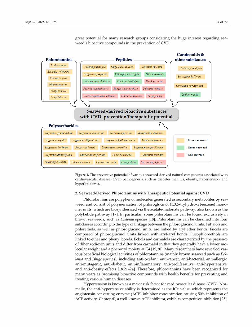
treatment and the links between cardiovascular and metabolic disorders (Figure 1). De‐
spite numerous efforts to improve the industrial use of bioactive seaweed compounds to
prevent CVD, there have been many limitations. The nutraceutical industry demands
more rigorous quality, standardization of components, and a clearer bioactivity mecha‐
nism than previously provided for seaweeds. To overcome this problem, it is necessary to
secure information on the characteristics and diversity of seaweed’s bioactive substances
and systematically understand their overall biological activities in the metabolic disorders
closely related to CVD. We believe this review can provide useful insights and open up
Appl. Sci. 2022, 12, 1025 3 of 27
great potential for many research groups considering the huge interest regarding sea‐
weed’s bioactive compounds in the prevention of CVD.
Figure 1. The preventive potential of various seaweed‐derived natural components associated with
cardiovascular disease (CVD) pathogenesis, such as diabetes mellitus, obesity, hypertension, and
hyperlipidemia.
2. Seaweed‐Derived Phlorotannins with Therapeutic Potential against CVD
Phlorotannins are polyphenol molecules generated as secondary metabolites by sea‐
weed and consist of polymerization of phloroglucinol (1,3,5‐tryhydroxybenzene) mono‐
mer units, which are biosynthesized via the acetate‐malonate pathway, also known as the
polyketide pathway [17]. In particular, some phlorotannins can be found exclusively in
brown seaweeds, such as Ecklonia species [18]. Phlorotannins can be classified into four
subclasses according to the type of linkage between the phloroglucinol units. Fuhalols and
phlorethols, as well as phloroglucinol units, are linked by aryl ether bonds. Fucols are
composed of phloroglucinol units linked with aryl‐aryl bonds. Fucophloroethols are
linked to ether and phenyl bonds. Eckols and carmalols are characterized by the presence
of dibenzodioxin units and differ from carmalol in that they generally have a lower mo‐
lecular weight and a phenoxyl moiety at C4 [19,20]. Many researchers have revealed var‐
ious beneficial biological activities of phlorotannins (mainly brown seaweed such as Eck‐
lonia and Ishige species), including anti‐oxidant, anti‐cancer, anti‐bacterial, anti‐allergic,
anti‐mutagenic, anti‐diabetic, anti‐inflammatiory, anti‐proliferatiive, anti‐hypertensive,
and anti‐obesity effects [18,21–24]. Therefore, phlorotannins have been recognized for
many years as promising bioactive compounds with health benefits for preventing and
treating various human diseases.
Hypertension is known as a major risk factor for cardiovascular disease (CVD). Nor‐
mally, the anti‐hypertensive ability is determined as the IC50 value, which represents the
angiotensin‐converting enzyme (ACE) inhibitor concentration causing 50% inhibition of
ACE activity. Captopril, a well‐known ACE inhibitor, exhibits competitive inhibition [25].
Appl. Sci. 2022, 12, 1025 4 of 27
Additionally, several previous studies have confirmed that phlorotannins such as eckol
[26,27], dieckol [26,27], 6,6′‐bieckol [28], phloroglucinol [26,27], phlorofucofuroeckol A
[27], triphlorethol‐A [26,27], eckstolonol [26,27], fucosterol [27], and octaphlorethol A [29],
isolated from Ecklonia cava, Ecklonia stolonifera, and Ishige foliacea, showed non‐competitive
ACE inhibition (Table 1). These phlorotannins demonstrated ACE inhibitory activity sim‐
ilar to or even higher than that of captopril.
In addition to ACE inhibition, vasodilators also contribute greatly to the anti‐hyper‐
tensive properties. NO is a well‐known vessel‐relaxing factor produced from L‐arginine
by endothelial nitric oxide synthase (eNOS) in the presence of oxygen and the cofactors
Ca2+ and calmodulin (CaM) [30]. A previous study has indicated that genetically deficient
eNOS mice are hypertensive with lower circulating NO levels, thus indicating the critical
role of eNOS and NO in CVD [5,9]. It has also been reported that multiple mechanisms
control NO production via eNOS activation [31].
The phosphatidylinositol 3‐kinase (PI3K) pathway members, including their down‐
stream molecule, protein kinase B (Akt), are essential regulators. Activated Akt would
directly phosphorylate ser1177 on eNOS, enhancing the [Ca2+]/CaM complex [32]. Second,
the concentration of intracellular Ca2+ ([Ca2+]i) in ECs and VSMCs is closely related to the
vascular tone and influences blood pressure [33]. Third, the L‐type calcium channel is one
of the critical ion channels that regulates vasoconstriction and vasodilation. Nitrodilators
promote vasodilation by increasing soluble guanylyl cyclase (cGMP) and decreasing the
[Ca2+]i levels in VSMCs. These actions would reduce the phosphorylation of the Ca2+‐sen‐
sitive myosin light chains, resulting in vasodilation [34]. One study demonstrated that Lu
et al. used the EA.hy926 cells and zebrafish model to systematically establish the potential
mechanisms of the vasodilation produced by diekol and diphlorethohydroxycarmalol iso‐
lated from Ecklonia cava (E. cava) and Ishige okamurae (I. okamurae) [35,36] (Table 1). In
addition, four major phlorotannins—dieckol, 2,7‐phloroglucinol‐6,6‐bieckol, phlorofuco‐
furoeckol A, and pyrogallol‐phloroglucinol‐6,6‐bieckol—isolated from Ecklonia cava effec‐
tively inhibited monocyte‐associated vascular inflammation and dysfunction by sup‐
pressing monocyte migration and protecting monocyte‐associated endothelial cell death
[37]. Moreover, pyrogallol‐phloroglucinol‐6,6′‐bieckol and dieckol isolated from E. Cava
have been shown to improve blood circulation in mice fed diets to induce obesity and
hypertension [36,38]. Therefore, while numerous factors contribute to hypertension, it is
well recognized that an increased vascular tone is always the ultimate goal.
The evidence mentioned above has great pharmaceutical potential, with some al‐
ready being used in the clinical trial phase. Clinical studies have shown cardiovascular
protection, supporting the benefits of polyphenols extracted from land plants [39]. Nev‐
ertheless, only a few epidemiological studies have discussed the association between the
consumption of specific components of seaweed and blood pressure. Despite investigat‐
ing the benefits of single compounds from seaweed, the general components, including
minerals, fiber, and peptides, were more popular in clinical trials. This might be because
high doses of these compounds have comparatively low side effects. A case control study
demonstrated the anti‐hypertensive effect of Undaria pinnatifida powder (5 g/capsule/day)
by significantly reducing blood pressure in elderly Japanese patients following 8 weeks
of administration [40]. However, Murray et al. reported that no reduction in blood pres‐
sure occurred in healthy adults who were administered the Fucus vesiculosus extract [41].
As mentioned above, several phlorotannins have been confirmed to possess effects
against hypertension by ACE inhibition, calcium regulation, antagonism of L‐type cal‐
cium channels, or activation of the critical pathway (PI3K/Akt/eNOS). Despite these mech‐
anisms being verified, many other physiological signaling pathways in vascular regula‐
tion remain unclear. It will be helpful to explore the cardiovascular protection or anti‐
hypertension properties of phlorotannin via its use as a potassium channel opener and
angiotensin receptor blocker.
Diabetes mellitus (DM) is a prime risk factor for dramatically increasing CVD, con‐
tributing to more than 3 million cardiovascular deaths worldwide each year [42]. Type 2
Appl. Sci. 2022, 12, 1025 5 of 27
diabetes mellitus (T2DM) is a complex metabolic disorder involving insulin resistance,
impaired insulin signaling and β‐cell dysfunction, and abnormal glucose and lipid metab‐
olism. Hyperglycemia is the most important criterion for all types of diabetes and is the
cause of diabetic complications such as CVD. Therefore, it is important to prevent or delay
the onset of diabetes by controlling blood glucose levels in diabetic patients, as hypergly‐
cemia increases the risk of developing CVD long before clinical diabetes begins [43]. In
humans, α‐glucosidase and α‐amylase in the small intestine play an important role in the
digesting dietary carbohydrates into glucose. Therefore, reducing postprandial hypergly‐
cemia by delaying the absorption of glucose in the body through the inhibition of α‐am‐
ylase and α‐glucosidase is an important approach for treating T2DM [44,45]. The enzyme
inhibitors used for this action suppress the digestion of carbohydrates and consequently
slow the postprandial plasma glucose rise, thereby delaying the rate of glucose absorption
[46]. The inhibition of starch‐digesting enzymes using synthetic drugs exhibiting antidia‐
betic effects with α‐glucosidase inhibitory properties, such as acarbose, voglibose,
miglitol, and emiglitate, is an important clinical strategy for controlling postprandial hy‐
perglycemia. However, these synthetic drugs have been reported to effectively lower
postprandial blood glucose levels but cause serious side effects, such as liver disorders
[45,47]. The use of enzyme inhibitors derived from natural products (terrestrial plants or
seaweeds) with a lower risk of the potential side effects caused by synthetic enzyme in‐
hibitors is recommended. Therefore, to avoid or reduce the side effects caused by cur‐
rently used synthetase inhibitors, the use of enzyme inhibitors (α‐amylase and α‐gluco‐
sidase) from natural products is considered the best alternative. It has been reported that
minor phlorotannin derivatives such as eckol, 2‐phloroeckol, 8,8′‐bieckol, 6,8′‐bieckol, and
2‐O‐(2,4,6‐trihydroxyphenyl)‐6,6′‐bieckol isolated from E. cava showed α‐glucosidase in‐
hibitory activity, with an IC50 value ranging from 2.3 to 59.8 μM (Table 1). Among the
minor phlorotannin derivatives, 8,8′‐bieckol and 6,8′‐bieckol exhibited the strongest α‐
glucosidase inhibitory activity. In addition, molecular docking studies revealed that both
phlorotannins act as α‐glucosidase inhibitors by competitive inhibition [48]. In addition,
several researchers have found that phlorotannins such as fucodiphloroethol G, dieckol,
6,6′‐bieckol, 7‐phloroeckol, phlorofucofuroeckol A, and 2,7′‐phloroglucinol‐6,6′‐bieckol
isolated from Ecklonia cava have inhibitory activity against α‐glucosidase and α‐amylase
[49–51]. Moon et al. revealed that phlorotannins, such as phloroglucinol, dioxinodehy‐
droeckol, eckol, phlorofucofuroeckol‐A, dieckol, and 7‐phloroeckol isolated from Eck‐
lonia stolinifera (E. stolinifera) have anti‐diabetic effects by inhibiting α‐glucosidase. In
addition, it was confirmed that these phlorotannins effectively inhibit protein tyrosine
phosphatase 1 B, an enzyme that plays an important role in the development of insulin
resistance, thereby preventing a rapid increase in postprandial blood glucose levels [52].
In addition, diphlorethohydroxycarmalol (DPHC), ishophloroglucin A (IPA), and oc‐
taphlorethol A (OPA) were isolated from I. okamurae and Ishige foliacea (I. foliacea) from
the Ishigeaceae family and showed α‐glucosidase and α‐amylase inhibitory activity
[53,54]. In particular, oral administration of 100 mg/kg DPHC significantly suppressed the
postprandial blood glucose levels in streptozotocin‐induced diabetic mice [55]. According
to Lee et al., molecular docking analysis revealed that OPA interacts with amino acid res‐
idues in the region close to the active site of α‐glucosidase. Hence, OPA has the potential
to be used as a non‐competitive inhibitor with a high‐affinity binding site for α‐gluco‐
sidase [54].
Insulin resistance and impaired glucose metabolism are the most common factors
promoting the development of type 2 diabetes mellitus. Type 2 diabetes mellitus is typi‐
cally caused by two factors: impaired insulin production by pancreatic β cells and a failure
of insulin‐sensitive tissues to respond appropriately to insulin [56]. Signaling pathways
involved in insulin secretion in β‐cells under physiological conditions can be divided into
a few stages. First, insulin release is primarily triggered by high glucose concentrations,
mainly via glucose transporter 2 (GLUT2). When glucose catabolism is stimulated, intra‐
cellular ATP levels increase and close the cell membrane potassium channels, increasing
Appl. Sci. 2022, 12, 1025 6 of 27
intracellular Ca2+ concentrations and amplifying insulin secretion [57,58]. Furthermore,
the AMP‐activated protein kinase (AMPK) system simulates the effect of insulin on glu‐
cose transport in the muscle and glucose production in the liver. Because the AMPK sys‐
tem plays an important role in glucose homeostasis, it is a key target for discovering anti‐
diabetic agents [59]. Some evidence has demonstrated that 2,7″‐phloroglucinol‐6,6′‐
bieckol isolated from E. cava protects against high glucose‐induced glucotoxicity and
apoptotic cell death in INS‐1 cells [60] (Table 1). In addition, in C57BL/KsJ‐db/db mice, the
oral administration of 20 mg/kg dieckol isolated from E. cava significantly reduced blood
glucose levels, serum insulin levels, and body weight [61]. Yang et al. demonstrated that
oral administration of ishophloroglucin A (IPA) and oxtaphlorethol A (OPA) isolated
from I. okamurae and I. foliacea significantly ameliorated glucose intolerance and the fasting
glucose levels in high‐fat diet (HFD)‐fed mice, thereby reducing fasting and 2 h blood
glucose levels, as well as stimulated GLUT4 in HFD mouse muscle [62,63]. Furthermore,
phlorotannins such as DPHC and IPA isolated from I. okamurae treatment exhibited an
anti‐angiogenic effect by interfering with the VEGFR‐2 signaling pathway [64,65]. These
phlorotannin compounds isolated from various seaweeds exhibit anti‐diabetic efficacy
through various mechanisms in vivo and in vitro, demonstrating the potential as natural
agents that can replace synthetic anti‐diabetic treatments. Despite the evidence suggesting
that type 2 diabetes mellitus might be caused by more complex molecular pathways im‐
plicated in cell biology, the main concepts of treatments still mainly focus on the pathway
we mentioned above, and clinical trials using single compounds isolated from seaweeds
have not been reported.
Despite its relatively simple definition, obesity, defined as excess body fat, is a com‐
plex condition resulting from a chronic positive energy balance when the dietary energy
intake exceeds energy expenditure. Excess energy is converted to triglycerides and stored
in adipose tissue depots, expanding in size, producing weight gain, and increasing body
fat. The adipose tissue includes several types of cells, such as mature adipocytes, pre‐adi‐
pocytes, fibroblasts, endothelial cells, and immune cells [66]. Adipose tissue undergoes
dynamic histological changes as obesity progresses, including adipocyte enlargement, en‐
hanced angiogenesis, immune cell infiltration, and extracellular matrix overproduction
[67]. In contrast, adipocytes undergo differentiation, and specific proteins, including sterol
regulatory element‐binding protein 1 (SREBP1c), CCAAT/enhancer‐binding protein
(C/EBPα), and peroxisome proliferator‐activated receptor γ (PPARγ) are involved in all
processes [68,69]. Several lines of evidence indicate that the phlorotannins contained in
seaweed such as E. cava (eckol, dieckol, triphlorethol‐A, 6,6′‐bieckol, and phlorofucoeckol
A), Ecklonia stolonifera (phloroglucinol, eckol, dieckol, dioxinodehydroeckol, and
phlorofucofuroeckol A), Eisenia bicyclis (6,6′‐bieckol, 6,8′‐bieckol, 8,8′‐bieckol, and dieckol,
phlorofucofuroeckol A), and I. okamurae (diphlorethohydroxycarmalol) exhibit anti‐obe‐
sity properties by inhibiting intracellular lipid accumulation, suppressing adipogenesis in
3T3‐L1 cells via inhibiting the expression of PPARγ, C/EBPα, SREBP‐1, and FABP4, and
activating AMP‐activated protein kinase (AMPK) and ACC phosphorylation [70–77] (Ta‐
ble 1). In addition to these mechanisms, some phlorotannins isolated from brown seaweed
exhibited anti‐obesity effects by reducing leptin resistance or inhibiting pancreatic lipase
[78,79]. Additionally, oral administration of phlorotannins such as eckol and dieckol iso‐
lated from E. cava and Ecklonia stolonifera showed antihyperlipidemic effects by decreasing
the total cholesterol levels, triglyceride levels, and low‐density lipoprotein levels and in‐
creasing the high‐density lipoprotein in the serum of high‐fat diet mice [80,81].
Appl. Sci. 2022, 12, 1025 7 of 27
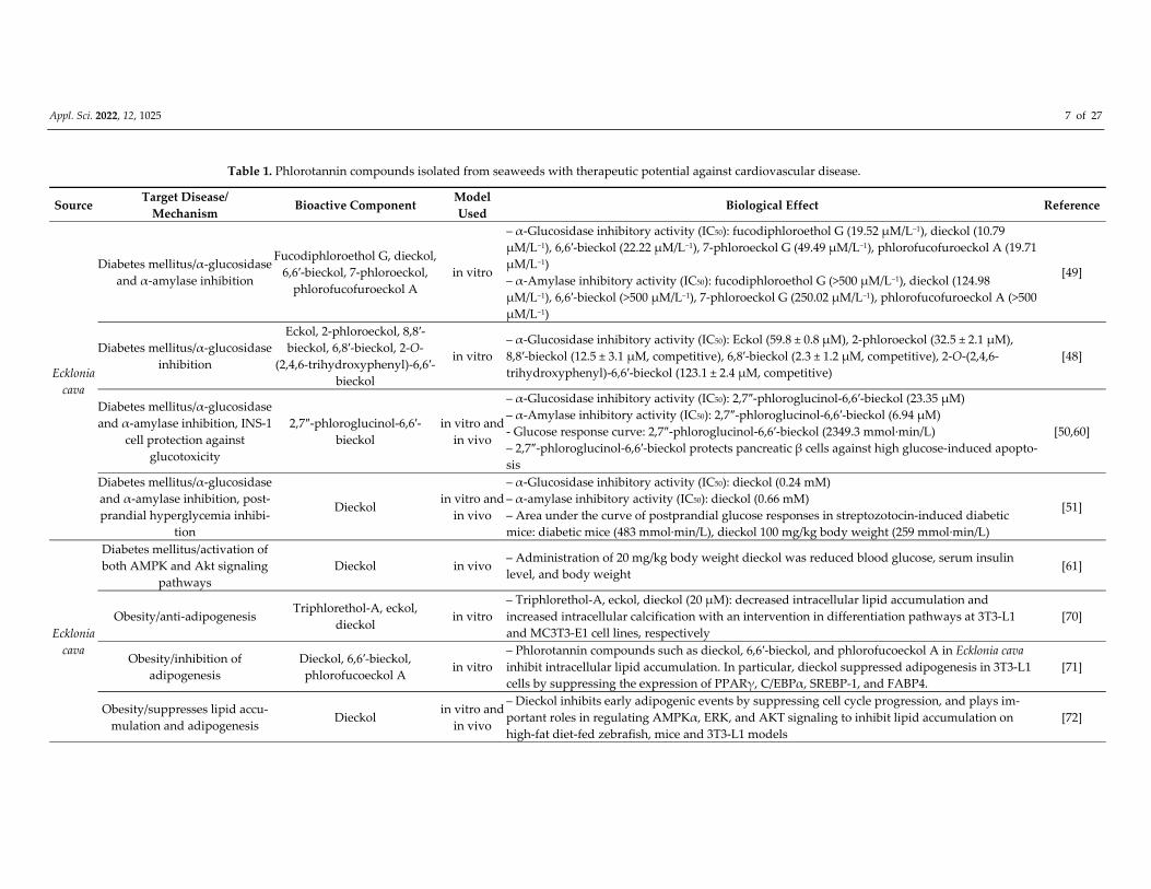
Table 1. Phlorotannin compounds isolated from seaweeds with therapeutic potential against cardiovascular disease.
Source Target Disease/
Mechanism Bioactive Component
Model
Used Biological Effect Reference
Ecklonia
cava
Diabetes mellitus/α‐glucosidase
and α‐amylase inhibition
Fucodiphloroethol G, dieckol,
6,6′‐bieckol, 7‐phloroeckol,
phlorofucofuroeckol A
in vitro
– α‐Glucosidase inhibitory activity (IC50): fucodiphloroethol G (19.52 μM/L−1), dieckol (10.79
μM/L−1), 6,6′‐bieckol (22.22 μM/L−1), 7‐phloroeckol G (49.49 μM/L−1), phlorofucofuroeckol A (19.71
μM/L−1)
– α‐Amylase inhibitory activity (IC50): fucodiphloroethol G (>500 μM/L−1), dieckol (124.98
μM/L−1), 6,6′‐bieckol (>500 μM/L−1), 7‐phloroeckol G (250.02 μM/L−1), phlorofucofuroeckol A (>500
μM/L−1)
[49]
Diabetes mellitus/α‐glucosidase
inhibition
Eckol, 2‐phloroeckol, 8,8′‐
bieckol, 6,8′‐bieckol, 2‐O‐
(2,4,6‐trihydroxyphenyl)‐6,6′‐
bieckol
in vitro
– α‐Glucosidase inhibitory activity (IC50): Eckol (59.8 ± 0.8 μM), 2‐phloroeckol (32.5 ± 2.1 μM),
8,8′‐bieckol (12.5 ± 3.1 μM, competitive), 6,8′‐bieckol (2.3 ± 1.2 μM, competitive), 2‐O‐(2,4,6‐
trihydroxyphenyl)‐6,6′‐bieckol (123.1 ± 2.4 μM, competitive)
[48]
Diabetes mellitus/α‐glucosidase
and α‐amylase inhibition, INS‐1
cell protection against
glucotoxicity
2,7″‐phloroglucinol‐6,6′‐
bieckol
in vitro and
in vivo
– α‐Glucosidase inhibitory activity (IC50): 2,7″‐phloroglucinol‐6,6′‐bieckol (23.35 μM)
– α‐Amylase inhibitory activity (IC50): 2,7″‐phloroglucinol‐6,6′‐bieckol (6.94 μM)
‐ Glucose response curve: 2,7″‐phloroglucinol‐6,6′‐bieckol (2349.3 mmol∙min/L)
– 2,7″‐phloroglucinol‐6,6′‐bieckol protects pancreatic β cells against high glucose‐induced apopto‐
sis
[50,60]
Diabetes mellitus/α‐glucosidase
and α‐amylase inhibition, post‐
prandial hyperglycemia inhibi‐
tion
Dieckol in vitro and
in vivo
– α‐Glucosidase inhibitory activity (IC50): dieckol (0.24 mM)
– α‐amylase inhibitory activity (IC50): dieckol (0.66 mM)
– Area under the curve of postprandial glucose responses in streptozotocin‐induced diabetic
mice: diabetic mice (483 mmol∙min/L), dieckol 100 mg/kg body weight (259 mmol∙min/L)
[51]
Ecklonia
cava
Diabetes mellitus/activation of
both AMPK and Akt signaling
pathways
Dieckol in vivo – Administration of 20 mg/kg body weight dieckol was reduced blood glucose, serum insulin
level, and body weight [61]
Obesity/anti‐adipogenesis Triphlorethol‐A, eckol,
dieckol in vitro
– Triphlorethol‐A, eckol, dieckol (20 μM): decreased intracellular lipid accumulation and
increased intracellular calcification with an intervention in differentiation pathways at 3T3‐L1
and MC3T3‐E1 cell lines, respectively
[70]
Obesity/inhibition of
adipogenesis
Dieckol, 6,6′‐bieckol,
phlorofucoeckol A in vitro
– Phlorotannin compounds such as dieckol, 6,6′‐bieckol, and phlorofucoeckol A in Ecklonia cava
inhibit intracellular lipid accumulation. In particular, dieckol suppressed adipogenesis in 3T3‐L1
cells by suppressing the expression of PPARγ, C/EBPα, SREBP‐1, and FABP4.
[71]
Obesity/suppresses lipid accu‐
mulation and adipogenesis Dieckol
in vitro and
in vivo
– Dieckol inhibits early adipogenic events by suppressing cell cycle progression, and plays im‐
portant roles in regulating AMPKα, ERK, and AKT signaling to inhibit lipid accumulation on
high‐fat diet‐fed zebrafish, mice and 3T3‐L1 models
[72]
Appl. Sci. 2022, 12, 1025 8 of 27
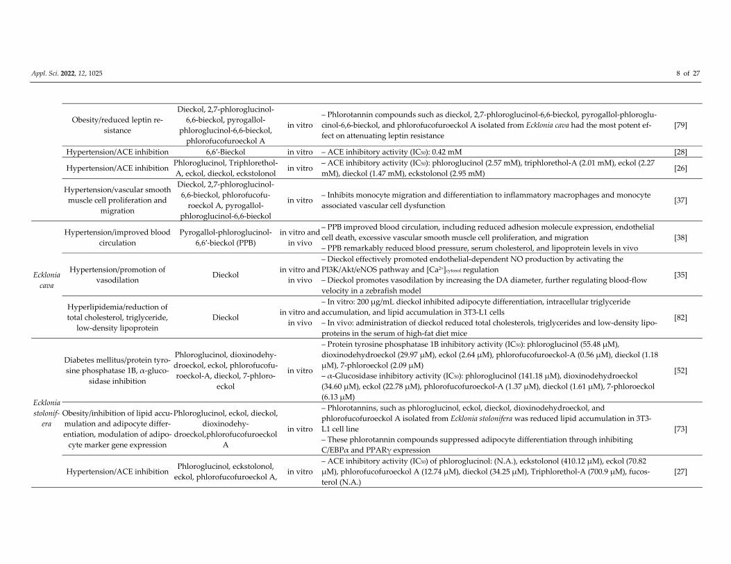
Obesity/reduced leptin re‐
sistance
Dieckol, 2,7‐phloroglucinol‐
6,6‐bieckol, pyrogallol‐
phloroglucinol‐6,6‐bieckol,
phlorofucofuroeckol A
in vitro
– Phlorotannin compounds such as dieckol, 2,7‐phloroglucinol‐6,6‐bieckol, pyrogallol‐phloroglu‐
cinol‐6,6‐bieckol, and phlorofucofuroeckol A isolated from Ecklonia cava had the most potent ef‐
fect on attenuating leptin resistance
[79]
Hypertension/ACE inhibition 6,6′‐Bieckol in vitro – ACE inhibitory activity (IC50): 0.42 mM [28]
Hypertension/ACE inhibition Phloroglucinol, Triphlorethol‐
A, eckol, dieckol, eckstolonol in vitro
– ACE inhibitory activity (IC50): phloroglucinol (2.57 mM), triphlorethol‐A (2.01 mM), eckol (2.27
mM), dieckol (1.47 mM), eckstolonol (2.95 mM) [26]
Hypertension/vascular smooth
muscle cell proliferation and
migration
Dieckol, 2,7‐phloroglucinol‐
6,6‐bieckol, phlorofucofu‐
roeckol A, pyrogallol‐
phloroglucinol‐6,6‐bieckol
in vitro – Inhibits monocyte migration and differentiation to inflammatory macrophages and monocyte
associated vascular cell dysfunction [37]
Ecklonia
cava
Hypertension/improved blood
circulation
Pyrogallol‐phloroglucinol‐
6,6′‐bieckol (PPB)
in vitro and
in vivo
– PPB improved blood circulation, including reduced adhesion molecule expression, endothelial
cell death, excessive vascular smooth muscle cell proliferation, and migration
– PPB remarkably reduced blood pressure, serum cholesterol, and lipoprotein levels in vivo
[38]
Hypertension/promotion of
vasodilation Dieckol
in vitro and
in vivo
– Dieckol effectively promoted endothelial‐dependent NO production by activating the
PI3K/Akt/eNOS pathway and [Ca2+]cytosol regulation
– Dieckol promotes vasodilation by increasing the DA diameter, further regulating blood‐flow
velocity in a zebrafish model
[35]
Hyperlipidemia/reduction of
total cholesterol, triglyceride,
low‐density lipoprotein
Dieckol in vitro and
in vivo
– In vitro: 200 μg/mL dieckol inhibited adipocyte differentiation, intracellular triglyceride
accumulation, and lipid accumulation in 3T3‐L1 cells
– In vivo: administration of dieckol reduced total cholesterols, triglycerides and low‐density lipo‐
proteins in the serum of high‐fat diet mice
[82]
Ecklonia
stolonif‐
era
Diabetes mellitus/protein tyro‐
sine phosphatase 1B, α‐gluco‐
sidase inhibition
Phloroglucinol, dioxinodehy‐
droeckol, eckol, phlorofucofu‐
roeckol‐A, dieckol, 7‐phloro‐
eckol
in vitro
– Protein tyrosine phosphatase 1B inhibitory activity (IC50): phloroglucinol (55.48 μM),
dioxinodehydroeckol (29.97 μM), eckol (2.64 μM), phlorofucofuroeckol‐A (0.56 μM), dieckol (1.18
μM), 7‐phloroeckol (2.09 μM)
– α‐Glucosidase inhibitory activity (IC50): phloroglucinol (141.18 μM), dioxinodehydroeckol
(34.60 μM), eckol (22.78 μM), phlorofucofuroeckol‐A (1.37 μM), dieckol (1.61 μM), 7‐phloroeckol
(6.13 μM)
[52]
Obesity/inhibition of lipid accu‐
mulation and adipocyte differ‐
entiation, modulation of adipo‐
cyte marker gene expression
Phloroglucinol, eckol, dieckol,
dioxinodehy‐
droeckol,phlorofucofuroeckol
A
in vitro
– Phlorotannins, such as phloroglucinol, eckol, dieckol, dioxinodehydroeckol, and
phlorofucofuroeckol A isolated from Ecklonia stolonifera was reduced lipid accumulation in 3T3‐
L1 cell line
– These phlorotannin compounds suppressed adipocyte differentiation through inhibiting
C/EBPα and PPARγ expression
[73]
Hypertension/ACE inhibition Phloroglucinol, eckstolonol,
eckol, phlorofucofuroeckol A, in vitro
– ACE inhibitory activity (IC50) of phloroglucinol: (N.A.), eckstolonol (410.12 μM), eckol (70.82
μM), phlorofucofuroeckol A (12.74 μM), dieckol (34.25 μM), Triphlorethol‐A (700.9 μM), fucos‐
terol (N.A.)
[27]
Appl. Sci. 2022, 12, 1025 9 of 27
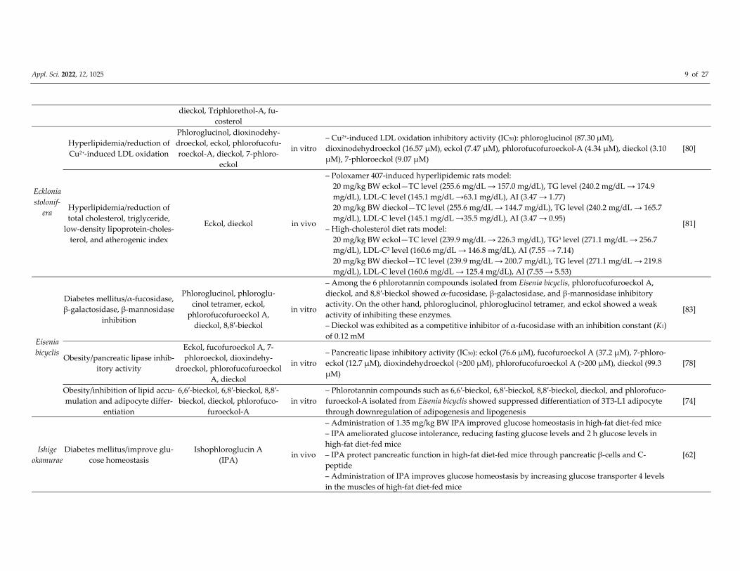
dieckol, Triphlorethol‐A, fu‐
costerol
Ecklonia
stolonif‐
era
Hyperlipidemia/reduction of
Cu2+‐induced LDL oxidation
Phloroglucinol, dioxinodehy‐
droeckol, eckol, phlorofucofu‐
roeckol‐A, dieckol, 7‐phloro‐
eckol
in vitro
– Cu2+‐induced LDL oxidation inhibitory activity (IC50): phloroglucinol (87.30 μM),
dioxinodehydroeckol (16.57 μM), eckol (7.47 μM), phlorofucofuroeckol‐A (4.34 μM), dieckol (3.10
μM), 7‐phloroeckol (9.07 μM)
[80]
Hyperlipidemia/reduction of
total cholesterol, triglyceride,
low‐density lipoprotein‐choles‐
terol, and atherogenic index
Eckol, dieckol in vivo
– Poloxamer 407‐induced hyperlipidemic rats model:
20 mg/kg BW eckol—TC level (255.6 mg/dL → 157.0 mg/dL), TG level (240.2 mg/dL → 174.9 mg/dL), LDL‐C level (145.1 mg/dL →63.1 mg/dL), AI (3.47 → 1.77) 20 mg/kg BW dieckol—TC level (255.6 mg/dL → 144.7 mg/dL), TG level (240.2 mg/dL → 165.7 mg/dL), LDL‐C level (145.1 mg/dL →35.5 mg/dL), AI (3.47 → 0.95)
– High‐cholesterol diet rats model:
20 mg/kg BW eckol—TC level (239.9 mg/dL → 226.3 mg/dL), TG3 level (271.1 mg/dL → 256.7 mg/dL), LDL‐C3 level (160.6 mg/dL → 146.8 mg/dL), AI (7.55 → 7.14) 20 mg/kg BW dieckol—TC level (239.9 mg/dL → 200.7 mg/dL), TG level (271.1 mg/dL → 219.8 mg/dL), LDL‐C level (160.6 mg/dL → 125.4 mg/dL), AI (7.55 → 5.53)
[81]
Eisenia
bicyclis
Diabetes mellitus/α‐fucosidase,
β‐galactosidase, β‐mannosidase
inhibition
Phloroglucinol, phloroglu‐
cinol tetramer, eckol,
phlorofucofuroeckol A,
dieckol, 8,8′‐bieckol
in vitro
– Among the 6 phlorotannin compounds isolated from Eisenia bicyclis, phlorofucofuroeckol A,
dieckol, and 8,8′‐bieckol showed α‐fucosidase, β‐galactosidase, and β‐mannosidase inhibitory
activity. On the other hand, phloroglucinol, phloroglucinol tetramer, and eckol showed a weak
activity of inhibiting these enzymes.
– Dieckol was exhibited as a competitive inhibitor of α‐fucosidase with an inhibition constant (K1)
of 0.12 mM
[83]
Obesity/pancreatic lipase inhib‐
itory activity
Eckol, fucofuroeckol A, 7‐
phloroeckol, dioxindehy‐
droeckol, phlorofucofuroeckol
A, dieckol
in vitro
– Pancreatic lipase inhibitory activity (IC50): eckol (76.6 μM), fucofuroeckol A (37.2 μM), 7‐phloro‐
eckol (12.7 μM), dioxindehydroeckol (>200 μM), phlorofucofuroeckol A (>200 μM), dieckol (99.3
μM)
[78]
Obesity/inhibition of lipid accu‐
mulation and adipocyte differ‐
entiation
6,6′‐bieckol, 6,8′‐bieckol, 8,8′‐
bieckol, dieckol, phlorofuco‐
furoeckol‐A
in vitro
– Phlorotannin compounds such as 6,6′‐bieckol, 6,8′‐bieckol, 8,8′‐bieckol, dieckol, and phlorofuco‐
furoeckol‐A isolated from Eisenia bicyclis showed suppressed differentiation of 3T3‐L1 adipocyte
through downregulation of adipogenesis and lipogenesis
[74]
Ishige
okamurae
Diabetes mellitus/improve glu‐
cose homeostasis
Ishophloroglucin A
(IPA) in vivo
– Administration of 1.35 mg/kg BW IPA improved glucose homeostasis in high‐fat diet‐fed mice
– IPA ameliorated glucose intolerance, reducing fasting glucose levels and 2 h glucose levels in
high‐fat diet‐fed mice
– IPA protect pancreatic function in high‐fat diet‐fed mice through pancreatic β‐cells and C‐
peptide
– Administration of IPA improves glucose homeostasis by increasing glucose transporter 4 levels
in the muscles of high‐fat diet‐fed mice
[62]
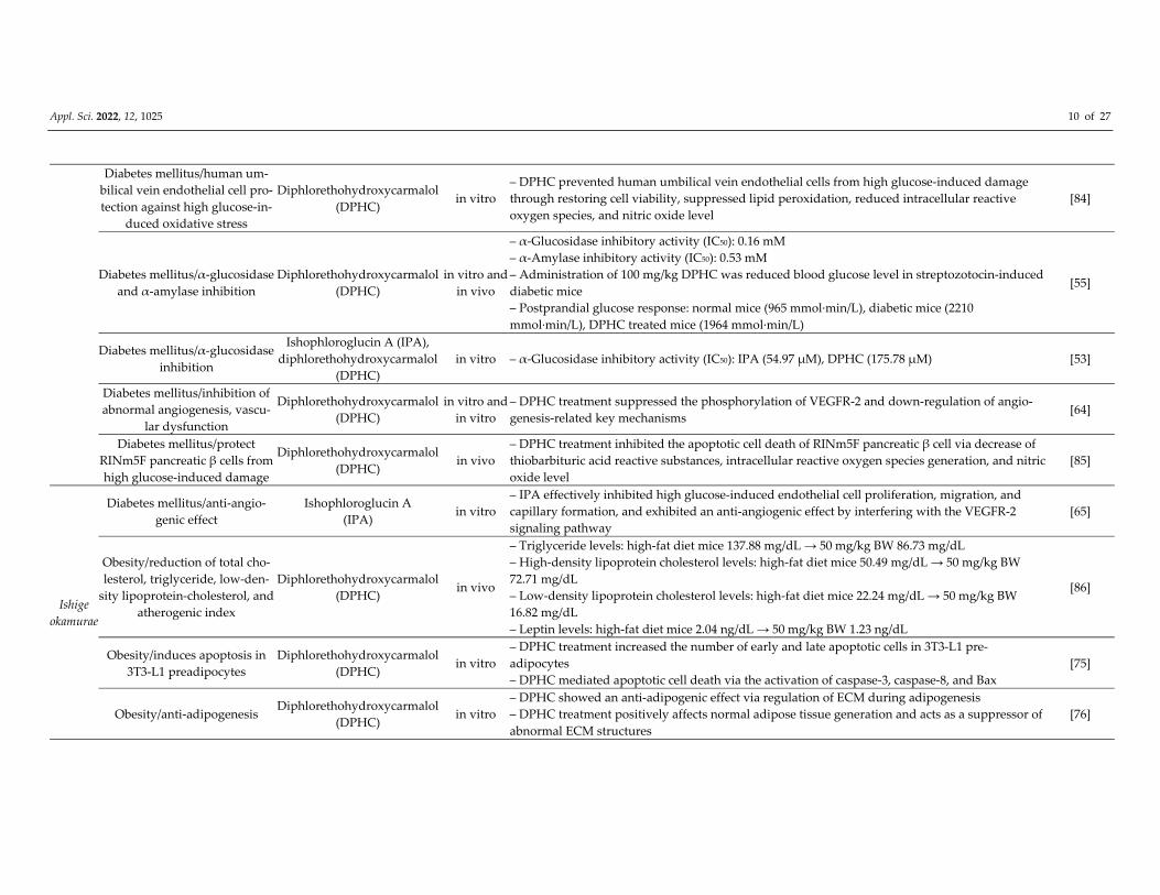
Appl. Sci. 2022, 12, 1025 10 of 27
Diabetes mellitus/human um‐
bilical vein endothelial cell pro‐
tection against high glucose‐in‐
duced oxidative stress
Diphlorethohydroxycarmalol
(DPHC) in vitro
– DPHC prevented human umbilical vein endothelial cells from high glucose‐induced damage
through restoring cell viability, suppressed lipid peroxidation, reduced intracellular reactive
oxygen species, and nitric oxide level
[84]
Diabetes mellitus/α‐glucosidase
and α‐amylase inhibition
Diphlorethohydroxycarmalol
(DPHC)
in vitro and
in vivo
– α‐Glucosidase inhibitory activity (IC50): 0.16 mM
– α‐Amylase inhibitory activity (IC50): 0.53 mM
– Administration of 100 mg/kg DPHC was reduced blood glucose level in streptozotocin‐induced
diabetic mice
– Postprandial glucose response: normal mice (965 mmol∙min/L), diabetic mice (2210
mmol∙min/L), DPHC treated mice (1964 mmol∙min/L)
[55]
Diabetes mellitus/α‐glucosidase
inhibition
Ishophloroglucin A (IPA),
diphlorethohydroxycarmalol
(DPHC)
in vitro – α‐Glucosidase inhibitory activity (IC50): IPA (54.97 μM), DPHC (175.78 μM) [53]
Diabetes mellitus/inhibition of
abnormal angiogenesis, vascu‐
lar dysfunction
Diphlorethohydroxycarmalol
(DPHC)
in vitro and
in vitro
– DPHC treatment suppressed the phosphorylation of VEGFR‐2 and down‐regulation of angio‐
genesis‐related key mechanisms [64]
Diabetes mellitus/protect
RINm5F pancreatic β cells from
high glucose‐induced damage
Diphlorethohydroxycarmalol
(DPHC) in vivo
– DPHC treatment inhibited the apoptotic cell death of RINm5F pancreatic β cell via decrease of
thiobarbituric acid reactive substances, intracellular reactive oxygen species generation, and nitric
oxide level
[85]
Ishige
okamurae
Diabetes mellitus/anti‐angio‐
genic effect
Ishophloroglucin A
(IPA) in vitro
– IPA effectively inhibited high glucose‐induced endothelial cell proliferation, migration, and
capillary formation, and exhibited an anti‐angiogenic effect by interfering with the VEGFR‐2
signaling pathway
[65]
Obesity/reduction of total cho‐
lesterol, triglyceride, low‐den‐
sity lipoprotein‐cholesterol, and
atherogenic index
Diphlorethohydroxycarmalol
(DPHC) in vivo
– Triglyceride levels: high‐fat diet mice 137.88 mg/dL → 50 mg/kg BW 86.73 mg/dL
– High‐density lipoprotein cholesterol levels: high‐fat diet mice 50.49 mg/dL → 50 mg/kg BW
72.71 mg/dL
– Low‐density lipoprotein cholesterol levels: high‐fat diet mice 22.24 mg/dL → 50 mg/kg BW
16.82 mg/dL
– Leptin levels: high‐fat diet mice 2.04 ng/dL → 50 mg/kg BW 1.23 ng/dL
[86]
Obesity/induces apoptosis in
3T3‐L1 preadipocytes
Diphlorethohydroxycarmalol
(DPHC) in vitro
– DPHC treatment increased the number of early and late apoptotic cells in 3T3‐L1 pre‐
adipocytes
– DPHC mediated apoptotic cell death via the activation of caspase‐3, caspase‐8, and Bax
[75]
Obesity/anti‐adipogenesis Diphlorethohydroxycarmalol
(DPHC) in vitro
– DPHC showed an anti‐adipogenic effect via regulation of ECM during adipogenesis
– DPHC treatment positively affects normal adipose tissue generation and acts as a suppressor of
abnormal ECM structures
[76]
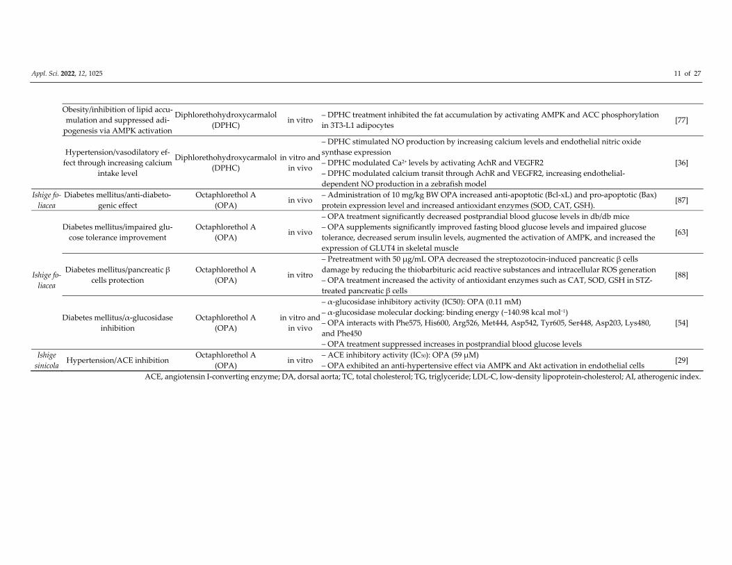
Appl. Sci. 2022, 12, 1025 11 of 27
Obesity/inhibition of lipid accu‐
mulation and suppressed adi‐
pogenesis via AMPK activation
Diphlorethohydroxycarmalol
(DPHC) in vitro
– DPHC treatment inhibited the fat accumulation by activating AMPK and ACC phosphorylation
in 3T3‐L1 adipocytes [77]
Hypertension/vasodilatory ef‐
fect through increasing calcium
intake level
Diphlorethohydroxycarmalol
(DPHC)
in vitro and
in vivo
– DPHC stimulated NO production by increasing calcium levels and endothelial nitric oxide
synthase expression
– DPHC modulated Ca2+ levels by activating AchR and VEGFR2
– DPHC modulated calcium transit through AchR and VEGFR2, increasing endothelial‐
dependent NO production in a zebrafish model
[36]
Ishige fo‐
liacea
Diabetes mellitus/anti‐diabeto‐
genic effect
Octaphlorethol A
(OPA) in vivo
– Administration of 10 mg/kg BW OPA increased anti‐apoptotic (Bcl‐xL) and pro‐apoptotic (Bax)
protein expression level and increased antioxidant enzymes (SOD, CAT, GSH). [87]
Ishige fo‐
liacea
Diabetes mellitus/impaired glu‐
cose tolerance improvement
Octaphlorethol A
(OPA) in vivo
– OPA treatment significantly decreased postprandial blood glucose levels in db/db mice
– OPA supplements significantly improved fasting blood glucose levels and impaired glucose
tolerance, decreased serum insulin levels, augmented the activation of AMPK, and increased the
expression of GLUT4 in skeletal muscle
[63]
Diabetes mellitus/pancreatic β
cells protection
Octaphlorethol A
(OPA) in vitro
– Pretreatment with 50 μg/mL OPA decreased the streptozotocin‐induced pancreatic β cells
damage by reducing the thiobarbituric acid reactive substances and intracellular ROS generation
– OPA treatment increased the activity of antioxidant enzymes such as CAT, SOD, GSH in STZ‐
treated pancreatic β cells
[88]
Diabetes mellitus/α‐glucosidase
inhibition
Octaphlorethol A
(OPA)
in vitro and
in vivo
– α‐glucosidase inhibitory activity (IC50): OPA (0.11 mM)
– α‐glucosidase molecular docking: binding energy (−140.98 kcal mol−1)
– OPA interacts with Phe575, His600, Arg526, Met444, Asp542, Tyr605, Ser448, Asp203, Lys480,
and Phe450
– OPA treatment suppressed increases in postprandial blood glucose levels
[54]
Ishige
sinicola Hypertension/ACE inhibition
Octaphlorethol A
(OPA) in vitro
– ACE inhibitory activity (IC50): OPA (59 μM)
– OPA exhibited an anti‐hypertensive effect via AMPK and Akt activation in endothelial cells [29]
ACE, angiotensin I‐converting enzyme; DA, dorsal aorta; TC, total cholesterol; TG, triglyceride; LDL‐C, low‐density lipoprotein‐cholesterol; AI, atherogenic index.
Appl. Sci. 2022, 12, 1025 12 of 27
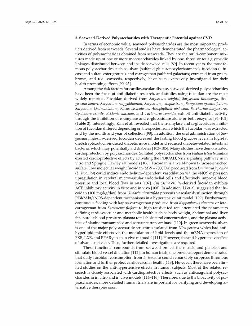
3. Seaweed‐Derived Polysaccharides with Therapeutic Potential against CVD
In terms of economic value, seaweed polysaccharides are the most important prod‐
ucts derived from seaweeds. Several studies have demonstrated the pharmacological ac‐
tivities of polysaccharides obtained from seaweeds. They are the multi‐component mix‐
tures made up of one or more monosaccharides linked by one, three, or four glycosidic
linkages distributed between and inside seaweed cells [89]. In recent years, the most fa‐
mous polysaccharides such as ulvan (sulfated glucuronoxylorhamnans), fucoidan (L‐fu‐
cose and sulfate ester groups), and carrageenan (sulfated galactans) extracted from green,
brown, and red seaweeds, respectively, have been extensively investigated for their
health‐promoting effects [90–93].
Among the risk factors for cardiovascular disease, seaweed‐derived polysaccharides
have been the focus of anti‐diabetic research, and studies using fucoidan are the most
widely reported. Fucoidan derived from Sargassum wightii, Sargassum thumbergii, Sra‐
gassum honeri, Sargassum ringgoldianum, Sargassum, siliquastrum, Sargassum graminifolium,
Sargassum kjellmanianum, Fucus vesiculosus, Ascophyllum nodosum, Saccharina longicruris,
Cystoseira crinite, Ecklonia maxima, and Turbinaria conoides exhibit anti‐diabetic activity
through the inhibition of α‐amylase and α‐glucosidase alone or both enzymes [94–102]
(Table 2). Interestingly, Kim et al. revealed that the α‐amylase and α‐glucosidase inhibi‐
tion of fucoidan differed depending on the species from which the fucoidan was extracted
and by the month and year of collection [98]. In addition, the oral administration of Sar‐
gassum fusiforme‐derived fucoidan decreased the fasting blood glucose levels in high‐fat
diet/streptozotocin‐induced diabetic mice model and reduced diabetes‐related intestinal
bacteria, which may potentially aid diabetes [103–105]. Many studies have demonstrated
cardioprotection by polysaccharides. Sulfated polysaccharides from Padina tetrastromatica
exerted cardioprotective effects by activating the PI3K/Akt/Nrf2 signaling pathway in in
vitro and Sprague Dawley rat models [106]. Fucoidan is a well‐known L‐fucose‐enriched
sulfate. Low molecular weight fucoidan (MW = 7000 Da) produced from Laminaria japonica
(L. japonica) could induce endothelium‐dependent vasodilation via the eNOS expression
upregulation in cerebral microvascular endothelial cells and effectively improve blood
pressure and local blood flow in rats [107]. Cystoseira crinite‐derived fucoidan exhibits
ACE inhibitory activity in vitro and in vivo [108]. In addition, Li et al. suggested that fu‐
coidan (100 mg/kg/day) from Undaria pinnatifida prevents vascular dysfunction through
PI3K/Akt/eNOS‐dependent mechanisms in a hypertensive rat model [109]. Furthermore,
continuous feeding with kappa‐carrageenan produced from Kappaphycus alvarezii or iota‐
carrageenan from Sarconema filiform to high‐fat diet‐fed rats attenuated the parameters
defining cardiovascular and metabolic health such as body weight, abdominal and liver
fat, systolic blood pressure, plasma total cholesterol concentrations, and the plasma activ‐
ities of alanine transaminase and aspartate transaminase [110]. In green seaweeds, ulvan
is one of the major polysaccharide structures isolated from Ulva pertusa which had anti‐
hyperlipidemic effects via the modulation of lipid levels and the mRNA expression of
FXR, LXR, and PPARγ in an in vivo rat model [111]. However, the anti‐hypertensive effect
of ulvan is not clear. Thus, further detailed investigations are required.
These functional compounds from seaweed protect the muscle and platelets and
stimulate blood vessel dilatation [112]. In human trials, one previous report demonstrated
that daily fucoidan consumption from L. japonica could remarkably suppress thrombus
formation and further protect cardiovascular health [113]. However, there have been lim‐
ited studies on the anti‐hypertensive effects in human subjects. Most of the related re‐
search is closely associated with cardioprotective effects, such as anticoagulant polysac‐
charides in in vitro and in vivo models [114–116]. Therefore, due to the bioactivity of pol‐
ysaccharides, more detailed human trials are important for verifying and developing al‐
ternative therapies soon.
Appl. Sci. 2022, 12, 1025 13 of 27
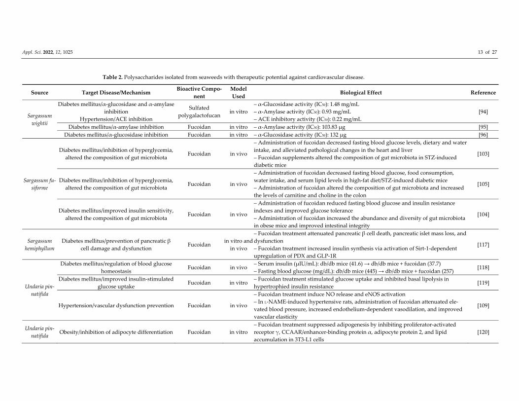
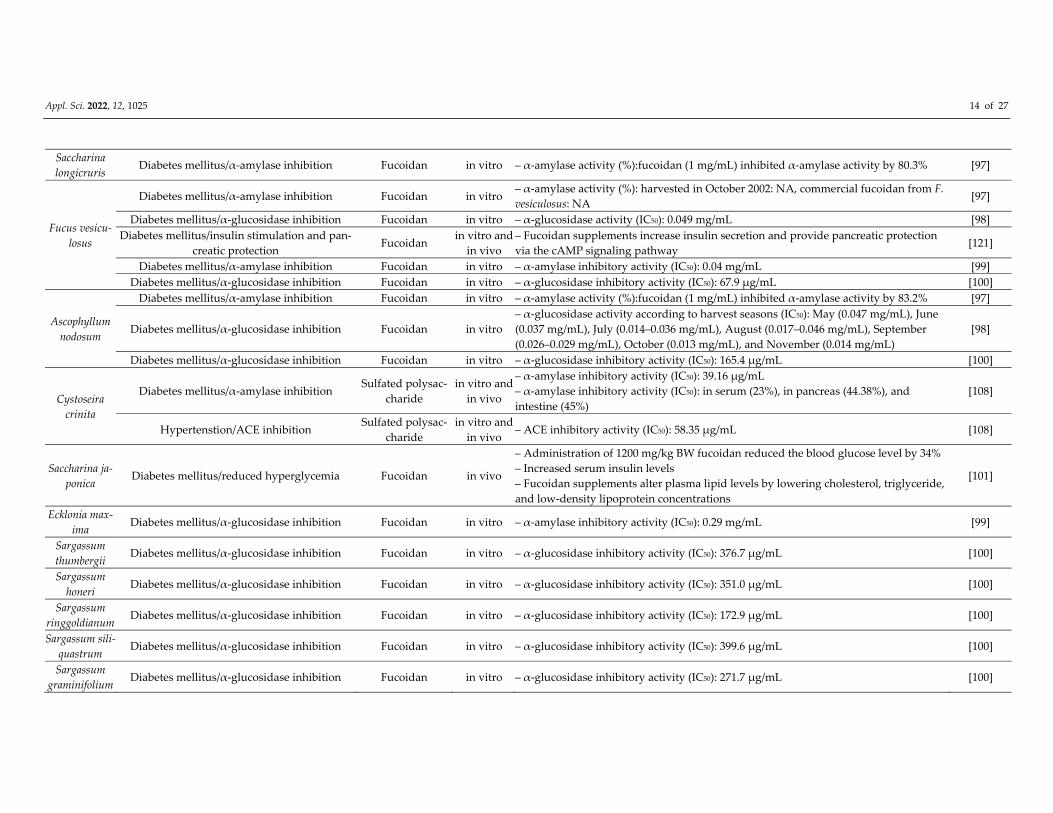
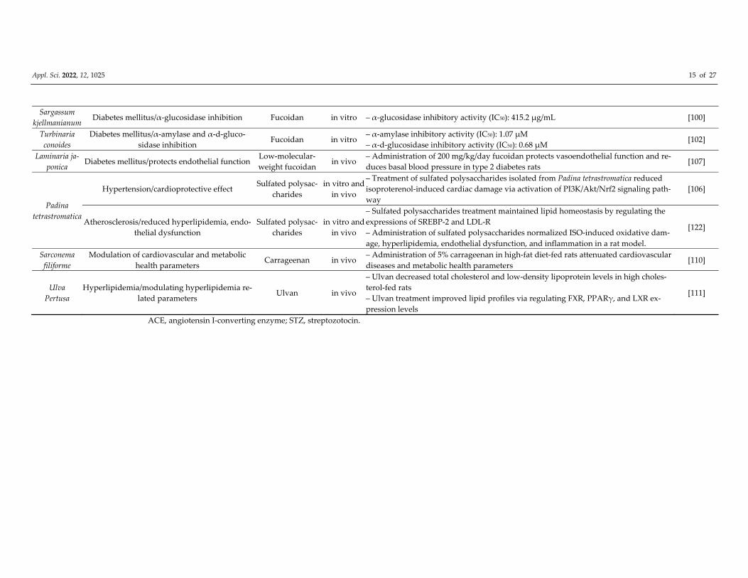
Table 2. Polysaccharides isolated from seaweeds with therapeutic potential against cardiovascular disease.
Source Target Disease/Mechanism Bioactive Compo‐
nent
Model
Used Biological Effect Reference
Sargassum
wightii
Diabetes mellitus/α‐glucosidase and α‐amylase
inhibition
Hypertension/ACE inhibition
Sulfated
polygalactofucan in vitro
– α‐Glucosidase activity (IC50): 1.48 mg/mL
– α‐Amylase activity (IC50): 0.93 mg/mL
– ACE inhibitory activity (IC50): 0.22 mg/mL
[94]
Diabetes mellitus/α‐amylase inhibition Fucoidan in vitro – α‐Amylase activity (IC50): 103.83 μg [95]
Diabetes mellitus/α‐glucosidase inhibition Fucoidan in vitro – α‐Glucosidase activity (IC50): 132 μg [96]
Sargassum fu‐
siforme
Diabetes mellitus/inhibition of hyperglycemia,
altered the composition of gut microbiota Fucoidan in vivo
– Administration of fucoidan decreased fasting blood glucose levels, dietary and water
intake, and alleviated pathological changes in the heart and liver
– Fucoidan supplements altered the composition of gut microbiota in STZ‐induced
diabetic mice
[103]
Diabetes mellitus/inhibition of hyperglycemia,
altered the composition of gut microbiota Fucoidan in vivo
– Administration of fucoidan decreased fasting blood glucose, food consumption,
water intake, and serum lipid levels in high‐fat diet/STZ‐induced diabetic mice
– Administration of fucoidan altered the composition of gut microbiota and increased
the levels of carnitine and choline in the colon
[105]
Diabetes mellitus/improved insulin sensitivity,
altered the composition of gut microbiota Fucoidan in vivo
– Administration of fucoidan reduced fasting blood glucose and insulin resistance
indexes and improved glucose tolerance
– Administration of fucoidan increased the abundance and diversity of gut microbiota
in obese mice and improved intestinal integrity
[104]
Sargassum
hemiphyllum
Diabetes mellitus/prevention of pancreatic β
cell damage and dysfunction Fucoidan
in vitro and
in vivo
– Fucoidan treatment attenuated pancreatic β cell death, pancreatic islet mass loss, and
dysfunction
– Fucoidan treatment increased insulin synthesis via activation of Sirt‐1‐dependent
upregulation of PDX and GLP‐1R
[117]
Undaria pin‐
natifida
Diabetes mellitus/regulation of blood glucose
homeostasis Fucoidan in vivo
– Serum insulin (μIU/mL): db/db mice (41.6) → db/db mice + fucoidan (37.7)
– Fasting blood glucose (mg/dL): db/db mice (445) → db/db mice + fucoidan (257) [118]
Diabetes mellitus/improved insulin‐stimulated
glucose uptake Fucoidan in vitro
– Fucoidan treatment stimulated glucose uptake and inhibited basal lipolysis in
hypertrophied insulin resistance [119]
Hypertension/vascular dysfunction prevention Fucoidan in vivo
– Fucoidan treatment induce NO release and eNOS activation
– In L‐NAME‐induced hypertensive rats, administration of fucoidan attenuated ele‐
vated blood pressure, increased endothelium‐dependent vasodilation, and improved
vascular elasticity
[109]
Undaria pin‐
natifida Obesity/inhibition of adipocyte differentiation Fucoidan in vitro
– Fucoidan treatment suppressed adipogenesis by inhibiting proliferator‐activated
receptor γ, CCAAR/enhancer‐binding protein α, adipocyte protein 2, and lipid
accumulation in 3T3‐L1 cells
[120]
Appl. Sci. 2022, 12, 1025 14 of 27
Saccharina
longicruris Diabetes mellitus/α‐amylase inhibition Fucoidan in vitro – α‐amylase activity (%):fucoidan (1 mg/mL) inhibited α‐amylase activity by 80.3% [97]
Fucus vesicu‐
losus
Diabetes mellitus/α‐amylase inhibition Fucoidan in vitro – α‐amylase activity (%): harvested in October 2002: NA, commercial fucoidan from F.
vesiculosus: NA [97]
Diabetes mellitus/α‐glucosidase inhibition Fucoidan in vitro – α‐glucosidase activity (IC50): 0.049 mg/mL [98]
Diabetes mellitus/insulin stimulation and pan‐
creatic protection Fucoidan
in vitro and
in vivo
– Fucoidan supplements increase insulin secretion and provide pancreatic protection
via the cAMP signaling pathway [121]
Diabetes mellitus/α‐amylase inhibition Fucoidan in vitro – α‐amylase inhibitory activity (IC50): 0.04 mg/mL [99]
Diabetes mellitus/α‐glucosidase inhibition Fucoidan in vitro – α‐glucosidase inhibitory activity (IC50): 67.9 μg/mL [100]
Ascophyllum
nodosum
Diabetes mellitus/α‐amylase inhibition Fucoidan in vitro – α‐amylase activity (%):fucoidan (1 mg/mL) inhibited α‐amylase activity by 83.2% [97]
Diabetes mellitus/α‐glucosidase inhibition Fucoidan in vitro
– α‐glucosidase activity according to harvest seasons (IC50): May (0.047 mg/mL), June
(0.037 mg/mL), July (0.014–0.036 mg/mL), August (0.017–0.046 mg/mL), September
(0.026–0.029 mg/mL), October (0.013 mg/mL), and November (0.014 mg/mL)
[98]
Diabetes mellitus/α‐glucosidase inhibition Fucoidan in vitro – α‐glucosidase inhibitory activity (IC50): 165.4 μg/mL [100]
Cystoseira
crinita
Diabetes mellitus/α‐amylase inhibition Sulfated polysac‐
charide
in vitro and
in vivo
– α‐amylase inhibitory activity (IC50): 39.16 μg/mL
– α‐amylase inhibitory activity (IC50): in serum (23%), in pancreas (44.38%), and
intestine (45%)
[108]
Hypertenstion/ACE inhibition Sulfated polysac‐
charide
in vitro and
in vivo – ACE inhibitory activity (IC50): 58.35 μg/mL [108]
Saccharina ja‐
ponica Diabetes mellitus/reduced hyperglycemia Fucoidan in vivo
– Administration of 1200 mg/kg BW fucoidan reduced the blood glucose level by 34%
– Increased serum insulin levels
– Fucoidan supplements alter plasma lipid levels by lowering cholesterol, triglyceride,
and low‐density lipoprotein concentrations
[101]
Ecklonia max‐
ima Diabetes mellitus/α‐glucosidase inhibition Fucoidan in vitro – α‐amylase inhibitory activity (IC50): 0.29 mg/mL [99]
Sargassum
thumbergii Diabetes mellitus/α‐glucosidase inhibition Fucoidan in vitro – α‐glucosidase inhibitory activity (IC50): 376.7 μg/mL [100]
Sargassum
honeri Diabetes mellitus/α‐glucosidase inhibition Fucoidan in vitro – α‐glucosidase inhibitory activity (IC50): 351.0 μg/mL [100]
Sargassum
ringgoldianum Diabetes mellitus/α‐glucosidase inhibition Fucoidan in vitro – α‐glucosidase inhibitory activity (IC50): 172.9 μg/mL [100]
Sargassum sili‐
quastrum Diabetes mellitus/α‐glucosidase inhibition Fucoidan in vitro – α‐glucosidase inhibitory activity (IC50): 399.6 μg/mL [100]
Sargassum
graminifolium Diabetes mellitus/α‐glucosidase inhibition Fucoidan in vitro – α‐glucosidase inhibitory activity (IC50): 271.7 μg/mL [100]
Appl. Sci. 2022, 12, 1025 15 of 27
Sargassum
kjellmanianum Diabetes mellitus/α‐glucosidase inhibition Fucoidan in vitro – α‐glucosidase inhibitory activity (IC50): 415.2 μg/mL [100]
Turbinaria
conoides
Diabetes mellitus/α‐amylase and α‐d‐gluco‐
sidase inhibition Fucoidan in vitro
– α‐amylase inhibitory activity (IC50): 1.07 μM
– α‐d‐glucosidase inhibitory activity (IC50): 0.68 μM [102]
Laminaria ja‐
ponica Diabetes mellitus/protects endothelial function
Low‐molecular‐
weight fucoidan in vivo
– Administration of 200 mg/kg/day fucoidan protects vasoendothelial function and re‐
duces basal blood pressure in type 2 diabetes rats [107]
Padina
tetrastromatica
Hypertension/cardioprotective effect Sulfated polysac‐
charides
in vitro and
in vivo
– Treatment of sulfated polysaccharides isolated from Padina tetrastromatica reduced
isoproterenol‐induced cardiac damage via activation of PI3K/Akt/Nrf2 signaling path‐
way
[106]
Atherosclerosis/reduced hyperlipidemia, endo‐
thelial dysfunction
Sulfated polysac‐
charides
in vitro and
in vivo
– Sulfated polysaccharides treatment maintained lipid homeostasis by regulating the
expressions of SREBP‐2 and LDL‐R
– Administration of sulfated polysaccharides normalized ISO‐induced oxidative dam‐
age, hyperlipidemia, endothelial dysfunction, and inflammation in a rat model.
[122]
Sarconema
filiforme
Modulation of cardiovascular and metabolic
health parameters Carrageenan in vivo
– Administration of 5% carrageenan in high‐fat diet‐fed rats attenuated cardiovascular
diseases and metabolic health parameters [110]
Ulva
Pertusa
Hyperlipidemia/modulating hyperlipidemia re‐
lated parameters Ulvan in vivo
– Ulvan decreased total cholesterol and low‐density lipoprotein levels in high choles‐
terol‐fed rats
– Ulvan treatment improved lipid profiles via regulating FXR, PPARγ, and LXR ex‐
pression levels
[111]
ACE, angiotensin I‐converting enzyme; STZ, streptozotocin.
Appl. Sci. 2022, 12, 1025 16 of 27
4. Seaweed‐Derived Peptides with Therapeutic Potential against CVD
As the importance of marine organisms as sources of novel bioactive substances is
growing, marine bioactive peptides have received much attention recently. Bioactive pep‐
tides are usually 2−20 amino acid residues [123]. Depending on the amino acid sequence,
they may be involved in various biological functions such as antioxidant, anti‐cancer, opi‐
oid agonists or antagonists, immunomodulatory, anti‐thrombotic, anti‐atherosclerotic,
and antimicrobial activities, in addition to nutrient utilization [123]. A pepsin‐hydrolyzed
peptide (VECYGPNRPQF) from Chlorella vulgaris protein waste possessed potent antiox‐
idant activity against various free radicals and exhibited gastrointestinal enzyme re‐
sistance. Still, no cytotoxicity was observed in human lung fibroblast cell lines (WI‐38)
[124]. The antitumor polypeptide Y2 was obtained from the trypsin digest of Spirulina
platensis proteins [125]. Cian et al. found that enzymatic hydrolysates from a phycobili
protein byproduct of Porphyra columbina exhibited immunosuppressive effects on rat sple‐
nocytes by enhancing IL‐10 production and inhibiting the production of TNF‐α and IFN‐
γ [126].
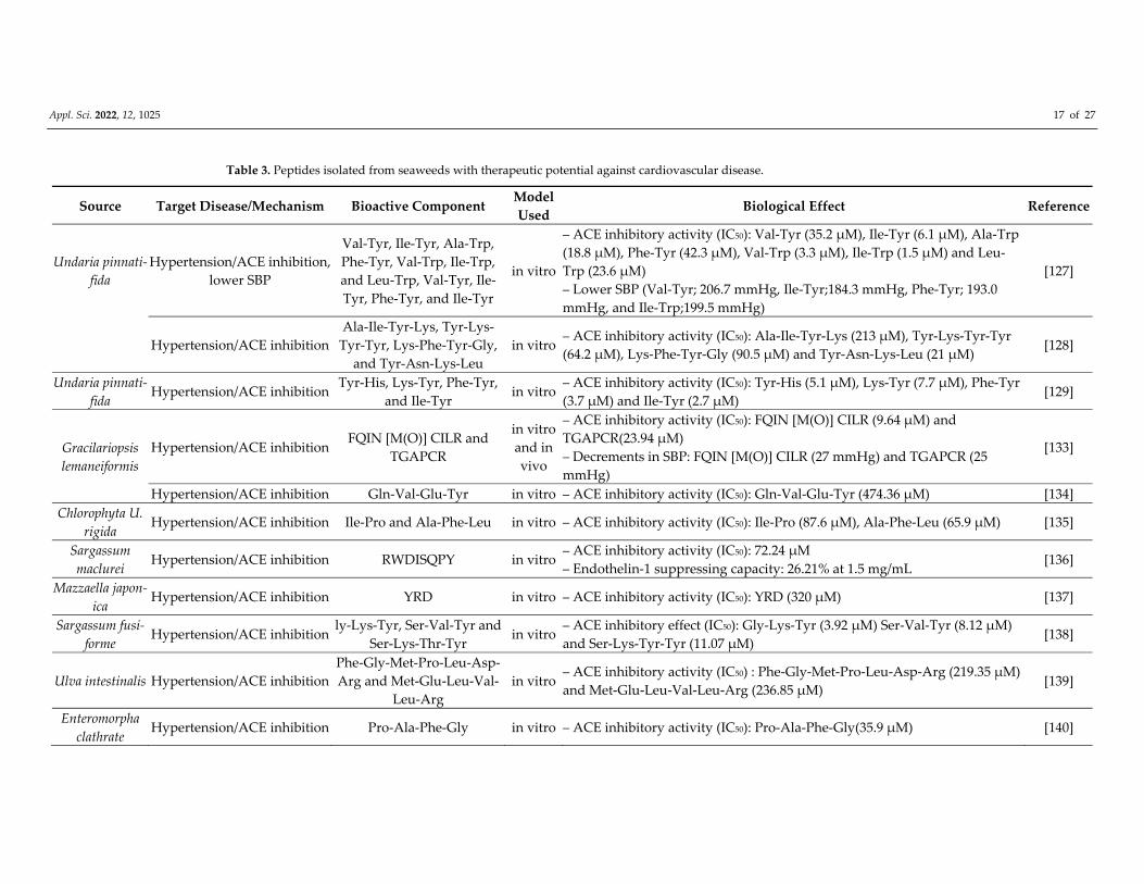
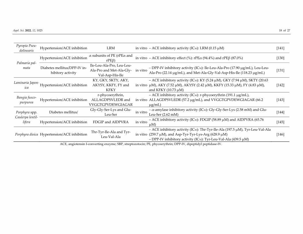
Several bioactive peptides have been identified from Undaria pinnatifida (U. pinnati‐
fida) and Palmaria palmate (P. palmate), which exerted an anti‐hypertensive effect in vitro
by showing potent ACE inhibition activity and a significant reduction in blood pressure
observed in an oral feeding rat model [127–131]. In particular, bioactive peptides derived
from various seaweeds (brown seaweed 10, red seaweed 10, and green seaweed 10) have
antihypertensive effects through ACE inhibition (Table 3). One mg/kg BW of Leu‐Trp,
Val‐Tyr, Ile‐Tyr, Phe‐Tyr, and Ile‐Tyr from U. pinnatifida decreased the systolic blood pres‐
sure in spontaneously hypertensive rats (SHRs) [127]. Furthermore, Suetsuna et al. re‐
ported that both single administration and repeated oral administration of synthetic di‐
peptides (Tyr‐His, Lys‐Tyr, Phe‐Tyr, and Ile‐Tyr) from U. pinnatifida significantly de‐
creased the blood pressure in spontaneously hypertensive rats (SHRs) [129]. The α and β
subunits of phycoerythrin (a major light‐harvesting protein pigment of red seaweed) from
P. palmate showed ACE inhibition activity [130]. Ile‐Leu‐Ala‐Pro, Leu‐Leu‐Ala‐Pro, and
Met‐Ala‐Gly‐Val‐Asp‐His‐Ile purified from P. palmate inhibited DPP‐Ⅳ, a novel bi‐omarker of ischemic cardiovascular disease, and had insulinotropic potency [131,132]. Ile‐
Pro and Ala‐Phe‐Leu isolated from Chlorophyta U. rigida exhibited ACE inhibition activity.
Ko et al. [25] demonstrated significant systolic blood pressure reduction in hypertensive
rats following 10 mg/kg body weight of Val‐Glu‐Gly‐Tyr administered orally. Gracilariop‐
sis lemaneiformis inhibited ACE‐I with IC50 values of 9.64. 23.94, and 474.36 μM for the
peptides FQIN [M(O)] CILR, TGAPCR, and Gln‐Val‐Glu‐Tyr, respectively [133,134]. The
administration of 10 mg/kg of FQIN [M(O)] CILR and TGAPCR had antihypertensive ef‐
fects in spontaneously hypertensive rats [133].
Appl. Sci. 2022, 12, 1025 17 of 27
Table 3. Peptides isolated from seaweeds with therapeutic potential against cardiovascular disease.
Source Target Disease/Mechanism Bioactive Component Model
Used Biological Effect Reference
Undaria pinnati‐
fida
Hypertension/ACE inhibition,
lower SBP
Val‐Tyr, Ile‐Tyr, Ala‐Trp,
Phe‐Tyr, Val‐Trp, Ile‐Trp,
and Leu‐Trp, Val‐Tyr, Ile‐
Tyr, Phe‐Tyr, and Ile‐Tyr
in vitro
– ACE inhibitory activity (IC50): Val‐Tyr (35.2 μM), Ile‐Tyr (6.1 μM), Ala‐Trp
(18.8 μM), Phe‐Tyr (42.3 μM), Val‐Trp (3.3 μM), Ile‐Trp (1.5 μM) and Leu‐
Trp (23.6 μM)
– Lower SBP (Val‐Tyr; 206.7 mmHg, Ile‐Tyr;184.3 mmHg, Phe‐Tyr; 193.0
mmHg, and Ile‐Trp;199.5 mmHg)
[127]
Hypertension/ACE inhibition
Ala‐Ile‐Tyr‐Lys, Tyr‐Lys‐
Tyr‐Tyr, Lys‐Phe‐Tyr‐Gly,
and Tyr‐Asn‐Lys‐Leu
in vitro – ACE inhibitory activity (IC50): Ala‐Ile‐Tyr‐Lys (213 μM), Tyr‐Lys‐Tyr‐Tyr
(64.2 μM), Lys‐Phe‐Tyr‐Gly (90.5 μM) and Tyr‐Asn‐Lys‐Leu (21 μM) [128]
Undaria pinnati‐
fida Hypertension/ACE inhibition
Tyr‐His, Lys‐Tyr, Phe‐Tyr,
and Ile‐Tyr in vitro
– ACE inhibitory activity (IC50): Tyr‐His (5.1 μM), Lys‐Tyr (7.7 μM), Phe‐Tyr
(3.7 μM) and Ile‐Tyr (2.7 μM) [129]
Gracilariopsis
lemaneiformis
Hypertension/ACE inhibition FQIN [M(O)] CILR and
TGAPCR
in vitro
and in
vivo
– ACE inhibitory activity (IC50): FQIN [M(O)] CILR (9.64 μM) and
TGAPCR(23.94 μM)
– Decrements in SBP: FQIN [M(O)] CILR (27 mmHg) and TGAPCR (25
mmHg)
[133]
Hypertension/ACE inhibition Gln‐Val‐Glu‐Tyr in vitro – ACE inhibitory activity (IC50): Gln‐Val‐Glu‐Tyr (474.36 μM) [134]
Chlorophyta U.
rigida Hypertension/ACE inhibition Ile‐Pro and Ala‐Phe‐Leu in vitro – ACE inhibitory activity (IC50): Ile‐Pro (87.6 μM), Ala‐Phe‐Leu (65.9 μM) [135]
Sargassum
maclurei Hypertension/ACE inhibition RWDISQPY in vitro
– ACE inhibitory activity (IC50): 72.24 μM
– Endothelin‐1 suppressing capacity: 26.21% at 1.5 mg/mL [136]
Mazzaella japon‐
ica Hypertension/ACE inhibition YRD in vitro – ACE inhibitory activity (IC50): YRD (320 μM) [137]
Sargassum fusi‐
forme Hypertension/ACE inhibition
ly‐Lys‐Tyr, Ser‐Val‐Tyr and
Ser‐Lys‐Thr‐Tyr in vitro
– ACE inhibitory effect (IC50): Gly‐Lys‐Tyr (3.92 μM) Ser‐Val‐Tyr (8.12 μM)
and Ser‐Lys‐Tyr‐Tyr (11.07 μM) [138]
Ulva intestinalis Hypertension/ACE inhibition
Phe‐Gly‐Met‐Pro‐Leu‐Asp‐
Arg and Met‐Glu‐Leu‐Val‐
Leu‐Arg
in vitro – ACE inhibitory activity (IC50) : Phe‐Gly‐Met‐Pro‐Leu‐Asp‐Arg (219.35 μM)
and Met‐Glu‐Leu‐Val‐Leu‐Arg (236.85 μM) [139]
Enteromorpha
clathrate Hypertension/ACE inhibition Pro‐Ala‐Phe‐Gly in vitro – ACE inhibitory activity (IC50): Pro‐Ala‐Phe‐Gly(35.9 μM) [140]
Appl. Sci. 2022, 12, 1025 18 of 27
Pyropia Pseu‐
dolinearis Hypertension/ACE inhibition LRM in vitro – ACE inhibitory activity (IC50): LRM (0.15 μM) [141]
Palmaria pal‐
mata
Hypertension/ACE inhibition α subunits of PE (rPEα and
rPEβ) in vitro – ACE inhibitory effect (%): rPEα (94.4%) and rPEβ (87.0%) [130]
Diabetes mellitus/DPP‐Ⅳ in‐hibitory activity
Ile‐Leu‐Ala‐Pro, Leu‐Leu‐
Ala‐Pro and Met‐Ala‐Gly‐
Val‐Asp‐His‐Ile
in vitro – DPP‐Ⅳ inhibitory activity (IC50): Ile‐Leu‐Ala‐Pro (17.90 μg/mL), Leu‐Leu‐
Ala‐Pro (22.14 μg/mL), and Met‐Ala‐Gly‐Val‐Asp‐His‐Ile (118.23 μg/mL) [131]
Laminaria Japon‐
ica Hypertension/ACE inhibition
KY, GKY, SKTY, AKY,
AKYSY, KKFY, FY and
KFKY
in vitro
– ACE inhibitory activity (IC50): KY (5.24 μM), GKY (7.94 μM), SKTY (20.63
μM), AKY (7.52 μM), AKYSY (2.42 μM), KKFY (15.33 μM), FY (4.83 μM),
and KFKY (10.73 μM)
[142]
Bangia fusco‐
purpurea Hypertension/ACE inhibition
r‐phycoerythrin,
ALLAGDPSVLEDR and
VVGGTGPVDEWGIAGAR
in vitro
– ACE inhibitory activity (IC50): r‐phycoerythrin (191.1 μg/mL),
ALLAGDPSVLEDR (57.2 μg/mL), and VVGGTGPVDEWGIAGAR (66.2
μg/mL)
[143]
Porphyra spp.
Caulerpa lentil‐
lifera
Diabetes mellitus/ Gly‐Gly‐Ser‐Lys and Glu‐
Leu‐Ser in vitro
– α‐amylase inhibitory activity (IC50): Gly‐Gly‐Ser‐Lys (2.58 mM) and Glu‐
Leu‐Ser (2.62 mM) [144]
Hypertension/ACE inhibition FDGIP and AIDPVRA in vitro – ACE inhibitory activity (IC50): FDGIP (58.89 μM) and AIDPVRA (65.76
μM) [145]
Porphyra dioica Hypertension/ACE inhibition Thr‐Tyr‐Ile‐Ala and Tyr‐
Leu‐Val‐Ala in vitro
– ACE inhibitory activity (IC50): Thr‐Tyr‐Ile‐Ala (197.5 μM), Tyr‐Leu‐Val‐Ala
(259.7 μM), and Asp‐Tyr‐Tyr‐Lys‐Arg (628.9 μM)
– DPP‐Ⅳ inhibitory activity (IC50): Tyr‐Leu‐Val‐Ala (439.5 μM)
[146]
ACE, angiotensin I‐converting enzyme; SBP, streptozotocin; PE, phycoerythrin; DPP‐Ⅳ, dipeptidyl peptidase‐Ⅳ.
Appl. Sci. 2022, 12, 1025 19 of 27
5. Seaweed‐Derived Carotenoids and Other Components with Therapeutic Potential
against CVD
All carotenoids, including fucoxanthin, carotene, lycopene, and siphonaxanthin, are
bioactive substances from seaweeds. Green seaweed extracts rich in carotenoids exhibit
significant antigenotoxic activities [147]. Fucoxanthin is a recognized secondary metabo‐
lite found in macroalgae, and its biological properties are well established [23]. Fucoxan‐
thin from brown seaweed exerts prebiotic and anti‐inflammatory activities in human in‐
testinal epithelial cells [148]. Fucoxanthin upregulates the expression of UCP1 in white
adipose tissue (WAT) in KK‐Ay mice [149]. Fucoxanthin and siphonaxanthin inhibit an‐
giogenesis by downregulating the FGF‐2‐mediated intracellular signals in vascular endo‐
thelial cells [150]. Siphonaxanthin has been reported to have anti‐angiogenic and anti‐in‐
flammatory effects [151]. Siphonaxanthin induces apoptosis by decreasing Bcl‐2 expres‐
sion and activating caspase‐3 in human leukemia (HL‐60) cells [152].
In Ae’s study, fucoxanthin extracted from Undaria pinnatifida (U. pinnatifida) in‐
creased the serum HDL. It decreased the triglyceride levels in high‐fat diet‐fed rats at a
0.2% diet dose for 4 weeks [153] (Table 4). In addition, the α‐amylase and α‐glucosidase
inhibitory activities of fucoxanthin were observed in Kawee‐Ai’s study [154]. The admin‐
istration of a 3‐g/kg 9‐cis β‐carotene diet disrupted the increases in plasma cholesterol and
LDL in LDL‐receptor knockout mice [155]. Feeding 8% 9‐Cis β‐carotene feed for 23 days
disrupted triglyceride elevation in 5‐week‐old female db/db mice [156]. Other carotenoids
have also been shown to have anti‐atherogenic effects. Zhuo et al. showed that si‐
phonaxanthin extracted from Codium fragile disrupts the elevation of the serum total cho‐
lesterol, triglyceride, and HDL in KK‐Ay mice [157]. In addition, (all‐E)‐lutein and (9‐Z)‐
zeaxanthin displayed anti‐diabetes mellitus potency via α‐glucosidase inhibition [158].
Seaweeds have a variety of physiologically active ingredients in addition to phloro‐
tannins, polysaccharides, peptides, and carotenoids. It has been reported that seaweeds
contain various sterol compounds [159]. Sterols are an important family of lipids present
in most eukaryotic cells. They are categorized into the steroid group, which contains the
same fused four‐ring core structure and has different biological roles than hormones and
signaling molecules. The search for natural bioactive sterols as safe alternatives from ma‐
rine seaweed is important in the food industry. Fucosterol isolated from the marine sea‐
weed Pelvetia siliquosa causes a significant elevation of free radical scavenging enzyme
activities, such as superoxide dismutase (SOD), catalase, and glutathione peroxidase
(GSH‐px) [160]. Moreover, 3 g,28 ξ‐dihydroxy‐24‐ethylcholesta‐5,23 Z‐dien and 24 ξ‐hy‐
droperoxy‐24‐vinylcholesterol isolated from the brown seaweed Sargassum carpophyllum
showed cytotoxic activity against human promyelocytic leukemia cells [161].
Zhen and Su studied bioactive compounds isolated from the edible brown seaweed
Sargassum fusiforme (S. fusiforme), and 24(S)‐saringosterol showed the strongest LXRβ‐me‐
diated transactivity among the seven phytosterols isolated from S. fusiforme containing
fucosterol, 24‐hydroperoxy‐24‐vinyl‐cholesterol, 29‐hydroperoxy‐stigmasta‐5,24(28)‐
dien‐3β‐ol, 24‐methylenecholesterol, 24‐keto‐cholesterol, and 5α,8α‐epidioxyergosta‐
6,22‐dien‐3β‐ol [162], while 18α‐glycyrrhetinic acid and 18β‐glycyrrhetinic acid were ex‐
tracted from S. fusiforme‐inhibited α‐glucosidase. Other compounds, including unsatu‐
rated FAs C20:4 (Δ7,9,11,13), C17:3 (Δ8,11,14), neolignan, and trace amines also potently inhib‐
ited α‐glucosidase [163]. Two plastoquinones (sargachromenol and sargaquinoic acid)
isolated from the active n‐hexane fraction of Sargassum serratifolium produced concentra‐
tion‐dependent inhibition against α‐glucosidase [164].
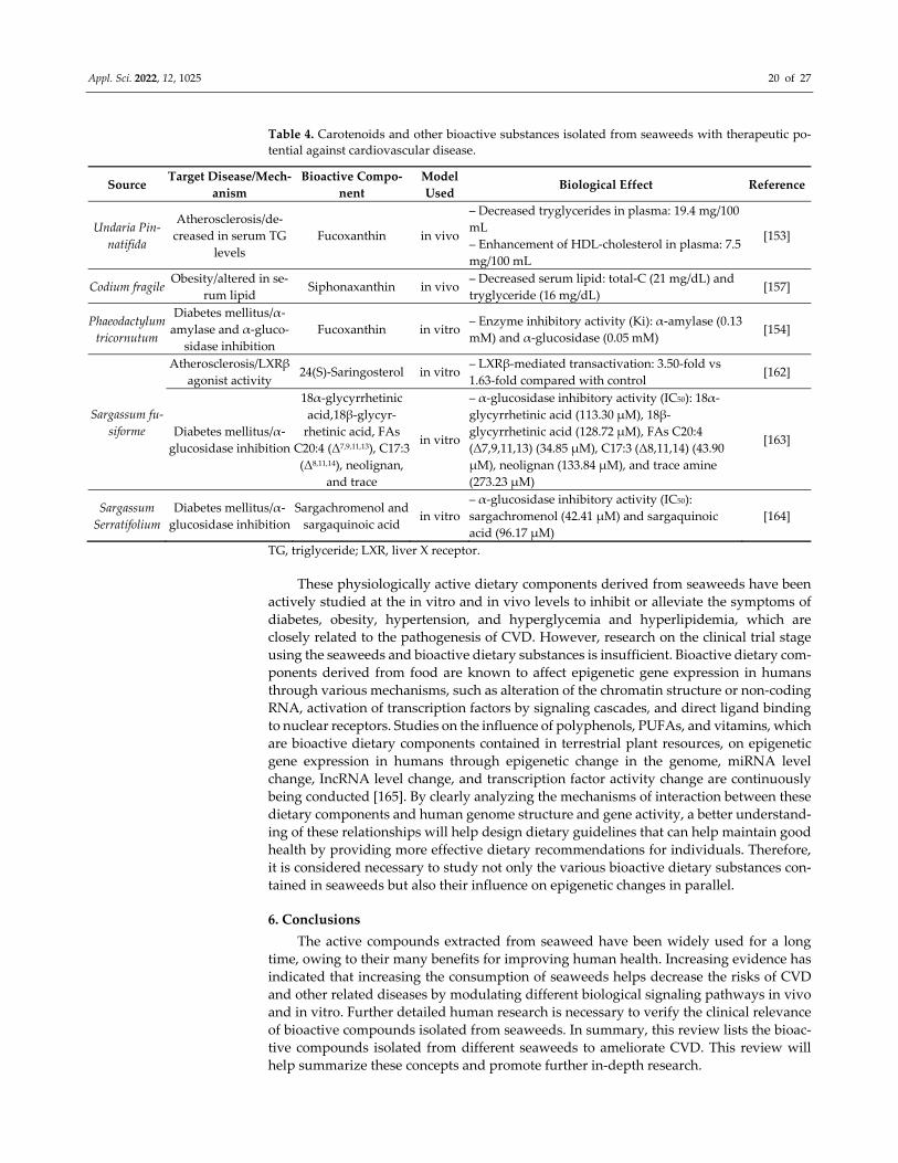
Appl. Sci. 2022, 12, 1025 20 of 27
Table 4. Carotenoids and other bioactive substances isolated from seaweeds with therapeutic po‐
tential against cardiovascular disease.
Source Target Disease/Mech‐
anism
Bioactive Compo‐
nent
Model
Used Biological Effect Reference
Undaria Pin‐
natifida
Atherosclerosis/de‐
creased in serum TG
levels
Fucoxanthin in vivo
– Decreased tryglycerides in plasma: 19.4 mg/100
mL
– Enhancement of HDL‐cholesterol in plasma: 7.5
mg/100 mL
[153]
Codium fragile Obesity/altered in se‐
rum lipid Siphonaxanthin in vivo
– Decreased serum lipid: total‐C (21 mg/dL) and
tryglyceride (16 mg/dL) [157]
Phaeodactylum
tricornutum
Diabetes mellitus/α‐
amylase and α‐gluco‐
sidase inhibition
Fucoxanthin in vitro – Enzyme inhibitory activity (Ki): α‐amylase (0.13
mM) and α‐glucosidase (0.05 mM) [154]
Sargassum fu‐
siforme
Atherosclerosis/LXRβ
agonist activity 24(S)‐Saringosterol in vitro
– LXRβ‐mediated transactivation: 3.50‐fold vs
1.63‐fold compared with control [162]
Diabetes mellitus/α‐
glucosidase inhibition
18α‐glycyrrhetinic
acid,18β‐glycyr‐
rhetinic acid, FAs
C20:4 (Δ7,9,11,13), C17:3
(Δ8,11,14), neolignan,
and trace
in vitro
– α‐glucosidase inhibitory activity (IC50): 18α‐
glycyrrhetinic acid (113.30 μM), 18β‐
glycyrrhetinic acid (128.72 μM), FAs C20:4
(Δ7,9,11,13) (34.85 μM), C17:3 (Δ8,11,14) (43.90
μM), neolignan (133.84 μM), and trace amine
(273.23 μM)
[163]
Sargassum
Serratifolium
Diabetes mellitus/α‐
glucosidase inhibition
Sargachromenol and
sargaquinoic acid in vitro
– α‐glucosidase inhibitory activity (IC50):
sargachromenol (42.41 μM) and sargaquinoic
acid (96.17 μM)
[164]
TG, triglyceride; LXR, liver X receptor.
These physiologically active dietary components derived from seaweeds have been
actively studied at the in vitro and in vivo levels to inhibit or alleviate the symptoms of
diabetes, obesity, hypertension, and hyperglycemia and hyperlipidemia, which are
closely related to the pathogenesis of CVD. However, research on the clinical trial stage
using the seaweeds and bioactive dietary substances is insufficient. Bioactive dietary com‐
ponents derived from food are known to affect epigenetic gene expression in humans
through various mechanisms, such as alteration of the chromatin structure or non‐coding
RNA, activation of transcription factors by signaling cascades, and direct ligand binding
to nuclear receptors. Studies on the influence of polyphenols, PUFAs, and vitamins, which
are bioactive dietary components contained in terrestrial plant resources, on epigenetic
gene expression in humans through epigenetic change in the genome, miRNA level
change, IncRNA level change, and transcription factor activity change are continuously
being conducted [165]. By clearly analyzing the mechanisms of interaction between these
dietary components and human genome structure and gene activity, a better understand‐
ing of these relationships will help design dietary guidelines that can help maintain good
health by providing more effective dietary recommendations for individuals. Therefore,
it is considered necessary to study not only the various bioactive dietary substances con‐
tained in seaweeds but also their influence on epigenetic changes in parallel.
6. Conclusions
The active compounds extracted from seaweed have been widely used for a long
time, owing to their many benefits for improving human health. Increasing evidence has
indicated that increasing the consumption of seaweeds helps decrease the risks of CVD
and other related diseases by modulating different biological signaling pathways in vivo
and in vitro. Further detailed human research is necessary to verify the clinical relevance
of bioactive compounds isolated from seaweeds. In summary, this review lists the bioac‐
tive compounds isolated from different seaweeds to ameliorate CVD. This review will
help summarize these concepts and promote further in‐depth research.
Appl. Sci. 2022, 12, 1025 21 of 27
Author Contributions: Conceptualization, C.‐H.C. and Y.‐A.L.; writing—original draft preparation,
C.‐H.C., Y.‐A.L. and M.‐Y.K.; writing—review and editing, Y.‐J.J. and S.‐H.L.; visualization, C.‐H.C.
and M.‐Y.K.; supervision, Y.‐J.J. and S.‐H.L.; project administration, S.‐H.L.; funding acquisition, S.‐
H.L. All authors have read and agreed to the published version of the manuscript.
Funding: This research was supported by a National Research Foundation of Korea (NRF) grant
funded by the Korean government (Ministry of Science and ICT, MSIT) (No. NRF‐
2020R1A2C2012608).
Institutional Review Board Statement: Not applicable.
Informed Consent Statement: Not applicable.
Data Availability Statement: Not applicable.
Conflicts of Interest: The authors declare no conflict of interest.
References
1. Roth, G.A.; Johnson, C.; Abajobir, A.; Abd‐Allah, F.; Abera, S.F.; Abyu, G.; Ahmed, M.; Aksut, B.; Alam, T. ; Alam K.; et al.
Global, Regional, and National Burden of Cardiovascular Diseases for 10 Causes, 1990 to 2015. J. Am. Coll. Cardiol. 2017, 70, 1–
25.
2. Pace, R.; Brazeau, A.‐S.; Meltzer, S.; Rahme E.; Dasgupta. K. Conjoint Associations of Gestational Diabetes and Hypertension
with Diabetes, Hypertension, and Cardiovascular Disease in Parents: A Retrospective Cohort Study. Am. J. Epidemiol. 2017, 186,
1115–1124.
3. Seravalle, G.; Grassi, G. Obesity and hypertension. Pharmacol. Res. 2017, 122, 1–7.
4. Leggio, M.; Lombardi, M.; Caldarone, E.; Severi, P.; D’emidio, S.; Armeni, M.; Bravi, V.;Bendini, M.G.; Mazza, A.;. The relation‐
ship between obesity and hypertension: An updated comprehensive overview on vicious twins. Hypertens. Res. 2017, 40, 947–
963.
5. Gheibi, S.; Jeddi, S.; Kashfi, K.; Ghasemi, A. Regulation of vascular tone homeostasis by NO and H2S: Implications in hyperten‐
sion. Biochem. Pharmacol. 2018, 149, 42–59.
6. Gutierrez, J.; Alloubani, A. ; Mari, M.; Alzaatreh, M. Cardiovascular disease risk factors: Hypertension, diabetes mellitus and
obesity among Tabuk citizens in Saudi Arabia. Open Cardiovasc. Med. J. 2018, 12, 41–49.
7. Leenen, F.H.H.; Nwachuku, C.E.; Black, H.R.; Cushman, W.C.; Davis, B.R.; Simpson, L.M.; Alderman, M.H.; Atlas, S.A.; Basile,
J.N.; Cuyjet, A.B.; et al. Chlinical events in high‐risk hypertensive patients randomly assigned to calcium channel blocker versus
angiotensin‐converting enzyme inhibitor in the antihypertensive and lipid‐lowering treatment to prevent heart attack trial.
Hypertension 2006, 48, 374–384.
8. Haller, H. Effective management of hypertension with dihydropyridine calcium channel blocker‐based combination therapy in
patients at high cardiovascular risk. Int. J. Clin. Pract. 2008, 62, 781–790.
9. Lin, Q.; Zhao, L.; Jing, R.; Trexler, C.; Wang, H.; Li, Y.; Tang, H.; Huang, F.; Zhang, F.; Fang, X.; et al. Inositol 1,4,5‐trisphosphate
receptors in endothelial cells play an essential role in vasodilation and blood pressure regulation. J. Am. Heart Assoc. 2019, 8,
e011704.
10. Dhargalkar, V.K.; Pereira, N. Seaweed: Promising plant of the millennium. Sci. Cult. 2005, 71, 60–66.
11. O’Sullivan, L.; Murphy, B.; McLoughlin, P.; Duggan, P.; Lawlor, P.G.; Hughes, H.; Gardiner, G.E. Prebiotics from marine
macroalgae for human and animal health applications. Mar. Drugs 2010, 8, 2038–2064.
12. Rengasamy, K.R.; Mahomoodally, M.F.; Aumeeruddy, M.Z.; Zengin, G.; Xiao, J.; Kim, D.H. Bioactive compounds in seaweeds:
An overview of their biological properties and safety. Food Chem. Toxicol. 2020, 135, 111013.
13. Ganesan, A.R.; Tiwari, U.; Rajauria, G. Seaweed nutraceuticals and their therapeutic role in disease prevention. Food Sci. Hum.
Wellness 2019, 8, 252–263.
14. Fernando, I.P.S.; Ryu, B.; Ahn, G.; Yeo, I.‐K.; Jeon, Y.‐J. Therapeutic potential of algal natural products against metabolic syn‐
drome: A review of recent developments. Trends Food Sci. Technol. 2020, 97, 286–299.
15. Samarakoon, K.; Jeon, Y.‐J. Bio‐vunctionalities of proteins derived from marine algae—A review. Food Res. Int. 2012, 48, 948–
960.
16. Plaza, M.; Cifuentes, A.; Ibáñez, E. In the search of new functional food ingredients from algae. Trends Food Sci. Technol. 2008, 19, 31–39.
17. Meslet‐Cladière, L.; Delage, L.; Leroux, C.J.‐J.; Goulitquer, S.; Leblanc, C.; Creis, E.; Gall, E.A.; Stiger‐Pouvreau, V.; Czjzek, M.;
Potin, P. Structure/function analysis of a type III polyketide synthase in the brown alga Ectocarpus Siliculosus reveals a biochem‐
ical pathway in phlorotannin monomer biosynthesis. Plant. Cell 2013, 25, 3089–3103.
18. Wikesekara, I.; Yoon, N.Y.; Kim, S.K. Phlorotannins from Ecklonia cava (Phaeophyceae): Biological activities and potential
health benefits. BioFactors 2010, 36, 408–414.
19. Singh, I.P.; Bharate, S.B. Phloroglucinol compounds of natural origin. Nat. Prod. Rep. 2005, 23, 558–591.
20. Arbenz, A.; Avérous, L. Chemical modification of tannins to elaborate aromatic biobased macromolecular architectures. Green
Chem. 2015, 17, 2626–2646.

Appl. Sci. 2022, 12, 1025 22 of 27
21. Meng, W.; Sun, T.M.H.; Garcia‐Vaquero, M. Phlorotannins: A review of extraction methods, structural characteristics, bioactiv‐
ities, bioavalability, and future trends. Algal Res. 2021, 60, 102484.
22. Shrestha, S.; Zhang, W.; Smid, S. Phlorotannins: A review on biosynthesis, chemistry and bioactivity. Food Biosci. 2021, 39, 100832.
23. Seca, A.M.L.; Pinto, D.C.G.A., Overview on the antihypertensive and anti‐obesity effects of secondary metabolites from sea‐
weeds. Mar. Drugs 2018, 16, 237.
24. Wijesinghe, W.A.J.P.; Jeon, Y.‐J. Exploiting biological activities of brown seaweed Ecklonia cava for potential industrial applica‐
tions: A review. Int. J. Food Sci. Nutr. 2012, 63, 225–235.
25. Cushman, D.W.; Ondetti, M.A. History of the design of captopril and related inhibitors of angiotensin converting enzyme.
Hypertension 1991, 17, 589–592.
26. Wijesinghe, W.A.J.P.; Ko, S.‐C.; Jeon, Y.‐J. Effect of phlorotannins isolated from Ecklonia cava on angiotensin I‐convering enzyme
(ACE) inhibitory activity. Nutr. Res. Pract. 2011, 5, 93–100.
27. Jung, H.A.; Hyun, S.K.; Kim, H.R.; Choi, J.S. Angiotensin‐converting enzyme I inhibitory activity of phlorotannins from Ecklonia
stolonifera. Fish. Sci. 2006, 72, 1292–1299.
28. Ko, S.‐C.; Kang, M.C.; Kang, N.; Kim, H.‐S.; Lee, S.‐H.; Ahn, G.; Jung, W.‐K.; Jeon, Y.‐J. Effect of angiotensin I‐convering enzyme
(ACE) inhibition and nitric oxide (NO) production of 6,6’‐bieckol, a marine algal polyphenol and its anti‐hypertensive effect in
spontaneously hypertensive rats. Process. Biochem. 2017, 58, 326–332.
29. Ko, S.‐C.; Jung, W.‐K.; Kang, S.‐M.; Lee, S.‐H.; Kang, M.C.; Heo, S.‐J.; Kang, K.‐H.; Kim,Y.‐T.; Park, S.‐J.; Jeong, Y.; et al. Angio‐
tensin I‐converting enzyme (ACE) inhibition and nitric oxide (NO)‐mediated antihypertensive effect of oxtaphorethol A iso‐
lated from Ishige sinicola: In vitro molecular mechanism and in vivo SHR model. J. Funct. Foods 2015, 18, 289–299.
30. Szabo, C., Hydrogen sulfide, an enhancer of vascular nitric oxide signaling: Mechanisms and implications. Am. J. Physiol. Cell
Physiol. 2017, 312, C3–C15.
31. Devika, N.T.; Ali, B.M.J. Analysing calcium dependent and independent regulation of eNOS in endothelium triggered by ex‐
tracelluar signalling events. Mol. BioSystems 2013, 9, 2653–2664.
32. Dudzinski, D.M.; Michel, T. Life history of eNOS: Partners and pathways. Cardiovasc. Res. 2007, 75, 247–260.
33. Raouf, A.K. Modulators of the vascular endothelin receptor in blood pressure regulation and hypertension. Curr. Mol. Pharmacol.
2011, 4, 176–186.
34. Nakamura, K.; Koga, Y.; Sakai, H.; Homma, K.; Ikebe, M. cGMP‐dependent relaxation of smooth muscle is coupled with the
change in the phosphorylatin of myosin phophatase. Circ. Res. 2007, 101, 712–722.
35. Lu, Y.‐A.; Je, J.‐G.; Hwang, J.; Jeon, Y.‐J.; Ryu, B.Ecklonia cava extract and its derivative dieckol promote vasodilation by modu‐
lating calcium signaling and PI3K/AKT/eNOS pathway in in vitro and in vivo model. Biomedicines 2021, 9, 438.
36. Lu, Y.A.; Jiang, Y.; Yang, H.‐W.; Hwang, J.; Jeon, Y.‐J.; Ryu, B. Diphlorethohydroxycarmalol isolated from Ishige okamurae exerts
vasodilatory effects via calcium signaling and PI3K/Akt/eNOS pathway. Int. J. Mol. Sci. 2021, 22, 1610.
37. Oh, S.; Son, M.; Lee, H.S.; Kim, H.‐S.; Jeon, Y.‐J.; Byun, K. Protective effect of pyrogallol‐phloroglucinol‐6,6‐bieckol from Eck‐
lonia cava on monocyte‐associated vascular dysfunction. Mar. Drugs 2018, 16, 441.
38. Son, M.; Oh, S.; Lee, H.S.; Ryu, B.; Jiang, Y.; Jang, J.T.; Jeon, Y.‐J.; Byun, K. Pyrogallol‐phloroglucinol‐6,6’‐bieckol from Ecklonia
cava improved blood circulation in diet‐induced obese and diet‐induced hypertension mouse model. Mar. Drugs 2019, 17, 272.
39. Gross, M. Flavonoids and cardiovascular disease. Pharm. Biol. 2004, 42, 21–35.
40. Hata, Y.; Nakajima, K.; Uchida, J.‐I.; Hidaka, H.; Nakano, T. Clinical effects of brown seaweed, Undaria pinnatifida (wakame),
on blood pressure in hypertensive subjects. J. Clin. Biochem. Nutr. 2001, 30, 43–53.
41. Murray, M.; Dordevic, A.L.; Ryan, L.; Bonham, M.P. The impact of a single dose of a polyphenol‐rich seaweed extract on post‐
prandial glycaemic control in healthy adults: A randomised cross‐over trial. Nutrients 2018, 10, 270.
42. Danaei, G.; Lawes, C.M. ; Hoorn, S.V.; Murray, C.J.; Ezzati, M. Global and regional mortality from ischaemic heart disease and
stroke attributable to higher‐than‐optimum blood glucose concentration: Comparative risk assessment. Lancet 2006, 368, 1651–
1659.
43. Bahadoran, Z.; Mirmiran, P.; Azizi, F. Dietary polyphenols as potential nutraceuticals in management of diabetes: A review. J.
Diabetes Metab. Disord. 2013, 12, 43.
44. Asgar, A. Anti‐diabetic potential of phenolic compounds: A review. Int. J. Food Prop. 2013, 16, 91–103.
45. Ghani, U. Re‐exploring promising α‐glucosidase inhibitors for potential development into oral anti‐diabetic drugs: Finding
needle in the haystack. Eur. J. Med. Chem. 2015, 103, 133–162.
46. Hossain, U.; Das, A.K.; Ghosh, S.; Sil, P.C. An overview on the role of bioactive α‐glucosidase inhibitors in ameliorating diabetic
complications. Food Chem. Toxicol. 2020, 145, 111738.
47. Dash, R.P.; Babu, R.J.; Srinivas, N.R. Reappraisal and perspectives of clinical drug‐drug interaction potential of α‐glucosidase
inhibitors such as acarbose, voglibose and miglitol in the treatment of type 2 diabetes mellitus. Xenobiotica 2018, 48, 89–108.
48. Park, S.R.; Kim, J.H.; Jang, H.D.; Yang, S.Y.; Kim, Y.H. Inhibitory activity of minor phlorotannins from Ecklonia cava on α‐
glucosidase. Food Chem. 2018, 257, 128–134.
49. Lee, S.‐H.; Karadeniz, F.; Kim, M.M.; Kim, S.K. α‐Glucosidase and α‐amylase inhibitory activities of phloroglucinal derivatives
from edible marine brown alga, Ecklonia cava. J. Sci. Food Agric. 2008, 89, 1552–1558.
50. Lee, H.‐A.; Lee, J.‐H.; Han, J.‐S. A phlorotannin constituent of Ecklonia cava alleviates postprandial hyperglycemia in diabetic
mice. Pharm. Biol. 2017, 55, 1149–1154.
Appl. Sci. 2022, 12, 1025 23 of 27
51. Lee, S.‐H.; Park, M.‐H.; Heo, S.‐J.; Kang, S.‐M.; Ko, S.‐C.; Han, J.‐S.; Jeon, Y.‐J. Dieckol isolated from Ecklonia cava inhibits α‐
glucosidase and α‐amylase in vitro and alleviates postprandial hyperglycemia in streptozotocin‐induced diabetic mice. Food
Chem. Toxicol. 2010, 48, 2633–2637.
52. Moon, H.E.; Islam, M.N.; Ahn, B.R.; Chowdhury, S.S.; Sohn, H.S.; Jung, H.A.; Choi, J.S. Protein tyrosine phosphatase 1B and α‐
glucosidase inhibitory phlorotannins from edible brown algae, Ecklonia stolonifera and Eisenia bicyclis. Biosci. Biotechnol. Biochem.
2011, 75, 1472–1480.
53. Ryu, B.; Jiang, Y.; Kim, H.‐S.; Hyun, J.‐M.; Lim, S.‐B.; Li, Y.; Jeon, Y.‐J. Ishophloroglucin A, a novel phlorotannin for standardiz‐
ing the anti‐α‐glucosidase activity of Ishige okamurae. Mar. Drugs 2018, 16, 436.
54. Lee, S.‐H.; Kang, S.‐M.; Ko, S.‐C.; Moon, S.‐H.; Jeon, B.‐T.; Lee, D.H.; Jeon, Y.‐J. Octaphlorethol A: A potent α‐glucosidase inhib‐
itor isolated from Ishige foliacea shows an anti‐hyperglycemic effect in mice with streptozotocin‐induced diabetes. Food Funct.
2014, 5, 2602–2608.
55. Heo, S.‐J.; Hwang, J.‐Y.; Choi, J.‐I.; Han, J.‐S.; Kim, H.‐J.; Jeon, Y.‐J. Diphlorethohydroxycarmalol isolated from Ishige okamurae,
a brown algae, a potent α‐glucosidase and α‐amylase inhibitor, alleviates postprandial hyperglycemia in diabetic mice. Eur. J.
Pharmacol. 2009, 615, 252–256.
56. Galicia‐Garcia, U.; Benito‐Vicente, A.; Jebari, S.; Larrea‐Sebal, A.; Siddiqi, H.; Uribe, K.B.; Ostolaza, H.; Martín, C. Pathophysi‐
ology of type 2 diabetes mellitus. Int. J. Mol. Sci. 2020, 21, 6275.
57. Doland, B.B.; Rhodes, C.J.; Grimsby, J.S. The dynamic plasticity of insulin production in β‐cells. Mol. Metab. 2017, 6, 958–973.
58. Fu, Z.; Gilbert. E.R.; Liu, D. Regulation of insulin synthesis and secretion and pancreatic beta‐cell dysfunction in diabetes. Curr.
Diabetes Rev. 2013, 9, 25–53.
59. Viollet, B.; Mounier, R.; Leclerc, J.; Yazigi, A.; Foretz, M.; Andreelli, F. Targeting AMP‐activated protein kinase as a novel ther‐
apeutic approach for the treatment of metabolic disorders. Diabetes Metab. 2007, 33, 395–402.
60. Lee, H.‐A.; Lee, J.‐H.; Han, J.‐S. 2,7”‐Phloroglucinol‐6,6’‐bieckol pretects INS‐1 cells against high glucose‐induced apoptosis.
Biomed. Pharmacother. 2018, 103, 1473–1481.
61. Kang, M.‐C.; Wijesinghe, W.; Lee, S.‐H.; Kang, S.‐M.; Ko, S.‐C.; Yang, X.; Kang, N.; Jeon,B.‐T.; Kim, J.; Lee, D.‐H.; et al. Dieckol isolated from brown seaweed Ecklonia cava attenuates type Ⅱ diabetes in db/db/ mouse model. Food Chem. Toxicol. 2013, 53, 294–298.
62. Yang, H.‐W.; Son, M.; Choi, J.; Oh, S.; Jeon, Y.‐J.; Byun, K.; Ryu, B. Effect of ishophloroglucin A, a component of Ishige okamurae,
on glucose homeostasis in the pancreas and muscle of high fat diet‐fed mice. Mar. Drugs 2019, 17, 608.
63. Lee, S.‐H.; Ko, S.‐C.; Kang, M.‐C.; Lee, D.H.; Jeon, Y.‐J. Octaphlorethol A, a marine algae product, exhibits antidiabetic effects
in type 2 diabetic mice by activating AMP‐activated protein kinase and upregulating the expression of glucose transporter 4.
Food Chem. Toxicol. 2016, 91, 58–64.
64. Fernando, K.H.N.; Yang, H.‐W.; Jiang, Y.; Jeon, Y.‐J.; Ryu, B. Diphlorethohydroxycarmalol isolated from Ishige okamurae re‐
presses high glucose‐induced angiogenesis in vitro and in vivo. Mar. Drugs 2018, 16, 375.
65. Fernando, K.H.N.; Yang, H.‐W.; Jiang, Y.; Jeon, Y.‐J.; Ryu, B. Ishige okamurae extract and its constituent ishophloroglucin A
attenuated in vitro and in vivo high glucose‐induced angiogenesis. Int. J. Mol. Sci. 2019, 20, 5542.
66. Tanaka, M.; Itoh, M.; Ogawa, Y.; Suganami, T. Molecular mechanism of obesity‐induced ‘metabolic’ tissue remodeling. J. Dia‐
betes Investig. 2017, 9, 256–261.
67. Sun, K.; Kusminski, C.M.; Scherer, P.E. Adipose tissue remodeling and obesity. J. Clin. Investig. 2011, 121, 2094–2101.
68. Kuri‐harcuch, W.; Velez‐delValle, C.; Vazquez‐Sandoval, A.; Hernández‐Mosqueira, C.; Fernandez‐Sanchez, V. A cellular per‐
spective of adipogenesis transcriptional regulation. J. Cell. Physiol. 2019, 234, 1111–1129.
69. Choi, K.‐M.; Jeon, Y.S.; Kim, W.; Lee, A.; Kim, Y.‐G.; Lee, J.H.; Kang, Y.E. ; Jung, J.‐C.; Lee, J.; Min, B.; et al. Xanthigen attenuates
high‐fat diet‐induced obesity through down‐regulation of PPARγ and activation of the AMPK pathway. Food Sci. Biotechnol.
2014, 23, 931–935.
70. Karadeniz, F.; Ahn, B.‐N.; Kim, J.‐A.; Seo, Y.; Jang, M.‐S.; Nam, K.‐H.; Kim, M.; Lee, S,‐H,; Kong, C.‐S.; et al. Phlorotannins
suppress adipogensis in pre‐adipocytes while enhancing osteoblastogenesis in pre‐osteoblasts. Arch. Pharmacal Res. 2015, 38,
2172–2182.
71. Ko, S.‐C.; Lee, M.; Lee, J.‐H.; Lee, S.‐H.; Lim, Y.; Jeon, Y.‐J. Dieckol, a phlorotannin isolated from a brown seaweed, Ecklonia
cava, inhibits adipogenesis through AMP‐activated protein kinase (AMPK) activation in 3T3‐L1 preadipocytes. Environ. Toxicol.
Pharmacol. 2013, 36, 1253–1260.
72. Choi, H.‐S.; Jeon, H.J.; Lee, O.H.; Lee, B.Y. Dieckol, a major phlorotannin in Ecklonia cava, suppresses lipid accumuation in the
adipocytes of high‐fat diet‐fed zebrafish and mice: Inhibition of early adipogensis via cell‐cycle arrest and AMPKα activation.
Mol. Nutr. Food Res. 2015, 59, 1458–1471.
73. Jung, H.A.; Jung, H.J.; Jeong, H.Y.; Kwon, H.J.; Ali, M.Y.; Choi, J.S. Phlorotannins isolated from the edible brown alga Ecklonia
stolonifera exert anti‐adipogenic activity on 3T3‐L1 adipocytes by downregulating C/EBPα and PPARγ. Fitoterapia 2014, 92, 260–
269.
74. Kwon, T.‐H.; Wu, Y.X.; Kim, J.S.; Woo, J.H.; Park, K.T.; Kwon, O.J.; Seo, H.‐J.; Kim, T.; Park, N.‐H. 6,6’‐Bieckol inhibits adipocyte
differentiation through downregulation of adipogenesis and lipogenesis in 3T3‐L1 cells. J. Sci. Food Agric. 2014, 95, 1830–1837.
75. Park, M.H.; Jeon, Y.J.; Kim, H.J.; Han, J.S. Effect of diphlorethohydroxycarmalol isolated from Ishige okamurae on apoptosis in
3T3‐L1 preadipocytes. Phytother. Res. 2013, 27, 931–936.
76. Jeon, Y.; Song, S.; Kim, H.; Cheon, Y.‐P. Diphlorethohydroxycarmalol of Ishige okamurae and caffeine modified the expression
of extracellular fibrillars during adipogenesis of mouse subcutaneous adipose derived stem cell. Dev. Reprod. 2013, 17, 275–287.
Appl. Sci. 2022, 12, 1025 24 of 27
77. Kang, M.‐C.; Ding, Y.; Kim, H.‐S.; Jeon, Y.‐J.; Lee, S.‐H. Inhibition of adipogenesis by diphlorethohydroxycarmalol (DPHC)
through AMPK activation in adipocytes. Mar. Drugs 2019, 17, 44.
78. Eom, S.‐H.; Lee, M.S.; Lee, E.W.; Kim, Y.M.; Kim, T.H. Pancreatic lipase inhibitory activity of phlorotannins isolated from Eisenia
bicyclis. Phytother. Res. 2013, 27, 148–151.
79. Oh, S.; Son, M.; Choi, J.; Choi, C.H.; Park, K.Y.; Son, K.H. Phlorotannins from Ecklonia cava attenuates plamitate‐induced en‐
doplasmic reticulum stress and leptin resistance in hypothalamic neurons. Mar. Drugs 2019, 17, 570.
80. Moon, H.E.; Ahn, B.R.; Jung, H.A.; Choi, J.S. Inhibitory activity of Ecklonia stolonifera and its isolated phlorotannins against Cu2+‐
induced low‐density lipoprotein oxidation. Fish. Sci. 2012, 78, 927–934.
81. Yoon, N.Y.; Kim, H.R., Chung, H.Y.; Choi, J.S. Anti‐hyperlipidemic effect of an edible brown algae, Ecklonia stolonifera, and its
constituents on polyxamer 407‐induced hyperlipidemic and choloesterol‐fed rats. Arch. Pharmacal Res. 2008, 31, 1564–1571.
82. Yeo, A.‐R.; Lee, J. ; Tae, I.H.; Park, S.‐R.; Cho, Y.H.; Lee, B.H. Anti‐hyperlipidemic effect of polyphenol extract (SeapolynolTM)
and dieckol isolated from Ecklonia cava in in vivo and in vitro models. Prev. Nutr. Food Sci. 2012, 17, 1–7.
83. Shibata, T.; Yamaguchi, K.; Nagayama, K.; Kawaguchi, S.; Nakamura, T. Inhibitory activity of brown algal phlorotannins against
glycosidase from the viscera of the turban shell Turbo cornutus. Eur. J. Phycol. 2002, 37, 493–500.
84. Heo, S.‐J.; Hwang, J.‐Y.; Choi, J.‐I.; Lee, S.‐H.; Park, P.‐J.; Kang, D.‐H.; Oh, C.; Kim, D.‐W.; Han, J.‐S.; Jeon, Y.‐J.; et al. Protective
effect of diphlorethohydroxycarmalol isolated from Ishige okamurae against high glucose‐induced‐oxidative stress in human
umbilical vein endothelial cells. Food Chem. Toxicol. 2010, 48, 1448–1454.
85. Lee, S.‐H.; Choi, J.‐I.; Heo, S.‐J.; Park, M.‐H.; Park, P.‐J.; Jeon, B.‐T. Se‐Kwon Kim, S.‐K.; Han, J.‐S.; Jeon, Y.‐J. Diphlorethohy‐
droxycarmalol isolated from Pae (Ishige okamurae) protects high glucose0induced damage in RINm5F pancreatic β cells via its
antioxidant effects. Food Sci. Biotechnol. 2012, 21, 239–246.
86. Ding, Y.; Wang, L.; Im, S.; Hwang, O.; Kim, H.‐S.; Kang, M.‐C.; Lee, S.‐H. Anti‐obesity effect of diphlorethohydroxycarmalol
isolated from brown alga Ishige okamurae in high‐fat diet‐induced obese mice. Mar. Drugs 2019, 17, 637.
87. Lee, S.‐H.; Kang, N.; Kim, E.‐A.; Heo, S.‐J.; Moon, S.‐H.; Jeon, B.‐T.; Jeon, Y.‐J. Antidiabetogenic and antioxidant effect of oc‐
taphlorethol A isolated from the brown algae Ishige foliacea in streptozotocin‐induced diabetic mice. Food Sci. Biotechnol. 2014,
23, 1261‐1266.
88. Lee, S.‐H.; Kang, S.‐M.; Ko, S.‐C.; Kang, M.‐C.; Jeon, Y.‐J. Octaphlorethol A, a novel phenolic compound isolated from Ishige
foliacea, protects against streptozotocin‐induced pancreatic β cell damage by reducing oxidative stress and apoptosis. Food
Chem. Toxicol. 2013, 59, 643–649.
89. Holdt, S.L.; Kraan, S. Bioactive compounds inseaweed: Functional food applications and legislation. J. Appl. Phycol. 2011, 23,
543–597.
90. Mo’o, F.R.C.; Wilar, G.; Devkota, H.P.; Wathoni, N. Ulvan, a polysaccharide from Macroalga Ulva sp.: A review of chemistry,
biological activities and potential for food and biomedical applications. Appl. Sci. 2020, 10, 5488.
91. Shen, P.; Yin, Z.; Qu, G.; Wang, C. Fucoidan and its health benefits. Bioact. Seaweeds Food Appl. Nat. Ingred. Healthy Diets 2018,
223‐238.
92. Wijesekara, I.; Pangestuti, R.; Kim, S.‐K. Biological activities and potential health benefits of sulfated polysaccharides derived
from marine algae. Carbohydr. Polym. 2011, 84, 14–21.
93. Pangestuti, R.; Kim, S.‐K. Chapter Seven‐Biological Activities of Carrageenan. Adv. Food Nutr. Res. 2014, 72, 113–124.
94. Maneesh, A.; Chakraborty, K. Pharmacological potential of sulfated polygalactopyranosyl‐fucopyranan from the brown sea‐
weed Sragassum wightii. J. Appl. Phycol. 2018, 30, 1971–1988.
95. Senthil, S.L.; Kumar, T.V.; Geetharmani, D.; Suja, G.; Yesudas, R.; Chacko, A. Fucoidan‐An α‐amylase inhibitor from Sargassum
wightii with relevance to NIDDM. Int. J. Biol. Marcromol. 2015, 81, 644–647.
96. Kumar, T.V.; Lakshmanasenthil, S.; Geetharamani, D.; Marudhupandi, T.; Suja, G.; Suganya, P. Fucoidan‐A α‐D‐glucosidase
inhibitor from Sargassum wightii with relevance to type 2 diabetes mellitus therapy. Int. J. Biol. Marcromol. 2015, 72, 1044–1047.
97. Kim, K.‐T.; Rioux, L.‐E.; Turgeon, S.L. Molecular weight and sulfate content modulate the inhibition of α‐amylase by fucoidan
relevant for type 2 diabetes management. PharmaNutrition 2015, 3, 108–114.
98. Kim, K.‐T.; Rioux, L.‐E.; Turgeon, S.L. Alpha‐amylase and alpha‐glucosidase inhibition is differentially modulated by fucoidan
obtained from Fucus vesiculosus and Ascophyllum nodosum. Phytochemistry 2014, 98, 27–33.
99. Baub, C.D.; Mabate, B.; Malgas, S.; Pletschke, B.I. Fucoidan from Eclonia maxima is a powerful inhibitor of the diabetes‐related
enzyme, α‐glucosidase. Int. J. Biol. Marcromol. 2020, 151, 412–420.
100. Shan, X.; Liu, X.; Hao, J.; Cai, C.; Fan, F.; Dun, Y.; Zhao, X.; Liu, X.; Li, C.; Yu, G. In vitro and in vivo hypoglycemic effects of
brown algal fucoidans. Int. J. Biol. Marcromol. 2016, 82, 249–255.
101. Wang, J.; Jin, W.; Zhang, W.; Hou, Y.; Zhang, H.; Zhang, Q. Hypoglycemic property of acidic polysaccharide extracted from
Saccharina japonica and its potential mechanism. Carbohydr. Polym. 2013, 95, 143–147.
102. Senthil, S.L.; Chandrasekaran, R.; Arjun, H.A.; Anantharaman, P. In vitro and in silico inhibition properties of fucoidan against
α‐amylase and α‐D‐glucosidase with relevance to type 2 diabetes mellitus. Carbohydr. Polym. 2019, 209, 350–355.
103. Cheng, Y.; Sibusiso, L.; Hou, L.; Jiang, H.; Chen, P.; Zhang, X.; Wu, M.; Tong, H. Sargassum fusiforme fucoidan modifies the gut
microbiota during alleivation of streptozotocin‐induced hyperglycemia in mice. Int. J. Biol. Marcromol. 2019, 131, 1162–1170.
104. Zhang, Y.; Zuo, J.; Yan, L.; Cheng, Y.; Li, Q.; Wu, S.; Chen, L.; Thring, R.W.; Yang, Y.; Gao, Y.; Wu, M.; Tong, H. Sargassum
fusiforme fucoidan alleviates high‐fat diet‐induced obesity and insulin resistance associated with the improvement of hepatic
oxidative stress and gut microbiota profile. J. Agric. Food Chem. 2020, 68, 10626–10638.
Appl. Sci. 2022, 12, 1025 25 of 27
105. Wu, Q.; Wu, S.; Cheng, Y.; Zhang, Z.; Mao, G.; Li, S.; Yang, Y.; Zhang, X.; Wu, M.; Tong, H. Sargassum fusiforme fucoidan modi‐
fied gut microbiota and intestinal metabolites during alleviation of hyperglycemia in type 2 diabetic mice. Food Funct. 2021, 12,
3572.
106. Lekshmi, V.S.; Arun, A.; Rauf, G.; Muraleedhara, K. Sulfated polysaccharides from the edible marine algae Padina tetrastromatica
attenuates isoproterenol‐induced oxidative damage via activation of PI3K/Akt/Nrf2 signaling pathway‐An in vitro and in vivo
approach. Chem. ‐Biol. Interact. 2019, 308, 258–268.
107. Cui, W.; Zheng, Y.; Zhang, Q.; Wang, J.; Wang, L.; Yang, W.; Guo, C.; Gao, W.; Wang, X.; Luo, D. Low‐molecular‐weight fu‐
coidan protects endothelial function and ameliorates basal hypertension in diabetic Goto‐Kakizaki rats. Lab. Investig. 2014, 94,
382–393.
108. Gara, A.B.; Kolsi, R.B.A.K.; Jardak, N.; Chaaben, R.; El‐Feki, A.; Fki, L.; Belghith, H.; Belghith, K. Inhibitory activities of Cystoseira
crinita sulfated polysaccharide on key enzymes related to diabetes and hypertension: In vitro and animal study. Arch. Physiol.
Biochem. 2017, 123, 31–42.
109. Li, X.; Li, J.; Li, Z.; Sang, Y.; Niu, Y.; Zhang, Q.; Ding, H.; Yin, S. Fucoidan from Undaria pinnatifida prevents vascular dysfunction
through PI3K/Akt/eNOS‐dependent mechanisms in the L‐NAME‐induced hypertensive rat model. Food Funct. 2016, 7, 2398–
2408.
110. Preez, R. d.; Paul, N.; Mouatt, P.; Majzoub, M.E.; Thomas, T.; Panchal, S.K.; Brown, L. Carrageenans from the red seaweed
Sarconema filiforme attenuate symptoms of diet‐induced metablic syndrome in rats. Mar. Drugs 2020, 18, 97.
111. Qi, H.; Sheng, J. The antihyperlipidemic mechanism of high sulfate content ulvan in rats. Mar. Drugs 2015, 13, 3407–3421.
112. Oak, M.‐H.; Auger, C.; Belcastro, E.; Park, S.‐H.; Lee, H.‐H.; Shini‐Kerth, V.B. Potential mechanisms underlying cardiovascular
protection by polyphenols: Role of the endothelium. Free Radic. Biol. Med. 2018, 122, 161–170.
113. Ren, R.; Azuma, Y.; Ojima, T.; Hashimoto, T.; Mizuno, M.; Nishitani, Y.; Yoshida, M.; Azuma, T.; Kanazawa, K. Modulation of
platelet aggregation‐related eicosanoid production by dietary F‐fucoidan from brown alga Laminaria japonica in human subjects.
Br. J. Nutr. 2013, 110, 880–890.
114. Faggio, C.; Pagano, M.; Morabito, M.; Minicante, S.A.; Arfuso, F.; Genovese, G. In vitro assessment of the effect of Undaria
pinnatifida extracts on erythrocytes membrane integrity and blood coagulation parameters of Equus Caballus. J. Coast. Life Med.
2014, 2, 614–616.
115. Favaloro, E.J.; Lippi, G.; Koutts, J. Laboratory testing of anticoagulants: The present and the future. Pathology 2011, 43, 682–692. 116. Casella, S.; Giannetto, C.; Giudice, E.; Marafioti, S.; Fazio, F.; Assenza, A.; Piccione, G. ADP‐induced platelet aggregation after
addition of tramadol in vitro in fed and fasted horses plasma. Res. Vet. Sci. 2013, 94, 325–330.
117. Yu, W.‐C.; Chen, Y.‐L.; Hwang, P.‐A.; Chen, T.‐H.; Chou, T.‐C. Fucoidan ameliorates pancreatic β ‐cell death and impaired
insulin synthesis in streptozotocin‐treated β cells and mice via a Sirt‐1‐dependent manner. Mol. Nutr. Food Res. 2017, 61, 1700136.
118. Kim, K.‐J.; Yoon, J.‐Y.; Lee, B.‐Y. Fucoidan regulate blood glucose homeostasis in C57BL/KSJ m+/+db and C57BL/KSJ db/db mice.
Fitoterapia 2012, 83, 1105–1109.
119. Sim, S.‐Y.; Shin, Y.‐E.; Kim, H.‐K. Fucoidan from Undaria pinnatifida has anti‐diabetic effects by stimulation of glucose uptake
and reduction of basal lipolysis in 3T3‐L1 adipocytes. Nutr. Res. Pract. 2019, 65, 54–62.
120. Kui‐Jin; Lee, B.‐Y. Fucoidan from the sporophyll of Undaria pinnatifida suppresses adipocyte differentiation by inhibition of inflammation‐related cytokines in 3T3‐L1 cells. Nutr. Res. 2012, 32, 439–447.
121. Juang, X.; Yu, J.; Ma, Z.; Zhang, H.; Xie, F. Effects of fucoidan on insulin stimulation and pancreatic protection via the cAMP
signaling pathway in vivo and in vitro. Mol. Med. Rep. 2015, 12, 4501–4507.
122. Lekshmi, V.S.; Kurup, G.M. Sulfated polysaccharides from the edible marine algae Padina tetrastromatica protects heart by ame‐
liorating hyperlipidemia, endothelial dysfunction and inflammation in isoproterenol induced experimental myocardial infarc‐
tion. J. Funct. Foods 2019, 54, 22–31.
123. Fan, X.; Bai, L.; Zhu, L.; Yang, L.; Zhang, X. Marine algae‐derived bioactive peptides for human nutrition and health. J. Agric.
Food Chem. 2014, 62, 9211–9222.
124. Sheih, I.‐C.; Wu, T.‐K.; Fang, T.J. Antioxidant properties of a new antioxidative peptide from algae protein waste hydrolysate
in different oxidation systems. Bioresour. Technol. 2009, 100, 3419–3425.
125. Zhang, B.; Zhang, X. Separation and nanoencapsulation of antitumor polypeptide from Spirulina platensis. Biotechnol. Prog. 2013,
29, 1230–1238.
126. Cian, R.E.; Martínez‐Augustin, O.; Drago, S.R. Bioactive properties of peptides obtained by enzymatic hydrolysis from protein
byproducts of Porphyra columbina. Food Res. Int. 2012, 49, 364–372.
127. Sato, M.; Hosokawa, T.; Yamaguchi, T.; Nakano, T.; Muramoto, K.; Kahara,T.; Funayama, K.; Kobayashi, A.; Nakano, T. Angi‐
otensin I‐converting enzyme inhibitory peptides derived from wakame (Undaria pinnatifida) and their antihypertensive effect
in spontaneously hypertensive rats. J. Agric. Food Chem. 2002, 50, 6245–6252.
128. Suetsuna, K.; Nakano, T. Identification of an antihypertensive peptide from peptic digest of wakame (Undaria pinnatifida). J.
Nutr. Biochem. 2000, 11, 450–454.
129. Suetsuna, K.; Maekawa, K.; Chen, J.‐R. Antihypertensive effects of Undaria pinnatifida (wakame) peptide on blood pressure in
spontaneously hypertensive rats. J. Nutr. Biochem. 2004, 15, 267–272.
130. Furuta, T.; Miyabe, Y.; Yasui, H.; Kinoshita, Y.; Kishimura, H. Angiotensin I converting enzyme inhibitory peptides derived
from phycobiliproteins of dulse Palmaria palmata. Mar. Drugs 2016, 14, 32.
Appl. Sci. 2022, 12, 1025 26 of 27
131. Harnedy, P.A.; O’Keeffe, M.B.; FitzGerald, R.J. Purification and identification of dipeptidyl peptidase (DPP) IV inhibitory pep‐
tides from the macroalga Palmaria palmata. Food Chem. 2015, 172, 400–406.
132. Lei, Y.; Hu, L.; Yang, G.; Piao, L.; Jin, M.; Cheng, X. Dipeptidyl peptidase‐IV inhibition for the treatment of cardiovascular
disease‐Recent insights focusing on angiogenesis and neovascularization. Circ. J. 2017, 81, 770–776.
133. Deng, Z.; Liu, Y.; Wang, J.; Wu, S.; Geng, L.; Sui, Z.; Zhang, Q. Antihypertensive Effects of Two Novel Angiotensin I‐Converting
Enzyme (ACE) Inhibitory Peptides from Gracilariopsis lemaneiformis (Rhodophyta) in Spontaneously Hypertensive Rats
(SHRs). Mar. Drugs 2018, 16, 299.
134. Cao, D.; Lv, X.; Xu, X.; Yu, H.; Sun, X.; Xu, N. Purification and identification of a novel ACE inhibitory peptide from marine
alga Gracilariopsis lemaneiformis protein hydrolysate. Eur. Food Res. Technol. 2017, 243, 1829–1837.
135. Carrizzo, A.; Conte, G.M.; Sommella, E.; Damato, A.; Ambrosio, M.; Sala, M.; Scala, M.C.; Aquino, R.P.; Lucia, M.D.; Madonna,
M. et al. Novel potent decameric peptide of Spirulina platensis reduces blood pressure levels through a PI3K/AKT/eNOS‐de‐
pendent mechanism. Hypertension 2019, 73, 449–457.
136. Kumagai, Y.; Kitade, Y.; Kobayashi, M.; Watanabe, K.; Kurita, H.; Takeda, H.; Yasui, H.; Kishimura, H. Identification of ACE
inhibitory peptides from red alga Mazzaella japonica. Eur. Food Res. Technol. 2020, 246, 2225–2231.
137. Suetsuna, K. Purification and identification of angiotensin I‐converting enzyme inhibitors from the red alga Porphyra yezoensis.
J. Mar. Biotechnol. 1998, 6, 163–167.
138. Suetsuna, K. Separation and identification of angiotensin I‐converting enzyme inhibitory peptides from peptic digest of Hizikia
fusiformis protein. Nippon Suisan Gakkaishi 1998, 64, 862–866.
139. Sun, S.; Xu, X.; Sung, X.; Zhang, X.; Chen, X.; Xu, N. Preparation and identification of ACE inhibitory peptides from the marine
macroalga Ulva intestinalis. Mar. Drugs 2019, 17, 179.
140. Pan, S.; Wang, S.; Jing, L.; Yao, D. Purification and characterisation of a novel angiotensin‐I converting enzyme (ACE)‐inhibitory
peptide derived from the enzymatic hydrolysate of Enteromorpha clathrata protein. Food Chem. 2016, 211, 423–430.
141. Kumagai, Y.; Toji, K.; Katsukura, S.; Morikawa, R.; Uji, T.; Yasui, H.; Shimizu, T.; Kishimura, H. Characterization of ACE Inhib‐
itory Peptides Prepared from Pyropia pseudolinearis Protein. Mar. Drugs 2021, 19, 200.
142. Chen, J.C.; Wang, J.; Zheng, B.‐D.; Pang, J.; Chen, L.‐J.; Lin, H.‐T.; Guo, X. Simultaneous Determination of 8 Small Antihyper‐
tensive Peptides with Tyrosine at the C‐Terminal in L aminaria japonica Hydrolysates by RP‐HPLC Method. J. Food Process.
Preserv. 2016, 40, 492–501.
143. Wu, Q.; Cai, Q.‐F.; Yoshida, A.; Sun, L.‐C.; Liu, Y.‐X.; Liu, G.‐M.; Su, W.‐J.; Cao, M.‐J. Purification and characterization of two
novel angiotensin I‐converting enzyme inhibitory peptides derived from r‐phycoerythrin of red algae (Bangia fusco‐purpurea).
Eur. Food Res. Technol. 2017, 243, 779–789.
144. Admassu, H.; Gasmalla, M.A.A.; Yang, R.; Zhao, W. Identification of bioactive peptides with α‐amylase inhibitory potential
from enzymatic protein hydrolysates of red seaweed (Porphyra spp). J. Agric. Food Chem. 2018, 66, 4872–4882.
145. Joel, C.H.; Sutopo, C.C.Y.; Prajitno, A.; Su, J.‐H.; Hsu, J.‐L. Screening of angiotensin‐I converting enzyme inhibitory peptides
derived from Caulerpa lentillifera. Molecules 2018, 23, 3005.
146. Cermeño, M.; Stack, J.; Tobin, P.R.; O’Keeffe, M.B.; Harnedy, P.A.; Stengel, D.B.; FitzGerald, R.J. Peptide identification from a
Porphyra dioica protein hydrolysate with antioxidant, angiotensin converting enzyme and dipeptidyl peptidase IV inhibitory
activities. Food Funct. 2019, 10, 3421–3429.
147. Bhagavathy, S.; Sumathi, P. Evaluation of antigenotoxic effects of carotenoids from green algae Chlorococcum humicola using
human lymphocytes. Asian Pac. J. Trop. Biomed. 2012, 2, 109–117.
148. Hwang, P.‐A.; Phan, N.N.; Lu, W.‐J.; Hieu, B.T.N.; Lin, Y.‐C. Low‐molecular‐weight fucoidan and high‐stability fucoxanthin
from brown seaweed exert prebiotics and anti‐inflammatory activities in Caco‐2 cells. Food Nutr. Res. 2016, 60, 32033.
149. Maeda, H.; Hosokawa, M.; Sashima, T.; Funayama, K.; Miyashita, K. Fucoxanthin from edible seaweed, Undaria pinnatifida,
shows antiobesity effect through UCP1 expression in white adipose tissues. Biochem. Biophys. Res. Commun. 2005, 332, 392–397.
150. Ganesan, P.; Matsubara, K.; Sugawara, T.; Hirata, T. Marine algal carotenoids inhibit angiogenesis by down‐regulating FGF‐2‐
mediated intracellular signals in vascular endothelial cells. Mol. Cell. Biochem. 2013, 380, 1–9.
151. Sugawara, T.; Ganesan, P.; Li, Zhuosi, Manabe, Y.; Hirata, T. Siphonaxanthin, a green algal carotenoid, as a novel functional
compound. Mar. Drugs 2014, 12, 3660–3668.
152. Ganesan, P.; Noda, K.; Manabe, Y.; Ohkubo, T.; Tanaka, Y.; Maoka, T.; Sugawara, T.; Hirata, T. Siphonaxanthin, a marine carot‐
enoid from green algae, effectively induces apoptosis in human leukemia (HL‐60) cells. Biochim. Biophys. Acta Gen. Subj. 2011,
1810, 497–503.
153. Ha, A.W.; Kim, W.K. The effect of fucoxanthin rich power on the lipid metabolism in rats with a high fat diet. Nutr. Res. Pract.
2013, 7, 287–293.
154. Kawee‐Ai, A.; Kim, A.T.; Kim, S.M. Inhibitory activities of microalgal fucoxanthin against α‐amylase, α‐glucosidase, and glu‐
cose oxidase in 3T3‐L1 cells linked to type 2 diabetes. J. Oceanol. Limnol. 2019, 37, 928–937.
155. Harari, A.; Harats, D.; Marko, D.; Cohen, H.; Barshack, I.; Kamari, Y.; Gonen, A.; Gerber, Y.; Ben‐Amotz, A.; Shaish, A. A 9‐cis
β‐carotene–enriched diet inhibits atherogenesis and fatty liver formation in LDL receptor knockout mice. J. Nutr. 2008, 138,
1923–1930.
156. Harari, A.; Harats, D.; Marko, D.; Cohen, H.; Barshack, I.; Gonen, A.; Ben‐Shushan, D.; Kamari, Y.; Ben‐Amotz, A.; Shaish, A.
Supplementation with 9‐cis β‐carotene‐rich alga Dunaliella improves hyperglycemia and adipose tissue inflammation in dia‐
betic mice. J. Appl. Phycol. 2013, 25, 687–693.
Appl. Sci. 2022, 12, 1025 27 of 27
157. Li, Z.‐S.; Noda, K.; Fujita, E.; Manabe, Y.; Hirata, T.; Sugawara, T. The green algal carotenoid siphonaxanthin inhibits adipogen‐
esis in 3T3‐L1 preadipocytes and the accumulation of lipids in white adipose tissue of KK‐Ay mice. J. Nutr. 2015, 145, 490–498.
158. Qi, J.; Kim, S.M. α‐Glucosidase inhibitory activities of lutein and zeaxanthin purified from green alga Chlorella ellipsoidea. J.
Ocean Univ. China 2018, 17, 983–989.
159. Sánchez‐Machado, D.I.; López‐Cervantes, J.; López‐Hernández, J.; Paseiro‐Losada, P. Fatty acids, total lipid, protein and ash contents of processed edible seaweeds. Food Chem. 2004, 85, 439–444.
160. Lee, S.; Lee, Y.S.; Kang, S.S.; Shin, K.H. Anti‐oxidant activities of fucosterol from the marine algae Pelvetia siliquosa. Arch. Phar‐
macal Res. 2003, 26, 719–722.
161. Tang, H.‐F.; Yi, Y.‐H.; Yao, X.‐S.; Xu, Q.‐Z.; Zhang, S.‐Y.; Lin, H.‐W. Bioactive steroids from the brown alga Sargassum carpophyl‐
lum. J. Asian Nat. Prod. Res. 2002, 4, 95–101.
162. Chen, Z.; Liu, J.; Fu, Z.; Ye, C.; Zhang, R.; Song, Y.; Zhang, Y.; Li, H.; Ying, H.; Liu, H. 24 (S)‐Saringosterol from edible marine
seaweed Sargassum fusiforme is a novel selective LXRβ agonist. J. Agric. Food Chem. 2014, 62, 6130–6137.
163. Seong, S.H.; Nguyen, D.H.; Wagle, A.; Woo, M.H.; Jug, H.A.; Choi, J.S. Experimental and computational study to reveal the
potential of non‐polar constituents from Hizikia fusiformis as dual protein tyrosine phosphatase 1B and α‐glucosidase inhibi‐
tors. Mar. Drugs 2019, 17, 302.
164. Ali, M.; Kim, D.H.; Seong, S.H.; Kim, H.‐R.; Jung, H.A.; Choi, J.S. α‐Glucosidase and protein tyrosine phosphatase 1B inhibitory
activity of plastoquinones from marine brown alga Sargassum serratifolium. Mar. Drugs 2017, 15, 368.
165. Mierziak, J.; Kostyn, K.; Boba, A.; Czemplik, M.; Kulma, A.; Wojtasik, W. Influence of the bioactive diet components on the gene
expression regulation. Nutrients 2021, 13, 3673.