Embed Size (px)
Citation preview
Front. Biosci. (Landmark Ed) 2022; 27(4): 134https://doi.org/10.31083/j.fbl2704134
Copyright: © 2022 The Author(s). Published by IMR Press.This is an open access article under the CC BY 4.0 license.
Publisher’s Note: IMR Press stays neutral with regard to jurisdictional claims in published maps and institutional affiliations.
Review
Quarter-Century Explorations of Bioactive Polyphenols: DiverseHealth BenefitsArthur J. Chu1,*1Department of Surgery, Medical School, Wayne State University, Detroit, MI 48202, USA*Correspondence: [email protected] (Arthur J. Chu)Academic Editor: Marcello IritiSubmitted: 14 December 2021 Revised: 18 February 2022 Accepted: 24 February 2022 Published: 20 April 2022
Abstract
Polyphenols, members of phytochemical superfamily rich in vegetables and fruits, include flavonoids, non-flavonoids, and phenolicacids. Their biological effects includes classical antioxidation (e.g., radical-scavenging, metal chelating, NOX inhibition, attenuation onmitochondrial respiration, inhibition on xanthine oxidase, and upregulations on endogenous antioxidant enzymes), multiple regulations oncell signaling (e.g., AMPK activation, SirT1 activation, eNOS activation, FOXO activation, NFκB inactivation, PI3K/AkT inhibition,mTORC1 inhibition, PKC inhibition, MAPK inhibition, ERK inhibition, JAK/STAT inhibition, IKK/JNK inhibition, PDE inhibition,β-catenin inactivation, downregulation on TLR expression, ACE inhibition, adiponectin elevation, attenuated ET-1 production, andK+ channel activation), and many other actions (e.g., inhibition on α-glucosidase, anticoagulation, γ-secretase inhibition, monoamineoxidase inhibition, LPL upregulation, ANGPTL4 suppression, upregulation on paraoxonase 1, PAI-1 downregulation, tPA upregulation,immunoregulation, epigenetic modulation, and altered gut microbiota). Such multi- targeting and functions exhibiting antioxidative stressand antiinflammation as major pillars along with many other antagonisms could not only afford healthy polyphenols suitable supplementsfor promoting health, but also advance them to therapeutic applications. This review aims to translate diverse polyphenolic biochemicalactions to clinical applications in fighting against non-communicable diseases such as CVD, cancer, diabetes, obesity, neurodegeneration,inflammatory diseases (e.g., IBD, IBS, NAFLD, etc.), AMD, allergy, and autoimmunity as well as communicable infection (e.g., bacteria,fungal, and viral).
Keywords: polyphenol; AMPK; antioxidation; ROS; CVD; cancer; diabetes; obesity; inflammation; infection; immunoregulation;neurodegeneration; communicable; non-communicable; PI3K; mTOR; NFκB
1. IntroductionPlants coexist with human beings on this planet, in part
replenishing some oxygen through photosynthesis. Plantproducts: vegetables and fruits supply us with micronutri-ents (e.g., vitamins) and macronutrients including carbohy-drates, proteins, and fats for daily lives. Plants also play“middleman” roles in delivering minerals to human (a fewexamples: as micronutrients for physiological functions(e.g., ion channels), as catalytic metals in active centers forproper biologically enzymatic reactions, and as electrolytesfor intra/extra- cellular fluid balance/osmosis).
The phytochemical superfamily consisting ofnitrogen-containing alkaloids (e.g., caffeine, mor-phine, nicotine, quinine, codeine, cocaine, cap-saicin, etc.), polyphenols (e.g., flavonoids, non-flavonoids, iso/flavanones, phenolic acids, etc.), andterpenoids/isoprenoids (e.g., saponins, lycopene, pacli-taxel, etc.) is plant secondary metabolites responsible forplant pigments, structures, and functions (e.g., chemicaldefense, pollinator attraction, and environmental adapta-tion (e.g., UV protection)). Nearly 80% of these secondarymetabolites are made by higher plants. The superfamily isunique in supporting plant survival and growth; these com-pounds also provide human balanced and healthy nutrients.
Other phytochemcials such as steroids, essential fattyacids, vitamins, and many other critical molecules are alsopivotal to human lives. For instance, Vitamin A/B/C/D/Eare vital to physiological functions. α-Linolenic acid (n-3C18:3) and linoleic acid (n-6 C18:2) known as vitamin Fare precursors for biosynthesis of long-chain polyunsatu-rated fatty acids (e.g., EPA, n-3 C20:5; DHA, n-3 C22:6)and arachidonic acid (n-6 C20:4), respectively, that areessential for human development. Linoleic acid is alsoan important component in building skin water barrierinteracting with ceramide. Phytosterols (e.g., β-sitosterol)compete with animal cholesterol absorption in the smallintestines. Phytoestrogens (e.g., genistein) are analogs toestrogen without estrogen harmful effects.
Polyphenols abundant in a wide variety of vegeta-bles and fruits are historically known as antioxidants. Thepolyphenolic antioxidant contents (e.g., quercetin, caffeicacid, epicatechin) could somewhat provide scientific in-sights into a myth & fiction type of old saying “an applea day keeps the doctor away” along with many other highlynutritious ingredients in apples. Furthermore, the Frenchparadox could certainly underscore the powerful polyphe-nolic antioxidant resveratrol in cardioprotection.

This translational review, if not exclusively, high-lights polyphenolic multi- targeting and functions inpromoting human health and fighting against non-communicable (metabolic syndromes: CVD, diabetes,obesity, NAFLD; cancer; neurodegeneration; autoim-mune; allergy; anemia; AMD; etc.) and communicable(viral infection) diseases. The anti-oxidative stress andanti-inflammatory effects as two major pillars plus diverseregulations on cell signaling and functions confer a broadspectrum of health benefits. The healthy polyphenols couldbecome suitable daily supplements in the upcoming era ofnutraceutics.
2. Biochemistry of Polyphenols2.1 Chemical Structure
As indicated by the name, bioactive polyphenolsare multiple-hydroxyl (-OH) aromatic phenolic phyto-chemicals. The complex polyphenols typically includethree categories: flavonoids, non-flavonoids, and phe-nolic acids. (a) Flavonoids can be subdivided into sixsub-classes: (i) flavonols (e.g., quercetin, kaempferol,myricetin, isorhamnetin); (ii) flavones (e.g., luteolin, api-genin); (iii) isoflavones (e.g., daidzein, genistein); (iv) fla-vanones (e.g., naringenin, hesperetin); (v) flavanols (e.g.,catechins, epicatechin (EC), gallocatechin (GC), and epi-gallocatechin (EGC) and their gallates (EGCG)), and (vi)anthocyanidins (e.g., malvidin, cyanidin). Proanthocyani-dins are traditionally considered to be condensed tannins.(b) Non-flavonoids are further classified into three sub-groups (stilbenoids, lignans, and diarylheptanoids) that in-clude resveratrol, curcumin, and coumarin as common ex-amples. (c) In the category of phenolic acids, they includeellagic acid, tannic acid, gallic acid, and caffeic acid as wellas many others (ferulic acid, syringic acid, sinapinic acid,etc.). Fig. 1 shows common typical polyphenols in the threecategories.
2.2 OccurrencePolyphenols are ubiquitously existing and abundant
in plants (vegetables and fruits). (a) In the category offlavonoids, (i) catechins are found in green and white tea,grapes, cocoa, lentils, berries, artichoke, celery, etc.; (ii)iso/flavanones (e.g., naringenin, genistein, hesperetin) arefound in oranges, grapefruit, lemon, etc.; flavanone genis-tein, a phytoestrogen, is rich in soybean; (iii) flavanols (e.g.,kaempferol, quercetin, myricetin, isorhamnetin) are foundin green vegetables, apples, berries, onions, chocolates, tea,red wine, etc.; (iv) quercetin is rich in fruits (cherries, ap-ples), vegetables (curly kale, Ginkgo biloba, broccoli, redonion, lettuce), olive oil, tea, nuts, red wine, etc.; (v) an-thocyanins are found in berries, red grapes, red wine, etc.,while proanthocyanidins are traditionally considered to becondensed tannins; (vi) anthocyanidins (plant pigments; thesugar-free counterparts of anthocyanins) and their deriva-tives can be found in pomegranate, blueberries, raspberries,
cranberries, rice, corn, cherries, etc. (b) In non-flavonoids,(i) resveratrol is mainly found in white hellebore, poly-gonum cupsidatun, cranberries, grape skin, red wine, nut,etc., (ii) curcumin is rich in turmeric plants, mustard, and(iii) coumarin is abundant in licorice, strawberries, apri-cots, cherries, cinnamon, etc. (c) In the category of pheno-lic acids, (i) ellagic acid is found in walnuts, strawberries,pomegranates, cranberries, blackberries, guava, or grapes,(ii) tannic acid is in nettles, tea, or berries, (iii) gallic acid isfound in tea leaves, mango, cranberries, strawberries, oakbark, gallnuts, sumac witch hazel rhubarb, soy, gallnuts,sumac witch hazel, etc., and (iv) caffeic acid widely ex-ists in coffee, spearmint, oregano, rosemary, sage, pepper-mint, bark, freshwater fern, mushroom, blueberries, kiwis,plums, cherries, apples, etc.
2.3 MetabolismPolyphenol oxidase (PPO) also known as tyrosinase is
responsible for polyphenol metabolism/oxidation in plants.The copper-containing enzyme typically catalyzes two dif-ferent reactions in the presence of molecular oxygen:the hydroxylation of monophenols to ortho-diphenol andthe oxidation of o-diphenol to o-quinone, which accountsfor darkening/browning of agricultural products affectingshelf-lives. The oxidation could infer the ability of polyphe-nols to sequester free radicals deriving from interaction withoxygen.
Ascorbic acid, citric acid, glutathione, cinnamic acid,cysteine, glycine, phytic acid, salicylic acid, unsaturatedfatty acids, isothiocyanate, β-cyclodextrin, NaCl, cold,high pressure CO2, hydroxylated naphthylchalcone, α/βnaphthol, UV-C (254 nm), γ-radiation, etc. inhibit PPO[1,2], suppressing fruit/vegetable discoloration of black,brown, red, green, etc. Interestingly, polyphenols (e.g.,flavonoids, phenolic acid, curcumin, quercetin, etc.) per seare effective inhibitors for PPO, undergoing substrate inhi-bition and quenching PPO activity by their Cu2+-chelatingcapacities resulting from the hydroxyl group(s) in combi-nation with the A and B rings in flavones and flavonols, forinstance. Such relevance reinforces the notion that polyphe-nols are powerful antioxidants.
2.4 Possible Polyphenol ReceptorLimited research remaining elusive is available con-
cerning polyphenol receptor(s); thus far, 67-kDa lamininreceptor functions as a cell-surface EGCG receptor andEGCG is able to activate this laminin receptor signaling[3,4]. It is possible that polyphenol affects a wide range ofcell functions through simple diffusion or membrane lipidraft. Some polyphenols are lipophilic; membrane lipid por-tioning could involve its entry or reception.
3. Mode of Polyphenolic ActionsIn addition to classical antioxidation and antiin-
flammation as major pillars, multiply targeting signal-
2
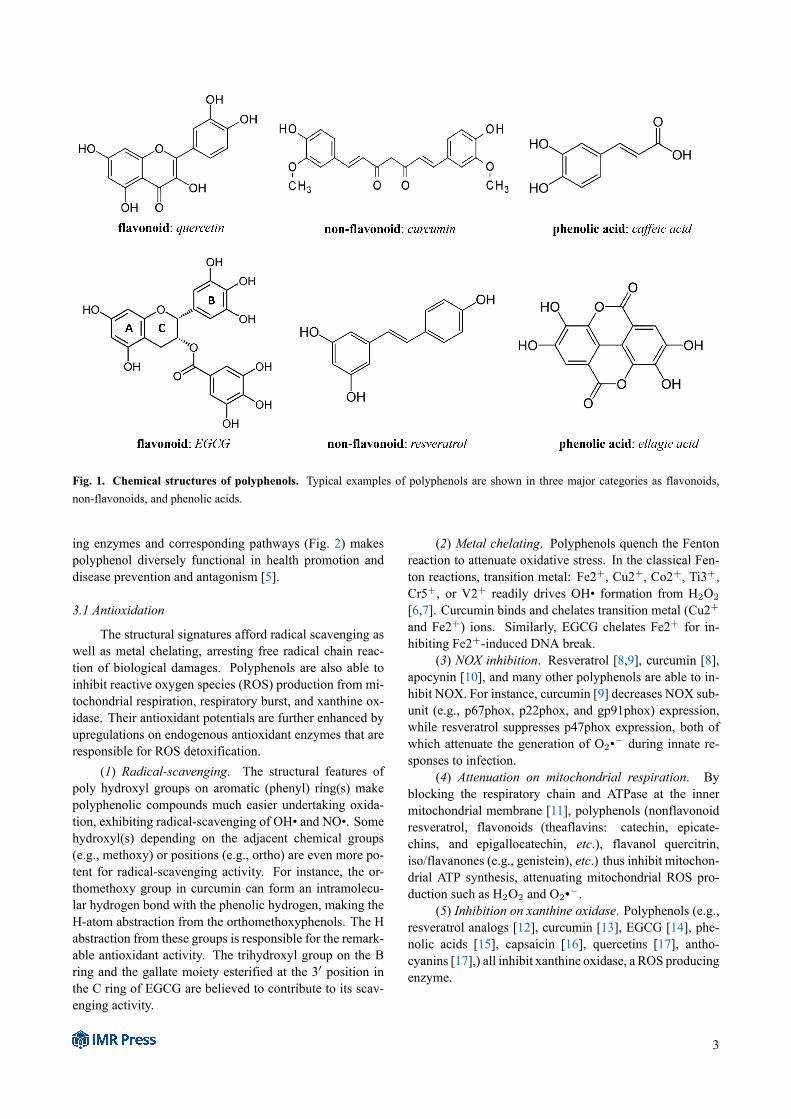
Fig. 1. Chemical structures of polyphenols. Typical examples of polyphenols are shown in three major categories as flavonoids,non-flavonoids, and phenolic acids.
ing enzymes and corresponding pathways (Fig. 2) makespolyphenol diversely functional in health promotion anddisease prevention and antagonism [5].
3.1 Antioxidation
The structural signatures afford radical scavenging aswell as metal chelating, arresting free radical chain reac-tion of biological damages. Polyphenols are also able toinhibit reactive oxygen species (ROS) production from mi-tochondrial respiration, respiratory burst, and xanthine ox-idase. Their antioxidant potentials are further enhanced byupregulations on endogenous antioxidant enzymes that areresponsible for ROS detoxification.
(1) Radical-scavenging. The structural features ofpoly hydroxyl groups on aromatic (phenyl) ring(s) makepolyphenolic compounds much easier undertaking oxida-tion, exhibiting radical-scavenging of OH• and NO•. Somehydroxyl(s) depending on the adjacent chemical groups(e.g., methoxy) or positions (e.g., ortho) are even more po-tent for radical-scavenging activity. For instance, the or-thomethoxy group in curcumin can form an intramolecu-lar hydrogen bond with the phenolic hydrogen, making theH-atom abstraction from the orthomethoxyphenols. The Habstraction from these groups is responsible for the remark-able antioxidant activity. The trihydroxyl group on the Bring and the gallate moiety esterified at the 3′ position inthe C ring of EGCG are believed to contribute to its scav-enging activity.
(2) Metal chelating. Polyphenols quench the Fentonreaction to attenuate oxidative stress. In the classical Fen-ton reactions, transition metal: Fe2+, Cu2+, Co2+, Ti3+,Cr5+, or V2+ readily drives OH• formation from H2O2
[6,7]. Curcumin binds and chelates transition metal (Cu2+and Fe2+) ions. Similarly, EGCG chelates Fe2+ for in-hibiting Fe2+-induced DNA break.
(3) NOX inhibition. Resveratrol [8,9], curcumin [8],apocynin [10], and many other polyphenols are able to in-hibit NOX. For instance, curcumin [9] decreases NOX sub-unit (e.g., p67phox, p22phox, and gp91phox) expression,while resveratrol suppresses p47phox expression, both ofwhich attenuate the generation of O2•− during innate re-sponses to infection.
(4) Attenuation on mitochondrial respiration. Byblocking the respiratory chain and ATPase at the innermitochondrial membrane [11], polyphenols (nonflavonoidresveratrol, flavonoids (theaflavins: catechin, epicate-chins, and epigallocatechin, etc.), flavanol quercitrin,iso/flavanones (e.g., genistein), etc.) thus inhibit mitochon-drial ATP synthesis, attenuating mitochondrial ROS pro-duction such as H2O2 and O2•−.
(5) Inhibition on xanthine oxidase. Polyphenols (e.g.,resveratrol analogs [12], curcumin [13], EGCG [14], phe-nolic acids [15], capsaicin [16], quercetins [17], antho-cyanins [17],) all inhibit xanthine oxidase, a ROS producingenzyme.
3
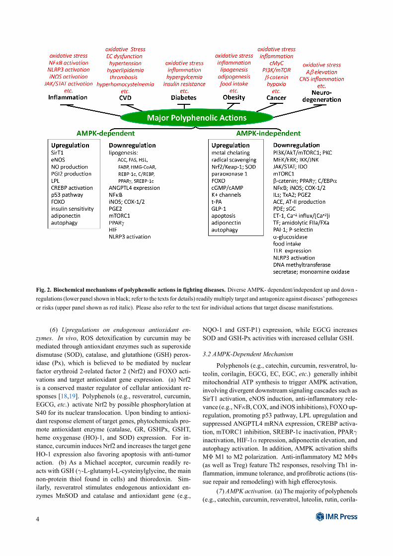
Fig. 2. Biochemical mechanisms of polyphenolic actions in fighting diseases. Diverse AMPK- dependent/independent up and down -regulations (lower panel shown in black; refer to the texts for details) readily multiply target and antagonize against diseases’ pathogenesesor risks (upper panel shown as red italic). Please also refer to the text for individual actions that target disease manifestations.
(6) Upregulations on endogenous antioxidant en-zymes. In vivo, ROS detoxification by curcumin may bemediated through antioxidant enzymes such as superoxidedismutase (SOD), catalase, and glutathione (GSH) perox-idase (Px), which is believed to be mediated by nuclearfactor erythroid 2-related factor 2 (Nrf2) and FOXO acti-vations and target antioxidant gene expression. (a) Nrf2is a conserved master regulator of cellular antioxidant re-sponses [18,19]. Polyphenols (e.g., resveratrol, curcumin,EGCG, etc.) activate Nrf2 by possible phosphorylation atS40 for its nuclear translocation. Upon binding to antioxi-dant response element of target genes, phytochemicals pro-mote antioxidant enzyme (catalase, GR, GSHPx, GSHT,heme oxygenase (HO)-1, and SOD) expression. For in-stance, curcumin induces Nrf2 and increases the target geneHO-1 expression also favoring apoptosis with anti-tumoraction. (b) As a Michael acceptor, curcumin readily re-acts with GSH (γ-L-glutamyl-L-cysteinylglycine, the mainnon-protein thiol found in cells) and thioredoxin. Sim-ilarly, resveratrol stimulates endogenous antioxidant en-zymes MnSOD and catalase and antioxidant gene (e.g.,
NQO-1 and GST-P1) expression, while EGCG increasesSOD and GSH-Px activities with increased cellular GSH.
3.2 AMPK-Dependent Mechanism
Polyphenols (e.g., catechin, curcumin, resveratrol, lu-teolin, corilagin, EGCG, EC, EGC, etc.) generally inhibitmitochondrial ATP synthesis to trigger AMPK activation,involving divergent downstream signaling cascades such asSirT1 activation, eNOS induction, anti-inflammatory rele-vance (e.g., NFκB, COX, and iNOS inhibitions), FOXO up-regulation, promoting p53 pathway, LPL upregulation andsuppressed ANGPTL4 mRNA expression, CREBP activa-tion, mTORC1 inhibition, SREBP-1c inactivation, PPARγinactivation, HIF-1α repression, adiponectin elevation, andautophagy activation. In addition, AMPK activation shiftsMΦ M1 to M2 polarization. Anti-inflammatory M2 MΦs(as well as Treg) feature Th2 responses, resolving Th1 in-flammation, immune tolerance, and profibrotic actions (tis-sue repair and remodeling) with high efferocytosis.
(7) AMPK activation. (a) The majority of polyphenols(e.g., catechin, curcumin, resveratrol, luteolin, rutin, corila-
4
gin, EGCG, EC, EGC, etc.) inhibit mitochondrial ATP syn-thesis by blocking the respiratory chain and ATPase at theinner mitochondrial membrane, thus activating AMPK. (i)Metabolically, AMPK activation mediates hypolipidemiceffects including suppressed lipogenic transcription factors(e.g., SREBP1/2, C/REBP, etc.) and enzymes (e.g., HMG-CoA reductase, acetyl-CoA carboxylase, etc.) for de novobiosyntheses of cholesterol and fatty acids and TG forma-tion. AMPK-mediated phosphorylation of the transcriptionfactors and lipogenic enzymes inactivates activities. (ii)Concerning cell signaling, AMPK activation leads to SirT1and FOXO activation as well as mTOR inhibition. (iii)AMPK activation could also shift MΦ M1 to M2 polariza-tion; anti-inflammatory M2 MΦs (as well as Treg) featureTh2 responses, resolving Th1 inflammation, immune tol-erance, and profibrotic actions (tissue repair and remodel-ing) with high efferocytosis. (b) Additionally, resveratrol[20] inhibits cAMP-degrading PDEs (e.g., PDE4), result-ing in accumulated cAMP that activates Epac1 for in turnstimulating PLCϵ. The resulting phosphorylated ryanodinereceptor 2 triggers Ca2+ channel releasing Ca2+ from ER.The activated CamKKβ then phosphorylates and activatesAMPK.
(8) SirT1 activation. SirT1 activation [20,21] trig-gers diverse signaling including AMPK activation, NFκBinactivation, eNOS activation, p53 activation, tumor sup-pressor FOXO upregulation for antioxidant enzyme (Mn-SOD) expression, early Treg differentiation (antiinflamma-tion), suppressed lipogenesis (PPARγ inactivation) [22],longevity, etc. Resveratrol triggers an array of signalcascade to exhibit metabolic benefits via elevated NAD+
and enhanced SirT1 activity, called AMPK-SirT1-PPARγcoactivator 1α (PGC-1α) axis. While phosporylating PGC-1α, AMPK increases NAD+ levels and activates SirT1that deacetylates PGC-1α. Thus, metabolic benefits suchas anti-aging, anti-diabetic, and increases in FA oxidation,gluconeogenesis, and mitochondrial biogenesis and func-tions could result from indirectly activated SirT1 via com-petitive inhibition of cAMP-phosphodiesterases (PDE4) bypolyphenol in red wine [20,21]. In these regards, resveratrolmimics caloric restriction, exercise, or short-term fasting infavoring longevity.
(9) FOXO activation. FOXO upregulation is largelya result of PI3K/AkT inhibition and AMPK/SirT1 activa-tion [22], which mediates NFκB inactivation, HIF repres-sion, antioxidant enzyme expression, and Treg differentia-tion. For further cancer protection, FOXO activation ex-tends its effects to proapoptosis and antagonism againstonco- gene/protein c-MyC; FOXO functions as a suppres-sive oncogene.
(10) eNOS activation. AMPK phosphorylates andactivates eNOS; NO is considered anti-inflammatory. Inaddition, polyphenols such as resveratrol via such eNOSactivation show benefits to insulin sensitivity and anti-hypertension. The resulting NO production activates
sGC for cGMP generation. Such eNOS activation me-diates Glut4 translocation and increased glucose up-take/utilization by muscle cells [23,24], mimicking insulinaction. In an EC-dependent vasodilation, resveratrol acti-vates eNOS activity, NO production, GC activation, andsubsequent cGMP production in addition to its ability tofunction as a non-selective PDE inhibitor. Taken together,resveratrol leads to cGMP accumulation and Ca2+ effluxesfor vascular dilation [25,26].
(11) NFκB, COX, iNOS inhibitions. (a) AMPKdownregulates hallmark inflammatory transcription fac-tor: NFκB [27] by at least two mechanisms. (i) AMPKincreases NAD+ for its consequent SirT1 activation.SirT1 deacetylates and activates PGC-1β while deacety-lating and inactivating NFκB p65, an inflammatory mas-ter transcription factor. (ii) AMPK directly inactivatesNFκB via mTORC1 inhibition (also see below section onmTORC1 inhibition); IκB kinase (IKK) phosphorylationby mTORC1 results in NFκB nuclear translocation and itstranscriptional activity. NFκB is recognized as a hallmarkof inflammation. (b) In addition to proinflammatory (TNF,IL-1, IL-6) genes, COX-2 and iNOS are important gene tar-gets of NFκB [27]. Therefore, inflammatory TNF, IL1/6,PGE2, and NO production are all suppressed by polyphe-nols. For instance, curcumin attenuates proinflammatorycytokine (e.g., IL-1β, IL-6, and TNF-α) expression andinhibits STAT3 phosphorylation and activation. In a sim-ilar manner, curcumin downregulates AP-1 and cytokine(IL-1α and TNF-α) expression. Importantly, NFκB is in-volved in cell proliferation and tumor cell survival, link-ing inflammation and cancer; thus, NFκB inactivation bearsanti-cancer action [27].
(12) Promoting p53 pathway. Mediated by AMPK ac-tivation, curcumin is able to phosphorylate at S15 in p53N-terminus [28–33], which attenuates p53 interaction withits negative regulator MDM2 for promoting p53 stabiliza-tion and nuclear translocation for its transcriptional activity.Such action favors cell apoptosis and suppresses cell prolif-eration. (a) p53 inhibits Bcl-2, while enhancing the intrinsicapoptotic pathway including elevated cytoplasmic proapo-totic proteins (PIDD, Bid) and mitochondrial proapoptoticproteins: Bax, Bak, Puma, and Noxa. (b) p53 also pro-motes the extrinsic pathway by elevating death receptors(Fas/Apo1, DR 5, etc.).
(13) LPL upregulation. AMPK phosphorylates andactivates LPL [33], a major circulating enzyme responsi-ble for TG-rich lipoprotein catabolism and TG degradationin lowering blood TG level.
(14) CREBP activation and BDNF expression. Me-diated by AMPK activation, polyphenols (e.g., curcumin,EGCG) phosphorylate CREBP that in turn activates brainderived neurotrophic factor (BDNF) expression. BDNF isrequired for long term potential and cognition process inhippocampus [31,32].
5
(15)mTORC1 inhibition. There are at least two mech-anisms by which AMPK inhibits mTORC1. (a) AMPKphosphorylates and activates TSC2, a negative upstreamregulator of mTORC1 [34–37]. (b) Alternatively, AMPKdirectly phosphorylates and inactivates raptor, an adap-tor protein in mTORC1 complex. Both actions ensuremTORC1 inhibition by AMPK activation. mTORC1 isresponsible for upregulating PPARγ, SREBP-1c, HIF, in-flammation (IKK phosphorylation and NFκB activation),and cell proliferation/differentiation/survival while down-regulating autophagy. Accordingly, AMPK-dependentmTORC1 inhibition results in downregulations on PPARγ,SREBP-1c, and HIF, which has been reported to be benefi-cial to aging related pathological conditions such as cogni-tion decline, Alzheimer’s (AD), cancer, and kidney, heart,and autoimmune diseases over the past 40 years.
mTORC1 inhibition presents HIF repression andNFκB inactivation. mTORC1 activation, otherwise, pro-motes glycolysis via upregulation of HIF1α and c-Myc (tu-morigenesis); stimulates lipid biosynthesis and the pentosephosphate pathway through sterol regulatory element bind-ing protein 1 (SREBP-1) (lipogenesis) [38]; and positivelycontrols glutamine metabolism by SIRT4 repression. Cur-cumin, resveratrol, EGCG, genistein, and caffeine readilyinhibit both mTORC1 and mTORC2 [39], which is a con-sequence of AMPK activation and/or PI3K/AkT inhibition.
(16) SREBP inactivation. mTORC1 inhibition in turninactivates SREBP-1c, resulting in suppressed lipogenesisincluding fatty acid synthesis (e.g., acetyl-CoA carboxy-lase) and TG formation (lipin 1) [37], characteristics offat accumulation in obesity. mTORC1 is responsible forSREBP-1c phosphorylation, cleavage, and its enhanced nu-clear translocation and transcriptional activity.
(17) PPARγ inactivation. As the result of mTORC1inhibition and SirT1 activation [40] by phytochemicals viaAMPK activation, PPARγ translation and transcriptionalactivity are significantly suppressed. PPARγ is a knownmaster gene for adipogenesis and adipocyte differentia-tion [37,40]; PPARγ inactivation thus blocks adipogene-sis, lipogenesis, and fat accumulation, contributing to anti-obesity.
(18) HIF-1α repression. mTORC1 inhibition down-regulates the expression of HIF-1α [34–36], contributingto antiinflammation and tumor suppression. HIF-1α isan inflammatory as well as angiogenic transcription fac-tor. (a) As an inflammatory trigger, hypoxia (HIFα) pro-motes Th17 expansion and IL-17 production while promot-ing degradation of Treg FOXp3 through VHL E3 ligase. (b)As an angiogenic factor, HIFα targets many genes includ-ing VEGF for cancer progression, metastasis, and cancerstem cell expansion.
(19) Suppressed ANGPTL4 expression. Upon AMPK-dependent downregulation on SREBP-1c, PPARγ, andHIFα, ANGPTL4 expression is suppressed. ANGPTL4, atarget gene of SREBP-1c, is a negative regulator of LPL.
ANGPTL4 expression could also be regulated by PPARγ,HIFα, and glucocordioid receptor; for instance, ANGPTL4is induced by fasting and hypoxia. Thus, its repression fa-cilitates LPL activity for TG degradation and hypoTG ac-tion.
(20) Autophagy upregulation. AMPK-dependentmTORC1 inhibition is able to upregulate autophagy.mTORC1 negatively regulates a complex consisting of es-sential autophagic proteins (e.g., ULK1, ATG13, ATG101,and FIP200). Alternatively independent of mTORC1 in-hibition, direct PI3K/AkT inhibition could block phospho-rylation of a crucial autophagic element: Beclin 1 thatotherwise dimerizes and recruits 14-3-3 further being se-questered to cytoskeletal actin vimentin and intermediatefilament complex. As a result of such blockage mediatedby PI3K/AkT inhibition, autophagosome assembly is ableto initiate autophagy.
Essentially, autophagy, an intracellular cleaning sys-tem including aggrephagy, xenophagy, mitophagy, andlipophagy, contributes to regenerating metabolic precur-sors, and cellular and tissue homeostasis by degrading long-lived proteins, protein aggregates, and defective organelles(e.g., mitochondria, ER, or peroxisomes), and cleaning sub-cellular debris.
Autophagy downregulates oxidation and prevents in-flammation (e.g., NLRP3 inflammasome activation). (a)Mitophagy limits NLRP3 activation by removing dam-aged mitochondria. (b) Autophagosomal Atg16L1 read-ily inhibits ROS production; ROS is essential for NLRP3activation (refer to as oxidation-inflammation axis). (c)Autophagy per se promotes lysosome (NLRP3 inflamma-some) degradation through ubiquitination involving au-tophagosomal components p62 and LC3. (d) Furthermore,removal of pro-IL1β/18 by autophagy for NLRP3-mediatedcaspase 1 cleavage ensures antiiflammation. Thus, au-tophagy protects from inflammasome (NLRP3) activationthat is essential for IL-1β/18 maturation and secretion. (e)Autophagy also plays role(s) in anti-viral (e.g., HIV), anti-bacteria (Mtb, Shigella flexneri), etc. (f) Limited informa-tion is available directly regarding autophagy upregulationin relation to cardioprotection. Apparently, resveratrol in-duces autophagy and thus possibly protects from MI.
(21) Adiponectin elevation and signaling. Increasedadiponectin is reported in response to resveratrol thatalso upregulates the expression of adiponectin receptor-1. Resveratrol promotes the posttranslational multimer-ization and stability of adiponectin by DsbA-L that is in-duced upon AMPK activation and AkT-mediated FOXO1activation [41]. (a) Adiponectin in contrast to its counter-parts (e.g., leptin) is of antiinflammation in nature. Forinstance, circulating adiponectin appreciably declines inobese. Adiponectin attenuates TNF-α and IL-6 produc-tion while inducing expression of anti-inflammatory cy-tokines (e.g., IL-10 and IL-1 receptor antagonist). (b)Adponectin signaling generally triggers S-1-P formation
6
and AMP, PPARα, and p38 MAPK activation. (c) Itsmajor metabolic functions include glucose homeostasis,insulin-sensitizing action, increased fatty acid oxidation,and downregulated hepatic gluconeogenesis, all of whichfight against metabolic symptoms.
In a clinical trial [42], grape resveratrol increasesserum adiponectin and downregulates inflammatory genes(PAI-1, IL-6, AP-1, JUN, CREBP, etc.). In an animal model[43], 7-O-galloyl-D-sedoheptulose increases adiponectinlevel while downregulating leptin, insulin, C-peptide, re-sistin, TNFα, and IL-6 in serum and proinflammatoryNFκB p65, COX-2, iNOS, JNK, phospho-JNK, AP-1,TGFβ1, and fibronectin.
3.3 AMPK-Independent MechanismsBy inhibiting multiple signaling enzymes or enzymes
per se, polyphenols downregulate corresponding signalingpathways and suppress metabolic activities, respectively.
A wide-range of AMPK-independent actions [44] in-clude PI3K/AkT inhibition, direct mTORC1 inhibition,β-catenin inactivation, FOXO activation, PDE inhibition,ACE inhibition, attenuated ET-1 expression, proapopto-sis, JNK or IKK inhibition, Nrf2 activation, Janus ki-nase (JAK)/signal transducer and activator of transcription(STAT) inactivation, PKC inhibition, and MAPK inactiva-tion. In addition, many polyphenolic effects concern non-signaling related enzyme inhibitions including secretase in-hibition, monoamine oxidase inhibition, α-glucosidase in-hibition, anticoagulation, and anti-thrombosis.
(22) PI3K/AkT inhibition. Polyphenols generally in-hibit PI3K/AkT signaling cascade; its downstream effectsinclude IKK inhibition and FOXO activation, exhibitingantiinflammation. Synergistically with AMPK activation,PI3K/AkT inhibition leads to mTORC1 inhibition. Thus,PI3K/AkT inhibition exhibits a broad spectrum of eventsincluding NFκB inactivation, FOXO upregulation, HIF re-pression, etc.
(23) β-catenin inactivation. By inhibiting PI3K/AkT,curcumin blocks axin/APC/GSK3β/β-catenin complex dis-assembly to cause β-catenin degradation. Moreover, cur-cumin directly inhibits GSK3β for quenching β-catenin re-lease and nuclear translocation [28–30]. As a result ofβ-catenin inactivation, transcription factors: PPARγ andC/EBPα are also downregulated. β-catenin inactivation isalso a target for attenuating cancers.
(24) FOXO activation. IP3k/AkT inhibition also leadsto FOXO activation. Transcription factor: FOXO, a knowntumor suppressor [45], is a positive downstream effectorof PI3K/AkT [46]. AkT phosphorylates FOXOs, whichblocks FOXO nuclear translocation and transcriptional ac-tivity. As the result of PI3K/AkT inhibition, FOXO dephos-phorylation encourages its nuclear translocation and tran-scription activity resulting in FOXO upregulation with atleast five-fold significance. (a) PI3K/AkT inhibition withFOXO3a upregulation decreases ROS production accom-
panied by ROS detoxification with elevated antioxidant en-zyme (e.g., SOD2, catalase, GSH-Px) expression [47] forpromoting antioxidant action. (b) Following the similar en-hanced FOXO3a transcriptional activity antagonizes anddestabilizes Myc oncogene [47], reflecting anti-cancers.(c) FOXO1 upregulation leads to enhanced apoptosis [45],while (d) FOXO1 activity is required for early phase in Tregdifferentiation, a component contributing to antiinflamma-tion. (e) FOXO proteins also negatively regulate HIF [45],implying its antagonism against inflammation, angiogene-sis, and possibly metastasis. Of particular interests, AMPKper se is proposed to be able to upregulate FOXO transcrip-tional activity independent of AkT modulation (please referto above AMPK-dependent mechanism for insights).
(25) Proapoptosis. The enhanced apoptosis is pro-posed to be largely mediated by AkT inhibition alone andFOXO upregulation [45] resulting from PI3K/AkT inhibi-tion along with SirT1 activation. Mechanistically, (a) Cur-cumin upregulates the extrinsic pathway [28–30,48] by (i)death receptor (e.g., DR4 and DR5) activation with bindingto the pro-caspase ligand and (ii) inducing apoptosis withincreased caspase-3/6/7/8. (b) In the intrinsic mitochon-drial pathway, curcumin downregulates apoptotic inhibitors(e.g., BcL2 and BcL-xL) while promoting mitochondrialcytochrome C release through PUMA, NOXA, Bak, andBAX activations. As a consequence, Apaf1 activation inturn leads to caspase-3/7/9 activation for apoptosis.
(26) NFκB inactivation. NFκB inactivation bypolyphenols is mainly mediated by PI3K/AkT inhibition,FOXO activation, and IKK inhibition. Curcumin, for in-stance, inhibits TLR4-induced IKKα/β phosphorylation;the resulting blocked release of IκB results in NFκB inacti-vation. Alternatively, curcumin inhibits AkT and its conse-quent IκB phosphorylation, similarly leading to NFκB in-activation and disfavoring cell proliferation [49–52].
(27) ERK inhibition. Curcumin inhibitsMAP3K/MAP2K and in turn MAPK (JNK, p38, andERK) activation, thereby downregulating AP-1-mediatedtranscription activity for TNFα, iNOS, and COX-2 expres-sion [49–52], presenting anti-inflammatory in addition toanti-proliferative activities.
(28) JAK/STAT inhibition. Curcumin could inhibitJAK that otherwise phosphorylate STAT3, thereby block-ing STAT3 dimerization and nuclear translocation. SuchJAK/STAT attenuation exhibits anti-inflammatory (e.g., re-pressed proinflammatory genes) and anti-cancer (e.g., in-duced apoptotic proteins, suppressed apoptotic inhibitorsand c-Myc oncogene) activities [49–52]. Similarly, resver-atrol prevents JAK phosphorylation, thereby inhibitingSTAT1 phosphorylation and transcriptional activity [53,54]. In a study by Noh et al. [55] has revealed thatsuch JAK/STAT1 inhibition by resveratrol extends to in-doleamine 2,3-dioxygenase (IDO) suppression for cancerimmunoprotection.
7
(29) IKK/JNK inhibition. EGCG, an example, in-hibits inflammatory serine/threonine kinase (e.g., JNK orIKK) directly or as a result of AkT inhibition to ensureantiinflammation (e.g., NFκB inactivation). In addition,such JNK/IKK inhibition attenuates insulin resistance; oth-erwise, JNK/IKK compete tyrosine phosphorylation on in-sulin receptor substrate (IRS).
(30) ACE inhibition. ACE inhibition by polyphe-nols presents two-fold significance in antiinflammation andantioxidation in view of ATII triggering ROS, cytokine,and chemokine production. Most flavonoids are reportedto be competitive inhibitors of ACE [56]. Anthocyanins,flavonols (e.g., quercetin, kaempferol, and myricetin), andflavanol (e.g., catechins, epicatechins, and their poly-mers) are effective ACE inhibitors. Flavonoid precursormolecules chalcones (butein) and their pyrazole derivativesalso dose-dependently inhibit ACE in vitro. Methylatedepigallocatechin-3-O-(3-O-methyl) gallate is much effec-tive inhibitory than its parent molecule epigallocatechin-3-O-gallate. ACE inhibitory properties of flavones remainunclear; however, isoflavones (e.g., genistein, daidzein,and glycetin) decrease ACE gene expression and enzymeactivity.
(31) α-glucosidase inhibition. Polyphenols (flavones,flavonols, flavanones, isoflavones, catechins, and antho-cyanidin) inhibiting α-glucosidase [57] reduces glucose in-puts from dietary carbohydrates, certainly lowering hyper-glycemic risk.
(32) PDE4 inhibition. Independent of inhibition onmitochondrial ATPase, resveratrol [20] inhibits cAMP-degrading PDEs (e.g., PDE4), resulting in accumulatedcAMP that activates Epac1 for in turn stimulating PLCϵ.PLCϵ-mediated CamKII activation phosphorylates RYR2,further activating CamKKβ for AMPK activation.
(33) Direct mTOR inhibition. There is evidence fordirect inhibitions on mTORC1 by phenolic phytochemi-cals. Resveratrol significantly increases the association be-tween mTOR and its negative regulator (DEPTOR), therebydownregulating mTOR activity [58]. Curcumin decreasestotal expression of mTOR, Raptor, and Rictor protein andmRNA levels [59]. Without affecting upstream kinase ac-tivities and TSC1/2 interaction, curcumin is also able to dis-sociate raptor from mTORC1 [60].
(34) Attenuated ET-1 production and signaling. Nu-clear exclusion of phosphorylated FOXO1 by EGCG resultsin downregulation of ET-1 promoter, thereby suppressingET-1 expression and its activity [61]. Similarly, hydrox-ysafflor yellow A (HSYA), resveratrol, and quercetin re-duce ET-1 production. In addition, green tea or EGCGdownregulates ETA receptor, blocking ET-1 signaling foranti-hypertension and hypertrophy.
(35) Anticoagulation. Significantly prolonged TT,aPTT, and PT have been reported in vitro, in vivo, or exvivo in response to polyphenols. (a) thrombin inhibition[62,63]. Curcumin and its derivative bisdemethoxycur-
cumin, cyanidin, quercetin, silybin, cyanin, (+)-catechinand (-)-epicatechin inhibit thrombin amidolytic activity; inaddition, cyanidin, quercetin, and silybin suppress throm-bin proteolytic activity. Aglycones act as competitivethrombin inhibitors, while chokeberry extract significantlyinhibits thrombin amidolytic activity. (b) FXa inhibition[63–65]. Flavonoids: procyanidin B2, cyanidin, quercetin,and silybin bind S1–S4 pockets in vicinity of the FXa ac-tive site and block access of substrates to Ser195, therebydirectly inhibiting FXa amidolytic activity. Curcuminand its derivative bisdemethoxycurcumin also inhibitFXa activity. (c) protection from FVII activation [66].Tannic acid, delphinidin, hamamelitannin, (-)-epicatechingallate, and 3,5-di-O-caffeoylquinic acid bind plasmahyaluronan-binding protein and inhibit FVII autoactivation(autoproteolysis). (d) TF suppression/encryption [67].Grape and its products with high content of polyphenolsexert anticoagulation by suppression of TF synthesis inblood mononuclear cells and VECs. In a recent personalcommunication, antiinflammatory HSYA (a phenolicrelated flavonoid component from Carthamus tinctoriusL.: 3,5,6-trihydroxy-2(E)-[1-oxo-3-(4-hydroxy phenyl)-2-propenyl]-4,6-bis[(2S,3R,4R,5S,6R)-3,4,5-trihydroxy-6-(hydroxyl-methyl) oxan-2-yl]-2,4 cyclo-exadien-1-one)suppresses OxLDL-induced TF expression in vitro/in vivomodels, which is mediated by PPARγ upregulation andattenuated p38 MAPK phosphorylation/activation. Rutin(flavonoid) has long been recognized as an anticoagulantfor possible prevention of heart attack and stroke, which ismediated by its inhibition on protein disulfide isomerasethat de-encrypts TF for initiating the extrinsic coagulationpathway and robusting thrombin formation. (e) otheractions. Aronia melanocarpa or seeds of Vitis viniferaprolong clotting time and decrease the maximal velocityof fibrin polymerization and FXIIIa amidolytic activityin human plasma [68]. However, there is no evidencethus far whether polyphenols have any effects on naturalanticoagulants (e.g., TFPI, APC, or AT III).
(36) PKC inhibition. Polyphenol superfamily gen-erally inhibits PKC by several mechanisms: competitionwith binding of Ca2+, ATP, the kinase catalytic domain,etc. as well as inhibition of PKC expression or transloca-tion to membrane [69]. (a) PKC downstream signaling in-cludes UDP-glucuronosyltransferase, MAPK/c-JUN, ERK,and EGFR, finally leading to upregulations on transcrip-tion factors: NFκB, AP-1, and early response gene-1. (b)PKC also involves in cell proliferation, which is mediatedby MAPK activation and the above upregulated transcrip-tion factors. Thus, PKC inhibition exhibits antiinflamma-tion, anti-proliferation, etc. as well as downregulated cellsignaling.
(37) MAPK inactivation. Curcumin inhibitsMAP3K/MAP2K and in turn MAPK (JNK, p38, andERK) inactivation, thereby downregulating AP-1-mediated transcription activity for TNFα, iNOS, and
8
COX-2 expressions [28–31]. Thus, MAPK inactivationpresents anti-inflammatory in addition to anti-proliferativeactivities.
(38) Secretase inactivation. Curcumin and otherpolyphenol [28–31,70] inactivates α/β/γ secretases thatotherwise cleave amyloid precursor protein (APP) intoamyloid β protein (Aβ). Aβ accumulation is a known char-acteristic of AD blocking neurotransmission along with in-tracellular neurofibrillary tangle formation.
(39) Monoamine oxidase inhibition. Resveratrol, cur-cumin, and quercetin directly inhibit monoamine oxidasethat otherwise catabolizes neurotransmitters (e.g., 5-HT,epinephrine, DOPA, dopamine, etc.); therefore, polyphe-nols exhibit antidepressive property and cognitive improve-ment [31,32].
3.4 Other ActionsPolyphenols are also able to modulate protein expres-
sion and proceed immunoregulations, achieving up or down-regulation for disease prevention and antagonism. Epige-netic modulation and gut microbiota alteration by polyphe-nols have also been reported.
(40) Downregulation on TLR expression. To arrestinflammatory initiation and propagation, polyphenols suchas curcumin, kaempferol-3-O-sophoroside, and EGCG in-hibit TLR2/4 expression [71], which could reduce wide-range of inflammatory responses such as LPS, IL-1α/β, IL-6, TNFα, and HMGB1 release/signaling.
(41) K+ channels activation. In an EC-independentfashion for vasodilation, resveratrol directly opens K+
channels including KATP and BKCa expressed on VSMCwhere extracellular Ca2+ influx and intracellular Ca2+ re-lease are also suppressed [72].
(42) Upregulation on paraoxonase 1. Quercetin in-creases paraoxonase 1 mRNA and protein expression, up-regulating paraoxonase 1 activity. Paraoxonase 1 is a HDL-associated enzyme displaying esterase and lactonase activ-ity; paraoxonase 1 metabolizes toxic OxLDL or OxHDL,thus protecting LDL and HDL from oxidation [73], show-ing cardioprotection.
(43) PAI downregulation. By stimulating binding ofupstream stimulatory factor-2 to two distinct E-box se-quences, quercetin downregulates PAI-1 promoter, thus re-sulting in suppressed PAI-1 expression in human coro-nary artery ECs [74]. Polyphenols including curcumin,quercetin, resveratrol, and EGCG and its derivatives (oc-taacetate and theaflavin digallate) act as potential PAI-1 in-hibitors to reduce PAI-1 production [75], favoring fibrinol-ysis and resolution of blood clots. Similarly, grape ingredi-ents suppress PAI-1 levels [76,77].
(44) tPA upregulation. Quercetin induces tPA expres-sion, which is mediated by functional Sp1-binding elementin tPA promoter and p38 MAPK pathway [78]. Similarly,catechin, epicatechin, and resveratrol in red wine also in-duce tPA and u-PA in vitro [79].
(45) Epigenetic modulations. Epigenetic modula-tions, including regulations on DNA methylation, histonemodification, and non-coding RNA (miRNA) effects, couldoffer cardioprotection and anti-cancer action, etc. EGCGinhibits DNA methyltransferase activity in various experi-mental and clinical studies [80–82]. Polyphenols also targethistone deacetylase 6-related pathways. EGCG decreasesthe expression of oncogenic miRNAs (miR-92, miR-93,and miR-106b) and to increase the expression of tumor-suppressor miRNAs (miR-7-1, miR-34a, and miR-99a) inhuman cancer cells, while curcumin up-regulates miR-22and down-regulates miR-199a, presenting improved canceroutcomes. Interestingly, polyphenolic antioxidation couldin part contribute to epigenetic modulation, which remainsto be elucidated.
(46) Immunoregulation. Polyphenols modulate im-mune responses in both the innate and adaptive systems,having either stimulatory or inhibitory effects includingself-tolerance, anergy, etc. [83–86]. (a) Pterostilbene, aresveratrol analogous, suppresses DC activation and pro-moting Treg cell development and green tea polyphe-nol EGCG induces Treg cells. Dietary polyphenols (b)downregulate DC antigen presentation by CD83, CD80,CD11c, and MHC II and immune-modulate MΦs (e.g.,antigen presentation, phagocytosis, cytokine production:M1/M2 polarization), (c) increase proliferation of B cells(antibody IgA/E/M/D production) and T cells (cytokineproduction, cytotoxic destruction, etc.), (d) suppress T dif-ferentiation into Th1, Th2, Th17, Th22, and Th9 cells, and(e) activate NKs (cytolytic perforin and granzyme B se-cretion). Accordingly, polyphenols confer immunomodu-latory effects against allergic reaction and autoimmune dis-ease largely by inhibition of autoimmune T cell prolifer-ation, downregulation of proinflammatory cytokines (e.g.,IL-6, IL-1, IFN-γ), enhanced Treg development, shiftedTh1/2 balance, and decreased Th17 cells.
(47) Altered gut microbiota. As a prebiotics, polyphe-nols could alter the landscape of gut microbiota/microbiotadiversity [87–89]. For instance, (a) resveratrol supplemen-tation suppresses Parabacteroides johnsonii, Alistipes pu-tredinis, and B. vulgatus induced by high-fat, which is pro-posed to enhance GLP-1 secretion [90]. (b) Pomegranateextract rich in gallic and ellagic acid enhances the totalgrowth of Bifidobacterium spp. and Lactobacillus spp.without affecting C. coccoides-E. rectale and the Clostrid-ium histolyticum groups [91]. (c) Green tea [92] increasesthe survival of Bifidobacteria. Bifidobacterium and Lac-tobacillus are known probiotics beneficial to food al-lergy. (d) Polyphenols decrease in Bacteroides acidifa-ciens, but increase in Ruminococcus gnavus and Akkerman-sia mucinphilia, [85] which in turn induces Tregs whilesuppressing inflammatory Th1/Th17 cells, confering anti-inflammation. (e) Curcumin is in favor of beneficial mi-crobiota (Bifidobacteria, Lactobacilli) that are butyrate-producing bacteria, while it reduces the abundance of the
9

pathogenic ones (Prevotellaceae, Coriobacterales, Enter-obacteria, Bacteroidaceae, and Rikenellaceae) that are of-ten associated to the onset of systemic diseases such as AD,CRC, etc. It is also noted that microbiota (Bifidobacterialongum, Bifidobacteria pseudocatenulaum, Enterococcusfaecalis, Lactobacillus acidophilus, and Lactobacillus ca-sei) could be able to metabolize curcumin. The reciprocalrelation between polyphenols and gut microbiota could beexpected to promote human health.
4. Biological/Physiological FunctionsPolyphenols with multiple targets (Fig. 2) readily offer
broad antagonisms against disease development and pro-gression including inflammation, CVD, diabetes, obesity,cancer, neurodegeneration, and infection; among which,oxidative stress and inflammation really play critical rolesin pathogenic developments. Table 1, if not exclusively,summarizes polyphenolic actions in comparison with com-mon approaches to combating pathological conditions.
4.1 Anti-Oxidative StressOxidative stress defines overload ROS/RNS and pro-
oxidants without appropriate/coordinated protection by an-tioxidants, possibly triggering pathologies. Biological sys-tem is constantly under oxidative stress, not only living in20% oxygen (O2) atmosphere, but also hypoxia (ischemia)stabilizing HIF1α to upregulate NADPH oxidase (NOX)(superoxide anion (O2•−) formation) or to turn on down-stream angiogenic gene (e.g., VEGF) expression. Oxida-tive stress serves as a molecular mechanism to mediate di-verse disease progression and pathogenesis.
In a classical view of singlet O2 metabolism, molecu-lar O2 is utilized by biological systems followed by a conse-quence of formations of O2•−, hydrogen peroxide (H2O2),hydroxyl radical (OH•), and H2O in step-wise one-electronsequential reductions [93]. O2•−, H2O2, and OH• are threemajor reactive oxygen species (ROS), all of which are cy-totoxic and exhibit damaging effects on biological compo-nents including DNA damage, lipid/cholesterol oxidation,lipoprotein oxidation, protein oxidation, and membrane dis-ruption [94].
ROS derives from either intrinsic or extrinsic sources.(a) Intrinsic ROS sources include that (i) ROS is by-products of mitochondrial respiration, especially in mito-chondrial dysfunctions in complex I/II/III or IV; (ii) dur-ing infection triggering innate immunity, O2•− is the mainproduct from NOX (also known as respiratory burst oxidasemainly in neutrophils) to kill invading pathogens. NOX cat-alyzes the one-electron reduction of O2 to generate O2•−in the presence of NADPH in microsomal electron trans-fer chain. In other signaling systems, TNF stimulates theformation of mitochondrial O2•−, while vascular smoothmuscle cells (VSMC) produces O2•− in response to AT II;(iii) endogenous H2O2 could derive from respiratory burstthrough NOX2 following infection; O2•− is then converted
to H2O2 by superoxide dismutase (SOD). MitochondrialH2O2 production can also be activated by defective res-piratory functions or blocking complex I (by retenone) orcomplex III (by antimycin A); (iv) endogenous H2O2 formsOH• and OH− anion through the non-enzymatic Fenton re-action when Fe2+ is oxidized to Fe3+ or during other tran-sition metal oxidations. H2O2 can also be decomposed bycatalase, GSH-Px, or peroxiredoxin to H2O; (v) xanthineoxidase is proposed to contribute to ROS production; (vi)other intrinsic sources could also consist of advanced gly-cation end-product (AGE), ATII, and pheomelanin. (b) Ex-trinsic ROS sources include smoking (e.g., some 1014 freeradicals per inhalation), alcohol (e.g., CYP2E1 inductionfor O2•− and H2O2 generation and reduced cellular GSHwhile inducing iron accumulation and TNF-α production),xenobiotic oxidation (cytochrome p450 reducing molecularoxygen in the proceeding of xenobiotic oxidation throughelectron transfer from NAD(P)H. O2•−, H2O2, and OH•are generated as intermediates when heme center undergo-ing oxidation with conversion of Fe2+ to Fe3+ for radicalformation), hypoxia/reperfusion (e.g., microsomal NOX in-duction for O2•−production), or infections (e.g., NOX ac-tivation), all of which participate in a series of enzymaticreactions in response to diverse environmental insults (ex-trinsic oxidative stress), initiating biological oxidations andelevating endogenous ROS. (c) Similarly, reactive nitrogenspecies (RNS) including reactive nitrogen intermediates(RNI) exhibit diverse biological damages often in cross-talk with ROS. RNS includes peroxynitrile (ONOO−), ni-troxyl (NO−), nitrosyl chloride (NOCl), and nitrogen diox-ide (NO2), all of which are toxic to biological functions.For instance, O2•− effectively reacts with NO; the result-ing OONO− undergoes notorious diverse radical reactionsincluding oxidations and nitrosations, which is even morebiological toxic and damaging (e.g., induced apoptosis, celldeath). (d) In addition, damaging radicals (electrophilic)undergoing non-enzymatic reactions in a fashion of chain-reaction with biomolecules (e.g., DNA, lipids, cholesterol,lipoproteins, proteins, and biomembranes) thus propagateradical formations and intensify oxidative stress.
Natural oxidative defense includes antioxidants (e.g.,vitamin C/E, GSH, α-lipolic acid, N-acetylcysteine,ubiquinol/CoQ, NO, Se, and many antioxidants (either 1° orchain breakers for radical chain reactions of propagation))and antioxidant enzymes (e.g., catalase, SOD, GSH reduc-tase, GSH-S-transferases, GSH-Px, quinone reductase, HO,paraoxonase, etc.), removing free radicals (scavenging orbreaking).
The classical antioxidant actions of polyphenols areable to scavenge radical, chelate metal, upregulate endoge-nous antioxidant enzymes for biological detoxification, andinhibit ROS production from mitochondrial respiration, res-piratory burst, and xanthine oxidase (please refer to 3.1 (1)to (6)). The protection from DNA damage, membrane dis-ruption, and lipid/cholesterol, lipoprotein, and carbohy-
10
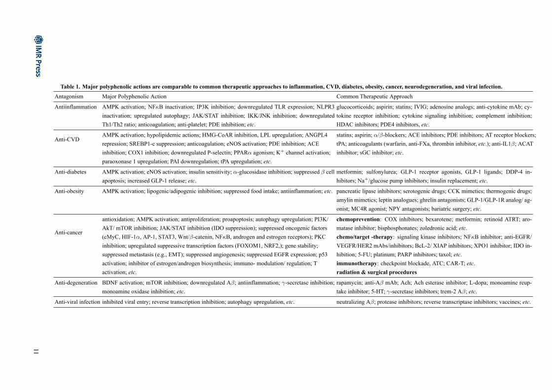
Table 1. Major polyphenolic actions are comparable to common therapeutic approaches to inflammation, CVD, diabetes, obesity, cancer, neurodegeneration, and viral infection.Antagonism Major Polyphenolic Action Common Therapeutic Approach
Antiinflammation AMPK activation; NFκB inactivation; IP3K inhibition; downregulated TLR expression; NLPR3inactivation; upregulated autophagy; JAK/STAT inhibition; IKK/JNK inhibition; downregulatedTh1/Th2 ratio; anticoagulation; anti-platelet; PDE inhibition; etc.
glucocorticoids; aspirin; statins; IVIG; adenosine analogs; anti-cytokine mAb; cy-tokine receptor inhibition; cytokine signaling inhibition; complement inhibition;HDAC inhibitors; PDE4 inhibitors, etc.
Anti-CVDAMPK activation; hypolipidemic actions; HMG-CoAR inhibition, LPL upregulation; ANGPL4repression; SREBP1-c suppression; anticoagulation; eNOS activation; PDE inhibition; ACEinhibition; COX1 inhibition; downregulated P-selectin; PPARα agonism; K+ channel activation;paraoxonase 1 upregulation; PAI downregulation; tPA upregulation; etc.
statins; aspirin; α/β-blockers; ACE inhibitors; PDE inhibitors; AT receptor blockers;tPA; anticoagulants (warfarin, anti-FXa, thrombin inhibitor, etc.); anti-IL1β; ACATinhibitor; sGC inhibitor; etc.
Anti-diabetes AMPK activation; eNOS activation; insulin sensitivity; α-glucosidase inhibition; suppressed β cellapoptosis; increased GLP-1 release; etc.
metformin; sulfonylurea; GLP-1 receptor agonists, GLP-1 ligands; DDP-4 in-hibitors; Na+/glucose pump inhibitors; insulin replacement; etc.
Anti-obesity AMPK activation; lipogenic/adipogenic inhibition; suppressed food intake; antiinflammation; etc. pancreatic lipase inhibitors; serotogenic drugs; CCK mimetics; thermogenic drugs;amylin mimetics; leptin analogues; ghrelin antagonists; GLP-1/GLP-1R analog/ ag-onist; MC4R agonist; NPY antagonists; bariatric surgery; etc.
Anti-cancer
antioxidation; AMPK activation; antiproliferation; proapoptosis; autophagy upregulation; PI3K/AkT/ mTOR inhibition; JAK/STAT inhibition (IDO suppression); suppressed oncogenic factors(cMyC, HIF-1α, AP-1, STAT3, Wnt/β-catenin, NFκB, androgen and estrogen receptors); PKCinhibition; upregulated suppressive transcription factors (FOXOM1, NRF2,); gene stability;suppressed metastasis (e.g., EMT); suppressed angiogenesis; suppressed EGFR expression; p53activation; inhibitor of estrogen/androgen biosynthesis; immuno- modulation/ regulation; Tactivation; etc.
chemoprevention: COX inhibitors; bexarotene; metformin; retinoid ATRT; aro-matase inhibitor; bisphosphonates; zoledronic acid; etc.chemo/target -therapy: signaling kinase inhibitors; NFκB inhibitor; anti-EGFR/VEGFR/HER2 mAbs/inhibitors; BcL-2/ XIAP inhibitors; XPO1 inhibitor; IDO in-hibition; 5-FU; platinum; PARP inhibitors; taxol; etc.immunotherapy: checkpoint blockade, ATC; CAR-T; etc.radiation & surgical procedures
Anti-degeneration BDNF activation; mTOR inhibition; downregulated Aβ; antiinflammation; γ-secretase inhibition;monoamine oxidase inhibition; etc.
rapamycin; anti-Aβ mAb; Ach; Ach esterase inhibitor; L-dopa; monoamine reup-take inhibitor; 5-HT; γ-secretase inhibitors; trem-2 Aβ; etc.
Anti-viral infection inhibited viral entry; reverse transcription inhibition; autophagy upregulation, etc. neutralizing Aβ; protease inhibitors; reverse transcriptase inhibitors; vaccines; etc.
11
drate/protein oxidations could exhibit anti-cancer, anti-diabetes, anti-obesity, anti-neurodegeneration, etc. More-over, it is well-established that ROS significantly con-tributes to the initiation of inflammation (refer to asoxidation-inflammation axis) [95–100]; therefore, polyphe-nols certainly complement their anti-inflammatory effortsby disrupting the ROS-inflammation axis. Concerningcardioprotection, for instance, the anti-oxidative stress ismainly achieved by the classical antioxidation, which isalso ensured by ACE inhibition interrupting ROS genera-tion in response to AT-II and by the anti-inflammatory ac-tions blocking the axis in view of ATII being an endogenoussource of ROS.
4.2 Antiinflammation
Historically, inflammation presents as heat, redness,swelling, and pain, which is now understood in responseto elevated cytokines and chemokines with major responsi-bilities for driving diverse non-communicable diseases in-cluding diabetes, obesity, CVD, neurodegeneration, non-alcoholic fatty liver disease (NAFLD), cancers, chronic kid-ney disease, inflammatory bowel diseases (IBD: Crohn’s,colitis), irritable bowel symptoms (IBS), etc.
4.2.1 Onset of Inflammation
(a) Upon infection (bacterial, viral, parasitic, etc.) rec-ognized by pattern recognition receptors (PRR; e.g., TLRs,RIRs, etc.), it often triggers inflammation with elevated cy-tokine and/or chemokine release by innate immune cellssuch as MΦs and neutrophils; in this regard, inflamma-tion is part of innate immune for activating and proceed-ing adaptive immunity. Without proper control, inflam-mation, however, often leads to pathological consequence.For instance, cytokine storm without proper antiinflamma-tion and resolution of inflammation damages tissues de-veloping pathological manifestations. (b) Non-infectiousconditions such as trauma, surgery, environmental insults,etc. also often trigger inflammatory responses. For in-stance, tissue injuries (e.g., ischemic heart attack or my-ocardial infarction) and the one triggered by microbes, of-ten cause necrotic/apoptotic cell death and matrix damages,which releases host danger products such as high mobilitygroup protein 1 (HMGB1), IL-1/33, mtDNA, or mitochon-drial N-formyl-peptide (f-Met-Leu-Phe; fMLP) for trig-gering local inflammation through DAMP receptors [101].Upon injury or infection, HMGB1 is released passivelyfrom necrotic cells or by active secretion from MΦs andmonocytes via IFNβ-mediated JAK/STAT pathway, whichis readily responsible for triggering inflammatory responsesin lethal endotoxemia and sepsis. (c) Autoimmunity haslong been proposed to lead to chronic inflammation. Au-toantibodies activate complements, which could contributeto acute/chronic inflammation. The elevated autoantibod-ies, for instance, anti-CRP in systemic lupus erythemato-sus, often target opsonins to form ternary pyrogenic im-
mune complex with apoptotic materials, which shifts fromclassical opsonin functions in facilitating phagocytosis ofapoptotic/necrotic cells toward promoting release of proin-flammatory cytokines (e.g., IL-8, TNF) by MΦs [102]. Inan experimental model, anti-CD3/CD28 (HIT3A/CD28.2)could result in IκBα degradation, an inflammatory prereq-uisite. (d) Furthermore, blood coagulation-inflammationaxis [103–106] and oxidative stress-induced inflammation[107] make inflammation occurring for diverse pathologi-cal manifestations. For instance, coagulants (FVIIa, FXa,FXIIa, KK, thrombin, etc.) trigger inflammatory cytokineelevation. Oxidation (ROS) readily contributes to the initi-ation of inflammation [95–100]; namely, ROS is essentialfor NLRP3 activation.
Inflammation occurs when pro- and anti- inflamma-tion systems are out of balance plus defects in resolu-tion of inflammation [108]. (a) Overwhelming proin-flammation includes signaling activations (upregulatedNFκB, HIF, mTORC1, PI3K/AkT, Ras/Raf/MEK/ERK,and JAK/STAT), complement activation, autophagy inacti-vation, and ER stress as well as elevated inflammatory me-diators (e.g., cytokines, chemokines, TNF, leptin, extracel-lular ATP, clotting factors, BK, arachidonate (AA) metabo-lites, ATII, AGE, PAF, CRP, plasmin, ROS, calpains,CD40/CD40L, growth factors, histamine, other endoge-nous DAMP (HMGB1, mtDNA, TSLP), etc. Interestingly,several extracellular matrix components such as MMP2,TNF converting enzyme, and proteoglycan play activatingroles in inflammation, while protease Omi suppresses in-flammation. (b) Anti-inflammatory events mainly involveAMPK activation, FOXO activation, SirT1 activation, au-tophagy activation, NFκB inactivation, mTORC1 inhibi-tion, M2 polarization, PI3K/AkT inhibition, JAK/STAT in-hibition, complement inhibition, anticoagulation, PAR in-hibition, PPAR agonism, HDAC inhibition, PDE4 inhi-bition, cytokine/mediator antagonisms (e.g., anti-TNF, re-ceptor antagonists, anti-cytokine, LPS antagonism), etc.(c) Resolution of inflammation is largely achieved by en-dogenous anti-inflammatory lipid mediators (lipoxin A4(LXA4) derived from AA, 15-epi-LXA4 derived fromAA, Rvs derived from EPA or DHA, RvD1/2/3/4 derivedfrom DHA, protectin and maresin derived from DHA).Other pro-resolving lipid mediators are also of resolu-tion and antiinflammation: (i) endogenous electrophilicnitrated fatty acids [109] (naturally occurring E-9/E-10NO2-oleic acid and E-10/E-12 NO2-linoleic acid) sup-pressing IKK phosphorylation, NFκB nuclear transloca-tion, TRAF6 recruitment (TLR4 signaling), STAT-1 phos-phorylation and nuclear translocation, neutrophil/plateletactivation, AT1 receptor, BcL-xL, xanthine oxidoreductase(O2•− production), NOX (p47phox and gp91phox), 5-LOX,and the expression of TLR4, cytokine (TNF-α and IL-1β),VCAM-1/ICAM-1, MMP, and iNOS. The abilities to ac-tivate Nrf2/keap1, PPARγ, AMPK, ERK1/2, CaMKKβ,caspase-8/9, Bad, MAPK phosphatase-1, eNOS phospho-
12
rylation at Ser1179, and the expression of eNOS, HO-1,and heat shock factors are consistent with the antiinflam-matory potentials. Independent of cGMP-mediated NO ac-tions, nitrated fatty acids undergoing nitroalkylation mod-ify protein functions and enzyme activities, which is sim-ilar to direct protein S-nitrosylation consequences in me-diating antiinflammation. Nitrated fatty acids are alsoproposed to release NO; (ii) lysophospholipid inactivatesERK/p38, thereby showing antiinflammation by the con-sequent suppression of NFκB activation and TNFα ex-pression. Sphingosine-1-phosphate promotes NO release,presenting its anti-inflammatory action; (iii) PGI2 blocksNFκB translocation/activation, while PGJ2 confers anti-inflammatory effect via inactivating NFκB by forming anadduct with NFκB; (iv) conjugated linoleic acid, a PPARαagonist, decreases TNFα and IL-6 production, which isaccompanied by FOXp3+ Treg expansion, increases inex vivo lymphocyte proliferation, and IL-2 or IFNγ pro-duction in stimulated T cells; (v) short chain fatty acids,main metabolic products of anaerobic bacteria fermenta-tion in the intestine, inhibit HDAC and act on leukocytesand endothelial cells through GPR41 and GPR43 recep-tors to reduce production of cytokines (TNFα, IL-2, IL-6,and IL-10), NO, and chemokines (e.g., MCP-1 and CX-CLs). Its suppression of HMGB1 release thereby attenu-ates septic risk; (vi) n-3 FA, n-6 AA, and PGD2-derivedcyclopentenone-containing lipid peroxidation products of-fer anti-inflammatory actions [110]; and (vii) possible anti-inflammatory and pro-resolving roles of PGF2α remainlargely unclear and elusive. PGF2α could reverse exac-erbation of inflammation by functioning as an endogenousagonist (selective FP receptor agonist fluprostenol) duringthe resolution phase after inhibition of COX-2 with a highlyselective COX-2 inhibitor.
4.2.2 Common Anti-Inflammatory Therapeutic Strategies
(a) Endogenous and exogenous glucocorticoids arecommon anti-inflammatory agents, reducing cytokine-induced genes or mediators [111]. (i) Glucocorticoids in-hibit the production of TSLP, cytokines, chemokines, ad-hesion molecules, and other inflammatory mediators. Theysuppress NFκB-dependent transcription by upregulatingMAPK phosphatase-1 to dephosphorylate p38 MAPK; oth-erwise, p38 MAPK transactivating NFκB via p65 serinephosphorylation in turn leading to NFκB-dependent tran-scription is essential for proinflammatory cytokine gene ex-pression. (ii) As a result of downregulated NFκB, glu-cocorticoids also suppress COX-2, iNOS, and ICAM-1expression. (iii) From immunology viewpoints, gluco-corticoids suppress T effectors; T effector proliferationrequires IL-2, IL-4, IL-5, IL-17, and IFNγ. (iv) Glu-cocorticoids activate MΦ phagocytosis of apoptotic cellswhile increasing the expression of IL-1 decoy receptorand promoting MΦ to release anti-inflammatory IL-10 andTGFβ. (v) Corticosteroids induce MAPK phosphatase
1, inhibit JNK, inactivate NFκB and AP-1, block PLA2,COX-2, and lipocortin-1, and reduce PG and LT biosyn-theses. As the result of suppressed production of IL-1,TNF-α, GM-CSF, IL-3, IL-4, IL-5, and CXCL 8, cor-ticosteroids readily exhibit antiinflammation. (b) Anti-inflammatory IVIG contains diverse soluble proteins thatcould neutralize cytokines and chemokines and antago-nize their corresponding receptors. Clinically, (i) IVIGreadily improves glucocorticoid response/sensitivity pos-sibly mediated by its improved receptor binding or sup-pressed proinflammatory cytokine production. (ii) Medi-ated by Fab, IVIG suppresses or neutralizes autoantibod-ies and cytokines, neutralizes activated complement com-ponents, restores idiotypic-antiidiotypic networks, blocksleukocyte-adhesion-molecule binding, targets specific im-mune cell-surface receptors, and modulates DC matura-tion and function. (iii) Through Fc domain, IVIG con-fers anti-inflammatory actions by blockade of the FcRn andFcγR activation, upregulation of inhibitory FcγRIIB, andimmunomodulation by anti-inflammatory sialylated IgGsegments. (iv) IVIG also lowers systemic HMGB1 re-lease [112]. (c) Extracellular adenosine dampens inflam-mation, which is mediated by four distinct adenosine re-ceptors: A1, A2A, A2B, and A3. Concerning clinicalanti-inflammatory functions, A1 receptor activation dur-ing intravenous administration of adenosine for the treat-ment of supraventricular tachycardia. A2A activation oninflammatory cells such as neutrophils or lymphocytes at-tenuates inflammation. A2B activation in response to tis-sue hypoxic adaptation suppresses ischemia and reperfu-sion. A3 adenosine receptor activation may relief inflam-matory dry eye syndrome. (d) Statins are recognized anti-inflammatory based upon AMPK activation [113], IKK in-hibition, IKK-independent NFκB inactivation, JAK/STATinhibition [114,115], PI3K/AkT/mTOR inhibition, FOXOupregulation, eNOS activation, Nrf2 activation, HO-1activation, increased IL-10, attenuated proinflammatorybiomarkers (e.g., CRP, IL-6, and TNF) [115,116], sup-pressed CD40 expression [117], decreased MHC II expres-sion, depressed tissue factor expression and its initiatedblood coagulation, and Treg accumulation [118]. Statinsalso promote efferocytosis and cysteine S-nitrosylation ofCOX-2 for Rvs (e.g., 15-epi-LXA4) production [119], bothof which are considerably anti-inflammatory. The abilityto promote S-nitrosylation of thioredoxin at Cys69 sub-sequently stimulates the antioxidative activity to facilitateROS scavenging. In the context of its classical effectson cholesterol lowering, statins eventually prevent inflam-masome (NLRP3) activation from cholesterol accumula-tion. Statins attenuate T cell activation by depleting mem-brane cholesterol and disrupting the integrity of lipid raftsthat are essential to TCR and costimulatory molecule as-semblies [120]. On the contrary, there is evidence forPI3K/AkT/mTOR and AkT/β-catenin activation by statins[121,122]; further research is needed to verify such discrep-
13
ancies in relation to anti-inflammatory mechanism(s). (e)Aspirin, a member of phytochemical family, is currentlyrecommended for cancer prevention, cardioprotection, andantiinflammation in addition to its classical roles in COXinhibition and minor pain/fever relief. Apart from COX in-hibition for attenuating inflammatory PGs and LTs species,aspirin effects include AMPK activation, suppressed TNFsecretion, and the serine acetylation of COX-2 for the for-mation of antiinflammatory Rvs (e.g., 15-epi-LXA4) [123].Other COX inhibitors (e.g., NS-398, celecoxib, etc.) blockPGE2 production; recent studies have revealed that COXinhibitors could relieve influenza infection [124]. (f) LowNO concentration (<400 nM or under hypoxia) essen-tially facilitates HIF degradation and impairs HIF1α sig-naling. NO antagonizes EC adhesion and inhibits cas-pase (suppressed IL-1β/18 expression) while enhancing Tcell expansion. The abilities of low level of NO to re-duce BcL-2 family member expression and increase cy-tochrome C release certainly contribute to proapoptosis,thereby representing resolution of inflammation. Apopo-totic immune cells are phagocytosized by MΦs, reduc-ing the production of inflammatory mediators. Mecha-nistically, post-translational modification (S-nitrosylation)not only mediates NO actions, but also serves as anti-inflammatory mechanisms [125–127]. (i) S-nitrosylationof NFκB p65 (Cys38)/p50 (Cys62) results in suppressedNFκB binding to iNOS promoter, thereby attenuating iNOSexpression. (ii) S-nitrosylation of AP-1 c-Jun and c-FosDNA binding domains blocks AP-1 binding to DNA pro-moters of various proinflammatory target genes. (iii) S-nitrosylated IKK at Cys179 inhibits IKK activity and sup-presses NFκB nuclear translocation, thus diminishing cy-tokine and COX expression. (iv) S-nitrosylation of MyD88at Cys216 blocks its recruitment to TLR for proceedingTLR signaling. (v) S-nitrosylation has negative effects onEGF receptor (Cys166 and Cys305) and AkT (Cys224),thereby attenuating growth factor-mediated inflammation.(vi) S-nitrosylation suppresses CD40L-induced CD40 ac-tivation, leading to attenuated IL-1β, IL-12, and TNFαproduction. (vii) S-nitrosylation enhances SOCS1 expres-sion. Clinically, endogenous or exogenous S-nitrosylatingagents (e.g., ethyl nitrite, S-nitroglutathione, etc.) and NOdonors (e.g., atorvastatin) are used for treating inflamma-tion such as Crohn’s disease, bronchopulmonary dyspla-sia, acute lung injury/acute respiratory distress syndrome,asthma, COPD, etc. (g) By blocking the ability of TRAF6to phosphorylate IKK, miR-146b and miR-155 serve asnegative feedback regulators in TLR-mediated signalingfollowing the canonical LPS/MyD88/IRAKs/TRAF6 path-way. miR-125b directly inhibits TNF-α expression andNFκB transcription, while miR-let7 and miR-21 targetTLR4 mRNA at the post-transcriptional level precedingMyD88 signaling. miR-21 also shows positive regula-tion on IL-10 production [128]. (h) Complement inhibi-tion attenuates tissue injury/destruction, septic shock, mul-
tiple organ failure, hyperacute graft rejection, and vari-ous disorders [129]. (i) Endogenous soluble C-1 inhibitoris an anti-inflammatory reagent with therapeutic poten-tial. (ii) Eculizumab and soliris (monoclonal antibod-ies against complement C5) suppress complement activa-tion. (iii) Other antagonisms include C1-recombinant sol-uble complement receptor, antibodies to C3/C5 blockingthe cascade reaction, neutralization of the complement-derived anaphylatoxin C3aR/C5aR/CD88, CD18/11b inter-ference with C3R, and regulatory membrane-bound com-plement receptors (e.g., CR1/CD35, complement receptor-related gene y (crry; CR2/CD21), membrane cofactor pro-tein (MCP/CD46), DAF/CD55, and CD59-protective re-ceptors) [129]. (i) Heat shock response attenuates proin-flammatory mechanisms and iNOS activity; it essentiallystabilizes IκBα by depleting IKK-α and phosphorylatedIKK-α. Such inhibition on NFκB-dependent transcriptionmakes HSP anti-inflammatory [130]. Accordingly, heatshock blocks AT II-induced expression of IL-6 and ICAM-1. Immunologically, Treg induction and maintenance pro-moted by stress-induced HSP certainly contributes to an-tiinflammation. Specifically, (i) HSP90 activates eNOS[131] with concomitant reduction in O2•−. (ii) In additionto reduced oxidative damages, HSP70 downregulates CD86and MHC II expression while inhibiting TNF-α production[131]. HSP70 can also inhibit IFN-γ production by mono-cytes. HSP70 through TLR2 activates MyD88 and subse-quent ERK phosphorylation that triggers IL-10 production[131]. HSP70 also exerts its anti-apoptotic function down-stream of caspase-3-like proteases. (iii) HSP60 facilitatesthe maturation of pro-caspase-3 to its active form, whileHSP32 functions as HO-1, an antioxidant enzyme.
HSP inducers include ischemia-reperfusion, physicalexercise, heavy metals, toxins, radiation, UV-light, laser,decreased ATP levels, and pH/osmolarity changes. Thepharmacological HSP inducers include bimoclomol, ger-anylgeranylacetone, α-lipoic acid, ansamycins, butyrate,prostaglandins, celastrol, terrecyclin-A, BRX-220, PLA2,and NO. TGFβ could induce HSP70 and HSP90 expression,which in part confers the antiinflammation of TGFβ [132].It is also noted that AT II induces HSP27 and HSP70 ex-pression and their phosphorylation; phosphorylated HSP27and HSP70 in turn protect against AT II-induced inflamma-tion [132,133]. (j) In the context of coagulation-triggeredinflammation [103–106], anticoagulation could arrest in-flammatory signaling. (i) Anticoagulants (e.g., inactivatedFVIIa, direct FXa inhibitors, direct thrombin inhibitors,LMWH, heparins, and natural anticoagulants (TFPI, acti-vated protein C (APC), and AT III) all suppress the extrinsiccoagulation pathway and the generation of proinflamma-tory coagulant mediators (e.g., FVIIa, FXa, and thrombin).APC directly inhibits FVa, FVIIIa, and PAI; it broadly tar-gets blood coagulation system including the extrinsic andintrinsic pathways as well as fibrinolysis, which makes itthe most efficient anti-sepsis. Decreased IL-6 production
14
and inhibited iNOS account for APC anti-inflammatory na-ture. Recombinant human APC (drotrecogin alfa; Dro-tAA) is recommended in severe sepsis and DIC, result-ing in dose-dependent reduced D-dimer and IL-6 withoutan increase in serious bleeding. APC inhibits HMGB1release and its receptor (TLR2/4 and RAGE) expression[112]. ATIII also attenuates HMGB1 accumulation. In-terestingly, anticoagulant protein soluble thrombomodulinfunctions as an antibody binding HMGB1, thereby reduc-ing HMGB1 transmission. (ii) Concerning the intrinsicpathway, PA (urokinase) readily downregulates contact sys-tem with the consequence of lowering BK production andcomplement inactivation, preventing inflammation. C-1 in-hibitor downregulates contact coagulation by inactivatingKK and FXIIa, showing antiinflammation. Eecallantide(DX88) is a potent and specific inhibitor of plasma KK;DX88 reverses the increased vascular permeability. Apro-tinin inhibits KK and suppresses BK release. ATIII-boundheparin and heparin sulfate inhibit FXII activation. Ecotinis a potent inhibitor for FXIIa and KK. Warfarin inhibit-ing vitamin K-dependent protease activations generally ex-hibits anti-inflammatory action [106]. (iii) PAR antago-nism blocks the signal transmission of coagulant mediatorsthat activates cells for eliciting proinflammatory cytokines,adhesion molecules, and growth factors. For instance,PAR-1 antagonist (SCH 79797) offsets plasmin-inducedIL-8 expression and PGE2 release [134]. Refluden® sup-presses MΦ adhesion [135]. A thrombin receptor antago-nist (E5510) diminishes VEGF [136] or PDGF [136] ex-pression. SCH79797 by blocking ERK activation also in-hibits lung inflammation and influenza A virus replica-tion [137], while PAR-2 antagonism via IFN-γ-dependentpathway prevents influenza infection [138]. PAR-2 pep-tide antagonists (FSLLRY-NH2 and LSIGRL-NH2) sup-press Serratia marcescens serralysin-induced IL-6/8 ex-pression [139]. Anti-PAR-2 antibodies and tryptase in-hibitors (GW-45 and GW-61) cause significant decreasesin IL-6 and IL-8 release from human peripheral bloodeosinophils [140]. ENMD-1068 suppresses cytokine pro-duction, benefiting to inflammatory arthritis [141]. FUT-175 (6-amidino-2-naphthyl 4-guanidino-benzoate) consis-tent with PAR-deficiency eases IBD/IBS [142]. PAR-4 an-tagonist (P4pal-10) dose-dependently diminishes the sever-ity of endotoxemia, systemic inflammation, and DIC [143].(k) PPARs are antiinflammatroy [144]. Clinically, PPARspresent protections from CNS, EC dysfunction, liver (e.g.,NAFLD), and white adipose tissue inflammation, endo-toxemia, LPS-induced cardiac and pulmonary inflamma-tion, IBD (e.g., Crohn’s disease), etc. (i) PPARα in-creases IκB expression and downregualtes NFκB, AP-1,and NFAT. PPARα favors switching to MΦ M2 polariza-tion. PPARα agonist (Wy) decreases mRNA of tnfa,mcp-1,mac-1, etc. For instance, conjugate linoleic acid shows an-tiinflammation via PPARα agonism. (ii) PPARβγ preventsLPS-induced NFκB activation by downregulating ERK1/2.
PPARβγ prevents M2 switching back to MΦ M1 polariza-tion. M1 MΦs display enhanced microbicidal capacity andsecrete high levels of proinflammatory cytokines (TNFα,IL-1β, and, IL-6) and increased O2•− and ROS/RNS rad-icals to increase their killing activity. In contrast, M2MΦs are pro-resolving and anti-inflammatory by dampen-ing proinflammatory cytokine levels, secreting ECM com-ponents, and promoting efferocytosis. (iii) PPARγ de-creases not only cytokine expression, but also PMN in-filtration. For instance, 15-deoxy-∆-(12,14)-PGJ2, a spe-cific ligand of the nuclear receptor PPARγ, reduces multi-ple organ failure and inhibits the expression of proinflam-matory genes. Pharmacological PPARγ ligand (rosigli-tazone) readily reduces the expression of iNOS, COX-2,ICAM-1, and P-selectin; thiazolidinediones (PPAR-γ ago-nists; e.g., rosiglitazone) reduces inflammation by activat-ing glucocorticoid nuclear translocation and/or downregu-lating NFκB-mediated pathways. (l) Histone deacetylase(HDAC) inhibitors (e.g., valproic acid, sodium butyrate,and suberylanilide hydroxamic acid) suppress cytokine pro-duction, exhibiting immunosuppression and antiinflamma-tion. HDAC inhibitors ensure acetylation of proiflamma-tory transcription factors (e.g., NFκB, AP-1, or NFAT-1)and their nuclear exclusion. It is also proposed that HDACinhibitor is involved in caspase-1 suppression for block-ing IL-1β release. (m) By increasing cAMP levels, PDE4inhibitors (e.g., rolipram, piclamilast, roflumilast, analogcilomilast, phthalazinones, etc.) present a broad spectrumof anti-inflammatory effects. (i) Notably, the inhibitorsattenuate LPS-induced TNF release from monocytes andMΦs. (ii) The inhibitors prevent NFκB from binding toDNA promoter and thus decrease VEGF expression and cy-tokine production. Clinically, they are used for treatmentof inflammatory asthma, COPD, psoriasis, IBD, RA, etc.(n) Anti-IL-6 mAb (sarilumab) or decoy could relief SARS-CoV2 symptom (cytokine storm).
4.2.3 Polyphenolic Actions
(a) The effective polyphenolic anti-oxidative stress(please refer to 3.1 (1) to (6) & 4.1) readily suppressesROS-inflammation axis, achieving antiinflammation. (b)Polyphenols target multiple inflammatory components[145] by antioxidant potentials (please refer to 3.1 (1) to(6)), AMPK activation (please refer to 3.2 (7)), inhibitionson PI3K/AkT, mTORC1, IKK/JNK, and JAK/STAT (pleaserefer to 3.3 (22), (33), (29), and (28), respectively), sup-pressed HMGB1 release (please refer to 3.4 (40)), and TLRsuppression (please refer to 3.4 (40)). As a result, polyphe-nols readily lead to NFκB, AP-1, HIF, and STAT inactiva-tion (please refer to 3.2 (11), 3.2 (18), 3.3 (26)) with re-duced proinflammatory mediators (e.g., PGE2, cytokines,adhesion molecules, growth factors, etc.). (c) Polyphe-nols sustain resolution of inflammation by SirT1 activa-tion (please refer to 3.2 (8)), eNOS activation (please re-fer to 3.2 (11)), FOXO upregulation (please refer to 3.2
15
(9), 3.3 (24)), PDE inhibition (please refer to 3.3 (32)), andadiponectin elevation (please refer to 3.2 (21)). (d) In ad-dition, polyphenol-induced anticoagulation (e.g., TF sup-pression, inhibited FVIIa/Xa amidolytic activities) and anti-platelet aggregation (e.g., COX inhibition; reduced TxA2)could arrest the coagulation-thrombosis-inflammation cir-cuit [103–106]. (e) Polyphenols are also able to de-crease Th1/Th2 for pro/anti-inflammatory cytokine secre-tion ratios in vitro, implying anti-immunoinflammatory po-tentials [86,145]. (f) Decrease in Bacteroides acidifaciens,but increase in Ruminococcus gnavus and Akkermansiamucinphilia [85] in turn induces Tregs while suppressinginflammatory Th1/Th17 cells, also showing antiinflamma-tion by polyphenols.
Such a wide range targeting the initiation (please re-fer to 4.2.1) and pathophysiology (please refer to 4.2.1)of inflammation by polyphenols (Fig. 2) is analogous tocommon pharmacological approaches (please refer to 4.2.2and Table 1) including glucocorticoids (suppressing NFκBfor COX-2, iNOS, and ICAM-1 expression, and T ef-fectors for IL-2, IL-4, IL-5, IL-17, and IFN), AMPKactivation-dependent aspirin and statins (inhibiting inflam-matory events: NFκB, mTOR, PGE2, HIF, etc.), oral anti-coagulants, PAR-2/4 antagonist, and complement inhibitors(suppressing coagulation-triggered inflammation, HMGB1release, etc.), PPAR angonists (downregulating NFκB/AP-1/NFAT, shifting to MΦ M2 polarization, etc.), PDE4 in-hibitors (downregulating NFκB, TNF release, etc.), etc.
4.3 Anti-CVD
CVD, a non-communicable disease, presents a groupof disorders of the heart (e.g., HF, MI, hypertrophy, ar-rhythmia including atrial fibrillation (AF), etc.) and bloodvessels (vascular diseases: e.g., atherosclerosis, hyperten-sion, and thrombosis). HF, cardiomyopathy, and cardiacarrhythmia often involve increased [Ca2+]i and abnormalmyocyte Ca2+ signaling, while cardiomyocytes apoptosismediates HF. Lack of cardiac energy involving defects insubstrate (e.g., fatty acid, glucose) utilization, mitochon-drial oxidative phosphorylation, and ATP transfer also playsa contributing role, being recognized as a chemical natureof HF. The interplays among different major CVD types(atherosclerosis, MI, cardiac hypertrophy, arrhythmia, AF,HF) forming feed-forward loops make CVD so compli-cated. As a metabolic syndrome, CVD significantly over-laps with other members including diabetes, obesity, andnon-alcoholic fatty liver disease (NAFLD), exhibiting di-verse risks and complexity. For instance, obesity and di-abetes have hiked recent CVD rate; otherwise, CVD hasbeen trending down during late 20th and early 21st century[146,147].
CVD common risks include oxidative stress, CVDfeatures (hyperlipidemia: hypercholesterolemia, hyper-triglyceridemia, and elevated Lp[a]; endothelial dys-function: elevated ET-1, reduced NO and PGI2, ele-
vated AT-II; thrombosis: hypercoagulation, platelet acti-vation/aggregation, hypofibrinolysis; hypertension; hyper-homocysteinemia), and other risks including inflammation,diabetes, and obesity.
4.3.1 Roles of Oxidation and Inflammation in CVD
(a) ROS initiates and progresses CVD. (i) ROS hasbeen proposed to mediate arrhythmia, while NOX playsa role in AF. (ii) ROS activates NFκB and favors hyper-trophic gene program. (iii) ROS can promote the initia-tion of coagulation by targeting the tissue factor (TF)-FVIIcomplex. (iv) ROS also inhibits the production of naturalanticoagulant APC, thus favoring coagulation and forma-tion of thrombin and thrombus. (v) ROS is a known factorfor endothelial dysfunction. (vi) Classical lipid hypothesisproposes lipid in the form of LDL accumulating in the in-tima; LDL-C is not only a classical biomarker, but also arisk factor and driver. LDL particle containing over 80%cholesterol and esters is oxidized. The uptake of OxLDLby MΦ CD36 scavenger receptor is not subject to choles-terol homeostasis; OxLDL activates PPARγ to stimulate itsown uptake. OxLDL including oxidized PC activates ROSgeneration, thereby promoting actin polymerization. As aresult, foam cells are immobilized and trapped in the intima[100,148]. Interestingly, OxLDL stimulates NOX [7,148];therefore, it establishes a forward-feed loop of oxidativestress refueling atherogenesis. AT II per se induces NOXand mitochondrial-derived ROS [149] in VSMCs, directlylinking hypertensive risk to lipid oxidative stress (OxLDL)in atherogenesis [100]. Atherogenesis typically featuressuch OxLDL uptake, which is the hallmark for foam cellformation in the intima (phase I, fatty streak formation inthe vascular lumen), which continues with phase II of fi-brous plaque formation involving various cell adhesion fol-lowed by phase III of plaque rupture involving inflamma-tion and matrix turnover. Severe atherosclerosis will lead toMI progressing as congestive heart failure. (b) Atheroscle-rosis is a chronic inflammatory disease [150,151]; inflam-mation sets in and results in monocyte adhesion followedby penetration into endothelial layer, and leukocyte recruit-ment by rolling and adhesion. Various inflammatory signalsactivate VSMC, EC, etc. (i) Not only does inflammationset in atherogenesis, but also OxLDL per se in the intimais proinflammatory including the stimulation of expressionof TNF, CRP, VCAM, ICAM, MCP-1, E-selectin, etc., allof which facilitate VSMC proliferation, fibrous cap forma-tion, etc. (ii) Saturated fats and accumulated intracellularcholesterol crystals are proinflammatory, which activatesinflammasome NLRP3 that in turn leads to procaspase-1activation, thereby consequently maturing pro-IL1β/18 fortheir secretion. NLRP3 activation also leads to pyroptosis,an inflammatory cell death. Such positive feedback loopsof inflammation result in severe atherosclerosis that couldlead to MI possibly followed by congestive heart failure[152,153]. (iii) Furthermore, other risk factor such as ATII
16
or CD40/D40L driving the “ROS-inflammation axis” [95–100] readily encourages CVD development.
4.3.2 Pharmacological Prevention and Treatment of CVDA growing list of innovative treatments (US-FDA
approved drugs) are available for CVD. (a) For hypolipi-demic actions, statins, ezetimibe, ApoB100 inhibitor,ApoC3 inhibitors, PCSK-9 inhibitors/mAb, bile acidsequestrants, hypoTG agents or LPL up-regulators (ApoC2and ApoA5 activators and ApoC1/3 (ISIS 304801)inhibition), ANGPTL3/4 blockade (anti-ANGPTL3mAb (evinacumab; REGN1500), anti-ANGPTL4 mAb(REGN1001)), microsomal triglyceride transfer protein(MTP) inhibitor (lomitapide/implitapide), and fibratederivatives (e.g., bezafibrate), and Apo[a] loweringagents (ISIS-Apo(a)Rx and PCSK-9 inhibitors). (b)In anti-thrombosis approaches: aspirin, anticoagulants(e.g., anti-FXa, heparin, low molecular weight heparin(LMWH), and warfarin), tissue plasminogen (tPA) acti-vators (e.g., streptokinase, urokinase, alteplase, reteplase,tenecteplase, anistreplase, desmoteplase, or viprinex),and antiplatelet agents (aspirin, clopidogrel, prasugrel,dipyridamol, abciximab, cilostazol, and ticagrelor aswell as ADP receptor inhibitors (ticagrelor, prasugrel,clopidogrel, or cangrelor) are often employed. In addi-tion, TM5007 [154] inhibits PAI-1 activity, while TAFIinhibitors [155] include guanidinoethyl-mercaptosuccinicacid, ϵ-amino caproic acid, potato tuber carboxypeptidaseinhibitor, DL-2-mercapto methyl-3-guanidinoethyl-thiopropanoic acid, leech carboxypeptidase inhibitor, tickcarboxypeptidase inhibitor, SAR-104772, compound-8/14,UK-396,082, AZD-9684, BX 528, EF6265, etc. (c)Anti-hypertension approaches involve drug combinations(renin inhibitor/calcium-channel blocker, AT1R antago-nist/diuretic, AT1R antagonist/calcium-channel blocker,AT1R antagonist/calcium-channel blocker/diuretic, orcalcium-channel blocker/renin inhibitor/diuretic), RAAS-targeting diuretics (e.g., hydrochlorothiazide), β-blockers,ACE inhibitors, AT-II inhibitors, rennin inhibitors, AT-IIreceptor blockers, Ca2+ channel blockers (e.g., amlodip-ine), α-blockers, and α/β-blockers. PDE5 inhibitors(sildenafil and vardenafil), PGI2 analogues (epoprostenolor iloprost), vasodialator (andrenomedullin), etc. (d) Inview of inflammation elevating Lp[a], anti-IL-6 mAb(tocilizumab) delivers efficicay in treating atherosclosis byblocking LPA promoter activity, thus suppressing hepaticapo[a] expression and Lp[a] synthesis. STAT3/JAK2inhibitor (WP1066) also diminishes IL-6-induced LPApromoter activity. (e) Others such as amiodarone anddronedarone are effective, while β-blockers, digoxin,ACE inhibitors, and AT receptor blockers are classicaltreatments for anti-arrhythmia.
4.3.3 Polyphenolic Actions
(a) Fig. 2 predicts that polyphenols offer a host ofbenefits to CVD, suppressing CVD features by antagoniz-ing hyperlipidemia, thrombosis, hypertension, and hyper-homocysteinia (please refer to 4.3.3.1 to 4.3.3.4). (b) Im-proved EC function (e.g., anti-hypertension, reduced PAI-1/2, ET-1 attenuation, and ACE inhibition), hypolipidemiceffects (please refer to 4.3.3.1), anti-oxidation (please re-fer to 4.1.3), antiinflammation (please refer to 4.2.3),and anti- hypertensive and thrombotic events readily fightagainst atherosclerosis, MI, and HF. (c) For protectionfrom hypertrophy, polyphenols alleviate its pathogenesis byFOXO upregulation (atrophic gene: atrogin-1 expression),PI3K/AkT/mTORC1 inhibition (please refer to 3.3 (22)), β-catenin inactivation (please refer to 3.3 (23)), and downreg-ulated [Ca2+]i (please refer to 3.2 (7)) and its consequentcalcineurin-dependent NFAT activation. The exhibited EC-dependent or independent VSMC relaxation and improvedEC functions attenuate hypertrophy. (d) The abilities to in-hibit ACE (please refer to 3.3 (30)), PDE (please refer to3.3 (32)), blood coagulation (please refer to 3.3 (35)), PAI-1production (please refer to 3.4 (43)), and platelet aggrega-tion (please refer to 3.2 (7) & (11)) along with K+ channelactivation (please refer to 3.4 (41)) and the classical anti-oxidative potentials (please refer to 4.1) are capable of com-bating fibrosis and arrhythmia and its manifestation (e.g.,AF and angina).
4.3.3.1 Hypolipidemic Actions. Polyphenol-inducedAMPK activation mediates hypolipidemic effects in-cluding suppressed lipogenic transcription factors (e.g.,SREBP1/2, C/REBP, etc.) (please refer to 3.2 (16))and enzymes (e.g., HMG-CoA reductase, acetyl-CoAcarboxylase, etc.) (please refer to 3.2 (7)) for de novobiosyntheses of cholesterol and fatty acids as well as TGformation. Concerning hypertriglyceridemia with elevatedcirculating TG level, polyphenols lead to LPL upregulationand suppressed ANGPTL4 mRNA expression (please referto 3.2 (19)). LPL is a key enzyme responsible for TGdegradation in TG-rich VLDL particles, while ANGPTL4inhibits LPL activity. Thus, polyphenols present hypo-TGaction.
4.3.3.2 Anti-Thrombosis. (a) Polyphenols’ anticoagula-tion (inhibition on FXa, thrombin, etc.; please refer to 3.3(35)), hypofibrinolytic (e.g., downregulated PAI-1, upregu-lated tPA; please refer to 3.4 (43) & (44)), and anti-plateletfunctions (suppressed TxA2, P-selectin, etc.) readily con-tribute to anti-thrombosis. (b) AMPK-dependent eNOSactivation (please refer to 3.2 (10)) in turn enhances NObioavailability for protecting platelets from activation andaggregation. In addition, the classical antioxidative poten-tials also improve EC function and NO bioavailability. (c)The anti-inflammatory potentials interrupt the coagulation-thrombosis-inflammation circuit [103–106], showing anti-
17
thrombosis. (d) NFκB inactivation (please refer to 3.2 (11))and consequent COX inhibition result in TxA2 suppression,which is also in line with anti-platelet.
4.3.3.3 Anti-Hypertension. (a) ACE inhibition targetsRAAS and AT-II-induced oxidative stress, sGC inhibi-tion, and ET-1 elevation (please refer to 3.3 (34)), largelypresenting anti-hypertension. (b) Improved EC function(e.g., reduced ET-1/ROS, enhanced NO bioavailability, andPGI2) exhibits EC-dependent relaxation, while direct K+
channel activation (please refer to 3.4 (41)) and PDE in-hibition (please refer to 3.3 (32)) result in EC-independentvasodilation.
4.3.3.4 Anti-Hyperhomocysteinemia. Although limitedinformation is known about the direct effects on homocys-teine level, the diverse polyphenolic actions could be ex-pected to significantly counteract hyperhomocysteinemiaconsequences.
It is also noted that polyphenols readily exhibit an-tagonisms against known CVD risks: inflammation, dia-betes, and obesity, all of which are elucidated and sum-marized in this review. It is not surprising that clin-ical trials have revealed polyphenolic actions in com-bating hyperlipidemia (elevated cholesterol, LDL, andTG), hypertension (vasoconstriction, elevated ATII, ET-1, and ROS), atherosclerosis-MI-HF axis (hyperlipidemia,oxidative stress, inflammation, hypertension, thrombo-sis, platelet aggregation, etc.), thrombosis (platelet ag-gregation, hypercoagulation, hypofibrinolysis), hypertro-phy (oxidative stress, hypertension, inflammation, etc.),arrhythmia (oxidative stress, fibrosis, channel defects,etc.), AF (arrhythmia, hypertension, thrombosis, etc.), is-chemia/reperfusion (oxidative stress, Ca2+ overload, MI,metabolic acidosis, inflammation, etc.), and angina (AF,plaque, blocked flow, ischemia, etc.) [156].
4.4 Anti-Diabetes
Both autoimmune insulin-dependent diabetes I andinsulin-resistance diabetes II feature hyperglycemia and el-evated AGE (HbA1c), a monitoring system for stable bloodglucose level, per se promoting diabetes progression. AGEsignaling promotes oxidative stress and inflammation, inturn feedforwarding and refueling diabetes exacerbation ina vicious cycle. Inflammatory AGE signaling through itsreceptor (AGER) is analogous to TLR4 signaling, posingmultiple health threats. In view of the ability of AGE to in-duce oxidative stress and inflammation for triggering manypathological manifestations, it is not surprising that dia-betes is associated with diverse complications. (1) Hy-perglycemia readily induces oxidative stress, inflammation,AT II formation, EC dysfunction, thrombosis, vessel calci-fication, etc. all of which lead to the development of dia-betic microvascular complications. AGE promotes calcifi-cation, a condition of blood vessel stiffness, as CVD risk
associated with atherosclerosis and hypertension as wellas thrombosis, for instance. (2) Diabetes is known to as-sociate with multiple electrolyte disorders, which conse-quently poses its closely associated risk of acid-base imbal-ance (i.e., ketoacidosis, ketone body overproduction). Ke-toacidosis and its treatment are often associated with brainedema. (3) Diabetes I & II present increased risk for bonefragility; increased osteoclastogenesis and suppressed os-teoblastogenesis favor osteoporosis. (4) Diabetic periph-eral neuropathy is one of the most common forms of neuro-pathic pain, with its incidence set to increase as the obesityand diabetes epidemics continue to grow, which is largelymediated by damage to the microvasculature that suppliesnerve fibers, blocked or damaged blood vessels causingdamaged nerve fiber. (4) Diabetes including diabetes I of-ten induces and exacerbates steatohepatitis, chronic viralhepatitis, and end-stage liver disease (cirrhosis and hepato-cellular carcinoma). (5) Diabetic macular edema, retinopa-thy, and nephropathy and impaired wound healing havebeen observed and reported. (6) Diabetes insipidus asso-ciated with defect antidiuretic vasopressin release exhibitsincreased urine flow and excess thirst/drinking. (7) Dia-betes II is often associated with hypoTH.
4.4.1 Roles of Oxidative Stress and Inflammation inDiabetes
(a) Hyperglycemia, through various mechanisms, perse leads to increased ROS production. Excessive ROScan feedback and contribute to the pathogenesis of insulin-resistance and impaired insulin secretion, not to mentionabout the oxidation-inflammation axis [95–100] in diabetesinitiation and progression. (b) Inflammation confers insulinresistance. Inflammation, cytokines (IL-1β, IL-6, TNF,IFN), and metabolic stress are capable of inducing β cellapoptosis mediated by IRS-2 ubiquitination, exhibiting in-sulin deficiency in diabetes II. Hyperglycemia and hyper-lipidemia lead to insulin resistance and β cell apoptosis,which is consistent with the notion that obesity likely de-velops diabetes II. (i) In fact, AGE per se is inflammatory;hyperglycemia with elevated AGE that induces EC apopto-sis, iNOS, and COX-2. AGE signal transduced through itsreceptor (RAGE) is similar to TLR-dependent cytokine sig-naling, which activates inflammatory kinase JNK or IKKand in turn blocks IRS tyrosine phosphorylation, leadingto insulin resistance. In addition, diabetes as manifesta-tion of obesity receiving adipocytokines (e.g., TNFα, IL-6, leptin, etc.) leads to insulin resistance. (ii) Proinfam-matory cytokines encourage insulin resistance by activatingsuppressor of cytokine signaling-3 (SOCS-3). SOCS-3 di-rectly binds insulin receptor and blocks the receptor recog-nition of IRS, thus suppressing signaling initiation. SOCS-3 is also able to bind IRS and function as E3 ligase, induc-ing IRS degradation and reducing insulin signaling. (iii)A nonreceptor-type phosphotyrosine phosphatase (PTP1B)dephosphorylates the receptor, limiting its activity. How-
18
ever, the receptor auto-phosphorylation and signaling pro-duces H2O2 that inhibits PTP1B, leading to prolonged in-sulin signaling. (iv) An SH2-containing adaptor protein,Grb10, binds and inhibits the receptor kinase activity. Re-cently, mTORC1 has been shown to phosphorylate and en-hance the inhibitory effect of Grb10 on the receptor. (v)Serine/threonine kinases (e.g., JNK or IKK) phosphorylateIRS in response to proinflammatory cytokines and thus in-hibit the recognition of IRS proteins by the receptor tyro-sine phosphorylation. Namely, inflammation (e.g., IL-6 orTNF) through its corresponding receptor autophosphoryla-tion activates JNK1 and STAT3 that in turn facilitates IRSserine phosphorylate to compete with IRS tyrosine phos-phorylation of initiating insulin signaling. Similarly, TLR-mediated IKK activation in response to inflammation com-petes with IRS tyrosine phosphorylation to block insulinsignaling [157].
4.4.2 Common Anti-Diabetes Therapeutic Strategies
Several common anti-diabetes strategies have beenreported [158]. (a) Sulfonylurea binds and closes K+
channels for β cell depolarization and insulin granule se-cretion. (b) Na+/glucose symport inhibitors (e.g., da-pagliflozin, canagliflozin and empagliflozin) block glu-cose reuptake/reabsorption from urine during Na+ re-covery from the kidney, lowering blood glucose levelfor a better glycemic control. (c) GLP-1 analogs (ex-enatide and liraglutide)/GLP-1 receptor agonists (liraglu-tide, semaglutide, lixisenatide, and once-weekly extended-release exenatide)/GLP-1 ligands mainly promote insulinsecretion. GLP-1 released in response to ingestion of nutri-ents essentially acts on pancreatic β cells to stimulate in-sulin secretion. Through GLP receptor, GLP-1 activatesadenylate cyclase causing increased levels of intracellularcAMP and activation of PKA. As a consequence, PKA ac-tivation closes K+ channel causing Ca2+ influx for insulinsecretion from β cells. In addition, (i) GLP-1 activatesAMPK, mediating insulin sensitivity via eNOS phosphory-lation and activation for Glut-4 translocation and resultingin enhanced glucose utilization by peripheral tissues (mus-cle, adipose tissue, liver, heart, etc.); (ii) GLP-1 inhibitsglucagon release from α cell; and (iii) GLP-1 receptor acti-vation and its downstream EPac2 recruitment to membrane(increased cAMP) cause natriuretic peptide secretion fromatrial cardiomyocytes; natriuretic peptide also stimulatesglucose-stimulated insulin secretion. (d) DPP-4 inhibitors(e.g., gliptins, saxagliptin, alogliptin, and sitagliptin) pre-vent GLP-1 rapid degradation by DPP-4. (e) In additionto AMPK activation, metformin inhibits cAMP production,blocking the action of glucagon, and thereby reducing fast-ing glucose levels. Metformin also induces a profound shiftin the microbiota profile that may contribute to its mode ofaction possibly through an effect on GLP-1 secretion. Apartfrom suppressing hepatic glucose production, metforminincreases insulin sensitivity, enhances peripheral glucose
uptake (inducing the phosphorylation of GLUT4 enhancerfactor), decreases insulin-induced suppression of fatty acidoxidation, and decreases the absorption of glucose fromthe GI tract. Increased peripheral use of glucose may be dueto improved insulin binding to insulin receptors. (f) Thiazo-lidinediones (PPAR-γ agonists; e.g., rosiglitazone) increasestorage of FFAs (carbohydrate utilization) in adipocytes,thus decreasing circulating glucose levels. (g) Basal/longlasting -insulin replacement supplements insulin and β celldeath-induced insulin deficiency in diabetes I and II, re-spectively.
4.4.3 Polyphenolic Actions(a) Polyphenols mimic/reinforce insulin action: (i)
AMPK-mediated eNOS activation (please refer to 3.2 (10))increases glucose uptake/utilization through Glut-4 translo-cation. (ii) AMPK induces adiponectin elevation (pleaserefer to 3.2 (21)) and increases GLP-1 production. GLP-1 and its ligand promote insulin secretion without weightgain. (iii) PI3K/AkT inhibition (please refer to 3.3 (22))accompanying with JNK inhibition (please refer to 3.3(29)) leads to suppressed insulin resistance and resultingmTORC1 inhibition promotes glycolysis (glucose utiliza-tion). (b) α-Glucosidase inhibition (please refer to 3.3 (31))reduces glucose inputs from dietary carbohydrates, cer-tainly lowering glycemic index. (c) Recent insights revealthat flavonoids promote proliferation and reducing apop-tosis of pancreatic β-cells. (d) As prebiotics (please referto 3.4 (47)), polyphenols could alter gut microbiota, whichcontributes to energy harvesting from diets, satiety, insulinsensitivity, etc.
Diverse antagonisms against the initiation (please re-fer to 4.4.1 (a) (b)) and pathophysiology (please refer to4.4.1) of diabetes by polyphenols (Fig. 2) are compatible tocommon pharmacological approaches (please refer to 4.4.2and Table 1) including GLP-1/GLP-1L analogs/mimetics(AMPK activation, K+ channel closure, Ca2+ influx, in-creased insulin release, eNOS activation, etc.), AMPK-dependent metformin (depressed hepatic gluconeogenesis,shifting microbiota profiles, etc.), etc.
4.5 Anti-Obesity
Obesity features chronic low-grade inflammation, ex-cessive food intake, and energy surplus in addition to ge-netic factors (e.g., melanocortin 4 receptor (MC4R), lep-tin deficiency). Obesity is a major risk factor for non-communicable diseases (e.g., diabetes II, hypertension,dyslipidemia, CVD, cancers, etc.). Hyperlipidemia per sein obesity lead to insulin resistance and β cell apoptosis,which is consistent with the notion that obesity likely de-velops diabetes II. For instance, lipid overload (obesity)promotes insulin resistance (diabetes II) also known as di-abesity via (1) proinflammation, (2) insulin receptor inter-nalization by resistin, (3) ER stress, (4) leptin deficiency,and (5) exosomal miR155 that is secreted by proinflamma-
19
tory M1 MΦs in adipose tissue. It is also noted that obesityas risk factors for pancreatitis, pancreatic cancer, and irondeficiency anemia induces NAFLD (overproduction of hep-atic VLDL-TG). A growing list of its pathologies could in-clude neuropsychiatric disorders (dementia, depression andanxiety).
4.5.1 Roles of Oxidative Stress and Inflammation inObesity
(a) Obesity is known as a low-grade inflammatory dis-ease [159–162], although it is not proposed that inflam-mation is an initial cause of obesity. Adipose tissue is alarge endocrine system; adipocytes produce a variety ofbiologically active molecules known as adipocytokines oradipokines, including PAI-1, visfatin, resistin, leptin, andadiponectin in addition to TNF-α, IL-6, MCP-1, and oth-ers. Metabolically, FFAs activate TLR-4 and excessivecellular ATP triggers inflammsome (NLRP3) activation,contributing to inflammation. Moreover, obesity per sedrives MΦs into M1 polarization that is characterized byiNOS and further produces TNFα, IL-1β, IL-6, MCP-1,and O2•−[162]. (b) In addition, increased ROS/RNS andROI/RNI in obesity readily refuel chronic inflammation.(i) During adipogenesis (differentiation of adipogenic pre-cursor cells (i.e., preadipocytes) into adipocytes), activatedNOX and increased mitochondrial biogenesis with complexI/II/III impairment readily result in excessive ROS produc-tion [163,164]. (ii) Consistent with such notion, Nrf2 andantioxidant enzyme (SOD2, catalase, GSH-Px) expressionare upregulated in response to the increased ROS in obesity[165]. Nrf2 then induces C/EBPb followed by turning onC/EBPα and PPARγ, both of which in concert are mastergenes for terminal adipogenesis [163,164]. (iii) Apart fromsuch intracellular ROS, extracellular redox state triggeringintrinsic ROS production could further promote adipogen-esis.
4.5.2 Common Anti-Obesity Therapeutic Strategies
The classical approaches include (a) Orlistat (Xeni-cal) inhibits pancreatic lipase, lowering dietary fat ab-sorption; (b) serotogenic drugs (sibutramine) suppress ap-petite; other incretin mimetics or analogues fluoxetineand sertraline in the treatment of depression such as (S)-fenfluramine, fluoxetine, and sertraline are developed forsatiety reducing food intake; (c) classical CCK mimet-ics (non-peptide benzodiapine and its derivative with in-dazole substitutions) block CCK signaling for food in-take; (d) thermogenic drugs (Bisphenyl ethylamines (BRL35135), triphenyl ethylamine (CL 316243), RO 40-2148,and [(S)-4(2-(hydroxyl-3-phenoxy propyl) amino)ethoxy]-N-(2-methoxyethyl)phenoxy acetamide (ZD 7114), a 1,4-dioxybenzene compound) increase thermogenesis burningfat in brown adipose tissues, thereby gradually reducingbody fat; (e) naltrexone/bupropion combination enhancesPOMC-mediated release of MSH for reduced food intake
and increased energy expenditure; (f) an MC4R agonist(setmelanotide) could be for weight loss, while amylinmimetics, leptin analogues, ghrelin antagonists, GLP-1analogues, GLP-1 agonist (liraglutide), and NPY antago-nists are also of anti-obesity in pre-clinical trials, show-ing promising results; and (g) bariatric surgery (e.g., gas-tric pouch reduction/bypass, Roux-en-Y gastric bypass) re-mains clinically effective by decreased ghrelin production;it also benefits to anti-diabetes by activating GLP-1 expres-sion.
4.5.3 Polyphenolic Actions
(a) The classical polyphenolic anti-oxidative stress(please refer to 3.1 (1) to (6) and 4.1) certainly blocks ROS-mediated inflammation [95–100], a common pathogenesisof obesity. (b) Polyphenols’ hypolipidemic effects (pleaserefer to 3.2 (7), (13), (16), etc.) also account for anti-obesity; accordingly, polyphenols reduce adipogenesis. (c)The AMPK activation-mediated consequence of SirT1 ac-tivation (please refer to 3.2 (8)) ensures the inhibitions onthe genes involved in adipocyte differentiation and TG ac-cumulation. (d) AMPK activation favors shifting MΦ M1to M2 polarization (please refer to 3.2 (7)); proinflamma-tory M1 polarization in white adipose tissues is one of obesecharacteristics. (e) Suppressed ADD1/SREBP-1c signals(please refer to 3.2 (16)) are associated with decreased lev-els of PPARγ as well as C/EBP α/δ mRNA levels duringadipogenesis resulting from mTORC1 inhibition [22,37].Inactivation of PPARγ, a known master gene for adipoge-nesis and adipocyte differentiation, thus blocks adipogene-sis, lipogenesis, and fat accumulation, contributing to anti-obesity [22,37]. (f) PPARγ and C/EBP α/δ inactivation(please refer to 3.2 (17)) also result from β-catenin inacti-vation independently of AMPK. (g) The AMPK-mediatedadiponection elevation (please refer to 3.2 (21)) leads tofood-intake suppression and weight loss in addition to an-tiinflammation. For instance, grape resveratrol increasesserum adiponectin and downregulates inflammatory genes(PAI-1, IL-6, AP-1, JUN, CREBP, etc.) [42]. 7-O-galloyl-D-sedoheptulose increases adiponectin level while down-regulating leptin, insulin, C-peptide, resistin, TNFα, andIL-6 in serum and proinflammatory NFκB p65, COX-2,iNOS, JNK, phospho-JNK, AP-1, TGFβ1, and fibronectin[43]. Green tea extract upregulates adiponectin and its sig-naling, promoting BAT thermogenesis accompanied by de-creasing final BW gain, adiposity index, adipocyte size andinsulin resistance, induced energy expenditure, and pro-moted fat browning in animal models [166]. (h) As pre-biotics (please refer to 3.4 (47)), polyphenols could altergut microbiota with suppressed “obese microbiota”. How-ever, it remains largely unknown whether polyphenols af-fect satiety, incretins, and food intake involving regulationson neuronal pathways.
Exception from genetic factors, polyphenols couldoffer a broad spectrum of anti-obesity. Antagonisms
20
against the adipogenesis/risk (please refer to 4.5.1 (a) (b))and pathophysiology (please refer to 4.5.1) of obesity bypolyphenols (Fig. 2) are similar to common pharmacologi-cal approaches (please refer to 4.5.2 and Table 1) includ-ing thermogenic drugs that mainly suppress energy pro-duction. Polyphenolic anti-diabetes actions (please refer to4.4.3) also extend to anti-obesity concerning GLP-1/GLP-1analogs/mimetics (please refer to 4.5.2 and Table 1).
4.6 Anti-Cancers
Oxidative stress and inflammation play major rolesin tumorigenesis and progression; oxidation-inflammationaxis [95–100] further ensures cancer initiation and activitiesranging from proliferation, angiogenesis, stemness, etc. tometastasis, all of which fall into cancer hallmarks.
4.6.1 Hallmarks of Cancer
Cancer is characterized with multiple cellular mal-functions. The major cancer hallmarks include sustainedproliferative signaling, evaded growth suppressors, resis-tance to cell death, replicative immortality, induced angio-genesis, activated invasion and metastasis, epigenetic dys-functions, reprogrammed energy metabolism (e.g., War-burg effect, serine consumption, glycine uptake, etc.), andescaping immune destruction in addition to uncontrolledsignaling upregulations. Tumor immunosuppressive mi-croenvironment simply allows immune silencing and en-courages T cell inactivation, further supporting cancerstemness, survival, and progression.
4.6.2 Roles of Oxidative Stress and Inflammation inCancer
(a) Oxidative stress ensures tumorigenic progression.Activated oncogenes could promote ROS production; theresulting DNA damage leading to genomic instability andmutations plays a major role in cancer initiation [7,101,167–170]. Further, the notion that tumor suppressors (e.g.,p53, FOXO, retinoblastoma, p21, and p16,) act as antiox-idants consistently supports the role of ROS in tumori-genesis [171]. It is also well established that ROS in-duces HIF1α, contributing to angiogenesis and metastasis.(i) Concerning DNA damages, purine nucleoside oxida-tion by OH• to form 8,5’-cyclo-2’-deoxyadenosine. Sim-ilarly, 8,5’-cyclo-2’-deoxyguanosine will result from de-oxyguanosine interaction with OH•. As a result, the rad-ical derivatives significantly induce gene mutations andalter gene transcriptions. In addition, H abstraction ofdeoxyribose leads to single strand breakage, while acti-vation of endonuclease cleaves phosphodiester bond trig-gering DNA fragmentation [7,101,167–170]. OH• dam-age to pyrimidines is also mutagenic. 5-Hydroxydeoxy-cytidine (5-hydroxy-dC) induces C→T and C→A muta-tions in vitro and C→T transitions in vivo. 5-Hydroxy-dCalso deaminates to 5-hydroxy-deoxy-uracil, which codes asT. This provides an additional mechanism for the induc-
tion of C→T transitions. Thymidine glycol causes T→Cmutations in vivo. Furthermore, DNA adducts with lipidperoxidation products, making more damaging DNA mod-ification [172]. Interestingly, independent of excessive UVradiation, apart from UV exposure, endogenous pheome-lanin is considered as an intrinsic carcinogen (intrinsicsource of ROS), leading to DNA lesion driving melanoma[173]. Clinically, elevated pheomelanin level often asso-ciated with “red-hair” susceptible to melanoma also linksto high frequency in BRAF V600E mutation, which sug-gests pheomelanin and the BRAF mutation together read-ily posing threats to melanoma risk in an UV-free environ-ment. (ii) Tobacco smoking remains a major health risk notonly limiting to lung cancer. Tobacco burning generatesRNS/RNI to proceed with radical chain-reactions. For in-stance, deoxyguanine (dG) undergoes nitration by ONOOHor ONOOCOO− to form unstable adduct 8-nitro-dG lead-ing to DNA strand cleavage. 8-nitro-dG further reacts withONOOH, resulting in 8-oxo-dG for mutation induction. Inaddition to single strand breakage, N2O3 readily reacts withdA/C/G forming diazo intermediates that are further hy-drolyzed to hypoxanthine, uracil, and xanthine, causingmispairing and G→A/T mutation [174]. (iii) Concerningoxidation-inflammation axis of forward feeding loop, ox-idative stress readily provides a tumorigenic momentum.(b) Inflammation certainly plays crucial roles in every phaseof tumorigenesis including cancer initiation, promotion,and progression [175,176]. Diverse damaging cytokinesand chemokines produced by immune cells during inflam-mation readily trigger signaling cascades and cell prolifer-ation/differentiation. Inflammation essentially represents alink between intrinsic (e.g., oncogenes, tumor suppressors,and genome stability genes) and extrinsic (e.g., immune andstromal components) factors contributing to tumor develop-ment [177]. (i) Inflammatory cells initiate cancer develop-ment. ROS/RNS/RNI produced by inflammatory cells pro-motes mutagenesis, causing mutations in neighboring ep-ithelial cells. Also, cytokines produced by inflammatorycells can elevate intracellular ROS/RNS/RNI in premalig-nant cells. In addition, inflammation upregulates NFκBand activation-induced cytidine deaminase, leading to ge-nomic instability and epigenetic changes that favor tumorinitiation. Moreover, STAT3 activation encourages stemcell reprogramming/renewal, while NFκB activation facil-itates survival and antiapoptosis by upregulating Wnt/β-catenin pathway, for instance, for colonic tumor growth. Itis also noted that oncoproteins (e.g., Ras, MyC, etc.) of ini-tiated tumor induce inflammatory cytokine (IL-6/8/1β) andchemokine (CCL2/20) production as a positive feedbackloop, ensuring tumor-associated inflammation to contributeto further ROS, RNI, and cytokine production [175,176].(ii) Inflammation readily prompts cancer progression. Proi-flammatory cytokines (e.g., TNFα, IL-23) produced bytumor-infiltrating immune cells activate key transcriptionfactors (NFκB or STAT3) in premalignant cells to control
21
numerous pro-tumorigenic processes in a paracrine fash-ion, promoting premalignant cells to a primary tumor. Asparts of positive feed-forward autocrine loops, NFκB, AP-1, and STAT3 activations induce production of chemokinesthat attract additional immune/inflammatory cells to sus-tain autocrine tumor-associated inflammation, leading tosurvival/antiapoptosis, proliferation, growth, angiogenesis,and invasion [175–177]. (iii) Inflammation enhances an-giogenesis as the result of NFκB, STAT3, and AP-1 ac-tivations and proangiogenic factors such as angiopoetin2,VEGF, IL-8, CXCL-1/8, including HIFα expression allbeing upregulated [176,177]. (iv) Inflammation also en-courages cancer metastasis. Inflammation engages in ev-ery steps of metastatic process. Mechanistically, TNF sig-naling represses E-cadherin transcription; the loss of Ecadherin increases tumor motility for invading epitheliallayer and basal membrane. Inflammatory prostaglandinsor MMPs increases vascular permeability, allowing tumorcells’ intravasation into blood circulation. In addition,chemokine receptors (CXCR4, CCR4/7/9/10) direct suchintravasation. Inflammatory cytokines (e.g., IL-6, TNFα,and epiregulin) promote tumor cell’s survival in blood cir-culation for micrometastasis and extravasation. Inflamma-tory extracellular matrix component: versican leads to MΦ
activation and induces TNF production in MΦs, ensuringadhesion and metastatic cell attachment in a new landscape.Furthermore, inflammation derived from IL-1/6 and TNFαfollowed by NFκB and STAT3 activations promotes MMPexpression for invasion. In addition, chemokine: CCL-9 induces MMP2/9 secretion [176,177] that contributes tometastasis, angiogenesis, migration, and beyond.
4.6.3 Pharmacologic Anti-Cancer Strategies
Cancer treatments remain most focused basic andclinical research. Classical cancer treatments includechemotherapies, radiation/surgery, and immunotherapies[178]. In addition to target/precision oncotherapies,many developing innovations include combinationsof oral chemo- prevention/therapeutic agents withDC/DNA/mRNA vaccines, significantly enhancing im-mune responses (e.g., Th1 enrichment, CD8+ T infiltration,etc.) for arresting tumor growth. Small molecules (e.g., CA170) are also developed for immune checkpoint blockadein addition to mAbs against PD-1/PDL-1 or CTLA-4.(a) Metformin, bexarotene, zoledronic acid and otherbisphosphonates, COX inhibitors, aromatase inhibitors,and retinoid ATRA are of cancer chemoprevention. Forinstance, aspirin is known CRC chemoprevention, whilearomatase inhibitors are breast cancer chemopreventionagents. (i) Metformin increases effector CD8+ T cellpopulations and resulting memory cells, but also increasesMHC-I expression on tumor cells, increasing visibility toeffector CD8+ T cells. (ii) Bexarotene inhibits apoptosis inT cells by increasing expression of BCL2. (iii) Zoledronicacid and other bisphosphonates increase phosphoantigens
in peripheral blood mononuclear cells and on cancercells, resulting in activation of anti-tumor γδ T cells.Zoledronic acid readily decreases populations of M2MΦs and may re-polarize them to the anti-tumor M1MΦs. (iv) COX inhibitors (aspirin, celecoxib, naproxen,and meloxicam) can reverse PGE2 effects on increasingimmunosuppressive Treg, MDSC, M2 MΦs, and evenTh2. The inhibition also blocks the PGE2 effect on Wnt/βcatenin signaling assembly for CRC development. (v)Aromatase inhibitors (letrozole) reduce Treg populationsand increased Th1-cytokine release; estrogen is known topromote a Th2 cytokine profile and expand Tregs. (vi)Retinoid (all trans retinoic acid; ATRA) can differentiateMDSCs into immature DCs, which may account forits ability to enhance proliferation of both effector andmemory CD8+ T cells. (vi) Preventive vaccines (e.g.,HPV vaccine) receives clinical efficicay. (b) Commonchemotherapies, mentioning a few, have been practicedacross different types of cancers. Chemotherapy-inducedoxidative stress reduces the rates of both the proliferationand the survival of cancer cells, resulting in responseand shrinkage of tumor volume. Chemotherapy leads tosenescent cells undergoing a permanent cell cycle arrest(antiproliferation). Some resistance to chemotherapies in-cluding MDR-efflux and toxicities have been reported. It isnoted that combined chemotherapies with cancer vaccinesachieve better outcomes and survival rates. (i) Inhibitorsof the Wnt signaling FRP (Frizzled-Related Protein), Cer(cerberus), WIF1 (Wnt-inhibitory factor-1), and Dkk1(Dickkopf-1) could downregulate Wnt signaling. (ii) Smallmolecule kinase inhibitors including ATP-competitive vs.non-ATP competitive and covalent vs. non-covalent in-hibitors suppress kinase activities by differential fashions,providing some specificity and reversibility for inhibitoryactions. For instance, non-Type I ATP-competivitiveerlotinib (quinazoline analog), dasatinib (pyrimidineanalog), suninib (urea analog), gefitinib (quinazolineanalog) target VEGFR, EGFR, FGFR, PDGFR, KIT,FLT3, ABL1/2, SRC, RET, and CSFR; Type II ATP-competivitive imatinib (pyridine/pyrimidine analog),nilotinib (pyridine/pyrimidine analog), sorafenib (pyridineanalog), vatalinib, lapatinib (quinazoline analog) couldinhibit ABL-1/2, PDGFR, TIE2, MET, EGFR, VEGFR,PDGFR, BRAF, ABL-2, KIT, FLT3, PDGFR, Raf kinase,and ErbB2; non-ATP-competitive rapamycin analogs(everolimus and temsirolimus) apparently are able tosuppress mTOR, CHK1, ABL, IKK, CDK2, and AkT. (iii)EGFR antagonists (e.g., Cetuximab & Panitumumab) areused as passive immunotherapy. (iv) Antagonism againstAR and ER signaling includes inhibitions of steriodoge-nesis, AR/ER ligand-binding, DNA binding, coactivatorbinding, and AR/ER breakdown, thereby attenuating estro-gen and androgen effects. For instance, aromatase inhibitor(anastrozole, letrozole), faslodex fulvestrant, tamoxifen,pyrrole imidazole polyamide, pyrimidines, guanylhydra-
22
zone, etc. attack ER signaling. Ketoconazole, abiraterone,finasteride, dutasteride, hydroxyflutamide, bicaltamide,pyrvinium maoate, polyamide, T3, flufenamic acid, T3acetate, HDAC inhibitor, HSP90 inhibitor, etc. target ARsignaling. (v) Bortezomib inhibits NF-κB, a central onco-gene associated with tumor proliferation/growth/migration,angiogenesis, EMT, and metastasis. (xvii) Fascin inhibitscell adhension. NFκB inactivation by IKKβ inhibitor(BMS-345541) could rescue resistance to tyrosine kinaseinhibitors (erlotinib) in addition to its anti-metastasis. (vi)Doxorubicin, all-trans retinoid acid, TNF, and bisphos-phonates reduce telomerase activity by suppressing theexpression of the catalytic RNA component. Antisenseolgionucleotides to such telomerase RNA componentsuppress the expression and activity. AZT accelerates bothapoptosis and telomere loss. (vii) Growth inhibition includeapoptotic inducer, promoted phagocytosis by MΦs, antag-onism against inhibitor of apoptosis proteins (IAP), andinhibition of anti-apoptotic GAS6 (ligand)/AXL (receptor)pathway. (viii) Synthetic MMP inhibitors (doxycyclineand chemically modified tetracyclines) inhibit the activityof MMPs, bevacizumab or VEGF inhibitors (sunitinib, pa-zopanib, and axitinib) targets VEGF and VEGFR, tyrosinekinase inhibitors (e.g., sorafenib or motesanib) supprerssVEGFR intracellular kinase activity, and AMG 386 targetsAng/Tie2 pathway all for anti- angiogenesis, invasion,tumorgenesis, and metastasis. (ix) For anti-hypoxia,aminoflavone inhibits HIF1α mRNA expression for theprotein synthesis; mTOR inhibitors, cardiac glycosides,microtubule targeting agents, topoisomerase inhibitors,synthetic oligonucleotides, and PX-478 inhibit HIF1αtranslation and protein synthesis; HSP90 inhibitors, HDACinhibitors, antioxidants, oligonucleotides, berberine,PX-12, and mTOR inhibitor/guanylate cyclase activatordestabilize HIF1α protein; acriflavine blocks HIF-1α/βdimerization; anthracyclines, echinomycin, and doxoru-bicin block HIF1 DNA binding; and borterzomib inhibitsHIF1 transactivation. (x) 5-FU (pro-drug capecitabine)inhibits thymidine synthase, presenting diverse impacts onpyrimidine, pruine, and one carbon metabolism along withelevated serum deoxyuridine/uridine represents overflowof nucleotide metabolism along with many other metaboliceffects including decreased ATP/ADP and GSH/GSSGratios and increased NADH/NAD+ ratio in cancer cells. Inaddition, 5-FU attenuates glycolysis and decreases FA ox-idation, favoring anti-cancer. (xi) For restoring/enhancingimmune responses, anti-CD47 Ab (CD47, a “don’t eat mesignal”) is as effective as anti-PD-1/PDL1, anti-solubleNKG2D ligand mAB facilitates NK cell function ineliminating tumors, and IDO inhibitors (epacadostat) de-activates DMSC suppression. (xii) Small molecule THZ1inhibits CDK7 for cell cycle proceeding. (xiii) High dosesof vitamin C inhibits DNA demethylation; (xiv) PARPinhibitors (olaparib, AZD2281, ABT-888, BSI-201, KU-0058948, and AG-014699) promote DNA repair (cell cycle
arrest). (xv) Small molecule antagonists inhibit oncogenicpathways (PI3K/AkT/mTORC1, Ras/Raf/Mek/Erk, etc.).(xvi) For autophagy inhibition, 3-MA, BafA, LC3-RNAi,ATG5/7RNAi, ATG 3/4C/5/12-RNAi result in autophagyinhibition for treating glioma, myeloma, cervical, breast,colon, and prostate cancer treatments. Chloroquine andhydroxychloroquine blocking lysosomal acidification andindirectly inhibiting autophagy are under clinical trialsof glioma, myeloma, cervical, breast, colon, lung (smallcell or non-small cell), leukemia, multiple myeloma, andprostate cancer. (xvii) Trastuzumab and pertuzumab are the2 current FDA-approved mAbs that inhibit the signaling ofHER2 as target therapies. (xviii) Classical platinum-based(cisplatin, carboplatin, and oxaliplatin) treatment since1978 basically restores DNA repair pathways. (xix) Taxol(paclitaxel) kills tumors by mitotic arrest across cancertypes. Others such as sunitinib, temozolomide, gemcitabin,bortezomib, gefitinib, erlotinib, tamoxifen, etc. are alsoincluded in this chemotherapy category. (c) Immunothera-pies include active/passive immune modulations, adoptiveT cells (ATC), CAR-T, CD8+ T activation, therapeuticDC/DNA/RNA vaccines, etc. (i) High dose IL-2 promotesT proliferation. (ii) PD-1/PDL-1 checkpoint blockade res-cues T exhaustion, while CLTA-4 Ab/inhibitor facilitatesCD8+ T infiltration. (iii) CAR-T enhances cancer infiltrat-ing T. (d) Radiation therapy achieves efficacies in cancerssuch as prostate, brain, breast, lung, etc. Radiation therapyactivates ATM that triggers p53 DNA repair mechanismand ROS production for killing tumors. (i) Radiation viaATM/ATR sensor induces p53 upregulation that elicitsexpression of CDK inhibitors to arrest cell cycle. (ii) ATMper se inhibits MDM2 (an E3 ligase) that degrades p53.(iii) ATM also phosphorylates Ser15 at N-terminus of p53,which facilitates dissociation with MDM2 resulting inp53 nuclear translocation. The resulting p53 effects favorapoptosis and suppresses cell proliferation. p53 inhibitsBcl-2 while enhancing the intrinsic apoptotic pathway in-cluding elevated cytoplasmic proapototic proteins (PIDD,Bid) and mitochondrial proapoptotic proteins: Bax, Bak,Puma, and Noxa. p53 also promotes the extrinsic pathwayby elevating death receptors (Fas/Apo1, DR 5, etc.). (e)Surgery is often highly recommended at earlier stage of thetreatment plan/sequence.
4.6.4 Polyphenolic Actions
In addition to the antioxidation and anti-inflammatorymechanisms by polyphenols, targeting specific tumori-genic signaling pathways/components certainly contributesto anticancer activities (Fig. 2), which has been generallydemonstrated by (a) antioxidant activity scavenging freeradicals (please refer to 3.1 (1)) and reducing oxidativestress, (b) inhibition of cell proliferation (e.g., NFκB inac-tivation, PKC inhibition, mTORC1 inhibition) (please referto 3.3 (26), (33), (36)), (c) inhibition of cell differentiation(e.g., NFκB inactivation, PKC inhibition, etc.) (please re-
23
fer to 3.2 (11) & 3.3 (36)), (d) inhibition of oncogene (e.g.,cMyC) expression (please refer to 3.3 (26)), (e) induction oftumor suppressor gene expression (e.g., p53, FOXO1, etc.)(please refer to 3.2 (9), (12), etc.), (f) induction of cell-cyclearrest, (g) induction of apoptosis (PKC inhibition (please re-fer to 3.3 (36)), (h) inhibition of oncogenic signaling path-ways (IP3K/AkT/mTOR, JAK/STAT, etc.) (please refer to3.3 (22), (28), etc.), (i) enzyme induction and enhancingdetoxification: (e.g., Phase II enzyme: glutathione peroxi-dase, catalase, SOD), (j) enzyme inhibition (e.g., Phase I en-zyme (blocked activation of carcinogens): COX-2, iNOS,xanthine oxidase) (please refer to 3.2 (7) & (11), 3.1 (5)), (k)enhancement of immune functions (please refer to 3.4 (47))and surveillance (M2/Treg downregulation) (please referto 3.1 (7), 4.2, etc.), (l) anti-angiogenesis (VEGF, TGFβ,NFκB inactivation, etc.) (please refer to 3.2 (7), 4.6.2 (b),etc.), (m) inhibition of cell adhesion and invasion (e.g., P-selectin), (n) suppressed metastasis (EMT, NFκB inactiva-tion, etc.) (please refer to 3.2 (11)& 4.6.2, etc.), (o) reducedstemness (NFκB inactivation) (please refer to 3.2 (11)), (p)inhibition of nitrosation and nitration (please refer to 4.1& 4.2 (c)), (q) prevention of DNA binding, (r) downreg-ulation of steroid hormone metabolism and signaling, (s)downregulation of estrogen metabolism, (t) a target for at-tenuating cancers, β-catenin inactivation (please refer to 3.3(23); curcumin blocking axin/APC/GSK3β/β-catenin com-plex disassembly for β-catenin degradation) by directly in-hibiting GSK3β for quenching β-catenin release and nu-clear translocation [28–30], also resulting in transcriptionfactors: PPARγ and C/EBPα being downregulated (pleaserefer to 3.2 (17)), (u) as epigenetic modifiers (please re-fer to 3.4 (45)), flavonoids, EGCG, genistein, resveratrol,and quercetin downregulating pro-cancer oncogene (e.g.,DNMT, HAT, HDAC, MECP2, etc.) while upregulatingacetylation and tumor-suppressor gene (e.g., p53, SCUBE2,BRCA1/2, ERa/b, EZH2, p300, ATP2A3, etc.) expression,and (v) anti-viral potentials (please refer to 4.8 (c)) reducingpathogenic risk.
Moreover, resveratrol and other dietary polyphenolsare inhibitors of estrogen metabolism in human breastcancer cells. JAK/STAT1 inhibition by resveratrol [55]extends its benefit to IDO suppression for cancer im-munoprotection; IDO plays roles in tumor immunosup-pressive microenvironment. Resveratrol and other di-etary polyphenols are inhibitors of estrogen metabolism inhuman breast cancer cells. Polyphenols also target HDAC6-related pathways. Curcumin induces Nrf2 and increasesthe target gene HO-1 expression, favoring apoptosis withanti-tumor action. Polyphenols even target cell senescenceor sphingolipid (ceramide)-mediated mechanisms, present-ing novel cancer preventions and therapeutic strategies.
Antagonisms against the progression (please refer to4.6.2 (a) (b), 4.6.4) and pathophysiology (please refer to4.6.1) of cancers by polyphenols offer broad coveragescompatible to current pharmacological approaches (please
refer to 4.6.3 & Table 1) including chemoprevention (e.g.,AMPK-dependent metformin & aspirin), target therapies(inhibiting PI3K/AkT/mTOR/ ERK/IKK/HIF/NFκB/AP1,suppressing JAK/STAT signaling, autophagy inducers, Th1activation, Treg/Th17 downregulation, etc.), etc.
4.7 Anti-Neurogeneration
Neurodegeneration including AD, PD, HD, ALS, etc.generally involve neuro- peptide/protein accumulation thatblocks neuro-transmission and functions. AD features toxicoligomeric Aβ protein accumulation, PD is characterizedby α-synuclein aggregation/acummulation, and HD showshuntingtin protein overexpression/ aggregation with polyQexpansion. Interestingly, MS in the CNS could fall intothis category, which is evidenced by white matter shrink-age/degeneration.
4.7.1 Roles of Oxidative Stress and Inflammation inNeurodegeneration
(a) Considering the role of oxidative stress in trig-gering neurodegeneration, largely deriving from activatedmicroglia (resident MΦs) and astrocytes through NOX,ROS targets cholinergic or dopaminergic neurons and posesrisks. (i) AD brain shows a decreased electron transportchain, especially decreased complex IV, thereby releas-ing ROS from mitochondria. AD is also reported with in-creased brain content of Fe2+ and Cu2+, both of which arecapable of stimulating free radical formation. ONOO− in-duces tau protein hyperphosphorylation, nitration, and ac-cumulation. Oxidative damage of lipids generates toxicaldehydes (4-HNE and MDA), leading to cholinergic neu-ron death. Interestingly, AGE per se could trigger ROSproduction from activated microglia, accounting for therisk by diabetes. (ii) Similarly, oxidative stress leads toPD pathogenesis. Mitochondrial damage has been asso-ciated with some PD incidence with deficit/impairment inmitochondrial complex I, resulting in reduced ATP pro-duction and enhanced free radical formation. Further-more, there are reduced GSH and elevated iron in sub-stantia nigra of PD patients. As a result, dopaminer-gic neuron apoptosis could occur. Environmental insultssuch as pesticides or insecticides are proposed to dam-age mitochrondria [179]. For instance, 1-methyl-4-phenyl-1,2,3,6-tetrahydropyridine known as MPTP inhibits thecomplex I; the impaired/inhibited mitochondrial complexI leads to oxidative stress. Among which, particularlyincrease in 3,4-dihydroxyphenylacetaldehyde (DOPAL) isthe critical endogenous toxin triggering dopaminergic neu-ron loss in PD. DOPAL reacts with H2O2 to generateOH• radicals triggering aggregation of toxic olgiomeric α-synuclein protein. The small acidic protein α-synucleinbinds loosely to the surface of vesicles, possibly playinga role in synaptic dynamics and initiating dopaminergicneuron death. (iii) Concerning HD, ROS worsens the ex-pansion of the CAG triplet repeat tract in postmitotic neu-
24
rons, resulting in a longer and more toxic polyglutamineexpansion in huntingtin, with possible consequences on dis-ease onset and progression. Interestingly, there is a pos-itive feedback loop refueling ROS production. The mu-tant huntingtin protein suppresses activity of several en-zymes involved in oxidative phosphorylation such as thecomplex I, II, III, and IV, leading to energy metabolismdefects and enhanced ROS production. In addition, up-regulated uncoupling protein 2 mRNA ensures inefficientcoupling of electron transport to ATP production. (iv) Ox-idative stress is also generally proposed to play a relevantpathogenic role in depression [180]. ROS and RNS havebeen demonstrated to modulate levels and activity of nora-drenaline (norepinephrine), serotonin, dopamine and gluta-mate, all of which are principal neurotransmitters involvedin the neurobiology of depression. Moreover, major de-pression has been associated with an impairment of the to-tal antioxidant status with lowered endogenous antioxidants(vitamin E, zinc, and coenzyme Q10), or antioxidant en-zymes (e.g., GSH-Px). (b) CNS inflammation as a risk fac-tor, enormous amounts of cytokines (e.g., TNFα, IL-1β,and IL-6) are produced by activated microglia and astro-cytes target cholinergic neurons. In addition, AGE is proin-flamatory to activate microglia. Apoptotic or necrotic neu-ron death secreting ATP further activates microglia. Such apositive feedback loop of inflammation ensures AD patho-genesis [181]. There are elevated TNFα, IL-1β, and IL-6in PD patients, which accompanies with caspase-3/8 acti-vation consistent with neuronal apoptosis for the pathogen-esis. Apart from elevated cytokines (e.g., IL-6 and TNFα),there are increased COX-2 expression and PGE2 level inPD patients. PGE2 diffuses into the brain parenchyma andactivates catecholaminergic and serotonergic brainstem nu-clei that innervate the paraventricular hypothalamus (PVH),bed nucleus of the stria terminalis (BNST), and centralnucleus of the amygdala (CeA). IL1β activates the vagusnerve that in turn stimulates primary and secondary projec-tion areas including the dorsal motor complex, PVH, BNSTand CeA.
4.7.2 Common Pharmacologic Approaches
(a) γ-Secretase inhibitors prevent amyloid precur-sor protein (APP) cleavage into pathological Aβ42. (b)Anti-Aβ mAb decreases Aβ oligomer/aggregation. (c)Acetylcholine (Ach) facilitates neurotransmission, while(d) Ach esterase inhibitor (e.g., donepezil, galantamine,rivastigmine) brings up Ach level for neurotransmis-sion. (e) L-DOPA supplies dopamine bioavailability. (f)Monoamine reuptake inhibitors maintain neurotransmitterlevel in synaptic cleft for continuous transmission. (g)Catechol-O-methyltransferase inhibitors (tolcapone and en-tacapone), and monoamine oxidase B inhibitors (rasagi-line, selegiline) reduce dopamine metabolism to improvedopamine bioavailability. (h) Pramipexole and talipexole(D2-receptor agonists) inhibit the in vitro α-synuclein ag-
gregation and cytochrome C release or Lewy body for-mation, limiting DOPAL toxicity and neuron death. (i)Tetrabenazine, a specific inhibitor of vesicular monoaminetransporter, decreases dopaminergic neurotransmission. (j)TREM-2 antibody activates TREM2 receptor; TREM-2 is responsible for Aβ42 internalization/trafficking andApoE reception, preventing Aβ deposition and delayingAβ pathology and neurofibrillary tangle formation. (k)Deep brain stimulation involves inhibiting cells and excit-ing fibers, changing firing rate of basal ganglia, electricalcurrent acting on synapses to trigger neighboring astrocytesto release neurotransmitters (adenosine and glutamate), in-creasing cerebral blood flow, and stimulating neurogenesis.
4.7.3 Polyphenolic Actions(a) Curcumin and EGCG phosphorylate CREBP that
in turn activates BDNF that is required for long termpotential and cognition process in hippocampus [31,32].(b) Curcumin inactivates α/β/γ secretases that otherwisecleave APP into Aβ, thereby suppressing of AD progres-sion along with intracellular neurofibrillary tangle forma-tion [28–31,70]. (c) Resveratrol, curcumin, and quercetindirectly inhibit monoamine oxidase that otherwise catab-olizes neurotransmitters (e.g., 5-HT, epinephrine, DOPA,dopamine, etc.), therefore exhibiting antidepressive prop-erty and cognitive improvement [31,70]. (d) The ability toinactivate p38 MAPK (e.g., NF-κB inactivation, reducedglutamate excitotoxicity, and recovering synaptic plasticity,anti-apoptosis, etc.) (please refer to 3.3 (27)) by polyphe-nols in part contributes to neuroprotection in easing ADpatholgy and Aβ toxicity [182]. (e) In the context of an-tioxidation (please refer to 4.1) in terms of both chemi-cal structures and cellular functions, polyphenols could tar-get the pathogenesis of neurodegeneration for full protec-tion. (f) In view of anti-inflammatory effects by polyphe-nols, SirT1 activation (please refer to 3.2 (8)), NFκB in-activation (please refer to 3.2 (11)), NLRP3 inactivation(please refer to 3.2 (20)) (autophagy upregulation), TLRsuppression (please refer to 3.4 (40)), and many others read-ily antagonize against CNS inflammation. (g) Resveratrolmarkedly reduces CSF, MMP9, IL-12p40, IL12p70, andRANTES while increasing macrophage-derived chemokine(IL-4), FGF-2, and plasma MMP10, showing induced adap-tive immunity.
In summary, fighting against the pathophysiology(please refer to 4.7.1 (a) & (b) of neurodegeneration bypolyphenols offer broad coverages compatible to currentpharmacological approaches (please refer to 4.7.2 and Ta-ble 1) including AMPK-dependent & mTOR-inhibiting ra-pamycin, γ-secretase inhibitors, monoamine reuptake in-hibitors, etc. Thus far, clinical trials have echoed protectionof neurodegeneration by polyphenols [183–191].
25
4.8 Anti-Infections
Recent research has illustrated that polyphenols dis-play antimicrobial potentials. The inhibitory mecha-nism(s), however, remain unclear. The mounting in vitrodata, however, pave the way to clinical trials for theirefficacies in human infections. (a) Resveratrol inhibitsG (+) bacterial growth including M. smegmatis, Bacil-lus cereus, Helicobacter pylori, Vibrio cholerae, Arcobac-ter cryaerophilus, Campylobacter coli, Mycobacterium tu-berculosis, Neisseria gonorrhoeae, etc., which is greaterthan their effects on G (-) bacteria. In addition, it al-ters bacterial expression of virulence including reducedtoxin production, inhibition of biofilm formation, reducedmotility, and interference with quorum sensing. Interest-ingly, resveratrol binds reversibly to ATP synthase, par-tially inhibiting both ATP hydrolysis and ATP synthe-sis functions of the ATP synthase in the facultative aer-obe (e.g., E. coli) [192], thus inhibiting oxidative phos-phorylation. ATP hydrolysis also being inhibited in My-cobacterium smegmatis [192], and the metabolic activ-ity of Arcobacter spp. being reduced [192]. (b) Infact, resveratrol displays better antifungal than antibacte-rial activity againstCandida albicans, Trichophyton menta-grophytes, Trichophyton tonsurans, Trichosporon beigelii,Trichophyton rubrum, Epidermophyton floccosum, Mi-crosporum gypseum, Saccharomyces cerevisiae, etc. (c)Apart from the anti-inflammatory effects on easing cy-tokine storm, polyphenols, especially green tea catechins(e.g., EC, ECG, EGC, and EGCG), show anti-viral activ-ities per se against both DNA (HBV, HSV, EBV, etc.)and RNA (HCV, HIV, influenza, ZKV, EBOV, rota/entero-, etc.) viruses by inhibiting their entry, genetic replica-tion, viral protein procession, etc. [193,194]. For instance,(i) inhibition of HBV RNA, DNA, and cccDNA synthe-sis and antigen expression contribute to antagonism againstHBV. EGCG as an antagonist of the farnesoid X receptoralpha thus downregulates the transcriptional activities of theHBV. EGCG also targets replicative intermediates of DNAsynthesis, interferes with transcription of the HBV corepromoter, inhibits different genotypes of HBV entry intohost cells, and reduces HBV replication by opposing HBV-induced incomplete autophagy. (ii) Flavonoids, EC, ECG,EGC, and EGCG show strong anti-HSV activity by destruc-tion of the virion structure and inhibition of HSV-1 attach-ment by interacting with the virion surface. (iii) The anti-EBV lytic infection mechanisms of EGCG could be asso-ciated with inhibition of the MEK/ERK1/2 and PI3K/AkTsignaling pathways (please refer to 3.3 (22), (27), etc.). (iv)The anti-HIV activities include inhibition of HIV reversetranscription, inhibition of viral entry into target cells byinterfering with the interaction of receptors with the HIVenvelope, inhibition of p24 antigen production, and EGCGstrongly binding to CD4 D1 domain reducing the forma-tion of the gp120/CD4 complex. (v) The anti-HCV activityis mainly mediated by inhibition of the HCV entry (e.g., im-
pairment of viral attachment by altering viral particle struc-ture), prevention of cell-to-cell transmission, suppressionof HCV RNA replication, interference with HCV replica-tion by downregulating COX-2 (please refer to 4.2 (7) &(11)), and targeting the HCV virion to prevent attachmentto heparan sulfate. (vi) EGCG anti-influenza actions in-clude as an influenza restriction factor, reduction of IAVand IBV by preventing viral adsorption to cell surface, in-hibition on acidification of endosomes and lysosomes, andreduction of viral neuraminidase and RNA synthesis of vi-ral genome. (vii) Polyphenols are even able to inhibit ACE2receptor binding activity in SARA-CoV2 infection. (viii)The fact that autophagy suppresses viral infection indepen-dent of STING pathway could readily switch on polyphe-nolic potential in anti-viral defense in general (please referto 3.2 (20) concerning AMPK activation/mTOR inhibition-dependent autophagy upregulation).
4.9 MiscellaneousIt is not surprising, the potentials of multi-targeting
by polyphenols confer wide antagonisms against diseaseprogression by either prevention or intervention measures.As a consequence of immunomodulation in addition to theclassical anti-cancer activities, polyphenols also show anti-autoimmunity (e.g., diabetes I, RA, MS, psoriasis) and anti-allergy (e.g., food allergy, asthma, eczema). For instance,EGCG, curcumin, quercetin, apigenin, silibinin, and black-berry polyphenols inhibit bone marrow-derived DC matu-ration and expression of MHC molecules, reducing antigenuptake and decreasing secretion of the proinflammatory cy-tokines IL-1/2/6/12.
4.9.1 Promoting LongevityIn view of the ability to inhibit mitochondrial ATPase
(please refer to 3.1 (4)) and activate AMPK (please referto 3.2 (7)) and its downstream SirT1 (please refer to 3.2(8)), polyphenols could be in line with calorie restrictionfor longevity. Moreover, the polyphenolic action on mTORinhibition (please refer to 3.2 (15), 3.3 (33)) readily supportsmetabolic downregulation for anti-aging approach.
4.9.2 NAFLD protectionNAFLD is generally characterized with inflamma-
tion, insulin resistance, hepatic TG/VLDL-TG over-load/accumulation (obesity), and oxidative stress. Theanti-oxidative (please refer to 3.1 (1) to (6) & 4.1), anti-inflammatory (please refer to 4.2.3), anti-diabetic (pleaserefer to 4.4.3), and hypolipidemic (anti-obesity; please re-fer to 4.5.3) capacities could certainly afford and makepolyphenols beneficial to NAFLD that could progress tononalcoholic steatohepatitis, liver fibrosis, or even hepato-cellular carcinoma.
26
4.9.3 Easing IBDIBD is a chronic inflammatory disorder caused by
deregulated immune responses in a genetically predisposedindividual. This is a complex process mediated by cy-tokines, chemokines, adhesion molecules, cytoplasm nu-clear receptors, among others. IBD pathogenesis presentsdisrupted intestinal homeostasis, including gut microbiotapopulation, barrier function, epithelial restitution, micro-bial defense, innate immune regulation (ROS generation,ER stress, autophagy, TLRs, NOD2, etc.), adaptive im-munity (T/B imbalances), and cellular signaling. Geneticmutations also contribute to IBD pathogenesis. Geneticsusceptibility, barrier defects, infection, sustained innateimmunity, and their defective regulations/coordination inconcert ensure the development of IBD. As a consequenceof enhanced luminal bacterial invasion, the production ofTNF-α and IL-1β/6/12/23 increases, triggering imbalancedT cells differentiation and further increased cytokine andchemokines for proinflammation. Consistent with the no-tion of elevated Th17 cells, IL-23 and its signaling on JAK-STAT activation mediate IBD. In these regards, it is plau-sible that IBD could share some genetic association withcertain autoimmune disease.
Polyphenolic (quercetin, isoflavones, flavones, an-thocyanins, etc.) benefits could readily extend to IBDthat features gut inflammation and microbiota leakage.(a) The anti-inflammatory effects (e.g., NFκB inactiva-tion, iNOS/COX2 downregulation, LOX-12 inhibition,suppressed cytokine TNFα, IL-1β/6/8, and IFNγ expres-sion, upregulated IL-10, favored Th1/2 balance, etc.) [194–196] naturally contribute to anti-IBD. (b) Immunomodu-lation by polyphenols could ease IBD episodes as well asprevent its progression. Polyphenols reduce inflammationby suppressing the proinflammatory cytokines in IBD byinducing Treg cells in the intestine, inhibition of TNF-α, induction of apoptosis, and decreasing DNA damage.(c) As prebiotics and a consequence of altered microbiotawith decreased species (Bacteroides acidifaciens) but in-creased species (Ruminococcus gnavus and Akkermansiamucinphilia), polyphenols induce Tregs while suppressinginflammatory Th1/Th17 cells, thereby preventing murinecolitis development [196].
4.9.4 Anti-AnemiaThe capabilities of polyphenols in anti-oxidation
(please refer to 3.1 (1) to (6) & 4.1), antiinflammation(please refer to 4.2.3), and anti-infection (please refer to4.8) are certainly responsible for fighting against iron de-ficiency anemia, a common form of anemia. Polyphenolichypolipidemic actions in anti-obesity (please refer to 4.5.3)also lower the risk for such iron deficiency anemia.
Iron is the only micronutrient known to have a reg-ulatory hormone (hepcidin) that responds to both nutrientstatus and infection [197]. Hepcidin, an antimicrobial pep-tide made in the liver, is a negative regulator of iron traf-
ficking; hepcidin binds iron exporter (ferroportin; FPN) tocause FPN internalization and degradation for consequentblocking not only iron release from cellular stores but alsoGI absorption of dietary iron [198,199]. Upregulated hep-cidin dictates low plasma iron and its reduced bioavail-ability for heme/hemoglobin syntheses and effective ery-thropoiosis for red blood cell production, major character-istics of anemia. (a) ROS readily drives iron deficiency;sustained endogenous H2O2 induces hepcidin expression.Similar to IL-6 signaling (see below section on anemiaof chronic inflammation), endogenous H2O2 mediates itspositive action on upregulation of hepcidin expression byJAK1/STAT3 signaling pathway. Such activation mediatedby H2O2 on hepcidin upregulation could provide mecha-nism by which infection or inflammation induces hepcidinexpression. Interestingly, H2O2 shows synergistic positiveeffects on hepcidin expression with IL-6 and BMP6 [200].(b) Inflammation upregulates hepcidin expression, result-ing in so-called anemia of chronic inflammation. IL-6, IL-22, and type I IFN stimulate hepcidin transcription throughSTAT3 signaling [103]. Several microbial-derived TLR lig-ands can induce hepcidin expression, likely via inductionof IL-6. SMAD and STAT3 signaling, which together alsoplay a role in Th17 responses. (c) Infection also upregu-lates hepcidin expression [201]. Except HCV, infection orstimuli in general that invoke a systemic inflammatory re-sponse are likely to induce liver hepcidin expression, reduceserum iron, and increase iron accumulation in reticuloen-dothelial cells. (i) In bacterial infection, local neutrophilsand MΦs also synthesize hepcidin in response to Gram-positive (group A Streptococcus) and Gram-negative (e.g.,Pseudomonas aeruginosa or Salmonella typhimurium) bac-teria in a TLR4-dependent fashion. Similarly in systemi-cally, P. aeruginosa increases liver hepcidin mRNA levels.(ii) During parasitic infection, increased systemic hepcidinlevels are observed during the blood stage of Plasmodiuminfection. Thus, lower serum iron levels and anemia arecommon in malaria. (iii) During viral infection, influenzaA and Candida albicans increase liver hepcidin with con-comitant reduction of transferrin saturation in animal mod-els in which pathogens likely increase hepcidin via an in-flammatory STAT3-mediated response [201].
4.9.5 Protection from Autoimmune APS
The antioxidative property (please refer to 3.1 (1) to(6) & 4.1) of polyphenols could protect against antiphos-pholipid syndrome (APS), a prothrombotic autoimmunedisease being characterized by elevated auto-antibodiesagainst phospholipid (cardiolipin), β2-glycoprotein I (β2GPI), and/or prothrombin. Oxidative stress plays a keycontributory role in APS pathogenicity of thrombosis,preeclampsia, and inflammation. (a) These antibodies acti-vate neutrophil NOX and impair mitochondrial respiration,both of which promote ROS production [202]. (b) Oxidizedβ2GPI has a disulfide bridge between C32 and C60 within
27
domain I near B-cell epitope in the N-terminus, while an-other disulfide bridge is formed between C326 and C288 inthe domain V near T-cell epitope in the C-terminus. The ox-idized β2GPI immune complex causes EC damage/injuryand activates protease disulfide isomerase; as a result, tis-sue factor is decrypted and activated, while FXI disulfidebridge is cleaved to become FXIa. Thus, APS manifestsas thrombosis, inflammation, and preeclampsia [202,203].The β2GPI immune complex also activates the classicalpathway of complement, which leads to not only thrombo-sis but preeclampsia.
4.9.6 Relieving AMDAge-related macular degeneration (AMD) is a multi-
factorial disease of the retina (e.g., changes in retinal vas-culature), featuring degeneration and loss of photorecep-tors and retinal pigment epithelium (RPE) cells. AMDpathogenesis includes oxidative stress for degeneration anddeath of retinal cells, which are mainly associated with ag-ing, ROS overproduction, reduced antioxidants and antiox-idant enzymes, and accumulation of damages to mitochon-drial DNA. The macula concentrates light and displays highmetabolic activity and high oxygen consumption associatedwith intense blood flow. In addition, inflammation in RPE,mutations in the complement factor H, epigenetic dysreg-ulation, angiogenesis (e.g., upregulated TGFBR1, VEGF-A), HDL-C pathway (e.g., ApoE, CETP, and LIPC upregu-lation), immune dysregulation, etc. are also involved. Cur-rent treatments involve VEGF inhibitors and diet supple-mentation with vitamins C and E, zinc, copper lutein, n-3FAs, and zeaxanthin.
Polyphenols with anti-oxidantive (please refer to 3.1(1) to (6) & 4.1), anti-inflammatory (please refer to 4.2.3),anti-angiogenic (please refer to 3.3 (23)), and anti- ag-ing/senescence (please refer to 3.2 (8) & (15), 3.3 (33))activities contribute to AMD relief. (a) The anti-oxidativestress is mainly achieved by induced Nrf2 activation (pleaserefer to 3.1 (6)), reduced A2E photooxidation, and sup-pressed mitochondrial dysfunction (please refer to 3.2 (7)).(b) The anti-inflammatory action in RPE cells is associatedwith downregulation of various IL signaling pathways, in-cluding IL-6/JAK2 (Janus kinase 2)/STAT3 (please refer to3.3 (28)) as well as suppressed expressions of iNOS, COX2,TNF (please refer to 3.2 (11)), and complement factor B. (c)The ability to inhibit IP3K/AkT (please refer to 3.3 (22))leads to BcL2 upregulation and Bax and caspase-3,9 inacti-vation for anti-apoptosis. (d) The upregulation on phagocy-tosis contributes to improve impaired cellular waste clear-ance, including AMD-specific deficient phagocytosis of theAβ42 peptide and autophagy. (e) The anti-angiogenesis(please refer to 3.3(22) PI3K/AkT/mTOR inhibition and ref.[36]) is accomplished by suppressed EMT and VEGF.
4.9.7 Anti-AutoimmunityIn addition to in vitro cellular actions [204,205], clin-
ical potentials of polyphenols (e.g., curcumin, resveratrol,EGCG, flavonoids, etc.) in treating autoimmune diseases(RA, psoriasis, MS, etc.) have recently been reported andreviewed [206–215]. (a) Concerning immunomodulationof Th17 downregulation, (i) EGCG reduces Th1 differ-entiation and numbers of Th17 and Th9 cells [204]. (ii)Grape seed pro-anthocyanidin extract exhibits anti-arthriticproperties by upregulating the number of Tregs that con-tribute to the maintenance of immune tolerance and, there-fore, the inhibition of autoimmunity [205] and by main-taining the balance between Th17/Treg for attenuating au-toimmunity. Green tea polyphenol EGCG also inducesTregs, which similarly easing autoimmunity [207]. (iii)Rresveratrol decreases Th17 cell numbers; inhibited Th17cell differentiation through the downregulation of phospho-STAT3/RORγt, whereas it promoted Treg differentiationby upregulating phospho-STAT5/Foxp3 [216]. Th17 read-ily marks autoimmunity [217]. (iv) Curcumin inhibits IL-17 production, easing autoimmune encephalomyelitis, epi-dermal hyperproliferation, abnormal keratinocyte differen-tiation, angiogenesis with blood vessel dilatation, and ex-cess Th-1 and Th-17 cell infiltration, main histopatholog-ical features of psoriasis [218]. (b) In view of autoim-munity often leading to chronic inflammation, vice versa[102], diverse polyphenolic anti-inflammatory effects nat-urally ease autoimmunity. For instance, downregulationon TLR expression, NFκB, and proinflammatory cytokines(TNFα, IL-1β/6) by curcumin in part contributes to anti-inflammation and ease autoimmunity. By downregulatinginflammatory cytokines such as IL-1β, IL-6, IL-12, TNF-α and IFN-γ and associated JAK-STAT, AP-1, and NF-κBsignaling pathways in immune cells, curcumin inhibits au-toimmune diseases [209]. (c) In conjunction with oxida-tive stress leading to autoimmunity (e.g., RA), the anti-oxidative stress (please refer to 3.1 (1) to (6) & 4.1) bypolyphenols could ease RA [219].
4.9.8 Anti-AllergyThe immuomodulation by polyphenols has been re-
ported as translational studies [220–225]. (a) In responseto the immunomodulation (please refer to 3.4 (46)), forinstance, shifting Th1/Th2 balance, (i) polyphenols (e.g.,flavonoids, caffeic acid, etc.) could affect allergic sensi-tization and re-exposure to the allergen, which is largelymediated by binding to allergic protein, suppressed MHCII expression or co-stimulatory molecules (CD80, CD86),decreased Th2 cytokines, T inactivation, etc.; (ii) polyphe-nols could also regulate the Th1/Th2 balance and in-hibit antigen-specific IgE antibody formation, showingasthma relieving; (iii) curcumin inhibits experimental aller-gic encephalomyelitis by blocking IL-12 signaling throughJAK/STAT pathway; (iv) polyphenols also improve aller-gic contact hypersensitivity by regulating the balance of
28
Th1/Th2/Th17/Treg cell subsets; (v) by activation of Th1and inhibition of Th2 and Th17 in a mouse model, gal-lic acid alleviates nasal inflammation of allergic rhinitis;and (vi) quercetin inhibits histamine production and proin-flammatory mediators and regulates the Th1/Th2 stabil-ity/balance with the potential effect on allergic diseases. (b)Polyphenols increase the growth of probiotics: Bifidobac-terium and Lactobacillus, known to have beneficial impactsin food allergies. (c) By targeting the pathogenesis of al-lergy [226–233], the effective polyphenolic anti-oxidativestress (please refer to 3.1 (1) to (6) & 4.1) readily eases al-lergic asthma, atopic dermatitis, and beyond.
5. PerspectivesThe quarter-century explorations have demonstrated
that polyphenols offer a broad spectrum of health bene-fits [5,44,108,156]. Table 1, if not exclusively, summa-rizes polyphenolic actions, which is somewhat compatibleto common pharmacological approaches to inflammation,CVD, diabetes, obesity, cancer, neurodegeneration, and vi-ral infection. Interestingly, polyphenols are multi-targetingand functional, nearly covering the most or combined drugactions in combating non-communicable metabolic symp-toms and others as well as communicable viral infection; itcertainly not only makes polyphenols suitable supplementsfor health prevention, but also advances them to therapeuticapplications.
5.1 Era of NutraceuticsNatural ingredients/products readily promote health
and serve as a platform for drug developments. In addi-tion to vitamins (A/B/C/D/E/F) are vital for life, naturallyoccurring bioactive substances such as aspirin, statins, ra-pamycin, metformin, paclitaxel, penicillin, etc. become ac-tive therapeutic applications for years.
5.1.1 Natural Ingredients Preventing Disease Progression(a) Till now, phytochemical aspirin [234] remains
mostly recommended for cardioprotection; the AmericanHeart Association strongly advocates low dose NSAID as-pirin (e.g., 80 mg) daily for healthy hearts. (i) Its classicalCOX1 inhibition arrests PGE2 and TxA2 production; PGE2is proinflammatory, while TxA2 is responsible for vasocon-striction and platelet aggregation. (ii) Such common blood-thinner aspirin essentially activates AMPK and promotesresolvin (Rv) formation from n-3 PUFAs [108,123], whichpresents a wide range of antiinflammation. (iii) In tran-scellular metabolisms, COX-2 acetylation by aspirin un-dergoing conformational changes leads to 18R-HpEPA or17R-HpDHA formation [235]. The R form derivatives arecontinually metabolized by 5-LOX and epoxide hydrolasein neutrophils; thus, RvE1 and at-RvD1 are formed fromEPA and DHA, respectively. In a close relation to car-dioprotection, RvE1 reduces ADP-stimulated platelet ag-gregation, TxA2 generation, P-selectin mobilization, and
actin polymerization in a calcium-independent manner;RvE1 counter-regulation of ADP activation is ChemR23-dependent [123]. (iv) Furthermore, acetylated COX-2 cat-alyzes AA conversion to 15(R)-HETE that then through 5-LOX reaction followed by hydrolysis results in epi-LXA4and epi-LXB4 derivatives, two potent anti-inflammatorymediators. Beyond cardioprotection, aspirin, a “wonderdrug”, has also been used for diabetes, colorectal cancer,inflammation, Alzheimer’s, and many other medical ap-plications. (b) Ever since compactin or mevastatin beingextracted from fungi by Endo group in 1976 [236], statinfamily compounds have been identified to competitivelyinhibit HMG-CoAR, a regulatory enzyme in the pathway,for effectively blocking cholesterol de novo biosynthesisin lowering blood cholesterol and offering cardioprotec-tion. Beyond hypocholesterolemic action, statins also ex-hibit anti-oxidation (NOX inhibition, HO-1 activation, andNrf2 activation), antiinflammation (CRP suppression [116],AMPK activation [113], eNOS activation, FOXO upregu-lation, IKK inhibition, NFκB inactivation, JAK/STAT in-hibition [114,115], PI3K/AkT/mTOR inhibition, NLPR3inactivation, increased IL-10, attenuated proinflammatorybiomarkers, etc.), anti-thrombosis (platelet aggregation,tPA induction, downregulation of PAI-1 and TF expres-sion, etc.), and anti-hypertension (decreased ET-1 expres-sion, upregulated eNOS and NO bioavailability, and down-regulated AT1R expression). (c) Metformin (e.g., galegineor isoamylene guanidine) originating from herb Galega of-ficinalis has been developed into anti-diabetes therapy, de-creasing blood glucose inputs (hepatic gluconeogenesis andintestinal glucose absorption) and improving insulin sen-sitivity (increased peripheral glucose uptake and utiliza-tion). In addition, the ability to activate AMPK readilyshows anti-oxidative stress and anti-inflammatory effects,being referred to as another “wonder drug” including can-cer chemoprevention. (d) Terpenoid paclitaxel, also knownas taxol from bark of pacific yew tree (Taxus brevifolia), isused for treating lung, ovarian, and breast cancer, as wellas Kaposi sarcoma, gastroesophageal, endometrial, cervi-cal, prostate, and head and neck cancers, in addition to sar-coma, lymphoma, and leukemia, killing tumors by mitoticarrest. In principle, paclitaxel stabilizes the microtubulepolymer and protects it from disassembly; chromosomesare unable to achieve a metaphase spindle configuration,thus blocking the progression of mitosis and prolonging ac-tivation of the mitotic checkpoint for triggering apoptosis orreversion to the G0-phase of the cell cycle without cell divi-sion. The mitotic checkpoint delays separation of the chro-mosomes, which enter mitosis as replicated pairs of sisterchromatids, until each pair has made stable attachments toboth poles of the mitotic spindle. The presence of a smallnumber of unattached kinetochores arrests cells in mitosis(e.g., metaphase and contain near-normal, bipolar spindles)by inhibiting the anaphase-promoting complex/cyclosome.Thus, such mitotic arrest results in either cell death during
29
mitosis or an abnormal exit from mitosis to form a tetraploidG1 cell without chromosome segregation or cytokinesis.In addition, it is proposed that taxol might directly causecell death in interphase without having an affected ear-lier phase in the mitosis. (e) Potent immunosuppressiveand anti-proliferative rapamycin disrupts cytokine signal-ing that promotes lymphocyte growth and differentiation.(i) Rapamycin impedes progression through the G1/S tran-sition of the proliferation cycle, resulting in a mid-to-lateG1 arrest. Initially, rapamycin binds to intracellular pro-teins (FKBP), thereby forming a unique effector molecularcomplex. The FKBP-rapamycin complex specifically tar-gets mTOR, a Ser/Thr kinase on the upstream of (p70s6K)and cdk2-cyclin E. Thus, rapamycin leads to p70s6k in-hibition or p27-induced cdk2-cyclin E inhibition, down-regulating cell growth and proliferation by cell cycle ar-rest. (ii) Its inhibition on mTORC1 naturally suppressespurine/pyrimidine biosynthesis, reducing protein expres-sion and cell proliferation. (iii) Its function in autophagy in-duction also exhibits anti-infections by decreased prolifera-tion and growth for pathogen clearance and death. The clin-ical application of rapamycin includes inflammatory (e.g.,obesity) and aging related pathological conditions such ascognition decline, Alzheimer’s, cancer, and kidney, heart,and autoimmune diseases [34–39,237]. (f) Named aftermould Penicillium notatum, penicillins, a family of broadspectrum β-lactam antibiotics, are effective against staphy-lococcal and pseudomonal bacterial infection by blockingpeptidoglycan biosynthesis (transpeptidases inhibition) andglycosylation in membrane/cell wall synthesis.
5.1.2 Drug Platform
The well-known modes of actions could buildpolyphenols suitable candidates for therapeutic drug de-velopment. Concerning drug biochemical actions, (a)polyphenol-induced AMPK activation is analogous to anti-inflammatory (e.g., aspirin, methotrexate, pemetrexed,acadesine, and salicylate) [238] and anti-diabetes (e.g.,metformin, phenformin, and A769662) agents [239]. Itis noted that AMPK activation is a key metabolic regu-lator for metabolic symptoms; (b) its direct and indirectmTORC1 inhibition could make polyphenols compatible torapamycin benefits in clinical practices. Such mTORC1inhibition could also lead to longevity benefits alongwith polyphenol-induced AMPK/SirT1 activation similarto caloric restriction; (c) polyphenol-mediated signaling en-zyme/pathway inhibition such as IP3K/AkT/mTOR, PKC,MAPK/ERK, NFκB, etc. readily confer anti-proliferationand pro-apoptosis in anti-cancer strategies, neuroprotec-tion, and beyond; and (d) in addition to powerful autophagicconsequence of antioxidation and antiinflammation, au-tophagy upregulation per se by polyphenols could play aunique role in combating a host of diseases such as inflam-mation, cancer, neurodegeneration, infection, etc.
5.2 Future Directions
Further basic research as well as clinical trials war-rant polyphenolics being healthy and combating diseasedevelopment and progression. In-depth understanding thepharmaco- kinetics and dynamics including half-lives, pHresistance, lability, bioavailability, shelf-lives, metabolites,etc. of polyphenolics will advocate nutraceutical applica-tions. Clinical trials could guarantee therapeutic benefitsespecially focusing on the bioavailability, dosage, effec-tiveness, and efficacy or toxicity, if any, of polyphenol in-takes.
(a) Although vegetable and fruit consumption arehealthy and highly recommended; a fundamental questionremains: how much is effectively enough for health promo-tion concerning polyphenol bioavailability? Whether couldsuch daily supplementation be transformed/extended or de-veloped into drug formulation even for therapeutic appli-cations? (b) Whether are polyphenolic candidates fulfill-ing potent antioxidation together with other multiple bio-logical targets in improving human health? Thus far, vi-tamin C and E are well-established antioxidants; vitaminC coupled with glutathione system fights against biologi-cal oxidation in the aqueous phases, while vitamin E (α-tocopherol) scavenges free radicals in the lipid phases. Itremains unclear whether there is any antioxidant as power-ful as vitamin C & E. Will be polyphenols as effective asvitamin C/E in human health promotion? Will lipophilicpolyphenols specifically ease membrane damage/oxidationfor a broad spectrum of actions? (c) It remains largely elu-sive if polyphenols have any profound effects on endocrinesystem that plays a key role in cell signaling dictating healthand diseases. For instance, thyroid hormone is critical formetabolic controls. Growth factor (e.g., EGF/VEGF) over-expression drives tumorigenesis. Insulin secretion could berelevant to many health statuses not only limiting to dia-betes issues. (d) Several nuclear receptors (e.g., PPARα,FXR, LXRα, GR, CAR, PXR, etc.) are the targets ofpolyphenols or phytochemicals for exerting metabolic im-pacts on carbohydrate, lipid, or drug metabolisms. The clin-ical relevance remains largely unclear as to whether suchaffected nuclear receptors are fine-tune mechanism(s) forprecise metabolic regulations along with diverse cell sig-naling regulations. (e) Immune system holds a crucial rolein health maintenance. Although research has shaded lightson the complex issues in the beginning phases, it remainslargely unclear whether polyphenols could consistently andwidely boost immunity in accordance with different patho-logical settings. (f) Polyphenols are able to alter micro-biota profiles/diversity [240,241]; however, it still remainsits infancy in understanding the complex microbiota issuesper se in human health and diseases. Microbiota diver-sity favors anti-obesity, anti-diabetes, anti-food allergy, an-tiinflammation, immune boosting, host energy harvesting,facilitating cancer immune checkpoint blockade efficacy,anti-autoimmunity, etc. Whether could altered microbiota
30
profiles consistently mediate polyphenolic beneficial ac-tions? In a related consideration of gut microbiota con-tributing to drug/xenobiotic metabolism(s), whether couldmicrobiota metabolize polyphenols? Whether could suchmetabolism(s)/metabolite(s) benefit or attenuate polyphe-nolic action(s)? (g) Antimicrobial potentials of polyphe-nols [242,243] have been reported primarily in vitrowithoutclearly established mode(s) of actions, which could how-ever pave the way to clinical trials for broadening theirhealthy applications. (h) The epigenetic (non-DNA mu-tation/chromatin modification) and miRNA (gene silenc-ing/mRNA degradation/ translational blockade) effects alsowarrant further research. Food components (e.g., folic acid,vitamin B12, choline, zinc, selenium, etc.) are known to un-dergo epigenetic manipulations as well as RNA ribozymemodifications [244]. EGCG decreases the expressionof oncogenic miRNAs (miR-92, miR-93, and miR-106b)and increases the expression of tumor-suppressor miRNAs(miR-7-1, miR-34a, and miR-99a) in human cancer cells,while curcumin up-regulates miR-22 and down-regulatesmiR-199a, showing improved cancer outcomes. The abil-ity to undergo epigenetic modifications and miRNA geneticmodulations [80–82] could be expected to substantially ex-tend polyphenolic benefits in fighting diseases [245] be-yond anti-cancer.
In summary, the research community (ba-sic/translational research and clinical trials) as a wholewelcomes innovative discoveries, looking forward tosignificant developments within the next quarter centuryfor shaping up polyphenol as a main stream in therapeuticapplications to improve/promote human health.
AbbreviationsACC, acetyl-CoA carboxylase; ACE, angiotensin
converting enzyme; Ach, acetylcholine; AF, atrial fib-rillation; AGE, advanced glycation end-product; AkT,protein kinase B; AMD, age-related macular degenera-tion; AMPK, AMP-activated protein kinase; ANGPTL,angiopoietin-like; AP-1, activated protein-1; APC, acti-vated protein C; Apo, apolipoprotein; APP, amyloid pre-cursor protein; aPTT, activated partial thrombin time; ATIII, antithrombin; AT, angiotensin; ATC, adoptive T cells;ATRA, all trans retinoic acid; AXL, receptor tyrosine ki-nase AXL (anexelekto; uncontrolled); BAS, bile acid se-questrants; C/REBP, cAMP response element-binding pro-tein; cAMP, cyclic adenosine monophosphate; cGMP,cyclic guanosine monophosphate; CM, chylomicron; COX,cyclooxygenase; CRP, C-reactive protein; CVD, cardiovas-cular disease; DHA, docosahexaenoic acid; DPP-4, dipep-tidyl peptidase 4; EC, endothelial cell; ECM, extracel-lular matrix; EGC, epigallocatechin; EGCG, EGC gal-lates; EPA, eicosapentaenoic acid; ERK, extracellular sig-nal regulated kinase; ET, endothelin; FAS, fatty acid syn-thase; FBG, fibrinogen; FH, familial hypercholesterolemia;FIIa, thrombin; FOXO, Forkhead box O; GAS6, growth
arrest-specific 6 protein; GC, gallocatechin; GI, gas-trointestine; GLP-1, glucogan-like protein-1; GP, glyco-protein; GPIHBP1, glycosylphosphatidylinositol-anchoredhigh-density lipoprotein binding protein 1; GPx1, glu-tathione peroxidase 1; GSK3β, glycogen synthase kinase3β; Hb, hemoglobin; HBV, hepatitis B virus; HCV, hep-atitis B virus; HDL, high density lipoprotein; HDL-C,HDL-cholesterol; HF, heart failure; HIF, hypoxia induciblefactor; HIV, human immunodeficiency virus; HMGB1,high mobility group box 1; HO-1, heme oxygenase-1;HSL, hormone sensitive lipase; HSYA, hydroxysafflor yel-low A; 5-hydroxy-dC, 5-hydroxydeoxycytidine; hyperTG,hypertriglyceridemia; IBD, inflammatory bowel disease;IDO, indoleamine 2,3-dioxygenasebe; Idol, inducible de-grader of LDLR; IFN, interferon; IKK, inhibitor kappaB kinase; IL, interleukin; iNOS, inducible NOS; I/R, is-chemia/reperfusion; IRS, insulin receptor substrate; IsoP,isoprostane; JAK, Janus kinase; JNK, Jun N-terminuskinase; LDL, low density lipoprotein; LDL-C, LDL-cholesterol; LDLR, LDL receptor; LMWH, low-molecular-weight heparin; Lp[a], lipoprotein [a]; LPL, lipoproteinlipase; LV, left ventricular ; LX, lipoxin; mAB, mon-oclonal antibody; MAPK, mitogen-activated protein ki-nase; MC4R, melanocortin 4 receptor; MCP-1, monocytechemoattractant protein 1; MI, myocardial infarction; miR,microRNA; MMP, matrix metalloprotease; mTORC, mam-malian/mechanistic target of rapamycin complex; MTP,microsomal triglyceride transfer protei; MΦ, macrophage;NAFLD, non-alcoholic fatty liver disease; NFAT, nu-clear factor activated T; NFκB, nuclear factor kappaB; NLRP, NOD-like receptor protein; NOS, nitric ox-ide synthase; NOX, NADPH oxidase; Nrf2, nuclear fac-tor erythroid 2-related factor 2; NSAID, non-steroid anti-inflammatory drug; NT-proBNP, N-terminal pro–brain na-triuretic peptide; OxLDL, oxidized LDL; PAF, plateletactivating factor; PAI, plasminogen activator inhibitor;PAR, protease-activated receptor; PCSK, proprotein con-vertase subtilisin kexin; PDE, phosphate diesterase; PGC-1α, peroxisome proliferator-activated receptor coactivator;PGE2, prostaglandin E2; PGI2, prostacyclin; PI3K, phos-phatidylinositol 3-kinase; PPAR, peroxisome proliferator-activated receptor; PPO, polyphenol oxidase; PT, partialthrombin time; PTEN, Phosphatase and tensin homolog;RAAS, rennin-angiotensin-aldosterone-system; RCT, re-verse cholesterol transport; ROS, reactive oxygen species;RPE, retinal pigment epithelium; Rv, resolvin; SCFA, shortchain fatty acid; sGC, soluble guanylate cyclase; SirT, sir-tuins; SOD, superoxide dismutase; SREBP, sterol responseelement binding protein; STAT, signal transducer and acti-vator of transcription; SVEP1, sushi, von Willebrand fac-tor type A, EGF and pentraxin domain containing 1; TAFI,thrombin activatable fibrinolysis inhibitor; TF, tissue fac-tor; TFPI, TF pathway inhibitor; TG, triglyceride; TLR,Toll-like receptor; TMA, trimethylamine; tPA, tissue plas-minogen activator; Treg, regulatory T cells; TSC, tuberous
31
sclerosis complex; TT, thrombin time; TxA2, thrombox-ane A2; UCP1, uncoupling protein 1; VLDL, very low den-sity lipoprotein; VSMC, vascular smooth muscle cell; vWF,von Willebrand factor.
Author ContributionsAJC conceived the study, prepared the figures, wrote
the manuscript, revised the manuscript, read and approvedthe final manuscript.
Ethics Approval and Consent to ParticipateNot applicable.
AcknowledgmentNot applicable.
FundingThis research received no external funding.
Conflict of InterestThe author declares no conflict of interest. AJC is
serving as one of the Editorial Board members of this jour-nal. We declare that AJC had no involvement in the peerreview of this article and has no access to information re-garding its peer review. Full responsibility for the editorialprocess for this article was delegated to MI.
References[1] Moon KM, Kwon EB, Lee B, Kim CY. Recent Trends in Con-
trolling the Enzymatic Browning of Fruit and Vegetable Prod-ucts. Molecules. 2020; 25: 2754–2766.
[2] Yu K, Zhou L, Sun Y, Zeng Z, Chen H, Liu J, et al. Anti-browning effect of Rosa roxburghii on apple juice and identifi-cation of polyphenol oxidase inhibitors. Food Chemistry. 2021;359: 129855.
[3] Gundimeda U, McNeill TH, Fan TK, Deng R, Rayudu D, ChenZ, et al. Green tea catechins potentiate the neuritogenic actionof brain-derived neurotrophic factor: role of 67-kDa laminin re-ceptor and hydrogen peroxide. Biochemical and Biophysical Re-search Communications. 2014; 445: 218–224.
[4] Mocanu MM, Ganea C, Georgescu L, Váradi T, Shrestha D,Baran I, et al. Epigallocatechin 3-O-gallate induces 67 kDalaminin receptor-mediated cell death accompanied by downreg-ulation of ErbB proteins and altered lipid raft clustering in mam-mary and epidermoid carcinoma cells. Journal of Natural Prod-ucts. 2014; 77: 250–257.
[5] . Leri M, Scuto M, Ontario ML, Calabrese V, Calabrese EJ, Buc-ciantini M, et al. Healthy Effects of Plant Polyphenols: Molec-ular Mechanisms. International Journal of Molecular Sciences.2020; 21: 1250. doi:10.3390/ijms21041250.
[6] Marnett LJ, Riggins JN, West JD. Endogenous generation of re-active oxidants and electrophiles and their reactions with DNAand protein. Journal of Clinical Investigation. 2003; 111: 583–593.
[7] Prousek J. Fenton chemistry in biology and medicine. Pure andApplied Chemistry. 2007; 79: 2325–2338.
[8] Deby-Dupont G, Mouithys-Mickalad A, Serteyn D, Lamy M,Deby C. Resveratrol and curcumin reduce the respiratory burstof Chlamydia-primed THP-1 cells. Biochemical and Biophysi-cal Research Communications. 2005; 333: 21–27.
[9] Chow SE, Hshu YC, Wang JS, Chen JK. Resveratrol attenu-ates oxLDL-stimulated NADPH oxidase activity and protectsendothelial cells from oxidative functional damages. Journal ofApplied Physiology. 2007; 102: 1520–1527.
[10] Petrônio MS, Zeraik ML, Fonseca LM, Ximenes VF. Apocynin:chemical and biophysical properties of a NADPH oxidase in-hibitor. Molecules. 2013; 18: 2821–2839.
[11] Zheng J, Ramirez VD. Inhibition of mitochondrial proton F0F1-ATPase/ATP synthase by polyphenolic phytochemicals. BritishJournal of Pharmacology. 2000; 130: 1115–1123.
[12] Huang XF, Li HQ, Shi L, Xue JY, Ruan BF, Zhu HL. Synthesisof resveratrol analogues, and evaluation of their cytotoxic andxanthine oxidase inhibitory activities. Chemistry and Biodiver-sity. 2008; 5: 636–642.
[13] Shen L, Ji H. Insights into the inhibition of xanthine oxidase bycurcumin. Bioorganic and Medicinal Chemistry Letters. 2009;19: 5990–5993.
[14] Aucamp J, Gaspar A, Hara Y, Apostolides Z. Inhibition of xan-thine oxidase by catechins from tea (Camellia sinensis). Anti-cancer Research. 1997; 17: 4381–4385.
[15] Schmidt AP, Böhmer AE, Antunes C, Schallenberger C, Por-ciúncula LO, Elisabetsky E, et al. Anti-nociceptive propertiesof the xanthine oxidase inhibitor allopurinol in mice: role ofA1 adenosine receptors. British Journal of Pharmacology. 2009;156: 163–172.
[16] Nguyen MT, Nguyen NT. Xanthine oxidase inhibitors fromVietnamese Blumea balsamifera L. Phytotherapy Research.2012; 26: 1178–1181.
[17] Bräunlich M, Slimestad R, Wangensteen H, Brede C, Mal-terud KE, Barsett H. Extracts, anthocyanins and procyanidinsfrom Aronia melanocarpa as radical scavengers and enzyme in-hibitors. Nutrients. 2013; 5: 663–678.
[18] Tebay LE, Robertson H, Durant ST, Vitale SR, Penning TM,Dinkova-Kostova AT, et al. Mechanisms of activation of thetranscription factor Nrf2 by redox stressors, nutrient cues, andenergy status and the pathways through which it attenuates de-generative disease. Free Radical Biology and Medicine. 2015;88: 108–146.
[19] Sporn MB, Liby KT. NRF2 and cancer: the good, the bad andthe importance of context. Nature Reviews Cancer. 2012; 12:564–571.
[20] Park S, Ahmad F, Philp A, Baar K, Williams T, Luo H, et al.Resveratrol ameliorates aging-related metabolic phenotypes byinhibiting cAMP phosphodiesterases. Cell. 2012; 148: 421–433.
[21] Mohar DS, Malik S. The Sirtuin system: The Holy Grail ofResveratrol? Journal of Clinical and Experimental Cardiology.2012; 3: 216.
[22] Giannakou ME, Partridge L. The interaction between FOXO andSIRT1: tipping the balance towards survival. Trends in Cell Bi-ology. 2004; 14: 408–412.
[23] Roy D, Perreault M, Marette A. Insulin stimulation of glucoseuptake in skeletal muscles and adipose tissues in vivo is NO de-pendent. American Journal of Physiology. 1998; 274: E692–E699.
[24] Fryer LGD, Hajduch E, Rencurel F, Salt IP, Hundal HS, HardieG, et al. Activation of glucose transport by AMP-activated pro-tein kinase via stimulation of nitric oxide synthase. Diabetes.2000; 49: 1978–1985.
[25] Wallerath T, Deckert G, Ternes T, Anderson H, Li H, Witte K, etal. Resveratrol, a polyphenolic phytoalexin present in red wine,enhances expression and activity of endothelial nitric oxide syn-thase. Circulation. 2002; 106: 1652–1658.
[26] Tsai CC, Tey SL, Lee MC, Liu CW, Su YT, Huang SC. Mecha-nism of resveratrol-induced relaxation of the guinea pig fundus.Phytomedicine. 2018; 43:55-59.
32
[27] Mantovani A. Molecular pathways linking inflammation andcancer. Current Molecular Medicine. 2010; 10: 369–373.
[28] Marchiani A, Rozzo C, Fadda A, Delogu G, Ruzza P. Cur-cumin and curcumin-like molecules: from spice to drugs. Cur-rent Medicinal Chemistry. 2013; 21: 204–222.
[29] Noorafshan A, Ashkani-Esfahani S. A review of therapeutic ef-fects of curcumin. Current Pharmaceutical Design. 2013; 19:2032–2046.
[30] Gupta SC, Prasad S, Kim JH, Patchva S, Webb LJ, PriyadarsiniIK, et al. Multitargeting by curcumin as revealed by molecularinteraction studies. Natural Product Reports. 2011; 28: 1937–1955.
[31] Shehzad A, Lee YS. Molecular mechanisms of curcumin action:signal transduction. BioFactors. 2013; 39: 27–36.
[32] Gomez-Pinilla F, Nguyen TTJ. Natural mood foods: the actionsof polyphenols against psychiatric and cognitive disorders. Nu-tritional Neuroscience. 2012; 15: 127–133.
[33] Wang GE, Li YF, Wu YP, Tsoi B, Zhang SJ, Cao LF, et al. Phlo-ridzin improves lipoprotein lipase activity in stress-loaded micevia AMPK phosphorylation. International Journal of Food Sci-ences and Nutrition. 2014; 65: 874–880.
[34] Laplante M, Sabatini DM. mTOR signaling in growth controland disease. Cell. 2012; 149: 274–293.
[35] Guertin DA, Sabatini DM. Defining the role of mTOR in cancer.Cancer Cell. 2007; 12: 9–22.
[36] Karar J, Maity A. PI3K/AKT/mTOR Pathway in Angiogenesis.Frontiers in Molecular Neuroscience. 2011; 4: 51–59.
[37] Laplante M, Sabatini DM. An emerging role of mTOR in lipidbiosynthesis. Current Biology. 2009; 19: R1046–R1052.
[38] Yecies JL, Manning BD. Transcriptional control of cellularmetabolism by mTOR signaling. Cancer Research. 2011; 71:2815–2820.
[39] Zhou H, Luo Y, Huang S. Updates of mTOR inhibitors. Anti-Cancer Agents in Medicinal Chemistry. 2010; 10: 571–581.
[40] Jiang S, Wang W, Miner J, Fromm M. Cross regulation of sir-tuin 1, AMPK, and PPARγ in conjugated linoleic acid treatedadipocytes. PLoS ONE. 2012; 7: e48874.
[41] Wang A, Liu M, Liu X, Dong LQ, Glickman RD, Slaga TJ, et al.Up-regulation of adiponectin by resveratrol: the essential rolesof the Akt/FOXO1 and AMP-activated protein kinase signalingpathways and DsbA-L. Journal of Biological Chemistry. 2011;286: 60–66.
[42] Tomé-Carneiro J, Gonzálvez M, Larrosa M, Yáñez-GascónMJ, García-Almagro FJ, Ruiz-Ros JA, et al. Grape resvera-trol increases serum adiponectin and downregulates inflamma-tory genes in peripheral blood mononuclear cells: a triple-blind,placebo-controlled, one-year clinical trial in patients with sta-ble coronary artery disease. Cardiovascular Drugs and Therapy.2013; 27: 37–48.
[43] Park CH, Tanaka T, Yokozawa T. Anti-diabetic action of 7-O-galloyl-D-sedoheptulose, a polyphenol from Corni Fructus,through ameliorating inflammation and inflammation-relatedoxidative stress in the pancreas of type 2 diabetics. Biologicaland Pharmaceutical Bulletin. 2013; 36: 723–732.
[44] Chu AJ. Chapter 11: Phytochemicals in Disease Preventions andInterventions: A Current View. In Martirosyan DM (ed.) Intro-duction to Functional Food Science: Textbook (Volume 1) (pp.155–194). Food Science Publishers, Inc: Richardson, TX, USA.2013.
[45] Dansen TB, Burgering BM. Unravelling the tumor-suppressivefunctions of FOXO proteins. Trends in Cell Biology. 2008; 18:421–429.
[46] Higuchi M, Dusting GJ, Peshavariya H, Jiang F, Hsiao ST, ChanEC, et al. Differentiation of human adipose-derived stem cellsinto fat involves reactive oxygen species and Forkhead box O1mediated upregulation of antioxidant enzymes. Stem Cells and
Development. 2013; 22: 878–888.[47] Peck B, Ferber EC, Schulze A. Antagonism between FOXO and
MYC Regulates Cellular Powerhouse. Frontiers in Oncology.2013; 3: 96.
[48] Verbrugge I, Johnstone RW, Smyth MJ. SnapShot: Extrinsicapoptosis pathways. Cell. 2010; 143: 1192.e1–1192.e2.
[49] Lin JK. Molecular targets of curcumin. Advances in Experimen-tal Medicine and Biology. 2007; 595: 227–43.
[50] Sun X, Liu Y, Li C, Wang X, Zhu R, Liu C, et al. Recent Ad-vances of Curcumin in the Prevention and Treatment of RenalFibrosis. Biomed Research International. 2017; 2418671.
[51] Rajagopal C, Lankadasari MB, Aranjani JM, Harikumar KB.Targeting oncogenic transcription factors by polyphenols: Anovel approach for cancer therapy. Pharmacological Research.2018; 130: 273–291.
[52] Fang G, Chen S, Huang Q, Chen L, Liao D. Curcumin sup-presses cardiac fibroblasts activities by regulating the prolifera-tion and cell cycle via the inhibition of the p38 MAPK/ERK sig-naling pathway. Molecular Medicine Reports. 2018; 18:1433–1438.
[53] Madan E, Prasad S, Roy P, George J, Shukla Y. Regulation ofapoptosis by resveratrol through JAK/STAT and mitochondriamediated pathway in human epidermoid carcinoma a431 cells.Biochemical and Biophysical Research Communications. 2008;377: 1232–1237.
[54] Chung EY, Kim BH, Hong JT, Lee CK, Ahn B, Nam S, etal. Resveratrol down-regulates interferon-γ-inducible inflam-matory genes in macrophages: molecular mechanism via de-creased STAT-1 activation. Journal of Nutritional Biochemistry.2011; 22: 902–909.
[55] Noh KT, Chae SH, Chun SH, Jung ID, Kang HK, Park Y.Resveratrol suppresses tumor progression via the regulation ofindoleamine 2,3-dioxygenase. Biochemical and Biophysical Re-search Communications. 2013; 431: 348–353.
[56] Actis-Goretta L, Ottaviani JI, Fraga CG. Inhibition of an-giotensin converting enzyme activity by flavanol-rich foods.Journal of Agricultural and Food Chemistry. 2006; 54: 229–234.
[57] Kumar S, Narwal S, Kumar V, Prakash O. α-glucosidase in-hibitors from plants: a natural approach to treat diabetes. Phar-macognosy Reviews. 2011; 5: 19–29.
[58] Liu M, Wilk SA, Wang A, Zhou L, Wang RH, Ogawa W, etal. Resveratrol inhibits mTOR signaling by promoting the in-teraction between mTOR and DEPTOR. Journal of BiologicalChemistry. 2010; 285: 36387–36394.
[59] Johnson SM, Gulhati P, Arrieta I, Wang X, Uchida T, Gao T, etal. Curcumin inhibits proliferation of colorectal carcinoma bymodulating Akt/mTOR signaling. Anticancer Research. 2009;29: 3185–3190.
[60] Beevers CS, Chen L, Liu L, Luo Y, Webster NJ, Huang S. Cur-cumin Disrupts the Mammalian Target of Rapamycin-RaptorComplex. Cancer Research. 2009; 69: 1000–1008.
[61] Reiter CE, Kim JA, Quon MJ. Green tea polyphenol epigallo-catechin gallate reduces endothelin-1 expression and secretionin vascular endothelial cells: roles for AMP-activated proteinkinase, Akt, and FOXO1. Endocrinology. 2010; 151: 103–114.
[62] Bijak M, Ziewiecki R, Saluk JS, Ponczek M, Pawlaczyk I,Krotkiewski H, et al. Thrombin inhibitory activity of somepolyphenolic compounds. Medicinal Chemistry Research. 2014;23: 2324–2337
[63] Kim DC, Ku SK, Bae J. Anticoagulant activities of curcuminand its derivative. BMB Reports. 2012; 45: 221–226.
[64] Bijak M, Ponczek MB, Nowak P. Polyphenol compounds be-longing to flavonoids inhibit activity of coagulation factor X.International Journal of Biological Macromolecules. 2014; 65:129–135.
[65] Chen XQ, Wang XB, Guan RF, Tu J, Gong ZH, Zheng N, et al.
33
Blood anticoagulation and antiplatelet activity of green tea (-)-epigallocatechin (EGC) in mice. Food and Function. 2013; 4:1521–1525.
[66] Yamamoto E, Nishimura N, Okada K, Sekido C, Yamamichi S,Hasumi K. Inhibitors of autoactivation of plasma hyaluronan-binding protein (factor VII activating protease). Biological andPharmaceutical Bulletin. 2011; 34: 462–470.
[67] Milella RA, Antonacci D, Crupi P, Incampo F, Carrieri C, Se-meraro N, et al. Skin extracts from 2 Italian table grapes (Italiaand Palieri) inhibit tissue factor expression by human bloodmononuclear cells. Journal of Food Science. 2012; 77: H154–H159.
[68] Bijak M, Bobrowski M, Borowiecka M, Podsędek A, GolańskiJ, Nowak P. Anticoagulant effect of polyphenols-rich extractsfrom black chokeberry and grape seeds. Fitoterapia. 2011; 82:811–817.
[69] Das J, Ramani R, Suraju MO. Polyphenol compounds and PKCsignaling. Biochimica et Biophysica Acta. 2016; 1860: 2107–2121.
[70] Davinelli S, Sapere N, Zella D, Bracale R, Intrieri M,Scapagnini G. Pleiotropic protective effects of phytochemi-cals in Alzheimer’s disease. Oxidative Medicine and CellularLongevity. 2012; 2012: 386527.
[71] Wang J, Fan SM, Zhang J. Epigallocatechin-3-gallate amelio-rates lipopolysaccharide-induced acute lung injury by suppres-sion of TLR4/NF-κB signaling activation. Brazilian Journal ofMedical and Biological Research. 2019; 52: e8092.
[72] Shen M, Zhao L, Wu RX, Yue SQ, Pei JM. The vasorelaxingeffect of resveratrol on abdominal aorta from rats and its under-lying mechanisms. Vascular Pharmacology. 2013; 58: 64–70.
[73] Costa LG, Vitalone A, Cole TB, Furlong CE. Modulationof paraoxonase (PON1) activity. Biochemical Pharmacology.2005; 69: 541–550.
[74] Olave NC, Grenett MH, Cadeiras M, Grenett HE, Higgins PJ.Upstream stimulatory factor-2 mediates quercetin-induced sup-pression of PAI-1 gene expression in human endothelial cells.Journal of Cellular Biochemistry. 2010; 111: 720–726.
[75] Cale JM, Li SH, Warnock M, Su EJ, North PR, Sanders KL, etal. Characterization of a novel class of polyphenolic inhibitors ofplasminogen activator inhibitor-1. Journal of Biological Chem-istry. 2010; 285: 7892–7902.
[76] Tomé-Carneiro J, Gonzálvez M, Larrosa M, Yáñez-Gascón MJ,García-Almagro FJ, Ruiz-Ros JA, et al. One-year consumptionof a grape nutraceutical containing resveratrol improves the in-flammatory and fibrinolytic status of patients in primary preven-tion of cardiovascular disease. American Journal of Cardiology.2012; 110: 356–363.
[77] Sandra D, Radha M, Harishkumar M, Yuichi N, Sayuri O, Ma-sugi M. Downregulation of urokinase-type plasminogen activa-tor and plasminogen activator inhibitor-1 by grape seed proan-thocyanidin extract. Phytomedicine. 2010; 17: 42–46.
[78] Pan W, Chang MJ, Booyse FM, Grenett HE, Bradley KM,Wolkowicz PE, et al. Quercetin induced tissue-type plasmino-gen activator expression is mediated through Sp1 and p38mitogen-activated protein kinase in human endothelial cells.Journal of Thrombosis and Haemostasis. 2008; 6: 976–985.
[79] Abou-Agag LH, Aikens ML, Tabengwa EM, Benza RL, ShowsSR, Grenett HE, et al. Polyphyenolics increase t-PA and u-PAgene transcription in cultured human endothelial cells. Alco-holism, Clinical and Experimental Research. 2001; 25: 155–162.
[80] Link A, Balaguer F, Goel A. Cancer chemoprevention by dietarypolyphenols: promising role for epigenetics. Biochemical Phar-macology. 2010; 80: 1771–1792.
[81] Thakur VS, Gupta K, Gupta S. The chemopreventive andchemotherapeutic potentials of tea polyphenols. Current Phar-
maceutical Biotechnology. 2012; 13: 191–199.[82] Selvakumar P, Badgeley A, Murphy P, Anwar H, Sharma U,
Lawrence K, et al. Flavonoids and Other Polyphenols Act asEpigenetic Modifiers in Breast Cancer. Nutrients. 2020; 12:761–779.
[83] Yashiro T, Yura S, Tobita A, Toyoda Y, Kasakura K, NishiyamaC. Pterostilbene reduces colonic inflammation by suppressingdendritic cell activation and promoting regulatory T cell devel-opment. FASEB Journal. 2020; 34: 14810–14819.
[84] Wong CP, Nguyen LP, Noh SK, Bray TM, Bruno RS, Ho E. In-duction of regulatory T cells by green tea polyphenol EGCG.Immunology Letters. 2011; 139: 7–13.
[85] Alrafas HR, Busbee PB, Nagarkatti M, Nagarkatti PS. Resver-atrol modulates the gut microbiota to prevent murine colitis de-velopment through induction of Tregs and suppression of Th17cells. Journal of Leukocyte Biology. 2019; 106: 467–480
[86] Shakoor H, Feehan J, Apostolopoulos V, Platat C, AI Dha-heri AS, Ali HI, et al. Immunomodulatory Effects of DietaryPolyphenols. Nutrients. 2021; 13: 728–746.
[87] Parkar SG, Stevenson DE, Skinner MA. The potential influenceof fruit polyphenols on colonic microflora and human gut health.International Journal of Food Microbiology. 2008; 124: 295–298.
[88] Selma MV, Espín JC, Tomás-Barberán FA. Interaction betweenPhenolics and Gut Microbiota: Role in Human Health. Journalof Agricultural and Food Chemistry. 2009; 57: 6485–6501.
[89] Tzounis X, Vulevic J, Kuhnle GGC, George T, Leonczak J, Gib-son GR, et al. Flavanol monomer-induced changes to the humanfaecal microflora. British Journal of Nutrition. 2008; 99: 782–792.
[90] Dao TA, Waget A, Klopp P, Serino M, Vachoux C, Pechere L,et al. Resveratrol increases glucose induced GLP-1 secretion inmice: a mechanism which contributes to the glycemic control.PLoS ONE. 2011; 6: e20700.
[91] Bialonska D, Ramnani P, Kasimsetty SG, Muntha KR, GibsonGR, Ferreira D. The influence of pomegranate by-product andpunicalagins on selected groups of human intestinal microbiota.International Journal of Food Microbiology. 2010; 140: 175–182.
[92] Vodnar DC, Socaciu C. Green tea increases the survival yieldof Bifidobacteria in simulated gastrointestinal environment andduring refrigerated conditions. Chemistry Central Journal. 2012;6: 61–73.
[93] Nauseef WM. B. Biological roles for the NOX family NADPHoxidases. Journal of Biological Chemistry. 2008; 283: 16961–16965.
[94] Harrison R. Physiological Roles of xanthine oxidoreductase.Drug Metabolism Reviews. 2004; 36: 363–375.
[95] Bryan N, Ahswin H, Smart N, Bayon Y, Wohlert S, HuntJA. Reactive oxygen species (ROS)- a family of fate decidingmolecules pivotal in constructive inflammation and wound heal-ing. European Cells and Materials. 2012; 24: 249–265.
[96] Naik E, Dixit VM. Mitochondrial reactive oxygen species driveproinflammatory cytokine production. Journal of ExperimentalMedicine. 2011; 208: 417–420.
[97] Bonizzi G, Piette J, Schoonbroodt S, Greimers R, Havard L,Merville MP, et al. Reactive oxygen intermediate-dependentNF-kappaB activation by interleukin-1beta requires 5-lipoxygenase or NADPH oxidase activity. Molecular andCellular Biology. 1999; 19: 1950–1960.
[98] Clark RA, Valente AJ. Nuclear factor kappa B activation byNADPH oxidases. Mechanisms of Ageing and Development.2004; 125: 799–810.
[99] Geiszt M, Leto TL. The Nox family of NAD(P)H oxidases: hostdefense and beyond. Journal of Biological Chemistry. 2004; 279:51715–51718.
34
[100] Harrison D, Griendling KK, Landmesser U, Hornig B, DrexlerH. Role of oxidative stress in atherosclerosis. American Journalof Cardiology. 2003; 91: 7A–11A.
[101] Barbusinski K. Fenton reaction-controversy concerning thechemistry. Ecological Chemistry and Engineerings. 2009; 16:347–358.
[102] Germolec DR, Shipkowski KA, Frawley RP, Evans E. Markersof Inflammation. Methods in Molecular Biology. 2018; 1803:57–79.
[103] Chu AJ. Blood coagulation as intrinsic source of proinflam-mation: a mini review. Inflammation and Allergy-Drug Targets.2010; 9: 32–44.
[104] Chu AJ. Role of tissue factor in thrombosis. Coagulation-inflammation-thrombosis circuit. Frontiers in Bioscience. 2006;11: 256–271.
[105] Chu AJ. Tissue factor mediates inflammation. Archives of Bio-chemistry and Biophysics. 2005; 440: 123–132.
[106] Chu AJ. Chapter II: Blood coagulation fuels inflammation im-plementing a blood coagulation-inflammation-thrombosis cir-cuit. In Tondre R, Lebegue C (eds.) Handbook of HematologyResearch (pp. 33–86). Nova Science Publishers: Hauppauge,NY, USA. 2010.
[107] Saha S, Buttari B, Panieri E, Profumo E, Saso L. An Overviewof Nrf2 Signaling Pathway and Its Role in Inflammation.Molecules. 2020; 25: 5474–5505.
[108] Chu AJ. Antagonisms by bioactive polyphenols against inflam-mation: a systematic view. Inflammation and Allergy-Drug Tar-gets. 2014; 13: 34–64.
[109] Trostchansky A, Bonilla L, González-Perilli L, Rubbo H.Nitro-fatty acids: formation, redox signaling, and therapeuticpotential. Antioxidants and Redox Signaling. 2013; 19: 1257–1265.
[110] Bannenberg GL. Therapeutic applicability of anti-inflammatory and proresolving polyunsaturated fatty acid-derived lipid mediators. The Scientific World Journal. 2010;10: 676–712.
[111] Rhen T, Cidlowski JA. Antiinflammatory action ofglucocorticoids-new mechanisms for old drugs. New EnglandJournal of Medicine. 2005; 353: 1711–1723.
[112] Bae JS. Role of high mobility group box 1 in inflammatorydisease: focus on sepsis. Archives of Pharmacal Research. 2012;35: 1511–1523.
[113] Woodard J, Platanias LC. AMP-activated kinase (AMPK)-generated signals in malignant melanoma cell growth and sur-vival. Biochemical and Biophysical Research Communications.2010; 398: 135–139.
[114] Bu D, Griffin G, Lichtman AH. Mechanisms for the anti-inflammatory effects of statins. Current Opinion in Lipidology.2011; 22: 165–170.
[115] Jougasaki M, Ichiki T, Takenoshita Y, Setoguchi M. Statinssuppress interleukin-6-induced monocyte chemo-attractantprotein-1 by inhibiting Janus kinase/signal transducers and acti-vators of transcription pathways in human vascular endothelialcells. British Journal of Pharmacology. 2010; 159: 1294–1303.
[116] Xing XQ, Duan S, Wu XW, Gan Y, Zhao SP, Chen P, etal. Atorvastatin reduces lipopolysaccharide-induced expressionof C-reactive protein in human lung epithelial cells. MolecularMedicine Reports. 2011; 4: 753–757.
[117] Townsend KP, Shytle DR, Bai Y, San N, Zeng J, Freeman M, etal. Lovastatin modulation of microglial activation via suppres-sion of functional CD40 expression. Journal of NeuroscienceResearch. 2004; 78: 167–176.
[118] Meng X, Zhang K, Li J, Dong M, Yang J, An G, et al. Statinsinduce the accumulation of regulatory T cells in atheroscleroticplaque. Molecular Medicine. 2012; 18: 598–605.
[119] Morita M, Kuba K, Ichikawa A, Nakayama M, Katahira J,
Iwamoto R, et al. T The lipid mediator protectin D1 inhibits in-fluenza virus replication and improves severe influenza. Cell.2013; 153: 112–125.
[120] Ehrenstein MR, Jury EC, Mauri C. Statins for atherosclerosis-as good as it gets? New England Journal of Medicine. 2005;352: 73–75.
[121] Nakao T, Shiota M, Tatemoto Y, Izumi Y, Iwao H. Pravastatininduces rat aortic endothelial cell proliferation and migration viaactivation of PI3K/Akt/mTOR/p70 S6 kinase signaling. Journalof Pharmacological Sciences. 2007; 105: 334–341.
[122] Yang D, Han Y, Zhang J, Chopp M, Seyfried DM. atins en-hance expression of growth factors and activate the PI3K/Akt-mediated signaling pathway after experimental intracerebralhemorrhage. World Journal of Neuroscience. 2012; 2: 74–80.
[123] Serhan CN, Chiang N. Resolution phase lipid mediators of in-flammation: agonists of resolution. Current Opinion in Pharma-cology. 2013; 13: 632–640.
[124] Li W, Liu Y, Mukhtar MM, Gong R, Pan Y, Rasool ST, etal. Activation of interleukin-32 proinflammatory pathway in re-sponse to influenza A virus infection. PLoS ONE. 2008; 3:e1985.
[125] Martínez-Ruiz A, Araújo IM, Izquierdo-Álvarez A,Hernansanz-Agustín P, Lamas S, Serrador JM. Specificity inS-nitrosylation: a short-range mechanism for NO signaling?Antioxidants and Redox Signaling. 2013; 19: 1220–1235.
[126] Anand P, Stamler JS. Enzymatic mechanisms regulating pro-tein S-nitrosylation: implications in health and disease. Journalof Molecular Medicine. 2012; 90: 233–244.
[127] López-Sánchez LM, López-Pedrera C, Rodríguez-Ariza A.Proteomics insights into deregulated protein S-nitrosylation anddisease. Expert Review of Proteomics. 2012; 9: 59–69.
[128] Curtale G, Mirolo M, Renzi TA, Rossato M, Bazzoni F, Lo-cati M. Negative regulation of Toll-like receptor 4 signaling byIL-10-dependent microRNA-146b. Proceedings of the NationalAcademy of Sciences of the United States of America. 2013;110: 11499–11504.
[129] Ricklin D, Lambris JD. Complement-targeted therapeutics. Na-ture Biotechnology. 2007; 25: 1265–1275.
[130] Pritchard KA Jr, Ackerman AW, Gross ER, Stepp DW, Shi Y,Fontana JT, et al. Heat shock protein 90 mediates the balance ofnitric oxide and superoxide anion from endothelial nitric-oxidesynthase. Journal of Biological Chemistry. 2001; 276: 17621–17624.
[131] Borges TJ, Wieten L, van Herwijnen MJ, Broere F, van derZee R, Bonorino C, et al. The anti-inflammatory mechanisms ofHsp70. Frontiers in Immunology. 2012; 3: 95.
[132] Sato S, Fujita N, Tsuruo T. Modulation of Akt kinase activ-ity by binding to Hsp90. Proceedings of the National Academyof Sciences of the United States of America. 2000; 97: 10832–10837.
[133] Whitesell L, Bagatell R, Falsey R. The stress response: impli-cations for the clinical development of hsp90 inhibitors. CurrentCancer Drug Targets. 2003; 3: 349–358.
[134] Kamio N, Hashizume H, Nakao S, Matsushima K, SugiyaH. Plasmin is involved in inflammation via protease-activatedreceptor-1 activation in human dental pulp. Biochemical Phar-macology. 2008; 75: 1974–1980.
[135] Arisato T, Sarker KP, Kawahara K, Nakata M, Hashiguchi T,Osame M, et al. The agonist of the protease-activated receptor-1 (PAR) but not PAR3 mimics thrombin-induced vascular en-dothelial growth factor release in human smooth muscle cells.Cellular and Molecular Life Sciences. 2003; 60: 1716–1724.
[136] Asokananthan N, Graham PT, Fink J, Knight DA, Bakker AJ,McWilliam AS, et al. Activation of Protease-Activated Recep-tor (PAR)-1, PAR-2, and PAR-4 Stimulates IL-6, IL-8, andprostaglandin E2 release from human respiratory epithelial cells.
35
Journal of Immunology. 2002; 168: 3577–3585.[137] Khoufache K, Berri F, Nacken W, Vogel AB, Delenne M,
Camerer E, et al. PAR1 contributes to influenza A viruspathogenicity in mice. Journal of Clinical Investigation. 2013;123: 206–214.
[138] Khoufache K, LeBouder F, Morello E, Laurent F, Riffault S,Andrade-Gordon P, et al. Protective role for protease-activatedreceptor-2 against influenza virus pathogenesis via an IFN-gamma-dependent pathway. Journal of Immunology. 2009; 182:7795–7802.
[139] Kida Y, Inoue H, Shimizu T, Kuwano K. Serratia marcescensserralysin induces inflammatory responses through protease-activated receptor 2. Infection and Immunity. 2007; 75: 164–174.
[140] Temkin V, Kantor B, Weg V, Hartman M, Levi-Schaffer F.Tryptase activates the mitogen-activated protein kinase/activatorprotein-1 pathway in human peripheral blood eosinophils, caus-ing cytokine production and release. Journal of Immunology.2002; 169: 2662–2669.
[141] Kelso EB, Lockhart JC, Hembrough T, Dunning L, PlevinR, Hollenberg MD, et al. Therapeutic promise of proteinase-activated receptor-2 antagonism in joint inflammation. Journalof Pharmacology and Experimental Therapeutics. 2006; 316:1017–1024.
[142] Cenac N, Andrews CN, Holzhausen M, Chapman K, CottrellG, Andrade-Gordon P, et al. Role for protease activity in visceralpain in irritable bowel syndrome. Journal of Clinical Investiga-tion. 2007; 117: 636–647.
[143] Slofstra SH, Bijlsma MF, Groot AP, Reitsma PH, Lindhout T,ten Cate H, et al. Protease-activated receptor-4 inhibition pro-tects from multiorgan failure in a murine model of systemic in-flammation. Blood. 2007; 110: 3176–3182.
[144] Mandard S, Patsouris D. nuclear control of the inflammatoryresponse in mammals by peroxisome proliferator-activated re-ceptors. PPAR Research. 2013; 2013: 613864.
[145] Yahfoufi N, Alsadi N, Jambi M, Matar C. The Immunomod-ulatory and Anti-Inflammatory Role of Polyphenols. Nutrients.2018; 10: 1618–1625.
[146] Ford ES, Ajani UA, Croft JB, Critchley JA, Labarthe DR, Kot-tke TE, et al. Explaining the decrease in U.S. deaths from coro-nary disease, 1980–2000. New England Journal of Medicine.2007; 356: 2388–2398.
[147] Yoon SS, Dillon CF, Illoh K, Carroll M. Trends in the Preva-lence of Coronary Heart Disease in the U.S.: National Healthand Nutrition Examination Survey, 2001–2012. American Jour-nal of Preventive Medicine. 2016; 51: 437–445.
[148] Peluso I, Morabito G, Urban L, Ioannone F, Serafini M. Ox-idative stress in atherosclerosis development: the central roleof LDL and oxidative burst. Endocrine, Metabolic and ImmuneDisorders-Drug Targets. 2012; 12: 351–360.
[149] Wilkinson-Berka JL, Rana I, Armani R, Agrotis A. Reactiveoxygen species, Nox and angiotensin II in angiogenesis: impli-cations for retinopathy. Clinical Science. 2013; 124: 597–615.
[150] Ross R. Atherosclerosis-an inflammatory disease. New Eng-land Journal of Medicine. 1999; 340: 115–126.
[151] Libby P. Inflammation in atherosclerosis. Arteriosclerosis,Thrombosis, and Vascular Biology. 2012; 32: 2045–2051.
[152] Swirski FK, Nahrendorf M. Leukocyte behavior in atheroscle-rosis, myocardial infarction, and heart failure. Science. 2013;339: 161–166.
[153] Tabas I, Glass CK. Anti-Inflammatory Therapy in Chronic Dis-ease: Challenges and Opportunities. Science. 2013; 339: 166–172.
[154] Izuhara Y, Takahashi S, Nangaku M, Takizawa S, Ishida H,Kurokawa K, et al. Inhibition of plasminogen activator inhibitor-1: Its mechanism and effectiveness on coagulation and fibrosis.
Arteriosclerosis, Thrombosis, and Vascular Biology. 2008; 28:672–677.
[155] Willemse JL, Heylen E, Nesheim ME, Hendriks DF. Car-boxypeptidase U (TAFIa): a new drug target for fibrinolytictherapy? Journal of Thrombosis and Haemostasis. 2009; 7:1962–1971.
[156] Chu AJ. Cardioprotection by Bioactive Polyphenols: AStrategic View. Austin Journal of Cardiovascular Disease andAtherosclerosis. 2018; 5: 1034–1065.
[157] Odegaard JI, Chawla A. Pleiotropic actions of insulin re-sistance and inflammation in metabolic homeostasis. Science.2013; 339: 172–177.
[158] Blair M. Diabetes Mellitus Review. Urologic Nursing. 2016;36: 27–36.
[159] Hotamisligil GS. Inflammation and metabolic disorders. Na-ture. 2006; 444: 860–867.
[160] Wellen KE, Hotamisligil GS. Inflammation, stress, and dia-betes. Journal of Clinical Investigation. 2005; 115: 1111–1119.
[161] Nathan C. Epidemic inflammation: pondering obesity. Molec-ular Medicine. 2008; 14: 485–492.
[162] Clària J, González-Périz A, López-Vicario C, Rius B, TitosE. New insights into the role of macrophages in adipose tissueinflammation and fatty liver disease: modulation by endoge-nous omega-3 fatty acid-derived lipid mediators. Frontiers inImmunology. 2011; 2: 49.
[163] Furukawa S, Fujita T, Shimabukuro M, Iwaki M, Yamada Y,Nakajima Y, et al. Increased oxidative stress in obesity and itsimpact on metabolic syndrome. Journal of Clinical Investiga-tion. 2004; 114: 1752–1761.
[164] Bournat JC, Brown CW. Mitochondrial dysfunction in obesity.Current Opinion in Endocrinology, Diabetes, and Obesity. 2010;17: 446–452.
[165] Vasileva LV, Savova MS, Amirova KM, Dinkova-KostovaAT, Georgiev MI. Obesity and NRF2-mediated cytoprotection:where is the missing link? Pharmacological Research. 2020;156: 104760.
[166] Bolin AP, Sousa-Filho CPB, Marinovic MP, RodriguesAC, Otton R. Polyphenol-rich green tea extract inducesthermogenesis in mice by a mechanism dependent onadiponectin signaling. J Nutr Biochem. 2020; 78:108322. doi:10.1016/j.jnutbio.2019.108322.
[167] Klaunig JE. Oxidative Stress and Cancer. Current Pharmaceu-tical Design. 2018; 24: 4771–4778.
[168] Hwang AB, Jeong DE, Lee SJ. Mitochondria and organismallongevity. Current Genomics. 2012; 13: 519–532.
[169] Nathan C, Ding A. SnapShot: Reactive Oxygen Intermediates(ROI). Cell. 2010; 140: 951–951.e2.
[170] Dizdaroglu M. Oxidatively induced DNA damage: mecha-nisms, repair and disease. Cancer Letters. 2012; 327: 26–47.
[171] Vurusaner B, Poli G, Basaga H. Tumor suppressor genes andROS: complex networks of interactions. Free Radical Biologyand Medicine. 2012; 52: 7–18.
[172] Blair IA. DNA adducts with lipid peroxidation products. Jour-nal of Biological Chemistry. 2008; 283: 15545–15549.
[173] Mitra D, Luo X, Morgan A, Wang J, Hoang MP, Lo J, et al.An ultraviolet-radiation-independent pathway to melanoma car-cinogenesis in the red hair/fair skin background. Nature. 2012;491: 449–453.
[174] Hwang ES, Kim GH. Biomarkers for oxidative stress status ofDNA, lipids, and proteins in vitro and in vivo cancer research.Toxicology. 2007; 229: 1–10.
[175] Mitchell S, Vargas J, Hoffmann A. Signaling via the NFκBsystem. Wiley Interdisciplinary Reviews: Systems Biology andMedicine. 2016; 8: 227–241.
[176] Grivennikov SI, Greten FR, Karin M. Immunity, inflammation,and cancer. Cell. 2010; 140: 883–899.
36
[177] Coussens LM, Zitvogel L, Palucka AK. Neutralizing tumor-promoting chronic inflammation: a magic bullet? Science.2013; 339: 286–291.
[178] Schirrmacher V. From chemotherapy to biological therapy: areview of novel concepts to reduce the side effects of systemiccancer treatment (Review). International Journal of Oncology.2019; 54: 407–419.
[179] Burke WJ. 3,4-dihydroxyphenylacetaldehyde: a potential tar-get for neuroprotective therapy in Parkinson’s disease. CurrentDrug Targets-CNS and Neurological Disorders. 2003; 2: 143–148.
[180] Lindqvist D, Dhabhar FS, James SJ, Hough CM, Jain FA,Bersani FS, et al. Oxidative stress, inflammation and treat-ment response in major depression. Psychoneuroendocrinology.2017; 76: 197–205.
[181] Glass CK, Saijo K, Winner B, Marchetto MC, Gage FH. Mech-anisms underlying inflammation in neurodegeneration. Cell.2010; 140: 918–934.
[182] Kheiri G, Dolatshahi M, Rahmani F, Rezaei N. Role ofp38/MAPKs in Alzheimer’s disease: implications for amyloidbeta toxicity targeted therapy. Reviews in the Neurosciences.2018; 30: 9–30.
[183] Ramesh BN, Rao TS, Prakasam A, Sambamurti K, Rao KS.Neuronutrition and Alzheimer’s disease. Journal of Alzheimer’sDisease. 2010; 19: 1123–1139.
[184] Elifani F, Amico E, Pepe G, Capocci L, Castaldo S, RosaP, et al. Curcumin dietary supplementation ameliorates diseasephenotype in an animal model of Huntington’s disease. HumanMolecular Genetics. 2019; 28: 4012–4021.
[185] Olajide OA, Sarker SD. Alzheimer’s disease: natural prod-ucts as inhibitors of neuroinflammation. Inflammopharmacol-ogy. 2020; 28: 1439–1455.
[186] Sousa JCE, Santana ACF, MagalhÃes GJP. Resveratrol inAlzheimer’s disease: a review of pathophysiology and therapeu-tic potential. Arquivos de Neuro-Psiquiatria. 2020; 78: 501–511.
[187] Khan H, Ullah H, Aschner M, Cheang WS, Akkol EK.Neuroprotective Effects of Quercetin in Alzheimer’s Disease.Biomolecules. 2019; 10: 59–68.
[188] Tang M, Taghibiglou C. The Mechanisms of Action of Cur-cumin in Alzheimer’s Disease. Journal of Alzheimer’s Disease.2017; 58: 1003–1016.
[189] Yan Y, Yang H, Xie Y, Ding Y, Kong D, Yu H. ResearchProgress on Alzheimer’s Disease and Resveratrol. Neurochemi-cal Research. 2020; 45: 989–1006.
[190] Sawikr Y, Yarla NS, Peluso I, Kamal MA, Aliev G, Bishayee A.Neuroinflammation in Alzheimer’s Disease: The Preventive andTherapeutic Potential of Polyphenolic Nutraceuticals. Advancesin Protein Chemistry and Structural Biology. 2017; 108: 33–57.
[191] Kou X, Chen N. Resveratrol as a Natural Autophagy Regulatorfor Prevention and Treatment of Alzheimer’s Disease. Nutrients.2017; 9: 927–936.
[192] Vestergaard M, Ingmer H. Antibacterial and antifungal prop-erties of resveratrol. International Journal of AntimicrobialAgents. 2019; 53: 716–723.
[193] Xu J, Xu Z, Zheng W. A Review of the Antiviral Role of GreenTea Catechins. Molecules. 2017; 22: 1337–1355.
[194] Chacko SM, Thambi PT, Kuttan R, Nishigaki I. Beneficial ef-fects of green tea: A literature review. Chinese Medicine. 2010;5: 13–14.
[195] Salaritabar A, Darvishi B, Hadjiakhoondi F, Manayi A, SuredaA, Nabavi SF, et al. Therapeutic potential of flavonoids in in-flammatory bowel disease: A comprehensive review. WorldJournal of Gastroenterology. 2017; 23: 5097–5114.
[196] Kikut J, Konecka N, Ziętek M, Kulpa D, Szczuko M. Dietsupporting therapy for inflammatory bowel diseases. EuropeanJournal of Nutrition. 2021; 60: 2275–2291.
[197] Tussing-Humphreys L, Pusatcioglu C, Pustacioglu C, NemethE, Braunschweig C. Rethinking iron regulation and assessmentin iron deficiency, anemia of chronic disease, and obesity: in-troducing hepcidin. Journal of the Academy of Nutrition and Di-etetics. 2012; 112: 391–400.
[198] Ganz T, Nemeth E. Hepcidin and iron homeostasis. Biochimicaet Biophysica Acta. 2012; 1823: 1434–1443.
[199] Ganz T. Hepcidin and iron regulation, 10 years later. Blood.2011; 117: 4425–4433.
[200] Sun CC, Vaja V, Babitt JL, Lin HY. Targeting the hepcidin-ferroportin axis to develop new treatment strategies for anemiaof chronic disease and anemia of inflammation. American Jour-nal of Hematology. 2012; 87: 392–400.
[201] Kwaan HC. Infection and anemia. Infectious Disorders-DrugTargets. 2011; 11: 40–44.
[202] Giannakopoulos B, Krilis SA. The pathogenesis of the an-tiphospholipid syndrome. New England Journal of Medicine.2013; 368: 1033–1044.
[203] Chu AJ. Tissue factor, blood coagulation, and beyond: anoverview. International Journal of Inflammation. 2011; 2011:367284.
[204] Wang J, Pae M, Meydani SN, Wu D. Green teaepigallocatechin-3-gallate modulates differentiation ofnaïve CD4+ T cells into specific lineage effector cells. Journalof Molecular Medicine. 2013; 91: 485–495.
[205] Feuerer M, Hill JA, Mathis D, Benoist C. Foxp3+ regulatory Tcells: differentiation, specification, subphenotypes. Nature Im-munology. 2009; 10: 689–695.
[206] Cavani A, Pennino D, Eyerich K. Th17 and Th22 in Skin Al-lergy. New Trends in Allergy and Atopic Eczema (Volume 96)(pp. 39–44). Karger Publishers: Basel, Switzerland. 2012.
[207] Wu D, Wang J, Pae M, Meydani SN. Green tea EGCG, T cells,and T cell-mediated autoimmune diseases. Molecular Aspects ofMedicine. 2012; 33: 107–118.
[208] Khan H, Sureda A, Belwal T, Çetinkaya S, Süntar İ, TejadaS, et al. Polyphenols in the treatment of autoimmune diseases.Autoimmunity Reviews. 2019; 18: 647–657.
[209] Bright JJ. Curcumin and autoimmune disease. Advances in Ex-perimental Medicine and Biology. 2007; 595: 425–451.
[210] Dahan S, Segal Y, Shoenfeld Y. Dietary factors in rheumaticautoimmune diseases: a recipe for therapy? Nature ReviewsRheumatology. 2017; 13: 348–358.
[211] Gianchecchi E, Fierabracci A. Insights on the Effects ofResveratrol and Some of Its Derivatives in Cancer and Autoim-munity: A Molecule with a Dual Activity. Antioxidants. 2020;9: 91–102.
[212] Oliveira ALB, Monteiro VVS, Navegantes-Lima KC, Reis JF,Gomes RDS, Rodrigues DVS, et al. Resveratrol Role in Autoim-mune Disease-A Mini-Review. Nutrients. 2017; 9: 1306–1312.
[213] Patel S. Phytochemicals for taming agitated immune-endocrine-neural axis. Biomedicine and Pharmacotherapy.2017; 91: 767–775.
[214] Sung S, Kwon D, Um E, Kim B. Could Polyphenols Help in theControl of Rheumatoid Arthritis? Molecules. 2019; 24: 1589.
[215] Rengasamy KRR, Khan H, Gowrishankar S, Lagoa RJL, Ma-homoodally FM, Khan Z, et al. The role of flavonoids in autoim-mune diseases: Therapeutic updates. Pharmacology and Thera-peutics. 2019; 194: 107–131.
[216] Delmas D, Limagne E, Ghiringhelli F, Aires V. Immune Th17lymphocytes play a critical role in the multiple beneficial prop-erties of resveratrol. Food and Chemical Toxicology. 2020; 137:111091–111099.
[217] Petro TM. Regulatory role of resveratrol on Th17 in autoim-mune disease. International Immunopharmacology. 2011; 11:310–318.
[218] Xie L, Li XK, Funeshima-Fuji N, Kimura H, Matsumoto Y,
37
Isaka Y, et al. Amelioration of experimental autoimmune en-cephalomyelitis by curcumin treatment through inhibition of IL-17 production. International Immunopharmacology. 2009; 9:575–581.
[219] Mateen S, Moin S, Zafar A, Khan AQ. Redox signaling inrheumatoid arthritis and the preventive role of polyphenols.Clinica Chimica Acta. 2016; 463: 4–10.
[220] Mlcek J, Jurikova T, Skrovankova S, Sochor J. Quercetin andits Anti-Allergic Immune Response. Molecules. 2016; 21: 623–631.
[221] Malaguarnera L. Influence of Resveratrol on the Immune Re-sponse. Nutrients. 2019; 11: 946.
[222] Jafarinia M, Sadat Hosseini M, kasiri N, Fazel N, Fathi F, Gan-jalikhani Hakemi M, et al. Quercetin with the potential effecton allergic diseases. Allergy, Asthma and Clinical Immunology.2020; 16: 36–45.
[223] Fan Y, Piao CH, Hyeon E, Jung SY, Eom J, Shin HS, et al. Gal-lic acid alleviates nasal inflammation via activation of Th1 andinhibition of Th2 and Th17 in a mouse model of allergic rhinitis.International Immunopharmacology. 2019; 70: 512–519.
[224] Shakoor H, Feehan J, Apostolopoulos V, Platat C, AlDhaheri AS, Ali HI, et al. Immunomodulatory Effectsof Dietary Polyphenols. Nutrients. 2021; 13(3):728. doi:10.3390/nu13030728.
[225] Moussa C, Hebron M, Huang X, Ahn J, Rissman RA, AisenPS, et al. Resveratrol regulates neuro-inflammation and inducesadaptive immunity in Alzheimer’s disease. Journal of Neuroin-flammation. 2017; 14: 1–8.
[226] Ji H, Li X. Oxidative Stress in Atopic Dermatitis. OxidativeMedicine and Cellular Longevity. 2016; 2016: 2721469.
[227] Li Y, Li GP. Oxidative stress in asthma: a distinct clinical andpathologic feature? Journal of Biological Regulators and Home-ostatic Agents. 2016; 30: 1053–1057.
[228] Sachdeva K, Do DC, Zhang Y, Hu X, Chen J, Gao P. Envi-ronmental Exposures and Asthma Development: Autophagy,Mitophagy, and Cellular Senescence. Frontiers in Immunology.2019; 10: 2787–2795.
[229] Ajith Y, Dimri U, Gopalakrishnan A, Madhesh E, Jhambh R,Joshi V, et al. Th1/Th2 immune responses and oxidative stress incaprine flea allergy dermatitis. Parasite Immunology. 2017; 39:e12500.
[230] Chen L, Zhong JL. MicroRNA and heme oxygenase-1 in al-lergic disease. International Immunopharmacology. 2020; 80:106132.
[231] Shibama S, Ugajin T, Yamaguchi T, Yokozeki H. Bilirubin ox-idation derived from oxidative stress is associated with disease
severity of atopic dermatitis in adults. Clinical and ExperimentalDermatology. 2019; 44: 153–160.
[232] Kleniewska P, Pawliczak R. The participation of oxidativestress in the pathogenesis of bronchial asthma. Biomedicine andPharmacotherapy. 2017; 94: 100–108.
[233] de Groot LES, van der Veen TA, Martinez FO, Hamann J, Lut-ter R, Melgert BN. Oxidative stress and macrophages: drivingforces behind exacerbations of asthma and chronic obstructivepulmonary disease? American Journal of Physiology-Lung Cel-lular and Molecular Physiology. 2019; 316: L369–L384.
[234] Weissmann G. Aspirin. Scientific American. 1991; 264: 84–90.
[235] Fredman G, Serhan CN. Specialized proresolving mediator tar-gets for RvE1 and RvD1 in peripheral blood and mechanisms ofresolution. Biochemical Journal. 2011; 437: 185–197.
[236] Endo A, Kuroda M, Tanzawa K. Competitive inhibition of3-hydroxy-3-methylglutaryl coenzyme A reductase 2 by ML-236A and ML-236B fungal metabolites, having hypocholes-terolemic activity. FEBS Letters. 1976; 72: 323–326.
[237] Li J, Kim SG, Blenis J. Rapamycin: one drug, many effects.Cell Metabolism. 2014; 19: 373–379.
[238] Hawley SA, Fullerton MD, Ross FA, Schertzer JD, ChevtzoffC, Walker KJ, et al. The ancient drug salicylate directly activatesAMP-activated protein kinase. Science. 2012; 336: 918–922.
[239] Shaw RJ, Cantley LC. Ancient Sensor for Ancient Drug. Sci-ence. 2012; 336: 813–814.
[240] Scazzocchio B, Minghetti L, D’Archivio M. Interaction be-tween Gut Microbiota and Curcumin: A New Key of Under-standing for the Health Effects of Curcumin. Nutrients. 2020;12: 2499–2458.
[241] Wiciński M, Gębalski J, Mazurek E, Podhorecka M, ŚniegockiM, Szychta P, et al. The Influence of Polyphenol Compounds onHuman Gastrointestinal Tract Microbiota. Nutrients. 2020; 12:350–357.
[242] Yamanaka-Okada A, Sato E, Kouchi T, Kimizuka R, Kato T,Okuda K. Inhibitory effect of cranberry polyphenol on cario-genic bacteria. The Bulletin of Tokyo Dental College. 2008; 49:107–112.
[243] Fathima A, Rao JR. Selective toxicity of Catechin-a naturalflavonoid towards bacteria. Applied Microbiology and Biotech-nology. 2016; 100: 6395–6402.
[244] Johnson JE Jr, Reyes FE, Polaski JT, Batey RT. B12 cofactorsdirectly stabilize an RNA regulatory switch. Nature. 2012; 492:133–137.
[245] Quiat D, Olson EN. MicroRNAs in cardiovascular disease:from pathogenesis to prevention and treatment. Journal of Clin-ical Investigation. 2013; 123: 11–18.
38


