Embed Size (px)
Citation preview

The Small Ribozymes: Common and Diverse Features Observedthrough the FRET Lens
Nils G. Walter and Shiamalee PerumalDepartment of Chemistry, Single Molecule Analysis Group, University of Michigan, Ann Arbor, MI48109
AbstractThe hammerhead, hairpin, HDV, VS and glmS ribozymes are the five known, naturally occurringcatalytic RNAs classified as the “small ribozymes”. They share common reaction chemistry incleaving their own backbone by phosphodiester transfer, but are diverse in their secondary andtertiary structures, indicating that Nature has found at least five independent solutions to acommon chemical task. Fluorescence resonance energy transfer (FRET) has been extensively usedto detect conformational changes in these ribozymes and dissect their reaction pathways. Commonand diverse features are beginning to emerge that, by extension, highlight general biophysicalproperties of non-protein coding RNAs.
X.1 IntroductionSince the discovery in the early 1980s that certain biological catalysts involved in theprocessing of genetic information are composed of RNA (Kruger et al. 1982; Guerrier-Takada et al. 1983), a number of such natural ribozymes have been discovered, and researchin the field has focused on elucidating their enzymatic mechanisms and secondary andtertiary structures. In recent years, the spotlight has been on emerging high-resolution crystalstructures that illustrate the precise manner in which ribozymes orient and align reactivegroups. The main challenge now lies in linking these static snapshots to the dynamicalfeatures of RNA structure to answer the outstanding question of how chemical catalysisarises. This chapter summarizes how the current application of fluorescence resonanceenergy transfer (FRET) has helped dissect the reaction mechanisms of the small ribozymes.Common and distinct features are beginning to emerge under the magnifying lens of FRET.
X.2 The class of small ribozymesX.2.1 Common mechanism and catalytic strategies
Biological evolution has produced and preserved five known, structurally distinct ribozymesthat promote non-hydrolytic phosphodiester backbone cleavage in RNA, the hammerhead,hairpin, hepatitis delta virus (HDV), Varkud satellite (VS), and glmS ribozymes. Given theirrelatively small size (<200 nt) and common reaction mechanism, these self-cleaving RNAsare grouped as the class of “small ribozymes” (Doudna and Lorsch 2005; Fedor andWilliamson 2005; Bevilacqua and Yajima 2006; Scott 2007). Unlike their largercounterparts, such as group I and II intron ribozymes or RNase P, the small ribozymes donot require an external nucleophile but site-specifically activate one of their own 2′-OHmoieties by deprotonation to attack the adjacent 3′,5′-phosphodiester and substitute the 5′-
Correspondence to: [email protected] the following an “X” is consistently used for the chapter number in lieu of the final one.
NIH Public AccessAuthor ManuscriptSpringer Ser Biophys. Author manuscript; available in PMC 2011 July 25.
Published in final edited form as:Springer Ser Biophys. 2009 January 1; 13: 103–127. doi:10.1007/978-3-540-70840-7_5.
NIH
-PA Author Manuscript
NIH
-PA Author Manuscript
NIH
-PA Author Manuscript

oxygen in an SN2-type phosphodiester transfer. As a result, the 5′- and 3′-reaction productscarry 2′,3′-cyclic phosphate and 5′-OH termini, respectively (Fig. X.1).
Although many protein-based ribonucleases catalyze the same reaction, the small ribozymesafford superior site-specificity by using base pairing and other interactions to align thecleavage site for in-line nucleophilic attack. Arguably, evolution chose these RNA-basedcatalysts not for their rate acceleration and processivity, which is higher for the proteinenzymes, but for their compact usage of genetic information to accomplish unique sequencespecificity. This sequence specificity is all the more surprising since the mechanismcatalyzed is the same as that of the non-enzymatic degradation of RNA (Zhou and Taira1998), which implies that a ribozyme has to avoid non-specific backbone cleavage whilepromoting the site-specific path.
While very different in chemical makeup, the small ribozymes are turning out to use thesame repertoire of catalytic strategies as their protein counterparts (Emilsson et al. 2003;Doudna and Lorsch 2005; Fedor and Williamson 2005; Bevilacqua and Yajima 2006; Scott2007; Walter 2007), including (Fig. X.1): positioning of the reacting functional groups in anoptimal in-line attack configuration (α); electrostatic catalysis to stabilize the enhancednegative charge on the phosphate oxygens in the transition state (β); general base catalysisby removing the proton from the attacking 2′-OH nucleophile (γ); general acid catalysis bydonating a proton to the 5′-oxygen leaving group (δ); and destabilization of the ground statestructure (ε).
In protein-based enzymes, the chemical versatility of the amino-acid side chains contributespolar, either charged or uncharged, side chains as obvious participants in general acid-baseand electrostatic catalysis. Initially, it was therefore less clear how ribozymes with theirmuch more limited chemical makeup may affect general acid-base catalysis at neutral pH,especially since ionization of the nucleobases and riboses in isolation only occurs atconsiderably acidic or basic pH. Consequently, the first proposals suspected externalcofactors such as liganded, partially hydrated Mg2+-ions as effectors of ribozyme chemistry(Pyle 1993), but the field has now embraced the idea of chemistry catalyzed by pKa-shiftednucleobases (Doudna and Lorsch 2005; Fedor and Williamson 2005; Bevilacqua and Yajima2006) and even considers mechanisms involving proton relays through structural watermolecules (Walter 2007). Intriguingly, through discovery of the glmS ribozyme the field hascome full circle, since this ribozyme indeed requires an external, small-molecule cofactor asan essential reaction participant (McCarthy et al. 2005; Hampel and Tinsley 2006; Klein andFerre-D'Amare 2006; Tinsley et al. 2007).
The three questions to be specifically addressed in this review are: (i) How can FRETdissect the folding and reaction pathways responsible for positioning the participants ofsmall ribozyme catalysis? (ii) How do small ribozymes direct catalytic power towards onespecific bond without sacrificing their overall backbone integrity? (iii) Which common anddiverse features emerge from a comparison of the five known, naturally occurring smallribozymes? We will address these questions by first summarizing for each small ribozymewhat insights FRET has helped reveal.
X.3 The Hammerhead RibozymeThe hammerhead ribozyme was first discovered in viroids and satellite RNAs of plantviruses, where it is thought to be essential for RNA self-cleavage and –ligation duringdouble-rolling circle replication of the parasitic pathogen (Prody et al. 1986; Forster andSymons 1987; Flores et al. 2004). It was subsequently also identified in the genomes of thenewt (Notophthalamus viridescens), schistosome trematodes, and cave crickets(Dolichopoda species) and emerges as the smallest and most common self-cleaving RNA
Walter and Perumal Page 2
Springer Ser Biophys. Author manuscript; available in PMC 2011 July 25.
NIH
-PA Author Manuscript
NIH
-PA Author Manuscript
NIH
-PA Author Manuscript

motif from in vitro evolution experiments, suggesting that Nature may have created itmultiple times by convergent evolution (Salehi-Ashtiani and Szostak 2001).
The hammerhead motif consists of a catalytic core of 11 conserved nucleotides flanked bythree helical stems that are arranged in a “Y-shape” by a sharp uridine (U-)turn in thebackbone that juxtaposes Stems I and II (Ruffner et al. 1990; Wedekind and McKay 1998)(Fig. X.2A). The ribozyme is catalytically active in the presence of either millimolardivalent or molar monovalent cations with similar sequence requirements, suggesting thatdivalents are not obligatory participants in phosphodiester transfer (Murray et al. 1998;Curtis and Bartel 2001; O'Rear et al. 2001). Most of the functional groups in the catalyticcore are essential for catalytic activity (McKay 1996), making it initially difficult todistinguish moieties important for folding from those directly involved in catalysis. Inaddition, a controversy ensued between early crystal structures and biochemical evidencethat suggested significant conformational changes from these structures were necessary toreach the catalytically active state (Blount and Uhlenbeck 2005).
Only in 2003 did it become clear that all naturally occurring hammerhead ribozymes havenon-conserved (and therefore initially overlooked) loop-loop interactions between Stems Iand II that further stabilize the Y-shape (Fig. X.2B) and significantly enhance catalysis,especially at near-physiologic concentrations of Mg2+ (0.5-1 mM free Mg2+) (De la Pena etal. 2003; Khvorova et al. 2003; Penedo et al. 2004). New crystal structures including suchloop-loop interactions then lifted the controversy by showing how these distal tertiarycontacts rearrange the catalytic core in a way much more consistent with the biochemicaldata, where the scissile phosphate is poised for general acid-base catalysis by functionalgroups of guanosines G8 and G12 (Martick and Scott 2006; Nelson and Uhlenbeck 2006)and potentially a structural water molecule (Martick et al. 2008) (please note that thesestructures are the focus of the preceding chapter by Scott). The current, unifying modelsuggests that both the “minimal” and “extended” hammerhead ribozymes dynamically adoptboth inactive and active conformations similar to the two types of crystal structures, albeitwith different bias, leading to the observed difference in catalytic activity (Martick and Scott2006; Nelson and Uhlenbeck 2008b; Nelson and Uhlenbeck 2008a).
The hammerhead ribozyme thus emerges as a small, yet very dynamic RNA motif, whichhas largely eluded high-resolution solution-phase structure determination by, for example,NMR spectroscopy (Simorre et al. 1997; Simorre et al. 1998; Bondensgaard et al. 2002;Furtig et al. 2008). A biophysical technique capable of probing conformational ensembles insolution is fluorescence spectroscopy. FRET, in particular, is a photon-less process thatoccurs at nanometer distances between a donor (D) fluorophore in the excited electronicstate and an acceptor (A) in the ground electronic state. The rate of energy transfer kT isdependent on the distance r between D and A, as well as the D excited state lifetime τD(Lakowicz 2006):
The distance R0 at which the FRET efficiency is 50% is called the Förster distance, afterTheodor Förster who first described the theory behind the phenomenon (Förster 1946;Förster 1948). The Förster distance typically ranges from 20 to 70 Å and depends primarilyon the spectral overlap of the D emission and A excitation, as well as the relative orientationof the transition dipole moments of the fluorophores. The FRET efficiency is most strongly
Walter and Perumal Page 3
Springer Ser Biophys. Author manuscript; available in PMC 2011 July 25.
NIH
-PA Author Manuscript
NIH
-PA Author Manuscript
NIH
-PA Author Manuscript
dependent on the D-A distance around the Förster distance, making it a well suitedmolecular ruler in biology (Stryer 1978):
Given the “Y-shaped” structure of the hammerhead ribozyme, its two proximal Stems I andII are suitable placement sites for a D-A pair. Most FRET-based structure probing so far wasreported for “minimal” hammerhead ribozyme constructs (Tuschl et al. 1994; Bassi et al.1997; Bassi et al. 1999; Rueda et al. 2003), but their similar global structures and apparentconformational exchange on the local structural level make these studies relevant also to theless studied “extended” versions (Penedo et al. 2004).
A commonly employed “minimal” hammerhead ribozyme termed HHL is shown in FigureX.2A. Lilley and co-workers used steady-state FRET (i.e., continuous fluorophore excitationcombined with inspection of the emission spectra (Walter 2001; Walter 2002)) to establishthat, upon Mg2+ titration, two sequential metal ion-induced structural transitions lead to itsadoption of the “Y-shaped” structure. The first transition leads to coaxial stacking of StemsII and III with an apparent half-titration point for Mg2+ of ∼0.1 mM, while the secondtransition presumably organizes the U-turn motif in the catalytic core such that Stems I andII become juxtaposed with an apparent Mg2+ half-titration point of ∼1 mM (Bassi et al.1997). In an extended hammerhead ribozyme, these two transitions collapse into a singleone at the lower Mg2+ concentration (Penedo et al. 2004), suggesting that the loop-loopinteractions of the extended hammerhead ribozyme reduce the Mg2+-requirement by anorder of magnitude for adopting the Y-shaped structure with folded U-turn of the core.
We utilized the minimal HHL ribozyme to reveal, by combining cleavage assays (Fig. X.2C) and time-resolved FRET (Figs. X.2D and X.2E), a third folding transition at even higherMg2+ concentrations (half-titration point of 90 mM), which finally activates the ribozyme(Rueda et al. 2003). For time-resolved FRET we site-specifically labeled Stems I and II withfluorescein donor (D) and tetramethylrhodamine acceptor (A), respectively, as indicated inFigure X.2A. To avoid flurophore quenching by adjacent guanines, two A:U base pairs wereplaced next to the fluorophores at the ends of the labeled stems.
Time-resolved FRET measures the donor fluorescence decay lifetimes (typically on theorder of a few nanoseconds) in both a donor-only and a donor-acceptor doubly-labeledsample to derive the fluorophore distance distribution from the relative decay acceleration inthe presence of the acceptor (Walter 2001). Relative to the timescale of the donorfluorescence decay, most molecular motions are slow so that time-resolved FRET takes asnapshot of the donor-acceptor distance distribution arising (at least in part) from structuralflexibility among the ensemble of molecules in the sample.
Given the placement of the fluorophores in the HHL construct, the mean Stem I-stem IIdistance and its full width at half-maximum (fwhm) were thus measured in standard buffer(50 mM Tris-HCl, pH 8.0) at 25°C as a function of Mg2+ concentration (Figs. X.2D and X.2E). A large global structural transition with a Mg2+ dissociation constant in thephysiological range of ∼1 mM was observed that brings Stems I and II closer together andsharpens their distance distribution (i.e., lowers the fwhm) (Fig. X.2E), essentially consistentwith the earlier studies (Bassi et al. 1997; Hammann and Lilley 2002). Na+ ions impair thisMg2+-induced transition. However, a previously undetected, relatively subtle globalrearrangement coincides with catalytic activation at ∼100-fold lower Mg2+ affinity. This
Walter and Perumal Page 4
Springer Ser Biophys. Author manuscript; available in PMC 2011 July 25.
NIH
-PA Author Manuscript
NIH
-PA Author Manuscript
NIH
-PA Author Manuscript

transition broadens the Stem I-II distance distribution and is not impaired by Na+ (Fig. X.2E). Notably, a catalytically more active hammerhead ribozyme (termed HHα) exhibits thesame general behavior with higher Mg2+ affinity and a larger fwhm than HHL, furthersupporting the notion that a shortening of the mean Stem I-II distance combined with largerflexibility at high Mg2+-concentrations is important for catalysis (Rueda et al. 2003).
X.4 The Hairpin RibozymeThe hairpin ribozyme is derived from the negative-polarity strand of the satellite RNAassociated with the tobacco ringspot virus, where it complements a hammerhead ribozymemotif in the positive-polarity strand to affect double-rolling circle replication of the satellite(Haseloff and Gerlach 1989; Feldstein et al. 1990; Walter and Burke 1998). A minimalhairpin ribozyme of ∼50 nucleotides can be segmented into two separate strands (termedRzA and RzB) that bind a substrate to form domain A, comprising helices H1 and H2 andthe symmetric internal loop A (Fig. X.3A). Domain A is connected by a flexible hinge todomain B, which comprises H3 and H4 and the asymmetric internal loop B. Site-specificcleavage and ligation occur in the substrate strand of domain A (Fig. X.3A) and require thepresence of either millimolar concentrations of divalent or molar concentrations ofmonovalent cations (Hampel and Cowan 1997; Nesbitt et al. 1997; Young et al. 1997;Murray et al. 1998). A number of crystal structures have highlighted the intricate network ofhydrogen bonding and stacking interactions that docks loops A and B to form the catalyticcore and single out a specific phosphodiester bond for cleavage (Fig. X.3B) (Rupert andFerre-D'Amare 2001; Rupert et al. 2002; Alam et al. 2005; Salter et al. 2006; Torelli et al.2007), presumably involving (electrostatic) transition state stabilization (Rupert et al. 2002;Kuzmin et al. 2004; Kuzmin et al. 2005) and/or general acid-base catalysis by RNAfunctional groups of A38 and perhaps G8 (Pinard et al. 2001; Bevilacqua 2003; Wilson et al.2006) and/or structural water molecules (Rhodes et al. 2006; Torelli et al. 2007).
To dissect steps on the reaction pathway of the hairpin ribozyme, the ensemble relaxationkinetics from the undocked to the docked conformational state upon substrate addition weremonitored by steady-state FRET between domain-terminal fluorophores (Walter et al.1998c). The results revealed that substrate binding (secondary structure formation) anddomain docking (tertiary structure formation) are two distinct steps on the reaction pathway.Upon docking, the ribozyme reversibly cleaves the substrate, followed by undocking andproduct release (Fig. X.3C). Docking of domains A and B, as monitored by FRET, is thusrequired and at least partially rate-limiting for both cleavage and ligation. Ensemble steady-state FRET assays also revealed that most modifications to the RNA or reaction conditionsknown to inhibit catalysis do in fact prevent the necessary domain docking (Walter et al.1998c).
To further characterize the folding intermediates, the undocked and docked conformers ofthe hairpin ribozyme were examined under varying Mg2+ concentrations by time-resolvedFRET (Walter et al. 1999). This analysis yielded the interdomain distance distributions, theirmean distances, and their fractional contributions, which define differences in docking freeenergy. The catalytically active tertiary structure was found to be stabilized by both specificdocking interactions between domains A and B and the topology of the intervening helicaljunction. In particular, at near-physiological Mg2+ concentrations the naturally occurringfour-way junction thermodynamically favors docking, whereas a nicked or connected two-way junction and a three-way junction favor the undocked conformer. These findingshighlighted the importance of the four-way junction as part of the hairpin ribozyme motiffound in the satellite RNA to bring domains A and B into proximity, independent of specificdocking interactions between them (Walter et al. 1999). This view is qualitatively supported
Walter and Perumal Page 5
Springer Ser Biophys. Author manuscript; available in PMC 2011 July 25.
NIH
-PA Author Manuscript
NIH
-PA Author Manuscript
NIH
-PA Author Manuscript

by ensemble steady-state FRET assays (Murchie et al. 1998; Walter et al. 1998a; Walter etal. 1998b).
Over the past few years, single molecule FRET has become increasingly popular forilluminating pathways of RNA folding and catalysis (Zhuang 2005; Ditzler et al. 2007).Using prism-based total internal reflection fluorescence microscopy (TIRFM) (Walter et al.2008), low background noise levels can be obtained during the observation of single, surfaceimmobilized hairpin ribozyme molecules that reproduce the catalytic rate constants of freesolution conditions (Zhuang et al. 2002). Since single molecules exist in either the undockedor the docked conformation, single molecule FRET can detect (given sufficient timeresolution) individual transitions between the two states (Fig. X.3.D). Statistics of the dwellor residency times in a specific state then yield the kinetics associated with theinterconversion of states (Zhuang et al. 2002; Ditzler et al. 2007), unlike in ensemblebehavior where the individual molecule behavior is averaged out and only non-equilibriumrelaxation kinetics can be measured.
Three single molecule FRET approaches have been used to dissect the reaction pathway oftwo- and four-way hairpin ribozymes (Fig. X.3.C) and provide access to their chemicalturnover rates (Ditzler et al. 2007): (i) Probing of inactivated ribozyme-substrate and -product complexes in combination with ensemble activity assays and mechanistic modeling(Rueda et al. 2004). This approach depends on the accuracy of assumptions necessary forthe kinetic modeling, such as that of identical folding behavior of the inactivated and activeribozyme complexes. (ii) Direct observation of catalytic turnover due to associated changesin FRET or dwell time constant (Nahas et al. 2004). This approach depends on the ability toclearly distinguish such changes from unrelated fluctuations. (iii) Indirect observation of theprobability for catalytic events using a succession of buffer exchanges to produce distincttime sequences of the single molecule FRET signal that serve as kinetic “fingerprints” ofspecific catalytic intermediates (Liu et al. 2007). This approach requires a very careful“sorting” of the resulting time traces by behavioral classes.
These three approaches showed that the rate of substrate cleavage is rate-limited by acombination of conformational transitions and reversible chemical equilibrium (Zhuang etal. 2002; Rueda et al. 2004). Adoption of the four-way junction and shortening of thesubstrate accelerate docking and product dissociation, respectively, and shift the ratelimitation largely toward reaction chemistry (Nahas et al. 2004). Strikingly, all studiesconsistently found evidence for non-interchanging sub-populations of the hairpin ribozymethat are readily distinguished by their dwell time in the docked state (Fig. X.3.D) (Zhuang etal. 2002; Bokinsky et al. 2003; Nahas et al. 2004; Okumus et al. 2004; Rueda et al. 2004;Liu et al. 2007). These complex structural dynamics quantitatively explain theheterogeneous cleavage kinetics common to many catalytic RNAs (Zhuang et al. 2002;Rueda et al. 2004), as evident from the fact that only the sum of the predicted contributionsof each sub-population can fully reproduce the biphasic kinetics of product formation.Figure X.3.E illustrates this point on exemplary wild-type (WT) and dA38 mutant cleavagetime courses.
These observations suggest that single molecule approaches are critical in delineating thecomplexities of RNA folding and function (Zhuang 2005; Ditzler et al. 2007). Furthermore,in conjunction with site-specific mutations, metal ion titrations, and computational modelingthey reveal that the domains of the hairpin ribozyme are in near-contact in the dockingtransition state even though the native tertiary contacts are at most partially formed(Bokinsky et al. 2003). More in-depth single molecule FRET analyses indicate that mostsite-specific RNA modifications affect the rate constants of docking, undocking, andchemistry even when distant from any direct docking and catalytic interactions (Rueda et al.
Walter and Perumal Page 6
Springer Ser Biophys. Author manuscript; available in PMC 2011 July 25.
NIH
-PA Author Manuscript
NIH
-PA Author Manuscript
NIH
-PA Author Manuscript

2004). This effect is likely due to a long-range network of hydrogen bonding and stackinginteractions that involves several structural water molecules in the catalytic core (Rhodes etal. 2006; Walter 2007).
X.5 The HDV RibozymeThe hepatitis delta virus (HDV) is a human pathogen and satellite of the hepatitis B virus;co-infection with both leads to enhanced liver damage. HDV contains a viroid-like genomicRNA that is thought to undergo replication via a double rolling-circle mechanism whereinboth the genomic and antigenomic RNAs self-cleave and re-ligate into circular monomers(Taylor 2006). The cleavage activities map to ∼85-nucleotide RNA motifs, of the genomicand antigenomic ribozymes, that have ∼75% sequence homology and share a commonsecondary and tertiary structure (Been 2006). The HDV ribozyme is comprised of fivehelices P1, P1.1, P2, P3, and P4 that are tight-knit into a double-nested pseudoknot (Fig. X.4.A) (Perrotta and Been 1991; Ferre-D'Amare et al. 1998), making this foldthermodynamically highly stable, perhaps as an adaptation to the conditions in mammaliancells (Perrotta and Been 1991). A structurally and biochemically closely related ribozymemotif of unknown biological function resides in an intron of the human CPEB3 gene,suggesting that HDV may have parasitically arisen from the human transcriptome (Salehi-Ashtiani et al. 2006).
The cleavage site of the HDV ribozyme is located at the junction of a single-stranded 5′-sequence and the G1:U37 wobble pair that closes the P1 helix (Fig. X.4.A). The HDVribozyme was the first (and arguably is still the clearest) example of an RNA enzyme wherestructural and kinetic data suggest a specific role for an RNA side chain in catalysis; just theexact nature of this role remains controversial. In particular, two kinetically equivalent roles,those of a general base and general acid catalyst, have been alternatively proposed fornucleobase C75 in the genomic ribozyme (or the equivalent C76 in the antigenomic form)(Perrotta et al. 1999; Nakano et al. 2000; Ke et al. 2004; Das 2005).
The crystal structures of the reaction precursor and product show that C75 is jutting from itslocation in the joiner between helices P4 and P2 (J4/2) toward the cleavage site (Ferre-D'Amare et al. 1998; Ferre-D'Amare and Doudna 2000; Ke et al. 2004) (Fig. X.4.B).Controversially, the conformation of the catalytic pocket in the precursor crystal led to amodel of general base involvement of C75 in the reaction mechanism (Ke et al. 2004), whilethe hydrogen bond between C75(N3) and the 5′-OH leaving group in the product crystal(Ferre-D'Amare et al. 1998; Ferre-D'Amare and Doudna 2000) is suggestive of general acidcatalysis. 13C-NMR spectroscopy yielded no clear evidence for the significant pKa shiftexpected for C75(N3) to play a direct role in catalysis (Luptak et al. 2001), whereas a recentRaman crystallography approach did (Gong et al. 2007). A hydrated Mg2+ ion, located nearthe cleavage site (Ke et al. 2004) is, while not absolutely obligatory for activity (Nakano etal. 2003), thought to assist C75 by providing the complementary function in general acid-base catalysis (Nakano et al. 2000; Nakano and Bevilacqua 2007). However, which specificrole is played by these two components has been difficult to discern by either mechanistic orstructural studies (Bevilacqua and Yajima 2006).
Studies of the HDV ribozyme mechanism are complicated by the fact that a conformationalchange accompanies catalysis (Fig. X.4.C). Probing of a synthetic three-strand form (Fig. X.4.A) by a combination of steady-state FRET (Fig. X.4.D), time-resolved FRET, andelectrophoretic mobility shift FRET assays (Fig. X.4.E) revealed that the distance betweenthe P2 and P4 termini is significantly shorter (by ∼15 Å) in the reaction precursor than in theproduct (Pereira et al. 2002). Complementary assays based on 2-aminopurine fluorescence(Harris et al. 2002) and terbium(III)-mediated footprinting (Jeong et al. 2003) highlighted
Walter and Perumal Page 7
Springer Ser Biophys. Author manuscript; available in PMC 2011 July 25.
NIH
-PA Author Manuscript
NIH
-PA Author Manuscript
NIH
-PA Author Manuscript

local conformational changes that accompany the global conformational change detected byFRET. Subsequently, the crystal structure of the genomic HDV ribozyme precursor (Ke etal. 2004) showed a similar, if less pronounced P2-P4 contraction relative to the productstructure (Ferre-D'Amare et al. 1998), validating the FRET observations on the three-strandform. Time-resolved FRET studies of the latter RNA in dependence of Mg2+ found that theprecursor shortens while the product expands with increasing divalent metal ionconcentration, thereby amplifying the structural differences observed in the crystal structures(Tinsley et al. 2004). Amplification of the conformational change may contribute to thesystematically lower cleavage rate constants observed for multi-strand (“trans-acting”)constructs relative to the contiguous (“cis-acting”) genomic and antigenomic HDVribozymes (Tinsley and Walter 2007). Finally, the Mg2+ affinity of the C75 wild-type asmonitored by time-resolved FRET is slightly (∼2-fold) lower than that of a C75U mutant,consistent with the notion that C75 binds in proximity to and competes with a divalent metalion (Tinsley et al. 2004), as also suggested by X-ray crystallography (Ke et al. 2004) andmechanistic studies (Nakano et al. 2000).
X.6 The VS RibozymeThe Varkud Satellite (VS) RNA is located within the mitochondria of the bread moldNeurospora, where it replicates similarly to a retrotransposon by reverse transcription into a(circular) DNA plasmid, transcription into multimeric VS RNA copies, and self-cleavageand re-ligation back into circular, monomeric VS RNA (Collins and Saville 1990; Savilleand Collins 1990). The VS ribozyme is the largest and most complex of the smallribozymes, and accordingly, least structurally understood. The secondary structure of the VSribozyme consists of six helical segments: Stem-loop I forms the substrate domain with thecleavage site and stem-loops II through VI comprise the catalytic domain. The catalyticdomain is organized into two three-way junctions (II-III-VI) and (III-IV-V) that share helixIII (Fig. X.5.A). It recognizes the substrate predominantly through tertiary interactions,particularly the Mg2+-dependent kissing-loop interaction between stems I and V (Rastogi etal. 1996; Andersen and Collins 2000), which exposes the substrate to the catalytic corearound the 730 loop of helix VI (Hiley et al. 2002) (Fig. X.5.A), wherein functional groupsof G638 and A756 appear to be involved in catalysis (Wilson et al. 2007; Jaikaran et al.2008). Cleavage activity is observed in the absence of divalent metal ions, consistent withthe sufficiency of RNA residues for catalysis (Murray et al. 1998). Models of the global foldof the VS ribozyme have been derived from ensemble steady-state FRET of the isolatedthree-way junctions (Lafontaine et al. 2001; Lafontaine et al. 2002), mutagenesis, native gelelectrophoresis, hydroxyl radical footprinting, and UV-induced crosslinking (Beattie et al.1995; Hiley and Collins 2001; Hiley et al. 2002).
To assess the folding dynamics of the VS ribozyme, single molecule FRET was performedon the well-studied G11 construct (Pereira et al. 2008). The 5′ end of G11 was labeled with aCy3 FRET donor by a ligation approach, whereas the non-essential closing loop of helix VIwas opened to attach a 5′-Cy5 FRET acceptor and a 3′-biotin for surface immobilization.This wild-type construct is designed to monitor global distance changes between thesubstrate stem-loop I and the catalytic core in helix VI (Fig. X.5.A). It exhibits dynamicthree-state folding, where especially a mid (M) and high FRET (H) state interconvert withrapid and heterogeneous kinetics (Fig. X.5.C), showing occasional excursions into a long-lived low FRET (L) state (Fig. X.5.D). Disruption of the kissing-loop interaction uponmutation of a single base pair completely eliminates both the H state and catalytic activity,while a second-site mutation to invert the base pair restores both, suggesting that the H stateis required for catalytic activity (Pereira et al. 2008). Kinetic modeling showed, however,that formation of the H state is not rate-limiting, suggesting that a slow and local
Walter and Perumal Page 8
Springer Ser Biophys. Author manuscript; available in PMC 2011 July 25.
NIH
-PA Author Manuscript
NIH
-PA Author Manuscript
NIH
-PA Author Manuscript
conformational change, hidden from FRET observation, must be traversed before theribozyme undergoes self-cleavage (Pereira et al. 2008).
Mutation of the II-III-VI junction leads to reduced catalytic activity as a consequence of lessfrequent H state access, suggesting that the II-III-VI junction acts as an important structuralscaffold onto which the I-V kissing-loop interaction is built. These observations provideevidence for hierarchical folding of the VS ribozyme as an example of a more complexribozyme with multiple structural motifs (Pereira et al. 2008). Notably, a change in topologythat connects stem-loop I with the 3′- rather than the 5′-end of the catalytic core leads to bothconsiderably faster cleavage activity, rate-limited by proton transfer (Smith and Collins2007), and more rapid and stable docking into the high FRET state (Pereira et al. 2008).
X.7 The glmS RibozymeA recently discovered class of gene regulatory RNAs, termed riboswitches, are commonlyfound in the 5′-untranslated regions of mRNAs in Gram-positive bacteria such as Bacillus,where they typically change conformation upon binding a specific metabolite, thus refoldingan adjacent expression platform that down- or sometimes up-regulates expression of thedownstream gene involved in biosynthesis or transport of the metabolite (Winkler andBreaker 2005; Coppins et al. 2007; Edwards et al. 2007; Al-Hashimi and Walter 2008)(please note that riboswitches are the focus of the accompanying chapters by Batey andSchwalbe and coworkers). Given that riboswitches are bacterial, highly selective receptorsfor small, drug-like metabolites, they may represent a new class of RNA targets for thedevelopment of antibiotics (Blount and Breaker 2006).
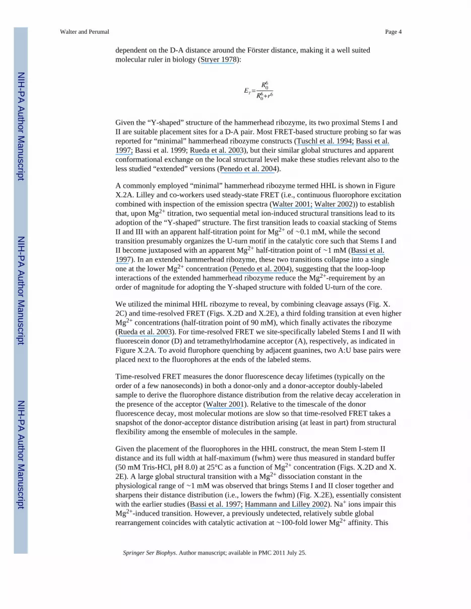
The riboswitch known as the glmS ribozyme is unique in that binding of its ligandglucosamine-6-phosphate (GlcN6P) induces self-cleavage (Winkler et al. 2004) andsubsequent intracellular degradation of the embedding glmS mRNA, which encodes GlcN6Psynthase (Collins et al. 2007). While ligand is absolutely required for catalytic activity andspecificity for GlcN6P is high, several structurally related amine-containing compoundswere found to partially activate the riboswitch, suggesting that the amino-group participatesin general acid–base catalysis rather than to function as an allosteric activator (McCarthy etal. 2005). Crystal structures of the glmS ribozyme (Figs. X.6.A and X.6.B) revealed threeparallel helical stacks with a doubly pseudoknotted core that binds GlcN6P in a mannerconsistent with general acid-base catalysis by GlcN6P, in conjunction with G40 of theribozyme and possibly structural water molecules (Klein and Ferre-D'Amare 2006;Cochrane et al. 2007; Walter 2007). Notably, the glmS ribozyme can fold and function in theabsence of divalent metal ions (Roth et al. 2006).
To address with maximal sensitivity the question of whether any conformationalrearrangements accompany ligand binding by the glmS ribozyme, the closing loop of P1 wasremoved to obtain a trans-acting ribozyme with external substrate for FRET labeling, whilethe downstream P3-P4.1 was truncated to obtain a minimal, slightly destabilized ribozymecore (Tinsley et al. 2007) (Figs. X.6.A and X.6.B). Fluorescein donor andtetramethylrhodamine acceptor fluorophores were attached to the substrate 5′- and 3′-termini, respectively, to detect changes in distance along the central P1:P2.2 helical axis thatencompasses the GlcN6P binding and cleavage sites.
Addition of an excess of ribozyme to the FRET labeled non-cleavable substrate analogresulted in a significant increase in donor fluorescence and corresponding decrease inacceptor:donor signal ratio (Fig. X.6.C top). These changes are expected as the binding ofthe ribozyme will cause an extension of the initially random-coil substrate in the complex.Upon further addition of 10 mM GlcN6P, however, no significant changes in fluorescencesignal and FRET efficiency were observed (Fig. X.6.C top). By contrast, a considerable
Walter and Perumal Page 9
Springer Ser Biophys. Author manuscript; available in PMC 2011 July 25.
NIH
-PA Author Manuscript
NIH
-PA Author Manuscript
NIH
-PA Author Manuscript

increase in donor signal and resulting decrease in FRET efficiency were evident uponaddition of the GlcN6P ligand to the ribozyme-cleavable substrate complex (Fig. X.6.Cbottom), as expected from ligand-induced substrate cleavage and dissociation of the 5′-product with attached donor fluorophore. These results are corroborated by time-resolvedFRET analysis, where the donor-acceptor distance in the absence and presence of GlcN6Pwas found to both be 52 Å (Tinsley et al. 2007). Taken together, these results support thenotion that the glmS ribozyme is fully folded in solution prior to binding its activating ligand(cofactor), which is consistent with precursor and product crystal structures (Klein andFerre-D'Amare 2006; Cochrane et al. 2007) and observations from hydroxyl radicalfootprinting in solution (Hampel and Tinsley 2006).
X.8 Common and Diverse Features: Attaining Site-Specificity in CleavageThe RNA backbone can spontaneously undergo acid-base catalyzed self-cleavage via thesame reaction chemistry utilized by the small ribozymes (Fig. X.1). Under physiologicconditions, spontaneous RNA degradation occurs at a rate constant estimated to average∼10-7 min-1 per bond (Emilsson et al. 2003), with a strong dependence on both sequenceand structure context (Kaukinen et al. 2002). In double-stranded (Watson-Crick base paired)RNA, the angle between the 2′O-P-5′O atoms is 65-70°, which is significantly different fromthe optimal angle for in-line attack of close to 180° (Min et al. 2007). This renders thebackbone of double-stranded RNA chemically quite inert. Strikingly, all crystal structures ofsmall ribozymes that are thought to be catalytically relevant find the cleavage site in an atleast partially not Watson-Crick paired (or single-stranded) RNA segment featuring abackbone kink (Rupert and Ferre-D'Amare 2001; Ke et al. 2004; Klein and Ferre-D'Amare2006; Martick and Scott 2006). In fact, the absence of such a backbone kink was used as anindication that the early crystal structures of the hammerhead ribozyme could only beactivated by a conformational change after which the cleavage site would adopt non-double-helical characteristics (Wedekind and McKay 1998; Blount and Uhlenbeck 2005).
The small ribozymes have convergently evolved to prepare a specific bond for a commonreaction chemistry, and while they utilize different strategies and structural contexts, in allcases the two nucleotides immediately upstream and downstream of the scissile phosphateare splayed apart in distinct hydrogen bonding and stacking interactions, enforcing unusualtorsion angles that kink the backbone (Sefcikova et al. 2007; Walter 2007). Estimatessuggest that adoption of an ideal in-line attack configuration accelerates spontaneousbackbone cleavage only by <100-fold (Emilsson et al. 2003; Min et al. 2007), supporting thenotion that backbone kinking is likely to serve more than one role: to facilitate in-line attack,to specify a bond near the kink for cleavage by exposure to the general acid and base thataffect catalysis, and to destabilize the ground state structure (Fig. X.1).
Specific nucleobases, structural water molecules, and – in the case of the HDV ribozyme – ahydrated divalent metal ion are generally proximal to the exposed scissile phosphate of thesmall ribozymes, placed there by the intricacies of the global and local RNA fold. Thenucleobases so far implicated in general acid-base (or electrostatic) catalysis are mostly thepurines G and A, perhaps due to their larger number of functional groups and more extendedaromatic system with larger surface area for stacking. The only exception to this rule is theHDV ribozyme that utilizes a cytosine (C75) for catalysis and is, perhaps not coincidentally,also the only small ribozyme strongly dependent on divalent metal ions for activity (Murrayet al. 1998). This and other roles of metal ions are the focus of the following segment.
X.9 Common and Diverse Features: The Roles of Metal IonsAll small ribozymes show at least residual catalytic function in the complete absence ofdivalent metal ions, as long as sufficient countercharge in the form of monovalents is
Walter and Perumal Page 10
Springer Ser Biophys. Author manuscript; available in PMC 2011 July 25.
NIH
-PA Author Manuscript
NIH
-PA Author Manuscript
NIH
-PA Author Manuscript

provided to neutralize the negatively charged sugar-phosphate backbone (Hampel andCowan 1997; Nesbitt et al. 1997; Young et al. 1997; Murray et al. 1998; Nakano et al. 2000;Curtis and Bartel 2001; O'Rear et al. 2001; Nakano et al. 2003; Roth et al. 2006). Thisobservation contrasts with ribozymes catalyzing more complex phosphodiester transfers,which consistently appear to require a pair of Mg2+ ions for catalysis (Walter 2007) (seealso accompanying chapters by Woodson, Pyle, and Harris and coworkers). The simplerchemistry executed by the small ribozymes typically requires neither an external nucleophile(as do most larger ribozymes) nor an external catalytic cofactor (except perhaps forubiquitous water molecules), making small ribozymes surprisingly self-sufficient andversatile, perhaps as an adaptation to their function in parasite replication. Exceptions are theglmS ribozyme, which requires GlcN6P as a cofactor and is not a parasite-derived ribozymebut is embedded in bacterial mRNA to control gene expression (Winkler et al. 2004), andthe HDV ribozyme, which not only is part of the HDV viroid but is also embedded in thehuman genome (Salehi-Ashtiani et al. 2006).
The role of metal ions in folding of the small ribozymes is much more unequivocal,especially under physiologic (low-ionic strength) conditions, where Mg2+ is the preferred,highest-affinity folding cofactor. Mg2+ is thought to bind RNA in three different modes, byspecific inner-sphere coordination (chelation), by outer-sphere binding where a single waterlayer influences the ion position through steric packing and hydrogen bonding, and bydiffuse association with the long-range RNA electrostatic field through multiple water layers(Draper et al. 2005). FRET studies have shown that tertiary structure folding is generallyaccelerated (and thermodynamically stabilized) and unfolding decelerated by increasingMg2+ concentrations, although differential saturation behavior of the two Mg2+ dependencycurves is observed for the hairpin ribozyme (Bokinsky et al. 2003). Resulting modelingstudies interpret this behavior as an indication that the folding transition state is compactedby Mg2+-based bridging of the two ribozyme domains, even before tertiary hydrogen bondand stacking interactions are extensively formed (Bokinsky et al. 2003). How general thisbehavior is for the small ribozymes remains to be seen.
X.10 Common and Diverse Features: RNA Structural DynamicsThe catalytic transition state of any enzyme is, by definition, a high-energy state along thereaction pathway that only exists on the timescale of a bond vibration. By contrast,structures observed in a crystalline and solution state by, for example, X-ray crystallographyand NMR, respectively, are ensemble-averaged, low-energy ground states, even if perturbedby employing transition state analogs. As a consequence, the available high-resolutioncrystal structures of ribozymes need to be animated with their dynamics to understand theinherently dynamic processes underlying catalysis.
Ensemble and single molecule FRET has extensively contributed to our understanding ofhow and why the small ribozymes fold or show intrinsic dynamics. With one exception,global structural changes that rearrange helical alignments for catalytic activation (as in thehammerhead ribozyme), “dock” substrate and catalytic core domains (as in the hairpin andVS ribozymes), or accompany and possibly control catalysis (as in the HDV ribozyme) havereadily been detected by FRET in all small ribozymes. It is tempting to speculate that suchlarge-scale conformational rearrangements from inactive to active states serve a biologicalrole by controlling the activity of the catalytic motif during satellite replication independence of intracellular cues. Notably, the glmS ribozyme is devoid of any suchsignificant conformational change, but is catalytically triggered by a different kind ofintracellular cue in form of a threshold concentration of its catalytic cofactor GlcN6P.
Walter and Perumal Page 11
Springer Ser Biophys. Author manuscript; available in PMC 2011 July 25.
NIH
-PA Author Manuscript
NIH
-PA Author Manuscript
NIH
-PA Author Manuscript

Protein enzymes are thought to exploit a global network of coupled molecular motions tofacilitate the chemical reaction they catalyze. This network comprises fast thermal motionsthat are at equilibrium while the reaction progresses along the reaction pathway; thesemotions lead to slower, larger-scale conformational changes that directly facilitate reactionchemistry (Hammes-Schiffer and Benkovic 2006). Whether ribozymes employ a similarstrategy to accelerate catalysis is not known, but single molecule FRET probing incombination with molecular dynamics simulations has revealed that at least the hairpinribozyme exhibits analogous networks of coupled molecular motions throughout its catalyticcore (Rueda et al. 2004; Rhodes et al. 2006). Reaction chemistry in an enzymatic reaction islargely of a local nature, suggesting that the dynamics contributing directly to catalysis inRNA would mostly entail vibrations, torsional librations, sugar re-puckering, andlongitudinal and lateral motions of bases, which all take place in the tens-of-femtoseconds tolow-nanosecond time regimes (Al-Hashimi and Walter 2008). It remains to be determinedwhether global structural changes at the nanosecond to minute timescale, as observable byFRET, couple to these local dynamics and contribute to the catalytic power of the smallribozymes.
In conclusion, many common features are emerging from a comparison of the five naturallyoccurring, structurally very distinct small ribozymes. Diversities between them probablyhighlight evolutionary adaptation to specific functional requirements. Future studies usingFRET and other biophysical techniques promise to elucidate more of the fundamentalprinciples underlying RNA catalysis, enabling an even broader comparison to enzymes,including those based on protein, and a potential utilization in human engineering.
ReferencesAl-Hashimi HM, Walter NG. RNA dynamics: It's about time. Curr Opin Struct Biol. 2008 in press.Alam S, Grum-Tokars V, Krucinska J, Kundracik ML, Wedekind JE. Conformational heterogeneity at
position U37 of an all-RNA hairpin ribozyme with implications for metal binding and the catalyticstructure of the S-turn. Biochemistry. 2005; 44:14396–408. [PubMed: 16262240]
Andersen AA, Collins RA. Rearrangement of a stable RNA secondary structure during VS ribozymecatalysis. Mol Cell. 2000; 5:469–78. [PubMed: 10882132]
Bassi GS, Murchie AI, Walter F, Clegg RM, Lilley DM. Ion-induced folding of the hammerheadribozyme: a fluorescence resonance energy transfer study. EMBO J. 1997; 16:7481–9. [PubMed:9405376]
Bassi GS, Mollegaard NE, Murchie AI, Lilley DM. RNA folding and misfolding of the hammerheadribozyme. Biochemistry. 1999; 38:3345–54. [PubMed: 10079078]
Beattie TL, Olive JE, Collins RA. A secondary-structure model for the self-cleaving region ofNeurospora VS RNA. Proc Natl Acad Sci U S A. 1995; 92:4686–90. [PubMed: 7753865]
Been MD. HDV ribozymes. Curr Top Microbiol Immunol. 2006; 307:47–65. [PubMed: 16903220]Bevilacqua PC. Mechanistic Considerations for General Acid-Base Catalysis by RNA: Revisiting the
Mechanism of the Hairpin Ribozyme. Biochemistry. 2003; 42:2259–65. [PubMed: 12600192]Bevilacqua PC, Yajima R. Nucleobase catalysis in ribozyme mechanism. Curr Opin Chem Biol. 2006;
10:455–64. [PubMed: 16935552]Blount KF, Uhlenbeck OC. The Structure-Function Dilemma of the Hammerhead Ribozyme. Annu
Rev Biophys Biomol Struct. 2005Blount KF, Breaker RR. Riboswitches as antibacterial drug targets. Nat Biotechnol. 2006; 24:1558–64.
[PubMed: 17160062]Bokinsky G, Rueda D, Misra VK, Rhodes MM, Gordus A, Babcock HP, Walter NG, Zhuang X.
Single-molecule transition-state analysis of RNA folding. Proc Natl Acad Sci U S A. 2003;100:9302–7. [PubMed: 12869691]
Walter and Perumal Page 12
Springer Ser Biophys. Author manuscript; available in PMC 2011 July 25.
NIH
-PA Author Manuscript
NIH
-PA Author Manuscript
NIH
-PA Author Manuscript

Bondensgaard K, Mollova ET, Pardi A. The global conformation of the hammerhead ribozymedetermined using residual dipolar couplings. Biochemistry. 2002; 41:11532–42. [PubMed:12269797]
Cochrane JC, Lipchock SV, Strobel SA. Structural investigation of the GlmS ribozyme bound to Itscatalytic cofactor. Chem Biol. 2007; 14:97–105. [PubMed: 17196404]
Collins JA, Irnov I, Baker S, Winkler WC. Mechanism of mRNA destabilization by the glmSribozyme. Genes Dev. 2007; 21:3356–68. [PubMed: 18079181]
Collins RA, Saville BJ. Independent transfer of mitochondrial chromosomes and plasmids duringunstable vegetative fusion in Neurospora. Nature. 1990; 345:177–9. [PubMed: 1970854]
Coppins RL, Hall KB, Groisman EA. The intricate world of riboswitches. Curr Opin Microbiol. 2007;10:176–81. [PubMed: 17383225]
Curtis EA, Bartel DP. The hammerhead cleavage reaction in monovalent cations. RNA. 2001; 7:546–52. [PubMed: 11345433]
Das S, Piccirilli J. General acid catalysis by the hepatitis delta virus ribozyme. Nature ChemicalBiology. 2005; 1:45–52.
De la Pena M, Gago S, Flores R. Peripheral regions of natural hammerhead ribozymes greatly increasetheir self-cleavage activity. EMBO J. 2003; 22:5561–70. [PubMed: 14532128]
Ditzler MA, Aleman EA, Rueda D, Walter NG. Focus on function: Single molecule RNAenzymology. Biopolymers. 2007; 87:302–16. [PubMed: 17685395]
Doudna JA, Lorsch JR. Ribozyme catalysis: not different, just worse. Nat Struct Mol Biol. 2005;12:395–402. [PubMed: 15870731]
Draper DE, Grilley D, Soto AM. Ions and RNA folding. Annu Rev Biophys Biomol Struct. 2005;34:221–43. [PubMed: 15869389]
Edwards TE, Klein DJ, Ferre-D'Amare AR. Riboswitches: small-molecule recognition by generegulatory RNAs. Curr Opin Struct Biol. 2007; 17:273–9. [PubMed: 17574837]
Emilsson GM, Nakamura S, Roth A, Breaker RR. Ribozyme speed limits. RNA. 2003; 9:907–18.[PubMed: 12869701]
Fedor MJ, Williamson JR. The catalytic diversity of RNAs. Nature Rev Mol Cell Biol. 2005; 6:399–412. [PubMed: 15956979]
Feldstein PA, Buzayan JM, van Tol H, deBear J, Gough GR, Gilham PT, Bruening G. Specificassociation between an endoribonucleolytic sequence from a satellite RNA and a substrateanalogue containing a 2′-5′ phosphodiester. Proc Natl Acad Sci U S A. 1990; 87:2623–7.[PubMed: 1690890]
Ferre-D'Amare AR, Zhou K, Doudna JA. Crystal structure of a hepatitis delta virus ribozyme. Nature.1998; 395:567–74. [PubMed: 9783582]
Ferre-D'Amare AR, Doudna JA. Crystallization and structure determination of a hepatitis delta virusribozyme: use of the RNA-binding protein U1A as a crystallization module. J Mol Biol. 2000;295:541–56. [PubMed: 10623545]
Flores R, Delgado S, Gas ME, Carbonell A, Molina D, Gago S, De la Pena M. Viroids: the minimalnon-coding RNAs with autonomous replication. FEBS Lett. 2004; 567:42–8. [PubMed: 15165891]
Forster AC, Symons RH. Self-cleavage of virusoid RNA is performed by the proposed 55-nucleotideactive site. Cell. 1987; 50:9–16. [PubMed: 3594567]
Förster T. Energiewanderung Und Fluoreszenz. Naturwissenschaften. 1946; 33:166–75.Förster T. Intermolecular energy migration and fluorescence. Ann Phys (Leipzig). 1948; 2:55–75.Furtig B, Richter C, Schell P, Wenter P, Pitsch S, Schwalbe H. NMR-spectroscopic Characterisation of
Phosphodiester Bond Cleavage catalysed by the minimal Hammerhead Ribozyme. RNA Biol.2008; 5
Gong B, Chen JH, Chase E, Chadalavada DM, Yajima R, Golden BL, Bevilacqua PC, Carey PR.Direct measurement of a pK(a) near neutrality for the catalytic cytosine in the genomic HDVribozyme using Raman crystallography. J Am Chem Soc. 2007; 129:13335–42. [PubMed:17924627]
Guerrier-Takada C, Gardiner K, Marsh T, Pace N, Altman S. The RNA moiety of ribonuclease P is thecatalytic subunit of the enzyme. Cell. 1983; 35:849–57. [PubMed: 6197186]
Walter and Perumal Page 13
Springer Ser Biophys. Author manuscript; available in PMC 2011 July 25.
NIH
-PA Author Manuscript
NIH
-PA Author Manuscript
NIH
-PA Author Manuscript

Hammann C, Lilley DM. Folding and activity of the hammerhead ribozyme. Chembiochem. 2002;3:690–700. [PubMed: 12203967]
Hammes-Schiffer S, Benkovic SJ. Relating protein motion to catalysis. Annu Rev Biochem. 2006;75:519–41. [PubMed: 16756501]
Hampel A, Cowan JA. A unique mechanism for RNA catalysis: the role of metal cofactors in hairpinribozyme cleavage. Chem Biol. 1997; 4:513–7. [PubMed: 9263639]
Hampel KJ, Tinsley MM. Evidence for preorganization of the glmS ribozyme ligand binding pocket.Biochemistry. 2006; 45:7861–71. [PubMed: 16784238]
Haran G. Noise reduction in single-molecule fluorescence trajectories of folding proteins. Chem Phys.2004; 307:137–45.
Harris DA, Rueda D, Walter NG. Local conformational changes in the catalytic core of the trans-acting hepatitis delta virus ribozyme accompany catalysis. Biochemistry. 2002; 41:12051–61.[PubMed: 12356305]
Haseloff J, Gerlach WL. Sequences required for self-catalysed cleavage of the satellite RNA oftobacco ringspot virus. Gene. 1989; 82:43–52. [PubMed: 2684775]
Hiley SL, Collins RA. Rapid formation of a solvent-inaccessible core in the Neurospora Varkudsatellite ribozyme. EMBO J. 2001; 20:5461–9. [PubMed: 11574478]
Hiley SL, Sood VD, Fan J, Collins RA. 4-thio-U cross-linking identifies the active site of the VSribozyme. EMBO J. 2002; 21:4691–8. [PubMed: 12198171]
Jaikaran D, Smith MD, Mehdizadeh R, Olive J, Collins RA. An important role of G638 in the cis-cleavage reaction of the Neurospora VS ribozyme revealed by a novel nucleotide analogincorporation method. Rna. 2008; 14:938–49. [PubMed: 18356538]
Jeong S, Sefcikova J, Tinsley RA, Rueda D, Walter NG. Trans-acting hepatitis delta virus ribozyme:catalytic core and global structure are dependent on the 5′ substrate sequence. Biochemistry. 2003;42:7727–40. [PubMed: 12820882]
Kaukinen U, Lyytikainen S, Mikkola S, Lonnberg H. The reactivity of phosphodiester bonds withinlinear single-stranded oligoribonucleotides is strongly dependent on the base sequence. NucleicAcids Res. 2002; 30:468–74. [PubMed: 11788709]
Ke A, Zhou K, Ding F, Cate JH, Doudna JA. A conformational switch controls hepatitis delta virusribozyme catalysis. Nature. 2004; 429:201–5. [PubMed: 15141216]
Khvorova A, Lescoute A, Westhof E, Jayasena SD. Sequence elements outside the hammerheadribozyme catalytic core enable intracellular activity. Nat Struct Biol. 2003; 10:708–12. [PubMed:12881719]
Klein DJ, Ferre-D'Amare AR. Structural basis of glmS ribozyme activation by glucosamine-6-phosphate. Science. 2006; 313:1752–6. [PubMed: 16990543]
Kruger K, Grabowski PJ, Zaug AJ, Sands J, Gottschling DE, Cech TR. Self-splicing RNA:autoexcision and autocyclization of the ribosomal RNA intervening sequence of Tetrahymena.Cell. 1982; 31:147–57. [PubMed: 6297745]
Kuzmin YI, Da Costa CP, Fedor MJ. Role of an active site guanine in hairpin ribozyme catalysisprobed by exogenous nucleobase rescue. J Mol Biol. 2004; 340:233–51. [PubMed: 15201049]
Kuzmin YI, Da Costa CP, Cottrell JW, Fedor MJ. Role of an active site adenine in hairpin ribozymecatalysis. J Mol Biol. 2005; 349:989–1010. [PubMed: 15907933]
Lafontaine DA, Norman DG, Lilley DM. Structure, folding and activity of the VS ribozyme:importance of the 2- 3-6 helical junction. EMBO J. 2001; 20:1415–24. [PubMed: 11250907]
Lafontaine DA, Norman DG, Lilley DM. The global structure of the VS ribozyme. EMBO J. 2002;21:2461–71. [PubMed: 12006498]
Lakowicz, JR. Principles of Fluorescence Spectroscopy. Third. Springer; New York: 2006.Liu S, Bokinsky G, Walter NG, Zhuang X. Dissecting the multistep reaction pathway of an RNA
enzyme by single-molecule kinetic “fingerprinting”. Proc Natl Acad Sci U S A. 2007; 104:12634–9. [PubMed: 17496145]
Luptak A, Ferre-D'Amare AR, Zhou K, Zilm KW, Doudna JA. Direct pK(a) measurement of theactive-site cytosine in a genomic hepatitis delta virus ribozyme. J Am Chem Soc. 2001; 123:8447–52. [PubMed: 11525650]
Walter and Perumal Page 14
Springer Ser Biophys. Author manuscript; available in PMC 2011 July 25.
NIH
-PA Author Manuscript
NIH
-PA Author Manuscript
NIH
-PA Author Manuscript

Martick M, Scott WG. Tertiary Contacts Distant from the Active Site Prime a Ribozyme for Catalysis.Cell. 2006; 126:309–20. [PubMed: 16859740]
Martick M, Lee TS, York DM, Scott WG. Solvent structure and hammerhead ribozyme catalysis.Chem Biol. 2008; 15:332–42. [PubMed: 18420140]
McCarthy TJ, Plog MA, Floy SA, Jansen JA, Soukup JK, Soukup GA. Ligand requirements for glmSribozyme self-cleavage. Chem Biol. 2005; 12:1221–6. [PubMed: 16298301]
McKay DB. Structure and function of the hammerhead ribozyme: an unfinished story. RNA. 1996;2:395–403. [PubMed: 8665407]
Min D, Xue S, Li H, Yang W. ‘In-line attack’ conformational effect plays a modest role in an enzyme-catalyzed RNA cleavage: a free energy simulation study. Nucleic Acids Res. 2007; 35:4001–6.[PubMed: 17553832]
Murchie AI, Thomson JB, Walter F, Lilley DM. Folding of the hairpin ribozyme in its naturalconformation achieves close physical proximity of the loops. Mol Cell. 1998; 1:873–81. [PubMed:9660970]
Murray JB, Seyhan AA, Walter NG, Burke JM, Scott WG. The hammerhead, hairpin and VSribozymes are catalytically proficient in monovalent cations alone. Chem Biol. 1998; 5:587–95.[PubMed: 9818150]
Nahas MK, Wilson TJ, Hohng S, Jarvie K, Lilley DM, Ha T. Observation of internal cleavage andligation reactions of a ribozyme. Nat Struct Mol Biol. 2004; 11:1107–13. [PubMed: 15475966]
Nakano S, Chadalavada DM, Bevilacqua PC. General acid-base catalysis in the mechanism of ahepatitis delta virus ribozyme. Science. 2000; 287:1493–7. [PubMed: 10688799]
Nakano S, Cerrone AL, Bevilacqua PC. Mechanistic Characterization of the HDV GenomicRibozyme: Classifying the Catalytic and Structural Metal Ion Sites within a Multichannel ReactionMechanism. Biochemistry. 2003; 42:2982–94. [PubMed: 12627964]
Nakano S, Bevilacqua PC. Mechanistic characterization of the HDV genomic ribozyme: a mutant ofthe C41 motif provides insight into the positioning and thermodynamic linkage of metal ions andprotons. Biochemistry. 2007; 46:3001–12. [PubMed: 17315949]
Nelson JA, Uhlenbeck OC. When to believe what you see. Mol Cell. 2006; 23:447–50. [PubMed:16916633]
Nelson JA, Uhlenbeck OC. Hammerhead redux: does the new structure fit the old biochemical data?RNA. 2008a; 14:605–15. [PubMed: 18287565]
Nelson JA, Uhlenbeck OC. Minimal and extended hammerheads utilize a similar dynamic reactionmechanism for catalysis. Rna. 2008b; 14:43–54. [PubMed: 17998291]
Nesbitt S, Hegg LA, Fedor MJ. An unusual pH-independent and metal-ion-independent mechanism forhairpin ribozyme catalysis. Chem Biol. 1997; 4:619–30. [PubMed: 9281529]
O'Rear JL, Wang S, Feig AL, Beigelman L, Uhlenbeck OC, Herschlag D. Comparison of thehammerhead cleavage reactions stimulated by monovalent and divalent cations. RNA. 2001; 7(4):537–45. [PubMed: 11345432]
Okumus B, Wilson TJ, Lilley DM, Ha T. Vesicle Encapsulation Studies Reveal that Single MoleculeRibozyme Heterogeneities Are Intrinsic. Biophys J. 2004; 87:2798–806. [PubMed: 15454471]
Penedo JC, Wilson TJ, Jayasena SD, Khvorova A, Lilley DM. Folding of the natural hammerheadribozyme is enhanced by interaction of auxiliary elements. RNA. 2004; 10:880–8. [PubMed:15100442]
Pereira MJ, Harris DA, Rueda D, Walter NG. Reaction pathway of the trans-acting hepatitis delta virusribozyme: a conformational change accompanies catalysis. Biochemistry. 2002; 41:730–40.[PubMed: 11790094]
Pereira MJB, Nikolova EN, Hiley SL, Collins RA, Walter NG. Single VS ribozyme molecules revealdynamic and heterogeneous hierarchical folding toward catalysis. J Mol Biol submitted. 2008
Perrotta AT, Been MD. A pseudoknot-like structure required for efficient self-cleavage of hepatitisdelta virus RNA. Nature. 1991; 350:434–6. [PubMed: 2011192]
Perrotta AT, Shih I, Been MD. Imidazole rescue of a cytosine mutation in a self-cleaving ribozyme.Science. 1999; 286:123–6. [PubMed: 10506560]
Walter and Perumal Page 15
Springer Ser Biophys. Author manuscript; available in PMC 2011 July 25.
NIH
-PA Author Manuscript
NIH
-PA Author Manuscript
NIH
-PA Author Manuscript
Pinard R, Hampel KJ, Heckman JE, Lambert D, Chan PA, Major F, Burke JM. Functionalinvolvement of G8 in the hairpin ribozyme cleavage mechanism. EMBO J. 2001; 20:6434–42.[PubMed: 11707414]
Prody GA, Bakos JT, Buzayan JM, Schneider IR, Bruening G. Autolytic Processing of Dimeric PlantVirus Satellite RNA. Science. 1986; 231:1577–80. [PubMed: 17833317]
Pyle AM. Ribozymes: a distinct class of metalloenzymes. Science. 1993; 261:709–14. [PubMed:7688142]
Rastogi T, Beattie TL, Olive JE, Collins RA. A long-range pseudoknot is required for activity of theNeurospora VS ribozyme. EMBO J. 1996; 15:2820–5. [PubMed: 8654379]
Rhodes MM, Reblova K, Sponer J, Walter NG. Trapped water molecules are essential to structuraldynamics and function of a ribozyme. Proc Natl Acad Sci U S A. 2006; 103:13380–5. [PubMed:16938834]
Roth A, Nahvi A, Lee M, Jona I, Breaker RR. Characteristics of the glmS ribozyme suggest onlystructural roles for divalent metal ions. RNA. 2006; 12:607–19. [PubMed: 16484375]
Rueda D, Wick K, McDowell SE, Walter NG. Diffusely bound Mg2+ ions slightly reorient stems Iand II of the hammerhead ribozyme to increase the probability of formation of the catalytic core.Biochemistry. 2003; 42:9924–36. [PubMed: 12924941]
Rueda D, Bokinsky G, Rhodes MM, Rust MJ, Zhuang X, Walter NG. Single-molecule enzymology ofRNA: essential functional groups impact catalysis from a distance. Proc Natl Acad Sci USA. 2004;101:10066–71. [PubMed: 15218105]
Ruffner DE, Stormo GD, Uhlenbeck OC. Sequence requirements of the hammerhead RNA self-cleavage reaction. Biochemistry. 1990; 29:10695–702. [PubMed: 1703005]
Rupert PB, Ferre-D'Amare AR. Crystal structure of a hairpin ribozyme-inhibitor complex withimplications for catalysis. Nature. 2001; 410:780–6. [PubMed: 11298439]
Rupert PB, Massey AP, Sigurdsson ST, Ferre-D'Amare AR. Transition state stabilization by a catalyticRNA. Science. 2002; 298:1421–4. [PubMed: 12376595]
Salehi-Ashtiani K, Szostak JW. In vitro evolution suggests multiple origins for the hammerheadribozyme. Nature. 2001; 414:82–4. [PubMed: 11689947]
Salehi-Ashtiani K, Luptak A, Litovchick A, Szostak JW. A genomewide search for ribozymes revealsan HDV-like sequence in the human CPEB3 gene. Science. 2006; 313:1788–92. [PubMed:16990549]
Salter J, Krucinska J, Alam S, Grum-Tokars V, Wedekind JE. Water in the active site of an all-RNAhairpin ribozyme and effects of Gua8 base variants on the geometry of phosphoryl transfer.Biochemistry. 2006; 45:686–700. [PubMed: 16411744]
Saville BJ, Collins RA. A site-specific self-cleavage reaction performed by a novel RNA inNeurospora mitochondria. Cell. 1990; 61:685–96. [PubMed: 2160856]
Scott WG. Ribozymes. Curr Opin Struct Biol. 2007; 17:280–6. [PubMed: 17572081]Sefcikova J, Krasovska MV, Sponer J, Walter NG. The genomic HDV ribozyme utilizes a previously
unnoticed U-turn motif to accomplish fast site-specific catalysis. Nucleic Acids Res. 2007;35:1933–46. [PubMed: 17337436]
Simorre JP, Legault P, Hangar AB, Michiels P, Pardi A. A conformational change in the catalytic coreof the hammerhead ribozyme upon cleavage of an RNA substrate. Biochemistry. 1997; 36:518–25.[PubMed: 9012667]
Simorre JP, Legault P, Baidya N, Uhlenbeck OC, Maloney L, Wincott F, Usman N, Beigelman L,Pardi A. Structural variation induced by different nucleotides at the cleavage site of thehammerhead ribozyme. Biochemistry. 1998; 37:4034–44. [PubMed: 9521724]
Smith MD, Collins RA. Evidence for proton transfer in the rate-limiting step of a fast-cleaving Varkudsatellite ribozyme. Proc Natl Acad Sci U S A. 2007; 104:5818–23. [PubMed: 17389378]
Stryer L. Fluorescence energy transfer as a spectroscopic ruler. Annu Rev Biochem. 1978; 47:819–46.[PubMed: 354506]
Taylor JM. Structure and replication of hepatitis delta virus RNA. Curr Top Microbiol Immunol. 2006;307:1–23. [PubMed: 16903218]
Walter and Perumal Page 16
Springer Ser Biophys. Author manuscript; available in PMC 2011 July 25.
NIH
-PA Author Manuscript
NIH
-PA Author Manuscript
NIH
-PA Author Manuscript

Tinsley RA, Harris DA, Walter NG. Magnesium dependence of the amplified conformational switch inthe trans-acting hepatitis delta virus ribozyme. Biochemistry. 2004; 43:8935–45. [PubMed:15248751]
Tinsley RA, Furchak JR, Walter NG. Trans-acting glmS catalytic riboswitch: locked and loaded. Rna.2007; 13:468–77. [PubMed: 17283212]
Tinsley RA, Walter NG. Long-range impact of peripheral joining elements on structure and function ofthe hepatitis delta virus ribozyme. Biol Chem. 2007; 388:705–15. [PubMed: 17570823]
Torelli AT, Krucinska J, Wedekind JE. A comparison of vanadate to a 2′-5′ linkage at the active site ofa small ribozyme suggests a role for water in transition-state stabilization. RNA. 2007; 13:1052–70. [PubMed: 17488874]
Tuschl T, Gohlke C, Jovin TM, Westhof E, Eckstein F. A three-dimensional model for thehammerhead ribozyme based on fluorescence measurements. Science. 1994; 266:785–9.[PubMed: 7973630]
Walter F, Murchie AI, Thomson JB, Lilley DM. Structure and activity of the hairpin ribozyme in itsnatural junction conformation: effect of metal ions. Biochemistry. 1998a; 37:14195–203.[PubMed: 9760257]
Walter F, Murchie AIH, Lilley DMJ. Folding of the four-way RNA junction of the hairpin ribozyme.Biochemistry. 1998b; 37:17629–36. [PubMed: 9860879]
Walter NG, Burke JM. The hairpin ribozyme: structure, assembly and catalysis. Curr Opin Chem Biol.1998; 2:24–30. [PubMed: 9667918]
Walter NG, Hampel KJ, Brown KM, Burke JM. Tertiary structure formation in the hairpin ribozymemonitored by fluorescence resonance energy transfer. EMBO J. 1998c; 17:2378–91. [PubMed:9545249]
Walter NG, Burke JM, Millar DP. Stability of hairpin ribozyme tertiary structure is governed by theinterdomain junction. Nat Struct Biol. 1999; 6:544–9. [PubMed: 10360357]
Walter NG. Structural dynamics of catalytic RNA highlighted by fluorescence resonance energytransfer. Methods. 2001; 25:19–30. [PubMed: 11558994]
Walter NG. Probing RNA structural dynamics and function by fluorescence resonance energy transfer(FRET). Curr Protoc Nucleic Acid Chem. 2002; 11.10:11.0.1–.0.23.
Walter NG. Ribozyme catalysis revisited: is water involved? Mol Cell. 2007; 28:923–9. [PubMed:18158891]
Walter NG, Huang C, Manzo AJ, Sobhy MA. Do-it-yourself guide: How to use the modern singlemolecule toolkit. Nat Methods. 2008; 5:475–489. [PubMed: 18511916]
Wedekind JE, McKay DB. Crystallographic structures of the hammerhead ribozyme: relationship toribozyme folding and catalysis. Annu Rev Biophys Biomol Struct. 1998; 27:475–502. [PubMed:9646875]
Wilson TJ, Ouellet J, Zhao ZY, Harusawa S, Araki L, Kurihara T, Lilley DM. Nucleobase catalysis inthe hairpin ribozyme. RNA. 2006; 12:980–7. [PubMed: 16601203]
Wilson TJ, McLeod AC, Lilley DM. A guanine nucleobase important for catalysis by the VSribozyme. Embo J. 2007; 26:2489–500. [PubMed: 17464286]
Winkler WC, Nahvi A, Roth A, Collins JA, Breaker RR. Control of gene expression by a naturalmetabolite-responsive ribozyme. Nature. 2004; 428:281–6. [PubMed: 15029187]
Winkler WC, Breaker RR. Regulation of bacterial gene expression by riboswitches. Annu RevMicrobiol. 2005; 59:487–517. [PubMed: 16153177]
Young KJ, Gill F, Grasby JA. Metal ions play a passive role in the hairpin ribozyme catalysedreaction. Nucleic Acids Res. 1997; 25:3760–6. [PubMed: 9380495]
Zhou DM, Taira K. The Hydrolysis of RNA: From Theoretical Calculations to the HammerheadRibozyme-Mediated Cleavage of RNA. Chem Rev. 1998; 98:991–1026. [PubMed: 11848922]
Zhuang X, Kim H, Pereira MJ, Babcock HP, Walter NG, Chu S. Correlating structural dynamics andfunction in single ribozyme molecules. Science. 2002; 296:1473–6. [PubMed: 12029135]
Zhuang X. Single-molecule RNA science. Annu Rev Biophys Biomol Struct. 2005; 34:399–414.[PubMed: 15869396]
Walter and Perumal Page 17
Springer Ser Biophys. Author manuscript; available in PMC 2011 July 25.
NIH
-PA Author Manuscript
NIH
-PA Author Manuscript
NIH
-PA Author Manuscript
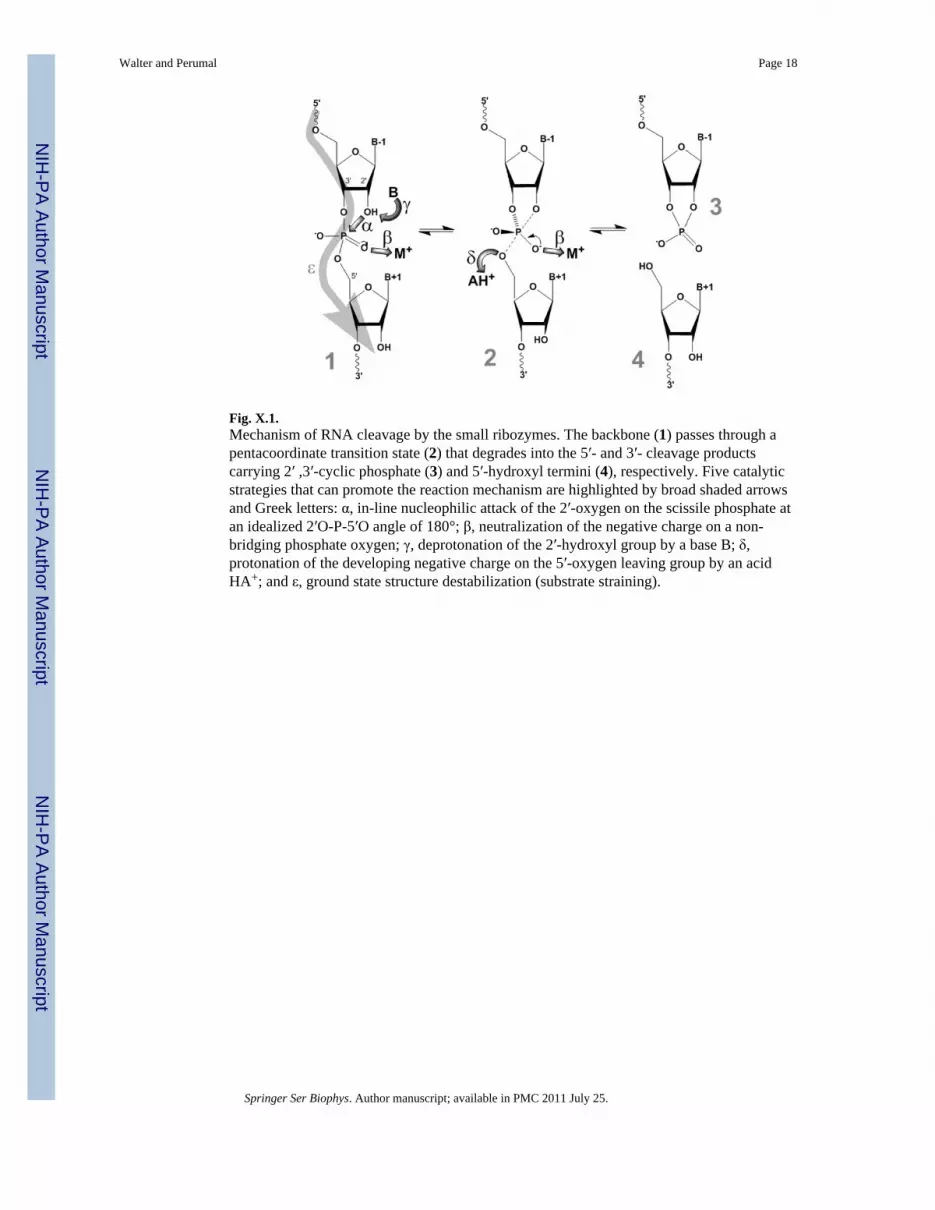
Fig. X.1.Mechanism of RNA cleavage by the small ribozymes. The backbone (1) passes through apentacoordinate transition state (2) that degrades into the 5′- and 3′- cleavage productscarrying 2′ ,3′-cyclic phosphate (3) and 5′-hydroxyl termini (4), respectively. Five catalyticstrategies that can promote the reaction mechanism are highlighted by broad shaded arrowsand Greek letters: α, in-line nucleophilic attack of the 2′-oxygen on the scissile phosphate atan idealized 2′O-P-5′O angle of 180°; β, neutralization of the negative charge on a non-bridging phosphate oxygen; γ, deprotonation of the 2′-hydroxyl group by a base B; δ,protonation of the developing negative charge on the 5′-oxygen leaving group by an acidHA+; and ε, ground state structure destabilization (substrate straining).
Walter and Perumal Page 18
Springer Ser Biophys. Author manuscript; available in PMC 2011 July 25.
NIH
-PA Author Manuscript
NIH
-PA Author Manuscript
NIH
-PA Author Manuscript

Fig. X.2.FRET studies of the hammerhead ribozyme. (A) Secondary structure of the hammerheadribozyme HHL utilized in these studies. Helices are color coded, the catalytic core ishighlighted in green, and the cleavage site in the substrate (outlined, S) is marked by aclosed red arrow. Stems I and II are labeled with donor (D) and acceptor (A) fluorophores,as indicated, which undergo FRET. In extended forms of the ribozyme, Stems I and IIinteract in the regions marked by an open double-arrow. (B) Cartoon representation of thecrystal structure of the extended Schistosoma hammerhead ribozyme (PDB ID: 2GOZ)(Martick and Scott 2006), color-coded as in panel A except for the silver capping loop that ispresent only in the extended form. The interaction of an internal and a capping loop in Stems
Walter and Perumal Page 19
Springer Ser Biophys. Author manuscript; available in PMC 2011 July 25.
NIH
-PA Author Manuscript
NIH
-PA Author Manuscript
NIH
-PA Author Manuscript
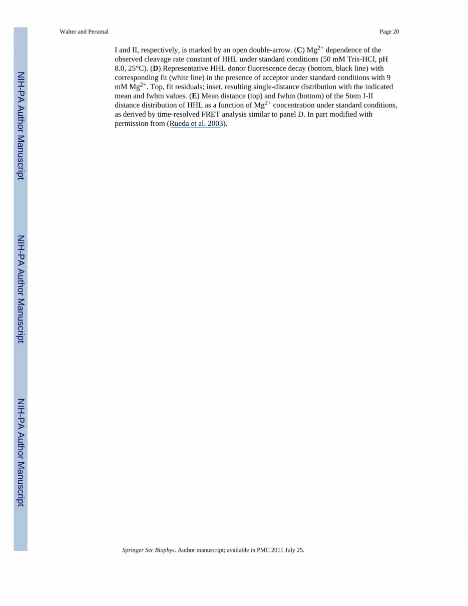
I and II, respectively, is marked by an open double-arrow. (C) Mg2+ dependence of theobserved cleavage rate constant of HHL under standard conditions (50 mM Tris-HCl, pH8.0, 25°C). (D) Representative HHL donor fluorescence decay (bottom, black line) withcorresponding fit (white line) in the presence of acceptor under standard conditions with 9mM Mg2+. Top, fit residuals; inset, resulting single-distance distribution with the indicatedmean and fwhm values. (E) Mean distance (top) and fwhm (bottom) of the Stem I-IIdistance distribution of HHL as a function of Mg2+ concentration under standard conditions,as derived by time-resolved FRET analysis similar to panel D. In part modified withpermission from (Rueda et al. 2003).
Walter and Perumal Page 20
Springer Ser Biophys. Author manuscript; available in PMC 2011 July 25.
NIH
-PA Author Manuscript
NIH
-PA Author Manuscript
NIH
-PA Author Manuscript
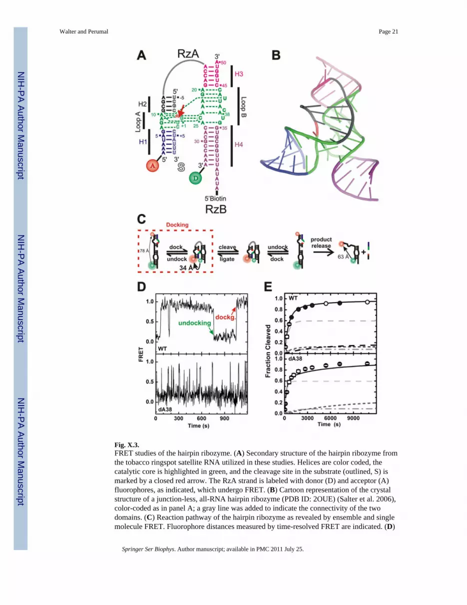
Fig. X.3.FRET studies of the hairpin ribozyme. (A) Secondary structure of the hairpin ribozyme fromthe tobacco ringspot satellite RNA utilized in these studies. Helices are color coded, thecatalytic core is highlighted in green, and the cleavage site in the substrate (outlined, S) ismarked by a closed red arrow. The RzA strand is labeled with donor (D) and acceptor (A)fluorophores, as indicated, which undergo FRET. (B) Cartoon representation of the crystalstructure of a junction-less, all-RNA hairpin ribozyme (PDB ID: 2OUE) (Salter et al. 2006),color-coded as in panel A; a gray line was added to indicate the connectivity of the twodomains. (C) Reaction pathway of the hairpin ribozyme as revealed by ensemble and singlemolecule FRET. Fluorophore distances measured by time-resolved FRET are indicated. (D)
Walter and Perumal Page 21
Springer Ser Biophys. Author manuscript; available in PMC 2011 July 25.
NIH
-PA Author Manuscript
NIH
-PA Author Manuscript
NIH
-PA Author Manuscript

Representative single molecule FRET time trace of wild-type (WT) and dA38 mutantribozymes after binding of non-cleavable (2′-OMe-A-1) substrate analog under standardconditions (50 mM Tris- HCl, pH 7.5, 12 mM Mg2+, 25°C). One undocking and onedocking event are indicated. (E) Experimental (symbols) and simulated (lines) cleavage timecourses of the WT and dA38 mutant ribozymes under standard conditions. The predictedcontributions from four and three experimentally detected molecule sub-populations,respectively, are indicated by gray and dashed lines. In part modified with permission from(Rueda et al. 2004).
Walter and Perumal Page 22
Springer Ser Biophys. Author manuscript; available in PMC 2011 July 25.
NIH
-PA Author Manuscript
NIH
-PA Author Manuscript
NIH
-PA Author Manuscript

Fig. X.4.FRET studies of the HDV ribozyme. (A) Secondary structure of the HDV ribozyme D1utilized in these studies. Helices are color coded, the catalytic core is highlighted in green,and the cleavage site in the substrate (outlined, S) is marked by a closed red arrow. Strand Bof the ribozyme is labeled with donor (D) and acceptor (A) fluorophores, as indicated, whichundergo FRET. Dashed gray lines indicate connections that are present in the naturallyoccurring, cis-acting, genomic and antigenomic HDV ribozymes. (B) Cartoon representationof the crystal structure of the genomic HDV ribozyme (PDB ID: 1SJ3) (Ke et al. 2004),color-coded as in panel A with connections removed for the trans-acting D1 construct shownin silver. Red sphere, presumably catalytically involved Mg2+ ion. (C) Reaction pathway of
Walter and Perumal Page 23
Springer Ser Biophys. Author manuscript; available in PMC 2011 July 25.
NIH
-PA Author Manuscript
NIH
-PA Author Manuscript
NIH
-PA Author Manuscript
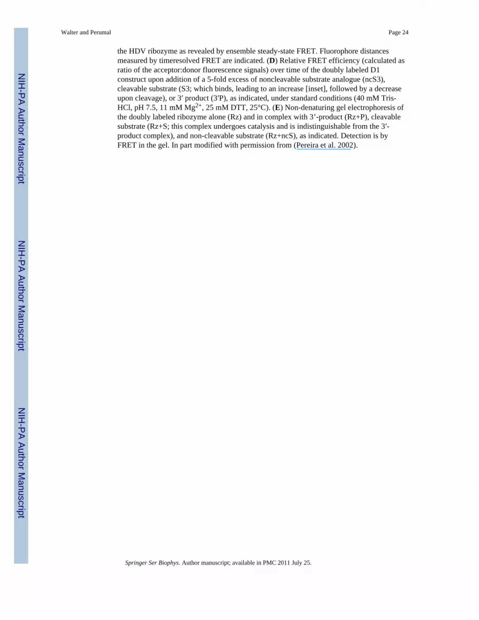
the HDV ribozyme as revealed by ensemble steady-state FRET. Fluorophore distancesmeasured by timeresolved FRET are indicated. (D) Relative FRET efficiency (calculated asratio of the acceptor:donor fluorescence signals) over time of the doubly labeled D1construct upon addition of a 5-fold excess of noncleavable substrate analogue (ncS3),cleavable substrate (S3; which binds, leading to an increase [inset], followed by a decreaseupon cleavage), or 3′ product (3′P), as indicated, under standard conditions (40 mM Tris-HCl, pH 7.5, 11 mM Mg2+, 25 mM DTT, 25°C). (E) Non-denaturing gel electrophoresis ofthe doubly labeled ribozyme alone (Rz) and in complex with 3’-product (Rz+P), cleavablesubstrate (Rz+S; this complex undergoes catalysis and is indistinguishable from the 3′-product complex), and non-cleavable substrate (Rz+ncS), as indicated. Detection is byFRET in the gel. In part modified with permission from (Pereira et al. 2002).
Walter and Perumal Page 24
Springer Ser Biophys. Author manuscript; available in PMC 2011 July 25.
NIH
-PA Author Manuscript
NIH
-PA Author Manuscript
NIH
-PA Author Manuscript

Fig. X.5.FRET studies of the VS ribozyme. (A) Secondary structure of the VS ribozyme G11 utilizedin these studies. Helices are color coded, the cleavage site is marked with a closed red arrow,and the FRET donor (D) and acceptor (A) and biotin labeling sites are indicated. (B)Tertiary structure model of the VS ribozyme , using the same color code as in panel A.Some strand connectivities are incomplete in the model; the ones in the I-V kissing loop areindicated by dashed lines. (C) Structural and kinetic model of the reaction pathway of theWT VS ribozyme as derived from single molecule FRET. The helix colors match those inpanel A. (D) Single molecule FRET time trace of a VS ribozyme (Hiley and Collins 2001).The raw Cy3 donor and Cy5 acceptor fluorescence signals are green and red, respectively(upper panel). Superimposed in gray are the data after applying a non-linear filter (Haran2004). The FRET ratio (lower panel, black trace) is calculated from the filtered data asacceptor/(donor+acceptor) and reveals three distinct states by Hidden Markov modeling (redline). In part modified with permission from (Pereira et al. 2008).
Walter and Perumal Page 25
Springer Ser Biophys. Author manuscript; available in PMC 2011 July 25.
NIH
-PA Author Manuscript
NIH
-PA Author Manuscript
NIH
-PA Author Manuscript

Fig. X.6.FRET studies of the glmS ribozyme. (A) Secondary structure of the trans-acting glmSribozyme utilized in these studies. Helices are color coded, the catalytic core is highlightedin green, the cleavage site in the substrate (outlined, S) is marked by a closed red arrow, andthe cofactor GlcN6P is shown as an orange oval interacting with several core residues(dashed orange lines). The substrate strand is labeled with donor (D) and acceptor (A)fluorophores, as indicated, which undergo FRET. Dashed gray lines indicate connectionsand a downstream pseudoknot that are present in the naturally occurring, cis-actingribozyme. (B) Cartoon representation of the crystal structure of the Thermoanaerobactertengcongensis glmS ribozyme (PDB ID: 2H0Z) (Klein and Ferre-D'Amare 2006), color-coded as in panel A with removed parts shown in silver. Red sphere, ligand-chelating Mg2+
ion; blue sphere, amino group of the ligand presumably involved in catalysis. (C) Changesover time in donor, acceptor, and acceptor:donor ratio signals upon addition of a 10-foldexcess of glmS ribozyme (Rz) and subsequently saturating (10 mM) GlcN6P ligand to eitherthe non-cleavable or cleavable substrate under standard conditions (50 mM HEPES-KOH,pH 7.5, 200 mM K+, 10 mM Mg2+, 25 mM DTT, 25°C), as indicated. In part modified withpermission from (Tinsley et al. 2007).
Walter and Perumal Page 26
Springer Ser Biophys. Author manuscript; available in PMC 2011 July 25.
NIH
-PA Author Manuscript
NIH
-PA Author Manuscript
NIH
-PA Author Manuscript