Embed Size (px)
Citation preview

Long-range tertiary interactions in single hammerhead
ribozymes bias motional sampling toward catalyticallyactive conformations
S. ELIZABETH MCDOWELL,1,2 JESSE M. JUN,2 and NILS G. WALTER2
1Biophysics, University of Michigan, Ann Arbor, Michigan 48109-1055, USA2Department of Chemistry, Single Molecule Analysis Group, University of Michigan, Ann Arbor, Michigan 48109-1055, USA
ABSTRACT
Enzymes generally are thought to derive their functional activity from conformational motions. The limited chemical variationin RNA suggests that such structural dynamics may play a particularly important role in RNA function. Minimal hammerheadribozymes are known to cleave efficiently only in ~10-fold higher than physiologic concentrations of Mg2+ ions. Extendedversions containing native loop–loop interactions, however, show greatly enhanced catalytic activity at physiologically relevantMg2+ concentrations, for reasons that are still ill-understood. Here, we use Mg2+ titrations, activity assays, ensemble, and singlemolecule fluorescence resonance energy transfer (FRET) approaches, combined with molecular dynamics (MD) simulations, toask what influence the spatially distant tertiary loop–loop interactions of an extended hammerhead ribozyme have on itsstructural dynamics. By comparing hammerhead variants with wild-type, partially disrupted, and fully disrupted loop–loopinteraction sequences we find that the tertiary interactions lead to a dynamic motional sampling that increasingly populatescatalytically active conformations. At the global level the wild-type tertiary interactions lead to more frequent, if transient,encounters of the loop-carrying stems, whereas at the local level they lead to an enrichment in favorable in-line attack angles atthe cleavage site. These results invoke a linkage between RNA structural dynamics and function and suggest that loop–loopinteractions in extended hammerhead ribozymes—and Mg2+ ions that bind to minimal ribozymes—may generally allow morefrequent access to a catalytically relevant conformation(s), rather than simply locking the ribozyme into a single active state.
Keywords: conformational change; fluorescence spectroscopy; fluorescence microscopy; molecular dynamics simulation;single molecule FRET
INTRODUCTION
Noncoding RNA plays a central role in a wide variety ofcellular processes (Amaral et al. 2008), providing compellingmotivation to understand how RNA function arises fromstructure and dynamics. Modern views of enzyme catalysisencompass notions such as induced fit, conformational cap-ture, active site preorganization, and substrate strain to ex-plain the efficient turnover of a substrate by an enzyme(Fersht 1999; Hammes-Schiffer and Benkovic 2006). Theseconcepts all invoke conformational flexibility (sampling) inboth enzyme and substrate, derived from their structuraldynamics, or the sum of all thermally activated global and
local motions. While quite well established for proteinenzymes (Fersht 1999; Hammes-Schiffer and Benkovic2006), the potentially intricate linkage between functionand structural dynamics is still much more tenuous for RNA(Al-Hashimi and Walter 2008).
In recent years, X-ray crystallography has increasinglyilluminated the intricate folds associated with the catalyticactivity of RNA enzymes, or ribozymes, yet such structuralsnapshots lack the animation that generally accompaniesenzyme action. The hammerhead ribozyme is a self-cleavingsmall noncoding RNA originally discovered in satellite RNAsassociated with plant viruses. More recently, hammerheadmotifs have been found in the genomes of the newt,schistosome trematodes, cave crickets, and rodents, suggest-ing that the motif has arisen multiple times by convergentevolution (Salehi-Ashtiani and Szostak 2001; Martick et al.2008a). Minimal hammerheads cleave efficiently only in highionic strength (Murray et al. 1998), while extended versionsof the ribozyme containing native, peripheral tertiary
Reprint requests to: Nils G. Walter, Department of Chemistry, SingleMolecule Analysis Group, University of Michigan, Ann Arbor, MI 48109-1055, USA; e-mail: [email protected]; fax: (734) 647-4865.
Article published online ahead of print. Article and publication date areat http://www.rnajournal.org/cgi/doi/10.1261/rna.1829110.
2414 RNA (2010), 16:2414–2426. Published by Cold Spring Harbor Laboratory Press. Copyright � 2010 RNA Society.
interactions show enhanced catalytic activity, particularly atlower, and thus more physiologically relevant, Mg2+ con-centrations (De la Pena et al. 2003; Khvorova et al. 2003;Canny et al. 2004). Recent crystal structures of an extendedhammerhead ribozyme (Martick and Scott 2006; Chi et al.2008; Martick et al. 2008b) resolved many long-standinginconsistencies with biochemical data (McKay 1996; Blountand Uhlenbeck 2005; Nelson and Uhlenbeck 2008a), andsuggested that the peripheral loop–loop interactions affectcleavage activity at a distance by changing the structure of thecatalytic core (Westhof 2007). Since the crystal structuresprovide only a static picture, however, details of how theloop–loop interactions affect the structural dynamics of theribozyme and how such effects may relate to catalytic activityhave largely remained elusive.
Understanding how dynamics influence function in RNAis crucial; RNA molecules have limited chemical diversity,suggesting their activity in many cases may depend partic-ularly strongly on conformational changes (Al-Hashimiand Walter 2008). Experimental studies on both the minimaland extended forms of the hammerhead, using mutagenesis(Nelson and Uhlenbeck 2008b) and biophysical techniquessuch as NMR, 2-aminopurine fluorescence, and X-ray crys-tallography (summarized in Blount and Uhlenbeck 2005)have demonstrated that this ribozyme, and particularly itsconserved catalytic core, is dynamic. Whether the dynamicnature of the ribozyme enhances or limits catalysis has beenunclear; an attractive model is that the minimal hammerheadis floppy whereas the tertiary contacts in the extended form‘‘lock’’ it into a catalytically active state. Similarly, whilemetal ions play key roles in RNA folding and dynamics, thereis little information describing how Mg2+ affects the dynamicsof the extended hammerhead ribozyme.
To investigate the role of global dynamics in the extendedhammerhead ribozyme at varying Mg2+ concentrations, wehere have used complementary time-resolved fluorescenceresonance energy transfer (tr-FRET) and single-moleculeFRET (sm-FRET) techniques to monitor the global struc-ture and dynamics of three hammerheads based on theAvocado Sunblotch Viroid (ASBV) sequence. Their loopsequences represent, respectively, wild-type, partially dis-rupted, and fully disrupted loop–loop interaction sequences.To monitor structural dynamics at an atomistic level, wefurthermore conducted MD simulations on three additionalvariants, based on the available Schistosoma mansoni crystalstructure of the hammerhead ribozyme. These S. mansonivariants are distinct from the ASBV ribozymes, but alsorepresent intact, partially disrupted, and fully disruptedloop–loop interaction sequences. We find that the wild-typeloops help populate, at both the global and the local levels,a conformation(s) that harbors catalytic activity. Our resultsbegin to establish a correlation between RNA structuraldynamics and function, suggesting that loop–loop interac-tions in extended hammerhead ribozymes, and Mg2+ ionsthat bind to minimal ribozymes, allow more frequent access
to catalytically relevant conformations, rather than lockingthe ribozyme into a single catalytically active state.
RESULTS
Design of hammerhead ribozymes for FRET
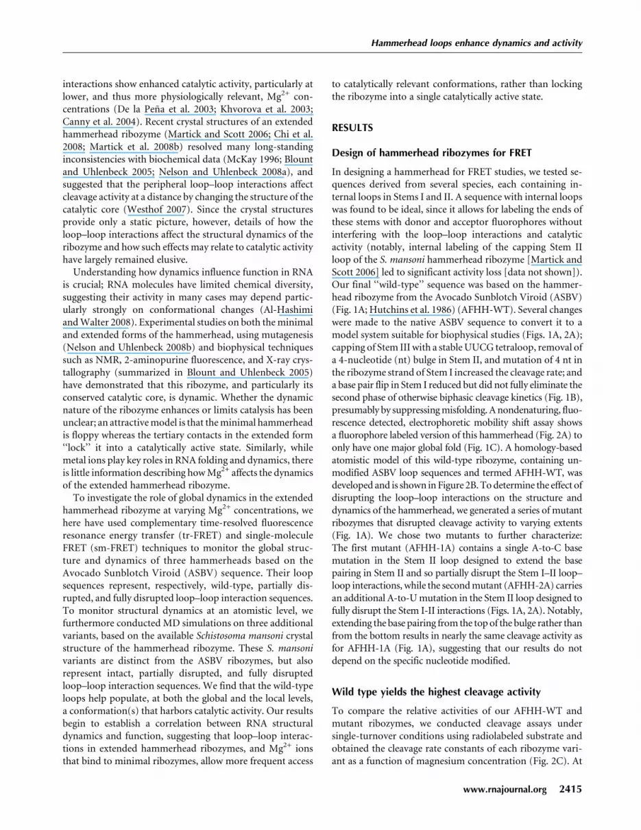
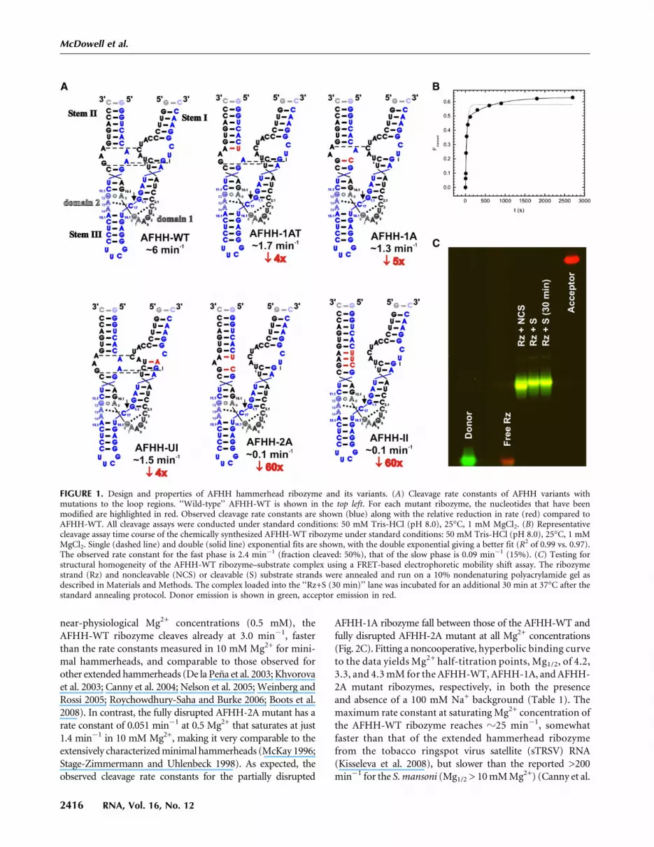
In designing a hammerhead for FRET studies, we tested se-quences derived from several species, each containing in-ternal loops in Stems I and II. A sequence with internal loopswas found to be ideal, since it allows for labeling the ends ofthese stems with donor and acceptor fluorophores withoutinterfering with the loop–loop interactions and catalyticactivity (notably, internal labeling of the capping Stem IIloop of the S. mansoni hammerhead ribozyme [Martick andScott 2006] led to significant activity loss [data not shown]).Our final ‘‘wild-type’’ sequence was based on the hammer-head ribozyme from the Avocado Sunblotch Viroid (ASBV)(Fig. 1A; Hutchins et al. 1986) (AFHH-WT). Several changeswere made to the native ASBV sequence to convert it to amodel system suitable for biophysical studies (Figs. 1A, 2A);capping of Stem III with a stable UUCG tetraloop, removal ofa 4-nucleotide (nt) bulge in Stem II, and mutation of 4 nt inthe ribozyme strand of Stem I increased the cleavage rate; anda base pair flip in Stem I reduced but did not fully eliminate thesecond phase of otherwise biphasic cleavage kinetics (Fig. 1B),presumably by suppressing misfolding. A nondenaturing, fluo-rescence detected, electrophoretic mobility shift assay showsa fluorophore labeled version of this hammerhead (Fig. 2A) toonly have one major global fold (Fig. 1C). A homology-basedatomistic model of this wild-type ribozyme, containing un-modified ASBV loop sequences and termed AFHH-WT, wasdeveloped and is shown in Figure 2B. To determine the effect ofdisrupting the loop–loop interactions on the structure anddynamics of the hammerhead, we generated a series of mutantribozymes that disrupted cleavage activity to varying extents(Fig. 1A). We chose two mutants to further characterize:The first mutant (AFHH-1A) contains a single A-to-C basemutation in the Stem II loop designed to extend the basepairing in Stem II and so partially disrupt the Stem I–II loop–loop interactions, while the second mutant (AFHH-2A) carriesan additional A-to-U mutation in the Stem II loop designed tofully disrupt the Stem I-II interactions (Figs. 1A, 2A). Notably,extending the base pairing from the top of the bulge rather thanfrom the bottom results in nearly the same cleavage activity asfor AFHH-1A (Fig. 1A), suggesting that our results do notdepend on the specific nucleotide modified.
Wild type yields the highest cleavage activity
To compare the relative activities of our AFHH-WT andmutant ribozymes, we conducted cleavage assays undersingle-turnover conditions using radiolabeled substrate andobtained the cleavage rate constants of each ribozyme vari-ant as a function of magnesium concentration (Fig. 2C). At
Hammerhead loops enhance dynamics and activity
www.rnajournal.org 2415
near-physiological Mg2+ concentrations (0.5 mM), theAFHH-WT ribozyme cleaves already at 3.0 min�1, fasterthan the rate constants measured in 10 mM Mg2+ for mini-mal hammerheads, and comparable to those observed forother extended hammerheads (De la Pena et al. 2003; Khvorovaet al. 2003; Canny et al. 2004; Nelson et al. 2005; Weinberg andRossi 2005; Roychowdhury-Saha and Burke 2006; Boots et al.2008). In contrast, the fully disrupted AFHH-2A mutant has arate constant of 0.051 min�1 at 0.5 Mg2+ that saturates at just1.4 min�1 in 10 mM Mg2+, making it very comparable to theextensively characterized minimal hammerheads (McKay 1996;Stage-Zimmermann and Uhlenbeck 1998). As expected, theobserved cleavage rate constants for the partially disrupted
AFHH-1A ribozyme fall between those of the AFHH-WT andfully disrupted AFHH-2A mutant at all Mg2+ concentrations(Fig. 2C). Fitting a noncooperative, hyperbolic binding curveto the data yields Mg2+ half-titration points, Mg1/2, of 4.2,3.3, and 4.3 mM for the AFHH-WT, AFHH-1A, and AFHH-2A mutant ribozymes, respectively, in both the presenceand absence of a 100 mM Na+ background (Table 1). Themaximum rate constant at saturating Mg2+ concentration ofthe AFHH-WT ribozyme reaches z25 min�1, somewhatfaster than that of the extended hammerhead ribozymefrom the tobacco ringspot virus satellite (sTRSV) RNA(Kisseleva et al. 2008), but slower than the reported >200min�1 for the S. mansoni (Mg1/2 > 10 mM Mg2+) (Canny et al.
FIGURE 1. Design and properties of AFHH hammerhead ribozyme and its variants. (A) Cleavage rate constants of AFHH variants withmutations to the loop regions. ‘‘Wild-type’’ AFHH-WT is shown in the top left. For each mutant ribozyme, the nucleotides that have beenmodified are highlighted in red. Observed cleavage rate constants are shown (blue) along with the relative reduction in rate (red) compared toAFHH-WT. All cleavage assays were conducted under standard conditions: 50 mM Tris-HCl (pH 8.0), 25°C, 1 mM MgCl2. (B) Representativecleavage assay time course of the chemically synthesized AFHH-WT ribozyme under standard conditions: 50 mM Tris-HCl (pH 8.0), 25°C, 1 mMMgCl2. Single (dashed line) and double (solid line) exponential fits are shown, with the double exponential giving a better fit (R2 of 0.99 vs. 0.97).The observed rate constant for the fast phase is 2.4 min�1 (fraction cleaved: 50%), that of the slow phase is 0.09 min�1 (15%). (C) Testing forstructural homogeneity of the AFHH-WT ribozyme–substrate complex using a FRET-based electrophoretic mobility shift assay. The ribozymestrand (Rz) and noncleavable (NCS) or cleavable (S) substrate strands were annealed and run on a 10% nondenaturing polyacrylamide gel asdescribed in Materials and Methods. The complex loaded into the ‘‘Rz+S (30 min)’’ lane was incubated for an additional 30 min at 37°C after thestandard annealing protocol. Donor emission is shown in green, acceptor emission in red.
McDowell et al.
2416 RNA, Vol. 16, No. 12

2004; Kim et al. 2005; Boots et al. 2008) and in vitro selectedRzB hammerheads (Mg1/2� 2 mM Mg2+) (Saksmerprome et al.2004). Such differences may be due to variations in the proto-cols used for analyzing cleavage activity (Stage-Zimmermannand Uhlenbeck 1998) and/or be true evidence for the catalyticdiversity of extended hammerhead ribozymes (Shepotinovskayaand Uhlenbeck 2008). Regardless, our work now establishesa fourth extended hammerhead particularly suitable forbiophysical studies, as demonstrated in the following.
AFHH-WT shows the broadest Stem I–IIdistance distribution
We used tr-FRET to monitor the global structure distribu-tion of our three hammerhead ribozymes. This approachallows us to derive equilibrium distance distributions be-tween the donor and acceptor fluorophores at the ends ofStems I and II as a function of Mg2+ concentration (Ruedaet al. 2003). Figure 3 shows the measured mean distance andfull width at half-maximum (FWHM) of each distance dis-tribution plotted against the Mg2+ concentration in eitherthe absence or presence of a 100 mM Na+ background (whichensures proper secondary structure formation even at lowionic strength). The primary effect of the sodium back-ground is a slightly smaller initial mean distance, indicatingthat some tertiary folding of the ribozymes occurs in thepresence of monovalents alone, similar to our previousobservations on minimal hammerheads (Rueda et al. 2003).
Under all conditions, we observed a two-phase decrease inthe mean distance accompanied by first a decrease then anincrease in FWHM with increasing Mg2+ concentration,which we interpret as a (at least) two-step folding process,similar to our previous results on minimal hammerheads(Rueda et al. 2003). We fit the Mg2+ dependences of distanceand FWHM in the absence of Na+ to a two-step Mg2+
binding model, and obtained Mg1/2 values of 0.14, 0.15, and0.19 mM for the first step of folding for the AFHH-WT,AFHH-1A, and AFHH-2A mutant ribozymes, respectively(with cooperativity coefficients of 1.1–1.4, close to unity)(Table 1). These apparent Mg2+ affinities are approximatelyfive- to 10-fold tighter (lower Mg1/2) than those previouslyobserved for the catalytically slower minimal ribozymes. (Asexpected, a background of 100 mM competing Na+ reducesthe Mg2+ affinity, by approximately two- to threefold) (Table1). These observations are generally consistent with previoussteady-state FRET measurements on extended hammerheads(Penedo et al. 2004; Boots et al. 2008) and suggest that allthree of our ribozymes fold at lower ionic strength than theirminimal counterparts (Rueda et al. 2003). Just as with theminimal hammerhead ribozymes (Rueda et al. 2003), how-ever, our combined cleavage and tr-FRET assays reveala lower-affinity, noncooperative folding step of the extendedribozymes that coincides with their catalytic activation inthe low-millimolar Mg2+ range (3–4 mM) (Table 1). Thisobservation may be consistent with observations of the
FIGURE 2. Modifications and cleavage activity of the hammerheadribozymes used in this study. (A) Secondary structure of the AFHH-WT (AFHH; blue), as well as the partially disrupted (AFHH-1A;green) and fully disrupted (AFHH-2A; red) mutants, based on thesequence from Avocado Sunblotch Viroid (ASBV). Several putativeinteractions between the internal loops of Stems I and II are indicatedas thin dashes. (B) Homology model showing possible orientation ofthe helices. Colors are the same as in A; the green base is the mutationin AFHH-1A, the red base is the additional mutation in AFHH-2A.(C) Characterization of cleavage rate dependence on Mg2+ concen-tration under standard conditions (50 mM Tris-HCl [pH 8.0], 25°C).
Hammerhead loops enhance dynamics and activity
www.rnajournal.org 2417

Mg2+-induced catalytic activation (Canny et al. 2004) andfolding of the Schistosoma hammerhead as monitored bysteady-state FRET (Boots et al. 2008). Boots et al. report asingle phase of folding with half-titration points of z1 mMMg2+ (in 100 mM NaCl) as monitored by steady-state FRET.Their data also show a rise in FRET beyond this initialtransition at tens of millimolar Mg2+, which could representthe second folding step that is observed here by tr-FRET andis related to catalytic activity. Our more detailed tr-FRETexperiments show that during this second folding step theStem I–II distance slightly decreases further, whereas theFWHM increases dramatically for AFHH-WT. We note thatdespite this qualitative agreement, our wild-type hammer-head ribozyme exhibits Mg2+ half-titration points for thefirst and second steps of folding of 0.14–0.19 mM (dependingon monovalent background) and 4.2 mM, respectively(Table 1), that are approximately five- to 20-fold tighter thanthose reported for the previously characterized extendedhammerhead ribozymes from sTRSV and S. mansoni (Cannyet al. 2004; Kim et al. 2005; Boots et al. 2008) and appearmore similar to those of the selected RzB hammerhead(Saksmerprome et al. 2004). We conclude that our wild typeis a hammerhead with comparably high Mg2+ affinity, andtherefore well suited for biophysical studies. Importantly, wefind our observations for the wild type to be less pronouncedfor the AFHH-1A and AFHH-2A mutants with partially andfully disrupted loop–loop interaction sequences, respectively(Fig. 3). The relative increase of the FWHM in AFHH-WTover that of the mutants suggests that in AFHH-WT the StemI–II distance distribution is the broadest.
AFHH-WT exhibits the most sm-FRET statesand the greatest interstem dynamics
To determine the source of the broad FWHMs measured bytr-FRET at low millimolar Mg2+ concentrations and probedynamics of individual molecules over time, we used total
internal reflection fluorescence (TIRF)-based sm-FRET. Incontrast to the similarly small hairpin ribozyme and anaptazyme based on the minimal hammerhead, which bothshow dynamics between only two sm-FRET states (Zhuanget al. 2002; Ditzler et al. 2008; de Silva and Walter 2009), weobserved at least four distinguishable states in our extendedhammerhead ribozymes (Fig. 4A). The individual FRET timetraces within a set of conditions showed considerable het-erogeneity with multiple types of dynamics between subsetsof states; examples for the AFHH-WT as well as AFHH-1Aand AFHH-2A mutants are found in Figure 5A–C. ‘‘Hetero-geneity’’ refers here to a lack of interconversion, within theaccessible observation window, between kinetically distinctmolecular species, also observed for other RNAs such as the
TABLE 1. Parameters describing the Mg2+ dependence of global folding of hammerhead ribozymes AFHH-WT, AFHH-1A, and AFHH-2A asmonitored by tr-FRET
Variant [Na+] (mM) RN (A) KL (mM) m RL (A) KH (mM) n RH (A)
AFHH-WT 0 91.6 6 0.1 0.14 6 0.01 1.4 6 0.2 81.0 6 0.5 4.2 1 71.2 6 0.3100 83.3 6 0.7 0.23 6 0.05 1.9 6 0.6 80.4 6 0.5 4.2 1 69.4 6 0.3
AFHH-1A 0 92 6 3 0.15 6 0.01 1.3 6 0.2 76.2 6 0.7 3.3 1 70.3 6 0.3100 81.4 6 0.4 0.5 6 0.3 0.8 6 0.2 74 6 2 3.3 1 71.2 6 0.4
AFHH-2A 0 95 6 2 0.19 6 0.01 1.14 6 0.06 75.0 6 0.5 4.3 1 68.8 6 0.3100 80.9 6 0.5 0.29 6 0.06 1.3 6 0.3 74.9 6 0.8 4.3 1 67.5 6 0.4
The apparent Mg2+ binding constants KL, associated cooperativity constant m, and mean distances RL and RH were derived from the fits inFigure 3 as described in Materials and Methods under standard cleavage conditions: single turnover, 50 mM Tris-HCl (pH 8.0) either with orwithout 100 mM NaCl. RN, the distance in the absence of Mg2+, was taken directly from the ‘‘no-Mg2+’’ tr-FRET measurement. KH and n, thedissociation constant and cooperativity coefficient of catalytic activation, respectively, were derived from our cleavage assays which wereconducted in 0 mM Na+, and this value was fixed as a parameter in the 100 mM Na+ tr-FRET fits as well. This approach is reasonably justifiedsince (1) we consistently obtain good fits with this KH value (Fig. 3); and (2) activity assays with the minimal hammerhead ribozyme yieldedidentical KH values in the presence and absence of 100 mM Na+ (Rueda et al. 2003). Errors are 1 standard deviation as determined by the fit, or,in the case of RN, derived from at least duplicates of the no-Mg2+ measurement.
FIGURE 3. Distance distributions between stems I and II of theAFHH-WT (solid line), as well as partially disrupted (AFHH-1A;dashed line) and fully disrupted (AFHH-2A; dotted line) hammerheadmutants monitored by tr-FRET. (A) Mean distance and full widthat half-maximum (FWHM) of the distance distributions (modeled as asingle Gaussian) as a function of Mg2+ concentration under standardconditions (50 mM Tris-HCl [pH 8.0], 25°C). (B) tr-FRET measure-ments as in A, but the buffer included 100 mM Na+.
McDowell et al.
2418 RNA, Vol. 16, No. 12

hairpin (Zhuang et al. 2002; Bokinsky et al. 2003; Rueda et al.2004; Ditzler et al. 2008) and group I intron ribozymes(Solomatin et al. 2010), as well as the tetraloop–receptortertiary interaction (Fiore et al. 2009). To analyze our data,we first plotted aggregate FRET histograms for each set ofconditions and fit them with Gaussians to identify FRETstates (Fig. 4A–C). These histograms define multiple FRETstates under all conditions, with more FRET states appearing
and the distribution among states becoming more even as theMg2+ concentration increases. Most significantly, we ob-served a high FRET state of z0.7 that becomes increasinglypopulated at 10 mM Mg2+, particularly in the catalyticallymost active AFHH-WT, suggesting that enhanced activitycorrelates with more frequent, yet transient visits to this state.
For a relative comparison, we mapped the distancesderived from the tr-FRET analysis onto the FRET ratios of
FIGURE 4. Single molecule FRET histograms, each constructed from 50–100 single molecule time traces prior to photobleaching of the acceptorunder standard conditions (50 mM Tris-HCl [pH 8.0], 25°C, with varying Mg2+ concentration). Vertical solid gray lines are FRET ratioscorresponding to the tr-FRET derived FWHMs. (A) AFHH-WT ribozyme; (top) 0.5 mM Mg2+, (middle) 1 mM Mg2+, (bottom) 10 mM Mg2+. (B)Partially disrupted mutant AFHH-1A; Mg2+ concentrations as in A. (C) Fully disrupted mutant AFHH-2A; Mg2+ concentrations as in A.
FIGURE 5. Single molecule FRET analysis. (A–C) Exemplary single molecule time traces showing donor (green) and acceptor (red) intensities,and the corresponding FRET ratio (black) with HaMMy fit (gray). Conditions are, 50 mM Tris-HCl (pH 8.0), 10 mM Mg2+, at 25°C. (A) AFHH-WT ribozyme. (B) Partially disrupted AFHH-1A ribozyme. (C) Fully disrupted AFHH-2A ribozyme. (D–F) Corresponding transition densityplots for the same conditions as in A–C, showing the FRET states, transitions between those states, and specific transition rate constantsdetermined by fitting time traces with the software HaMMy (McKinney et al. 2006) for AFHH-WT AFHH (80 molecules; D), partially disruptedmutant AFHH-1A (84 molecules; E) and fully disrupted mutant AFHH-2A (49 molecules; F).
Hammerhead loops enhance dynamics and activity
www.rnajournal.org 2419

our sm-FRET derived histograms, using similar strategiesand assumptions as described previously (Pereira et al. 2008).This comparison shows the general consistency between thetwo types of data. At increasing Mg2+ concentrations, we ob-served broader tr-FRET distance distributions with smallermean Stem I–II distances, which were correlated with broadersm-FRET histograms and enhanced occurrence of high FRETstates (Fig. 4A–C). This comparison also illustrates the dif-ficulty of ensemble approaches to discern multiple over-lapping distance distributions, which appear only as a broadrange of tr-FRET derived distances but are readily dissected bycomplementary sm-FRET analyses.
To more quantitatively characterize the dynamics of singlehammerhead ribozymes, we used hidden Markov modeling(HMM, as implemented in the software HaMMY [McKinneyet al. 2006]) to determine the most probable path traversed ineach sm-FRET trajectory among up to four FRET states (Fig.5A–C). HMM also yields transition density contour maps thatwe find to be highly symmetric with respect to the maindiagonal for all ribozymes and Mg2+
concentrations, indicative of the fact thatmost transitions occur reversibly betweenpairs of FRET states (Fig. 5D–F) as ex-pected for an RNA at thermal equilibrium(Abelson et al. 2010). In addition, themaps reveal more frequent (color scalein Fig. 5D–F) and faster (AFHH-WT,0.14 sec�1; AFHH-1A, 0.04 sec�1; AFHH-2A, 0.09 sec�1) transitions of the AFHH-WT ribozyme to the 0.7 FRET state com-pared to its two mutant counterparts, whilethe mutants show more frequent andfaster (AFHH-WT, 0.11 sec�1; AFHH-1A,0.27 sec�1; AFHH-2A, 0.17 sec�1) transi-tions away from the 0.7 FRET state, furthersupporting the notion that the most activeribozyme transiently accesses a state(s) withdiminished Stem I–II distance(s) mostfrequently and for the longest time period.
Taken together, our experimental dataconsistently suggest that the wild-typeloop sequences specifically lead to fre-quent, transient encounters of Stems I andII, as well as enhanced catalytic activity ofthe AFHH-WT ribozyme. Both interstemencounters and catalytic activity are sig-nificantly reduced by even a single nucle-otide mutation designed to disrupt theloop–loop interactions. Increasing Mg2+
concentrations also enhance both thefrequency of Stem I–II encounters andcatalytic activity, but cannot fully com-pensate for the loss of specific intersteminteractions. These global dynamics, how-ever, cannot explain how loop–loop in-
teractions between Stems I and II impact the cleavage site overa distance of >10 A (Fig. 2B).
MD simulations show more active site dynamicsin the wild type
To examine whether the patterns of hammerhead globaldynamics observed by FRET are reflected at the active site, weconducted MD simulations on three variants also represent-ing intact, partially disrupted, and fully disrupted loop–loopinteraction sequences (Fig. 6A). Since MD simulationsrequire an accurate starting geometry as provided by ahigh-resolution crystal structure (McDowell et al. 2007), wechose the S. mansoni hammerhead ribozyme structure (PDBID 2GOZ) as our wild type (SCH-WT) (Martick and Scott2006). Our partially disrupted loop mutant is based on thisSCH-WT, but we replaced each nucleotide in the Stem IIloop with uracil (Fig. 6A), creating a mutant (SCH-UL)
FIGURE 6. Molecular dynamics simulations of three S. mansoni hammerheads reveal dif-ferences in global structural dynamics. (A) Secondary structure of the three variants representingSCH-WT (crystal structure sequence [Martick and Scott 2006]), partially disrupted (SCH-UL;green), and fully disrupted mutants (SCH-NoL; red). (B) Global residue correlation differencemap (SCH-UL minus SCH-WT) for the first 5 nsec of simulation time. Circled areas show wherecorrelation between the loop and the bulge is lost for the U-loop mutant, SCH-UL. (C) Normalmode analysis. Porcupine plots (visualized using VMD) depict the motion of each backboneatom for the first normal mode of SCH-NoL (left) and SCH-WT (right) over the first 10 nsecof MD simulation. Arrows vectorially represent the magnitude and direction of movement.The right-most panel shows snapshots of the Pymol movie visualizing the first normal mode ofSCH-WT; the two shades of blue represent the two extremes of the motion. (D) Alignment ofsimulated variants to the crystal structure (orange). The simulated structures are averages overthe time period of 10.5–10.6 nsec. The core residues are aligned to the crystal structure core usingPymol. SCH-WT, blue; SCH-UL, green; SCH-NoL, red.
McDowell et al.
2420 RNA, Vol. 16, No. 12

shown experimentally to have significantly reduced self-cleavage activity (Osborne et al. 2005). Finally, our fullydisrupted loop mutant (SCH-NoL) was created by deletingthe loop–loop regions (all nucleotides of the Stem II loop aswell as all bases of Stem I above the A1.6–U2.6 base pair) (Fig.6A), leaving essentially a minimal hammerhead.
Standard MD simulations were performed on these threeribozyme variants in explicit solvent over 20 nsec each, usingAMBER8 and the parm99 Cornell et al. force field as pre-viously described (McDowell et al. 2007; Sefcikova et al. 2007).Cross-correlation matrices were derived from AMBER forevery 5-nsec segment of each trajectory, highlighting bothcorrelated and anti-correlated motions among individualnucleotides across the entire RNA, as previously described(Rhodes et al. 2006). A difference map of these global residuecross-correlations of our wild-type (SCH-WT) and U-loopmutant (SCH-UL) simulations suggests that disruption of theloop–loop interaction reduces long-range structural correla-tions, especially between the residues of the Stem I and Stem IIloops, already over the first 5 nsec of simulation time (ob-served as the circled dark regions in Fig. 6B). Complementarynormal mode analysis was performed (calculating the eigen-vectors from a covariance matrix) for eachsimulation to highlight the dominantglobal motions in each RNA molecule.In this analysis, large-scale (global) mo-tions are represented by the lowest-fre-quency eigenvectors. For example, inspec-tion of ‘‘porcupine’’ plots (representingthe vectorial directions of motion asarrow heads) of the first normal modesover the first 10 nsec of the SCH-WT andSCH-NoL simulations indicates a similarpattern of global motion in these mole-cules as experimentally observed by sm-FRET in our AFHH sequences. In partic-ular, comparing the first normal modes ofthese variants shows large-scale motion inStem I of the SCH-WT, while the motionin Stems I and II of the SCH-NoL isrelatively dampened (Fig. 6C). Theselarge-scale motions of Stem I of SCH-WT represent a bending of the whole helix,rather than a fraying of individual basepairs, and are therefore consistent withbeing on-path to the dissociation of theloop–loop interactions, which must occurgiven the transient nature of the Stem I–IIinteractions observed in our experimentalsm-FRET studies. Despite this apparentcorrespondence of the relative global mo-tions in our MD simulations and experi-mental studies it has to be noted, however,that the timescales at which these motionsoccur are very different (nanoseconds for
the MD simulations, >100 msec for the sm-FRET studies). Thefast MD simulation timescale therefore has to be thought of asexploring local fluctuations within a minimum of the foldingfree energy landscape, whereas the much slower experimentaltimescale captures much larger segments of the landscape.
Overlays comparing the simulated structures (averagedover the time period of 10.5–10.6 nsec) to the crystal struc-ture show that while the overall global fold of each simulationremains similar to the starting structure, some differences doappear (Fig. 6D). In particular, in the absence of loop–loopinteractions, the relative helical twists of the three stems ofthe SCH-NoL mutant significantly change relative to thestarting crystal structure and the simulated SCH-WT struc-ture (Fig. 6D, cf. SCH-NoL and SCH-WT).
The cleavage reaction of the hammerhead ribozyme pro-ceeds through in-line nucleophilic attack by the 29-oxygenof C17 on the phosphorous of C1.1 (van Tol et al. 1990).The ideal nucleophilic in-line attack angle (IAA, 29O-P-59O)for this reaction is 180° (Soukup and Breaker 1999). Analignment of the active sites of the simulated structures(averaged over the time period of 10.5–10.6 nsec in about themiddle of the trajectory) to the crystal structure (Fig. 7A–C)
FIGURE 7. MD simulations also reveal differences in local structural dynamics. (A–C) Close-up view of the alignment of simulated variants to the crystal structure. Boxed regions fromFigure 6D are shown in more detail to highlight the cleavage site; (A) SCH-WT, blue; (B) SCH-UL, green; (C) SCH-NoL, red. (D) Nucleophilic in-line attack angle (IAA) for SCH-WT (blue),SCH-UL (green) and SCH-NoL (red) mutants. Histograms were constructed from anglesmeasured every 100 psec over 10 nsec of simulation time. (Inset) The in-line fitness parameterwas calculated as described (Soukup and Breaker 1999) and is plotted as a histogram. (E) IAAsfor SCH-WT (blue), SCH-UL (green), and SCH-NoL (red) over 10 nsec of simulation time.
Hammerhead loops enhance dynamics and activity
www.rnajournal.org 2421
highlights the significant effect of the loop–loop interactionson the IAA at long range; while the SCH-WT cleavage siteremains in a geometry similar to that in the crystal structure,with a relatively favorable IAA of 160° (Fig. 7A), modifica-tion of the loop region leads to more frequent adoption ofless favorable IAAs. In the snapshots shown in Figure 7B,C,the SCH-UL and SCH-NoL mutants adopt IAAs of 132° and129°, respectively. Figure 7D shows histograms of nucleo-philic IAAs for the SCH-WT, SCH-UL, and SCH-NoLvariants constructed from the angles measured every 100psec over 10 nsec of simulation time. Each distribution showsa dominant peak around a low IAA of 125°, but also has ashoulder consisting of IAAs closer to the ideal 180°. Strik-ingly, access to the higher, catalytically most favorable IAAangles is enhanced in the SCH-WT with wild-type loopsequences, and least prevalent in the SCH-NoL mutant, withthe partially disrupted SCH-UL mutant falling in between(Fig. 7D). The same trend is seen in the in-line fitnessparameter (Soukup and Breaker 1999; Lee et al. 2008), whichincludes both the IAA and the O29-P attack distance (Fig. 7D,inset). Finally, Figure 7E reveals the dynamic nature of theIAA distribution by plotting time courses of nucleophilicIAAs over the first 10 nsec. Taken together, our observationssupport the notion that wild-type Stem I–II loop–loopinteractions lead to both global and local structural dynamicsthat are correlated with increased catalytic activity.
DISCUSSION
Here we have combined cleavage assays, Mg2+ titrationsmonitored by tr- and sm-FRET, and MD simulations toprobe the global and local structural dynamics of extendedhammerhead ribozymes with wild-type, partially, or fullydisrupted Stem I–II loop–loop interaction sequences. Wefind that wild-type loop sequences and increased Mg2+ con-centrations act to enhance both cleavage activity and struc-tural dynamics and heterogeneity of the ribozyme. It has beenappreciated since 2003 that the peripheral loop–loop in-teractions increase ribozyme activity (De la Pena et al. 2003;Khvorova et al. 2003). Our results are consistent with thenotion that the Stem I–II distance in our wild-type ham-merhead ribozyme is surprisingly dynamic. These motionshelp populate catalytically relevant in-line attack angles at thedistal cleavage site. Our observations therefore indicate thatthe extended hammerhead ribozyme is not restricted rigidlyto a single (or few) catalytically relevant conformation(s).
Our tr-FRET monitored Mg2+ titrations show a biphasicdecrease in mean distances (Fig. 3), consistent with our pre-vious results on minimal hammerheads (Rueda et al. 2003),revealing significant similarities between both forms of theribozyme. Similarly, recent mechanistic studies comparingminimal and extended hammerheads have suggested funda-mental similarities between the two ribozyme forms as theircatalytic cores are in a dynamic equilibrium between similarinactive and active conformations, albeit with different bias
(Scott 2007; Nelson and Uhlenbeck 2008a,b). Furthermore,our tr-FRET results reveal that the mean Stem I–II distancesof the AFHH-WT ribozyme are slightly larger than those ofthe mutants, inconsistent with the wild-type loops simplyholding Stems I and II closer together (Fig. 3). We generallyobserved broad tr-FRET derived distribution widths (FWHMvalues) that may reflect either global conformational dynamics(rapid conformational isomerization) or heterogeneity (dis-tinct, slowly interconverting conformers), or both. All ourmeasured widths increase at high Mg2+ concentrations (Fig.3), consistent with both our previous work on minimalhammerheads (Rueda et al. 2003) and with data on a Diels-Alderase ribozyme that likewise showed increased widths ofintramolecular distance distributions with increasing Mg2+
concentration (Kobitski et al. 2007). Notably, the distributionof distances measured for the AFHH-WT hammerhead issignificantly wider than those of the mutants (Fig. 3). The factthat the fluorophore attachment sites are identical for all threevariants and distal from the mutations suggests that thesedifferences are significant. Our tr-FRET data are contrary tothe hypothesis that the hammerhead loop–loop interactionslock the ribozyme in a single (or few) active structure(s)(Martick and Scott 2006).
Our sm-FRET approach reports on the detailed structuraldynamics underlying the broad FWHM values observed intr-FRET, indicating that both Mg2+ and wild-type loop–loopinteraction sequences bias the folding free energy landscapetoward more disparate stem–stem distances by making newconformations accessible and/or by populating previouslyrare conformations (Fig. 4). Because the ribozyme morefrequently accesses and spends more time in the z0.7 FRETstate under conditions and in variants that show highercatalytic activity (Figs. 4, 5), this state exhibits propertiesexpected for an active conformation. Conversion of thedistance distributions from tr-FRET to apparent sm-FRETratios finds good agreement in the general trends (Fig. 4, solidvertical lines), but suggests that high FRET states remainrelatively underestimated by the tr-FRET approach. Recentwork has suggested that relating sm-FRET values to absolutedistances (as derived by tr-FRET) can be complicated by thepotential of Cy3 and Cy5 to stack on opposite ends of a singlenucleic acid helix (Iqbal et al. 2008). In our labeling scheme,Cy5 is internally attached, the two fluorophores are coupled todifferent helices (Stems I and II) (Fig. 2A), and long linkers areused on both fluorophores, suggesting that helix-end stackingshould be less dominant. In addition, such complicationsshould not affect relative comparisons of our three variantsgiven that they are all labeled in the same manner. Clearly, tr-FRET and sm-FRET complement and cross-validate eachother, further supporting the notion that our wild-type loopsenhance, rather than quench, the conformational sampling ofthe helical stems that bracket the catalytic core.
While our FRET techniques allow us to experimentallymeasure global dynamics between the interacting Stems I andII, they can report only on specific fluorophore distances.
McDowell et al.
2422 RNA, Vol. 16, No. 12

MD simulations allow us to calculate complementary, moredetailed dynamic information, albeit at a much shorter time-scale. Normal mode analysis of the SCH-WT and SCH-NoLsimulations shows increased motion between Stems I and II ofthe wild-type hammerhead relative to a loop-disrupted mutant(Fig. 6). Thus our FRET-based observations are echoed in thesesimulations, using distinct hammerhead sequences and a verydifferent methodology. MD simulations allow us to focus onlocal motions, and suggest that the loop–loop interactionshave a significant long-range effect on the cleavage site. Morespecifically, we observe that the presence of wild-type loopsequences biases the nucleophilic IAA toward more frequentand extended visits to catalytically favorable values near 180°,while mutations that disrupt the Stem I–II interactions disfavorcatalytically relevant IAAs proportionally to their experimen-tally observed adverse impact on catalysis (Fig. 7). Again, theloop–loop interactions do not lock the hammerhead active siteinto a single conformation but instead lead to dynamics thatsample favorable IAAs. These observations are consistent withthe notion that distal tertiary interactions and global dynamicscouple with local dynamics and catalytic function of thehammerhead ribozyme. Given that the timescales of ourMD and FRET approaches are very different (10 nsec and100 msec, respectively), exploring different fractions of thefolding free energy landscapes (Rhodes et al. 2006; McDowellet al. 2007; Ditzler et al. 2010), the consistency of observingspecific differences between wild-type and mutant ribozymesis remarkable.
Thus, with our specific wild-type loop–loop interactionswe observe extensive global and local dynamics in the ham-merhead, with different patterns of dynamics for each ribo-zyme variant and at each magnesium concentration. While it isnot likely that all dynamically visited states are ‘‘on-pathway’’for catalysis, at both the global and local scales we findstructural dynamics enhanced in parallel with ribozymeactivity. A favorable IAA has been predicted to enhance therate of ribozyme catalysis between 10- and 100-fold (Emilssonet al. 2003; Min et al. 2007), consistent with our observations.More specifically, we measure a 16-fold enhancement ofAFHH-WT cleavage rate constants over our fully disruptedmutant in 10 mM Mg2+ (Fig. 2C), and our computationalresults suggest that the SCH-WT simulation samples IAAswith values >170° z12-times more frequently than does theminimal hammerhead simulation (Fig. 7D). Even keeping inmind the limitations posed by their disparate timescales, ourFRET and MD data thus jointly support the notion that theperipheral loop–loop interactions exert a long-range effect onactive site dynamics of the hammerhead ribozyme.
In summary, our results provide evidence for the notionthat the dynamic nature of at least the hammerhead ribo-zymes studied here is conducive, rather than disruptive, tocatalysis. Conformational flexibility and sampling of thefolding free-energy landscape is altered in the presence ofperipheral loop–loop interactions and/or increasing Mg2+
concentrations, in a way that populates catalytically relevant
in-line attack angles in the distal active site. Our findings areconsistent across the two hammerhead species studied here,and are consistent with observations from other RNAs,providing a potentially generalizable model for how periph-eral structures influence RNA dynamics and function. NMRstudies reporting on both global and local dynamics of HIVTAR (Al-Hashimi et al. 2003) show that increasing Mg2+
concentrations lead to a stabilization of global helix stacking,but in parallel increase local motions. We observe a similarphenomenon here, where an increase in Mg2+ concentrationfavors an overall global structure while promoting localstructural ‘‘relaxation,’’ with conformational states sampledmore broadly, populating states favorable to in-line nucle-ophilic attack of a scissile phosphate. Similarly, the hairpinribozyme has been observed to populate more catalyticallyrelevant ‘‘docked’’ states in the presence of increasingMg2+ concentrations and stabilizing architectural elements(Zhuang et al. 2002; Bokinsky et al. 2003; Tan et al. 2003). Apicture emerges wherein the concepts of induced fit, con-formational capture, active site preorganization, and sub-strate strain (Fersht 1999) begin to gain a dynamic dimensionfor an RNA enzyme, suggesting that specific ribozyme vari-ants can, to varying degrees, bias molecular motions towardcatalytically active conformations, analogous to currentnotions in protein enzymology (Hammes-Schiffer andBenkovic 2006). Future work needs to examine the mecha-nism of the observed linkage of global and local dynamics, aswell as ask whether global collective motions are directlycoupled to the enzymatic function of RNA, as is thought to bethe case in protein enzymes (Hammes-Schiffer and Benkovic2006; Henzler-Wildman et al. 2007) and as has been pro-posed for the hairpin ribozyme (Rhodes et al. 2006). To thisend, a true integration of experimental and computationalsingle molecule studies into a single conceptual framework,as foreshadowed here, will likely be necessary.
MATERIALS AND METHODS
Preparation of RNA oligonucleotides
For cleavage assays, RNA oligonucleotides were transcribed invitro using T7 RNA polymerase (Milligan and Uhlenbeck 1989).Deprotected RNA was purified as previously described (Walter2002). 59-Fluorescein, Cy3, and biotin were attached to the RNAby the manufacturer, while 39-tetramethylrhodamine and Cy5 wereincorporated post-synthetically as previously described (Walter2002). RNA concentrations were calculated from absorption at260 nm and, in the case of the tr-FRET HHRz strand, corrected forthe additional absorption of fluorescein and tetramethylrhodamineby using the relations A260/A492 = 0.3 and A260/A554 = 0.49, re-spectively. The following sequences were used: (1) HHRz strand:59-GGUUCCCAUCAUCUAUCCCUGAAGAGACGAAGUGACCC-39; (2) HHS strand: GGGUCACAAGUCGAAACUCCUUCGGGAGUCGGAUAGUCGGAACC; (3) HH39A strand: GGGUCACACGUCGAAACUCCUUCGGGAGUCGGAUAGUCGGAACC; and(4) HH2A strand: GGGUCACUCGUCGAAACUCCUUCGGGAG
Hammerhead loops enhance dynamics and activity
www.rnajournal.org 2423

UCGGAUAGUCGGAACC. For tr-FRET and sm-FRET, RNAoligonucleotides were purchased with 29-protection groups fromthe Howard Hughes Medical Institute Biopolymer/Keck Founda-tion Biotechnology Resource RNA Laboratory at the Yale Uni-versity School of Medicine and were deprotected as suggested bythe manufacturer (http://keck.med.yale.edu/oligos/). The followingsequences were used: (1-a) tr-FRET HHRz strand: 59-(F-AU)GUUCCCAUCAUCUAUCCCUGAAGAGACGAAGUGACC(UA–TMR)-39;(1-b) sm-FRET HHRz strand: 59-(Cy3-AU)GUUCCCAUCAUCUAUCCCUGAAGAGACGAAGUGACC(UAT(Cy5)AUAU-biotin)-39;(2) HHS strand: UAGGUCACAAGUCGAAACUCCUUCGGGAGUCGGAUAGUCGGAACAU; (3) HH39A strand: UAGGUCACACGUCGAAACUCCUUCGGGAGUCGGAUAGUCGGAACAU; and(4) HH2A strand: UAGGUCACUCGUCGAAACUCCUUCGGGAGUCGGAUAGUCGGAACAU. F represents 59-fluorescein and TMR39-tetramethylrhodamine; to obtain noncleavable substrate analogsthe underlined C nucleotide was modified with a 29-methoxy group.
Cleavage reactions
All cleavage reactions were conducted under single-turnover (pre-steady-state) conditions in standard buffer (50 mM Tris-HCl, pH8.0) and analyzed as previously described (Rueda et al. 2003;Sefcikova et al. 2007). 59-32P-labeled substrates were prepared byphosphorylation with T4 polynucleotide kinase and [g-32P]ATP.Standard buffer was 50 mM Tris-HCl (pH 8.0). Mg2+ concentra-tions were varied as shown in Figure 2. Final RNA concentrationswere 1 mM for the Rz strand, and trace amounts (<1 nM) of theradiolabeled substrates. Error bars stem from at least two indepen-dent cleavage assays. Time traces of product formation were fit witha double-exponential equation y(t) = y0 + A1[1 – e^(�kobs1t)] +A2[1 � e^(�kobs2t)], employing Marquardt-Levenberg nonlinearleast-squares regression (Igor Pro), where A1 and A2 are the extentsof cleavage and kobs1 and kobs2 are observed rate constants. The firstphases of the time traces were dependent on Mg2+ concentration,and a set of cleavage assay time courses (typically four) under thesame conditions were globally fit to obtain a single fast-phase cleav-age rate constant (first-phase amplitudes and second-phase rates andamplitudes were free parameters for each time course). The secondphases were not dependent on Mg2+ concentration, were gen-erally at least an order of magnitude slower, contributed typicallyless than one-fourth of the total substrate cleaved, and were notfurther analyzed. Mg2+ dependencies of the kobs1 rate constantwere fit with the binding equation kobs = ksat(Mgn)/[(Mgn) + (kH
n)]to yield the cleavage rate constant ksat under saturating Mg2+
conditions, the apparent metal ion dissociation constant KH, andthe cooperativity coefficient n.
FRET electrophoretic mobility shift assays
FRET electrophoretic mobility shift (gel FRET) assays were con-ducted as previously described (Pereira et al. 2002), to test thehomogeneity of ribozyme–substrate complexes. Nondenaturinggel electrophoresis was performed in 10% (w/v) polyacrylamide(acrylamide:bisacrylamide 19:1), 50 mM Tris-HOAc (pH 8.0) andeither 1 or 10 mM Mg(OAc)2. Ten pmol doubly fluorophorelabeled ribozyme strand and 30 pmol unlabeled substrate strandwere annealed in 50 mM Tris-HOAc (pH 8.0) and either 1 mM or10 mM Mg(OAc)2, and 10% (v/v) glycerol, by heating for 2 minto 90°C and cooling to room temperature. The electrophoresis
unit was assembled and pre-equilibrated at 4°C, the sampleswere loaded onto the gel, and an electric field of 6 V/cm wasimmediately applied. After electrophoresis overnight, the gel wasscanned between its low-fluorescence glass plates in a FluorImagerSI fluorescence scanner with ImageQuant software (MolecularDynamics). RNAs labeled with only fluorescein and only tetra-methylrhodamine were included as color calibration standards.
Time-resolved FRET
The global structure of the hammerhead ribozyme was studied asa function of Mg2+ concentration by tr-FRET analysis of ribozymecomplexes doubly labeled with fluorescein and tetramethylrhodamineas previously described (Rueda et al. 2003). The preannealedribozyme-noncleavable substrate complex (80 mL; 1 mM Rz and3 mM S) was incubated at 25°C for at least 15 min in standardbuffer either with or without 100 mM NaCl prior to collectionof time-resolved emission profiles of the donor fluorescein usingtime-correlated single-photon counting similar to previously de-scribed procedures (Rueda et al. 2003). Mg2+ was titrated by in-cremental additions of 0.5 mL aliquots of appropriate MgCl2 stocksolutions in standard buffer, taking into account the volumechange. Fluorescein emission decays were analyzed as previouslydescribed, with distance distributions modeled as single Gaussians(Rueda et al. 2003).
Single-molecule FRET
Single-molecule experiments were performed as previously de-scribed (Rueda et al. 2004; Pereira et al. 2008; de Silva and Walter2009). The biotinylated and Cy3-Cy5-labeled ribozyme and non-cleavable substrate strands (Fig. 2A) were annealed at a concen-tration of 1 mM each in a buffer containing 50 mM Tris-HCl (pH8.0) and 1% (v/v) 2-mercaptoethanol. The annealing solutionwas heated to 90°C for 2 min before cooling to room temperatureover >5 min. The annealed complex was diluted to <20 pM andbound to a streptavidin-coated quartz slide surface via the biotin–streptavidin interaction. The donor (ID) and acceptor (IA) fluo-rescence signals of optically resolved single molecules (character-ized by single-step photobleaching) were detected on a total internalreflection fluorescence microscope as described (Rueda et al. 2004;Pereira et al. 2008). The FRET ratio [defined as IA/(IA + ID)] wasfollowed in real time for each individual molecule. Measurementswere performed under standard buffer conditions (50 mM Tris-HCl[pH 8.0] and either 0.5, 1, or 10 mM MgCl2) at 25°C, with anoxygen scavenging system consisting of 10% (w/v) glucose, 2% (v/v)2-mercaptoethanol, 750 mg/mL glucose oxidase, and 90 mg/mLcatalase to reduce photobleaching. Traces that showed very highFRET (z0.9) were not analyzed, since this FRET level correspondedto an enzyme strand that did not form a complex with a substrate(data not shown). sm-FRET intensity traces were analyzed using anonlinear filter (Haran 2004), and fit with HaMMy software(McKinney et al. 2006), which uses hidden Markov modeling todetermine FRET states and transitions between states. HaMMy fitswere also used to generate transition density plots (McKinneyet al. 2006).
Comparison of tr-FRET distances to sm-FRET ratios
The apparent FRET efficiency was calculated using a scaling factorc = 0.68 as in Sabanayagam et al. (2005), then the FRET ratio was
McDowell et al.
2424 RNA, Vol. 16, No. 12

calculated from this apparent FRET efficiency (Lakowicz 2006),in an analogous manner to previously described calculations(Pereira et al. 2008). Since our observed sm-FRET ratio is z0.2in the absence of acceptor fluorophore, this is the lower limit ofour calculations.
Homology modeling
The homology model in Figure 2B was constructed based onmodels of Stems I and II kindly provided by Dr. John SantaLucia(Wayne State University) using his structural bioinformaticsapproaches. These stems were appended to the core of the ex-tended hammerhead crystal structure (Martick and Scott 2006)using ERNA-3D software (Pentafolium-Soft), after which themodel was minimized using AMBER 8 (Case et al. 2004).
MD simulations
Explicit solvent MD simulations using AMBER8 with the parm99Cornell et al. force field were conducted as previously described(Sefcikova et al. 2007). Initial structures were based on the full-length crystal structure of the hammerhead ribozyme, PDB ID2GOZ (Martick and Scott 2006). The SCH-WT structure waschanged only by removing the bromine atoms and replacing the29-O-methyl at the cleavage site with a 29-OH. The partiallydisrupted U-Loop mutant, SCH-UL, was constructed from theSCH-WT structure by using InsightII to replace each of the sixbases in the Stem II loop with a U. The fully disrupted SCH-NoLmutant was constructed by simply deleting the six nucleotides ofthe Stem II loop and all nucleotides of Stem I above the A1.6–U2.6base pair. Explicit solvent MD simulations, using particle meshEwald electrostatics with a 9 A cutoff, were conducted usingAMBER8 (Case et al. 2004) with the parm99 Cornell et al. forcefield as previously described (Sefcikova et al. 2007). Each of thethree variants (SCH-WT, SCH-UL, and SCH-NoL) was simulatedfor 20 nsec. MD trajectories were analyzed using the ptraj moduleof AMBER8, cross-correlation matrices (Rhodes et al. 2006) wereplotted using MATLAB7.5, structures were visualized using PyMOL(DeLano Scientific), and histograms were created in IGOR Pro(WaveMetrics). Normal mode analysis was conducted using soft-ware written and developed by Meyer et al. (2006) and Haider et al.(2008). Normal modes were calculated for the first 10 nsec ofsimulations using the backbone atoms P, O59, C59, C49, C39, andO39. Movies were visualized using PyMOL (DeLano Scientific), andporcupine plots representing these global motions were createdusing VMD (University of Illinois).
ACKNOWLEDGMENTS
We thank Dr. John SantaLucia, Wayne State University, formodeling Stems I and II of our extended hammerhead ribozymeand Dr. Anastasia Khvorova (Dharmacon Inc. and Thermo FisherScientific) for the original sequence of the ASBV hammerhead.
Received July 16, 2009; accepted August 30, 2010.
REFERENCES
Abelson J, Blanco M, Ditzler MA, Fuller F, Aravamudhan P, Wood M,Villa T, Ryan DE, Pleiss JA, Maeder C, et al. 2010. Conformational
dynamics of single pre-mRNA molecules during in vitro splicing.Nat Struct Mol Biol 17: 504–512.
Al-Hashimi HM, Walter NG. 2008. RNA dynamics: It is about time.Curr Opin Struct Biol 18: 321–329.
Al-Hashimi H, Pitt S, Majumdar A, Xu W, Patel D. 2003. Mg2+-induced variations in the conformation and dynamics of HIV-1TAR RNA probed using NMR residual dipolar couplings. J MolBiol 329: 867–873.
Amaral PP, Dinger ME, Mercer TR, Mattick JS. 2008. The eukaryoticgenome as an RNA machine. Science 319: 1787–1789.
Blount KF, Uhlenbeck OC. 2005. The structure-function dilemma ofthe hammerhead ribozyme. Annu Rev Biophys Biomol Struct 34:415–440.
Bokinsky G, Rueda D, Misra VK, Rhodes MM, Gordus A, BabcockHP, Walter NG, Zhuang X. 2003. Single-molecule transition-stateanalysis of RNA folding. Proc Natl Acad Sci 100: 9302–9307.
Boots JL, Canny MD, Azimi E, Pardi A. 2008. Metal ion specificitiesfor folding and cleavage activity in the Schistosoma hammerheadribozyme. RNA 14: 2212–2222.
Canny MD, Jucker FM, Kellogg E, Khvorova A, Jayasena SD, Pardi A.2004. Fast cleavage kinetics of a natural hammerhead ribozyme.J Am Chem Soc 126: 10848–10849.
Case DA, Darden TA, Cheatham TE III, Simmerling CL, Wang J,Duke RE, Luo R, Merz KM, Wang B, Pearlman DA, et al. 2004.AMBER 8. University of California, San Francisco.
Chi YI, Martick M, Lares M, Kim R, Scott WG, Kim SH. 2008.Capturing hammerhead ribozyme structures in action by modu-lating general base catalysis. PLoS Biol 6: e234. doi: 10.1371/journal.pbio.0060234.
De la Pena M, Gago S, Flores R. 2003. Peripheral regions of naturalhammerhead ribozymes greatly increase their self-cleavage activity.EMBO J 22: 5561–5570.
de Silva C, Walter NG. 2009. Leakage and slow allostery limitperformance of single drug-sensing aptazyme molecules basedon the hammerhead ribozyme. RNA 15: 76–84.
Ditzler MA, Rueda D, Mo J, Hakansson K, Walter NG. 2008. A ruggedfree energy landscape separates multiple functional RNA foldsthroughout denaturation. Nucleic Acids Res 36: 7088–7099.
Ditzler MA, Otyepka M, Sponer J, Walter NG. 2010. Moleculardynamics and quantum mechanics of RNA: Conformational andchemical change we can believe in. Acc Chem Res 43: 40–47.
Emilsson GM, Nakamura S, Roth A, Breaker RR. 2003. Ribozymespeed limits. RNA 9: 907–918.
Fersht A. 1999. Structure and mechanism in protein science: A guide toenzyme catalysis and protein folding. Freeman, New York.
Fiore JL, Kraemer B, Koberling F, Edmann R, Nesbitt DJ. 2009.Enthalpy-driven RNA folding: Single-molecule thermodynamicsof tetraloop–receptor tertiary interaction. Biochemistry 48: 2550–2558.
Haider S, Parkinson GN, Neidle S. 2008. Molecular dynamics andprincipal components analysis of human telomeric quadruplexmultimers. Biophys J 95: 296–311.
Hammes-Schiffer S, Benkovic SJ. 2006. Relating protein motion tocatalysis. Annu Rev Biochem 75: 519–541.
Haran G. 2004. Noise reduction in single-molecule fluorescencetrajectories of folding proteins. Chem Phys 307: 137–145.
Henzler-Wildman KA, Lei M, Thai V, Kerns SJ, Karplus M, Kern D.2007. A hierarchy of timescales in protein dynamics is linked toenzyme catalysis. Nature 450: 913–916.
Hutchins CJ, Rathjen PD, Forster AC, Symons RH. 1986. Self-cleavageof plus and minus RNA transcripts of avocado sunblotch viroid.Nucleic Acids Res 14: 3627–3640.
Iqbal A, Arslan S, Okumus B, Wilson TJ, Giraud G, Norman DG, HaT, Lilley DM. 2008. Orientation dependence in fluorescent energytransfer between Cy3 and Cy5 terminally attached to double-stranded nucleic acids. Proc Natl Acad Sci 105: 11176–11181.
Khvorova A, Lescoute A, Westhof E, Jayasena SD. 2003. Sequenceelements outside the hammerhead ribozyme catalytic core enableintracellular activity. Nat Struct Biol 10: 708–712.
Hammerhead loops enhance dynamics and activity
www.rnajournal.org 2425

Kim NK, Murali A, DeRose VJ. 2005. Separate metal requirements forloop interactions and catalysis in the extended hammerheadribozyme. J Am Chem Soc 127: 14134–14135.
Kisseleva N, Khvorova A, Westhof E, Schiemann O, Wolfson AD.2008. The different role of high-affinity and low-affinity metal ionsin cleavage by a tertiary stabilized cis hammerhead ribozyme fromtobacco ringspot virus. Oligonucleotides 18: 101–110.
Kobitski A, Nierth A, Helm M, Jaschke A, Nienhaus G. 2007. Mg2+-dependent folding of a Diels-Alderase ribozyme probed by single-molecule FRET analysis. Nucleic Acids Res 35: 2047–2059.
Lakowicz JR. 2006. Principles of fluorescence spectroscopy. Springer,New York.
Lee TS, Silva Lopez C, Giambasu GM, Martick M, Scott WG, YorkDM. 2008. Role of Mg2+ in hammerhead ribozyme catalysis frommolecular simulation. J Am Chem Soc 130: 3053–3064.
Martick M, Scott WG. 2006. Tertiary contacts distant from the activesite prime a ribozyme for catalysis. Cell 126: 309–320.
Martick M, Horan LH, Noller HF, Scott WG. 2008a. A discontinuoushammerhead ribozyme embedded in a mammalian messengerRNA. Nature 454: 899–902.
Martick M, Lee TS, York DM, Scott WG. 2008b. Solvent structure andhammerhead ribozyme catalysis. Chem Biol 15: 332–342.
McDowell SE, Spackova N, Sponer J, Walter NG. 2007. Moleculardynamics simulations of RNA: An in silico single molecule ap-proach. Biopolymers 85: 169–184.
McKay DB. 1996. Structure and function of the hammerheadribozyme: An unfinished story. RNA 2: 395–403.
McKinney SA, Joo C, Ha T. 2006. Analysis of single-molecule FRETtrajectories using hidden Markov modeling. Biophys J 91: 1941–1951.
Meyer T, Ferrer-Costa C, Prez A, Rueda M, Bidon-Chanal A, LuqueFJ, Laughton CA, Orozco M. 2006. Essential dynamics: A tool forefficient trajectory compression and management. J Chem TheoryComput 2: 251–258.
Milligan JF, Uhlenbeck OC. 1989. Synthesis of small RNAs using T7RNA polymerase. Methods Enzymol 180: 51–62.
Min D, Xue S, Li H, Yang W. 2007. ‘In-line attack’ conformationaleffect plays a modest role in an enzyme-catalyzed RNA cleavage: Afree energy simulation study. Nucleic Acids Res 35: 4001–4006.
Murray JB, Seyhan AA, Walter NG, Burke JM, Scott WG. 1998. Thehammerhead, hairpin and VS ribozymes are catalytically proficientin monovalent cations alone. Chem Biol 5: 587–595.
Nelson JA, Uhlenbeck OC. 2008a. Hammerhead redux: Does the newstructure fit the old biochemical data? RNA 14: 605–615.
Nelson JA, Uhlenbeck OC. 2008b. Minimal and extended hammer-heads utilize a similar dynamic reaction mechanism for catalysis.RNA 14: 43–54.
Nelson JA, Shepotinovskaya I, Uhlenbeck OC. 2005. Hammerheadsderived from sTRSV show enhanced cleavage and ligation rateconstants. Biochemistry 44: 14577–14585.
Osborne EM, Schaak JE, Derose VJ. 2005. Characterization of a nativehammerhead ribozyme derived from schistosomes. RNA 11: 187–196.
Penedo JC, Wilson TJ, Jayasena SD, Khvorova A, Lilley DM. 2004.Folding of the natural hammerhead ribozyme is enhanced byinteraction of auxiliary elements. RNA 10: 880–888.
Pereira MJ, Harris DA, Rueda D, Walter NG. 2002. Reaction pathwayof the trans-acting hepatitis delta virus ribozyme: A conforma-tional change accompanies catalysis. Biochemistry 41: 730–740.
Pereira MJ, Nikolova EN, Hiley SL, Jaikaran D, Collins RA, WalterNG. 2008. Single VS ribozyme molecules reveal dynamic andhierarchical folding toward catalysis. J Mol Biol 382: 496–509.
Rhodes MM, Reblova K, Sponer J, Walter NG. 2006. Trapped watermolecules are essential to structural dynamics and function ofa ribozyme. Proc Natl Acad Sci 103: 13381–13385.
Roychowdhury-Saha M, Burke DH. 2006. Extraordinary rates oftransition metal ion-mediated ribozyme catalysis. RNA 12:1846–1852.
Rueda D, Wick K, McDowell SE, Walter NG. 2003. Diffusely boundMg2+ ions slightly reorient stems I and II of the hammerheadribozyme to increase the probability of formation of the catalyticcore. Biochemistry 42: 9924–9936.
Rueda D, Bokinsky G, Rhodes MM, Rust MJ, Zhuang X, Walter NG.2004. Single-molecule enzymology of RNA: Essential functionalgroups impact catalysis from a distance. Proc Natl Acad Sci 101:10066–10071.
Sabanayagam C, Eid J, Meller A. 2005. Using fluorescence resonanceenergy transfer to measure distances along individual DNAmolecules: Corrections due to nonideal transfer. J Chem Phys122: 061103. doi: 10.1063/1.1854120.
Saksmerprome V, Roychowdhury-Saha M, Jayasena S, Khvorova A,Burke DH. 2004. Artificial tertiary motifs stabilize trans-cleavinghammerhead ribozymes under conditions of submillimolar di-valent ions and high temperatures. RNA 10: 1916–1924.
Salehi-Ashtiani K, Szostak JW. 2001. In vitro evolution suggestsmultiple origins for the hammerhead ribozyme. Nature 414: 82–84.
Scott WG. 2007. Morphing the minimal and full-length hammerheadribozymes: Implications for the cleavage mechanism. Biol Chem388: 727–735.
Sefcikova J, Krasovska M, Sponer J, Walter N. 2007. The genomicHDV ribozyme utilizes a previously unnoticed U-turn motif toaccomplish fast site-specific catalysis. Nucleic Acids Res 35: 1933–1946.
Shepotinovskaya IV, Uhlenbeck OC. 2008. Catalytic diversity ofextended hammerhead ribozymes. Biochemistry 47: 7034–7042.
Solomatin SV, Greenfeld M, Chu S, Herschlag D. 2010. Multiplenative states reveal persistent ruggedness of an RNA foldinglandscape. Nature 463: 681–684.
Soukup GA, Breaker RR. 1999. Relationship between internucleotidelinkage geometry and the stability of RNA. RNA 5: 1308–1325.
Stage-Zimmermann TK, Uhlenbeck OC. 1998. Hammerhead ribo-zyme kinetics. RNA 4: 875–889.
Tan E, Wilson TJ, Nahas MK, Clegg RM, Lilley DM, Ha T. 2003. Afour-way junction accelerates hairpin ribozyme folding via a dis-crete intermediate. Proc Natl Acad Sci 100: 9308–9313.
van Tol H, Buzayan JM, Feldstein PA, Eckstein F, Bruening G. 1990.Two autolytic processing reactions of a satellite RNA proceed withinversion of configuration. Nucleic Acids Res 18: 1971–1975.
Walter NG. 2002. Probing RNA structural dynamics and function byfluorescence resonance energy transfer (FRET). Curr ProtocNucleic Acid Chem 11.10: 11.10.11–11.10.23.
Weinberg MS, Rossi JJ. 2005. Comparative single-turnover kineticanalyses of trans-cleaving hammerhead ribozymes with naturallyderived non-conserved sequence motifs. FEBS Lett 579: 1619–1624.
Westhof E. 2007. A tale in molecular recognition: The hammerheadribozyme. J Mol Recognit 20: 1–3.
Zhuang X, Kim H, Pereira MJ, Babcock HP, Walter NG, Chu S. 2002.Correlating structural dynamics and function in single ribozymemolecules. Science 296: 1473–1476.
McDowell et al.
2426 RNA, Vol. 16, No. 12


