Embed Size (px)
Citation preview
Continuing Education Course Approved by the American Board of Opticianry
Lens Aberrations – Lower Order Aberrations
National Academy of Opticianry
8401 Corporate Drive #605
Landover, MD 20785
800-229-4828 phone
301-577-3880 fax
www.nao.org
Copyright© 2015 by the National Academy of Opticianry. All rights reserved.
No part of this text may be reproduced without permission in writing from the publisher.

2

3
National Academy of Opticianry
PREFACE: This continuing education course was prepared under the auspices of the National Academy of Opticianry and is designed to be convenient, cost effective and practical for the Optician. The skills and knowledge required to practice the profession of Opticianry will continue to change in the future as advances in technology are applied to the eye care specialty. Higher rates of obsolescence will result in an increased tempo of change as well as knowledge to meet these changes. The National Academy of Opticianry recognizes the need to provide a Continuing Education Program for all Opticians. This course has been developed as a part of the overall program to enable Opticians to develop and improve their technical knowledge and skills in their chosen profession. The National Academy of Opticianry
INSTRUCTIONS: Read and study the material. After you feel that you understand the material thoroughly take the test following the instructions given at the beginning of the test. Upon completion of the test, mail the answer sheet to the National Academy of Opticianry, 8401 Corporate Drive, Suite 605, Landover, Maryland 20785 or fax it to 301-577-3880. Be sure you complete the evaluation form on the answer sheet. Please allow two weeks for the grading and a reply.
CREDITS: The American Board of Opticianry has approved this course for one (1) Continuing Education Credit toward certification renewal. To earn this credit, you must achieve a grade of 80% or higher on the test. The Academy will notify all test takers of their score and mail the credit certificate to those who pass. You must mail the appropriate section of the credit certificate to the ABO and/or your state licensing board to renew your certification/licensure. One portion is to be retained for your records.
AUTHORS: Diane F. Drake, LDO, ABOM, FCLSA, FNAO David F. Meldrum, LDO, ABOM, FNAO Randall L. Smith, M.S., ABOM, NCLEC, FNAO
COURSE LEVEL: Technical, Advanced

4
COURSE DESCRIPTION: This course will present information regarding lower order lens aberrations. It will explain the role lower order lens aberrations have played in lens design and the effect these aberrations have on patient vision.
LEARNING OBJECTIVES/OUTCOME: At the completion of this subsection, the student should be able to:
• Explain lower order aberrations
• Identify the effects of aberration on patient vision
• List the changes in lens design to reduce the effects of these aberrations
National Academy of Opticianry 8401 Corporate Drive #605
Landover, MD 20785 800-229-4828 phone
301-577-3880 fax www.nao.org
Copyright© 2015 by the National Academy of Opticianry. All rights reserved.
No part of this text may be reproduced without permission in writing from the publisher.

5
Lens Aberrations
Lower and Higher Order Aberrations
Diane F. Drake, LDO, ABOM, FCLSA, FNAO David F. Meldrum, LDO, ABOM, FNAO
Randall L. Smith, M.S., ABOM, NCLEC, FNAO
Under ideal circumstances, a spectacle lens would reproduce a perfect image on the retina. The ideal, however, is not the rule and as a consequence, the lens designer must deal with a variety of lens aberrations Aberrations are what we call the errors that lenses may have, compared to reality. There are two basic types of lower order (ABBE value) aberrations; monochromatic aberrations and chromatic aberrations. Chromatic aberrations are a result of the refractive index of the lens and are actually a function of the wavelength of light. The most obvious example of this is a prism. Light entering a prism is split into its individual wavelengths and is called dispersion. This is because blue light experiences a higher refractive index than red light. Hence the blue light is refracted at a greater angle. Chromatic Aberration Referring back to the basics, it should be understood that an optical prism will bend white light towards its base. White light is a combination of all colors, and the degree to which the light is bent is proportional to the wavelength of the color. The shorter, higher energy wavelengths are bent more readily and to a greater degree than the longer, lower energy wavelengths. This phenomenon is responsible for the dispersion of white light through a prism into its component parts. The lens designer must deal with this aberration by selecting a lens material with a low dispersive value (Abbe value). Since lenses are a combination of prisms, this uneven bending of the various wavelengths results in separate focal points. This will manifest itself to the wearer as a blur. When white light (e.g., sunlight) is refracted, it breaks down into a rainbow. This is referred to as “dispersion,” which results in a chromatic aberration. Abbe value is the measure of a material’s characteristic of breaking light into its component colors. It has been a constant concern with high index lens materials. If the color aberration is significant enough, the lens wearer will likely see some reduction in vision quality and possibly colored ghost images around objects. Ernest Abbe, working with Carl Zeiss and Otto Schott, developed a mathematical formula to determine the amount of chromatic aberration a lens material will produce. The higher the number, the better the optics of the material due to the decrease of the amount of chromatic aberration caused by light passing a refractive medium and breaking down into its component colors (dispersion). The Abbe value of a lens material (sometimes referred to as Nu value or V number) is the reciprocal of the dispersive power of the lens. Dispersive power being the characteristic of a lens that breaks up white light into its component colors, a rainbow. Abbe numbers range from 20 - 60. The higher the Abbe number, the less chromatic dispersion occurs.

6
The following diagram illustrates the two focal points formed by the violet and red wavelengths refracted by a spectacle lens:
Monochromatic aberrations are effects that impact the imaging ability of lenses. Some can deform field curvature and distortion. Others may degrade spherical aberration, coma, and astigmatism. Light travels in rays. A group of rays is considered a pencil; a group of pencils is a beam that can be classified as either a narrow beam or a wide beam. Spherical Aberrations
Spherical aberration is the name given to the effect where the focal length of a lens will vary depending on how far you are from the center of the lens. It occurs when a pencil of light is refracted in a large aperture optical system, which occurs because different zones of the aperture have different focal lengths. Spherical aberration affects the sharpness of image points. What this means, in reality, is that a parallel ray of light entering the lens near the center of the lens will be focused less or more than a parallel ray entering near the edges of the lens.
Below is a graphic example of this effect. Note that the rays near the edge of the lens are refracted more than those near the center:

7
This aberration results in a slight blurring of the image due to the fact that a lens will have the tendency to focus the light rays from the periphery sooner than the rays passing through the central portion of the lens. This again results in separate foci which prevent a sharp clear image on the retina. In general, this aberration is not a great problem to the lens designer in that the small aperture of the pupil is effective in cutting out the aberrant peripheral rays. This is the reason why a pinhole occluder is used when best corrected acuity is not optimal. The small pinhole occludes outside rays, only focusing on inside rays. Coma
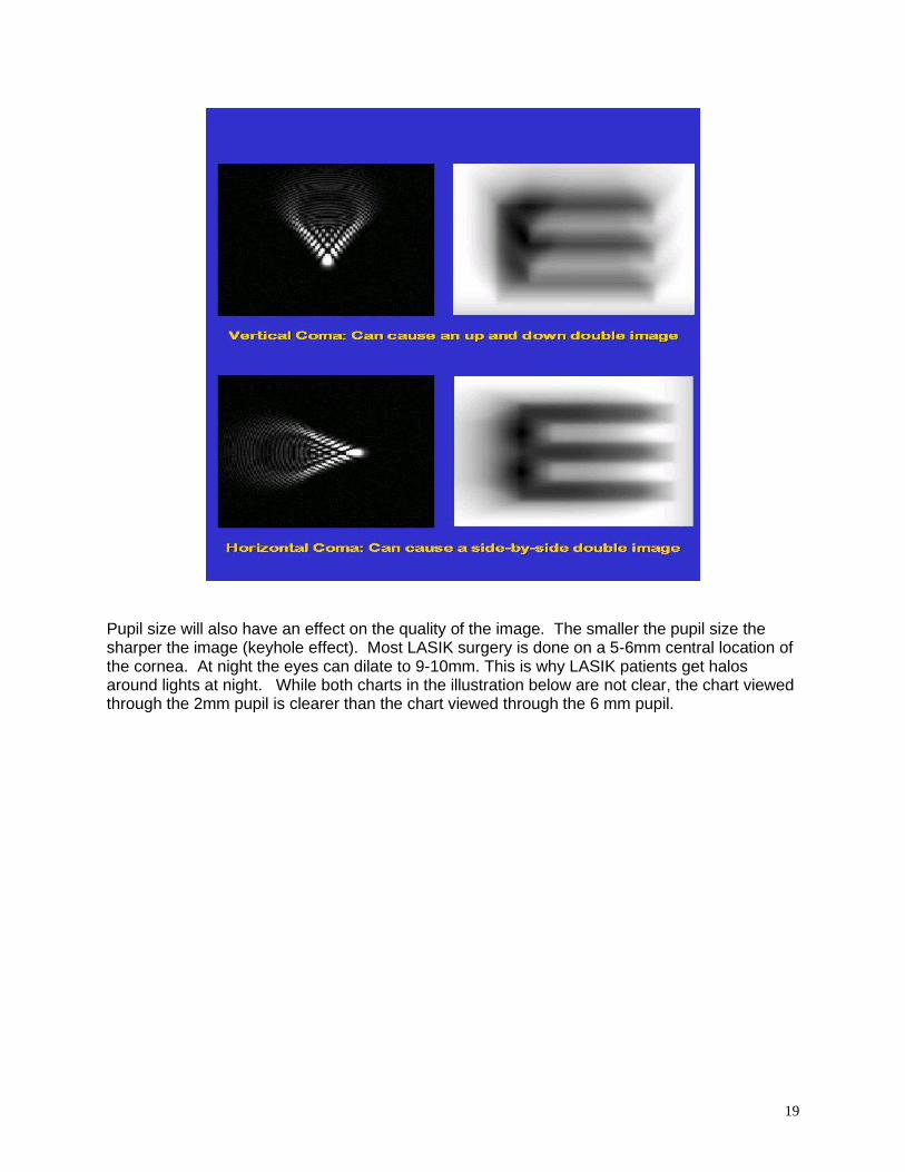
Coma is similar to spherical aberration. It applies to rays entering the lens at an angle. The focal point of the lens will vary the further away the ray hits the lens from the center. This can cause blurring of the image the further off-axis you go. A single lens can be chosen that will give no coma but only at a set distance from the object you wish to view. Coma occurs when oblique rays are refracted by a large-aperture optical system. Coma also affects the sharpness of image points.
This aberration is produced when a wide beam of light passes obliquely through a lens. Depending upon the angle of incidence the rays at the lower end of the beam will be bent to a different degree than the rays at the upper end. This will cause multiple focuses resulting in a blurred image as opposed to a singular sharp image. Like spherical aberration, the small aperture of the pupil is effective in limiting the aberrant peripheral rays. Consequently, the lens designer pays little attention to this particular aberration. Coma is an aberration which causes rays from the off-axis
point of light in the object plane to create a trailing comet-like blur directed away from the optic axis. A lens with considerable coma may produce a sharp image in the center of the field, but become increasingly blurred toward the edges. For a single lens, coma can be partially corrected by bending the lens. More complete correction can be achieved by using a combination of lenses symmetric about a central stop.

8
Marginal Astigmatism Astigmatism (marginal or oblique, depending upon the source) arises because if an object point is a distance from the optical axis, then the cone of rays from that point will strike the lens asymmetrically. In other words, the focal length of the lens will vary depending on where the beam of light hits the lens. This will lead to rays which are less parallel to the optical axis being focused differently from those which are parallel or almost parallel to the optical axis. This means that for some points the object will always be blurred, such as when you can focus the light for some of the rays you cannot focus it for all of them. Oblique astigmatism occurs when oblique rays are refracted by a small-aperture system and affects both sharpness of image points and image position. Even when oblique astigmatism is corrected, curvature of field is still present. Marginal Astigmatism is a real concern for the lens designer in that it involves narrow beams of parallel rays that strike the lens at an oblique angle. Unlike spherical or chromatic aberrations that are wide beam oriented, marginal astigmatism is more difficult to deal with in that the pupil does not limit the entrance of these narrow beams into the eye. When a narrow beam of parallel rays strikes a lens obliquely there is a tendency for the rays in the two opposite meridians to focus at different points. The distance between the two points of focus equals the degree of astigmatism created. The presence of this troublesome error led manufacturers to the development of corrected curve lens series. The idea here was that if the specific curvatures were controlled for specific corrections then marginal astigmatism could be controlled.

9
Curvature of Field For rays entering the lens on or near the optical axis (paraxial rays) the focal length of the lens, barring other aberrations, is constant. This leads to the problem of field curvature. As the distance from the center of the lens to the focus point is constant, then the image described by the lens is going to be a curved surface, not a flat one. Curvature of field manifests itself as a curved field surface for a flat object surface, and primarily affects image position. However, if the curvature of the image surface does not match that of the screen on which the image is formed, peripheral portions of the image are blurred.
In effect, curvature of field refers to the phenomenon that the eye viewing an object through a lens does not create a true reproduction of the object. A flat plane may have a tendency to look slightly curved. Curvature of field causes a planar object to project a curved (non-planar) image. It can be thought of as arising from a power error for rays at a large angle.
Those rays see thin lenses as having an effectively smaller diameter and an effectively higher power, forming the image of the off-axis points closer to the lens.

10
Distortion The focal length of the lens and hence the magnification/minification it causes varies over the surface of the lens (i.e. a ray hitting the lens at one spot will be focused more or less than that at another). This leads to distortion. Distortion is where parts of the image are magnified more or less than others. The most common distortions are barrel distortion, where the center of the image is bigger than the edges, and pincushion distortion, where the edges are bigger than the center. These can commonly be seen on television and computer monitors.
Distortion occurs when the magnification of an extended object varies with its distance from the optical axis. Distortion effects image shape and lateral position, but not image clarity. As the rays approach the edge the lens’ increasing magnification causes a distortion of the image. In the case of strong plus lens, the distortion is seen as an inward curvature, known as pincushion effect. In the case of a strong minus lens, the distortion is seen as an outward bulging, known as barrel distortion. Since each of you will visualize the description of distortions differently, the following illustrations may help you to understand the effects of distortion.

11

12
Lens Design and Aberrations Spectacle lenses suffer from both chromatic and monochromatic aberrations, but these aberrations are not all equally detrimental. Chromatic aberration is usually of little importance and is mostly ignored. Because the eye itself suffers from chromatic aberration, adaptation to this aberration very likely occurs. In addition, since the eye has its greatest sensitivity in the central portion of the visible spectrum, the extremes of the visible spectrum are relatively ineffective. Correction by a doublet results in both a heavy and an unattractive lens. Spherical aberration and coma are dependent on the size of the pupil. Because of the small effective pupil size at the center of rotation of the eye, these aberrations have relatively small effects in comparison with those of the other monochromatic aberrations. Distortion is a problem only for high-powered lenses and for larger amounts of anisometropia. The aberrations of greatest importance in the design of ophthalmic lenses are marginal/oblique astigmatism and curvature of field. Of the two, marginal/oblique astigmatism is usually of greater concern. It is mainly controlled by varying the curve design of the lens. In the process, curvature of field can also be modified. The lens designer may address the correction of either oblique astigmatism or curvature of field or may make a compromise between the two. Corrected-curve and best-form are adjectives used to describe a lens that is designed to control oblique astigmatism, curvature of image, or both, for oblique directions of view through the lens. A corrected-curve lens should have a rationale underlying its design. Two schools of thought exist in the design of ophthalmic lenses. One approach is to concentrate solely on the reduction of oblique astigmatism and to ignore the residual curvature of field error. Lenses made on the basis of this approach are known as point-focal lenses. On the-other hand, if primary consideration is given to reduction of curvature of field and allowing a residual amount of marginal/oblique astigmatism to exist, the lens design conforms to Percival's design philosophy. Percival, an English ophthalmologist, believed that the correction of curvature of field was more important than the elimination of oblique astigmatism. The lens designer is not faced with an arbitrary choice between the point-focal and the Percival principles of corrections. There is room for compromise. For example, a designer could set an arbitrary limit for the amount of the uncorrected oblique astigmatism and use this latitude to reduce the mean oblique (curvature of image) error. Today’s digitally-surfaced lenses will eliminate most of these lower order aberrations because of the combinations of the sophisticated curves that can be made on the front and back surfaces of lenses.

13
Magnification and Minification As compared to the image created on the retina of an emmetropic eye, plus lenses enlarge (magnify) the image, and minus lenses make the image smaller (minify). The greater the plus power, the greater the magnification. The greater the minus power, the greater the minification is. Although minification is a minor annoyance at best, magnification can be a very useful aberration when image enlargement is desirable, for example, with low vision lenses. The negative effects are of little consequence with lens powers less than 4 diopters. The exception would be the patient who has a plus lens correction for one eye and a minus lens correction for the other eye. A significant image size difference makes it difficult to maintain fusion and binocular vision. Magnification and minification are affected significantly by the vertex distance. The greater the vertex distance is, the greater the degree of magnification and minification. There is a practical application of the vertex effect upon magnification and minification. Image size changes are minimized when the vertex distance is zero. This is the case when the patient wears contact lenses. When checking the vision of a high myope above 8D, keep in mind the minification factor. Some of these patients cannot see the 20/20 letters because they make such a small image on the retina. For a -15.00D myope, 20/25 vision can be considered to be equivalent to 20/20 vision for an emmetrope. High Order Aberrations Clinically, higher order aberrations have a predictable effect on vision:
• Halos around bright lights at night - spherical aberration
• Comet-shaped ghost image (a tail) around the light - coma
• Triple images of the light - trefoil A refractionist measures the patient’s sphere power, cylinder power, and axis along with any prism imbalances. These are lower order aberrations. They are focusing errors. Higher order aberrations are also present. A refraction cannot detect these. Patients may have focusing aberrations which affect the eye’s ability to obtain sharp vision. If uncorrected, the patient’s vision is not as sharp and clear as it could be. Higher order aberrations reduce vision quality even in patients with 20/20 Snellen acuity. They reduce contrast, a significant part of quality vision A traditional refraction is conducted by:
• Snellen chart or similar chart is used
• Solid black letters on a white background
• Room lighting is dim
• Chart is projected with high contrast
14
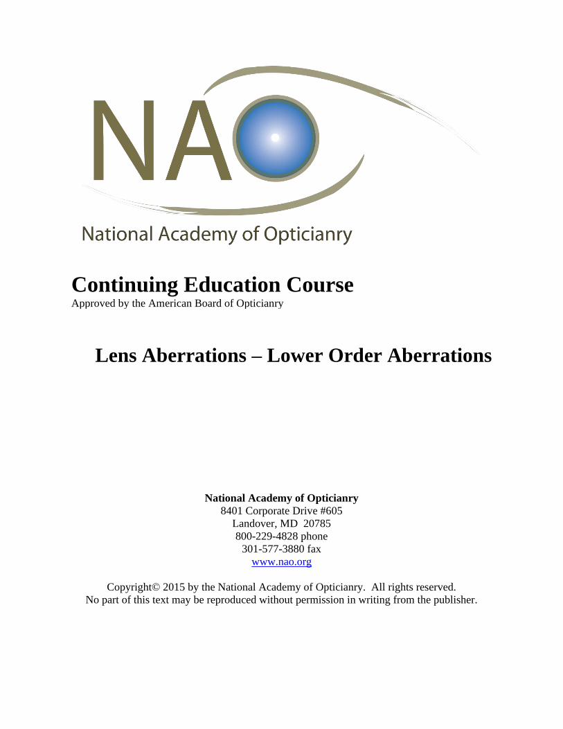
The patient is asked if they can read the letters, not how boldly they see them. Quality of vision is not the intent of the Snellen chart; it’s the quantity of vision. Higher order aberrations effects on patient’s vision:
• Reduce contrast
• Halos or comet-shaped flare around bright lights at night
• Ghost images
• Low contrast sensitivity
• Reduced color perception
• Reduced brightness
• Reduced depth perception
• Distorted imagery
• Glare sensitivity
• Poor low light and night driving vision
• Up to 20 percent of the eye’s vision is accountable to higher order aberrations
• Correcting these aberrations can significantly improve vision for most patients
• Research indicates the potential for vision is as high as 20/10. With sharp, crisp detail, high contrast, deep color saturation and more when both lower and higher order aberrations are corrected

15
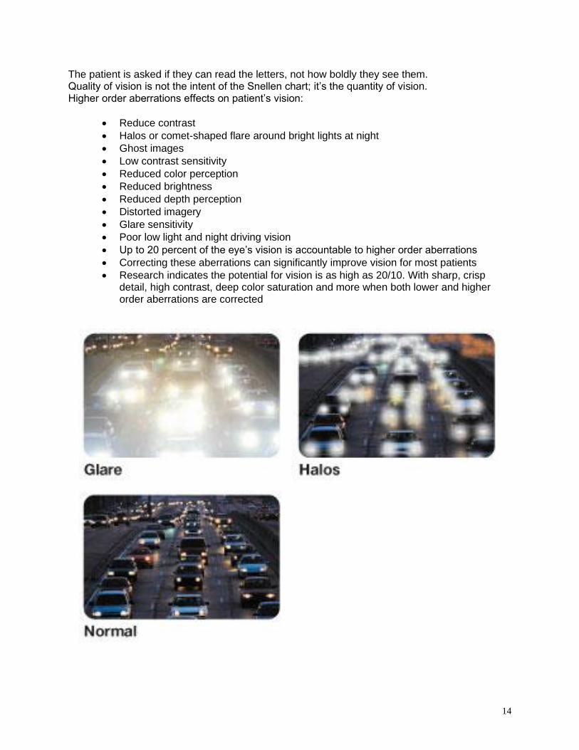
Aberrations are ranked using Zernike Polynomials. They are used to illustrate lower and higher order aberrations. The aberrations are arranged in levels or orders:
• First - tilt
• Second - defocus
• Middle image is hyperopic and myopic defocus
• Images to the right and left represent with-the-rule and against-the-rule astigmatism
• Third (central) - coma bordered by trefoil
• Fourth (central) - spherical aberration bordered by secondary astigmatism
• Levels five and six are usually less clinically significant
• The central aberrations of each line have the most impact on the quality of vision
These are the equations used to calculate the aberrations.
The following are contour pictures of the aberrations according to the Zernike Polynomials in lenses. Green represents little to no aberration and red represents a large presence of aberration. The pictures below would be comparable to viewing a corneal topography map. Red would denote the higher aberration. Blue would be lower aberration.
( )
( )
( )( )
( )( )( )
)4(c10 4 4 14
mAstigmatisSecondary )2cos(3410 2 4 13
Defocus ,Aberration Spherical 1665 0 4 12
mAstigmatisSecondary )2sin(3410 2- 4 11
)4(sin10 4- 4 10
)3(c8 3 3 9
axis- xalong Coma )cos(238 1 3 8
axis-y along Coma )sin(238 1- 3 7
)3(sin8 3- 3 6
90or 0at axis with mAstigmatis )2(c6 2 2 5
Defocus curvature, Field 123 0 2 4
45at axis with mAstigmatis )2(sin6 2- 2 3
Distortion direction,-in xTilt )cos(2 1 1 2
Distortion direction,-yin Tilt )(sin2 1- 1 1
Pistonor erm,Constant t 1 0 0 0
Meaning ,
frequencyorder mode
4
24
24
24
4
3
3
3
3
2
2
2
os
os
os
Zmnj m
n
−
+−
−
−
−
−
16

17
Above are the maps of the aberrations. Notice as the angle of viewing increases from the center, the greater the aberration. That is why most aberrations are considered off-axis viewing errors, as the patient isn’t using the optical center of the lens for viewing.
Examples of higher-order aberration maps from eyes with four different clinical conditions. The darker the color, the higher the aberration. (A) Dry eye, (B) Keratoconus, (C) LASIK surgery, (D) Cataract.

18
19
Pupil size will also have an effect on the quality of the image. The smaller the pupil size the sharper the image (keyhole effect). Most LASIK surgery is done on a 5-6mm central location of the cornea. At night the eyes can dilate to 9-10mm. This is why LASIK patients get halos around lights at night. While both charts in the illustration below are not clear, the chart viewed through the 2mm pupil is clearer than the chart viewed through the 6 mm pupil.

20
Aberrations are a given concern with spectacle lenses, but improvements in lens design such as free-form and wavefront technology will continue to improve the quality of lenses and therefore the patient’s quality of vision.

21
National Academy of Opticianry 8401 Corporate Drive #605
Landover, MD 20785 800-229-4828 phone
301-577-3880 fax www.nao.org
Copyright© 2015 by the National Academy of Opticianry. All rights reserved.
No part of this text may be reproduced without permission in writing from the publisher.