Embed Size (px)
Citation preview
Medical Terminology:
Basics
Before we can start in with some new and interesting medical terms, you need to
learn a few fundamentals of how medical terminology is constructed as a
language.
There are three basic parts to medical terms:
- a word root (usually the middle of the word and its central meaning),
- a prefix (comes at the beginning and usually identifies some subdivision or
part of the central meaning), and
- a suffix (comes at the end and modifies the central meaning as to what or
who is interacting with it or what is happening to it). An example may make
better sense.
WORD ROOT
therm = heat
hypothermia (less heat), thermometer (measuring heat)
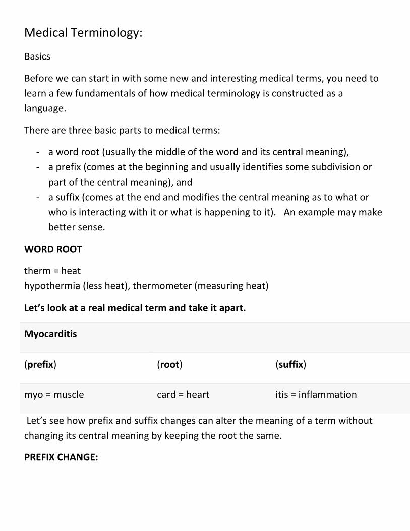
Let’s look at a real medical term and take it apart.
Myocarditis
(prefix) (root) (suffix)
myo = muscle card = heart itis = inflammation
Let’s see how prefix and suffix changes can alter the meaning of a term without
changing its central meaning by keeping the root the same.
PREFIX CHANGE:
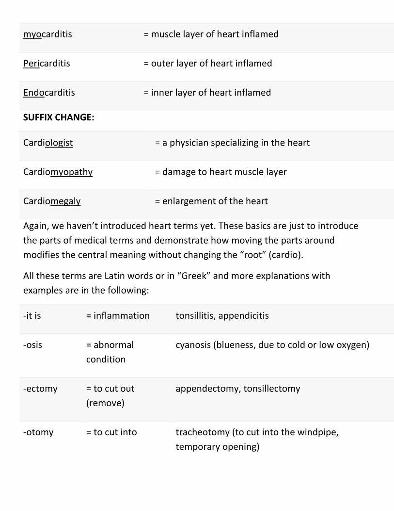
myocarditis = muscle layer of heart inflamed
Pericarditis = outer layer of heart inflamed
Endocarditis = inner layer of heart inflamed
SUFFIX CHANGE:
Cardiologist = a physician specializing in the heart
Cardiomyopathy = damage to heart muscle layer
Cardiomegaly = enlargement of the heart
Again, we haven’t introduced heart terms yet. These basics are just to introduce
the parts of medical terms and demonstrate how moving the parts around
modifies the central meaning without changing the “root” (cardio).
All these terms are Latin words or in “Greek” and more explanations with
examples are in the following:
-it is = inflammation tonsillitis, appendicitis
-osis = abnormal
condition
cyanosis (blueness, due to cold or low oxygen)
-ectomy = to cut out
(remove)
appendectomy, tonsillectomy
-otomy = to cut into tracheotomy (to cut into the windpipe,
temporary opening)

-ostomy = to make a
“mouth”
colostomy (to make a permanent opening in colon)
a/an = without, none anemia (literally no blood but means few red cells)
Micro = small microstomia (abnormally small mouth, see “stomy” in
colostomy above?)
Macro = large macrostomia (abnormally large mouth)
mega/ -
megaly
= enlarged megacolon (abnormally large colon = large intestine)
-scopy/ -
scopic
= to look, observe colonoscopy (look into colon)
Just a few more that you will see and hear over and over again.
-graphy/ -graph = recording
an image
mammography (imaging the breasts)
-gram = the image (X-ray) Mammogram
Whenever you see these endings, -graphy, -graph, -gram, they relate to recording
an image such as an X-ray, CT (computed tomography scan) or MRI
(magnetic resonance imaging) scan or a written recording with pen and moving
paper.
Mammography is the process of recording, i.e. the machine and procedure.
Mammogram is the image itself, the X-ray.
A recording of heart activity is called an electrocardiogram (ECG) using an
electrocardiograph (machine).
A recording of brain activity is an electroencephalogram and the medical
procedure and machine is called electroencephalography (machine).
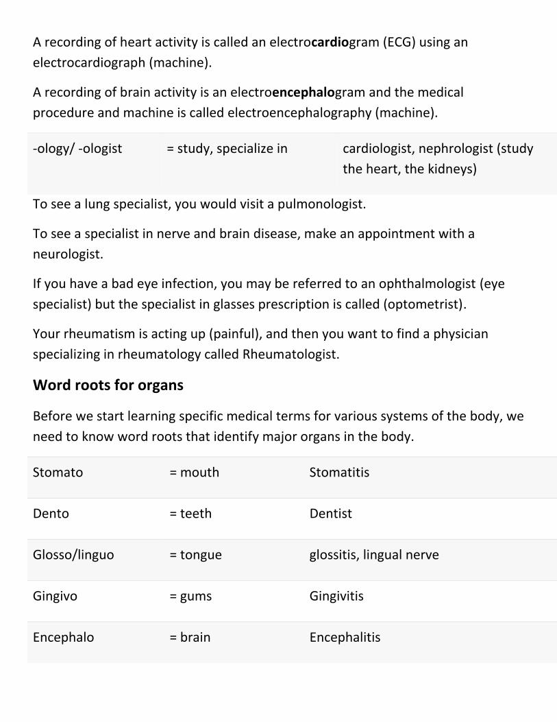
-ology/ -ologist = study, specialize in cardiologist, nephrologist (study
the heart, the kidneys)
To see a lung specialist, you would visit a pulmonologist.
To see a specialist in nerve and brain disease, make an appointment with a
neurologist.
If you have a bad eye infection, you may be referred to an ophthalmologist (eye
specialist) but the specialist in glasses prescription is called (optometrist).
Your rheumatism is acting up (painful), and then you want to find a physician
specializing in rheumatology called Rheumatologist.
Word roots for organs
Before we start learning specific medical terms for various systems of the body, we
need to know word roots that identify major organs in the body.
Stomato = mouth Stomatitis
Dento = teeth Dentist
Glosso/linguo = tongue glossitis, lingual nerve
Gingivo = gums Gingivitis
Encephalo = brain Encephalitis
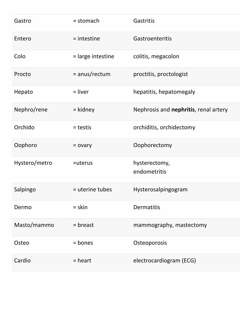
Gastro = stomach Gastritis
Entero = intestine Gastroenteritis
Colo = large intestine colitis, megacolon
Procto = anus/rectum proctitis, proctologist
Hepato = liver hepatitis, hepatomegaly
Nephro/rene = kidney Nephrosis and nephritis, renal artery
Orchido = testis orchiditis, orchidectomy
Oophoro = ovary Oophorectomy
Hystero/metro =uterus hysterectomy,
endometritis
Salpingo = uterine tubes Hysterosalpingogram
Dermo = skin Dermatitis
Masto/mammo = breast mammography, mastectomy
Osteo = bones Osteoporosis
Cardio = heart electrocardiogram (ECG)
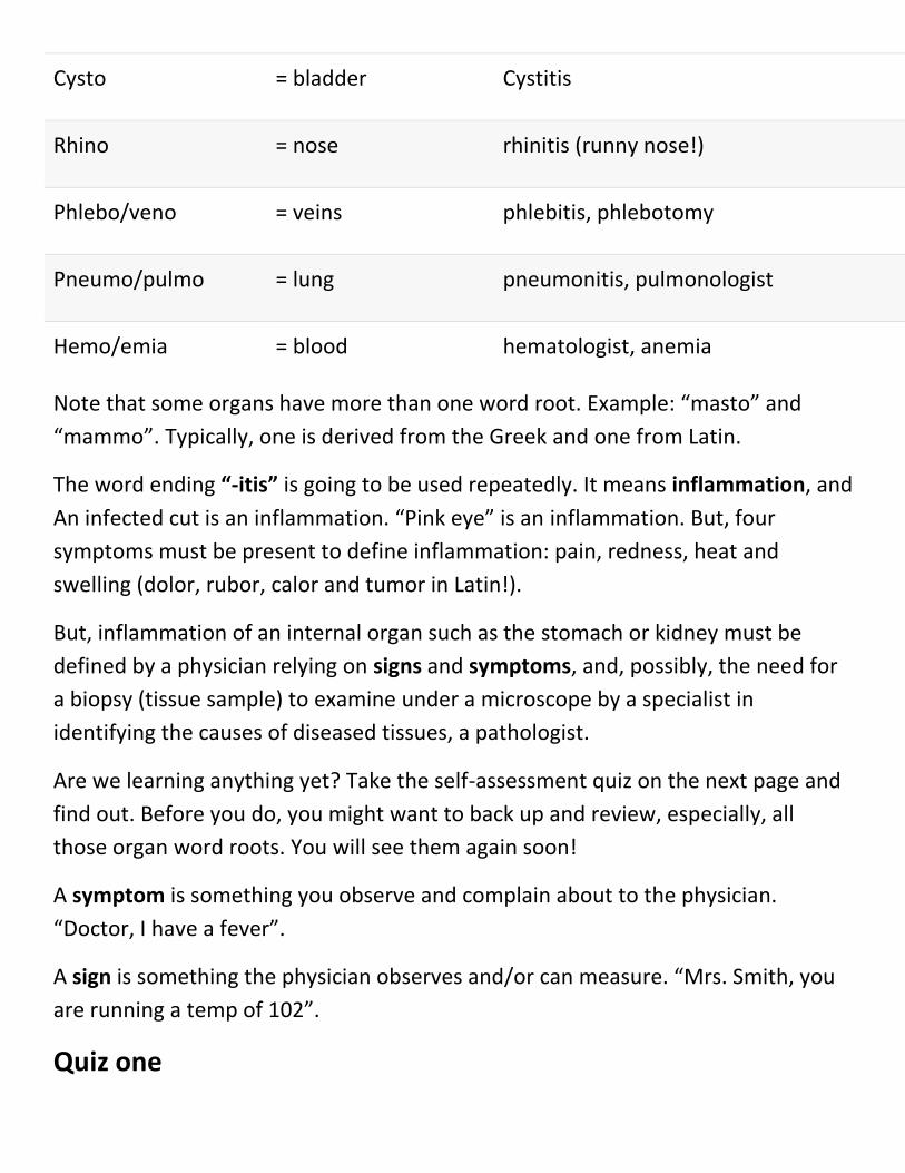
Cysto = bladder Cystitis
Rhino = nose rhinitis (runny nose!)
Phlebo/veno = veins phlebitis, phlebotomy
Pneumo/pulmo = lung pneumonitis, pulmonologist
Hemo/emia = blood hematologist, anemia
Note that some organs have more than one word root. Example: “masto” and
“mammo”. Typically, one is derived from the Greek and one from Latin.
The word ending “-itis” is going to be used repeatedly. It means inflammation, and
An infected cut is an inflammation. “Pink eye” is an inflammation. But, four
symptoms must be present to define inflammation: pain, redness, heat and
swelling (dolor, rubor, calor and tumor in Latin!).
But, inflammation of an internal organ such as the stomach or kidney must be
defined by a physician relying on signs and symptoms, and, possibly, the need for
a biopsy (tissue sample) to examine under a microscope by a specialist in
identifying the causes of diseased tissues, a pathologist.
Are we learning anything yet? Take the self-assessment quiz on the next page and
find out. Before you do, you might want to back up and review, especially, all
those organ word roots. You will see them again soon!
A symptom is something you observe and complain about to the physician.
“Doctor, I have a fever”.
A sign is something the physician observes and/or can measure. “Mrs. Smith, you
are running a temp of 102”.
Quiz one
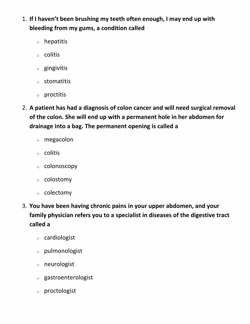
1. If I haven’t been brushing my teeth often enough, I may end up with
bleeding from my gums, a condition called
o hepatitis
o colitis
o gingivitis
o stomatitis
o proctitis
2. A patient has had a diagnosis of colon cancer and will need surgical removal
of the colon. She will end up with a permanent hole in her abdomen for
drainage into a bag. The permanent opening is called a
o megacolon
o colitis
o colonoscopy
o colostomy
o colectomy
3. You have been having chronic pains in your upper abdomen, and your
family physician refers you to a specialist in diseases of the digestive tract
called a
o cardiologist
o pulmonologist
o neurologist
o gastroenterologist
o proctologist
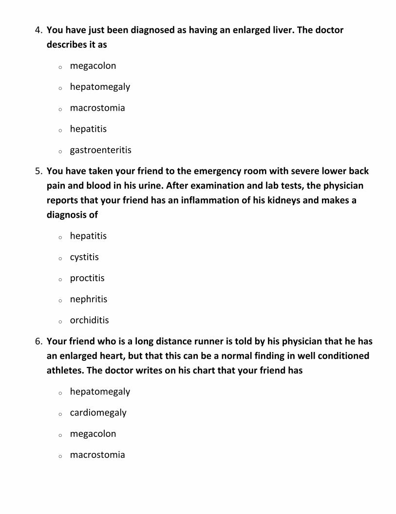
4. You have just been diagnosed as having an enlarged liver. The doctor
describes it as
o megacolon
o hepatomegaly
o macrostomia
o hepatitis
o gastroenteritis
5. You have taken your friend to the emergency room with severe lower back
pain and blood in his urine. After examination and lab tests, the physician
reports that your friend has an inflammation of his kidneys and makes a
diagnosis of
o hepatitis
o cystitis
o proctitis
o nephritis
o orchiditis
6. Your friend who is a long distance runner is told by his physician that he has
an enlarged heart, but that this can be a normal finding in well conditioned
athletes. The doctor writes on his chart that your friend has
o hepatomegaly
o cardiomegaly
o megacolon
o macrostomia
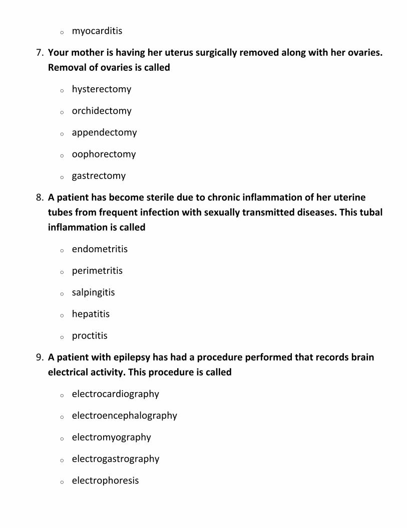
o myocarditis
7. Your mother is having her uterus surgically removed along with her ovaries.
Removal of ovaries is called
o hysterectomy
o orchidectomy
o appendectomy
o oophorectomy
o gastrectomy
8. A patient has become sterile due to chronic inflammation of her uterine
tubes from frequent infection with sexually transmitted diseases. This tubal
inflammation is called
o endometritis
o perimetritis
o salpingitis
o hepatitis
o proctitis
9. A patient with epilepsy has had a procedure performed that records brain
electrical activity. This procedure is called
o electrocardiography
o electroencephalography
o electromyography
o electrogastrography
o electrophoresis
10. A female patient has a special X-ray procedure of the breasts
performed. The X-ray image is called a
o mammoplasty
o mammoplasia
o mammography
o mastectomy
o mammogram
Odds and ends
It is advisable to cover a few more items that don’t fit into any particular system.
TECHNICOLOR TERMS
Leuk/o = white leukemia (overabundance of white blood cells)
melan/o = black melanoma (black tumor of the skin)
cyan/o = blue cyanosis (blueness may be due to cold or not enough oxygen in blood)
xanth/o = yellow xanthoma (yellow tumor)
TUMOR Terminology
Adding – oma (a swelling) to organ and tissue word roots names tumors.
It is Not all tumors are malignant (cancerous). Many are benign (not life-
threatening).
Aden/o = gland adenoma
Lip/o = fat lipoma
My/o = muscle myoma
Lymph/o = lymph tissue lymphoma
Carcin/o = malignant carcinoma
Osteo/o = bone osteoma
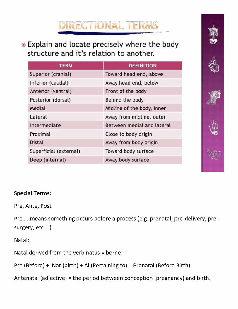
DIRECTIONS Terms:
Endo = within, inside
of
endoscopy (to inspect the inside of an organ or space with a lighted
instrument)
Peri = around perianal (around the anus)
Circum = around circumcise (cut around)
Retro = behind retrosternal (behind the breastbone)
Epi = upon, on top epidermis (the top or outermost layer of skin)
Trans = through transurethral (through the urinary exit duct)
Intra = within intravenous (inside the veins, e.g. IV fluids)
Sub = below subclavian (below the clavicle = collar bone)
In review, the word parts that make up medical terminology are prefixes, suffixes
and word roots. The most typical sequence is prefix, word root, suffix with the
word root being central but this is not always the case. In the interests of
simplification, putting a hyphen in front of a suffix to indicate it is added to the end
of a word, example, -itis. Prefixes and word roots which shown as freestanding
word parts.
Sometimes it is added a slash and a vowel, example, melan/o. These are called
combining forms which make it easier to attach to other word parts, and,
hopefully, making them easier to pronounce. Just so you know!
SIGNS AND SYMPTOMS – (EVER WONDER WHAT’S THE DIFFERENCE?)
As it mentioned earlier, symptom is something you observe and complain about to
the physician (doctor) such as fever, etching, chest pain, etc….
A sign is something the physician observes (noticeable) and/or can measure like
temp above or under 37 C, running nose, bleeding from the nose, blood pressure.
Circulatory system
CIRCULATORY SYSTEM TERMS
Cardi/o = heart Endocarditis, myocarditis, pericarditis (inflammation of the lining,
the muscle layer, the outer layer of the heart)
Brady/tachy =
slow/fast
Bradycardia (rate<60) tachycardia (rate>100)
Angi/o = vessel Angiography, angiogram (X-ray of artery)
Veno/phlebo = vein Venogram (X-ray of veins), phlebitis (inflammation of veins)
-stasis = to stop Hemostasis (to stop bleeding), hemostat (a clamp-like instrument)
-cyte = cell Erythrocytes, leukocytes (red, white blood cells)
Hem/o, -
emia
= blood Hypoxemia (low oxygen), hematosalpinx (blood in the uterine
tubes)
Directional Terms:
Special Terms:
Pre, Ante, Post
Pre…..means something occurs before a process (e.g. prenatal, pre-delivery, pre-
surgery, etc….)
Natal:
Natal derived from the verb natus = borne
Pre (Before) + Nat (birth) + Al (Pertaining to) = Prenatal (Before Birth)
Antenatal (adjective) = the period between conception (pregnancy) and birth.
Prenatal = The period during pregnancy before birth (delivery or labour).
labour (labor) = the process of delivering the baby and placenta.
This process includes 3 stages:
1. Birth delivery signs: the period lasts from the onset (beginning) of pains to
full contraction of the muscles of the womb (uterus)
and wall of the abdomen.
2. Widening (dilatation) of the cervix (neck of the uterus) and
delivery (expulsion) of the baby.
3. Delivery of the placenta.
Placenta: is a temporary connecting cord (tube) that joins the mother and fetus,
transferring oxygen and nutrients from the mother to the fetus and permitting the
release of CO2 and waste products from the fetus.
Neonate = a newborn child (infant), or one in its first 28 days
Newborn = a recently borne child (infant) or called neonate
Stillbirth: the death of a fetus at any time after the twentieth week of pregnancy.
Postnatal: the period immediately after birth.
A suitable subdivision is: early postnatal within 48 hours
of birth; delayed postnatal 2 to 7 days; late postnatal—1 to 4 weeks.
The following are health and medical definitions of terms:
Arms: An appendage in anatomy and in clinical trials.
Bilirubin: A yellow-orange compound that is produced by the breakdown of
hemoglobin from red blood cells.
Blood clot: A mass of coagulated blood. A blood clot can block a major blood
vessel, causing heart attack.
Blood count: The calculated number of white or red blood cells (WBCs or RBCs) in
a cubic millimeter
Brain: The portion of the central nervous system that is located within the skull.
Breastfeeding: Feeding a child human breast milk.
Otherwise formula feeding through prepared milk in a bottle.
Breathing: The process of respiration, during which air is inhaled into the lungs.
Cell: The basic structural and functional unit of any living organism.
Consultant: An individual to whom one refers for expert advice or services.
CPR: Cardiopulmonary resuscitation.
Fever: Although a fever technically is any body temperature above the normal of
98.6 F (37 C)
Heart: The muscle that pumps blood received from veins into arteries throughout
the body.
Hemolysis: The destruction of red blood cells which leads to the release of
hemoglobin from the blood
Infant: A young baby, from birth to 12 months of age.
Jaundice: Yellow staining of the skin and sclerae (the whites of the eyes) by
abnormally high blood levels of the bile pigment bilirubin.
Lactation: The process of milk production. Human milk is secreted by the
mammary glands
Kidney: One of a pair of organs located in the right and left side of the abdomen
and each a size of a closed fist. The kidneys remove waste products from the blood
and produce urine.
The right kidney sits slightly higher than the left one because of the position of the
liver. The kidneys are about 4 1/2 inches long and 2 1/2 inches wide.
Function: As blood flows through the kidneys, the kidneys filter waste products,
chemicals, and unneeded water from the blood. Urine collects in the middle of
each kidney, in an area called the Renal Pelvis: is the area (basin) at the center of
the kidney. Urine collects in the renal pelvis and is funneled into the ureter.
It then drains from the kidney through a long tube, the ureter, to the bladder,
where it is stored until elimination. The kidneys also make substances that help
control blood pressure and regulate the formation of red blood cells.
The nephron is the tiny filtering basic structure in your kidneys. Its chief function is
to regulate the concentration of water and soluble substances like sodium salts by
filtering the blood. Each of your kidneys contains more than a million tiny filtering
nephrons that help clean your blood.
The main parts of the kidney are:
1. Renal Capsule – outer membrane that surrounds the kidney; it is thin but
tough and fibrous
2. Renal Internal Structures:
a. Medulla (collecting chamber),
b. Renal pelvis (Basin to the urter inner region which receives urine through
the major calyces from medulla).
c. Urter: collects filtrate and urine from renal pelvis and takes it to the
bladder for urination
3. Renal Blood Vessels:
a. Renal Artery – branches off of the aorta bringing waste-filled blood about
20% into the kidney for filtering in the nephrons;
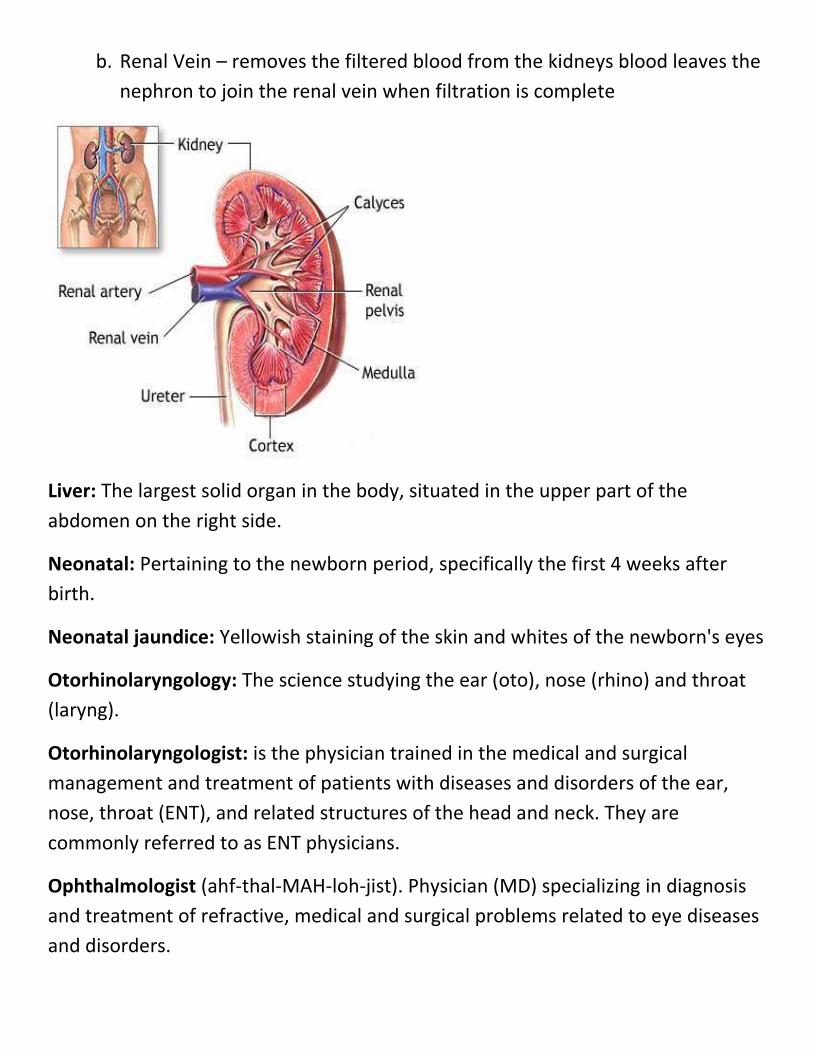
b. Renal Vein – removes the filtered blood from the kidneys blood leaves the
nephron to join the renal vein when filtration is complete
Liver: The largest solid organ in the body, situated in the upper part of the
abdomen on the right side.
Neonatal: Pertaining to the newborn period, specifically the first 4 weeks after
birth.
Neonatal jaundice: Yellowish staining of the skin and whites of the newborn's eyes
Otorhinolaryngology: The science studying the ear (oto), nose (rhino) and throat
(laryng).
Otorhinolaryngologist: is the physician trained in the medical and surgical
management and treatment of patients with diseases and disorders of the ear,
nose, throat (ENT), and related structures of the head and neck. They are
commonly referred to as ENT physicians.
Ophthalmologist (ahf-thal-MAH-loh-jist). Physician (MD) specializing in diagnosis
and treatment of refractive, medical and surgical problems related to eye diseases
and disorders.
Optic: issue related to vision.
Outpatient: A patient who is not hospitalized, but instead comes to a physician’s
office.
Phototherapy: Treatment with light. For example, a newborn with jaundice may
be put under sun or room light.
Physiologic: Something that is normal, that is due neither to anything pathologic
nor sign of infectious agent.
Polycythemia: Too many red blood cells. The opposite of anemia. polycythemia
formally exis...
Prognosis: The forecast (expectation) of the probable outcome or course of a
disease development in infected person.
Red blood cell (erhtrocytes): The blood cell that carries oxygen. Red cells contain
hemoglobin.
Red cells: Short for red blood cells, the oxygen/carbon dioxide carrying cells in
blood.
Reticulocyte: A young red blood cell that usually remains in the bone marrow with
only a f...
Reticulocyte count: The number of reticulocytes (young red blood cells) circulating
in blood stream.
Sclera: The tough white outer coat over the eyeball that covers approximately the
posterior of the ball
Serum: The clear liquid that can be separated from clotted blood. Serum differs
from plasma.
Syndrome: A combination of symptoms and signs that together represent a
disease process.
Transfusion: The transfer of blood or blood products from one person (the donor)
to a patient (recipient).
Endocrine Glands:
The endocrine system is the collection of glands of an organism
that secrete hormones directly into the circulatory system to be carried towards
distant target organs.
The major endocrine glands include the pineal gland, pituitary
gland, pancreas, ovaries, testes, thyroid gland, parathyroid gland, and adrenal
glands.
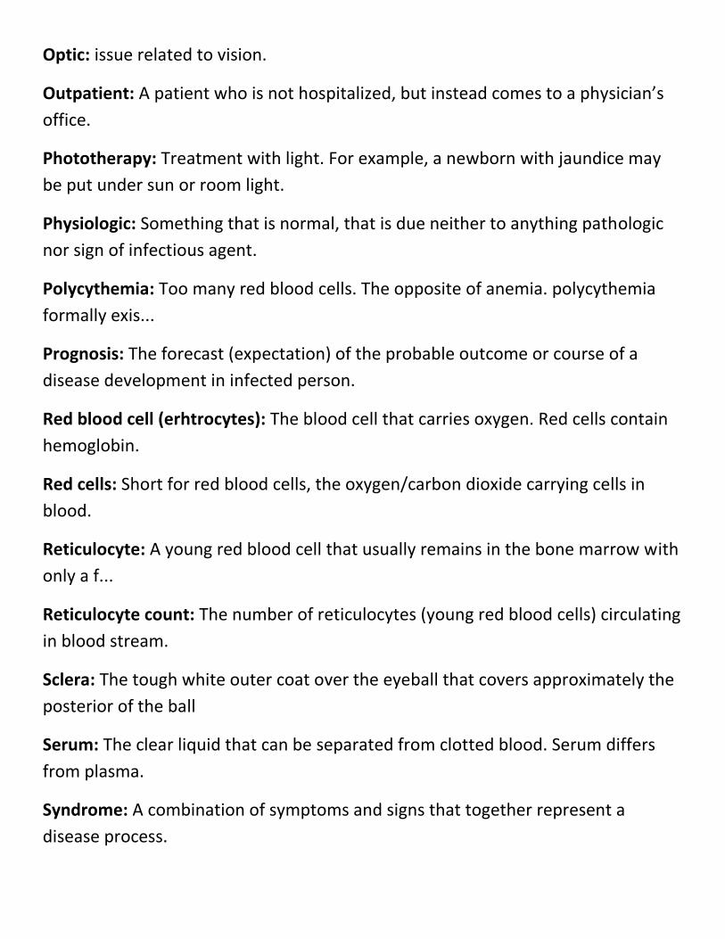
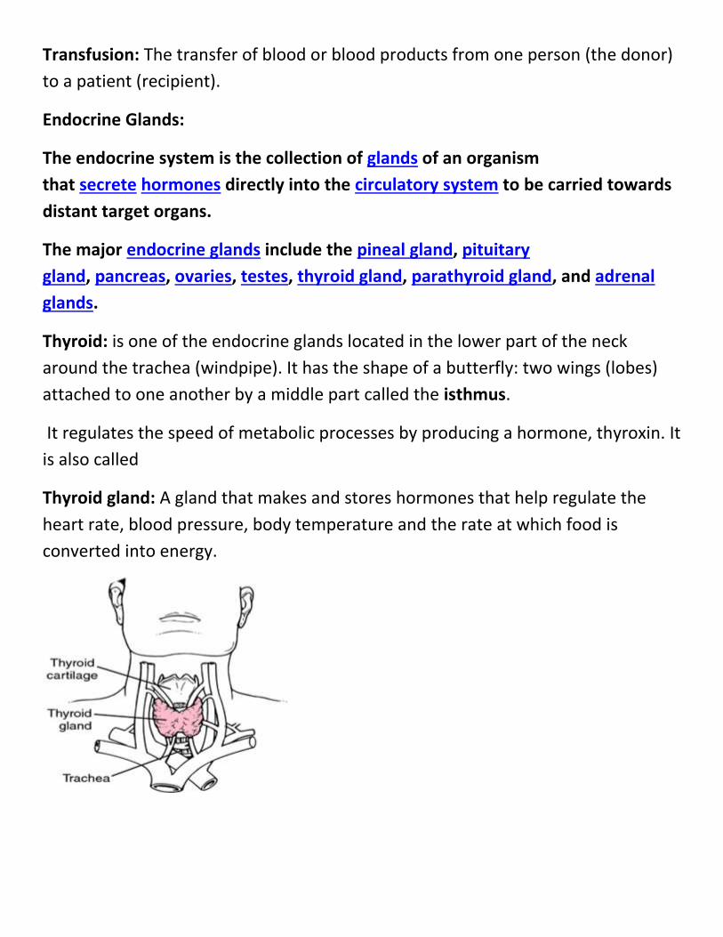
Thyroid: is one of the endocrine glands located in the lower part of the neck
around the trachea (windpipe). It has the shape of a butterfly: two wings (lobes)
attached to one another by a middle part called the isthmus.
It regulates the speed of metabolic processes by producing a hormone, thyroxin. It
is also called
Thyroid gland: A gland that makes and stores hormones that help regulate the
heart rate, blood pressure, body temperature and the rate at which food is
converted into energy.
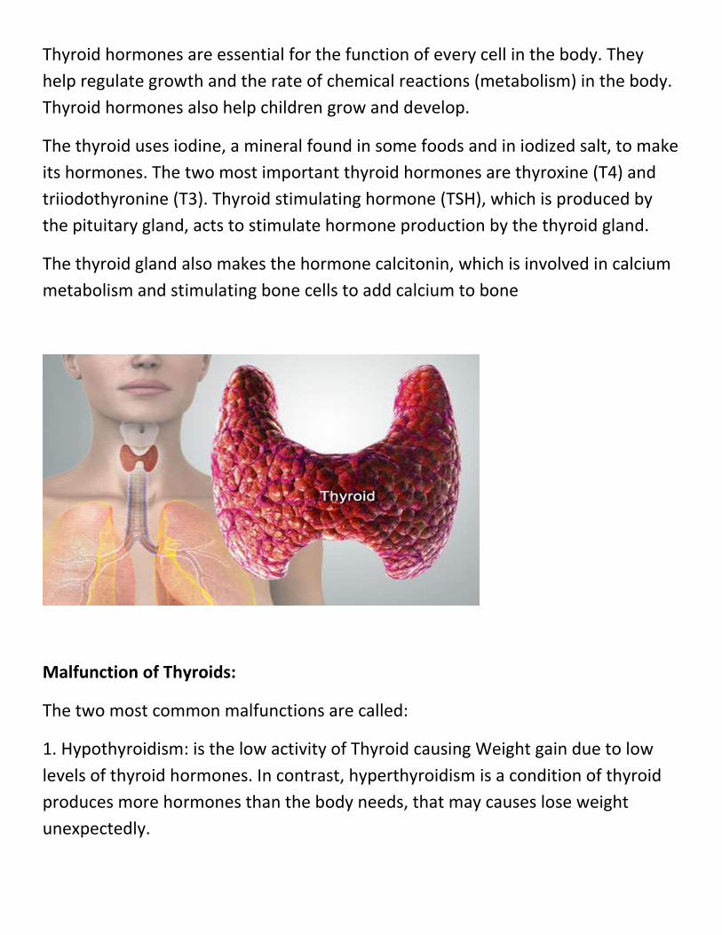
Thyroid hormones are essential for the function of every cell in the body. They
help regulate growth and the rate of chemical reactions (metabolism) in the body.
Thyroid hormones also help children grow and develop.
The thyroid uses iodine, a mineral found in some foods and in iodized salt, to make
its hormones. The two most important thyroid hormones are thyroxine (T4) and
triiodothyronine (T3). Thyroid stimulating hormone (TSH), which is produced by
the pituitary gland, acts to stimulate hormone production by the thyroid gland.
The thyroid gland also makes the hormone calcitonin, which is involved in calcium
metabolism and stimulating bone cells to add calcium to bone
Malfunction of Thyroids:
The two most common malfunctions are called:
1. Hypothyroidism: is the low activity of Thyroid causing Weight gain due to low
levels of thyroid hormones. In contrast, hyperthyroidism is a condition of thyroid
produces more hormones than the body needs, that may causes lose weight
unexpectedly.
2. Hyperthyroidism:
Hyperthyroidism is a medical condition that results from an excess of thyroid
hormone in the blood. Thyroid hormones control most metabolic processes in the
body. In cases of hyperthyroidism, these processes are often sped up causing
symptoms of hyperthyroidism, which will be discussed later in this slide show.
Thyrotoxicosis is an extreme version of hyperthyroidism that can cause severe or
life-threatening symptoms.
What Are Thyroid Hormones?
Thyroid hormones control most metabolic processes in the body. They are
produced by the thyroid gland located in the anterior (front) part of the neck.
These hormones affect many organs and biochemical systems in your body.
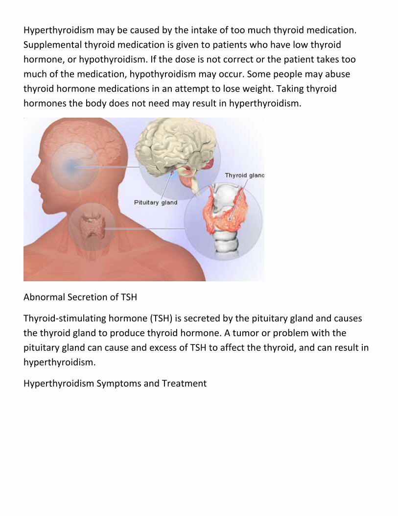
Thyroid Hormone Regulation – The Chain of Command
Complex biochemical processes in the body control the thyroid gland's production
of thyroid hormones. Two other glands – the hypothalamus and the pituitary gland
– both have a biochemical effect on the thyroid. The hypothalamus (the "master
gland") releases a hormone called thyrotropin-releasing hormone (TRH), which
sends a signal to the pituitary to release thyroid-stimulating hormone (TSH). In
turn, TSH sends a signal to the thyroid to release thyroid hormones. A problem
with any of these three glands may cause an over-production of thyroid hormone
and can cause hyperthyroidism.
What Causes Hyperthyroidism?
Some common causes of hyperthyroidism that will be covered in the following
slides include:
Graves' Disease
Functioning adenoma ("hot nodule") and Toxic Multinodular Goiter (TMNG)
Excessive intake of thyroid hormones
Abnormal secretion of TSH
Thyroiditis (inflammation of the thyroid gland)
Excessive iodine intake
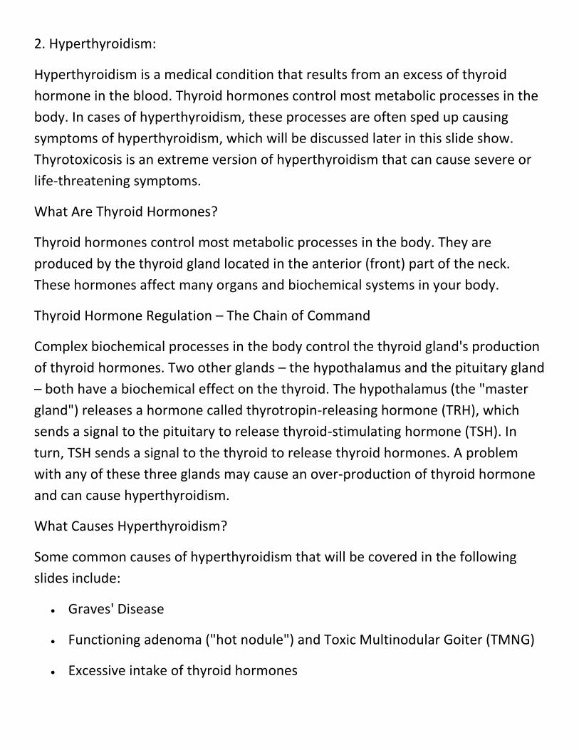
Graves' Disease
The most common cause of hyperthyroidism is Graves' disease. The thyroid gland
itself over-produces thyroid hormone and is no longer able to respond to the
pituitary and hypothalamus. Graves' disease is five times more common in women
and runs in families. Risk factors for Graves' disease include smoking, viral
illnesses, radiation to the neck, and medications. The condition is associated with
an eye disease called Graves' ophthalmopathy and skin lesions called dermopathy.
Diagnosis of Graves' disease is made by blood tests, and a nuclear medicine
thyroid scan.
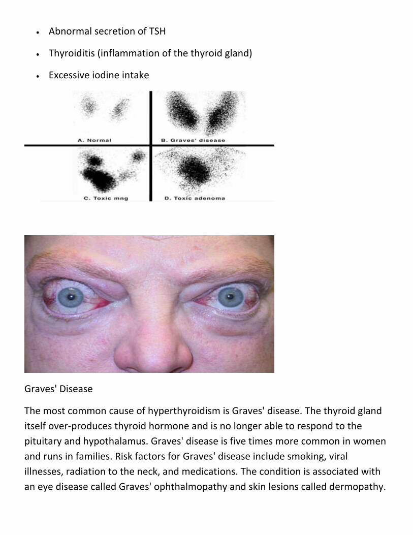
Functioning Adenoma and Toxic Multinodular Goiter
When the thyroid gland tissue overgrows, either in individual nodules (the
functioning adenoma) or in multiple clusters (multinodular goiter), it is generally
termed a "goiter." Goiters appear as large, swollen areas in the front of the neck
near the Adam's apple. These goiters may over-produce thyroid hormone, causing
symptoms of hyperthyroidism.
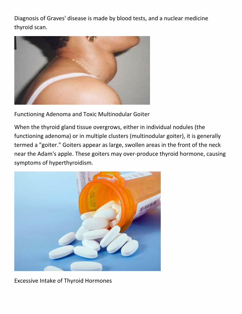
Excessive Intake of Thyroid Hormones
Hyperthyroidism may be caused by the intake of too much thyroid medication.
Supplemental thyroid medication is given to patients who have low thyroid
hormone, or hypothyroidism. If the dose is not correct or the patient takes too
much of the medication, hypothyroidism may occur. Some people may abuse
thyroid hormone medications in an attempt to lose weight. Taking thyroid
hormones the body does not need may result in hyperthyroidism.
Abnormal Secretion of TSH
Thyroid-stimulating hormone (TSH) is secreted by the pituitary gland and causes
the thyroid gland to produce thyroid hormone. A tumor or problem with the
pituitary gland can cause and excess of TSH to affect the thyroid, and can result in
hyperthyroidism.
Hyperthyroidism Symptoms and Treatment
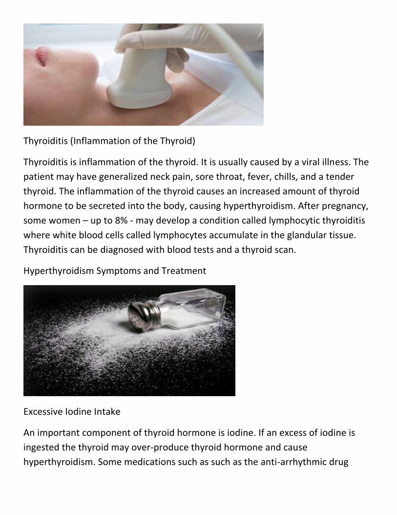
Thyroiditis (Inflammation of the Thyroid)
Thyroiditis is inflammation of the thyroid. It is usually caused by a viral illness. The
patient may have generalized neck pain, sore throat, fever, chills, and a tender
thyroid. The inflammation of the thyroid causes an increased amount of thyroid
hormone to be secreted into the body, causing hyperthyroidism. After pregnancy,
some women – up to 8% - may develop a condition called lymphocytic thyroiditis
where white blood cells called lymphocytes accumulate in the glandular tissue.
Thyroiditis can be diagnosed with blood tests and a thyroid scan.
Hyperthyroidism Symptoms and Treatment
Excessive Iodine Intake
An important component of thyroid hormone is iodine. If an excess of iodine is
ingested the thyroid may over-produce thyroid hormone and cause
hyperthyroidism. Some medications such as such as the anti-arrhythmic drug
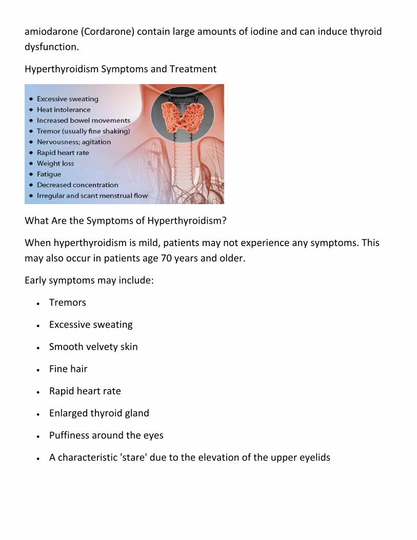
amiodarone (Cordarone) contain large amounts of iodine and can induce thyroid
dysfunction.
Hyperthyroidism Symptoms and Treatment
What Are the Symptoms of Hyperthyroidism?
When hyperthyroidism is mild, patients may not experience any symptoms. This
may also occur in patients age 70 years and older.
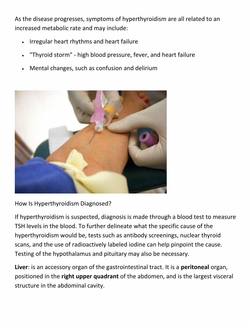
Early symptoms may include:
Tremors
Excessive sweating
Smooth velvety skin
Fine hair
Rapid heart rate
Enlarged thyroid gland
Puffiness around the eyes
A characteristic 'stare' due to the elevation of the upper eyelids
As the disease progresses, symptoms of hyperthyroidism are all related to an
increased metabolic rate and may include:
Irregular heart rhythms and heart failure
"Thyroid storm" - high blood pressure, fever, and heart failure
Mental changes, such as confusion and delirium
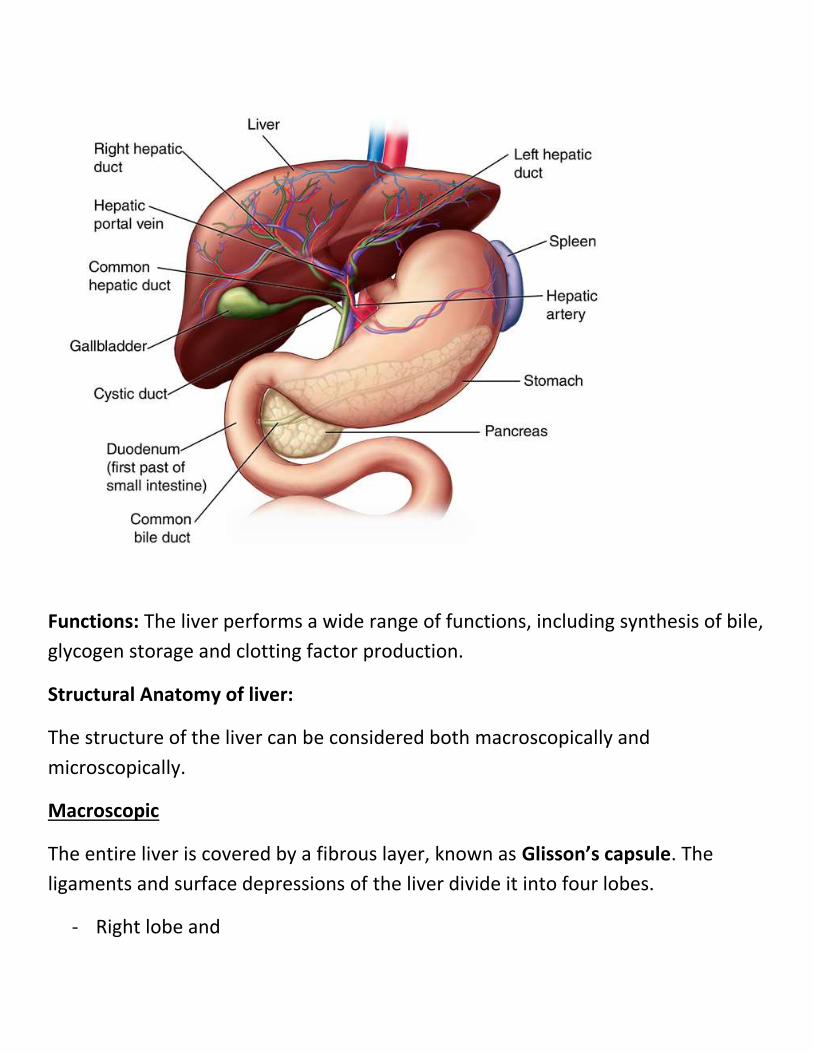
How Is Hyperthyroidism Diagnosed?
If hyperthyroidism is suspected, diagnosis is made through a blood test to measure
TSH levels in the blood. To further delineate what the specific cause of the
hyperthyroidism would be, tests such as antibody screenings, nuclear thyroid
scans, and the use of radioactively labeled iodine can help pinpoint the cause.
Testing of the hypothalamus and pituitary may also be necessary.
Liver: is an accessory organ of the gastrointestinal tract. It is a peritoneal organ,
positioned in the right upper quadrant of the abdomen, and is the largest visceral
structure in the abdominal cavity.
Functions: The liver performs a wide range of functions, including synthesis of bile,
glycogen storage and clotting factor production.
Structural Anatomy of liver:
The structure of the liver can be considered both macroscopically and
microscopically.
Macroscopic
The entire liver is covered by a fibrous layer, known as Glisson’s capsule. The
ligaments and surface depressions of the liver divide it into four lobes.
- Right lobe and
- Left lobe by the attachment of the falciform ligament (a fold of peritoneum
that attaches the liver to the anterior abdominal wall).
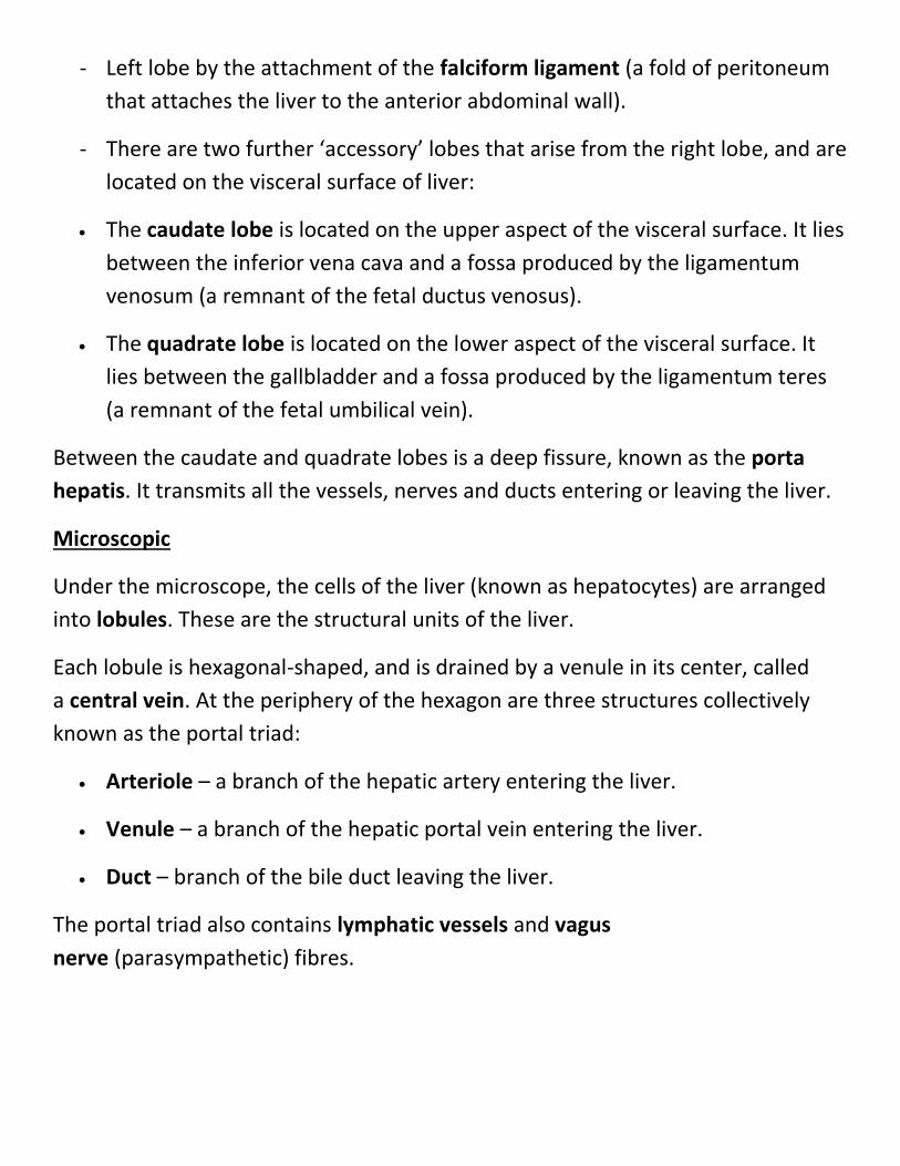
- There are two further ‘accessory’ lobes that arise from the right lobe, and are
located on the visceral surface of liver:
The caudate lobe is located on the upper aspect of the visceral surface. It lies
between the inferior vena cava and a fossa produced by the ligamentum
venosum (a remnant of the fetal ductus venosus).
The quadrate lobe is located on the lower aspect of the visceral surface. It
lies between the gallbladder and a fossa produced by the ligamentum teres
(a remnant of the fetal umbilical vein).
Between the caudate and quadrate lobes is a deep fissure, known as the porta
hepatis. It transmits all the vessels, nerves and ducts entering or leaving the liver.
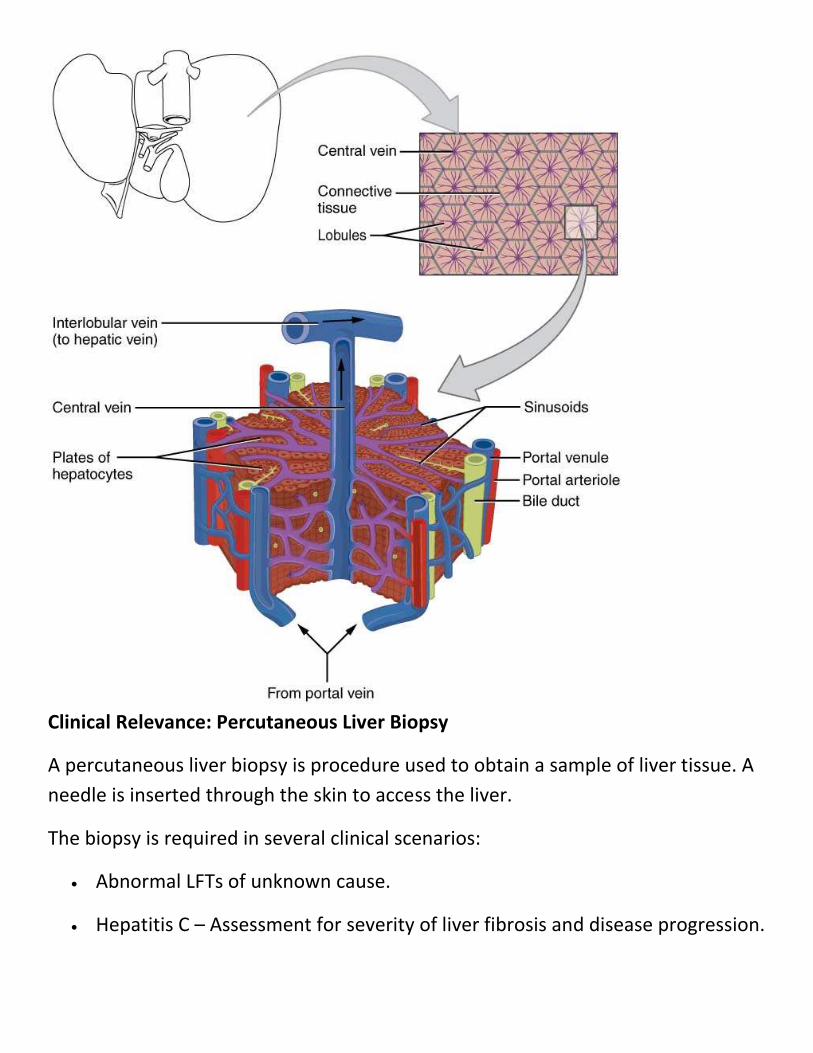
Microscopic
Under the microscope, the cells of the liver (known as hepatocytes) are arranged
into lobules. These are the structural units of the liver.
Each lobule is hexagonal-shaped, and is drained by a venule in its center, called
a central vein. At the periphery of the hexagon are three structures collectively
known as the portal triad:
Arteriole – a branch of the hepatic artery entering the liver.
Venule – a branch of the hepatic portal vein entering the liver.
Duct – branch of the bile duct leaving the liver.
The portal triad also contains lymphatic vessels and vagus
nerve (parasympathetic) fibres.
Clinical Relevance: Percutaneous Liver Biopsy
A percutaneous liver biopsy is procedure used to obtain a sample of liver tissue. A
needle is inserted through the skin to access the liver.
The biopsy is required in several clinical scenarios:
Abnormal LFTs of unknown cause.
Hepatitis C – Assessment for severity of liver fibrosis and disease progression.
Other liver conditions (such as Hereditary Haemochromatosis and
Autoimmune Hepatitis).
Following liver transplantation.
During the procedure, the liver is located via ultrasound. Local anaesthetic is
injected on the mid-axillary line, where on percussion there is dullness. The
patient is asked to deeply expire (avoiding damage to the lungs), and the needle
biopsy is taken during held expiration.
If a patient has abnormal clotting (a contraindication for the procedure),
a transvenous liver biopsy can be attempted. This involves cannulating the
internal jugular vein, and passing the biopsy needle through to the hepatic veins,
allowing for a biopsy sample to be taken.
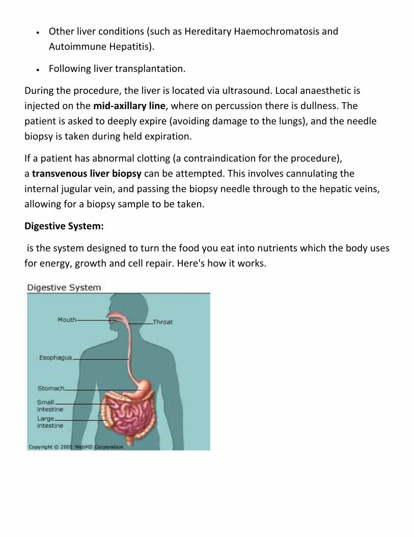
Digestive System:
is the system designed to turn the food you eat into nutrients which the body uses
for energy, growth and cell repair. Here's how it works.
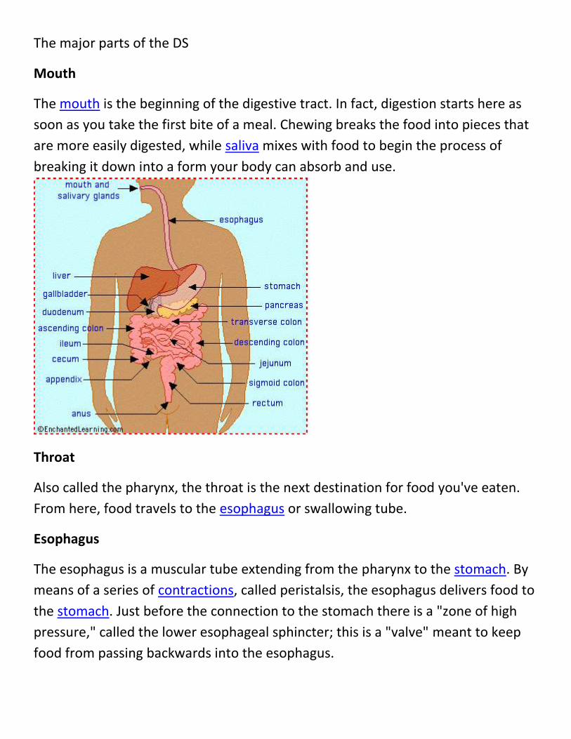
The major parts of the DS
Mouth
The mouth is the beginning of the digestive tract. In fact, digestion starts here as
soon as you take the first bite of a meal. Chewing breaks the food into pieces that
are more easily digested, while saliva mixes with food to begin the process of
breaking it down into a form your body can absorb and use.
Throat
Also called the pharynx, the throat is the next destination for food you've eaten.
From here, food travels to the esophagus or swallowing tube.
Esophagus
The esophagus is a muscular tube extending from the pharynx to the stomach. By
means of a series of contractions, called peristalsis, the esophagus delivers food to
the stomach. Just before the connection to the stomach there is a "zone of high
pressure," called the lower esophageal sphincter; this is a "valve" meant to keep
food from passing backwards into the esophagus.
Stomach
The stomach is a sac-like organ with strong muscular walls. In addition to holding
the food, it's also a mixer and grinder. The stomach secretes acid and powerful
enzymes that continue the process of breaking down the food. When it leaves the
stomach, food is the consistency of a liquid or paste. From there the food moves to
the small intestine.
Small Intestine
Made up of three segments, the duodenum, jejunum, and ileum, the small
intestine is a long tube loosely coiled in the abdomen (spread out, it would be
more than 20 feet long). The small intestine continues the process of breaking
down food by using enzymes released by the pancreas and bile from the liver. Bile
is a compound that aids in the digestion of fat and eliminates waste products from
the blood. Peristalsis (contractions) is also at work in this organ, moving food
through and mixing it up with digestive secretions. The duodenum is largely
responsible for continuing the process of breaking down food, with the jejunum
and ileum being mainly responsible for the absorption of nutrients into the
bloodstream.
Three organs play a pivotal role in helping the stomach and small intestine digest
food:
Pancreas
Among other functions, the oblong pancreas secretes enzymes into the small
intestine. These enzymes break down protein, fat, and carbohydrates from the
food we eat.
Liver
The liver has many functions, but two of its main functions within the digestive
system are to make and secrete bile, and to cleanse and purify the blood coming
from the small intestine containing the nutrients just absorbed.
Gallbladder
The gallbladder is a pear-shaped reservoir that sits just under the liver and stores
bile. Bile is made in the liver then if it needs to be stored travels to the gallbladder
through a channel called the cystic duct. During a meal, the gallbladder contracts,
sending bile to the small intestine.
Once the nutrients have been absorbed and the leftover liquid has passed through
the small intestine, what is left of the food you ate is handed over to the large
intestine, or colon.
Colon (Large Intestine)
The colon is a 5- to 6-foot-long muscular tube that connects the cecum (the first
part of the large intestine to the rectum (the last part of the large intestine). It is
made up of the cecum, the ascending (right) colon, the transverse (across) colon,
the descending (left) colon, and the sigmoid colon (so-called for its "S" shape; the
Greek letter for S is called the sigma), which connects to the rectum.
Stool, or waste left over from the digestive process, is passed through the colon by
means of peristalsis (contractions), first in a liquid state and ultimately in solid
form as the water is removed from the stool. A stool is stored in the sigmoid colon
until a "mass movement" empties it into the rectum once or twice a day. It
normally takes about 36 hours for stool to get through the colon. The stool itself is
mostly food debris and bacteria. These bacteria perform several useful functions,
such as synthesizing various vitamins, processing waste products and food
particles, and protecting against harmful bacteria. When the descending colon
becomes full of stool, or feces, it empties its contents into the rectum to begin the
process of elimination.
Rectum
The rectum (Latin for "straight") is an 8-inch chamber that connects the colon to
the anus. It is the rectum's job to receive stool from the colon, to let you know
there is stool to be evacuated, and to hold the stool until evacuation happens.
When anything (gas or stool) comes into the rectum, sensors send a message to
the brain. The brain then decides if the rectal contents can be released or not. If
they can, the sphincters (muscles) relax and the rectum contracts, expelling its
contents. If the contents cannot be expelled, the sphincters contract and the
rectum accommodates, so that the sensation temporarily goes away.
Anus
The anus is the last part of the digestive tract. It consists of the pelvic floor muscles
and the two anal sphincters (internal and external muscles). The lining of the
upper anus is specialized to detect rectal contents. It lets us know whether the
contents are liquid, gas, or solid. The pelvic floor muscle creates an angle between
the rectum and the anus that stops stool from coming out when it is not supposed
to. The anal sphincters provide fine control of stool. The internal sphincter keeps
us from going to the bathroom when we are asleep, or otherwise unaware of the
presence of stool. When we get an urge to go to the bathroom, we rely on our
external sphincter to keep the stool in until we can get to the toilet.
Stomach Acidity:
A typical adult human stomach will secrete about 1.5 liters of gastric acid daily.
There are three phases in the secretion of gastric acid which increase the secretion
rate in order to digest a meal:[3]
1. The cephalic phase: Thirty percent of the total gastric acid secretions to be
produced is stimulated by anticipation of eating and the smell or taste of
food. This signalling occurs from higher centres in the brain through
the vagus nerve (Cranial Nerve X). It activates parietal cells to release acid
and ECL cells to release histamine. The vagus nerve (CN X) also
releases gastrin releasing peptide onto G cells. Finally, it also
inhibits somatostatin release from D cells.[6]
2. The gastric phase: About fifty percent of the total acid for a meal is secreted
in this phase. Acid secretion is stimulated by distension of the stomach and
by amino acids present in the food.
3. The intestinal phase: The remaining 10% of acid is secreted
when chyme enters the small intestine, and is stimulated by small intestine
distension and by amino acids. The duodenal cells release entero-
oxyntin which acts on parietal cells without affecting gastrin


