Embed Size (px)
Citation preview
NeuroImage 86 (2014) 536–543
Contents lists available at ScienceDirect
NeuroImage
j ourna l homepage: www.e lsev ie r .com/ locate /yn img
The helmet head restraint system: A viable solution for resting statefMRI in awake monkeys
Fadila Hadj-Bouziane a,b,⁎,1, Elisabetta Monfardini a,b,c,1, Carole Guedj a,b,1, Gislène Gardechaux a,b,Clément Hynaux a,b, Alessandro Farnè a,b, Martine Meunier a,b
a INSERM, U1028, CNRS UMR5292, Lyon Neuroscience Research Center, ImpAct Team, Lyon F-69000, Franceb University UCBL Lyon 1, F-69000, Francec Institut de Médecine Environnementale, Paris F-75007, France
⁎ Corresponding author at: INSERM U1028, CNRSResearch Center, ImpAct Team, 16 avenue Doyen Lépine,
E-mail address: [email protected] (F. Ha1 These authors contributed equally to this work.
1053-8119/$ – see front matter © 2013 Elsevier Inc. All rihttp://dx.doi.org/10.1016/j.neuroimage.2013.09.068
a b s t r a c t
a r t i c l e i n f oArticle history:Accepted 25 September 2013Available online 8 October 2013
In monkey neuroimaging, head restraint is currently achieved via surgical implants. Eradicating such invasivehead restraint fromotherwise non-invasivemonkey studies could represent a substantial progress in terms of Re-duction and Refinement. Two non-invasive helmet-basedmethods are available but they are used exclusively bythe pioneering research groups who designed them. In the absence of independent replication, they have had lit-tle impact in replacing the surgical implants. Here, we built a modified version of the helmet system proposed bySrihasam et al. (2010 NeuroImage, 51(1), 267–73) and tested it for resting state fMRI in awake monkeys. Ex-tremely vulnerable tomotion artifacts, resting state fMRI represents a decisive test for non-invasive head restraintsystems.We compared twomonkeys restrainedwith the helmet to onemonkeywith a surgically implanted headpost using both a seed-based approach and an independent component analysis. Technically, the helmet systemproved relatively easy to develop. Scientifically, although it allowed more extensive movements than the headpost system, the helmet proved viable for resting state fMRI, in particular when combined with the independentcomponent analysis that deals more effectively with movement-related noise than the seed-based approach. Wealso discuss the pros and cons of such device in light of the European Union new 2013 regulation on non-humanprimate research and its firm Reduction and Refinement requests.
© 2013 Elsevier Inc. All rights reserved.
Introduction
Awake monkey fMRI emerged at the turn of the century (Dubowitzet al., 1998; Logothetis et al., 1999; Stefanacci et al., 1998) as a uniquetool to bridge the gap between human (generally whole-brain) andmonkey (often single-cell) data. From the start, it was reputed for itstechnical challenges, a major one being the control of motion duringscanning (Goense et al., 2010; Orban, 2002). In the European Union,awake monkey fMRI now faces an additional obstacle. Since January2013, a new regulation on the protection of animals used for scientificpurposes is being implemented. Muchmore restrictive than the preced-ing 1986Directive, the new2010/63/EUDirective requires strict compli-ancewith the Reduction and Refinement requirements in projects usingnon-human primates (http://eur-lex.europa.eu/LexUriServ/LexUriServ.do?uri=OJ:L:2010:276:0033:0079:en:PDF). How can we comply withthis new regulation in monkey fMRI experiments while minimizingmotion-induced artifacts?
Over the years, motion-induced signal loss and distortion have beensuccessfully minimized by improving MRI sequences, coils, as well as
UMR5292, Lyon Neuroscience69500 Bron, France.dj-Bouziane).
ghts reserved.
animals' training procedures (Chen et al., 2011; Stoewer et al., 2012).The one thing that remained unchanged is head restraint. In their vastmajority, the studies have stuck to surgically implanted head posts, a de-vice that decades of practice around the world have optimized. Brainmotion being the archenemy, it made sense not to tamper with a devicethat reliably controls head movements, most of the time without ad-verse reactions. Achieving head restraint without surgery would, how-ever, 1) allow re-use of the animal and 2) suppress a potential sourceof pain, suffering, and distress for the animal, thereby meeting boththe Reduction and the Refinement requirements of Directive 2010/63/EU. This prompted us to test a non-invasive way to limit head move-ments in our current awake monkey fMRI studies.
Two non-invasive head restraint methods are already available inmonkeys: a helmet filled with individually-molded foam developed byHowell et al. (2001) and a vacuum helmet maintained by mild suctiondeveloped by Srihasam et al. (2010). In both cases, the method's viabil-ity was confirmed in subsequent studies (see e.g. Murnane and Howell,2010; Srihasam et al., 2012). Yet, to the best of our knowledge, no teamaround theworld ever followed suit. Here, we provide the first indepen-dent support to the helmet approach pioneered by these two groups.Using a modified version of Srihasam et al.'s vacuum helmet, we com-pared the estimated movements and functional data obtained in twohelmet-restrained monkeys with those obtained in one head post-restrained monkey.
537F. Hadj-Bouziane et al. / NeuroImage 86 (2014) 536–543
Our objective was threefold, technical, scientific, and ethical. Thefirst aim was to test whether the system was technically easy to repro-duce. The second aimwas to determinewhether it was compatiblewithresting state fMRI (rs-fMRI). Increasingly used in both humans and ma-caques (e.g. Birn, 2012; Hutchison et al., 2011), rs-fMRI extracts slowsignal fluctuations (b0.1 Hz) from raw time series typically acquiredover long (6–15min) scanning epochs. Extremely vulnerable to physio-logical noise including movements, rs-fMRI thus represents a decisivetest for alternative non-invasive head restraint systems. Several analy-ses are available for resting state data; we used both a seed-based ap-proach and an independent component analysis. The third aim was toevaluate whether the systemhelps meet the Reduction and Refinementprinciples that are now central to monkey research in the EuropeanUnion.
Methods
Subjects
Three female rhesus monkeys (Macaca mulatta) participated in thestudy. Monkey CE (age: 10 yrs; weight: 5.5 kg) had a plastic head postsecured by plastic screws and bone cement. Monkeys CA (12 yrs;5.5kg) and CI (5yrs; 6kg)were trainedwith thehelmet system.MonkeyCA had a head post two years before the present experiment and wastested for 6months with this head-restraint system before her implantwas rejected. Training with the helmet took place 6months later, aftershe had fully recovered. Monkey CI never underwent surgery, and thehelmet was the first head-restraint system that she ever experienced.
Ethics
This study was carried out in strict accordance with Directive 2010/63/UE of the European Parliament and of the Council of 22 September2010 on the protection of animals used for scientific purposes that hasbeen transposed into French law in February 2013. In accordance withthe French transposition texts of Directive 2010/63/UE, the projectwas authorized by the French Ministry for Higher Education and Re-search (project no. 20-12-0401-005). This authorization was based onthe ethical evaluation of the French Committee on the Ethics of Experi-ments in Animals (C2EA) CELYNE registered at the national level asC2EA number 42.
Helmet system (Fig. 1)
Srihasam et al. (2010) used a “one size fits all” vacuum helmet andno body restraint, whereas Howell et al. (2001) used individually fittedfoam-filled helmets coupled with body restraint. We opted for an indi-vidually fitted vacuum helmet and no body restraint. The helmet was
Fig. 1. The vacuum helmet — left: 3D model of a helmet-restrained monkey showing the inner(blue). Middle: The inner piece of the helmet was individually customized to fit each animal's slaser sintering of PA12, a polyamide powder previously tested for magnetic susceptibility. Righ
composed of two pieces. The inner piece fitted each monkey's skullshape as reconstructed from anatomical MR images using the Brainsightneuronavigation system (http://www.rogue-resolutions.com/neuronavigation/brainsight). The outer piece served to anchor the system to themonkey chair and to connect it to the vacuum blower via a plastic tube(internal diameter: 25mm). It also held the surface coil. Three ports forsuction and two concentric grooves for silicone gaskets (diameter:3mm) applied a mild suction (approximately 2 psi) that, with the helpof a chin piece, held the animal's head. Nova Simulation (http://nova-simulations.com) created the digital model of the two-piece helmetwith the Catia software (http://www.3ds.com/products/catia/). The hel-met was then manufactured by E2R (http://www.e2r-rp.com) usinglaser sintering of PA12, a polyamide powder that we had previously test-ed for magnetic susceptibility.
Training and scanning procedures
The helmet-restrained monkeys underwent 2–5 training sessionsperweek for 6 (monkey CI) to 12 (monkey CA)months before the pres-ent data were collected. These intervals were used to optimize the hel-met and familiarized the animals to it. The monkey with the head postused for comparison was an extensively trained animal with morethan two years of experience with both mock and real scanners. As inHadj-Bouziane et al. (2008, 2012), the scanswere performed after injec-tion of an exogenous contrast agent (Feraheme®, 10mg/kg) into the sa-phenous vein to increase the contrast-to-noise ratio and optimize thelocalization of the fMRI signal. During the scans, the alert monkey wasplaced into the magnet bore, in the dark. Imaging data were collectedon a 1.5 T Siemens Magnetom (Siemens AG, Erlangen, Germany) scan-ner. Functional data from thewhole brainwere acquiredwith a gradientecho sequence lasting 13.33 min and a custom-made 9 cm receivesurface coil (Rapid Biomed) (TR = 2 s; TE = 27 ms; 2 × 2 × 3 mm;400 TRs). Typically, RSN studies use 6 minutes-scans as correlationvalues stabilize after 6 min (Van Dijk et al., 2010). We chose longer,13minutes-scans to test the new head-restraint system because theselonger scans result in greater intra-session reliability scores (Birnet al., 2013). Two thousand functional volumes were collected in mon-keys CE and CI (5 runs), 1600 in monkey CA (4 runs) during a singlescanning session. Monkeys were rewarded in between runs. Althoughhelmet-restrained animals showed no overt sign of pain, an analgesictreatment (paracetamol, 30mg/kg) was given at the end of each train-ing and scanning session as a precautionary measure.
Preprocessing
Data were analyzed using the AFNI software (Cox, 1996). The pre-processing pipeline included: despiking, motion correction, temporalfiltering to extract the spontaneous, slowly-fluctuating brain activity
(gray) and outer (black) piece of the helmet, the surface coil (white), and the coil holderkull shape as reconstructed from an anatomical scan. The helmet wasmanufactured usingt: Picture of an alert monkey placed in the helmet.
538 F. Hadj-Bouziane et al. / NeuroImage 86 (2014) 536–543
(0.01–0.1Hz), linear regression to remove nuisance variables (i.e. the sixparameter estimates for head motion, linear and quadratic trends, andthe CSF signal) and spatial smoothingwith a 3-mmFWHMGaussian ker-nel. Data from all runs were concatenated for each monkey. Data werethen aligned onto a MRI-based atlas of the rhesus macaque (McLarenet al., 2009), normalized to the Saleem and Logothetis (2007) stereotaxicatlas and projected onto an inflated version of a single macaque corticalsurface (F99, packaged with CARET).
Estimated brain motion and temporal signal-to-noise ratio (TSNR)
For each animal, we estimated six possible movements (three rota-tions and three translations) based on themotion correction algorithm.First, we computed cumulative motion normalized to the first volume.Then, we computed volume-to-volume displacements (i.e. derivatives;
A
B
Fig. 2. Estimated brain motion, expressed as cumulative motion normalized to the first volumemovements (in millimeters) are shown on the top panels and rotation movements (in degreeswith the helmet (middle: monkey CI and right: monkey CA). The x-axis is the image number; ostandard error to the mean (dotted lines).
Van Dijk et al., 2012; Power et al., 2012). In both cases, we calculatedone value per volume. For each animal and each run, we also quantifiedTSNR as the ratio of themean signal in each voxel to the temporal stan-dard deviation, using a whole brain mask. Estimated motion and TSNRwere compared across monkeys using one way ANOVAs with themon-keys as a factor followed by Tukey's HSD test for pairwise comparisons.
Identification of resting-state networks (RSNs)
Wefirst used a seed-based approach to uncover the attention fronto-parietal network in each monkey, which was among the first RSNs de-scribed in themonkey (Vincent et al., 2007). We selected 4 seed regions(diameter=2mm): the left and right LIP and FEF (Kagan et al., 2010).For each animal, we computed the correlation maps from each seed re-gion and averaged them to unveil the fronto-parietal functional
(A) or as volume-to-volume displacements (i.e. derivatives, B). In both cases, translation) in the bottom panels, for the monkey (CE) with the head post (left) and for the monkeysne image was taken every 2 s. Each scan consisted of 400 images. Mean (solid lines)+/−
0.4 0.7z-scores
0.5 0.9z-scores
0.6 1.2z-scores
q = 2.9 x 10 -8 q = 1.1 x 10⁻⁵ q = 11.6 x 10 -4
FEF
LIP
HEAD-POST HELMET
Co
rrel
atio
nm
aps
(See
d)
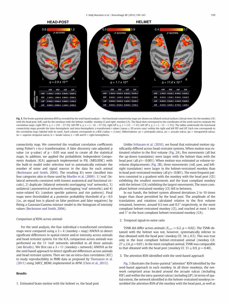
Fig. 3.The fronto-parietal attention RSNas revealed by the seed-based analysis— the functional connectivitymaps are shownon inflated cortical surfaces (dorsal view) for themonkey (CE)with the head post (left) and for themonkeys with the helmet (middle: monkey CI and right: monkey CA). The black dots correspond to the coordinates of the seeds used to compute thecorrelation maps (right FEF x, y, z= [16 −27 25]; left FEF x, y, z = [−16 −27 25]; right LIP x, y, z= [12 −1 31]; left LIP x, y, z = [−12 −1 31]). The tables underneath the functionalconnectivity maps provide the inter-hemispheric and intra-hemispheric z-transformed r values (mean± SD across runs) within the right and left FEF and LIP. Each row corresponds tothe correlation map (labeled with its seed). Each column corresponds to a ROI (radius = 2mm). Abbreviations: ps= principalis sulcus, ars = arcuate sulcus, ips = intraparietal sulcus,sts= superior temporal sulcus, ls= lunate sulcus, L= left and R= right hemispheres.
539F. Hadj-Bouziane et al. / NeuroImage 86 (2014) 536–543
connectivity map. We converted the resultant correlation coefficientsusing Fisher's r-to-z transformation. A false discovery rate-adjusted pvalue (or q-value) of p b 0.05 was used to create all the statisticalmaps. In addition, we applied the probabilistic Independent Compo-nents Analysis (ICA) approach implemented in FSL (MELODIC) withthe built-in model order selection tool to automatically estimate thenumber of noise and signal sources in the data for each animal(Beckmann and Smith, 2004). The resulting ICs were classified intofour categories akin to those used by Moeller et al. (2009): 1) ‘real’ (bi-lateral networks consistent with known anatomical and functional cir-cuits), 2) duplicate (bilateral networks overlapping ‘real’ networks), 3)unilateral (asymmetrical networks overlapping ‘real’ networks) and 4)noise-related ICs (random speckle patterns and rim patterns). Finalmaps were thresholded at a posterior probability threshold of p b 0.5(i.e., an equal loss is placed on false positives and false negatives) byfitting a Gaussian/Gamma mixture model to the histogram of intensityvalues (Beckmann and Smith, 2004).
Comparison of RSNs across animals
For the seed analysis, the four individual z-transformed correlationmaps were compared using a 3 × 4 (monkey ×map) ANOVA to detectsignificant differences in spatial extent and/or intensity across animalsand head restraint system. For the ICA, comparison across animals wasperformed on the 11 ‘real’ networks identified in all three animals(see Results). We first ran a 3× 11 (monkey×network) ANOVA as forthe seed based approach to detect significant differences across animalsand head restraint system. Then we ran an intra-class correlation (ICC)to study reproducibility in fMRI data as proposed by Thomason et al.(2011) using 3dICC_REML implemented in AFNI (Chen et al., 2013).
Results
1. Estimated brain motion with the helmet vs. the head post
Unlike Srihasam et al. (2010), we found that estimated motion sig-nificantly differed across head-restraint systems. When motion was es-timated relative to the first volume (Fig. 2A), five movements (all butthe up-down translation) were larger with the helmet than with thehead post (all p's b 0.001). When motion was estimated as volume-to-volume displacements (Fig. 2B), three movements (roll, yaw, and left-right translation) were larger in the helmet-restrained monkey thanin head post-restrainedmonkey (all p'sb0.001). Themost frequent pat-tern consisted in a gradient with the monkey with the head post (CE)exhibiting the smallest movements and the least compliant monkeywith the helmet (CA) exhibiting the largestmovements. Themore com-pliant helmet-restrained monkey (CI) fell in between.
In our hands, the helmet system allowed deviations 2 to 10 timeslarger than those permitted by the head post. The amplitude of thetranslations and rotations calculated relative to the first volumeremained, however, around 0.5mm and 0.5° respectively, in the mostcompliant helmet-restrained monkey (CI), and reached at most 1mmand 1° in the least compliant helmet-restrained monkey (CA).
2. Temporal signal-to-noise ratio
TSNR did differ across animals (F2,11=6.2, p=0.02). The TSNR ob-tained with the helmet was not, however, systematically inferior tothat obtained with the head post (monkey CE: 34±0.5). This was trueonly in the least compliant helmet-restrained animal (monkey CA:27±2.6, p=0.01). In themost compliant animal, TSNRwas comparableto that obtained with the head post (monkey CI: 31±0.9, p=0.40).
3. The attention RSN identified with the seed-based approach
Fig. 3 illustrates the fronto-parietal “attention” RSN identified by theseed-based approach in each monkey. In all three monkeys, the net-work comprised areas located around the arcuate sulcus (includingFEF) andwithin the intra-parietal sulcus (including LIP). In termsof spa-tial extent, the network identified in the helmet-restrainedmonkeys re-sembled the attention RSN of the monkey with the head post, as well as
Somatosensory (SS)
Peripheral visual (PV)
Fronto -parietal (FP)
Foveal visual (FV)
Somatomotor(SM)
Superior temporal sulcus (STS)
Executive (Ex)
Default-mode (DMN)
CORTICAL NETWORKS
Cerebellum (Cereb )
Basal ganglia (BG)
Thalamus (Th)
HEAD POST HELMET
SUB-CORTICAL NETWORKS
540 F. Hadj-Bouziane et al. / NeuroImage 86 (2014) 536–543
A B C
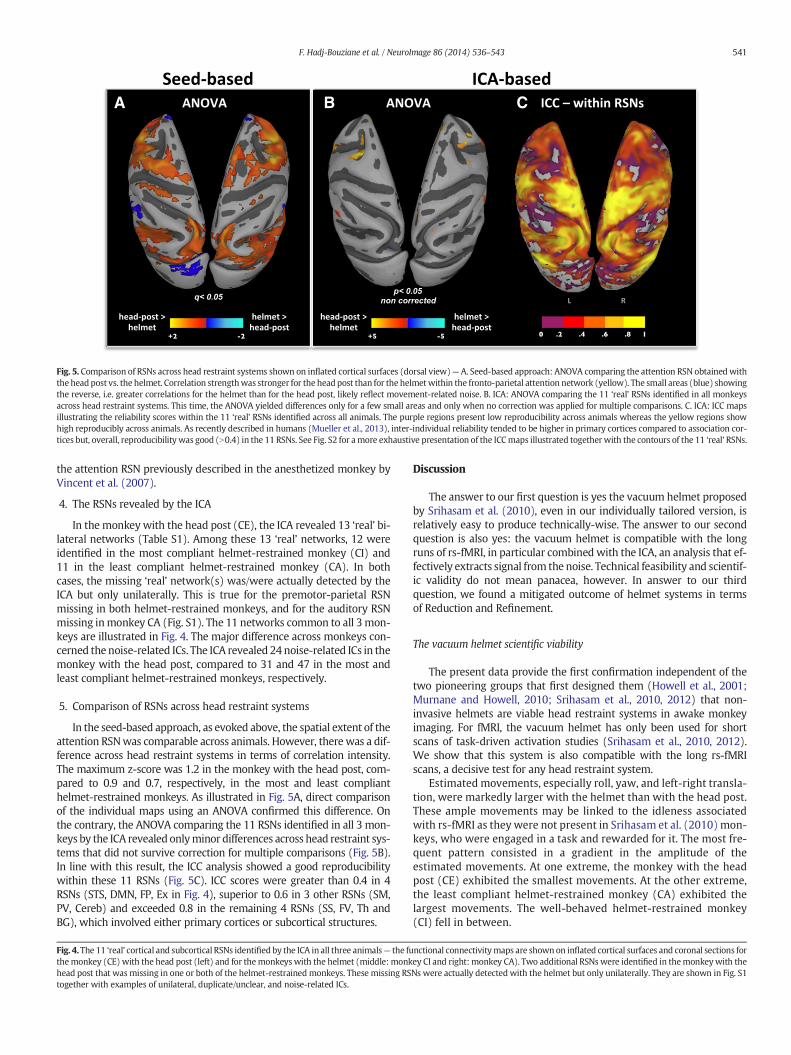
Fig. 5. Comparison of RSNs across head restraint systems shown on inflated cortical surfaces (dorsal view)— A. Seed-based approach: ANOVA comparing the attention RSN obtainedwiththe headpost vs. the helmet. Correlation strengthwas stronger for the headpost than for the helmetwithin the fronto-parietal attention network (yellow). The small areas (blue) showingthe reverse, i.e. greater correlations for the helmet than for the head post, likely reflect movement-related noise. B. ICA: ANOVA comparing the 11 ‘real’ RSNs identified in all monkeysacross head restraint systems. This time, the ANOVA yielded differences only for a few small areas and only when no correction was applied for multiple comparisons. C. ICA: ICC mapsillustrating the reliability scores within the 11 ‘real’ RSNs identified across all animals. The purple regions present low reproducibility across animals whereas the yellow regions showhigh reproducibly across animals. As recently described in humans (Mueller et al., 2013), inter-individual reliability tended to be higher in primary cortices compared to association cor-tices but, overall, reproducibility was good (N0.4) in the 11 RSNs. See Fig. S2 for amore exhaustive presentation of the ICCmaps illustrated together with the contours of the 11 ‘real’ RSNs.
541F. Hadj-Bouziane et al. / NeuroImage 86 (2014) 536–543
the attention RSN previously described in the anesthetized monkey byVincent et al. (2007).
4. The RSNs revealed by the ICA
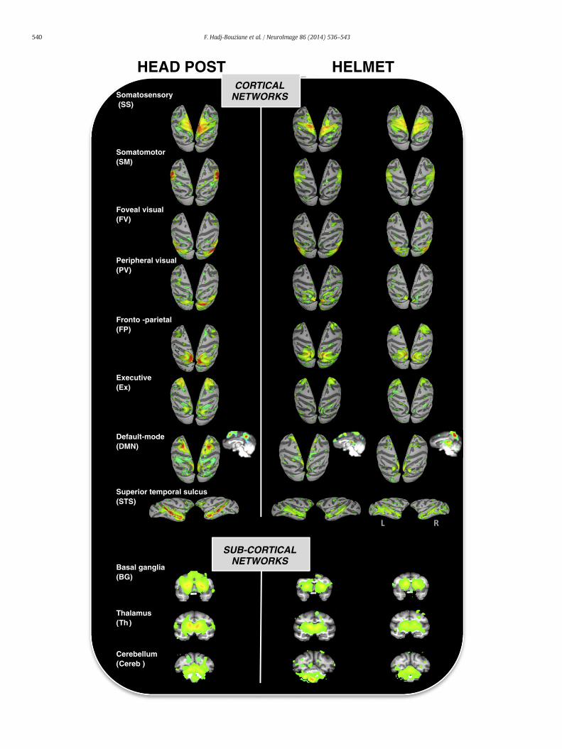
In the monkey with the head post (CE), the ICA revealed 13 ‘real’ bi-lateral networks (Table S1). Among these 13 ‘real’ networks, 12 wereidentified in the most compliant helmet-restrained monkey (CI) and11 in the least compliant helmet-restrained monkey (CA). In bothcases, the missing ‘real’ network(s) was/were actually detected by theICA but only unilaterally. This is true for the premotor-parietal RSNmissing in both helmet-restrained monkeys, and for the auditory RSNmissing inmonkey CA (Fig. S1). The 11 networks common to all 3mon-keys are illustrated in Fig. 4. The major difference across monkeys con-cerned the noise-related ICs. The ICA revealed 24 noise-related ICs in themonkey with the head post, compared to 31 and 47 in the most andleast compliant helmet-restrained monkeys, respectively.
5. Comparison of RSNs across head restraint systems
In the seed-based approach, as evoked above, the spatial extent of theattention RSNwas comparable across animals. However, there was a dif-ference across head restraint systems in terms of correlation intensity.The maximum z-score was 1.2 in the monkey with the head post, com-pared to 0.9 and 0.7, respectively, in the most and least complianthelmet-restrained monkeys. As illustrated in Fig. 5A, direct comparisonof the individual maps using an ANOVA confirmed this difference. Onthe contrary, the ANOVA comparing the 11 RSNs identified in all 3 mon-keys by the ICA revealed onlyminor differences across head restraint sys-tems that did not survive correction for multiple comparisons (Fig. 5B).In line with this result, the ICC analysis showed a good reproducibilitywithin these 11 RSNs (Fig. 5C). ICC scores were greater than 0.4 in 4RSNs (STS, DMN, FP, Ex in Fig. 4), superior to 0.6 in 3 other RSNs (SM,PV, Cereb) and exceeded 0.8 in the remaining 4 RSNs (SS, FV, Th andBG), which involved either primary cortices or subcortical structures.
Fig. 4.The 11 ‘real’ cortical and subcortical RSNs identifiedby the ICA in all three animals— the futhemonkey (CE) with the head post (left) and for themonkeys with the helmet (middle: monkhead post that was missing in one or both of the helmet-restrained monkeys. These missing RStogether with examples of unilateral, duplicate/unclear, and noise-related ICs.
Discussion
The answer to our first question is yes the vacuum helmet proposedby Srihasam et al. (2010), even in our individually tailored version, isrelatively easy to produce technically-wise. The answer to our secondquestion is also yes: the vacuum helmet is compatible with the longruns of rs-fMRI, in particular combinedwith the ICA, an analysis that ef-fectively extracts signal from the noise. Technical feasibility and scientif-ic validity do not mean panacea, however. In answer to our thirdquestion, we found a mitigated outcome of helmet systems in termsof Reduction and Refinement.
The vacuum helmet scientific viability
The present data provide the first confirmation independent of thetwo pioneering groups that first designed them (Howell et al., 2001;Murnane and Howell, 2010; Srihasam et al., 2010, 2012) that non-invasive helmets are viable head restraint systems in awake monkeyimaging. For fMRI, the vacuum helmet has only been used for shortscans of task-driven activation studies (Srihasam et al., 2010, 2012).We show that this system is also compatible with the long rs-fMRIscans, a decisive test for any head restraint system.
Estimated movements, especially roll, yaw, and left-right transla-tion, were markedly larger with the helmet than with the head post.These ample movements may be linked to the idleness associatedwith rs-fMRI as they were not present in Srihasam et al. (2010) mon-keys, who were engaged in a task and rewarded for it. The most fre-quent pattern consisted in a gradient in the amplitude of theestimated movements. At one extreme, the monkey with the headpost (CE) exhibited the smallest movements. At the other extreme,the least compliant helmet-restrained monkey (CA) exhibited thelargest movements. The well-behaved helmet-restrained monkey(CI) fell in between.
nctional connectivitymaps are shown on inflated cortical surfaces and coronal sections forey CI and right: monkey CA). Two additional RSNs were identified in themonkeywith theNs were actually detected with the helmet but only unilaterally. They are shown in Fig. S1
542 F. Hadj-Bouziane et al. / NeuroImage 86 (2014) 536–543
Not surprisingly, as motion induces more noise in the fMRI signal,the more extensive movements permitted by the helmet impacted thefMRI results. In the seed-based approach, the gradient observed acrossmonkeys for the amplitude of the movements was paralleled by a de-crease in the correlation strength. In the ICA approach, the movementgradient was paralleled by an increase in the number of noise-relatedICs, with the least compliant animal (CA) showing twice as manynoise-induced ICs as monkey CE (47 compared to 24). The other conse-quence of the movement-related noise in the ICA was that some RSNsidentified bilaterally with the head post were detected only unilaterallywith the helmet. This, however, occurred for only one RSN (out of 13) inmonkey CI (the most compliant) and a maximum of 2 RSNs in monkeyCA (the least compliant). Whether this represents a limit of the helmethead restraint system or can be optimized remains to be determined.
The important point is that despite these differences, we successful-ly identified RSNs in our two helmet-restrained monkeys. Using theseed-based approach, we uncovered the fronto-parietal attention RSN,originally identified in anesthetized monkeys (Hutchison et al., 2011;Vincent et al., 2007). Using the ICA approach, we identified in thethree animals, regardless of the head restraint system, 11 RSNs, compa-rable to those previously described in both anesthetized and awakemonkeys (Hutchison et al., 2011; Moeller et al., 2009). In addition, theICC analysis showed good inter-subject reproducibility for these 11RSNs. Interestingly, the degree of inter-individual reliability in RSNsfollowed a pattern similar as the one recently described in humans(Mueller et al., 2013). Inter-individual reliability tended to be higherin primary cortices compared to association cortices. Inter-individualvariability per se may therefore also have contributed to the variationsseen in our data in addition to the head restraint system.
Overall, both data analyses allowed us to reveal RSNs regardless ofthe head restraint system. The seed-based approach proved a valuabletool but ICA may be the method of choice with non-invasive head re-straint devices. ICA decomposes the signal and eliminates movement-related noise, thereby resulting in cleaner correlation maps.
Reduction and Refinement
We initiated the present study with the Reduction and Refinementrequirements put forward by the European Directive 2010/63/EU inmind. Across the EU, monkeys can be submitted to no more than twomoderate severity procedures such as surgical implants. Non-surgicalhead restraints downgrade brain imaging into a mild severity proce-dure. The vacuum helmet, like any non-surgical head restraint, there-fore constitutes a substantial progress in terms of compliance with theReduction requirement.
For the Refinement requirement, the picture is moremixed. The hel-met does spare the animals the per- and post-surgery pain associatedwith head implants but this may not be a major refinement given thecurrent, very efficient, management of surgical pain (Flecknell, 2008).Second, like Srihasam et al. (2010), we observed nobruising or swelling,as long as thepressurewas kept at ~2psi, but onemonkey did show skinrashes (that receded with rest and prednisolone spray). Third, we de-tected no overt pain signs after helmet removal but this is to be takenwith caution as headaches can easily go unnoticed in monkeys.
Helmet disadvantages and limits
The vacuumhelmet usedhere presents twodisadvantages. Cost-wise,the laser sintering technique is expensive; other solutions may becheaper, e.g. individually-molded foam cradles (Howell et al., 2001) orthermoplastic masks (see http://www.nc3rs.org.uk/researchportfolio/showcatportfolio.asp?id=319). In our hands, the system also provedtime-consuming, with one monkey still presenting ample movementsafter 12 months of training. Adding a restraining device (e.g. foam orbean bags) to limit bodymotion, as Howell et al. (2001) did, may reduce
the training period, and also limit the amplitude of movements duringscanning.
Helmet systems have two other important limits that areworth not-ing. First, they entailmore bite risks for the experimenters than the headpost due to the handling they require around the animal's mouth. Thisexplains why most imaging data published to date using helmet sys-tems in awake monkeys have been collected in females or juvenilemales (Srihasam et al., 2010, 2012; Howell et al., 2001; Murnane andHowell, 2010; present study). Second, helmet systems are adaptedwhen neuroimaging is used on its own, or together with other non-invasive procedures such as systemic drug injections. In our opinion,they are not suitable when intra-cerebral devices such as recordingelectrodes or injection needles are needed. In the latter cases, head rota-tions or translation of 0.5 to 1 mm or degree, or a rupture of the suctionseal, could lead to brain damage.
Conclusion
Head posts have been optimized thanks to decades of cooperationamong research teams involved in monkey research. Comparatively, lit-tle effort has been invested in developing non-invasive head restraintsystems. Here, we provide firm evidence that helmet restraint systemsare viable for rs-fMRI in awakemonkeys.Webelieve this evidence shouldencourage researchers worldwide to consider implementing helmet sys-tems in the future. Though challenging, this endeavor is worth pursuingin order to precisely delimit the extent to which these systems can im-prove the welfare of non-human primates involved in neuroimagingstudies without jeopardizing data quality.
Supplementary data to this article can be found online at http://dx.doi.org/10.1016/j.neuroimage.2013.09.068.
Acknowledgments
We thank Ivan Balansard and Emmanuel Procyk for performingmon-key CE's surgery, Valérie Gaveau, Jean Christophe Comte, DanielleIbarrola and Dominique Sappey-Marinier for their invaluable help withcollecting imaging data, Camille Lamy for help with processing anatomi-cal data, Cindy O'Mondays and Gang Chen for help with the statisticalanalyses, and Frederic Volland and Roméo Salemme for technical and en-gineering assistance. This work was funded by the French NationalResearchAgency (ANR)ANR-08-BLAN-0068-1 grant. Itwas also support-ed by ANR grant no. RPV08085CSA, the NEURODIS Foundation and theJames S. McDonnell Scholar award. It was performed within the frame-work of the LABEX CORTEX (ANR-11-LABX-0042) of Lyon Universitywithin the program “Investissements d'Avenir” (ANR-11-IDEX-0007)operated by the ANR.
Conflict of interest
The authors certify that no actual or potential conflict of interest inrelation to this article exists.
References
Beckmann, C.F., Smith, S.M., 2004. Probabilistic independent component analysis for func-tional magnetic resonance imaging. IEEE Trans. Med. Imaging 23, 137–152.
Birn, R.M., 2012. The role of physiological noise in resting-state functional connectivity.Neuroimage 62, 864–870.
Birn, R.M., Molloy, E.K., Patriat, R., Parker, T., Meier, T.B., Kirk, G.R., Nair, V.A., Meyerand,M.E., Prabhakaran, V., 2013. The effect of scan length on the reliability of resting-state fMRI connectivity estimates. Neuroimage 83C, 550–558.
Chen, G., Wang, F., Dillenburger, B.C., Friedman, R.M., Chen, L.M., Gore, J.C., Avison, M.J.,Roe, A.W., 2011. Functional magnetic resonance imaging of awake monkeys: someapproaches for improving imaging quality. Magn. Reson. 30, 36–47.
Chen, G., Saad, Z.S., Britton, J.C., Pine, D.S., Cox, R.W., 2013. Linear mixed-effects modelingapproach to FMRI group analysis. Neuroimage 73, 176–190.
Cox, R.W., 1996. AFNI: software for analysis and visualization of functional magnetic res-onance neuroimages. Comput. Biomed. Res. 29 (3), 162–173.
Dubowitz, D., Chen, D., Atkinson, D., 1998. Functional magnetic resonance imaging inma-caque cortex. Neuroreport 9, 2213–2218.
543F. Hadj-Bouziane et al. / NeuroImage 86 (2014) 536–543
Flecknell, P., 2008. Analgesia from a veterinary perspective. Br. J. Anaesth. 101, 121–124.Goense, J.B.M., Whittingstall, K., Logothetis, N.K., 2010. Functional magnetic resonance
imaging of awake behaving macaques. Methods 50, 178–188.Hadj-Bouziane, F., Bell, A.H., Knusten, T. a, Ungerleider, L.G., Tootell, R.B.H., 2008. Percep-
tion of emotional expressions is independent of face selectivity in monkey inferiortemporal cortex. Proc. Natl. Acad. Sci. U. S. A. 105, 5591–5596.
Hadj-Bouziane, F., Liu, N., Bell, A.H., Gothard, K.M., Luh,W.-M., Tootell, R.B.H., Murray, E.A.,Ungerleider, L.G., 2012. Amygdala lesions disrupt modulation of functional MRI activ-ity evoked by facial expression in the monkey inferior temporal cortex. Proc. Natl.Acad. Sci. U. S. A. 109, E3640–E3648.
Howell, L.L., Hoffman, J.M., Votaw, J.R., Landrum, A.M., Jordan, J.F., 2001. An apparatus andbehavioral training protocol to conduct positron emission tomography (PET) neuro-imaging in conscious rhesus monkeys. J. Neurosci. Methods 106, 161–169.
Hutchison, R.M., Leung, L.S., Mirsattari, S.M., Gati, J.S., Menon, R.S., Everling, S., 2011.Resting-state networks in the macaque at 7 T. Neuroimage 56, 1546–1555.
Kagan, I., Iyer, A., Lindner, A., Andersen, R.A., 2010. Space representation for eye move-ments is more contralateral in monkeys than in humans. Proc. Natl. Acad. Sci. U. S. A.107, 7933–7938.
Logothetis, N.K., Guggenberger, H., Peled, S., Pauls, J., 1999. Functional imaging of themonkey brain. Nat. Neurosci. 2, 555–562.
McLaren, D.G., Kosmatka, K.J., Oakes, T.R., Kroenke, C.D., Kohama, S.G., Matochik, J.A.,Ingram, D.K., Johnson, S.C., 2009. A population-average MRI-based atlas collectionof the rhesus macaque. Neuroimage 45, 52–59.
Moeller, S., Nallasamy, N., Tsao, D.Y., Freiwald, W.A., 2009. Functional connectivity ofthe macaque brain across stimulus and arousal states. J. Neurosci. 6;29 (18),5897–5909.
Mueller, S., Wang, D., Fox, M.D., Yeo, B.T., Sepulcre, J., Sabuncu, M.R., Shafee, R., Lu, J., Liu,H., 2013. Individual variability in functional connectivity architecture of the humanbrain. Neuron 77 (3), 586–595.
Murnane, K.S., Howell, L.L., 2010. Development of an apparatus and methodol-ogy for conducting functional magnetic resonance imaging (fMRI) with
pharmacological stimuli in conscious rhesus monkeys. J. Neurosci. Methods191, 11–20.
Orban, G., 2002. Functional MRI in the awake monkey: the missing link. J. Cogn. Neurosci.14, 965–969.
Power, J.D., Barnes, K.A., Snyder, A.Z., Schlaggar, B.L., Petersen, S.E., 2012. Spurious but sys-tematic correlations in functional connectivity MRI networks arise from subject mo-tion. Neuroimage 59, 2142–2154.
Saleem, K.S., Logothetis, N.K., 2007. A combined MRI and Histology Atlas of the RhesusMonkey Brain in Stereotaxic Coordinates. Academic, London.
Srihasam, K., Sullivan, K., Savage, T., Livingstone, M.S., 2010. Noninvasive functional MRIin alert monkeys. Neuroimage 51, 267–273.
Srihasam, K., Mandeville, J.B., Morocz, I.A., Sullivan, K.J., Livingstone,M.S., 2012. Behavioraland anatomical consequences of early versus late symbol training in macaques. Neu-ron 73, 608–619.
Stefanacci, L., Reber, P., Costanza, J., Wong, E., Buxton, R., Zola, S., Squire, L., Albright, T.,1998. fMRI of monkey visual cortex. Neuron 20, 1051–1057.
Stoewer, S., Goense, J., Keliris, G., Bartels, A., Logothetis, N.K., Duncan, J., Sigala, N., 2012.An analysis approach for high-field fMRI data from awake non-human primates.PLoS One 7, e29697.
Thomason, M.E., Dennis, E.L., Joshi, A.A., Joshi, S.H., Dinov, I.D., Chang, C., Henry, M.L.,Johnson, R.F., Thompson, P.M., Toga, A.W., Glover, G.H., Van Horn, J.D., Gotlib, I.H.,2011. Resting-state fMRI can reliably map neural networks in children. Neuroimage55, 165–175.
Van Dijk, K.R., Hedden, T., Venkataraman, A., Evans, K.C., Lazar, S.W., Buckner, R.L., 2010.Intrinsic functional connectivity as a tool for human connectomics: theory, proper-ties, and optimization. J. Neurophysiol. 103, 297–321.
Van Dijk, K.R., Sabuncu, M.R., Buckner, R.L., 2012. The influence of head motion on intrin-sic functional connectivity MRI. Neuroimage 59, 431–438.
Vincent, J.L., Patel, G.H., Fox, M.D., Snyder, A.Z., Baker, J.T., Van Essen, D.C., Zempel, J.M.,Snyder, L.H., Corbetta, M., Raichle, M.E., 2007. Intrinsic functional architecture in theanaesthetized monkey brain. Nature 447, 83–86.