Embed Size (px)
Citation preview
NATURE | VOL 415 | 21 FEBRUARY 2002 | www.nature.com 871
articles
The genome sequence ofSchizosaccharomyces pombeV. Wood1, R. Gwilliam1, M.-A. Rajandream1, M. Lyne1, R. Lyne1, A. Stewart2, J. Sgouros2, N. Peat3, J. Hayles3, S. Baker1, D. Basham1,S. Bowman1, K. Brooks1, D. Brown1, S. Brown1, T. Chillingworth1, C. Churcher1, M. Collins1, R. Connor1, A. Cronin1, P. Davis1, T. Feltwell1,A. Fraser1, S. Gentles1, A. Goble1, N. Hamlin1, D. Harris1, J. Hidalgo1, G. Hodgson1, S. Holroyd1, T. Hornsby1, S. Howarth1, E. J. Huckle1,S. Hunt1, K. Jagels1, K. James1, L. Jones1, M. Jones1, S. Leather1, S. McDonald1, J. McLean1, P. Mooney1, S. Moule1, K. Mungall1,L. Murphy1, D. Niblett1, C. Odell1, K. Oliver1, S. O'Neil1, D. Pearson1, M. A. Quail1, E. Rabbinowitsch1, K. Rutherford1, S. Rutter1,D. Saunders1, K. Seeger1, S. Sharp1, J. Skelton1, M. Simmonds1, R. Squares1, S. Squares1, K. Stevens1, K. Taylor1, R. G. Taylor1,A. Tivey1, S. Walsh1, T. Warren1, S. Whitehead1, J. Woodward1, G. Volckaert4, R. Aert4, J. Robben4, B. Grymonprez4, I. Weltjens4,E. Vanstreels4, M. Rieger5, M. SchaÈ fer5, S. MuÈ ller-Auer5, C. Gabel5, M. Fuchs5, C. Fritzc6, E. Holzer6, D. Moestl6, H. Hilbert6, K. Borzym7,I. Langer7, A. Beck7, H. Lehrach7, R. Reinhardt7, T. M. Pohl8, P. Eger8, W. Zimmermann9, H. Wedler9, R. Wambutt9, B. Purnelle10,A. Goffeau10, E. Cadieu11, S. DreÂano11, S. Gloux11, V. Lelaure11, S. Mottier11, F. Galibert11, S. J. Aves12, Z. Xiang12, C. Hunt12, K. Moore12,S. M. Hurst12, M. Lucas13, M. Rochet13, C. Gaillardin13, V. A. Tallada14,15, A. Garzon14,15, G. Thode14, R. R. Daga14,15, L. Cruzado14,J. Jimenez14,15, M. SaÂnchez16, F. del Rey16, J. Benito16, A. DomõÂnguez16, J. L. Revuelta16, S. Moreno16, J. Armstrong17, S. L. Forsburg18,L. Cerrutti1, T. Lowe19, W. R. McCombie20, I. Paulsen21, J. Potashkin22, G. V. Shpakovski23, D. Ussery24, B. G. Barrell1 & P. Nurse3
............................................................................................................................................................................................................................................................................
We have sequenced and annotated the genome of ®ssion yeast (Schizosaccharomyces pombe), which contains the smallestnumber of protein-coding genes yet recorded for a eukaryote: 4,824. The centromeres are between 35 and 110 kilobases (kb) andcontain related repeats including a highly conserved 1.8-kb element. Regions upstream of genes are longer than in budding yeast(Saccharomyces cerevisiae), possibly re¯ecting more-extended control regions. Some 43% of the genes contain introns, of whichthere are 4,730. Fifty genes have signi®cant similarity with human disease genes; half of these are cancer related. We identifyhighly conserved genes important for eukaryotic cell organization including those required for the cytoskeleton,compartmentation, cell-cycle control, proteolysis, protein phosphorylation and RNA splicing. These genes may have originatedwith the appearance of eukaryotic life. Few similarly conserved genes that are important for multicellular organization wereidenti®ed, suggesting that the transition from prokaryotes to eukaryotes required more new genes than did the transition fromunicellular to multicellular organization.
We report here the completion of the fully annotated genomesequence of the simple eukaryote Schizosaccharomyces pombe, a®ssion yeast. It becomes the sixth eukaryotic genome to besequenced, following Saccharomyces cerevisiae1, Caenorhabditiselegans2, Drosophila melanogaster3, Arabidopsis thaliana4 andHomo sapiens5,6. The entire sequence of the unique regions of thethree chromosomes is complete, with gaps in the centromericregions of about 40 kb, and about 260 kb in the telomeric regions.The completion of this sequence, the availability of sophisticatedresearch methodologies, and the expanding community working onS. pombe, will accelerate the use of S. pombe for functional andcomparative studies of eukaryotic cell processes.
Schizosaccharomyces pombe is a single-celled free living archias-comycete fungus sharing many features with cells of more compli-cated eukaryotes. From gene sequence comparisons andphylogenetic analyses, it has been suggested that ®ssion yeastdiverged from budding yeast around 330±420 million years (Myr)ago, and from Metazoa and plants around 1,000±1,200 Myr ago7,although a more recent estimate has put these times at 1,144 and1,600 Myr, respectively8. Some gene sequences are as equally
diverged between the two yeasts as they are from their humanhomologues, probably re¯ecting a more rapid evolution withinfungal lineages than in the Metazoa. S. pombe was ®rst described inthe 1890s and has been extensively studied since the 1950s9,10,resulting in the characterization of around 1,200 genes (http://www.genedb.org/pombe). The ease with which it can be geneticallymanipulated is second only to S. cerevisiae among eukaryotes and ithas served as an excellent model organism for the study of cell-cyclecontrol, mitosis and meiosis11, DNA repair and recombination12,and the checkpoint controls important for genome stability13.
The 13.8-Mb genome of S. pombe is distributed between chro-mosomes I (5.7 Mb), II (4.6 Mb) and III (3.5 Mb)14, together with a20-kb mitochondrial genome15. Tandem arrays of 100±120 repeatsof a 10.4-kb fragment containing the 5.8S, 18S and 25S ribosomalRNA genes account for around 1.1 Mb16. The three centromeres are35, 65 and 110 kb long for chromosomes I, II and III, respectively,totalling 0.2 Mb. This leaves about 12.5 Mb of unique sequence,similar in size to that of S. cerevisiae, and substantially smaller thanthose of the three other sequenced model eukaryotes, C. elegans(97 Mb), Arabidopsis (125 Mb) and Drosophila (137 Mb). All of the
1The Wellcome Trust Sanger Institute, The Wellcome Trust Genome Campus, Hinxton, Cambridge
CB10 1SA, UK. 2Cancer Research UK London Research Institute, Computational Genome AnalysisLaboratory, 44 Lincoln's Inn Fields, London WC2A 3PX, UK. 3Cancer Research UK London Research
Institute, Cell Cycle Laboratory, 44 Lincoln's Inn Fields, London, WC2A 3PX, UK. 4Katholieke
Universiteit Leuven, Faculty of Agricultural and Applied Biological Sciences, Laboratory of Gene
Technology, Kardinaal Mercierlaan 92 Blok F, B-3001 Leuven, Belgium. 5Genotype GmbH, Molecular
Biology and Biotech Research, Angelhofweg 39, D-69259 Wilhelmsfeld, Germany. 6QIAGEN GmbH, MaxVolmer Str. 4, D-40724 Hilden, Germany. 7Max-Planck-Institut fuÈr molekulare Genetik, Ihnestrasse 73,
D-14195 Berlin, Germany. 8GATC Biotech AG, Jakob-Stadler-Platz 7, D-78467 Konstanz, Germany.9AGOWA GmbH, Glienicker Weg 185, D-12489 Berlin, Germany. 10Universite de Louvain, Unite de
Biochimie Physiologique, Place Croix du Sud 2-20, B1348 Louvain-la-Neuve, Belgium. 11UMR 6061
CNRS Genetique et developpement, Faculte de MeÂdecine, 2 avenue du Professeur LeÂon Bernard, F-35043Rennes Cedex, France. 12University of Exeter, School of Biological Sciences, Washington Singer Labora-
tories, Perry Road, Exeter EX4 4QG, UK. 13GeÂneÂtique MoleÂculaire et Cellulaire, CNRS URA1925 INRA
UMR216, Institut National Agronomique Paris-Grignon, 78850 Thiverval Grignon, France. 14Departa-
mento de Genetica, Facultad de Ciencias, Universidad de Malaga, Spain. 15Laboratorio Andaluz deBiologia, Universidad Pablo de Olavide, Sevilla, Spain. 16Instituto de MicrobiologõÂa y BioquõÂmica,
Departamento de MicrobiologõÂa y GeneÂtica, CSIC/Universidad de Salamanca, Edi®cio Departamental,
Campus Miguel de Unamuno, 37007 Salamanca, Spain. 17University of Sussex, Falmer, Brighton BN1
9QG, UK. 18Molecular & Cell Biology Laboratory, Salk Institute for Biological Studies, 10010 North
Torrey Pines Road, La Jolla, California 92037-1099, USA. 19Stanford University, Stanford UniversitySchool of Medicine, Department of Genetics, CCSR Room 2255b, 269 Campus Drive, Stanford, California
94305, USA. 20Cold Spring Harbor Laboratory, PO Box 100, 1 Bungtown Road, Cold Spring Harbor, New
York 11724, USA. 21TIGR, 9712 Medical Center Drive, Rockville, Maryland 20850, USA. 22The Chicago
Medical School, 3333 Green Bay Road, North Chicago, Illinois 60064, USA. 23Shemyakin-Ovchinnikov
Institute of Bioorganic Chemistry, Russian Academy of Sciences, Ul. Miklukho-Maklaya 16/10, 117997Moscow, Russia. 24Center for Biological Sequence Analysis, BioCentrum-DTU, The Technical University
of Denmark, Building 208, DK-2800 Kgs. Lyngby, Denmark.
© 2002 Macmillan Magazines Ltd
unique sequence and most of the three centromeres of the UrsLeupold 972h- strain9 have been sequenced by the Wellcome TrustSanger Institute and the 13 other laboratories that make up theS. pombe European Sequencing Consortium (EUPOM), togetherwith 100 kb of sequence generated by the Cold Spring HarborLaboratory (GenBank accession numbers AL355920, AL355921,AL391034 and AL391016). Here, we present and discuss thegenome sequence and composition, and carry out an initial over-view of gene function, making comparisons with other eukaryoticorganisms, particularly S. cerevisiae.
Mapping, sequencing and sequence analysisA clone map was generated by the integration of the two pre-existing maps17,18. End sequencing and restriction digestion ofcosmids were used to construct a minimal tile path for sequencing.Problems with the earlier maps included the existence of chi-maeric clones, mismapped cosmids, bacterial insertion elementsand un®lled gaps. Small gaps were covered using a long-rangepolymerase chain reaction (PCR) strategy, plasmid libraries, and abacterial arti®cial chromosome (BAC) library provided clonesfor gap closure across regions not represented in the cosmidlibraries. The ®nal 12.5-Mb sequence of the S. pombe genome is acomposite of 452 cosmids, 22 plasmids, 15 BAC clones and 13PCR products.
Most sequencing was performed using random sequencing ofsub-cloned DNA followed by directed sequencing19. DNA fromclones was shattered (usually by sonication) and fragments of 1.4±2 kb were cloned, typically, into M13 or pUC18. Random sub-clones were sequenced with dye-terminator chemistry and analysedon automated sequencers. Most laboratories used Phred softwarefor sequence base calling and Phrap or Gap4 for contig assembly20.Gaps and low-quality regions of the sequence were resolved usingprimer walking, PCR and re-sequencing clones, under conditionsthat gave increased read lengths. Some laboratories also used directblotting procedures, classical radioactive sequencing and nesteddeletions. All sequences were ®nished to a high degree of accuracy,with at least two high-quality reads on each strand, or, if this couldnot be accomplished, an additional read on the same strand using analternative chemistry. The depth of coverage was on average eight-fold. Sequences were collected centrally at the Wellcome TrustSanger Institute, where the quality was examined by comparisonof overlapping regions and by checking for frameshifts in codingregions. The sequencing error rate was less than 1 in 180,000 basepairs (bp), calculated from the number of single-base differencesobserved in overlapping sequences from different sources. Allidenti®ed sequencing errors have been resolved with the exceptionof four single-base differences found in homopolymeric tractslocated outside coding regions, possibly generated by slippageduring DNA replication.
Gene prediction was carried out with GENEFINDER (P. Greenand L. Hillier, unpublished software) trained on experimentallycon®rmed S. pombe genes to recognize intronic and coding regions.Additional information was provided using a Hidden MarkovModel trained on intron sequences using HMMER (http://hmmer. wustl.edu/hmmer-html/). Searches were performed againstpublic databases (SWISS-PROT and TrEMBL21, EMBL22 andPfam23), using BLAST24, MSPcrunch25, FASTA26 and Genewise27.The predictions were re®ned manually within the Artemis analysisand annotation tool28 using protein homology and expressedsequence tag (EST) data29. Because most S. pombe genes have aprospective homologue in other organisms, putative functions wereassigned on the basis of similarities to known genes, using theSWISS-PROT21, Pfam23, Proteome30, SGD31 and MIPS databases32.Identi®cation of transfer RNA was carried out using the tRNA scan-SE software33.
Prediction of genes in ®ssion yeast is a problem of intermediatecomplexity. It is more dif®cult than the analysis of tightly packed
genomes that have little or no splicing, as found in prokaryotes andbudding yeast, but less dif®cult than gene prediction in multi-cellular eukaryotes, which have lower gene density, high levels ofsplicing, and long introns. There are 4,730 con®rmed and predictedintrons in S. pombe, many more than the 272 now predicted for S.cerevisiae. S. pombe introns average only 81 nucleotides in lengthand so are shorter and easier to predict than those found in Metazoaand plants. Of the 4,730 introns in S. pombe, 638 have beencon®rmed experimentally by messenger RNA and EST data29, andmany more by homology.
Genome contentWe predicted a maximum of 4,940 protein coding genes (including11 mitochondrial genes) and 33 pseudogenes. The three gene mapsshowing these predictions can be viewed at ftp://ftp.sanger.ac.uk/pub/yeast/pombe/GeneMaps/. All open reading frames (ORFs) over100 amino acids with an initiator methionine and not overlappingwith other known genes are included in this set. Also included are147 con®rmed or predicted protein-coding sequences of 25±99amino acids. Any remaining undiscovered genes are likely to haveeither a highly spliced structure with small exons, or to be smallerthan 100 amino acids. There are a further 116 questionable proteinsconsidered less likely to be coding because they are small, have nodetectable homologies, and display low coding potential. Removalof these questionable genes reduces the predicted gene complementfrom 4,940 to 4,824.
Even our upper estimate of 4,940 genes for S. pombe is substan-tially less than the 5,570±5,651 genes predicted for S. cerevisiae34,35,the 6,752 genes predicted for Mesorhizobium loti, the largestpublished prokaryote genome sequence to date36, and the 7,825genes estimated in the 8.67-Mb genome of the prokaryoteStreptomyces coelicolor (J. Parkhill and S. Bentley, personal commu-nication). We conclude that a free-living eukaryotic cell can beconstructed with fewer than 5,000 genes, and that the distinctionbetween eukaryotic and prokaryotic cell organization is not deter-mined simply by total number of genes but depends on the types ofgenes present and how they interact with each other and theenvironment. Comparing the genome content of species at differentlevels of organization, it seems that fewer than 500 genes aresuf®cient to generate a parasitic prokaryotic cell such asMycoplasma genitalium37, about 1,500 genes for a free-living pro-karyotic cell such as Aquifex aeolicus38, 5,000 genes for a free-livingeukaryotic cell (S. cerevisiae and S. pombe; ref. 39 and this paper),and around 15,000 genes for multicellular eukaryotic organismssuch as Drosophila and C. elegans2,3, whereas 30,000±40,000 genesgives rise to human consciousness5,6.
Gene density is similar for chromosomes I and II, with one geneevery 2,483 and 2,457 bp respectively, but is less dense for chromo-some III, at one gene every 2,790 bp. This is not due to differences inthe average length of the genes, which are similar (1,407±1,446 bp)for all three chromosomes (Table 1). Protein-coding genes areabsent from the centromeres, although tRNA genes are found inthese regions. Gene density is also lower at the telomeres. The genedensity for the complete genome is one gene every 2,528 bp,compared with one gene every 2,088 bp for S. cerevisiae. Theprotein-coding sequence is predicted to occupy 60.2% (57%excluding introns) of the sequenced portion of the S. pombegenome, compared with 71% in S. cerevisiae (70.5% excludingintrons). The overall guanine and cytosine (GC) content is 36.0%,compared with 38.3% in S. cerevisiae, and for the protein-codingportion is identical in the two yeasts at 39.6%.
We have identi®ed a total of 174 tRNAs, 45 of which have introns;all the tRNA families needed to decode all codons are present. Thespliceosomal RNAs (U1±U6) are found together with 16 smallnuclear RNA genes (snRNAs) and 33 small nucleolar RNAs (sno-RNAs). These are dispersed mostly as singletons throughout thegenome. The 5.8S, 18S and 26S ribosomal RNA genes are grouped
articles
872 NATURE | VOL 415 | 21 FEBRUARY 2002 | www.nature.com© 2002 Macmillan Magazines Ltd
together as 100±120 tandem repeats in two arrays on chromosomeIII40, but the thirty 5S ribosomal RNA genes are distributedthroughout the genome41, providing opportunities for unequalcrossing over when they are in tandem orientation and closeproximity. This can lead to local duplications and deletions ofgenes located between the 5S RNA genes42. There are 11 intacttransposable elements (Tf2 type) (Table 1), accounting for 0.35% ofthe genome. This is signi®cantly less than the 2.4% (59 elements)found in S. cerevisiae43 and the 10% found in Arabidopsis4, and isalso likely to be much less than the numbers in Drosophila andhumans44,45. There are 25 wtf elements (`with tf1- or tf2-type' longterminal repeats, LTRs), which appear to be spliced membraneproteins of S. pombe. These elements are often ¯anked by LTRs, andso may have been duplicated by retrotransposition. There are also180 solo LTRs, marking former transposition events, compared with268 found in S. cerevisiae. The density of transposable elementremnants on chromosome III of S. pombe is twice that of chromo-somes I and II (Table 1).
We examined 73 genetically and physically mapped genes fromthe three gene maps; comparison of these maps shows that they areessentially co-linear and that the level of recombination is similarthroughout the three chromosomes. More detailed comparisons ofthe genetic and physical maps may reveal subtle variations inrecombination around centromeres, telomeres, the mating-typelocus, and sites of meiotic DNA double-strand breaks. Severalinconsistencies in the genetic maps were identi®ed, including thereversal of a chromosome II fragment near the telomere between
trp1 and spo4 (ref. 46), the relocation of cut1 and wee1 from thetelomere region to the centromere region of chromosome III, andchanges in position of lys1 and top1.
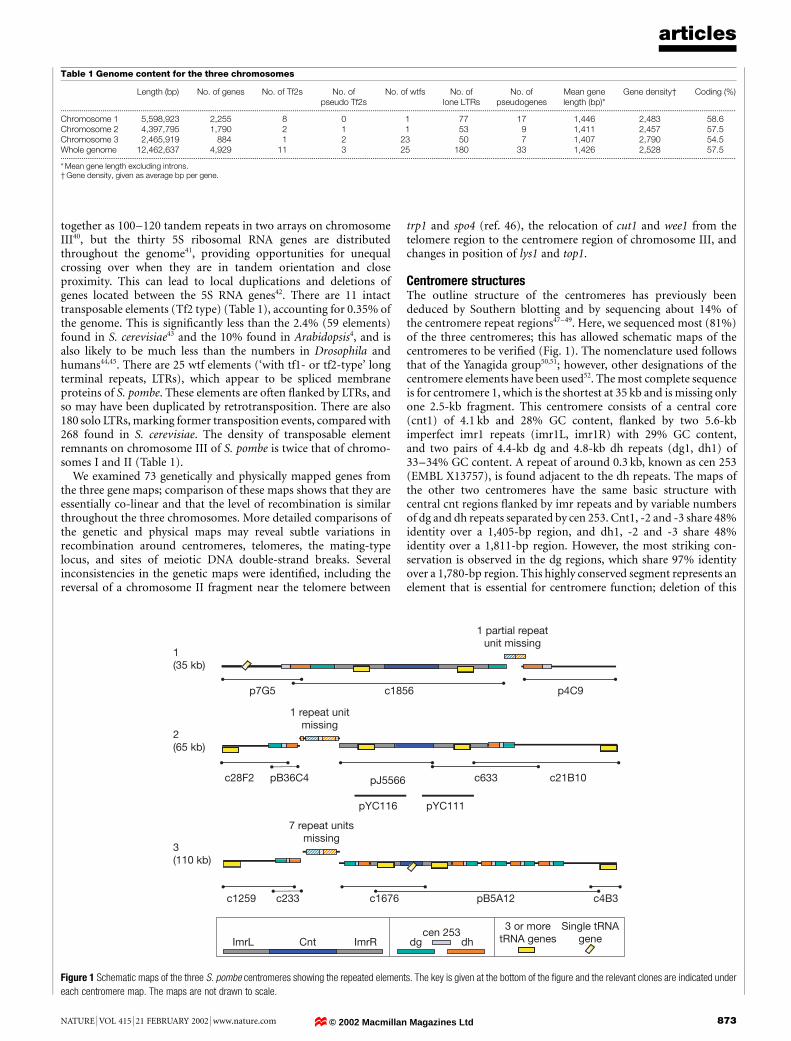
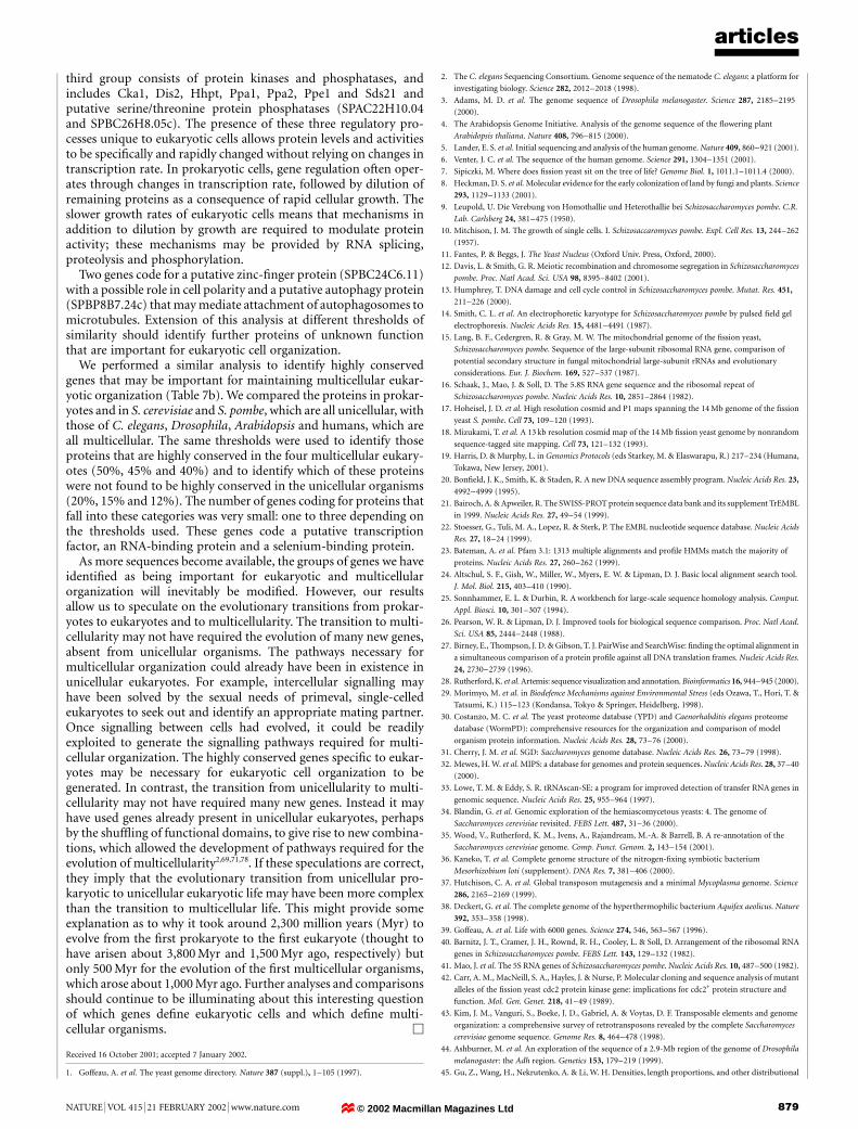
Centromere structuresThe outline structure of the centromeres has previously beendeduced by Southern blotting and by sequencing about 14% ofthe centromere repeat regions47±49. Here, we sequenced most (81%)of the three centromeres; this has allowed schematic maps of thecentromeres to be veri®ed (Fig. 1). The nomenclature used followsthat of the Yanagida group50,51; however, other designations of thecentromere elements have been used52. The most complete sequenceis for centromere 1, which is the shortest at 35 kb and is missing onlyone 2.5-kb fragment. This centromere consists of a central core(cnt1) of 4.1 kb and 28% GC content, ¯anked by two 5.6-kbimperfect imr1 repeats (imr1L, imr1R) with 29% GC content,and two pairs of 4.4-kb dg and 4.8-kb dh repeats (dg1, dh1) of33±34% GC content. A repeat of around 0.3 kb, known as cen 253(EMBL X13757), is found adjacent to the dh repeats. The maps ofthe other two centromeres have the same basic structure withcentral cnt regions ¯anked by imr repeats and by variable numbersof dg and dh repeats separated by cen 253. Cnt1, -2 and -3 share 48%identity over a 1,405-bp region, and dh1, -2 and -3 share 48%identity over a 1,811-bp region. However, the most striking con-servation is observed in the dg regions, which share 97% identityover a 1,780-bp region. This highly conserved segment represents anelement that is essential for centromere function; deletion of this
articles
NATURE | VOL 415 | 21 FEBRUARY 2002 | www.nature.com 873
Table 1 Genome content for the three chromosomes
Length (bp) No. of genes No. of Tf2s No. ofpseudo Tf2s
No. of wtfs No. oflone LTRs
No. ofpseudogenes
Mean genelength (bp)*
Gene density² Coding (%)
...................................................................................................................................................................................................................................................................................................................................................................
Chromosome 1 5,598,923 2,255 8 0 1 77 17 1,446 2,483 58.6Chromosome 2 4,397,795 1,790 2 1 1 53 9 1,411 2,457 57.5Chromosome 3 2,465,919 884 1 2 23 50 7 1,407 2,790 54.5Whole genome 12,462,637 4,929 11 3 25 180 33 1,426 2,528 57.5...................................................................................................................................................................................................................................................................................................................................................................* Mean gene length excluding introns.² Gene density, given as average bp per gene.
p7G5 c1856 p4C9
c633c28F2 pB36C4
pYC111pYC116
c21B10pJ5566
c1259 pB5A12 c4B3c1676c233
1(35 kb)
2(65 kb)
3(110 kb)
1 partial repeatunit missing
1 repeat unitmissing
7 repeat unitsmissing
ImrL Cnt dg dhImrRcen 253
3 or moretRNA genes
Single tRNAgene
Figure 1 Schematic maps of the three S. pombe centromeres showing the repeated elements. The key is given at the bottom of the ®gure and the relevant clones are indicated under
each centromere map. The maps are not drawn to scale.
© 2002 Macmillan Magazines Ltd
region from the dg repeat, termed the K/K0 repeat by the Clarkegroup, results in a complete loss of centromere activity in bothmitosis and meiosis53. There must be a special mechanism tomaintain such a high level of sequence conservation between thedifferent centromeres. The total calculated lengths of centromeres 1,2 and 3 are respectively 35, 65 and 110 kb, inversely proportional tothe lengths of the chromosomes at 5.7, 4.6 and 3.5 Mb. Possiblymore extended centromeric regions are required for proper mitoticand meiotic behaviour when the chromosome arms are shorter. Asnoted above there are no protein-coding genes in the centromericregion but there are many tRNA genes (Fig. 1). tRNA clusters ¯ankcentromeres 2 and 3 and are also found within the imr regions of allthree centromeres50. These tRNA genes might contribute to cen-tromere function by de®ning domain boundaries important forcentromere activity54.
The S. pombe centromeres are considerably longer than theirS. cerevisiae equivalents, which contain a core region suf®cient forcentromere activity of only 120 bp55,56 and a nuclease-protectedregion of 150±160 bp including the 120-bp conserved core57. It isnot clear why S. pombe centromeres are 300±1,000 times larger thantheir S. cerevisiae equivalents, but one possibility is that theirkinetochore structures are different.
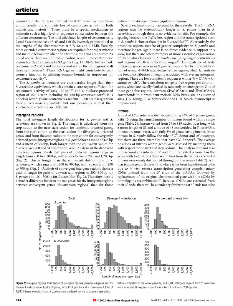
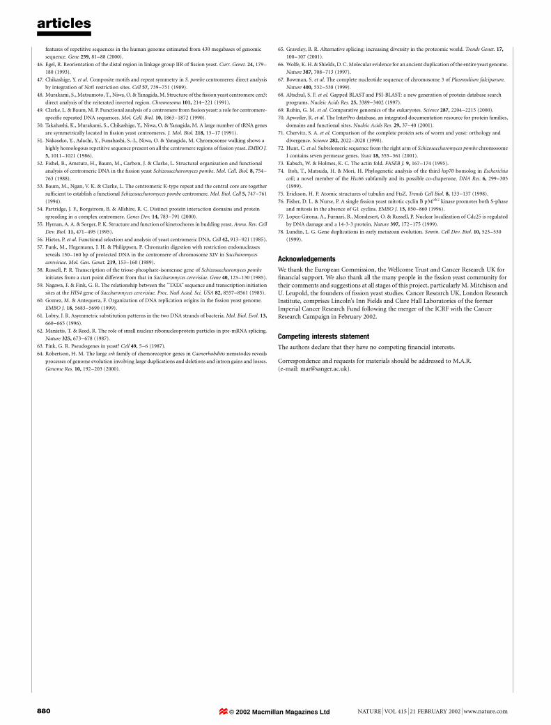
Intergene regionsThe total intergene length distributions for S. pombe and S.cerevisiae are shown in Fig. 2. The length is calculated from thestop codon to the next start codon for tandemly oriented genes,from the start codon to the start codon for divergently orientedgenes, and from the stop codon to the stop codon for convergentlyoriented genes. Intergenic regions in S. pombe have a mode of 423 bpand a mean of 952 bp, both longer than the equivalent values forS. cerevisiae (200 and 515 bp respectively). Analysis of the divergentintergene regions reveals that pairs of upstream regions range inlength from 200 to 2,100 bp, with a peak between 200 and 1,200 bp(Fig. 2). This is longer than the equivalent distributions in S.cerevisiae, which range from 200 to 900 bp, with a peak from 200to 700 bp (Fig. 2). Analysis of convergent intergene regions shows apeak in length for pairs of downstream regions of 200±800 bp forS. pombe and 100±500 bp for S. cerevisiae (Fig. 2). Therefore there isa smaller difference between the two yeasts for the intergenic regionsbetween convergent genes (downstream regions) than for those
between the divergent genes (upstream regions).Several explanations can account for these results. The 59 mRNA
regions may be systematically longer in S. pombe than in S.cerevisiae, although there is no evidence for this. For example, thespacing between the TATA-box region and the transcriptional startin S. pombe is shorter than that in S. cerevisiae58,59. Alternatively, thepromoter regions may be of greater complexity in S. pombe andtherefore longer. Again there is no direct evidence to support thisview, but there are other examples of more-extended organizationof chromatin elements in S. pombe, including larger centromeresand regions of DNA replication origin60. The existence of trulyintergenic spacer regions in S. pombe is supported by the identi®ca-tion of several 4±8-kb extended gene-free regions, which fall outsidethe broad distribution of lengths associated with average intergenicregions. These are low complexity sequences with a (G - C)/(G + C)strand switch61. There are about ten gene-free regions per chromo-some, which are usually ¯anked by tandemly oriented genes. One ofthese gene-free regions, between SPAC4G8.03c and SPAC4G8.04,corresponds to a prominent meiotic DNA break site or cluster ofsites (J. A. Young, R. W. Schreckhise and G. R. Smith, manuscript inpreparation).
IntronsA total of 4,730 introns is distributed among 43% of S. pombe genes,with 15 being the largest number of introns found within a singlegene (Table 2). Introns varied from 29 to 819 nucleotides long, witha mean length of 81 and a mode of 48 nucleotides. In S. cerevisiae,introns are much rarer, with only 5% of genes having introns. Mostintrons in S. pombe follow the rule of GT donor and AG acceptor,but there are three examples that have GC donors62. The averagepositions of introns within genes were assessed by mapping themwith respect to the start and stop codons. This analysis does not takeinto account any introns in 59 and 39 untranslated regions. For thegenes with 1±6 introns there is a 59 bias from the values expected ifintrons were evenly distributed throughout the genes (Table 2). A 59bias is also seen in S. cerevisiae, where it has been hypothesized to bedue to in vivo reverse transcription generating complementaryDNAs primed from the 39 ends of the mRNAs, followed byreplacement of the original chromosomal gene with the cDNA byhomologous recombination63. Because cDNAs are extended fromtheir 39 ends, there will be a tendency for introns at 59 ends not to be
articles
874 NATURE | VOL 415 | 21 FEBRUARY 2002 | www.nature.com
S. pombe
Total Divergent orientation Convergent orientation
S. cerevisiae
<–2
00 100
500
900
1,30
0
1,70
0
2,10
0
2,50
0
2,90
0
≥3,2
00
0100200300400500600700800900
<–2
00
100
500
900
1,30
0
1,70
0
2,10
0
2,50
0
2,90
0
≥3,2
00
0
500
1,000
1,500
2,000
2,500
<–2
00
100
500
900
1,30
0
1,70
0
2,10
0
2,50
0
2,90
0
≥3,2
00
020406080
100120140160
<–2
00 100
500
900
1,30
0
1,70
0
2,10
0
2,50
0
2,90
0
≥3,2
00
050
100150200250300350400450
<–2
00 100
500
900
1,30
0
1,70
0
2,10
0
2,50
0
2,90
0
≥3,2
00
050
100150200250300350400450
<–2
00
100
500
900
1,30
0
1,70
0
2,10
0
2,50
0
2,90
0
≥3,2
00
0100200300400500600700
Length of intergene region (bp)
No.
of i
nter
gene
reg
ions
Figure 2 Intergene regions. Distribution of intergene regions given for all genes and for
divergent and convergent pairs of genes, for both S. pombe and S. cerevisiae. A total of
4,890 intergene regions from S. pombe were analysed from a database prepared just
before completion of the whole genome, and 5,788 intergene regions from S. cerevisiae
were analysed. Histograms show the number of regions in 200-bp bins.
© 2002 Macmillan Magazines Ltd
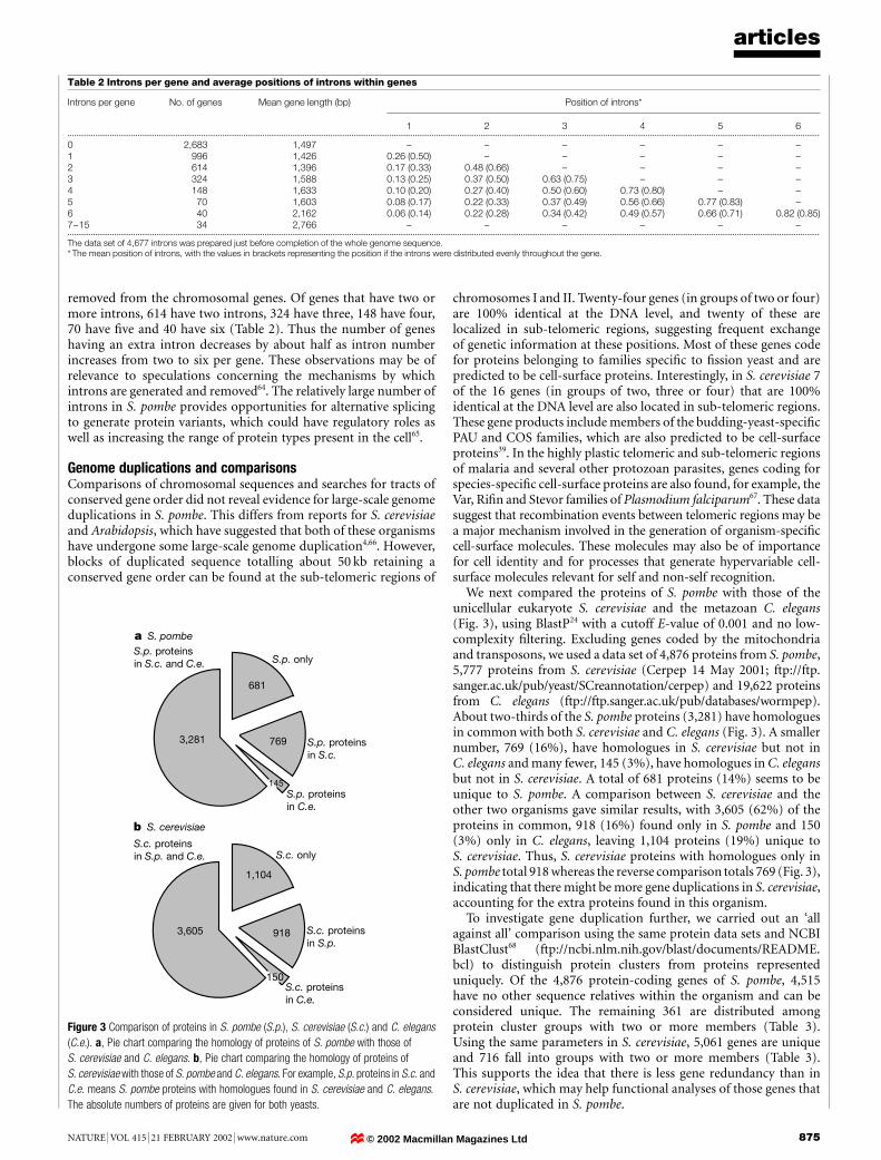
removed from the chromosomal genes. Of genes that have two ormore introns, 614 have two introns, 324 have three, 148 have four,70 have ®ve and 40 have six (Table 2). Thus the number of geneshaving an extra intron decreases by about half as intron numberincreases from two to six per gene. These observations may be ofrelevance to speculations concerning the mechanisms by whichintrons are generated and removed64. The relatively large number ofintrons in S. pombe provides opportunities for alternative splicingto generate protein variants, which could have regulatory roles aswell as increasing the range of protein types present in the cell65.
Genome duplications and comparisonsComparisons of chromosomal sequences and searches for tracts ofconserved gene order did not reveal evidence for large-scale genomeduplications in S. pombe. This differs from reports for S. cerevisiaeand Arabidopsis, which have suggested that both of these organismshave undergone some large-scale genome duplication4,66. However,blocks of duplicated sequence totalling about 50 kb retaining aconserved gene order can be found at the sub-telomeric regions of
chromosomes I and II. Twenty-four genes (in groups of two or four)are 100% identical at the DNA level, and twenty of these arelocalized in sub-telomeric regions, suggesting frequent exchangeof genetic information at these positions. Most of these genes codefor proteins belonging to families speci®c to ®ssion yeast and arepredicted to be cell-surface proteins. Interestingly, in S. cerevisiae 7of the 16 genes (in groups of two, three or four) that are 100%identical at the DNA level are also located in sub-telomeric regions.These gene products include members of the budding-yeast-speci®cPAU and COS families, which are also predicted to be cell-surfaceproteins39. In the highly plastic telomeric and sub-telomeric regionsof malaria and several other protozoan parasites, genes coding forspecies-speci®c cell-surface proteins are also found, for example, theVar, Ri®n and Stevor families of Plasmodium falciparum67. These datasuggest that recombination events between telomeric regions may bea major mechanism involved in the generation of organism-speci®ccell-surface molecules. These molecules may also be of importancefor cell identity and for processes that generate hypervariable cell-surface molecules relevant for self and non-self recognition.
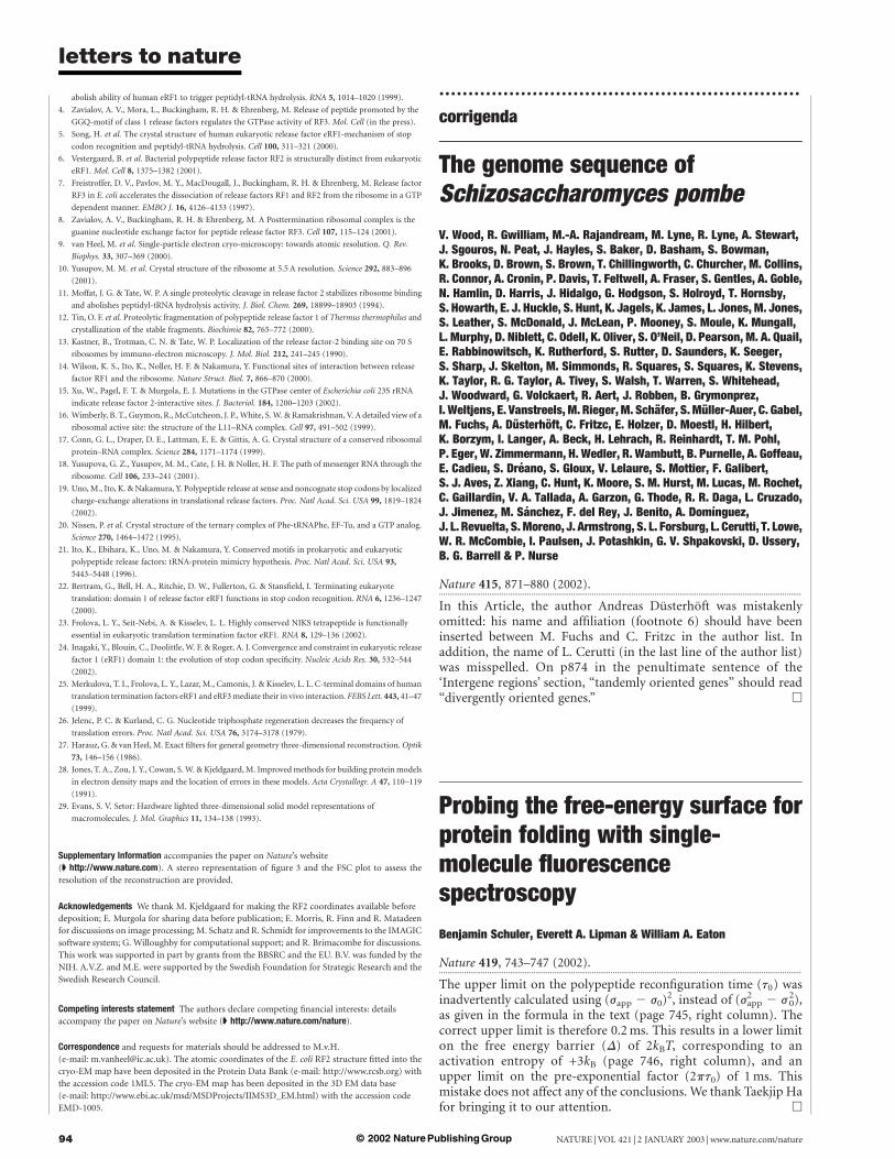
We next compared the proteins of S. pombe with those of theunicellular eukaryote S. cerevisiae and the metazoan C. elegans(Fig. 3), using BlastP24 with a cutoff E-value of 0.001 and no low-complexity ®ltering. Excluding genes coded by the mitochondriaand transposons, we used a data set of 4,876 proteins from S. pombe,5,777 proteins from S. cerevisiae (Cerpep 14 May 2001; ftp://ftp.sanger.ac.uk/pub/yeast/SCreannotation/cerpep) and 19,622 proteinsfrom C. elegans (ftp://ftp.sanger.ac.uk/pub/databases/wormpep).About two-thirds of the S. pombe proteins (3,281) have homologuesin common with both S. cerevisiae and C. elegans (Fig. 3). A smallernumber, 769 (16%), have homologues in S. cerevisiae but not inC. elegans and many fewer, 145 (3%), have homologues in C. elegansbut not in S. cerevisiae. A total of 681 proteins (14%) seems to beunique to S. pombe. A comparison between S. cerevisiae and theother two organisms gave similar results, with 3,605 (62%) of theproteins in common, 918 (16%) found only in S. pombe and 150(3%) only in C. elegans, leaving 1,104 proteins (19%) unique toS. cerevisiae. Thus, S. cerevisiae proteins with homologues only inS. pombe total 918 whereas the reverse comparison totals 769 (Fig. 3),indicating that there might be more gene duplications in S. cerevisiae,accounting for the extra proteins found in this organism.
To investigate gene duplication further, we carried out an `allagainst all' comparison using the same protein data sets and NCBIBlastClust68 (ftp://ncbi.nlm.nih.gov/blast/documents/README.bcl) to distinguish protein clusters from proteins representeduniquely. Of the 4,876 protein-coding genes of S. pombe, 4,515have no other sequence relatives within the organism and can beconsidered unique. The remaining 361 are distributed amongprotein cluster groups with two or more members (Table 3).Using the same parameters in S. cerevisiae, 5,061 genes are uniqueand 716 fall into groups with two or more members (Table 3).This supports the idea that there is less gene redundancy than inS. cerevisiae, which may help functional analyses of those genes thatare not duplicated in S. pombe.
articles
NATURE | VOL 415 | 21 FEBRUARY 2002 | www.nature.com 875
Table 2 Introns per gene and average positions of introns within genes
Introns per gene No. of genes Mean gene length (bp) Position of introns*
1 2 3 4 5 6...................................................................................................................................................................................................................................................................................................................................................................
0 2,683 1,497 ± ± ± ± ± ±1 996 1,426 0.26 (0.50) ± ± ± ± ±2 614 1,396 0.17 (0.33) 0.48 (0.66) ± ± ± ±3 324 1,588 0.13 (0.25) 0.37 (0.50) 0.63 (0.75) ± ± ±4 148 1,633 0.10 (0.20) 0.27 (0.40) 0.50 (0.60) 0.73 (0.80) ± ±5 70 1,603 0.08 (0.17) 0.22 (0.33) 0.37 (0.49) 0.56 (0.66) 0.77 (0.83) ±6 40 2,162 0.06 (0.14) 0.22 (0.28) 0.34 (0.42) 0.49 (0.57) 0.66 (0.71) 0.82 (0.85)7±15 34 2,766 ± ± ± ± ± ±...................................................................................................................................................................................................................................................................................................................................................................
The data set of 4,677 introns was prepared just before completion of the whole genome sequence.* The mean position of introns, with the values in brackets representing the position if the introns were distributed evenly throughout the gene.
150
918
1,104
3,605
3,281
681
769
145
S.p. proteinsin S.c. and C.e.
S.c. proteinsin S.p. and C.e.
S.c. proteinsin S.p.
S.c. proteinsin C.e.
S.c. only
S.p. only
S.p. proteinsin S.c.
S.p. proteinsin C.e.
a S. pombe
b S. cerevisiae
Figure 3 Comparison of proteins in S. pombe (S.p.), S. cerevisiae (S.c.) and C. elegans
(C.e.). a, Pie chart comparing the homology of proteins of S. pombe with those of
S. cerevisiae and C. elegans. b, Pie chart comparing the homology of proteins of
S. cerevisiae with those of S. pombe and C. elegans. For example, S.p. proteins in S.c. and
C.e. means S. pombe proteins with homologues found in S. cerevisiae and C. elegans.
The absolute numbers of proteins are given for both yeasts.
© 2002 Macmillan Magazines Ltd
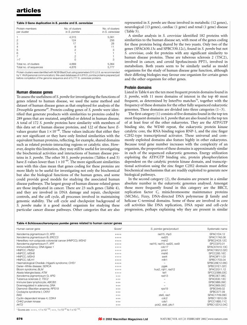
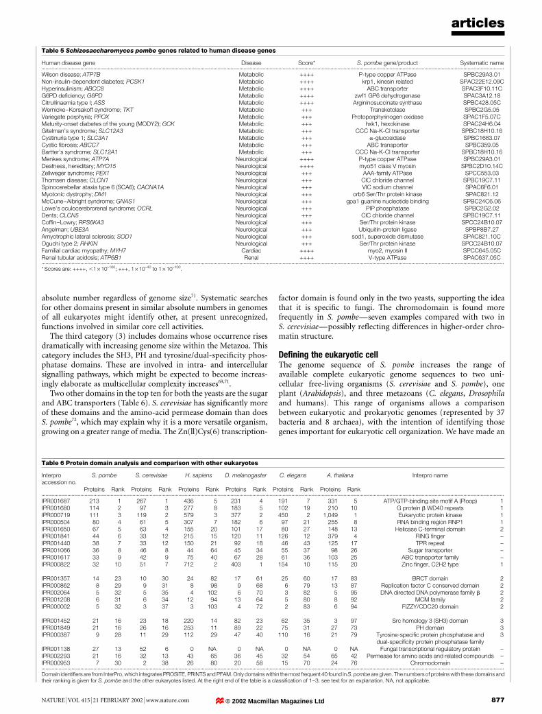
Human disease genesTo assess the usefulness of S. pombe for investigating the functions ofgenes related to human disease, we used the same method anddataset of human disease genes as that employed for analysis of theDrosophila genome69. Protein-coding genes of S. pombe were iden-ti®ed that generate products with similarities to proteins coded by289 genes that are mutated, ampli®ed or deleted in human disease.A total of 172 S. pombe proteins have similarity with members ofthis data set of human disease proteins, and 122 of these have E-values greater than 1 ´ 10-40. These values indicate that either theyare not signi®cant or they have only limited similarities with theequivalent human proteins, re¯ecting, for example, shared domainssuch as related protein-interacting regions or catalytic sites. How-ever, despite this limitation, they may still be useful for investigatingthe biochemical activities and interactions of human disease pro-teins in S. pombe. The other 50 S. pombe proteins (Tables 4 and 5)have E-values lower than 1 ´ 10-40. The more signi®cant similaritiesseen with this class mean that genes coding for these proteins aremore likely to be useful for investigating not only the biochemicalbut also the biological functions of the human genes, and somecould provide good models for studying the associated humandisease pathways. The largest group of human disease-related genesare those implicated in cancer. There are 23 such genes (Table 4),and they are involved in DNA damage and repair, checkpointcontrols, and the cell cycle, all processes involved in maintaininggenomic stability. The cell cycle and checkpoint background ofS. pombe make it a good model organism for studying theseparticular cancer disease pathways. Other categories that are also
represented in S. pombe are those involved in metabolic (12 genes),neurological (13 genes), cardiac (1 gene) and renal (1 gene) disease(Table 5).
A similar analysis in S. cerevisiae identi®ed 182 proteins withsimilarities to the human disease set, with most of the genes codingfor these proteins being shared by the two yeasts. Only two of thegenes (SPAC630.13c and SPBC530.12c), found in S. pombe but notS. cerevisiae, code for proteins with any signi®cant similarity tohuman disease proteins. These are tuberous sclerosis 2 (TSC2),involved in cancer, and ceroid lipofuscinosis PPT1, involved inmetabolism. Both yeasts seem to be similarly useful as modelorganisms for the study of human disease gene function, althoughtheir differing biologies may favour one organism for certain genesand the other organism for other genes.
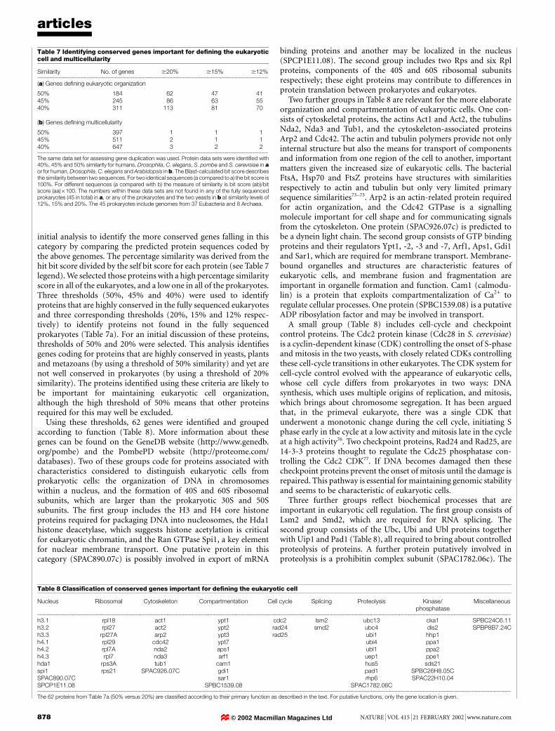
Protein domainsListed in Table 6 are the ten most frequent protein domains found inS. pombe, with 11 more domains of interest in the top 40 mostfrequent, as determined by InterPro matches70, together with thefrequency of these domains for the other fully sequenced eukaryoticgenomes. These domains are divided into three categories (1±3).
The ®rst category (1) consists of ®ve domains found in the top tenmost frequent domains in S. pombe that are also found in the top tenof at least four of the other eukaryotes. They are the ATP/GTPbinding site, the WD40 repeat, the eukaryotic protein kinasecatalytic core, the RNA binding region RNP-1, and the zinc ®ngerC2H2-type transcriptional activator. These universal and com-monly exploited domains also feature highly in other eukaryotes.Because total gene number increases with the complexity of anorganism, the proportion of these domains is approximately similarin each of the sequenced eukaryotic genomes. Energy utilizationexploiting the ATP/GTP binding site, protein phosphorylationdependent on the catalytic protein kinase domain, and transcrip-tional activation using the zinc ®nger C2H2 domain must de®nebiochemical mechanisms that are readily exploited to generate newbiological pathways.
In the second category (2), the domains are present in a similarabsolute number in the eukaryotic genomes analysed. Amongstthose more frequently found in this category are the BRCT,replication factor C, minichromosome maintenance proteins(MCMs), Fizzy, DNA-directed DNA polymerase b family andhelicase C-terminal domains. Some of these are involved in corecell activities like DNA replication, DNA repair and cell-cycleprogression, perhaps explaining why they are present in similar
articles
876 NATURE | VOL 415 | 21 FEBRUARY 2002 | www.nature.com
Table 3 Gene duplication in S. pombe and S. cerevisiae
Protein membersper cluster
No. of clustersin S. pombe
No. of clustersin S. cerevisiae
.............................................................................................................................................................................
1 4,515 5,0612 124 2563 17 284 8 115 2 36 1 17 2 1.7 0 3
Total no. of clusters 4,669 5,364Total no. of sequences 4,876 5,777.............................................................................................................................................................................
Protein clusters were identi®ed with NCBI BlastClust using parameters S10.L0.9, as recommendedby Y. Wolf (personal communication). We used databases of 4,876 S. pombe proteins prepared justbefore completion of the genome sequence and of 5,777 S. cerevisiae proteins.
Table 4 Schizosaccharomyces pombe genes related to human cancer genes
Human cancer gene Score* S. pombe gene/product Systematic name...................................................................................................................................................................................................................................................................................................................................................................
Xeroderma pigmentosum D; XPD ++++ rad15, rhp3 SPAC1D4.12Xeroderma pigmentosum B; ERCC3 ++++ rad25 SPAC17A5.06Hereditary non-polyposis colorectal cancer (HNPCC); MSH2 ++++ msh2 SPBC24C6.12CXeroderma pigmentosum F; XPF ++++ rad16, rad10, rad20, swi9 SPCC970.01Immunode®ciency; DNA ligase 1 ++++ cdc17 SPAC57A10.13CHNPCC; PMS2 ++++ pms1 SPAC19G12.02CHNPCC; MSH6 ++++ msh6 SPCC285.16CHNPCC; MSH3 ++++ swi4 SPAC8F11.03HNPCC; MLH1 ++++ mlh1 SPBC1703.04Haematological Chediak±Higashi syndrome; CHS1 ++++ ± SPBC28E12.06CDarier±White disease; SERCA ++++ pgak SPBC31E1.02CBloom syndrome; BLM ++++ hus2, rqh1, rad12 SPAC2G11.12Ataxia telangiectasia; ATM ++++ tel1 SPCC23B6.03CXeroderma pigmentosum G; XPG +++ rad13 SPBC3E7.08CTuberous sclerosis 2; TSC2 +++ ± SPAC630.13CImmune bare lymphocyte; ABCB3 +++ ± SPBC9B6.09CDownregulated in adenoma; DRA +++ ± SPAC869.05CDiamond±Blackfan anaemia; RPS19 +++ rps19 SPBC649.02Cockayne syndrome I; CKN1 +++ ± SPBC577.09RAS +++ ste5, ras1 SPAC17H9.09CCyclin-dependent kinase 4; CDK4 +++ cdc2 SPBC11B10.09CHK2 protein kinase +++ cds1 SPCC18B5.11CAKT2 +++ pck2, sts6, pkc1 SPBC12D12.04C...................................................................................................................................................................................................................................................................................................................................................................
* Scores are: ++++, ,1 ´ 10-100; +++, 1 ´ 10-40 to 1 ´ 10-100.
© 2002 Macmillan Magazines Ltd
absolute number regardless of genome size71. Systematic searchesfor other domains present in similar absolute numbers in genomesof all eukaryotes might identify other, at present unrecognized,functions involved in similar core cell activities.
The third category (3) includes domains whose occurrence risesdramatically with increasing genome size within the Metazoa. Thiscategory includes the SH3, PH and tyrosine/dual-speci®city phos-phatase domains. These are involved in intra- and intercellularsignalling pathways, which might be expected to become increas-ingly elaborate as multicellular complexity increases69,71.
Two other domains in the top ten for both the yeasts are the sugarand ABC transporters (Table 6). S. cerevisiae has signi®cantly moreof these domains and the amino-acid permease domain than doesS. pombe72, which may explain why it is a more versatile organism,growing on a greater range of media. The Zn(ll)Cys(6) transcription-
factor domain is found only in the two yeasts, supporting the ideathat it is speci®c to fungi. The chromodomain is found morefrequently in S. pombeÐseven examples compared with two inS. cerevisiaeÐpossibly re¯ecting differences in higher-order chro-matin structure.
De®ning the eukaryotic cellThe genome sequence of S. pombe increases the range ofavailable complete eukaryotic genome sequences to two uni-cellular free-living organisms (S. cerevisiae and S. pombe), oneplant (Arabidopsis), and three metazoans (C. elegans, Drosophilaand humans). This range of organisms allows a comparisonbetween eukaryotic and prokaryotic genomes (represented by 37bacteria and 8 archaea), with the intention of identifying thosegenes important for eukaryotic cell organization. We have made an
articles
NATURE | VOL 415 | 21 FEBRUARY 2002 | www.nature.com 877
Table 5 Schizosaccharomyces pombe genes related to human disease genes
Human disease gene Disease Score* S. pombe gene/product Systematic name...................................................................................................................................................................................................................................................................................................................................................................
Wilson disease; ATP7B Metabolic ++++ P-type copper ATPase SPBC29A3.01Non-insulin-dependent diabetes; PCSK1 Metabolic ++++ krp1, kinesin related SPAC22E12.09CHyperinsulinism; ABCC8 Metabolic ++++ ABC transporter SPAC3F10.11CG6PD de®ciency; G6PD Metabolic ++++ zwf1 GP6 dehydrogenase SPAC3A12.18Citrullinaemia type I; ASS Metabolic ++++ Argininosuccinate synthase SPBC428.05CWernicke±Korsakoff syndrome; TKT Metabolic +++ Transketolase SPBC2G5.05Variegate porphyria; PPOX Metabolic +++ Protoporphyrinogen oxidase SPAC1F5.07CMaturity-onset diabetes of the young (MODY2); GCK Metabolic +++ hxk1, hexokinase SPAC24H6.04Gitelman's syndrome; SLC12A3 Metabolic +++ CCC Na-K-Cl transporter SPBC18H10.16Cystinuria type 1; SLC3A1 Metabolic +++ a-glucosidase SPBC1683.07Cystic ®brosis; ABCC7 Metabolic +++ ABC transporter SPBC359.05Bartter's syndrome; SLC12A1 Metabolic +++ CCC Na-K-Cl transporter SPBC18H10.16Menkes syndrome; ATP7A Neurological ++++ P-type copper ATPase SPBC29A3.01Deafness, hereditary; MYO15 Neurological ++++ myo51 class V myosin SPBC2D10.14CZellweger syndrome; PEX1 Neurological +++ AAA-family ATPase SPCC553.03Thomsen disease; CLCN1 Neurological +++ ClC chloride channel SPBC19C7.11Spinocerebellar ataxia type 6 (SCA6); CACNA1A Neurological +++ VIC sodium channel SPAC6F6.01Myotonic dystrophy; DM1 Neurological +++ orb6 Ser/Thr protein kinase SPAC821.12McCune±Albright syndrome; GNAS1 Neurological +++ gpa1 guanine nucleotide binding SPBC24C6.06Lowe's oculocerebrorenal syndrome; OCRL Neurological +++ PIP phosphatase SPBC2G2.02Dents; CLCN5 Neurological +++ ClC chloride channel SPBC19C7.11Cof®n±Lowry; RPS6KA3 Neurological +++ Ser/Thr protein kinase SPCC24B10.07Angelman; UBE3A Neurological +++ Ubiquitin-protein ligase SPBP8B7.27Amyotrophic lateral sclerosis; SOD1 Neurological +++ sod1, superoxide dismutase SPAC821.10COguchi type 2; RHKIN Neurological +++ Ser/Thr protein kinase SPCC24B10.07Familial cardiac myopathy; MYH7 Cardiac ++++ myo2, myosin II SPCC645.05CRenal tubular acidosis; ATP6B1 Renal ++++ V-type ATPase SPAC637.05C...................................................................................................................................................................................................................................................................................................................................................................
* Scores are: ++++, ,1 ´ 10-100; +++, 1 ´ 10-40 to 1 ´ 10-100.
Table 6 Protein domain analysis and comparison with other eukaryotes
Interproaccession no.
S. pombe S. cerevisiae H. sapiens D. melanogaster C. elegans A. thaliana Interpro name
Proteins Rank Proteins Rank Proteins Rank Proteins Rank Proteins Rank Proteins Rank...................................................................................................................................................................................................................................................................................................................................................................
IPR001687 213 1 267 1 436 5 231 4 191 7 331 5 ATP/GTP-binding site motif A (Ploop) 1IPR001680 114 2 97 3 277 8 183 5 102 19 210 10 G protein b WD40 repeats 1IPR000719 111 3 119 2 579 3 377 2 450 2 1,049 1 Eukaryotic protein kinase 1IPR000504 80 4 61 5 307 7 182 6 97 21 255 8 RNA binding region RNP1 1IPR001650 67 5 63 4 155 20 101 17 80 27 148 13 Helicase C-terminal domain 2IPR001841 44 6 33 12 215 15 120 11 126 12 379 4 RING ®nger ±IPR001440 38 7 33 12 150 21 92 18 46 43 125 17 TPR repeat ±IPR001066 36 8 46 8 44 64 45 34 55 37 98 26 Sugar transporter ±IPR001617 33 9 42 9 75 40 67 28 61 36 103 25 ABC transporter family ±IPR000822 32 10 51 7 712 2 403 1 154 10 115 20 Zinc ®nger, C2H2 type 1
IPR001357 14 23 10 30 24 82 17 61 25 60 17 83 BRCT domain 2IPR000862 8 29 9 31 8 98 9 68 6 79 13 87 Replication factor C conserved domain 2IPR002064 5 32 5 35 4 102 6 70 3 82 5 95 DNA directed DNA polymerase family b 2IPR001208 6 31 6 34 12 94 13 64 5 80 8 92 MCM family 2IPR000002 5 32 3 37 3 103 4 72 2 83 6 94 FIZZY/CDC20 domain 2
IPR001452 21 16 23 18 220 14 82 23 62 35 3 97 Src homology 3 (SH3) domain 3IPR001849 21 16 26 16 253 11 89 22 75 31 27 73 PH domain 3IPR000387 9 28 11 29 112 29 47 40 110 16 21 79 Tyrosine-speci®c protein phosphatase and
dual-speci®city protein phosphatase family3
IPR001138 27 13 52 6 0 NA 0 NA 0 NA 0 NA Fungal transcriptional regulatory protein ±IPR002293 21 16 32 13 43 65 36 45 32 54 65 42 Permease for amino acids and related compounds ±IPR000953 7 30 2 38 26 80 20 58 15 70 24 76 Chromodomain ±...................................................................................................................................................................................................................................................................................................................................................................
Domain identi®ers are from InterPro, which integrates PROSITE, PRINTS and PFAM. Only domains within the most frequent 40 found in S. pombe are given. The numbers of proteins with these domains andtheir ranking is given for S. pombe and the other eukaryotes listed. At the right end of the table is a classi®cation of 1±3; see text for an explanation. NA, not applicable.
© 2002 Macmillan Magazines Ltd
initial analysis to identify the more conserved genes falling in thiscategory by comparing the predicted protein sequences coded bythe above genomes. The percentage similarity was derived from thehit bit score divided by the self bit score for each protein (see Table 7legend). We selected those proteins with a high percentage similarityscore in all of the eukaryotes, and a low one in all of the prokaryotes.Three thresholds (50%, 45% and 40%) were used to identifyproteins that are highly conserved in the fully sequenced eukaryotesand three corresponding thresholds (20%, 15% and 12% respec-tively) to identify proteins not found in the fully sequencedprokaryotes (Table 7a). For an initial discussion of these proteins,thresholds of 50% and 20% were selected. This analysis identi®esgenes coding for proteins that are highly conserved in yeasts, plantsand metazoans (by using a threshold of 50% similarity) and yet arenot well conserved in prokaryotes (by using a threshold of 20%similarity). The proteins identi®ed using these criteria are likely tobe important for maintaining eukaryotic cell organization,although the high threshold of 50% means that other proteinsrequired for this may well be excluded.
Using these thresholds, 62 genes were identi®ed and groupedaccording to function (Table 8). More information about thesegenes can be found on the GeneDB website (http://www.genedb.org/pombe) and the PombePD website (http://proteome.com/databases). Two of these groups code for proteins associated withcharacteristics considered to distinguish eukaryotic cells fromprokaryotic cells: the organization of DNA in chromosomeswithin a nucleus, and the formation of 40S and 60S ribosomalsubunits, which are larger than the prokaryotic 30S and 50Ssubunits. The ®rst group includes the H3 and H4 core histoneproteins required for packaging DNA into nucleosomes, the Hda1histone deacetylase, which suggests histone acetylation is criticalfor eukaryotic chromatin, and the Ran GTPase Spi1, a key elementfor nuclear membrane transport. One putative protein in thiscategory (SPAC890.07c) is possibly involved in export of mRNA
binding proteins and another may be localized in the nucleus(SPCP1E11.08). The second group includes two Rps and six Rplproteins, components of the 40S and 60S ribosomal subunitsrespectively; these eight proteins may contribute to differences inprotein translation between prokaryotes and eukaryotes.
Two further groups in Table 8 are relevant for the more elaborateorganization and compartmentation of eukaryotic cells. One con-sists of cytoskeletal proteins, the actins Act1 and Act2, the tubulinsNda2, Nda3 and Tub1, and the cytoskeleton-associated proteinsArp2 and Cdc42. The actin and tubulin polymers provide not onlyinternal structure but also the means for transport of componentsand information from one region of the cell to another, importantmatters given the increased size of eukaryotic cells. The bacterialFtsA, Hsp70 and FtsZ proteins have structures with similaritiesrespectively to actin and tubulin but only very limited primarysequence similarities73±75. Arp2 is an actin-related protein requiredfor actin organization, and the Cdc42 GTPase is a signallingmolecule important for cell shape and for communicating signalsfrom the cytoskeleton. One protein (SPAC926.07c) is predicted tobe a dynein light chain. The second group consists of GTP bindingproteins and their regulators Ypt1, -2, -3 and -7, Arf1, Aps1, Gdi1and Sar1, which are required for membrane transport. Membrane-bound organelles and structures are characteristic features ofeukaryotic cells, and membrane fusion and fragmentation areimportant in organelle formation and function. Cam1 (calmodu-lin) is a protein that exploits compartmentalization of Ca2+ toregulate cellular processes. One protein (SPBC1539.08) is a putativeADP ribosylation factor and may be involved in transport.
A small group (Table 8) includes cell-cycle and checkpointcontrol proteins. The Cdc2 protein kinase (Cdc28 in S. cerevisiae)is a cyclin-dependent kinase (CDK) controlling the onset of S-phaseand mitosis in the two yeasts, with closely related CDKs controllingthese cell-cycle transitions in other eukaryotes. The CDK system forcell-cycle control evolved with the appearance of eukaryotic cells,whose cell cycle differs from prokaryotes in two ways: DNAsynthesis, which uses multiple origins of replication, and mitosis,which brings about chromosome segregation. It has been arguedthat, in the primeval eukaryote, there was a single CDK thatunderwent a monotonic change during the cell cycle, initiating Sphase early in the cycle at a low activity and mitosis late in the cycleat a high activity76. Two checkpoint proteins, Rad24 and Rad25, are14-3-3 proteins thought to regulate the Cdc25 phosphatase con-trolling the Cdc2 CDK77. If DNA becomes damaged then thesecheckpoint proteins prevent the onset of mitosis until the damage isrepaired. This pathway is essential for maintaining genomic stabilityand seems to be characteristic of eukaryotic cells.
Three further groups re¯ect biochemical processes that areimportant in eukaryotic cell regulation. The ®rst group consists ofLsm2 and Smd2, which are required for RNA splicing. Thesecond group consists of the Ubc, Ubi and Ubl proteins togetherwith Uip1 and Pad1 (Table 8), all required to bring about controlledproteolysis of proteins. A further protein putatively involved inproteolysis is a prohibitin complex subunit (SPAC1782.06c). The
articles
878 NATURE | VOL 415 | 21 FEBRUARY 2002 | www.nature.com
Table 7 Identifying conserved genes important for de®ning the eukaryoticcell and multicellularity
Similarity No. of genes $20% $15% $12%.............................................................................................................................................................................
(a) Genes de®ning eukaryotic organization
50% 184 62 47 4145% 245 86 63 5540% 311 113 81 70
(b) Genes de®ning multicellularity
50% 397 1 1 145% 511 2 1 140% 647 3 2 2.............................................................................................................................................................................
The same data set for assessing gene duplication was used. Protein data sets were identi®ed with40%, 45% and 50% similarity for humans, Drosophila, C. elegans, S. pombe and S. cerevisiae in aor for human, Drosophila, C. elegans and Arabidopsis in b. The Blast-calculated bit score describesthe similarity between two sequences. For two identical sequences (a compared to a) the bit score is100%. For different sequences (a compared with b) the measure of similarity is bit score (ab)/bitscore (aa) ´ 100. The numbers within these data sets are not found in any of the fully sequencedprokaryotes (45 in total) in a, or any of the prokaryotes and the two yeasts in b at similarity levels of12%, 15% and 20%. The 45 prokaryotes include genomes from 37 Eubacteria and 8 Archaea.
Table 8 Classi®cation of conserved genes important for de®ning the eukaryotic cell
Nucleus Ribosomal Cytoskeleton Compartmentation Cell cycle Splicing Proteolysis Kinase/phosphatase
Miscellaneous
...................................................................................................................................................................................................................................................................................................................................................................
h3.1 rpl18 act1 ypt1 cdc2 lsm2 ubc13 cka1 SPBC24C6.11h3.2 rpl27 act2 ypt2 rad24 smd2 ubc4 dis2 SPBP8B7.24Ch3.3 rpl27A arp2 ypt3 rad25 ubi1 hhp1h4.1 rpl29 cdc42 ypt7 ubi4 ppa1h4.2 rpl7A nda2 aps1 ubl1 ppa2h4.3 rpl7 nda3 arf1 uep1 ppe1hda1 rps3A tub1 cam1 hus5 sds21spi1 rps21 SPAC926.07C gdi1 pad1 SPBC26H8.05CSPAC890.07C sar1 rhp6 SPAC22H10.04SPCP1E11.08 SPBC1539.08 SPAC1782.06C...................................................................................................................................................................................................................................................................................................................................................................
The 62 proteins from Table 7a (50% versus 20%) are classi®ed according to their primary function as described in the text. For putative functions, only the gene location is given.
© 2002 Macmillan Magazines Ltd
third group consists of protein kinases and phosphatases, andincludes Cka1, Dis2, Hhpt, Ppa1, Ppa2, Ppe1 and Sds21 andputative serine/threonine protein phosphatases (SPAC22H10.04and SPBC26H8.05c). The presence of these three regulatory pro-cesses unique to eukaryotic cells allows protein levels and activitiesto be speci®cally and rapidly changed without relying on changes intranscription rate. In prokaryotic cells, gene regulation often oper-ates through changes in transcription rate, followed by dilution ofremaining proteins as a consequence of rapid cellular growth. Theslower growth rates of eukaryotic cells means that mechanisms inaddition to dilution by growth are required to modulate proteinactivity; these mechanisms may be provided by RNA splicing,proteolysis and phosphorylation.
Two genes code for a putative zinc-®nger protein (SPBC24C6.11)with a possible role in cell polarity and a putative autophagy protein(SPBP8B7.24c) that may mediate attachment of autophagosomes tomicrotubules. Extension of this analysis at different thresholds ofsimilarity should identify further proteins of unknown functionthat are important for eukaryotic cell organization.
We performed a similar analysis to identify highly conservedgenes that may be important for maintaining multicellular eukar-yotic organization (Table 7b). We compared the proteins in prokar-yotes and in S. cerevisiae and S. pombe, which are all unicellular, withthose of C. elegans, Drosophila, Arabidopsis and humans, which areall multicellular. The same thresholds were used to identify thoseproteins that are highly conserved in the four multicellular eukary-otes (50%, 45% and 40%) and to identify which of these proteinswere not found to be highly conserved in the unicellular organisms(20%, 15% and 12%). The number of genes coding for proteins thatfall into these categories was very small: one to three depending onthe thresholds used. These genes code a putative transcriptionfactor, an RNA-binding protein and a selenium-binding protein.
As more sequences become available, the groups of genes we haveidenti®ed as being important for eukaryotic and multicellularorganization will inevitably be modi®ed. However, our resultsallow us to speculate on the evolutionary transitions from prokar-yotes to eukaryotes and to multicellularity. The transition to multi-cellularity may not have required the evolution of many new genes,absent from unicellular organisms. The pathways necessary formulticellular organization could already have been in existence inunicellular eukaryotes. For example, intercellular signalling mayhave been solved by the sexual needs of primeval, single-celledeukaryotes to seek out and identify an appropriate mating partner.Once signalling between cells had evolved, it could be readilyexploited to generate the signalling pathways required for multi-cellular organization. The highly conserved genes speci®c to eukar-yotes may be necessary for eukaryotic cell organization to begenerated. In contrast, the transition from unicellularity to multi-cellularity may not have required many new genes. Instead it mayhave used genes already present in unicellular eukaryotes, perhapsby the shuf¯ing of functional domains, to give rise to new combina-tions, which allowed the development of pathways required for theevolution of multicellularity2,69,71,78. If these speculations are correct,they imply that the evolutionary transition from unicellular pro-karyotic to unicellular eukaryotic life may have been more complexthan the transition to multicellular life. This might provide someexplanation as to why it took around 2,300 million years (Myr) toevolve from the ®rst prokaryote to the ®rst eukaryote (thought tohave arisen about 3,800 Myr and 1,500 Myr ago, respectively) butonly 500 Myr for the evolution of the ®rst multicellular organisms,which arose about 1,000 Myr ago. Further analyses and comparisonsshould continue to be illuminating about this interesting questionof which genes de®ne eukaryotic cells and which de®ne multi-cellular organisms. M
Received 16 October 2001; accepted 7 January 2002.
1. Goffeau, A. et al. The yeast genome directory. Nature 387 (suppl.), 1±105 (1997).
2. The C. elegans Sequencing Consortium. Genome sequence of the nematode C. elegans: a platform for
investigating biology. Science 282, 2012±2018 (1998).
3. Adams, M. D. et al. The genome sequence of Drosophila melanogaster. Science 287, 2185±2195
(2000).
4. The Arabidopsis Genome Initiative. Analysis of the genome sequence of the ¯owering plant
Arabidopsis thaliana. Nature 408, 796±815 (2000).
5. Lander, E. S. et al. Initial sequencing and analysis of the human genome. Nature 409, 860±921 (2001).
6. Venter, J. C. et al. The sequence of the human genome. Science 291, 1304±1351 (2001).
7. Sipiczki, M. Where does ®ssion yeast sit on the tree of life? Genome Biol. 1, 1011.1±1011.4 (2000).
8. Heckman, D. S. et al. Molecular evidence for the early colonization of land by fungi and plants. Science
293, 1129±1133 (2001).
9. Leupold, U. Die Verebung von Homothallie und Heterothallie bei Schizosaccharomyces pombe. C.R.
Lab. Carlsberg 24, 381±475 (1950).
10. Mitchison, J. M. The growth of single cells. I. Schizosaccaromyces pombe. Expl. Cell Res. 13, 244±262
(1957).
11. Fantes, P. & Beggs, J. The Yeast Nucleus (Oxford Univ. Press, Oxford, 2000).
12. Davis, L. & Smith, G. R. Meiotic recombination and chromosome segregation in Schizosaccharomyces
pombe. Proc. Natl Acad. Sci. USA 98, 8395±8402 (2001).
13. Humphrey, T. DNA damage and cell cycle control in Schizosaccharomyces pombe. Mutat. Res. 451,
211±226 (2000).
14. Smith, C. L. et al. An electrophoretic karyotype for Schizosaccharomyces pombe by pulsed ®eld gel
electrophoresis. Nucleic Acids Res. 15, 4481±4491 (1987).
15. Lang, B. F., Cedergren, R. & Gray, M. W. The mitochondrial genome of the ®ssion yeast,
Schizosaccharomyces pombe. Sequence of the large-subunit ribosomal RNA gene, comparison of
potential secondary structure in fungal mitochondrial large-subunit rRNAs and evolutionary
considerations. Eur. J. Biochem. 169, 527±537 (1987).
16. Schaak, J., Mao, J. & Soll, D. The 5.8S RNA gene sequence and the ribosomal repeat of
Schizosaccharomyces pombe. Nucleic Acids Res. 10, 2851±2864 (1982).
17. Hoheisel, J. D. et al. High resolution cosmid and P1 maps spanning the 14 Mb genome of the ®ssion
yeast S. pombe. Cell 73, 109±120 (1993).
18. Mizukami, T. et al. A 13 kb resolution cosmid map of the 14 Mb ®ssion yeast genome by nonrandom
sequence-tagged site mapping. Cell 73, 121±132 (1993).
19. Harris, D. & Murphy, L. in Genomics Protocols (eds Starkey, M. & Elaswarapu, R.) 217±234 (Humana,
Tokawa, New Jersey, 2001).
20. Bon®eld, J. K., Smith, K. & Staden, R. A new DNA sequence assembly program. Nucleic Acids Res. 23,
4992±4999 (1995).
21. Bairoch, A. & Apweiler, R. The SWISS-PROT protein sequence data bank and its supplement TrEMBL
in 1999. Nucleic Acids Res. 27, 49±54 (1999).
22. Stoesser, G., Tuli, M. A., Lopez, R. & Sterk, P. The EMBL nucleotide sequence database. Nucleic Acids
Res. 27, 18±24 (1999).
23. Bateman, A. et al. Pfam 3.1: 1313 multiple alignments and pro®le HMMs match the majority of
proteins. Nucleic Acids Res. 27, 260±262 (1999).
24. Altschul, S. F., Gish, W., Miller, W., Myers, E. W. & Lipman, D. J. Basic local alignment search tool.
J. Mol. Biol. 215, 403±410 (1990).
25. Sonnhammer, E. L. & Durbin, R. A workbench for large-scale sequence homology analysis. Comput.
Appl. Biosci. 10, 301±307 (1994).
26. Pearson, W. R. & Lipman, D. J. Improved tools for biological sequence comparison. Proc. Natl Acad.
Sci. USA 85, 2444±2448 (1988).
27. Birney, E., Thompson, J. D. & Gibson, T. J. PairWise and SearchWise: ®nding the optimal alignment in
a simultaneous comparison of a protein pro®le against all DNA translation frames. Nucleic Acids Res.
24, 2730±2739 (1996).
28. Rutherford, K. et al. Artemis: sequence visualization and annotation. Bioinformatics 16, 944±945 (2000).
29. Morimyo, M. et al. in Biodefence Mechanisms against Environmental Stress (eds Ozawa, T., Hori, T. &
Tatsumi, K.) 115±123 (Kondansa, Tokyo & Springer, Heidelberg, 1998).
30. Costanzo, M. C. et al. The yeast proteome database (YPD) and Caenorhabditis elegans proteome
database (WormPD): comprehensive resources for the organization and comparison of model
organism protein information. Nucleic Acids Res. 28, 73±76 (2000).
31. Cherry, J. M. et al. SGD: Saccharomyces genome database. Nucleic Acids Res. 26, 73±79 (1998).
32. Mewes, H. W. et al. MIPS: a database for genomes and protein sequences. Nucleic Acids Res. 28, 37±40
(2000).
33. Lowe, T. M. & Eddy, S. R. tRNAscan-SE: a program for improved detection of transfer RNA genes in
genomic sequence. Nucleic Acids Res. 25, 955±964 (1997).
34. Blandin, G. et al. Genomic exploration of the hemiascomycetous yeasts: 4. The genome of
Saccharomyces cerevisiae revisited. FEBS Lett. 487, 31±36 (2000).
35. Wood, V., Rutherford, K. M., Ivens, A., Rajandream, M.-A. & Barrell, B. A re-annotation of the
Saccharomyces cerevisiae genome. Comp. Funct. Genom. 2, 143±154 (2001).
36. Kaneko, T. et al. Complete genome structure of the nitrogen-®xing symbiotic bacterium
Mesorhizobium loti (supplement). DNA Res. 7, 381±406 (2000).
37. Hutchison, C. A. et al. Global transposon mutagenesis and a minimal Mycoplasma genome. Science
286, 2165±2169 (1999).
38. Deckert, G. et al. The complete genome of the hyperthermophilic bacterium Aquifex aeolicus. Nature
392, 353±358 (1998).
39. Goffeau, A. et al. Life with 6000 genes. Science 274, 546, 563±567 (1996).
40. Barnitz, J. T., Cramer, J. H., Rownd, R. H., Cooley, L. & Soll, D. Arrangement of the ribosomal RNA
genes in Schizosaccharomyces pombe. FEBS Lett. 143, 129±132 (1982).
41. Mao, J. et al. The 5S RNA genes of Schizosaccharomyces pombe. Nucleic Acids Res. 10, 487±500 (1982).
42. Carr, A. M., MacNeill, S. A., Hayles, J. & Nurse, P. Molecular cloning and sequence analysis of mutant
alleles of the ®ssion yeast cdc2 protein kinase gene: implications for cdc2+ protein structure and
function. Mol. Gen. Genet. 218, 41±49 (1989).
43. Kim, J. M., Vanguri, S., Boeke, J. D., Gabriel, A. & Voytas, D. F. Transposable elements and genome
organization: a comprehensive survey of retrotransposons revealed by the complete Saccharomyces
cerevisiae genome sequence. Genome Res. 8, 464±478 (1998).
44. Ashburner, M. et al. An exploration of the sequence of a 2.9-Mb region of the genome of Drosophila
melanogaster: the Adh region. Genetics 153, 179±219 (1999).
45. Gu, Z., Wang, H., Nekrutenko, A. & Li, W. H. Densities, length proportions, and other distributional
articles
NATURE | VOL 415 | 21 FEBRUARY 2002 | www.nature.com 879© 2002 Macmillan Magazines Ltd
features of repetitive sequences in the human genome estimated from 430 megabases of genomic
sequence. Gene 259, 81±88 (2000).
46. Egel, R. Reorientation of the distal region in linkage group IIR of ®ssion yeast. Curr. Genet. 24, 179±
180 (1993).
47. Chikashige, Y. et al. Composite motifs and repeat symmetry in S. pombe centromeres: direct analysis
by integration of NotI restriction sites. Cell 57, 739±751 (1989).
48. Murakami, S., Matsumoto, T., Niwa, O. & Yanagida, M. Structure of the ®ssion yeast centromere cen3:
direct analysis of the reiterated inverted region. Chromosoma 101, 214±221 (1991).
49. Clarke, L. & Baum, M. P. Functional analysis of a centromere from ®ssion yeast: a role for centromere-
speci®c repeated DNA sequences. Mol. Cell. Biol. 10, 1863±1872 (1990).
50. Takahashi, K., Murakami, S., Chikashige, Y., Niwa, O. & Yanagida, M. A large number of tRNA genes
are symmetrically located in ®ssion yeast centromeres. J. Mol. Biol. 218, 13±17 (1991).
51. Nakaseko, Y., Adachi, Y., Funahashi, S.-I., Niwa, O. & Yanagida, M. Chromosome walking shows a
highly homologous repetitive sequence present on all the centromere regions of ®ssion yeast. EMBO J.
5, 1011±1021 (1986).
52. Fishel, B., Amstutz, H., Baum, M., Carbon, J. & Clarke, L. Structural organization and functional
analysis of centromeric DNA in the ®ssion yeast Schizosaccharomyces pombe. Mol. Cell. Biol. 8, 754±
763 (1988).
53. Baum, M., Ngan, V. K. & Clarke, L. The centromeric K-type repeat and the central core are together
suf®cient to establish a functional Schizosaccharomyces pombe centromere. Mol. Biol. Cell 5, 747±761
(1994).
54. Partridge, J. F., Borgstrom, B. & Allshire, R. C. Distinct protein interaction domains and protein
spreading in a complex centromere. Genes Dev. 14, 783±791 (2000).
55. Hyman, A. A. & Sorger, P. K. Structure and function of kinetochores in budding yeast. Annu. Rev. Cell
Dev. Biol. 11, 471±495 (1995).
56. Hieter, P. et al. Functional selection and analysis of yeast centromeric DNA. Cell 42, 913±921 (1985).
57. Funk, M., Hegemann, J. H. & Philippsen, P. Chromatin digestion with restriction endonucleases
reveals 150±160 bp of protected DNA in the centromere of chromosome XIV in Saccharomyces
cerevisiae. Mol. Gen. Genet. 219, 153±160 (1989).
58. Russell, P. R. Transcription of the triose-phosphate-isomerase gene of Schizosaccharomyces pombe
initiates from a start point different from that in Saccharomyces cerevisiae. Gene 40, 125±130 (1985).
59. Nagawa, F. & Fink, G. R. The relationship between the ` TATA'' sequence and transcription initiation
sites at the HIS4 gene of Saccharomyces cerevisiae. Proc. Natl Acad. Sci. USA 82, 8557±8561 (1985).
60. Gomez, M. & Antequera, F. Organization of DNA replication origins in the ®ssion yeast genome.
EMBO J. 18, 5683±5690 (1999).
61. Lobry, J. R. Asymmetric substitution patterns in the two DNA strands of bacteria. Mol. Biol. Evol. 13,
660±665 (1996).
62. Maniatis, T. & Reed, R. The role of small nuclear ribonucleoprotein particles in pre-mRNA splicing.
Nature 325, 673±678 (1987).
63. Fink, G. R. Pseudogenes in yeast? Cell 49, 5±6 (1987).
64. Robertson, H. M. The large srh family of chemoreceptor genes in Caenorhabditis nematodes reveals
processes of genome evolution involving large duplications and deletions and intron gains and losses.
Genome Res. 10, 192±203 (2000).
65. Graveley, B. R. Alternative splicing: increasing diversity in the proteomic world. Trends Genet. 17,
100±107 (2001).
66. Wolfe, K. H. & Shields, D. C. Molecular evidence for an ancient duplication of the entire yeast genome.
Nature 387, 708±713 (1997).
67. Bowman, S. et al. The complete nucleotide sequence of chromosome 3 of Plasmodium falciparum.
Nature 400, 532±538 (1999).
68. Altschul, S. F. et al. Gapped BLAST and PSI-BLAST: a new generation of protein database search
programs. Nucleic Acids Res. 25, 3389±3402 (1997).
69. Rubin, G. M. et al. Comparative genomics of the eukaryotes. Science 287, 2204±2215 (2000).
70. Apweiler, R. et al. The InterPro database, an integrated documentation resource for protein families,
domains and functional sites. Nucleic Acids Res. 29, 37±40 (2001).
71. Chervitz, S. A. et al. Comparison of the complete protein sets of worm and yeast: orthology and
divergence. Science 282, 2022±2028 (1998).
72. Hunt, C. et al. Subtelomeric sequence from the right arm of Schizosaccharomyces pombe chromosome
I contains seven permease genes. Yeast 18, 355±361 (2001).
73. Kabsch, W. & Holmes, K. C. The actin fold. FASEB J. 9, 167±174 (1995).
74. Itoh, T., Matsuda, H. & Mori, H. Phylogenetic analysis of the third hsp70 homolog in Escherichia
coli; a novel member of the Hsc66 subfamily and its possible co-chaperone. DNA Res. 6, 299±305
(1999).
75. Erickson, H. P. Atomic structures of tubulin and FtsZ. Trends Cell Biol. 8, 133±137 (1998).
76. Fisher, D. L. & Nurse, P. A single ®ssion yeast mitotic cyclin B p34cdc2 kinase promotes both S-phase
and mitosis in the absence of G1 cyclins. EMBO J. 15, 850±860 (1996).
77. Lopez-Girona, A., Furnari, B., Mondesert, O. & Russell, P. Nuclear localization of Cdc25 is regulated
by DNA damage and a 14-3-3 protein. Nature 397, 172±175 (1999).
78. Lundin, L. G. Gene duplications in early metazoan evolution. Semin. Cell Dev. Biol. 10, 523±530
(1999).
Acknowledgements
We thank the European Commission, the Wellcome Trust and Cancer Research UK for®nancial support. We also thank all the many people in the ®ssion yeast community fortheir comments and suggestions at all stages of this project, particularly M. Mitchison andU. Leupold, the founders of ®ssion yeast studies. Cancer Research UK, London ResearchInstitute, comprises Lincoln's Inn Fields and Clare Hall Laboratories of the formerImperial Cancer Research Fund following the merger of the ICRF with the CancerResearch Campaign in February 2002.
Competing interests statement
The authors declare that they have no competing ®nancial interests.
Correspondence and requests for materials should be addressed to M.A.R.(e-mail: [email protected]).
articles
880 NATURE | VOL 415 | 21 FEBRUARY 2002 | www.nature.com© 2002 Macmillan Magazines Ltd
abolish ability of human eRF1 to trigger peptidyl-tRNA hydrolysis. RNA 5, 1014–1020 (1999).
4. Zavialov, A. V., Mora, L., Buckingham, R. H. & Ehrenberg, M. Release of peptide promoted by the
GGQ-motif of class 1 release factors regulates the GTPase activity of RF3. Mol. Cell (in the press).
5. Song, H. et al. The crystal structure of human eukaryotic release factor eRF1-mechanism of stop
codon recognition and peptidyl-tRNA hydrolysis. Cell 100, 311–321 (2000).
6. Vestergaard, B. et al. Bacterial polypeptide release factor RF2 is structurally distinct from eukaryotic
eRF1. Mol. Cell 8, 1375–1382 (2001).
7. Freistroffer, D. V., Pavlov, M. Y., MacDougall, J., Buckingham, R. H. & Ehrenberg, M. Release factor
RF3 in E. coli accelerates the dissociation of release factors RF1 and RF2 from the ribosome in a GTP
dependent manner. EMBO J. 16, 4126–4133 (1997).
8. Zavialov, A. V., Buckingham, R. H. & Ehrenberg, M. A Posttermination ribosomal complex is the
guanine nucleotide exchange factor for peptide release factor RF3. Cell 107, 115–124 (2001).
9. van Heel, M. et al. Single-particle electron cryo-microscopy: towards atomic resolution. Q. Rev.
Biophys. 33, 307–369 (2000).
10. Yusupov, M. M. et al. Crystal structure of the ribosome at 5.5 A resolution. Science 292, 883–896
(2001).
11. Moffat, J. G. & Tate, W. P. A single proteolytic cleavage in release factor 2 stabilizes ribosome binding
and abolishes peptidyl-tRNA hydrolysis activity. J. Biol. Chem. 269, 18899–18903 (1994).
12. Tin, O. F. et al. Proteolytic fragmentation of polypeptide release factor 1 of Thermus thermophilus and
crystallization of the stable fragments. Biochimie 82, 765–772 (2000).
13. Kastner, B., Trotman, C. N. & Tate, W. P. Localization of the release factor-2 binding site on 70 S
ribosomes by immuno-electron microscopy. J. Mol. Biol. 212, 241–245 (1990).
14. Wilson, K. S., Ito, K., Noller, H. F. & Nakamura, Y. Functional sites of interaction between release
factor RF1 and the ribosome. Nature Struct. Biol. 7, 866–870 (2000).
15. Xu, W., Pagel, F. T. & Murgola, E. J. Mutations in the GTPase center of Escherichia coli 23S rRNA
indicate release factor 2-interactive sites. J. Bacteriol. 184, 1200–1203 (2002).
16. Wimberly, B. T., Guymon, R., McCutcheon, J. P., White, S. W. & Ramakrishnan, V. A detailed view of a
ribosomal active site: the structure of the L11–RNA complex. Cell 97, 491–502 (1999).
17. Conn, G. L., Draper, D. E., Lattman, E. E. & Gittis, A. G. Crystal structure of a conserved ribosomal
protein–RNA complex. Science 284, 1171–1174 (1999).
18. Yusupova, G. Z., Yusupov, M. M., Cate, J. H. & Noller, H. F. The path of messenger RNA through the
ribosome. Cell 106, 233–241 (2001).
19. Uno, M., Ito, K. & Nakamura, Y. Polypeptide release at sense and noncognate stop codons by localized
charge-exchange alterations in translational release factors. Proc. Natl Acad. Sci. USA 99, 1819–1824
(2002).
20. Nissen, P. et al. Crystal structure of the ternary complex of Phe-tRNAPhe, EF-Tu, and a GTP analog.
Science 270, 1464–1472 (1995).
21. Ito, K., Ebihara, K., Uno, M. & Nakamura, Y. Conserved motifs in prokaryotic and eukaryotic
polypeptide release factors: tRNA-protein mimicry hypothesis. Proc. Natl Acad. Sci. USA 93,
5443–5448 (1996).
22. Bertram, G., Bell, H. A., Ritchie, D. W., Fullerton, G. & Stansfield, I. Terminating eukaryote
translation: domain 1 of release factor eRF1 functions in stop codon recognition. RNA 6, 1236–1247
(2000).
23. Frolova, L. Y., Seit-Nebi, A. & Kisselev, L. L. Highly conserved NIKS tetrapeptide is functionally
essential in eukaryotic translation termination factor eRF1. RNA 8, 129–136 (2002).
24. Inagaki, Y., Blouin, C., Doolittle, W. F. & Roger, A. J. Convergence and constraint in eukaryotic release
factor 1 (eRF1) domain 1: the evolution of stop codon specificity. Nucleic Acids Res. 30, 532–544
(2002).
25. Merkulova, T. I., Frolova, L. Y., Lazar, M., Camonis, J. & Kisselev, L. L. C-terminal domains of human
translation termination factors eRF1 and eRF3 mediate their in vivo interaction. FEBS Lett. 443, 41–47
(1999).
26. Jelenc, P. C. & Kurland, C. G. Nucleotide triphosphate regeneration decreases the frequency of
translation errors. Proc. Natl Acad. Sci. USA 76, 3174–3178 (1979).
27. Harauz, G. & van Heel, M. Exact filters for general geometry three-dimensional reconstruction. Optik
73, 146–156 (1986).
28. Jones, T. A., Zou, J. Y., Cowan, S. W. & Kjeldgaard, M. Improved methods for building protein models
in electron density maps and the location of errors in these models. Acta Crystallogr. A 47, 110–119
(1991).
29. Evans, S. V. Setor: Hardware lighted three-dimensional solid model representations of
macromolecules. J. Mol. Graphics 11, 134–138 (1993).
Supplementary Information accompanies the paper on Nature’s website
(ç http://www.nature.com). A stereo representation of figure 3 and the FSC plot to assess the
resolution of the reconstruction are provided.
Acknowledgements We thank M. Kjeldgaard for making the RF2 coordinates available before
deposition; E. Murgola for sharing data before publication; E. Morris, R. Finn and R. Matadeen
for discussions on image processing; M. Schatz and R. Schmidt for improvements to the IMAGIC
software system; G. Willoughby for computational support; and R. Brimacombe for discussions.
This work was supported in part by grants from the BBSRC and the EU. B.V. was funded by the
NIH. A.V.Z. and M.E. were supported by the Swedish Foundation for Strategic Research and the
Swedish Research Council.
Competing interests statement The authors declare competing financial interests: details
accompany the paper on Nature’s website (ç http://www.nature.com/nature).
Correspondence and requests for materials should be addressed to M.v.H.
(e-mail: [email protected]). The atomic coordinates of the E. coli RF2 structure fitted into the
cryo-EM map have been deposited in the Protein Data Bank (e-mail: http://www.rcsb.org) with
the accession code 1ML5. The cryo-EM map has been deposited in the 3D EM data base
(e-mail: http://www.ebi.ac.uk/msd/MSDProjects/IIMS3D_EM.html) with the accession code
EMD-1005.
..............................................................
corrigenda
The genome sequence ofSchizosaccharomyces pombe
V. Wood, R. Gwilliam, M.-A. Rajandream, M. Lyne, R. Lyne, A. Stewart,J. Sgouros, N. Peat, J. Hayles, S. Baker, D. Basham, S. Bowman,K. Brooks, D. Brown, S. Brown, T. Chillingworth, C. Churcher, M. Collins,R. Connor, A. Cronin, P. Davis, T. Feltwell, A. Fraser, S. Gentles, A. Goble,N. Hamlin, D. Harris, J. Hidalgo, G. Hodgson, S. Holroyd, T. Hornsby,S. Howarth, E. J. Huckle, S. Hunt, K. Jagels, K. James, L. Jones, M. Jones,S. Leather, S. McDonald, J. McLean, P. Mooney, S. Moule, K. Mungall,L. Murphy, D. Niblett, C. Odell, K. Oliver, S. O’Neil, D. Pearson, M. A. Quail,E. Rabbinowitsch, K. Rutherford, S. Rutter, D. Saunders, K. Seeger,S. Sharp, J. Skelton, M. Simmonds, R. Squares, S. Squares, K. Stevens,K. Taylor, R. G. Taylor, A. Tivey, S. Walsh, T. Warren, S. Whitehead,J. Woodward, G. Volckaert, R. Aert, J. Robben, B. Grymonprez,I. Weltjens, E. Vanstreels, M. Rieger, M. Schafer, S. Muller-Auer, C. Gabel,M. Fuchs, A. Dusterhoft, C. Fritzc, E. Holzer, D. Moestl, H. Hilbert,K. Borzym, I. Langer, A. Beck, H. Lehrach, R. Reinhardt, T. M. Pohl,P. Eger, W. Zimmermann, H. Wedler, R. Wambutt, B. Purnelle, A. Goffeau,E. Cadieu, S. Dreano, S. Gloux, V. Lelaure, S. Mottier, F. Galibert,S. J. Aves, Z. Xiang, C. Hunt, K. Moore, S. M. Hurst, M. Lucas, M. Rochet,C. Gaillardin, V. A. Tallada, A. Garzon, G. Thode, R. R. Daga, L. Cruzado,J. Jimenez, M. Sanchez, F. del Rey, J. Benito, A. Domınguez,J. L. Revuelta, S. Moreno, J. Armstrong, S. L. Forsburg, L. Cerutti, T. Lowe,W. R. McCombie, I. Paulsen, J. Potashkin, G. V. Shpakovski, D. Ussery,B. G. Barrell & P. Nurse
Nature 415, 871–880 (2002)..............................................................................................................................................................................
In this Article, the author Andreas Dusterhoft was mistakenlyomitted: his name and affiliation (footnote 6) should have beeninserted between M. Fuchs and C. Fritzc in the author list. Inaddition, the name of L. Cerutti (in the last line of the author list)was misspelled. On p874 in the penultimate sentence of the‘Intergene regions’ section, “tandemly oriented genes” should read“divergently oriented genes.” A
..............................................................
corrigendum
Probing the free-energy surface forprotein folding with single-molecule fluorescencespectroscopy
Benjamin Schuler, Everett A. Lipman & William A. Eaton
Nature 419, 743–747 (2002)..............................................................................................................................................................................
The upper limit on the polypeptide reconfiguration time (t0) wasinadvertently calculated using (japp 2 j0)2, instead of (japp
2 2 j02),
as given in the formula in the text (page 745, right column). Thecorrect upper limit is therefore 0.2 ms. This results in a lower limiton the free energy barrier (D) of 2kBT, corresponding to anactivation entropy of +3kB (page 746, right column), and anupper limit on the pre-exponential factor (2pt0) of 1 ms. Thismistake does not affect any of the conclusions. We thank Taekjip Hafor bringing it to our attention. A
letters to nature
NATURE | VOL 421 | 2 JANUARY 2003 | www.nature.com/nature94 © 2002 Nature Publishing Group


