Embed Size (px)
Citation preview

STUDENTSYMPOSIUM
Sponsors

i
A Letter From The Organizers Dear Attendees, Welcome to the 16th Annual School of Biomedical Engineering & Sciences Graduate Student Research Symposium hosted by the VT-WFU Biomedical Engineering Society Student Chapter! The Virginia Tech-Wake Forest University School of Biomedical Engineering & Sciences (SBES) is a joint graduate program formed in 2003 to bring together three prestigious academic institutions: the College of Engineering at Virginia Tech, the Wake Forest University School of Medicine, and the Regional Virginia-Maryland College of Veterinary Medicine. Each university contributes diverse educational and research opportunities to the students, providing a unique graduate experience. On August 11, 2014, Virginia Tech announced a new collaboration between SBES and the Engineering Science and Mechanics department to form the new Department of Biomedical Engineering and Mechanics. This department fosters important networking opportunities across the Virginia Tech campus as well as provides the framework towards an undergraduate biomedical engineering degree and strong vision for the future. The VT-WFU Biomedical Engineering Society (BMES) Student Chapter was founded to foster communication and collaboration among various research groups. Our mission is to encourage the development, dissemination, integration, and utilization of knowledge in biomedical engineering, as well as interact with the scientific community. The chapter offers unique ways for students to become involved in outreach projects, research collaborations, and social events with other students, faculty, and industry. We are involved in many service opportunities within our local communities, and participate annually in the BMES National meeting. The SBES Graduate Student Research Symposium was developed to provide students and faculty the opportunity to interact and exchange research ideas with colleagues and industry personnel. The VT-WFU BMES Student Chapter would like to thank our sponsors Cook Medical, Wake Forest Innovations, Medtronic, and BMES for their generous support. We greatly appreciate your participation and hope that this symposium will promote enhanced discussion and collaboration among researchers. Thank you for your attendance! The VT-WFU BMES Executive Committee
Alexandra Hyler James Gaewsky
Marc Thompson Kelli Simms
Roy Anderson Derek Jones
Grace Wusk Berkan Guleyopoglu
Presidents Vice Presidents Treasurers Secretaries
Please visit our website for more information www.sbes.vt.edu/bmes/
Scott Verbridge Ashley Weaver VT Faculty Advisor WFU Faculty Advisor

BIOMATERIALS DEVELOPMENT &
CHARACTERIZATION
THE TUMOR & TISSUE
MICROENVIRONMENTSMODELING THE HUMAN BODY
Smithfield Duck Pond Drillfield
DEVELOPING NOVEL MEDICAL DEVICES EVALUATIONS OF HEAD INJURY LOWER EXTREMITY BIOMECHANICS
Smithfield Duck Pond Drillfield
BIOELECTRICAL SYSTEMS CRASH INJURY & PREVENTION
Smithfield Duck Pond
3:15-3:45AWARDS & CLOSING
Latham A
11:15-11:45POSTER SESSION B
Latham B
11:45-1:00LUNCH
Latham C, D, E, F
1:00-2:00
2:00-2:15AFTERNOON BREAK
Latham Foyer
2:15-3:00
9:30-10:30
10:30-10:45MORNING BREAK
Latham Foyer
10:45-11:15POSTER SESSION A
Latham B
8:30-9:15
REGISTRATION, Latham Foyer
POSTER SET UP, Latham B
REFRESHMENTS, Latham Foyer
9:15-9:30WELCOME, Alex Hyler & Jamie Gaewsky, VT-WFU BMES Presidents
Latham A
ii

Page Number
13
BME
23
MS
9
MS
22
PhD
Page Number
5
PhD
15
PhD
3
MS
8
PhD
Page Number
25
PhD
2
MS
28
PhD
Drillfield
9:30
9:45
10:00
Development of Subject-Specific Proximal Femur and Lumbar Spine Finite Element Models of Obese, Older
Adults to Evaluate the Effects of Weight Loss On Bone Strength
Chair: Derek Jones
Finite Element Based Pelvic Injury Metric Creation and Validation in Lateral Impact for a Human Body
9:30-10:30
9:30-10:30
BIOMATERIALS DEVELOPMENT & CHARACTERIZATION
Smithfield
Duck PondTHE TUMOR & TISSUE MICROENVIRONMENTS
MODELING THE HUMAN BODY
9:30-10:30
Chair: Marc Thompson
Chair: Kelli Simms
9:30
Analyzing Hypoxia Induced Epigenetic Variations in Cell Subpopulations in the Tumor Microenvironment
Megan C. Cox1, Chengyu Deng
2, Yan Zhu
2, Yuan-Pang Hsieh
2, Chang Lu
2, and Scott S. Verbridge
1
1Virginia Tech Department of Biomedical Engineering and Mechanics, Blacksburg, VA
2Virginia Tech, Department of Chemical
Engineering, Blacksburg, VA
Craniofacial Bone Regeneration Guided by 3D Printed Architecture
Allison M. Pekkanen1*
, Callie Zawaski2, Andre T. Stevenson Jr
3, Ross Dickerman
4, Abby R. Whittington
1,3,4, Christopher B.
Williams2,5
, and Timothy E. Long
Macromolecules Innovation Institute, Department of Chemistry, Virginia Tech, Blacksburg, VA 1Biomedical Engineering and
Mechanics, Virginia Tech, Blacksburg, VA 2
Department of Mechanical Engineering, Virginia Tech, Blacksburg, VA 3Materials
Science and Engineering, Virginia Tech, Blacksburg, VA 4Department of Chemical Engineering, Virginia Tech, Blacksburg, VA
10:15
10:00
9:451Virginia Tech-Wake Forest School of Biomedical Engineering, Winston Salem, NC
2Department of Biochemistry and Molecular
Biology, University of Oklahoma Health Sciences Center, Oklahoma City, OK 3
Institute for Regenerative Medicine, Wake Forest
University School of Medicine Winston Salem, NC 4Comprehensive Cancer Center, Wake Forest University, School of
Biomedical Medicine, Winston Salem, NC
Effect of Preservation Fluid Type on the Failure Material Properties of Bovine Liver Parenchyma with
Increasing Post Mortem Time
Kristin M. Dunford1 and Andrew R. Kemper
1
1Virginia Tech Department of Biomedical Engineering and Mechanics, Blacksburg, VA
Poly(ether ester) Ionomers as Water-Soluble Polymers for Material Extrusion Additive Manufacturing
Modeling Thrombus Formation and Growth in Stenotic Flow
Hamid Hosseinzadegan1 and Danesh K. Tafti
1
1Mechanical Engineering, Virginia Tech, Blacksburg, VA
Molecular Analysis of Hyaluronic Acid with Solid-State Nanopores
Felipe Rivas1, Osama Zahid
1, Paul L. DeAngelis
2, Aleksander Skardal
1,3,4, Adam R. Hall
1,3,4, Elaheh Rahbar
1
Rheology vs Alignment: Two Operators of the Mechanical Tumor Microenvironment
Mahesh Devarasetty1,2
, Aleksander Skardal1,2
, and Shay Soker1,2
9:30
9:45
10:00
10:15
Carlos V. Kengla1,2
, Anthony Atala1,2
, James J. Yoo1,2
, and Sang Jin Lee1,2
1Wake Forest University, Institute for Regenerative Medicine, School of Biomedical Engineering and Sciences, Winston-Salem,
NC 2Virginia Tech School of Biomedical Engineering and Sciences, Winston-Salem, NC
N-(3-oxododecanoyl)-L-homoserine Lactone Interactions in the Breast Tumor Microenvironment:
Implications for Breast Cancer Viability and Proliferation In Vitro
Brittany N. Balhouse1, Logan Patterson
2,3, Eva M. Schmelz
4, Daniel J. Slade
2 and Scott S. Verbridge
1
1School of Biomedical Engineerng and Sciences, Virginia Tech-Wake Forest Blacksburg, VA
2Department of Biochemistry,
Virginia Tech, Blacksburg, VA 3
Department of Pathology, University of Virginia, Charlottesville, VA 4Department of Human
Nutrition, Foods and Exercise, Virginia Tech, Blacksburg, VA
Caitlin M. Weaver1,2,3
, Alexander M. Baker1,2
, Matthew L. Davis1,2
, Anna N. Miller4, and Joel D. Stitzel
1,2
1Virginia Tech-Wake Forest University, Center for Injury Biomechanics
2Wake Forest University, School of Medicine
3US Army
Research Laboratory, Soldier Protection Sciences Branch 4Washington University, Department of Orthopaedic Surgery
Samantha L. Schoell1, Ashley A. Weaver
1, Dan P. Beavers
2, Joel D. Stitzel
1, and Kristen M. Beavers
3
1Wake Forest School of Medicine, Department of Biomedical Engineering
2Wake Forest School of Medicine, Department of
Biostatistical Sciences 3Wake Forest School of Medicine, Department of Health and Exercise Science
Development and Validation of a Finite Element Model of the WIAMan Lower Extremity
Wade A. Baker1, Costin Untaroiu
1, and Mostafiz Chowhurdy
2
1Virginia Tech Department of Biomedical Engineering and Mechanics, Blacksburg, VA
2Army Research Laboratory
1Wake Forest University, Institute for Regenerative Medicine, Winston-Salem, NC
2Virginia Tech-Wake Forest School of
Biomedical Engineering and Sciences, Winston-Salem, NC
iii

Page Number
11
BME
18
PhD
20
MS
19
PhD
Page Number
1
PhD
26
MS
7
MS
4
MS
Page Number
12
MS
27
MS
17
MS
DEVELOPING NOVEL MEDICAL DEVICES
Smithfield
EVALUATIONS OF HEAD INJURY
Duck Pond
LOWER EXTREMITY BIOMECHANICS
Drillfield
Chair: Berkan Guleyupoglu
Chair: Roy Anderson
Chair: Grace Wusk
1:00-2:00
1:00-2:00
1:00-2:00
Assessing and Optimizing Smart Textile Systems for Human Activity Monitoring
M.I. Mokhlespour Esfahani1, Maury Nussbaum
1
Ryan D.M. Packett1, Philip J. Brown
1, Gautam S. Popli
1, and F. Scott Gayzik
2
1Wake Forest University, Department of Biomedical Engineering, Winston-Salem, NC
2Wake Forest Baptist Medical Center,
Department of Neurology, Winston-Salem, NC
Advanced Statistical Process Control Techniques for Analysis of Medical Linear Accelerator Performance
Callistus M. Nguyen1,2
, Alan H. Baydush1, Charles M. Able
3, and Michael T. Munley
1,2
1Radiation Oncology (Wake Forest School of Medicine, Radiation Oncology)
2Biomedical Engineering (Virginia Tech – Wake
Forest School of Biomedical Engineering and Sciences, Biomedical Engineering) 3Radiation Oncology (Florida Cancer
Specialists, Radiation Oncology)
1Virginia Tech Department of Industrial and Systems Engineering, Blacksburg, VA
Using fMRI Dynamic Networks in a Hypergraph Learning Model for Predicting the Success of Lifestyle
Weight Loss Interventions In Obese Older Adults
Fatemeh Mokhtari1,2
, Jonathan H. Burdette1, Anthony P. Marsh
2-5, W. Jack Rejeski
1, 3-5, Paul J. Laurienti
1-3
1Laboratory for Complex Brain Networks, Department of Radiology, Wake Forest School of Medicine
2Virginia Tech-Wake
Forest University School of Biomedical Engineering and Sciences 3Translational Science Center, Wake Forest University
4Department of Health and Exercise Science, Wake Forest University
5Department of Geriatric Medicine, Wake Forest
University, Winston-Salem, NC
Development and Validation of a Brain Phantom for Therapeutic Cooling Devices
Modular use of Human Body Models of Varying Levels of Complexity: Validation of Head Kinematics
William Decker1,2
, Bharath Koya2, Matthew L. Davis
1,2, and F. Scott Gayzik
1,2
1Wake Forest University School of Medicine
2Virginia Tech – Wake Forest University Center for Injury Biomechanics
High Magnitude Head Impact Exposure in Youth Football
Epigenetic Mechanisms In Blast Induced Neurotrauma
Zachary S. Bailey1, Pamela J. VandeVord
1,2
1Virginia Tech, Biomedical Engineering and Mechanics
2Salem Veterans Affairs Medical Center
STAR for Youth and Varsity Football Helmets: Characterizing Helmet Performance Using Linear and
Rotational Head Acceleration
David W. Sproule1, Eamon T. Campolettano
1, Steven Rowson
1
1:00
1Wake Forest School of Medicine, Department of Orthopaedics
2Virginia Tech, Center for Injury Biomechanics
3Ohio State
University, Injury Biomechanics Research Center
Effects of Achilles Tendon Taping on Joint Energetics in Jumping and Landing
Evan P. McConnell1, Philip Hernandez
1, and Robin M. Queen
1
1:00
1:15
1:30
1:45
1Department of Biomedical Engineering and Mechanics, Virginia Tech, Blacksburg, VA
1:30
1:15
Recovery of Balance In Total Ankle Replacement Patients Across Time
Jonathan R. Gladish1, Robin M. Queen
1
1Kevin P. Granata Biomechanics Lab, Department of Biomedical Engineering and Mechanics, Virginia Tech, Blacksburg, VA
Foot and Ankle Response in the Underbody-Blast Environment
Laura C. Watkins1, Aaron T. Scott
1, Andrew R. Kemper
2, John H. Bolte IV
3, Warren N. Hardy
2, Kerry A. Danelson
1
Eamon T. Campolettano1, Ryan A. Gellner
1, and Steven Rowson
1
1Biomedical Engineering and Mechanics, Virginia Tech, Blacksburg, VA
1:00
1:15
1:30
1:45
1Virginia Tech School of Biomedical Engineering and Sciences, Blacksburg, VA
iv
Page Number
10
PhD
16
MS
14
PhD
Page Number
21
PhD
6
PhD
24
MS
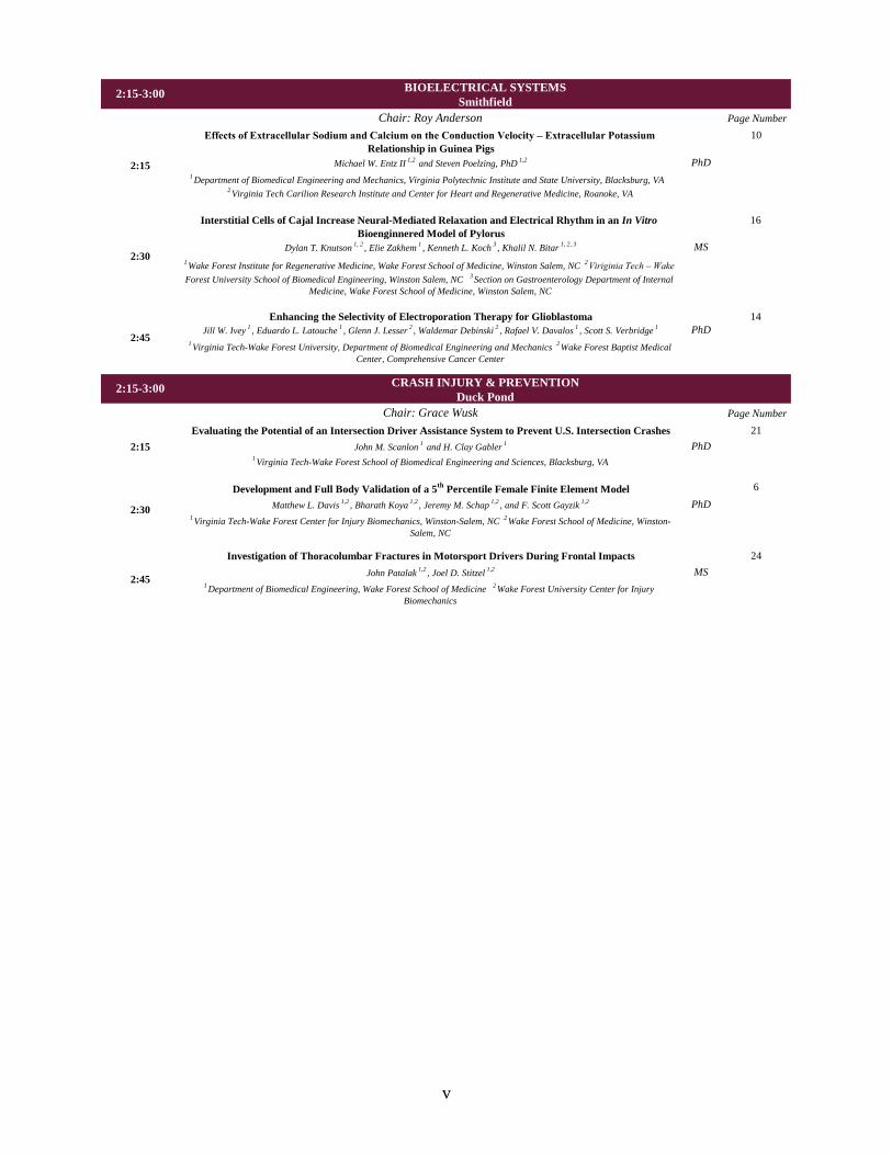
Chair: Grace Wusk
2:15-3:00
BIOELECTRICAL SYSTEMS
Smithfield
CRASH INJURY & PREVENTION
Duck Pond
Chair: Roy Anderson
2:15-3:00
1Department of Biomedical Engineering, Wake Forest School of Medicine
2Wake Forest University Center for Injury
Biomechanics
2:45
2:30
Evaluating the Potential of an Intersection Driver Assistance System to Prevent U.S. Intersection Crashes
John M. Scanlon1 and H. Clay Gabler
1
1Virginia Tech-Wake Forest School of Biomedical Engineering and Sciences, Blacksburg, VA
Development and Full Body Validation of a 5th
Percentile Female Finite Element Model
Matthew L. Davis1,2
, Bharath Koya1,2
, Jeremy M. Schap1,2
, and F. Scott Gayzik1,2
2:15
2:30
2:45
Effects of Extracellular Sodium and Calcium on the Conduction Velocity – Extracellular Potassium
Relationship in Guinea Pigs
Michael W. Entz II1,2
and Steven Poelzing, PhD1,2
1Department of Biomedical Engineering and Mechanics, Virginia Polytechnic Institute and State University, Blacksburg, VA
2Virginia Tech Carilion Research Institute and Center for Heart and Regenerative Medicine, Roanoke, VA
Interstitial Cells of Cajal Increase Neural-Mediated Relaxation and Electrical Rhythm in an In Vitro
Bioenginnered Model of Pylorus
Dylan T. Knutson1, 2
, Elie Zakhem1, Kenneth L. Koch
3, Khalil N. Bitar
1, 2, 3
1Wake Forest Institute for Regenerative Medicine, Wake Forest School of Medicine, Winston Salem, NC
2Viriginia Tech – Wake
Forest University School of Biomedical Engineering, Winston Salem, NC 3Section on Gastroenterology Department of Internal
Medicine, Wake Forest School of Medicine, Winston Salem, NC
Enhancing the Selectivity of Electroporation Therapy for Glioblastoma
Jill W. Ivey1, Eduardo L. Latouche
1, Glenn J. Lesser
2, Waldemar Debinski
2, Rafael V. Davalos
1, Scott S. Verbridge
1
1Virginia Tech-Wake Forest University, Department of Biomedical Engineering and Mechanics
2Wake Forest Baptist Medical
Center, Comprehensive Cancer Center
2:15
1Virginia Tech-Wake Forest Center for Injury Biomechanics, Winston-Salem, NC
2Wake Forest School of Medicine, Winston-
Salem, NC
Investigation of Thoracolumbar Fractures in Motorsport Drivers During Frontal Impacts
John Patalak1,2
, Joel D. Stitzel1,2
v
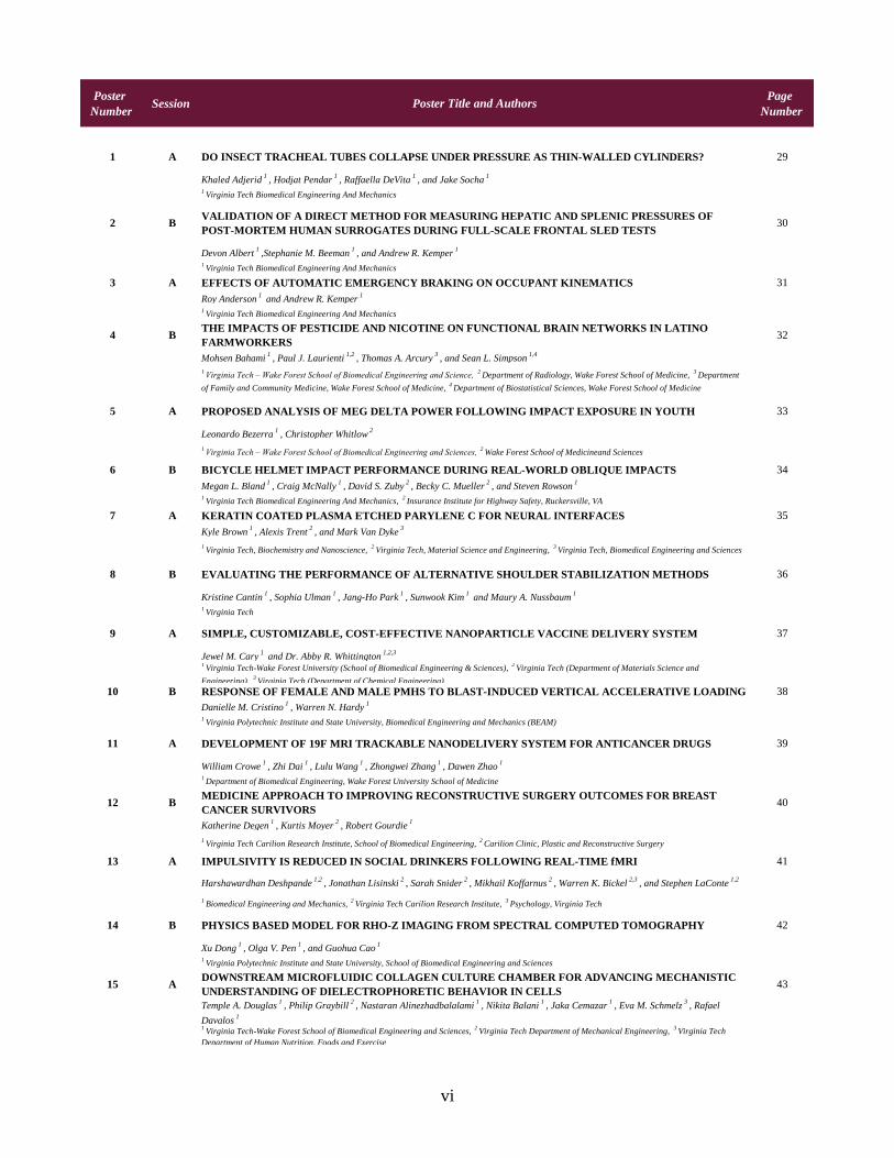
Poster
NumberSession Poster Title and Authors
Page
Number
1 A DO INSECT TRACHEAL TUBES COLLAPSE UNDER PRESSURE AS THIN-WALLED CYLINDERS? 29
Khaled Adjerid1
, Hodjat Pendar1
, Raffaella DeVita1
, and Jake Socha1
1Virginia Tech Biomedical Engineering And Mechanics
2 BVALIDATION OF A DIRECT METHOD FOR MEASURING HEPATIC AND SPLENIC PRESSURES OF
POST-MORTEM HUMAN SURROGATES DURING FULL-SCALE FRONTAL SLED TESTS30
Devon Albert1
,Stephanie M. Beeman1
, and Andrew R. Kemper1
1Virginia Tech Biomedical Engineering And Mechanics
3 A EFFECTS OF AUTOMATIC EMERGENCY BRAKING ON OCCUPANT KINEMATICS 31
Roy Anderson1
and Andrew R. Kemper1
1Virginia Tech Biomedical Engineering And Mechanics
4 BTHE IMPACTS OF PESTICIDE AND NICOTINE ON FUNCTIONAL BRAIN NETWORKS IN LATINO
FARMWORKERS32
Mohsen Bahami1
, Paul J. Laurienti1,2
, Thomas A. Arcury3
, and Sean L. Simpson1,4
1Virginia Tech – Wake Forest School of Biomedical Engineering and Science,
2Department of Radiology, Wake Forest School of Medicine,
3Department
of Family and Community Medicine, Wake Forest School of Medicine, 4
Department of Biostatistical Sciences, Wake Forest School of Medicine
5 A PROPOSED ANALYSIS OF MEG DELTA POWER FOLLOWING IMPACT EXPOSURE IN YOUTH 33
Leonardo Bezerra1
, Christopher Whitlow2
1Virginia Tech – Wake Forest School of Biomedical Engineering and Sciences,
2Wake Forest School of Medicineand Sciences
6 B BICYCLE HELMET IMPACT PERFORMANCE DURING REAL-WORLD OBLIQUE IMPACTS 34
Megan L. Bland1
, Craig McNally1
, David S. Zuby2
, Becky C. Mueller2
, and Steven Rowson1
1Virginia Tech Biomedical Engineering And Mechanics,
2Insurance Institute for Highway Safety, Ruckersville, VA
7 A KERATIN COATED PLASMA ETCHED PARYLENE C FOR NEURAL INTERFACES 35
Kyle Brown1
, Alexis Trent2
, and Mark Van Dyke3
1Virginia Tech, Biochemistry and Nanoscience,
2Virginia Tech, Material Science and Engineering,
3Virginia Tech, Biomedical Engineering and Sciences
8 B EVALUATING THE PERFORMANCE OF ALTERNATIVE SHOULDER STABILIZATION METHODS 36
Kristine Cantin1
, Sophia Ulman1
, Jang-Ho Park1
, Sunwook Kim1
and Maury A. Nussbaum1
1Virginia Tech
9 A SIMPLE, CUSTOMIZABLE, COST-EFFECTIVE NANOPARTICLE VACCINE DELIVERY SYSTEM 37
Jewel M. Cary1
and Dr. Abby R. Whittington1,2,3
1Virginia Tech-Wake Forest University (School of Biomedical Engineering & Sciences),
2Virginia Tech (Department of Materials Science and
Engineering), 3
Virginia Tech (Department of Chemical Engineering)
10 B RESPONSE OF FEMALE AND MALE PMHS TO BLAST-INDUCED VERTICAL ACCELERATIVE LOADING 38
Danielle M. Cristino1
, Warren N. Hardy1
1Virginia Polytechnic Institute and State University, Biomedical Engineering and Mechanics (BEAM)
11 A DEVELOPMENT OF 19F MRI TRACKABLE NANODELIVERY SYSTEM FOR ANTICANCER DRUGS 39
William Crowe1
, Zhi Dai1
, Lulu Wang1
, Zhongwei Zhang1
, Dawen Zhao1
1Department of Biomedical Engineering, Wake Forest University School of Medicine
12 BMEDICINE APPROACH TO IMPROVING RECONSTRUCTIVE SURGERY OUTCOMES FOR BREAST
CANCER SURVIVORS40
Katherine Degen1
, Kurtis Moyer2
, Robert Gourdie1
1Virginia Tech Carilion Research Institute, School of Biomedical Engineering,
2Carilion Clinic, Plastic and Reconstructive Surgery
13 A IMPULSIVITY IS REDUCED IN SOCIAL DRINKERS FOLLOWING REAL-TIME fMRI 41
Harshawardhan Deshpande1,2
, Jonathan Lisinski2
, Sarah Snider2
, Mikhail Koffarnus2
, Warren K. Bickel2,3
, and Stephen LaConte1,2
1Biomedical Engineering and Mechanics,
2Virginia Tech Carilion Research Institute,
3Psychology, Virginia Tech
14 B PHYSICS BASED MODEL FOR RHO-Z IMAGING FROM SPECTRAL COMPUTED TOMOGRAPHY 42
Xu Dong1
, Olga V. Pen1
, and Guohua Cao1
1Virginia Polytechnic Institute and State University, School of Biomedical Engineering and Sciences
15 ADOWNSTREAM MICROFLUIDIC COLLAGEN CULTURE CHAMBER FOR ADVANCING MECHANISTIC
UNDERSTANDING OF DIELECTROPHORETIC BEHAVIOR IN CELLS43
Temple A. Douglas1
, Philip Graybill2
, Nastaran Alinezhadbalalami1
, Nikita Balani1
, Jaka Cemazar1
, Eva M. Schmelz3
, Rafael
Davalos1
1Virginia Tech-Wake Forest School of Biomedical Engineering and Sciences,
2Virginia Tech Department of Mechanical Engineering,
3Virginia Tech
Department of Human Nutrition, Foods and Exercise
vi
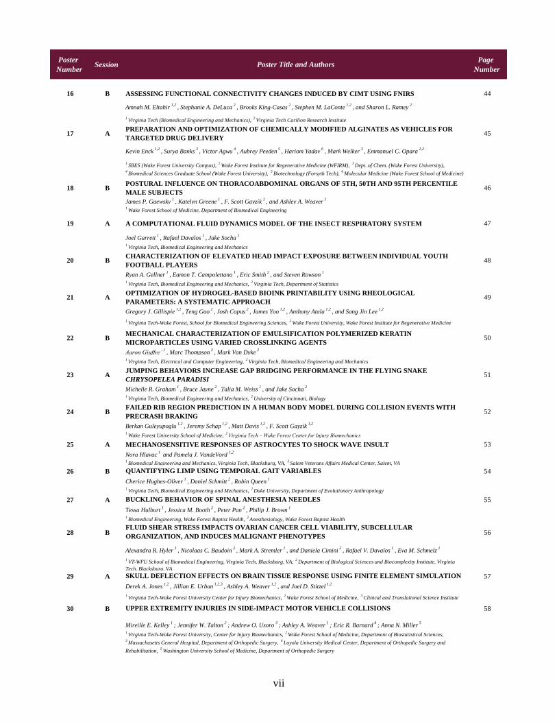
Poster
NumberSession Poster Title and Authors
Page
Number
16 B ASSESSING FUNCTIONAL CONNECTIVITY CHANGES INDUCED BY CIMT USING FNIRS 44
Amnah M. Eltahir1,2
, Stephanie A. DeLuca2
, Brooks King-Casas2
, Stephen M. LaConte1,2
, and Sharon L. Ramey2
1Virginia Tech (Biomedical Engineering and Mechanics),
2Virginia Tech Carilion Research Institute
17 APREPARATION AND OPTIMIZATION OF CHEMICALLY MODIFIED ALGINATES AS VEHICLES FOR
TARGETED DRUG DELIVERY45
Kevin Enck1,2
, Surya Banks3
, Victor Agwu4
, Aubrey Peeden5
, Hariom Yadav6
, Mark Welker3
, Emmanuel C. Opara1,2
1SBES (Wake Forest University Campus),
2Wake Forest Institute for Regenerative Medicine (WFIRM),
3Dept. of Chem. (Wake Forest University),
4Biomedical Sciences Graduate School (Wake Forest University),
5Biotechnology (Forsyth Tech),
6Molecular Medicine (Wake Forest School of Medicine)
18 BPOSTURAL INFLUENCE ON THORACOABDOMINAL ORGANS OF 5TH, 50TH AND 95TH PERCENTILE
MALE SUBJECTS46
James P. Gaewsky1
, Katelyn Greene1
, F. Scott Gayzik1
, and Ashley A. Weaver1
1Wake Forest School of Medicine, Department of Biomedical Engineering
19 A A COMPUTATIONAL FLUID DYNAMICS MODEL OF THE INSECT RESPIRATORY SYSTEM 47
Joel Garrett1
, Rafael Davalos1
, Jake Socha1
1Virginia Tech, Biomedical Engineering and Mechanics
20 BCHARACTERIZATION OF ELEVATED HEAD IMPACT EXPOSURE BETWEEN INDIVIDUAL YOUTH
FOOTBALL PLAYERS48
Ryan A. Gellner1
, Eamon T. Campolettano1
, Eric Smith2
, and Steven Rowson1
1Virginia Tech, Biomedical Engineering and Mechanics,
2Virginia Tech, Department of Statistics
21 AOPTIMIZATION OF HYDROGEL-BASED BIOINK PRINTABILITY USING RHEOLOGICAL
PARAMETERS: A SYSTEMATIC APPROACH49
Gregory J. Gillispie1,2
, Teng Gao2
, Josh Copus2
, James Yoo1,2
, Anthony Atala1,2
, and Sang Jin Lee1,2
1Virginia Tech-Wake Forest, School for Biomedical Engineering Sciences,
2Wake Forest University, Wake Forest Institute for Regenerative Medicine
22 BMECHANICAL CHARACTERIZATION OF EMULSIFICATION POLYMERIZED KERATIN
MICROPARTICLES USING VARIED CROSSLINKING AGENTS50
Aaron Giuffre’1
, Marc Thompson2
, Mark Van Dyke2
1Virginia Tech, Electrical and Computer Engineering,
2Virginia Tech, Biomedical Engineering and Mechanics
23 AJUMPING BEHAVIORS INCREASE GAP BRIDGING PERFORMANCE IN THE FLYING SNAKE
CHRYSOPELEA PARADISI51
Michelle R. Graham1
, Bruce Jayne2
, Talia M. Weiss1
, and Jake Socha2
1Virginia Tech, Biomedical Engineering and Mechanics,
2University of Cincinnati, Biology
24 BFAILED RIB REGION PREDICTION IN A HUMAN BODY MODEL DURING COLLISION EVENTS WITH
PRECRASH BRAKING52
Berkan Guleyupoglu1,2
, Jeremy Schap1,2
, Matt Davis1,2
, F. Scott Gayzik1,2
1Wake Forest University School of Medicine,
2Virginia Tech – Wake Forest Center for Injury Biomechanics
25 A MECHANOSENSITIVE RESPONSES OF ASTROCYTES TO SHOCK WAVE INSULT 53
Nora Hlavac1
and Pamela J. VandeVord1,2
1Biomedical Engineering and Mechanics, Virginia Tech, Blacksburg, VA,
2Salem Veterans Affairs Medical Center, Salem, VA
26 B QUANTIFYING LIMP USING TEMPORAL GAIT VARIABLES 54
Cherice Hughes-Oliver1
, Daniel Schmitt2
, Robin Queen1
1Virginia Tech, Biomedical Engineering and Mechanics,
2Duke University, Department of Evolutionary Anthropology
27 A BUCKLING BEHAVIOR OF SPINAL ANESTHESIA NEEDLES 55
Tessa Hulburt1
, Jessica M. Booth2
, Peter Pan2
, Philip J. Brown1
1Biomedical Engineering, Wake Forest Baptist Health,
2Anesthesiology, Wake Forest Baptist Health
28 BFLUID SHEAR STRESS IMPACTS OVARIAN CANCER CELL VIABILITY, SUBCELLULAR
ORGANIZATION, AND INDUCES MALIGNANT PHENOTYPES56
Alexandra R. Hyler1
, Nicolaas C. Baudoin2
, Mark A. Stremler1
, and Daniela Cimini2
, Rafael V. Davalos1
, Eva M. Schmelz1
1VT-WFU School of Biomedical Engineering, Virginia Tech, Blacksburg, VA,
2Department of Biological Sciences and Biocomplexity Institute, Virginia
Tech, Blacksburg, VA
29 A SKULL DEFLECTION EFFECTS ON BRAIN TISSUE RESPONSE USING FINITE ELEMENT SIMULATION 57
Derek A. Jones1,2
, Jillian E. Urban1,2,3
, Ashley A. Weaver1,2
, and Joel D. Stitzel1,2
1Virginia Tech-Wake Forest University Center for Injury Biomechanics,
2Wake Forest School of Medicine,
3Clinical and Translational Science Institute
30 B UPPER EXTREMITY INJURIES IN SIDE-IMPACT MOTOR VEHICLE COLLISIONS 58
Mireille E. Kelley1
; Jennifer W. Talton2
; Andrew O. Usoro3
; Ashley A. Weaver1
; Eric R. Barnard4
; Anna N. Miller5
1Virginia Tech-Wake Forest University, Center for Injury Biomechanics,
2Wake Forest School of Medicine, Department of Biostatistical Sciences,
3Massachusetts General Hospital, Department of Orthopedic Surgery,
4Loyola University Medical Center, Department of Orthopedic Surgery and
Rehabilitation, 5
Washington University School of Medicine, Department of Orthopedic Surgery
vii
Poster
NumberSession Poster Title and Authors
Page
Number
31 A HOW TEMPERATURE INFLUENCES THE VISCOSITY OF HORNWORM HEMOLYMPH 59
Melissa C. Kenny1
, Matthew N. Giarra2
, and John J. Socha1
1Virginia Tech, Department of Biomedical Engineering and Mechanics,
2Virginia Tech, Department of Mechanical Engineering
32 B ENGINEERING THE GUT-LIVER-BRAIN AXIS TO INVESTIGATE CHEMICAL TOXICITY 60
Anjaney Kothari1
, Rebekah Less1
and Padma Rajagopalan1, 2, 3
1School of Biomedical Engineering and Sciences,
2Department of Chemical Engineering,
3ICTAS Center for Systems Biology of Engineered Tissues
(ISBET)
33 A INTEGRATED IN VITRO GUT-LIVER MODEL FOR THE INVESTIGATION OF FIRST PASS METABOLISM 61
Rebekah Less1
, Anjaney Kothari1
, Padma Rajagopalan1, 2, 3
1School of Biomedical Engineering and Sciences, Virginia Tech, Blacksburg VA, 24060,
2Chemical Engineering, Virginia Tech, Blacksburg VA, 24060,
3ICTAS Center for Systems Biology of Engineered Tissues, Virginia Tech, Blacksburg VA, 24060
34 BIRREVERSIBLE ELECTROPORATION FOR THE ABLATION OF PANCREATIC MALIGNANCIES: A
PATIENT-SPECIFIC METHODOLOGY62
Melvin F. Lorenzo1
, Eduardo L. Latouche1
, Michael B. Sano2,3
, Robert C. G. Martin II4
, Rafael V. Davalos1
1Biomedical Engineering and Mechanics, Virginia Polytechnic Institute and State University, Blacksburg, Virginia,
2Radiation Oncology, Stanford
University School of Medicine, Stanford, California, 3
UNC/NCSU Joint Department of Biomedical Engineering, Chapel Hill, North Carolina, 4
Surgery,
Division of Surgical Oncology, University of Louisville, Louisville, Kentucky
35 ACELL TYPE-SPECIFIC MOUSE BRAIN METHYLOMES PROFILED USING AN ULTRALOW-INPUT
MICROFLUIDIC DEVICE63
Sai Ma1
, Zhixiong Sun2
, Chen Sun1
, Travis W. Murphy3
, Hehuang Xie2,4
, Chang Lu2
1School of Biomedical Engineering and Sciences, Virginia Tech,
2Department of Biological Sciences, Virginia Tech,
3Chemical Engineering, Virginia
Tech, 4
Biocomplexity Institute, Virginia Tech
36 B 3D PRINTING OF CANCER ORGANOIDS FOR HIGH THROUGHPUT SCREENING 64
Andrea Mazzocchi1
, Shay Soker1
, Aleksander Skardal1,2
1Wake Forest Institute for Regenerative Medicine, Wake Forest School of Medicine, Winston-Salem, NC,
2Comprehensive Cancer Center of Wake Forest
School of Medicine, Winston-Salem, NC
37 AHYPERTHERMIA-INDUCING NANOPARTICLES AS ADJUVANT THERAPY FOR OXALIPLATIN-BASED
CHEMOTHERAPY IN COLORECTAL CANCER65
Bryce D. McCarthy1
and Nicole H. Levi-Polyachenko1
1Wake Forest School of Medicine, Department of Plastic and Reconstructive Surgery
38 BAN INVESTIGATION INTO THE VALIDATION AND FUTURE USE OF FE HYBRID III, THOR, AND
GHBMC M50-OS FOR SPACEFLIGHT CONFIGURATION TESTING66
Kyle P. McNamara1,2
, Derek A. Jones1,2
, James P. Gaewsky1,2
, F. Scott Gayzik1,2
, Ashley A. Weaver1,2
, Joel D. Stitzel1,2
1Wake Forest School of Medicine,
2Virginia Tech-Wake Forest University Center for Injury Biomechanics
39 ACHARACTERIZING HEAD IMPACT EXPOSURE IN YOUTH FEMALE SOCCER WITH A CUSTOM-
INSTRUMENTED MOUTHPIECE67
Logan E. Miller1
, Elizabeth Pinkerton1
, Lyndia Wu2
, Katie Fabian1
, David Camarillo2
, Joel D. Stitzel1
, and Jillian E. Urban1
1Department of Biomedical Engineering, Wake Forest School of Medicine,
2Department of Bioengineering, Stanford University
40 B AGENT-BASED MODELING TO PREDICT THE EFFECT OF ELECTROCHEMOTHERAPY ON TUMORS 68
Maryam Moarefian1
, Luke Achenie2
1Virginia Polytechnic Institute and State University, Mechanical Engineering Department,
2Virginia Polytechnic Institute and State University, Chemical
Engineering Department
41 A NOVEL DEVICE FOR PREVENTING RODENT WOUND SPLINT REMOVAL 69
Jade Montgomery1,2
, Linda J. Jourdan2
, and Robert G. Gourdie1,2
1Virginia Tech, School of Biomedical Engineering and Sciences,
2Virginia Tech Carilion Research Institute
42 B MITIGATING BIOFILM FORMATION WITH SURFACE TOPOGRAPHY MODIFICATIN 70
Carolyn Y. Mottley1
, Zhou Ye2
, AhRam Kim2
, and Bahareh Behkam1,2
1Virginia Tech, Department of Mechanical Engineering,
2Virginia Tech, School of Biomedical Engineering and Sciences
43 ATHE DEVELOPMENT OF AN INTELLIGENT SURGICAL PROBE FOR REAL-TIME MONITORING OF
ELECTROPORATION BASED TREATMENTS71
Timothy J. O’Brien1
, Mohammad Bonakdar2
, and Rafael V. Davalos1
1Virginia Tech, Biomedical Engineering and Mechanics,
2Virginia Tech, Mechanical Engineering
44 BCORTICAL THINNING AND STRUCTURAL BONE CHANGES IN NON-HUMAN PRIMATES FOLLOWING
72
Catherine Okoukoni, Ph.D.1,2,3
, Michael Farris, M.D.1
, Emory R. McTyre, M.D.1
, J. Daniel Bourland, Ph.D.1,2
, J. Mark Cline, DVM
Ph.D.4
, Greg Dugan DVM4
, Jeffrey S. Willey, Ph.D.1,3
1Wake Forest University, Department of Radiation Oncology,
2Wake Forest University, Department of Physics,
3Wake Forest University, Pathology
Department, 4
Wake Forest University School of Medicine
viii
Poster
NumberSession Poster Title and Authors
Page
Number
45 AA FINITE ELEMENT MODEL OF A SMALL-STATURE FEMALE PEDESTRIAN FOR SIMULATING
TRAFFIC ACCIDENTS73
Wansoo Pak1
, Costin D. Untaroiu1
, Scott Gayzik2
1Department of Biomedical Engineering and Mechanics, Virginia Tech, Blacksburg, VA, USA,
2Department of Biomedical Engineering, Wake Forest
University School of Medicine, Winston-Salem, NC, USA
46 BQUANTITATIVE ANALYSIS OF THE EPIDERMAL NECROSIS IN CUTANEOUS RADIATION INJURIES
INDUCED BY BETA-SOURCE IRRADIATION74
Olga V. Pen1,2
, Peter A. Antinozzi3
, Nancy D. Kock4
, J. Daniel Bourland1,2
1Department of Radiation Oncology, Wake Forest School of Medicine, Wake Forest University,
2Department of Biomedical Engineering, VT-WFU SBES,
Wake Forest University, 3
Department of Biochemistry, Wake Forest University, 4
Department of Pathology and Comparative Medicine, Wake Forest
School of Medicine, Wake Forest University
47 ACHARACTERISTIC DIFFERENCES BETWEEN WATER DIALYZED AND BUFFER DIALYZED KERATOSE
BIOMATERIAL75
Nils A. Potter1
, Mark Van Dyke1
1Virginia Tech, Department of Biomedical Engineering and Mechanics
48 B 3D CELL CULTURE STUDIES IN MICROFLUIDIC SYSTEMS FOR TRANSLATIONAL APPLICATIONS 76
Shiny Rajan1,2,3
, Parker Hambright1
, Aleksander Skardal1,3,4
, and Adam R. Hall1,2,3,4
1Wake Forest University,
2Virginia Tech – Wake Forest School of Biomedical Engineering and Sciences,
3Wake Forest Institute of Regenerative Medicine,
4Comprehensive Cancer Institute, Wake Forest
49 A CHANGES IN LIMB SYMMETRY DURING A 2 MILE OUTDOOR RUN 77
Kristen E. Renner1
, Robin M Queen1
1Virginia Tech, Department of Biomedical Engineering and Mechanics
50 BEFFECT OF NON-STEROIDAL ANTI-INFLAMMATORY DRUGS ON ACHILLES TENDINOPATHY IN A
MURINE MODEL78
Sabah N. Rezvani1
, Adam Bitterman2
, Anna Plaas3
, Jun Li3
, Vincent Wang1
1Virginia Tech, Biomedical Engineering and Mechanics,
2Northwell Health, Huntington, NY,
3Rush University Medical Center, Chicago, IL
51 AHIGH-FREQUENCY IRREVERSIBLE ELECTROPORATION TARGETS MALIGNANT CELLS IN OVARIAN
CANCER MODEL79
Andrea Rolong1
, Eva M. Schmelz2
, and Rafael V. Davalos1
1Virginia Tech – Wake Forest University School of Biomedical Engineering and Sciences,
2Virginia Tech, Department of Human Nutrition, Foods, and
Exercise
52 BPELVIC ANATOMICAL CHARACTERIZATION AND COMPARISON BY SEX AND HISTORY OF
PREGNANCY80
Mona Saffarzadeh1,2
, R. Caresse Hightower1,2
, Ashley A. Weaver1,2
1Virginia Tech-Wake Forest University Center for Injury Biomechanics,
2Wake Forest University School of Medicine, Biomedical Engineering Department
53 AEFFECTS OF FEBUXOSTAT ON SOCIAL INTERACTION AND REPETITIVE GROOMING IN AUTISM
MICE 81
Vince Sannicalos1
, Molly Accord2
, Jaegu (Richard) Yea3
, Sukyoung (Chloe) Kim4
, Jamelle Simmons5
, and Yong Woo Lee5
1Virginia Tech, School of Neuroscience,
2Virginia Tech, Department of Chemical Engineering (ChemE),
3Virginia Tech, Department of Biological
Systems Engineering (BSE), 4
Virginia Tech, Department of Biochemistry, 5
Virginia Tech, Biomedical Engineering and Mechanics (BEAM)
54 B OPTICAL FIBER BASED IMAGING OF BIOENGINEERED TISSUE CONSTRUCT 82
Etai Sapoznik1,2
, Guoguang Niu2
, Peng Lu3
, Yu Zhou2
, Yong Xu3
, Shay Soker1,2
1Virginia Tech-Wake Forest Univ. School of Biomedical Engineering and Sciences,
2Wake Forest Institute for Regenerative Medicine,
3Virginia Tech,
Bradley Department of Electrical and Computer Engineering
55 ATRANSITIONAL LIVER MODELS FOR THE INVESTIGATION OF CHEMICAL AND MECHANICAL CUES
DURING THE PROGRESSION OF FIBROSIS83
Scott-Eugene Saverot1
, Sophia Orbach2
, Andrew Ford2
, Padmavathy Rajagopalan1, 2, 3
1Virginia Tech, School of Biomedical Engineering and Sciences,
2Virginia Tech, Department of Chemical Engineering,
3Virginia Tech, ICTAS Center for
Systems Biology of Engineered Tissues
56 BDIFFUSION MODEL ACROSS A BLOOD-BRAIN BARRIER (BBB) MIMIC FOR THE TREATMENT OF
AUTISM SPECTRUM DISORDER (ASD)84
Jamelle Simmons1
, Luke Achenie2,3
, and Yong W. Lee1,3
1Virginia Tech, Department of Biomedical Engineering and Mechanics (BEAM),
2Virginia Tech, Department of Chemical Engineering,
3Virginia Tech
Center for Autism Research (VTCAR)
57 A A SIMPLE TARGETED METHOD FOR THE REMOVAL OF FREE HEMOGLOBIN 85
Kelli N. Simms1
, Martin Guthold2
, Daniel B. Kim-Shapiro2
, Elaheh Rahbar1
*1
Department of Biomedical Engineering, Wake Forest School of Medicine, Winston Salem, NC, 2
Department of Physics, Wake Forest University, Winston
Salem, NC
58 BUSING VIDEOTAPED MOTION CODING TO IMPROVE UNDERSTAND OF F-NIRS SIGNAL CHANGES
86
Ben Stephens1
, Amnah Eltahir2
, Stephen LaConte3
, and Stephanie DeLuca3
1Virginia Tech Carilion School of Medicine,
2Virginia Tech, Biomedical Engineering,
3Virginia Tech Carillion Research Institute
59 A CALCULATION OF THE DIFFUSIVE PERMABILITY OF CELL MEMBRANES TO PROPIDIUM 87
Daniel C. Sweeney1
, James C. Weaver2
, Rafael V. Davalos1
1Virginia Tech, Dpeartment of Biomedical Engineering and Mechanics,
2MIT, Harvard-MIT Division of Health Sciences and Technology
ix
Poster
NumberSession Poster Title and Authors
Page
Number
60 B REAR IMPACT CRASHES IN THE UNITED STATES: HOW DANGEROUS ARE THEY? 88
Whitney M. Tatem1
and H. Clay Gabler1
1Virginia Tech, Department of Biomedical Engineering and Mechanics
61 BA MECHANISTIC EVALUATION OF INTRINSIC CROSSLINKING PROPERTIES AND SYNTHESIS
PROCEDURES FOR KERATIN-BASED MICROPARTICLES89
Marc Thompson1
, Aaron Giuffre’2
, Mark Van Dyke1
1Virginia Tech. Blacksburg, VA, Biomedical Engineering and Mechanics,
2Virginia Tech. Blacksburg, VA, Electrical and Computer Engineering
62 ACELL-SUBSTRATE INTERACTIONS ON CHARACTERIZED KERATIN COATING FOR PERCUTANEOUS
PROSTHETIC APPLICATIONS90
Alexis Trent1
, Mark Van Dyke2
1Department of Material Science and Engineering, Virginia Tech, Blacksburg, VA,
2Department of Biomedical Engineering and Mechanics, Virginia Tech,
Blacksburg, VA
63 B ANALYSIS OF BILIARY STENT HINGES USING ABAQUS 91
Aaron R. Van Gorkom1
, Greg J. Gillispie1
, Clifford Howard Jr2
, Philip J. Brown1
1Biomedical Engineering, Wake Forest Baptist Health,
2Wake Forest University Baptist Medical Center
64 ALASER INDUCED PRECISION HEATING FOR IMPROVED BACTERIAL DESTRUCTION WITH
GENTAMICIN 92
Kenneth A. Vogel1
, Anila Pullagura1
, Nicole Levi-Polyachenko1
1Wake Forest University (School of Medicine, Plastic and Reconstructive Surgery)
65 BSHIFTS IN CIRCULATING POLYUNSATURATED FATTY ACID LEVELS IN CHILDREN WITH
TRAUMATIC BRAIN INJURY93
Charlotte Mae K. Waits1
, Steven C. Kosmach2
, Susan Sergeant3
, Floyd H. Chilton4
, Charles S. Cox, Jr2
, and Elaheh Rahbar1
1Department of Biomedical Engineering, Wake Forest School of Medicine,
2Department of Pediatric Surgery, McGovern Medical School at the University
of Texas Health Sciences Center at Houston, 3
Department of Biochemistry, Wake Forest School of Medicine, 4
Department of Physiology and
Pharmacology, Wake Forest School of Medicine
66 AHIGHLY SPECIFIC AND MODULAR AFFINITY LABELING OF EPIGENETIC MODIFICATIONS
94
Fanny Wang1
, Osama K. Zahid1
, Brandi E. Swaibe2
, and Adam R. Hall1,3
1Virginia Tech-Wake Forest School of Biomedical Engineering and Sciences,
2Wake Forest University, Department of Physics,
3Wake Forest University
School of Medicine, Comprehensive Cancer Center
67 BFABRICATING AND TUNING AN ELASTOMERIC BLOOD VESSEL FOR USE IN CORONARY ARTERY
BYPASS SURGERIES95
Harleigh J. Warner1
and William D. Wagner1
1Plastic and Reconstructive Surgery and School of Biomedical Engineering and Sciences, Wake Forest University School of Medicine, Winston-Salem, NC
68 AA RAT MODEL OF SEVERE OSTEOPOROSIS FOR TESTING BONE REGENERATION CONSTRUCTS
96
Michele Waters1
, Nils Potter1
, Marc Thompson1
, Alexis Trent2
, Pamela VandeVord1
and Mark Van Dyke1
1Virginia Tech, Biomedical Engineering and Mechanics,
2Virginia Tech, Materials Science and Engineering
69 BNON-INVASIVE DETECTION OF RESPIRATION AND HEART RATE WITH A VEHICLE SEAT WEIGHT
SENSOR: A FEASIBILITY STUDY97
Grace C. Wusk1
and Hampton C. Gabler1
1Virginia Tech, Biomedical Engineering
70 AINVESTIGATION OF MLL3 COMPLEX IN DNA REPAIR MECHANISM IN LUNG ADENOCARCINOMA
98
Ted G. Xiao1
, Wei Zhang2
1Wake Forest School of Medicine, Virginia Tech-Wake Forest School of Biomedical Engineering and Sciences,
2Wake Forest School of Medicine,
Department of Cancer Biology
71 BDEVELOPMENT AND VALIDATION OF A GOTTINGEN MINIATURE PIG FINITE ELEMENT MODEL TO
INVESTIGATE INJURY SCALING TECHNIQUES99
Keegan M. Yates1
, Costin D. Untaroiu1
1Virginia Tech, Center for Injury Biomechanics
72 ANUMERICAL INVESTIGATION OF LOWER EXTREMITY INJURIES IN FRONTAL CRASH
RECONSTRUCTION 100
Xin Ye1,2
, James Gaewsky1,2
, Derek Jones1,2
, Logan Miller1,2
, Mireille Kelly1,2
, Jeff Suhey1,2
, Bharath Koya1,2
, Ashley Weaver1,2
and Joel Stitzel1,2
1Wake Forest University School of Medicine
73 B PERICYTE CELL LINE ISOLATION, VALIDATION AND APPLICATIONS 101
Huaning Zhao1
, John C. Chappell1,2
1School of Biomedical Engineering and Science,
2Center for Heart and Regenerative Medicine, Virginia Tech Carilion Research Institute
x

xi

xii
Thank you to Wake Forest Innovations for their gracious sponsorship of the 2017 SBES Graduate Student Research Symposium.
Wake Forest Innovations improves health through collaborative innovation so that important scientific
discoveries can become life-improving realities.
We accomplish this through our three Centers:
Center for Technology Innovation & Commercialization – accelerates the development and
commercialization of inventions.
Center for Industry Research Collaboration – expedites access to specialized clinical and
research capabilities.
Center for Applied Learning – promotes best clinical practices through experiential training
We believe collaborative innovation between our faculty and staff and with industry is based on sharing
expertise, knowledge, risk and reward at all stages of product research and development.
For more information, please visit their website at www.WakeForestInnovations.com.
Wake Forest Innovations 575 North Patterson Avenue, Suite 550 Winston-Salem, NC 27101 [email protected]
xiii
Thank you to Medtronic for their gracious sponsorship of the 2017 SBES Graduate Student Research Symposium. Visit Medtronic’s website to discover how they are creating life-changing therapies to help people with chronic diseases.
As a global leader in medical technology, services and solutions, Medtronic improves the health
and lives of millions of people each year. We believe our deep clinical, therapeutic and
economic expertise can help address the complex challenges — such as rising costs, aging
populations and the burden of chronic disease — faced by families and healthcare systems
today. But no one can do it alone. That’s why we’re committed to partnering in new ways and
developing powerful solutions that deliver better patient outcomes.
Founded in 1949 as a medical repair company, we're now among the world's largest medical
technology, services and solutions companies, employing more than 85,000 people worldwide,
serving physicians, hospitals and patients in more than 155 countries. Join us in our
commitment to take healthcare Further, Together. Learn more at Medtronic.com.
Medtronic Vascular Innovations 3576 Unocal Place Santa Rosa, CA 95403-1774

xiv
Thank you to the Biomedical Engineering Society for their gracious sponsorship of the 2017 SBES Graduate Student Research Symposium. Please visit their website to learn how they have become the leading society of professionals devoted to developing and using engineering to advance human health and well being.
The Biomedical Engineering Society (BMES) is the professional society for biomedical engineering and
bioengineering. Founded in early 1968, the Society now boasts over 7,000 members and is growing, rapidly.
BMES serves as the lead society and professional home for biomedical engineering and bioengineering. Our
leadership in accreditation, potential licensure, publications, scientific meetings, global programs, and diversity
initiatives, as well as our commitment to ethics, all serve our mission to promote and enhance knowledge and
education in biomedical engineering and bioengineering worldwide and its utilization for human health and well-
being.
The Vision of BMES is to serve as the world's leading society of professionals devoted to developing and using
engineering and technology to advance human health and well-being.
The Mission of BMES is to build and support the biomedical engineering community, locally, nationally and
internationally, with activities designed to communicate recent advances, discoveries, and inventions; promote
education and professional development; and integrate the perspectives of the academic, medical, governmental,
and business sectors.
Leading and emerging researchers use BMES as a platform for sharing the latest information and research in the
profession. BMES provides industry members exposure to an expanding market. Advertising and sponsorship
opportunities can help increase your company’s publicity year-round.
For more information, please visit the BMES website at www.bmes.org BMES 8201 Corporate Drive, Suite 1125 Landover, MD 20785-2224 [email protected]

16th Annual Graduate Student
Research Symposium
Student Abstracts
1 Evaluations of Head Injury Oral: Drillfield, 1:00
16th Annual Graduate Student Research Symposium, May 10, 2017
EPIGENETIC MECHANISMS IN BLAST INDUCED NEUROTRAUMA
Zachary S. Bailey1, Pamela J. VandeVord1,2
1. Virginia Tech, Biomedical Engineering and Mechanics 2. Salem Veterans Affairs Medical Center
Corresponding Author: Zachary Bailey, Email: [email protected] INTRODUCTION More than 25% of the Veterans returning from Operations Enduring and Iraqi Freedom, and New Dawn (OEF/OIF/OND) are suffering from closed head injuries due to blast exposure1. Despite its frequency, no cure exists and the injury mechanisms are poorly understood. Blast waves typically originate following an explosion. The rapid expansion of combustion products from the detonation leads to the outward, supersonic, flow of the surrounding air, deemed the blast wave. The displacement of air from the source leads to areas of negative pressure relative to ambient conditions. This triggers the reversal of flow towards to blast origin and re-establishes ambient pressures. At a distant observation point from the blast origin, these complex flow dynamics will lead to a pressure profile characterized by a rapid rise in pressure followed by an exponential decay and a brief negative phase before returning to ambient conditions. Clinical manifestations of blast induced neurotrauma (BINT) involve long-term functional and psychological impairments driven by underlying neuropathology that including oxidative stress, blood brain barrier disruption and reactive astrocytosis. The overall hypothesis of this work is that epigenetic regulatory mechanisms contribute to the progression of the BINT pathology and neurological impairments. Epigenetic mechanisms, including DNA methylation and histone acetylation, are important processes by which cells respond to various environmental stimuli and regulate transcription. To date, the role of epigenetics in BINT remains unknown. METHODOLOGY The blast wave was generated using a custom Advanced Blast Simulator that consists of three distinct sections to create, develop, and dissipate the blast wave. The wave originates following helium-driven rupture of calibrated acetate membranes. Pressure measurements were collected at 250 kHz using a Dash 8HF data acquisition system. Analysis of pressure profiles was conducted using a custom Matlab script. Following blast exposure, animals
underwent established behavioral tests to assess functional and psychological impairments. Reverse-transcription polymerase chain reaction, Western Blot, and immunofluorescence techniques were used to assess protein levels/modifications, gene expression, and DNA modifications at various time points following blast. RESULTS The progression of the BINT pathology is manifested as motor function deficits, increased anxiety, and decreased memory function. The underlying neuropathology involves both DNA methylation and histone acetylation changes. Analysis of DNA methylation levels following blast exposure elucidated time-dependent DNA methylation decreases in the hippocampus, a critical region in memory function. No DNA methylations changes were observed in the acute stages of injury but were prevalent in the sub-acute stages which indicate a role in secondary injury cascades. We also observed histone acetylation decreases for histones H2b, H3, and H4. After observing significant increases in astrocyte activation, immunoflourescence showed decreased H3 acetylation in astrocytes following blast exposure. Western blot analysis of intra-cellular signaling molecules responsible for regulating histone acetylation showed increased nuclear localization. CONCLUSIONS The observed changes to DNA methylation and histone acetylation have a potentially broad impact cellular function which may be important to the injury progression. Chronically, BINT increases risk for Alzheimer’s disease and post-traumatic stress disorder which may also involve such changes2. Continuing to increase our understanding of BINT will mitigate current clinical obstacles by making the development of treatment strategies more efficient. REFERENCES 1. MacGregor et al., (2011). J Head Trauma Rehabil. 2. Hoge et al. (2008). N Engl. J Med.
Oral: Duckpond, 9:45 Modeling the Human Body 2
16th Annual Graduate Student Research Symposium, May 10, 2017
DEVELOPMENT AND VALIDATION OF A FINITE ELEMENT MODEL OF THE WIAMAN LOWER
EXTREMITY
Wade A. Baker1, Costin Untaroiu1, and Mostafiz Chowhurdy2
1. Virginia Tech, BEAM 2. Army Research Laboratory
INTRODUCTION Improvised explosive devices were extensively used by insurgents to target occupants of military vehicles during the conflicts in Iraq and Afghanistan. Casualties of an IED attack are highly susceptible to lower extremity injuries. In a survey of 3,575 extremity wounds, explosive munitions accounted for 75% of injuries [1]. Automotive anthropomorphic test devices (ATD) have been optimized to mimic the kinematic and dynamic responses of a human during the frontal and side impacts characteristic of automotive collisions. However, when subjected to vertical loading conditions these ATDs exhibit poor biofidelity [2]. In order to assess vehicle safety and make informed improvements to vehicle design, a novel Anthropomorphic Test Device (ATD) was developed and optimized for vertical loading. The main objective of this study was to develop and validate a finite element (FE) model of the ATD lower extremity. METHODOLOGY A numerical model of the lower extremity was developed for LS-DYNA based on geometry of the physical dummy. All connections, including joints and hardware, were explicitly modeled and deformable. The soft materials in the dummy, which have the greatest influence on biofidelity, were characterized through uniaxial tension and compression tests. Non-linear strain rate dependent materials were implemented in the FE model based on characterization results. Experiments conducted on the Vertically Accelerated Load Transfer System (VALTS) were used to validate the lower extremity model. Validation was performed with and without the presence of a military combat boot. Comparison between simulation outputs and associated test data was used to validate the FE model. Correlation scores between the simulations and experiments were calculated using the objective rating system Corrlelation and Analysis (CorA). RESULTS
The proposed numerical models of materials exhibiting viscoelastic responses show good correlation to the specimen test data at both high and low strain rates. An example validation is shown in Figure 1 for the unbooted model at an impact rate of 2 m/s. The unbooted model was validated at four different loading rates (max 6m/s) and the booted model was validated at five loading rates (max 12m/s).
Figure 1: Comparison of FE to experiment at the load
cell built in to the tibia shaft of the ATD. CONCLUSIONS Simulations of the entire WIAMan-LX correlate well to the WIAMan physical dummy tests. Good results of the model backed by high objective rating scores lead to the recommendation to use it in numerical studies related to dummy design improvements. ACKNOWLEDGMENTS The authors would like to thank Army Research Laboratory for their support REFERENCES [1] Owens BD, Kragh JF, Wenke JC, Macaitis J, Wade CE, Holcomb JB. Combat wounds in operation Iraqi Freedom and operation Enduring Freedom. J Trauma 2008 [2] Bir C, Barbir A, Dosquet F, Wilhelm M, van der Horst M, Wolfe G. Validation of lower limb surrogates as injury assessment tools in floor impacts due to anti-vehicular land mines. Mil Med 2008
Tibi
a Fo
rce
(kN
)
3 The Tumor & Tissue Microenvironments Oral: Drillfield, 10:00
16th Annual Graduate Student Research Symposium, May 10, 2017
N-(3-OXODODECANOYL)-L-HOMOSERINE LACTONE INTERACTIONS IN THE BREAST TUMOR
MICROENVIRONMENT: IMPLICATIONS FOR BREAST CANCER VIABILITY AND PROLIFERATION IN VITRO
Brittany N. Balhouse1, Logan Patterson
2,3, Eva M. Schmelz
4, Daniel J. Slade
2 and Scott S. Verbridge
1
1. School of Biomedical Engineering and Sciences, Virginia Tech-Wake Forest University, Blacksburg, VA, USA
2. Department of Biochemistry, Virginia Tech, Blacksburg, VA, USA
3. Department of Pathology, University of Virginia, Charlottesville, VA, USA
4. Department of Human Nutrition, Foods and Exercise, Virginia Tech, Blacksburg, VA, USA
Corresponding Author: Brittany N. Balhouse, Email: [email protected]
INTRODUCTION
Hypoxia and dense tumor stroma in the breast tumor
microenvironment predispose cancer progression and
therapy resistance, yet the role of the breast resident
microbiome in this interplay remains uncharacterized. We
hypothesized that the effect of breast relevant bacterial
molecules on breast cell viability and proliferation would
be cell type and culture condition dependent.
METHODOLOGY
We used viability, proliferation, and apoptosis/necrosis
assays in order to assess the effect of bacteria quorum-
sensing molecule N-(3-oxododecanoyl)-L-homoserine
lactone (OdDHL) on human breast adenocarcinoma cells
(MDA-MB-231, MCF-DCIS.com) and non-malignant
breast epithelial cells (MCF-10A) in normoxia and
hypoxia (1% O2) in 2D and in a normoxic 3D
environment (type I collagen hydrogel tissue mimic).
RESULTS
OdDHL selectively decreases viability in breast cancer
cells and has no significant effect on the viability of
normal breast epithelial cells (Fig 1). The effect of
OdDHL is dependent both on cell type and the culture
condition to which the cells are exposed (Fig 1).
Figure 1: The effect of OdDHL is cell type and culture
condition dependent.
It was found that OdDHL treatment not only decreases
proliferation of MDA-MB-231 cells (data not shown), but
also selectively increases MDA-MB-231 necrosis in all
culture conditions (Fig 2). OdDHL also decreased
apoptosis and necrosis for MCF-10A cells in the
3D/normoxia condition (Fig 2).
Figure 2: OdDHL selectively induces necrosis in MDA-
MB-231 cells in all culture conditions.
Thus, the decrease in MDA-MB-231 viability associated
with the OdDHL treatment (Fig 1) is caused by both
decreased proliferation and induction of necrosis (Fig 2).
CONCLUSIONS
This study demonstrates the importance of microbiome-
produced molecules in the tumor microenvironment.
Though the effect of OdDHL was blunted with hypoxia
and 3D culture, it still significant decreased MDA-MB-
231 viability. OdDHL and other microbiome-produced
molecules may hold promise as anti-cancer therapies.
ACKNOWLEDGMENTS
We would like to thank Sara Peterson, Dr. Akanksha
Kanitkar, Dr. Ann Stevens for their assistance.
Oral: Drillfield, 1:45 Evaluations of Head Injury 4
16th Annual Graduate Student Research Symposium, May 10, 2017
HIGH MAGNITUDE HEAD IMPACT EXPOSURE IN YOUTH FOOTBALL
Eamon T. Campolettano1, Ryan A. Gellner1, and Steven Rowson1
1. Biomedical Engineering and Mechanics (Virginia Tech) Corresponding Author: Eamon Campolettano, Email: [email protected]
INTRODUCTION Most research quantifying head impact exposure in football has focused on high school, collegiate, or professional populations despite youth football players representing 70% of all players in the United States. Biomechanical investigations of youth football have been invaluable in promoting player safety, but the specific causation of higher risk head impacts remains unknown.1,2 The objective of this study was to quantify high magnitude head impact exposure in youth football games and compare that to practice. METHODOLOGY This study investigated head impact exposure and included 45 players (age 10.7 ± 1.1), who received helmets instrumented with accelerometer arrays associated with the HIT System. Players wore the instrumented helmets at each practice and game throughout the season. There were two teams in this study that represented different age groups: Juniors (age 9.9 ± 0.6) and Seniors (age 11.9 ± 0.6). Games and practices were filmed to facilitate video verification of head impacts. All impacts exceeding 40 g were categorized based on the part of the field it occurred in (open field or line of scrimmage), the cause of impact (blocking or tackling), and the specific practice activity (Blocking, Offense vs Defense, Tackling – No Blocker, or Tackling – Blocker), if applicable. Impact rates were compared between games and practice. ANCOVA was used to determine the effect of the factors position and team on high magnitude head impact exposure, while controlling for the continuous covariates of age, weight, and number of practices. RESULTS Among a total of 7590 impacts, 571 (8%) exceeded 40 g. These high magnitude impacts were comprised of 381 (67%) practice impacts and 190 (33%) game impacts. In practice, 4.5% of all impacts for Juniors were high magnitude, while 9.0% of practice impacts for Seniors
exceeded 40 g. For games, 6.6% and 11.4% of impacts for Juniors and Seniors respectively were categorized as high magnitude. Impact rates for most activities were higher for Seniors than Juniors (Figure 1).
Figure 1: For both teams, games were associated with a
higher impact rate than practices. CONCLUSIONS Knowledge of the specific impact scenarios that most frequently occur in games would allow coaches and leagues to construct practice drills that more effectively mimic these impacts. The practice conducted by Seniors was 2x as likely to produce high magnitude impacts as Juniors’ practice. This alludes to increased intensity of Seniors’ practice relative to Juniors’. How practice activities are conducted contributes towards the overall high magnitude head impact exposure for practice, not just the practice activity itself. ACKNOWLEDGMENTS The authors are grateful to the NIH-NINDS under award number R01NS094410 for supporting this work. REFERENCES [1] Daniel, R. et al., Ann. Biomed. Eng., 40(4):976– 981, 2012. [2] Cobb, B. et al., Ann. Biomed. Eng., 41(12): 2463-2473, 2013.
5 The Tumor & Tissue Microenvironments Oral: Drillfield, 9:30
16th Annual Graduate Student Research Symposium, May 10, 2017
ANALYZING HYPOXIA INDUCED EPIGENETIC VARIATIONS IN CELL SUBPOPULATIONS IN THE TUMOR
MICROENVIRONMENT
Megan C. Cox1, Chengyu Deng2, Yan Zhu2, Yuan-Pang Hsieh2, Chang Lu2, and Scott S. Verbridge1
1. Virginia Tech, Department of Biomedical Engineering and Mechanics 2. Virginia Tech, Department of Chemical Engineering
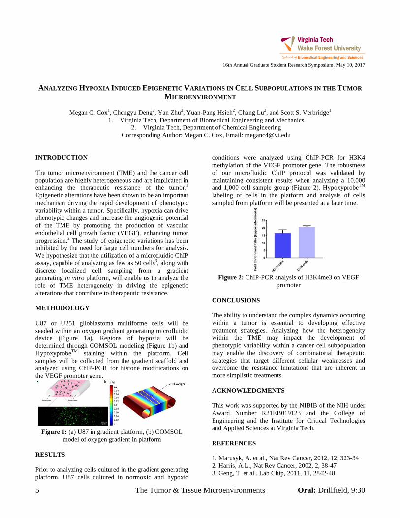
Corresponding Author: Megan C. Cox, Email: [email protected] INTRODUCTION The tumor microenvironment (TME) and the cancer cell population are highly heterogeneous and are implicated in enhancing the therapeutic resistance of the tumor.1 Epigenetic alterations have been shown to be an important mechanism driving the rapid development of phenotypic variability within a tumor. Specifically, hypoxia can drive phenotypic changes and increase the angiogenic potential of the TME by promoting the production of vascular endothelial cell growth factor (VEGF), enhancing tumor progression.2 The study of epigenetic variations has been inhibited by the need for large cell numbers for analysis. We hypothesize that the utilization of a microfluidic ChIP assay, capable of analyzing as few as 50 cells3, along with discrete localized cell sampling from a gradient generating in vitro platform, will enable us to analyze the role of TME heterogeneity in driving the epigenetic alterations that contribute to therapeutic resistance. METHODOLOGY U87 or U251 glioblastoma multiforme cells will be seeded within an oxygen gradient generating microfluidic device (Figure 1a). Regions of hypoxia will be determined through COMSOL modeling (Figure 1b) and HypoxyprobeTM staining within the platform. Cell samples will be collected from the gradient scaffold and analyzed using ChIP-PCR for histone modifications on the VEGF promoter gene.
Figure 1: (a) U87 in gradient platform, (b) COMSOL
model of oxygen gradient in platform RESULTS Prior to analyzing cells cultured in the gradient generating platform, U87 cells cultured in normoxic and hypoxic
conditions were analyzed using ChIP-PCR for H3K4 methylation of the VEGF promoter gene. The robustness of our microfluidic ChIP protocol was validated by maintaining consistent results when analyzing a 10,000 and 1,000 cell sample group (Figure 2). HypoxyprobeTM labeling of cells in the platform and analysis of cells sampled from platform will be presented at a later time.
Figure 2: ChIP-PCR analysis of H3K4me3 on VEGF
promoter
CONCLUSIONS The ability to understand the complex dynamics occurring within a tumor is essential to developing effective treatment strategies. Analyzing how the heterogeneity within the TME may impact the development of phenotypic variability within a cancer cell subpopulation may enable the discovery of combinatorial therapeutic strategies that target different cellular weaknesses and overcome the resistance limitations that are inherent in more simplistic treatments. ACKNOWLEDGMENTS This work was supported by the NIBIB of the NIH under Award Number R21EB019123 and the College of Engineering and the Institute for Critical Technologies and Applied Sciences at Virginia Tech. REFERENCES 1. Marusyk, A. et al., Nat Rev Cancer, 2012, 12, 323-34 2. Harris, A.L., Nat Rev Cancer, 2002, 2, 38-47 3. Geng, T. et al., Lab Chip, 2011, 11, 2842-48
Oral: Duckpond, 2:30 Crash Injury & Prevention 6
16th Annual Graduate Student Research Symposium, May 10, 2017
DEVELOPMENT AND FULL BODY VALIDATION OF A 5TH PERCENTILE
FEMALE FINITE ELEMENT MODEL
Matthew L. Davis1,2, Bharath Koya1,2, Jeremy M. Schap1,2, and F. Scott Gayzik1,2
1. Virginia Tech-Wake Forest Center for Injury Biomechanics 2. Wake Forest School of Medicine
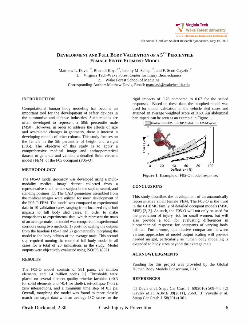
Corresponding Author: Matthew Davis, Email: [email protected] INTRODUCTION Computational human body modeling has become an important tool for the development of safety devices in the automotive and defense industries. Such models are often developed to represent a 50th percentile male (M50). However, in order to address the effects of size and sex-related changes in geometry, there is interest in developing models of other cohorts. This study focuses on the female in the 5th percentile of height and weight (F05). The objective of this study is to apply a comprehensive medical image and anthropometrical dataset to generate and validate a detailed finite element model (FEM) of the F05 occupant (F05-O). METHODOLOGY The F05-O model geometry was developed using a multi-modality medical image dataset collected from a representative small female subject in the supine, seated, and standing postures [1]. The CAD geometries assembled from the medical images were utilized for mesh development of the F05-O FEM. The model was compared to experimental data in 10 validation cases ranging from localized rigid hub impacts to full body sled cases. In order to make comparisons to experimental data, which represent the mass of an average male, the model was compared to experimental corridors using two methods: 1) post-hoc scaling the outputs from the baseline F05-O and 2) geometrically morphing the model to the body habitus of the average male. This second step required running the morphed full body model in all cases for a total of 20 simulations in the study. Model outputs were objectively evaluated using ISO/TS 18571. RESULTS The F05-O model consists of 981 parts, 2.6 million elements, and 1.4 million nodes [1]. Thresholds were placed on several element quality criteria: Jacobian (>0.3 for solid elements and >0.4 for shells), tet-collapse (>0.2), zero intersections, and a minimum time step of 0.1 µs. Overall, morphing the model was found to more closely match the target data with an average ISO score for the
rigid impacts of 0.76 compared to 0.67 for the scaled responses. Based on these data, the morphed model was used for model validation in the vehicle sled cases and attained an average weighted score of 0.69. An abdominal bar impact can be seen as an example in Figure 1.
Figure 1: Example of F05-O model response.
CONCLUSIONS This study describes the development of an anatomically representative small female FEM. The F05-O is the third in the GHBMC family of detailed occupant models (M50, M95) [2, 3]. As such, the F05-O will not only be used for the prediction of injury risk for small women, but will also provide a tool for evaluating differences in biomechanical response for occupants of varying body habitus. Furthermore, quantitative comparison between various approaches of model output scaling will provide needed insight, particularly as human body modeling is extended to body sizes beyond the average male. ACKNOWLEDGMENTS Funding for this project was provided by the Global Human Body Models Consortium, LLC REFERENCES [1] Davis et al. Stapp Car Crash J. 60(2016) 509-44. [2] Gayzik et al. ABME 39(2011), 2568. [3] Vavalle et al. Stapp Car Crash J. 58(2014) 361.
7 Evaluations of Head Injury Oral: Drillfield, 1:30
16th Annual Graduate Student Research Symposium, May 10, 2017
MODULAR USE OF HUMAN BODY MODELS OF VARYING LEVELS OF COMPLEXITY: VALIDATION OF
HEAD KINEMATICS
William Decker1,2, Bharath Koya2, Matthew L. Davis1,2, and F. Scott Gayzik1,2
1. Wake Forest University School of Medicine 2. Virginia Tech – Wake Forest University Center for Injury Biomechanics
Corresponding Author: F. Scott Gayzik, Email: [email protected] INTRODUCTION The significant computational resources required to execute detailed human body finite element models has motivated the development of faster running simplified models (e.g. GHBMC M50-OS). Previous studies have demonstrated the ability to modularly incorporate the validated GHBMC M50-O brain model into the simplified model (GHBMC M50-OS+B), which allows for localized analysis of the brain in a fraction of the computation time required for the detailed model. The objective of this study is to validate the head and neck kinematics of the GHBMC M50-O and M50-OS (detailed and simplified versions of the same model), as well as the M50-OS+B, against human volunteer test data in frontal and lateral loading.
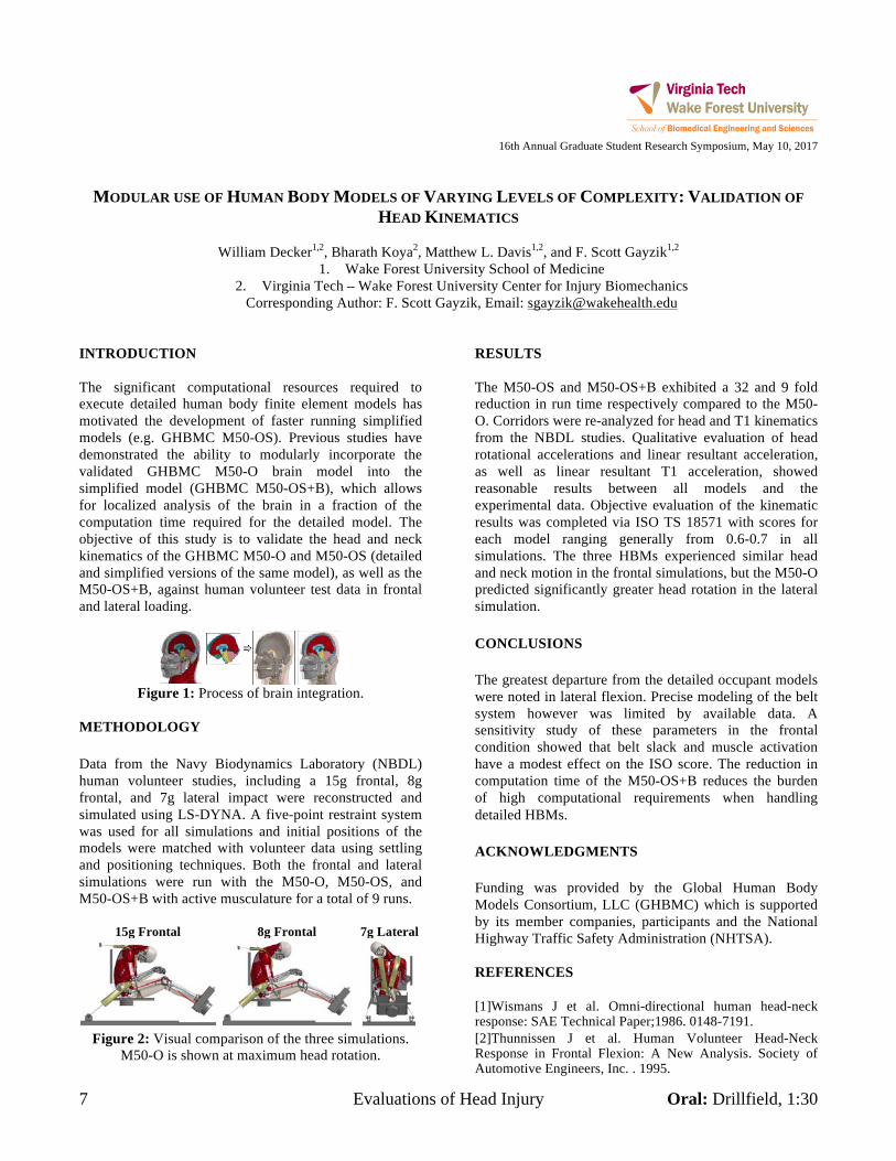
Figure 1: Process of brain integration.
METHODOLOGY Data from the Navy Biodynamics Laboratory (NBDL) human volunteer studies, including a 15g frontal, 8g frontal, and 7g lateral impact were reconstructed and simulated using LS-DYNA. A five-point restraint system was used for all simulations and initial positions of the models were matched with volunteer data using settling and positioning techniques. Both the frontal and lateral simulations were run with the M50-O, M50-OS, and M50-OS+B with active musculature for a total of 9 runs.
15g Frontal 8g Frontal 7g Lateral
Figure 2: Visual comparison of the three simulations.
M50-O is shown at maximum head rotation.
RESULTS The M50-OS and M50-OS+B exhibited a 32 and 9 fold reduction in run time respectively compared to the M50-O. Corridors were re-analyzed for head and T1 kinematics from the NBDL studies. Qualitative evaluation of head rotational accelerations and linear resultant acceleration, as well as linear resultant T1 acceleration, showed reasonable results between all models and the experimental data. Objective evaluation of the kinematic results was completed via ISO TS 18571 with scores for each model ranging generally from 0.6-0.7 in all simulations. The three HBMs experienced similar head and neck motion in the frontal simulations, but the M50-O predicted significantly greater head rotation in the lateral simulation. CONCLUSIONS The greatest departure from the detailed occupant models were noted in lateral flexion. Precise modeling of the belt system however was limited by available data. A sensitivity study of these parameters in the frontal condition showed that belt slack and muscle activation have a modest effect on the ISO score. The reduction in computation time of the M50-OS+B reduces the burden of high computational requirements when handling detailed HBMs. ACKNOWLEDGMENTS Funding was provided by the Global Human Body Models Consortium, LLC (GHBMC) which is supported by its member companies, participants and the National Highway Traffic Safety Administration (NHTSA). REFERENCES [1]Wismans J et al. Omni-directional human head-neck response: SAE Technical Paper;1986. 0148-7191. [2]Thunnissen J et al. Human Volunteer Head-Neck Response in Frontal Flexion: A New Analysis. Society of Automotive Engineers, Inc. . 1995.
Oral: Drillfield, 10:15 The Tumor & Tissue Microenvironments 8
16th Annual Graduate Student Research Symposium, May 10, 2017
RHEOLOGY VS ALIGNMENT: TWO OPERATORS OF THE MECHANICAL TUMOR MICROENVIRONMENT
Mahesh Devarasetty1,2, Aleksander Skardal1,2, and Shay Soker1,2
1. Wake Forest University, Institute for Regenerative Medicine 2. Virginia Tech Wake Forest University, School of Biomedical Engineering and Sciences
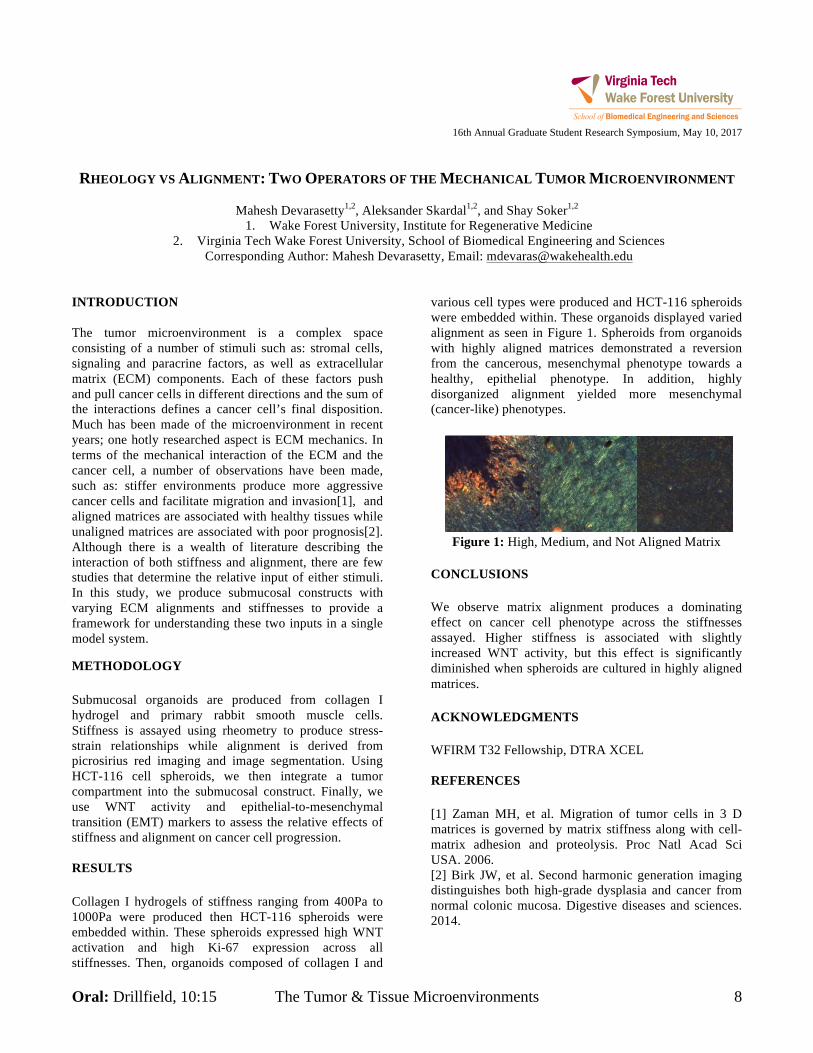
Corresponding Author: Mahesh Devarasetty, Email: [email protected] INTRODUCTION The tumor microenvironment is a complex space consisting of a number of stimuli such as: stromal cells, signaling and paracrine factors, as well as extracellular matrix (ECM) components. Each of these factors push and pull cancer cells in different directions and the sum of the interactions defines a cancer cell’s final disposition. Much has been made of the microenvironment in recent years; one hotly researched aspect is ECM mechanics. In terms of the mechanical interaction of the ECM and the cancer cell, a number of observations have been made, such as: stiffer environments produce more aggressive cancer cells and facilitate migration and invasion[1], and aligned matrices are associated with healthy tissues while unaligned matrices are associated with poor prognosis[2]. Although there is a wealth of literature describing the interaction of both stiffness and alignment, there are few studies that determine the relative input of either stimuli. In this study, we produce submucosal constructs with varying ECM alignments and stiffnesses to provide a framework for understanding these two inputs in a single model system. METHODOLOGY Submucosal organoids are produced from collagen I hydrogel and primary rabbit smooth muscle cells. Stiffness is assayed using rheometry to produce stress-strain relationships while alignment is derived from picrosirius red imaging and image segmentation. Using HCT-116 cell spheroids, we then integrate a tumor compartment into the submucosal construct. Finally, we use WNT activity and epithelial-to-mesenchymal transition (EMT) markers to assess the relative effects of stiffness and alignment on cancer cell progression. RESULTS Collagen I hydrogels of stiffness ranging from 400Pa to 1000Pa were produced then HCT-116 spheroids were embedded within. These spheroids expressed high WNT activation and high Ki-67 expression across all stiffnesses. Then, organoids composed of collagen I and
various cell types were produced and HCT-116 spheroids were embedded within. These organoids displayed varied alignment as seen in Figure 1. Spheroids from organoids with highly aligned matrices demonstrated a reversion from the cancerous, mesenchymal phenotype towards a healthy, epithelial phenotype. In addition, highly disorganized alignment yielded more mesenchymal (cancer-like) phenotypes.
Figure 1: High, Medium, and Not Aligned Matrix CONCLUSIONS We observe matrix alignment produces a dominating effect on cancer cell phenotype across the stiffnesses assayed. Higher stiffness is associated with slightly increased WNT activity, but this effect is significantly diminished when spheroids are cultured in highly aligned matrices. ACKNOWLEDGMENTS WFIRM T32 Fellowship, DTRA XCEL REFERENCES [1] Zaman MH, et al. Migration of tumor cells in 3 D matrices is governed by matrix stiffness along with cell-matrix adhesion and proteolysis. Proc Natl Acad Sci USA. 2006. [2] Birk JW, et al. Second harmonic generation imaging distinguishes both high-grade dysplasia and cancer from normal colonic mucosa. Digestive diseases and sciences. 2014.
9 Biomaterials Development & Characterization Oral: Smithfield, 10:00
16th Annual Graduate Student Research Symposium, May 10, 2017
EFFECT OF PRESERVATION FLUID TYPE ON THE FAILURE MATERIAL PROPERTIES OF BOVINE LIVER
PARENCHYMA WITH INCREASING POST MORTEM TIME
Kristin M. Dunford1 and Andrew R. Kemper
1
1. Virginia Tech, Department of Biomedical Engineering and Mechanics
Corresponding Author: Kristin M. Dunford, Email: kmcamp.edu
INTRODUCTION
The liver is the second most frequently injured abdominal
organ in motor vehicle collisions (MVCs). Lacerations or
crushing of the liver can result in potentially life
threatening internal bleeding. Although current
anthropomorphic test devices are used to predict injuries
in MVCs, they are not equipped to represent abdominal
organs or assess abdominal organ injury risk.
Consequently, finite element models are frequently used
to assess abdominal organ injury risk in MVCs. The
material response of soft tissue can change considerably
after death. Previous studies have observed changes in
cellular structure within a few hours postmortem. Liver
tissue has also been shown to increase in stiffness with
postmortem time1. However, such studies did not report
failure properties of liver tissue parenchyma. Other
studies have investigated the effects of various conditions
on the tensile failure properties of liver parenchyma. It
was found that freezing the tissue prior to testing resulted
in significantly lower strains2. Also, tensile strain rate
significantly affects the failure stress and strain3.
However, there have been no studies that have evaluated
the change in tensile failure properties of liver
parenchyma between 6 and 48 hours postmortem.
METHODOLOGY
Twelve bovine livers were acquired from a local
slaughterhouse immediately after death. Multiple samples
of the parenchyma were obtained from each liver for
testing at three time points. The three time points were 6
hours, 24 hours, and 48 hours postmortem.
A custom slicing jig and blade assembly was used to
obtain thin slices of parenchyma. The slices were then
stamped to create dog-bone shaped samples. Samples
were prepared and tested immediately after the liver
arrived at the laboratory to obtain data for the first time
point. Remaining liver tissue was divided into large
sections, immersed in preservation fluid, and sealed in
plastic containers until the later time points. Tissue from
six livers were stored in DMEM, while tissue from the
remaining six livers were stored in normal saline.
Uniaxial tension tests were conducted on the dog-bone
samples. A mounting and testing procedure was followed
to minimize variation in specimen alignment and initial
specimen slack. Optical markers were placed on the gage
region of the specimen. The testing system was operated
with a multi-axis controller that simultaneously moved
the top and bottom grips at a constant velocity. Specimens
were pulled to failure at a rate of 1s-1
. Data was collected
for the duration of the test from high-speed video, load
cells, potentiometers, and accelerometers. The optical
markers placed on the front of the sample were tracked
using a motion analysis software. The displacements of
the markers surrounding the failure tear were used to
calculate the Green strain. The 2nd
Piola Kirchhoff Stress
was calculated at each time point during the test. The
average failure stresses and strains were compared across
time points within each liver, and across all twelve livers.
RESULTS
A Block ANOVA was conducted to compare the failure
properties at each of the three time points. The failure
stresses and strains did not significantly change with
respect to time for livers that were preserved in DMEM.
Analysis is underway to determine if there are any
changes in failure properties for the livers stored in saline.
CONCLUSIONS
Uniaxial tension tests were conducted on the liver
parenchyma of twelve bovine livers. Neither the failure
stresses nor strains changed significantly between 6 and
48 hours postmortem for tissue stored in DMEM. Future
analysis will elucidate the effects of postmortem time on
the failure properties for tissue stored in saline.
REFERENCES [1] B. Tay, J. Kim, and M. Srinivasan, “In vivo mechanical behavior of intra-
abdominal organs,” IEEE Transactions on Biomedical Engineering, vol. 53,
no. 11, pp. 2129-2138, 2006.
[2] A.C. Santago, A.R. Kemper, C. McNally, J.L. Sparks, and S.M. Duma,
“Freezing affects the mechanical properties of bovine liver,” Biomedical
Sciences Instrumentation, vol. 45, pp.24-29, 2009.
[3] A.R. Kemper, A.C. Santago, J.D. Stitzel, J.L. Sparks, and S.M. Duma,
“Biomechanical response of human liver in tensile loading,” Ann. Adv.
Automot. Med., vol. 54, pp. 15-26, 2010.
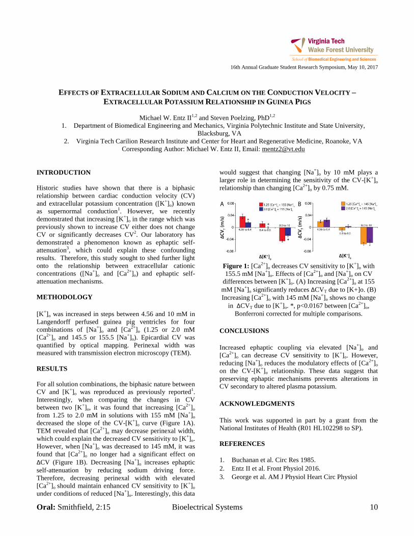
Oral: Smithfield, 2:15 Bioelectrical Systems 10
16th Annual Graduate Student Research Symposium, May 10, 2017
EFFECTS OF EXTRACELLULAR SODIUM AND CALCIUM ON THE CONDUCTION VELOCITY –
EXTRACELLULAR POTASSIUM RELATIONSHIP IN GUINEA PIGS
Michael W. Entz II1,2
and Steven Poelzing, PhD1,2
1. Department of Biomedical Engineering and Mechanics, Virginia Polytechnic Institute and State University,
Blacksburg, VA
2. Virginia Tech Carilion Research Institute and Center for Heart and Regenerative Medicine, Roanoke, VA
Corresponding Author: Michael W. Entz II, Email: [email protected]
INTRODUCTION
Historic studies have shown that there is a biphasic
relationship between cardiac conduction velocity (CV)
and extracellular potassium concentration ([K+]o) known
as supernormal conduction1. However, we recently
demonstrated that increasing [K+]o in the range which was
previously shown to increase CV either does not change
CV or significantly decreases CV2. Our laboratory has
demonstrated a phenomenon known as ephaptic self-
attenuation3, which could explain these confounding
results. Therefore, this study sought to shed further light
onto the relationship between extracellular cationic
concentrations ([Na+]o and [Ca
2+]o) and ephaptic self-
attenuation mechanisms.
METHODOLOGY
[K
+]o was increased in steps between 4.56 and 10 mM in
Langendorff perfused guinea pig ventricles for four
combinations of [Na+]o and [Ca
2+]o (1.25 or 2.0 mM
[Ca2+
]o and 145.5 or 155.5 [Na+]o). Epicardial CV was
quantified by optical mapping. Perinexal width was
measured with transmission electron microscopy (TEM).
RESULTS
For all solution combinations, the biphasic nature between
CV and [K+]o was reproduced as previously reported
1.
Interestingly, when comparing the changes in CV
between two [K+]o, it was found that increasing [Ca
2+]o
from 1.25 to 2.0 mM in solutions with 155 mM [Na+]o
decreased the slope of the CV-[K+]o curve (Figure 1A).
TEM revealed that [Ca2+
]o may decrease perinexal width,
which could explain the decreased CV sensitivity to [K+]o.
However, when [Na+]o was decreased to 145 mM, it was
found that [Ca2+
]o no longer had a significant effect on
ΔCV (Figure 1B). Decreasing [Na+]o increases ephaptic
self-attenuation by reducing sodium driving force.
Therefore, decreasing perinexal width with elevated
[Ca2+
]o should maintain enhanced CV sensitivity to [K+]o
under conditions of reduced [Na+]o. Interestingly, this data
would suggest that changing [Na+]o by 10 mM plays a
larger role in determining the sensitivity of the CV-[K+]o
relationship than changing [Ca2+
]o by 0.75 mM.
Figure 1: [Ca
2+]o decreases CV sensitivity to [K
+]o with
155.5 mM [Na+]o. Effects of [Ca
2+]o and [Na
+]o on CV
differences between [K+]o. (A) Increasing [Ca
2+]o at 155
mM [Na+]o significantly reduces ΔCVT due to [K+]o. (B)
Increasing [Ca2+
]o with 145 mM [Na+]o shows no change
in ΔCVT due to [K+]o. *, p<0.0167 between [Ca
2+]o,
Bonferroni corrected for multiple comparisons.
CONCLUSIONS
Increased ephaptic coupling via elevated [Na+]o and
[Ca2+
]o can decrease CV sensitivity to [K+]o. However,
reducing [Na+]o reduces the modulatory effects of [Ca
2+]o
on the CV-[K+]o relationship. These data suggest that
preserving ephaptic mechanisms prevents alterations in
CV secondary to altered plasma potassium.
ACKNOWLEDGMENTS
This work was supported in part by a grant from the
National Institutes of Health (R01 HL102298 to SP).
REFERENCES
1. Buchanan et al. Circ Res 1985.
2. Entz II et al. Front Physiol 2016.
3. George et al. AM J Physiol Heart Circ Physiol
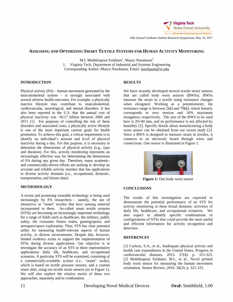
11 Developing Novel Medical Devices Oral: Smithfield, 1:00
16th Annual Graduate Student Research Symposium, May 10, 2017
ASSESSING AND OPTIMIZING SMART TEXTILE SYSTEMS FOR HUMAN ACTIVITY MONITORING
M.I. Mokhlespour Esfahani1, Maury Nussbaum
1
1. Virginia Tech, Department of Industrial and Systems Engineering
Corresponding Author: Maury Nussbaum, Email: [email protected]
INTRODUCTION
Physical activity (PA) – human movement generated by the
musculoskeletal system – is strongly associated with
several adverse health outcomes. For example, a physically
inactive lifestyle may contribute to musculoskeletal,
cardiovascular, neurological, and mental disorders. It has
also been reported in the U.S. that the annual cost of
physical inactivity was ~$117 billion between 2006 and
2011 [1]. For purposes of controlling the risk of these
disorders and associated costs, a physically active lifestyle
is one of the most important current goals for health
promotion. To achieve this goal, a critical requirement is to
identify an individual’s amount and level of physical
inactivity during a day. For this purpose, it is necessary to
determine the dimensions of physical activity (e.g., type
and duration). For this, activity monitoring represents an
increasingly effective way for determining the dimensions
of PA during any given day. Therefore, many academic-
and commercially-driven efforts are seeking to develop an
accurate and reliable activity monitor that has applications
to diverse activity domains (i.e., occupational, domestic,
transportation, and leisure time).
METHODOLOGY
A recent and promising wearable technology is being used
increasingly by PA researchers— namely, the use of
interactive or “smart” textiles that have sensing material
incorporated in them. So-called smart textile systems
(STSs) are becoming an increasingly important technology
for a range of fields such as healthcare, the military, public
safety, the consumer fitness realm, gaming/sports, and
aerospace/space exploration. Thus, STS has clear potential
utility for measuring health-relevant aspects of human
activity, in diverse environments. Despite this, however,
limited evidence exists to support the implementation of
STSs during diverse applications. Our objective is to
investigate the accuracy of an STS in three representative
applications: daily life, healthcare, and occupational
scenarios. A particular STS will be examined, consisting of
a commercially-available system (i.e., “smart” socks),
which is based on textile pressure sensors, and a custom
smart shirt, using our textile strain sensors (as in Figure 1).
We will also explore the relative merits of these two
approaches, separately and in combination.
RESULTS
We have recently developed several textile strain sensors
that are called body worn sensors (BWSs). BWSs
measure the strain in a textile using resistance changes
when elongated. Working as a potentiometer, the
resistance range is between 2kΩ and 70kΩ, which linearly
corresponds to zero tension and 50% maximum
elongation, respectively. The size of the BWS to be used
here is 20×40 mm, and its performance is not affected by
humidity [2]. Specific details about manufacturing a body
worn sensor can be obtained from our recent study [2].
Since a BWS is designed to measure strain in textiles, it
connects to an electronic board through wires and
connections. One sensor is illustrated in Figure 1.
Figure 1: One body worn sensor
CONCLUSIONS
The results of this investigation are expected to
demonstrate the potential performance of an STS for
activity monitoring in three broad domains: activities of
daily life, healthcare, and occupational scenarios. We
also expect to identify specific combinations or
configurations of STSs that could provide the most useful
and efficient information for activity recognition and
detection.
REFERENCES
[1] Carlson, S.A., et al., Inadequate physical activity and
health care expenditures in the United States. Progress in
cardiovascular diseases, 2015. 57(4): p. 315-323.
[2] Mokhlespour Esfahani, M.I., et al., Novel printed
body worn sensor for measuring the human movement
orientation. Sensor Review, 2016. 36(3): p. 321-331.
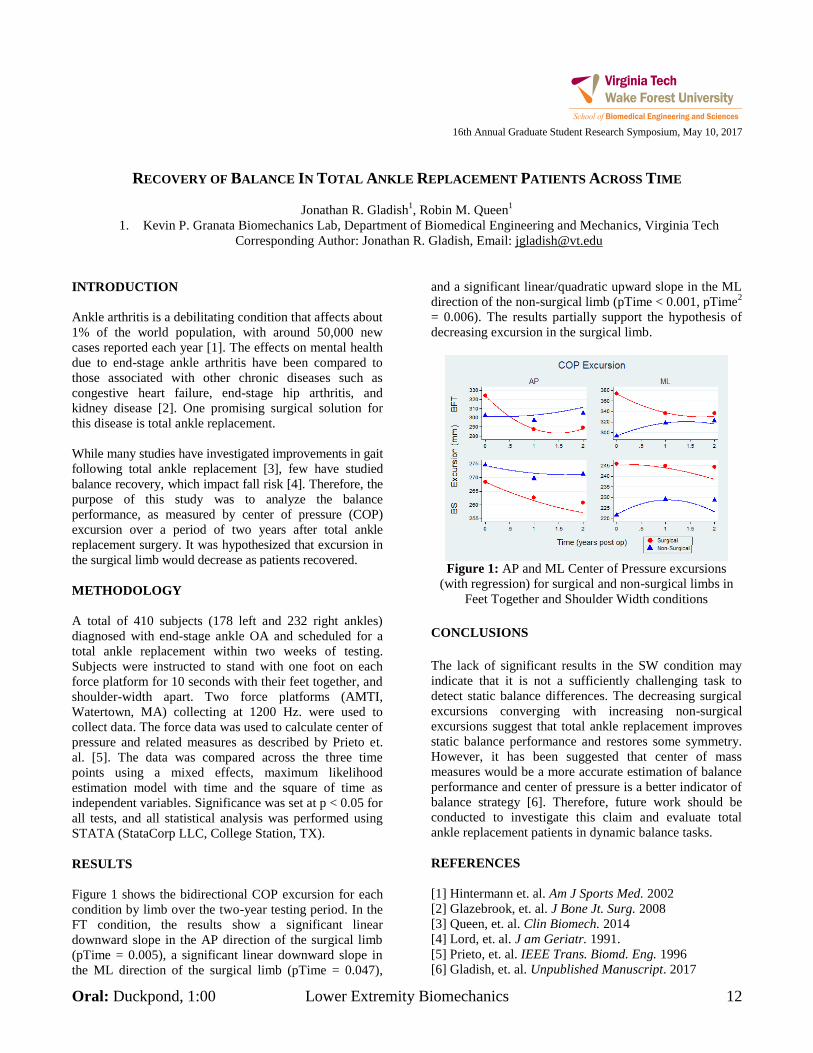
Oral: Duckpond, 1:00 Lower Extremity Biomechanics 12
16th Annual Graduate Student Research Symposium, May 10, 2017
RECOVERY OF BALANCE IN TOTAL ANKLE REPLACEMENT PATIENTS ACROSS TIME
Jonathan R. Gladish1, Robin M. Queen
1
1. Kevin P. Granata Biomechanics Lab, Department of Biomedical Engineering and Mechanics, Virginia Tech
Corresponding Author: Jonathan R. Gladish, Email: [email protected]
INTRODUCTION
Ankle arthritis is a debilitating condition that affects about
1% of the world population, with around 50,000 new
cases reported each year [1]. The effects on mental health
due to end-stage ankle arthritis have been compared to
those associated with other chronic diseases such as
congestive heart failure, end-stage hip arthritis, and
kidney disease [2]. One promising surgical solution for
this disease is total ankle replacement.
While many studies have investigated improvements in gait
following total ankle replacement [3], few have studied
balance recovery, which impact fall risk [4]. Therefore, the
purpose of this study was to analyze the balance
performance, as measured by center of pressure (COP)
excursion over a period of two years after total ankle
replacement surgery. It was hypothesized that excursion in
the surgical limb would decrease as patients recovered.
METHODOLOGY
A total of 410 subjects (178 left and 232 right ankles)
diagnosed with end-stage ankle OA and scheduled for a
total ankle replacement within two weeks of testing.
Subjects were instructed to stand with one foot on each
force platform for 10 seconds with their feet together, and
shoulder-width apart. Two force platforms (AMTI,
Watertown, MA) collecting at 1200 Hz. were used to
collect data. The force data was used to calculate center of
pressure and related measures as described by Prieto et.
al. [5]. The data was compared across the three time
points using a mixed effects, maximum likelihood
estimation model with time and the square of time as
independent variables. Significance was set at p < 0.05 for
all tests, and all statistical analysis was performed using
STATA (StataCorp LLC, College Station, TX).
RESULTS
Figure 1 shows the bidirectional COP excursion for each
condition by limb over the two-year testing period. In the
FT condition, the results show a significant linear
downward slope in the AP direction of the surgical limb
(pTime = 0.005), a significant linear downward slope in
the ML direction of the surgical limb (pTime = 0.047),
and a significant linear/quadratic upward slope in the ML
direction of the non-surgical limb (pTime < 0.001, pTime2
= 0.006). The results partially support the hypothesis of
decreasing excursion in the surgical limb.
Figure 1: AP and ML Center of Pressure excursions
(with regression) for surgical and non-surgical limbs in
Feet Together and Shoulder Width conditions
CONCLUSIONS
The lack of significant results in the SW condition may
indicate that it is not a sufficiently challenging task to
detect static balance differences. The decreasing surgical
excursions converging with increasing non-surgical
excursions suggest that total ankle replacement improves
static balance performance and restores some symmetry.
However, it has been suggested that center of mass
measures would be a more accurate estimation of balance
performance and center of pressure is a better indicator of
balance strategy [6]. Therefore, future work should be
conducted to investigate this claim and evaluate total
ankle replacement patients in dynamic balance tasks.
REFERENCES
[1] Hintermann et. al. Am J Sports Med. 2002
[2] Glazebrook, et. al. J Bone Jt. Surg. 2008
[3] Queen, et. al. Clin Biomech. 2014
[4] Lord, et. al. J am Geriatr. 1991.
[5] Prieto, et. al. IEEE Trans. Biomd. Eng. 1996
[6] Gladish, et. al. Unpublished Manuscript. 2017
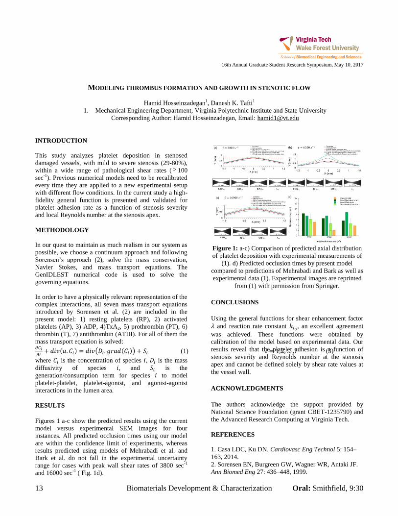
13 Biomaterials Development & Characterization Oral: Smithfield, 9:30
16th Annual Graduate Student Research Symposium, May 10, 2017
MODELING THROMBUS FORMATION AND GROWTH IN STENOTIC FLOW
Hamid Hosseinzadegan1, Danesh K. Tafti
1
1. Mechanical Engineering Department, Virginia Polytechnic Institute and State University
Corresponding Author: Hamid Hosseinzadegan, Email: [email protected]
INTRODUCTION
This study analyzes platelet deposition in stenosed
damaged vessels, with mild to severe stenosis (29-80%),
within a wide range of pathological shear rates ( 100
sec-1
). Previous numerical models need to be recalibrated
every time they are applied to a new experimental setup
with different flow conditions. In the current study a high-
fidelity general function is presented and validated for
platelet adhesion rate as a function of stenosis severity
and local Reynolds number at the stenosis apex.
METHODOLOGY
In our quest to maintain as much realism in our system as
possible, we choose a continuum approach and following
Sorensen’s approach (2), solve the mass conservation,
Navier Stokes, and mass transport equations. The
GenIDLEST numerical code is used to solve the
governing equations.
In order to have a physically relevant representation of the
complex interactions, all seven mass transport equations
introduced by Sorensen et al. (2) are included in the
present model: 1) resting platelets (RP), 2) activated
platelets (AP), 3) ADP, 4)TxA2, 5) prothrombin (PT), 6)
thrombin (T), 7) antithrombin (ATIII). For all of them the
mass transport equation is solved: 𝜕𝐶𝑖
𝜕𝑡+ 𝑑𝑖𝑣(𝑢. 𝐶𝑖) = 𝑑𝑖𝑣(𝐷𝑖 . 𝑔𝑟𝑎𝑑(𝐶𝑖)) + 𝑆𝑖 (1) 𝑖 = 1,2, … ,7 (3)
where 𝐶𝑖 is the concentration of species 𝑖, 𝐷𝑖 is the mass
diffusivity of species 𝑖, and 𝑆𝑖 is the
generation/consumption term for species 𝑖 to model
platelet-platelet, platelet-agonist, and agonist-agonist
interactions in the lumen area.
RESULTS
Figures 1 a-c show the predicted results using the current
model versus experimental SEM images for four
instances. All predicted occlusion times using our model
are within the confidence limit of experiments, whereas
results predicted using models of Mehrabadi et al. and
Bark et al. do not fall in the experimental uncertainty
range for cases with peak wall shear rates of 3800 sec-1
and 16000 sec-1
( Fig. 1d).
Figure 1: a-c) Comparison of predicted axial distribution
of platelet deposition with experimental measurements of
(1). d) Predicted occlusion times by present model
compared to predictions of Mehrabadi and Bark as well as
experimental data (1). Experimental images are reprinted
from (1) with permission from Springer.
CONCLUSIONS
Using the general functions for shear enhancement factor
𝜆 and reaction rate constant 𝑘𝑖0 , an excellent agreement
was achieved. These functions were obtained by
calibration of the model based on experimental data. Our
results reveal that the platelet adhesion is a function of
stenosis severity and Reynolds number at the stenosis
apex and cannot be defined solely by shear rate values at
the vessel wall.
ACKNOWLEDGMENTS
The authors acknowledge the support provided by
National Science Foundation (grant CBET-1235790) and
the Advanced Research Computing at Virginia Tech.
REFERENCES
1. Casa LDC, Ku DN. Cardiovasc Eng Technol 5: 154–
163, 2014.
2. Sorensen EN, Burgreen GW, Wagner WR, Antaki JF.
Ann Biomed Eng 27: 436–448, 1999.
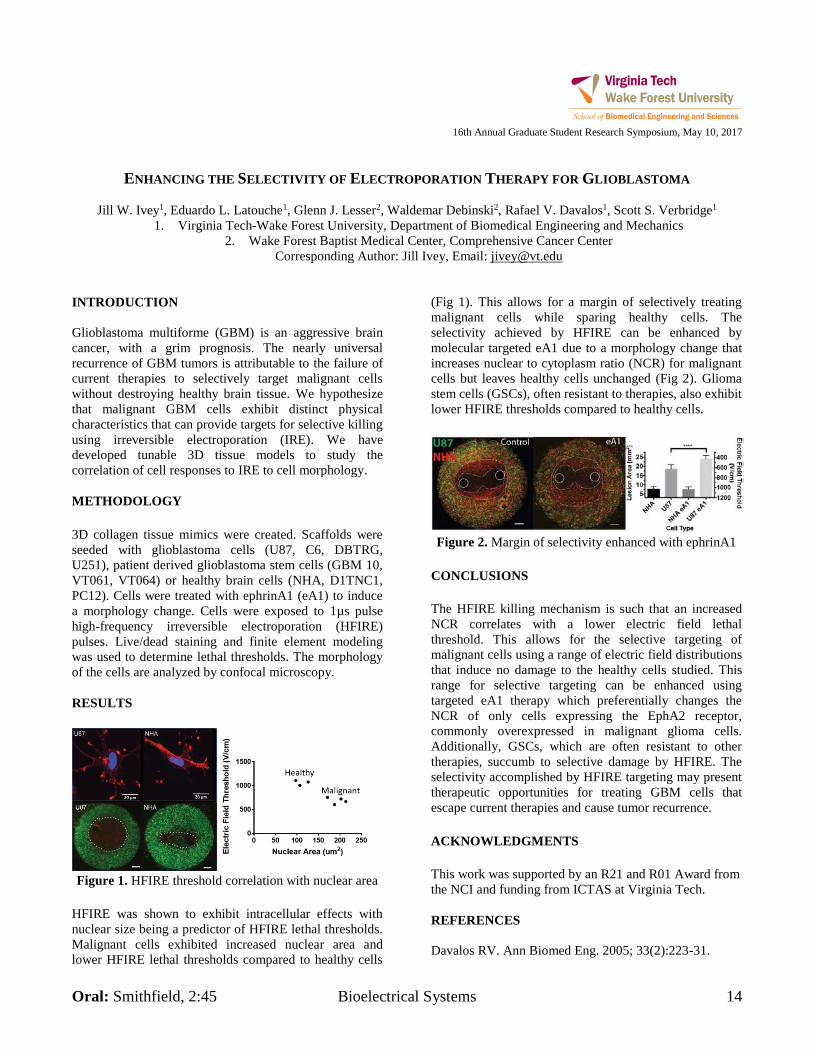
Oral: Smithfield, 2:45 Bioelectrical Systems 14
16th Annual Graduate Student Research Symposium, May 10, 2017
ENHANCING THE SELECTIVITY OF ELECTROPORATION THERAPY FOR GLIOBLASTOMA
Jill W. Ivey1, Eduardo L. Latouche1, Glenn J. Lesser2, Waldemar Debinski2, Rafael V. Davalos1, Scott S. Verbridge1
1. Virginia Tech-Wake Forest University, Department of Biomedical Engineering and Mechanics 2. Wake Forest Baptist Medical Center, Comprehensive Cancer Center
Corresponding Author: Jill Ivey, Email: [email protected] INTRODUCTION Glioblastoma multiforme (GBM) is an aggressive brain cancer, with a grim prognosis. The nearly universal recurrence of GBM tumors is attributable to the failure of current therapies to selectively target malignant cells without destroying healthy brain tissue. We hypothesize that malignant GBM cells exhibit distinct physical characteristics that can provide targets for selective killing using irreversible electroporation (IRE). We have developed tunable 3D tissue models to study the correlation of cell responses to IRE to cell morphology. METHODOLOGY 3D collagen tissue mimics were created. Scaffolds were seeded with glioblastoma cells (U87, C6, DBTRG, U251), patient derived glioblastoma stem cells (GBM 10, VT061, VT064) or healthy brain cells (NHA, D1TNC1, PC12). Cells were treated with ephrinA1 (eA1) to induce a morphology change. Cells were exposed to 1µs pulse high-frequency irreversible electroporation (HFIRE) pulses. Live/dead staining and finite element modeling was used to determine lethal thresholds. The morphology of the cells are analyzed by confocal microscopy. RESULTS
Figure 1. HFIRE threshold correlation with nuclear area
HFIRE was shown to exhibit intracellular effects with nuclear size being a predictor of HFIRE lethal thresholds. Malignant cells exhibited increased nuclear area and lower HFIRE lethal thresholds compared to healthy cells
(Fig 1). This allows for a margin of selectively treating malignant cells while sparing healthy cells. The selectivity achieved by HFIRE can be enhanced by molecular targeted eA1 due to a morphology change that increases nuclear to cytoplasm ratio (NCR) for malignant cells but leaves healthy cells unchanged (Fig 2). Glioma stem cells (GSCs), often resistant to therapies, also exhibit lower HFIRE thresholds compared to healthy cells.
Figure 2. Margin of selectivity enhanced with ephrinA1
CONCLUSIONS The HFIRE killing mechanism is such that an increased NCR correlates with a lower electric field lethal threshold. This allows for the selective targeting of malignant cells using a range of electric field distributions that induce no damage to the healthy cells studied. This range for selective targeting can be enhanced using targeted eA1 therapy which preferentially changes the NCR of only cells expressing the EphA2 receptor, commonly overexpressed in malignant glioma cells. Additionally, GSCs, which are often resistant to other therapies, succumb to selective damage by HFIRE. The selectivity accomplished by HFIRE targeting may present therapeutic opportunities for treating GBM cells that escape current therapies and cause tumor recurrence. ACKNOWLEDGMENTS This work was supported by an R21 and R01 Award from the NCI and funding from ICTAS at Virginia Tech. REFERENCES Davalos RV. Ann Biomed Eng. 2005; 33(2):223-31.
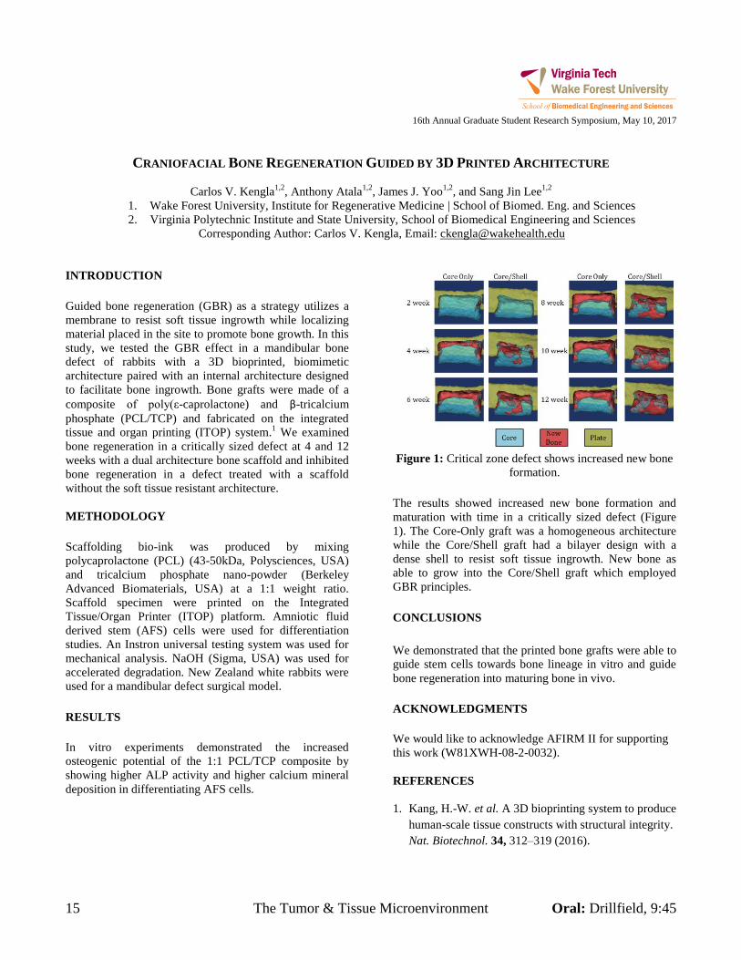
15 The Tumor & Tissue Microenvironment Oral: Drillfield, 9:45
16th Annual Graduate Student Research Symposium, May 10, 2017
CRANIOFACIAL BONE REGENERATION GUIDED BY 3D PRINTED ARCHITECTURE
Carlos V. Kengla1,2
, Anthony Atala1,2
, James J. Yoo1,2
, and Sang Jin Lee1,2
1. Wake Forest University, Institute for Regenerative Medicine | School of Biomed. Eng. and Sciences
2. Virginia Polytechnic Institute and State University, School of Biomedical Engineering and Sciences
Corresponding Author: Carlos V. Kengla, Email: [email protected]
INTRODUCTION
Guided bone regeneration (GBR) as a strategy utilizes a
membrane to resist soft tissue ingrowth while localizing
material placed in the site to promote bone growth. In this
study, we tested the GBR effect in a mandibular bone
defect of rabbits with a 3D bioprinted, biomimetic
architecture paired with an internal architecture designed
to facilitate bone ingrowth. Bone grafts were made of a
composite of poly(ε-caprolactone) and β-tricalcium
phosphate (PCL/TCP) and fabricated on the integrated
tissue and organ printing (ITOP) system.1 We examined
bone regeneration in a critically sized defect at 4 and 12
weeks with a dual architecture bone scaffold and inhibited
bone regeneration in a defect treated with a scaffold
without the soft tissue resistant architecture.
METHODOLOGY
Scaffolding bio-ink was produced by mixing
polycaprolactone (PCL) (43-50kDa, Polysciences, USA)
and tricalcium phosphate nano-powder (Berkeley
Advanced Biomaterials, USA) at a 1:1 weight ratio.
Scaffold specimen were printed on the Integrated
Tissue/Organ Printer (ITOP) platform. Amniotic fluid
derived stem (AFS) cells were used for differentiation
studies. An Instron universal testing system was used for
mechanical analysis. NaOH (Sigma, USA) was used for
accelerated degradation. New Zealand white rabbits were
used for a mandibular defect surgical model.
RESULTS
In vitro experiments demonstrated the increased
osteogenic potential of the 1:1 PCL/TCP composite by
showing higher ALP activity and higher calcium mineral
deposition in differentiating AFS cells.
Figure 1: Critical zone defect shows increased new bone
formation.
The results showed increased new bone formation and
maturation with time in a critically sized defect (Figure
1). The Core-Only graft was a homogeneous architecture
while the Core/Shell graft had a bilayer design with a
dense shell to resist soft tissue ingrowth. New bone as
able to grow into the Core/Shell graft which employed
GBR principles.
CONCLUSIONS
We demonstrated that the printed bone grafts were able to
guide stem cells towards bone lineage in vitro and guide
bone regeneration into maturing bone in vivo.
ACKNOWLEDGMENTS
We would like to acknowledge AFIRM II for supporting
this work (W81XWH-08-2-0032).
REFERENCES
1. Kang, H.-W. et al. A 3D bioprinting system to produce
human-scale tissue constructs with structural integrity.
Nat. Biotechnol. 34, 312–319 (2016).
Oral: Smithfield, 2:30 Bioelectrical Systems 16
16th Annual Graduate Student Research Symposium, May 10, 2017
INTERSTITIAL CELLS OF CAJAL INCREASE NEURAL-MEDIATED RELAXATION
AND ELECTRICAL RHYTHM IN AN IN VITRO BIOENGINNERED MODEL OF PYLORUS
Dylan T. Knutson1, 2, Elie Zakhem1, Kenneth L. Koch3, Khalil N. Bitar1, 2, 3
1Wake Forest Institute for Regenerative Medicine, Wake Forest School of Medicine, Winston Salem, NC 2Viriginia Tech – Wake Forest University School of Biomedical Engineering, Winston Salem, NC
3Section on Gastroenterology Department of Internal Medicine, Wake Forest School of Medicine, Winston Salem, NC Corresponding Author: Dylan T. Knutson, Email: [email protected]
INTRODUCTION Motility in the gut is coordinated by the neuromuscular apparatus, a complex of smooth muscle, enteric neurons and interstitial cells of Cajal (ICC). Gastrointestinal neuromuscular disorders are associated with the depletion of neurons and ICC. In vitro models to study the effect of depletion or restoration of these key cells on neuromuscular function are lacking. The aim of this study was to produce different combinations of neuromuscular cells to measure physiological function of these combinations. METHODOLOGY Smooth muscle cells (SMC) were isolated from human pylorus, ICCs were isolated from GFP-mice small intestines, and neural progenitor cells (NPC) were isolated from rat small intestine. Pyloric constructs were engineered using the following combinations: (1) SMC, (2) SMC+ICC, (3) SMC+NPC and (4) SMC+ICC+NPC. Tissues were characterized by immunohistochemistry and physiological functionality using force and electrical measurements. RESULTS 1. Isolated ICC exhibited normal morphology and stained positive for Ano1. 2. Microscopy of tissues revealed GFP-positive ICC in respective constructs. 3. Organ bath studies were conducted on the engineered neuro-muscular tissues: (i) Basal tone, and subsequent contraction induced by potassium chloride was similar in rate, peak, and magnitude (area) among all tissues (367.5 µN and 348.4 µN peak on average, p>.05, n=4 per group). (ii) Electrical field stimulation (EFS) induced relaxation only in the muscular tissues that contained NPC (-230 ± 32.97 µN. p<.05, n=10). (iii) Relaxation from EFS was blocked by inhibitors Tetrodotoxin and Lω-Nitro Arginine, confirming dependence on a functional nNOS neuron
population to relax muscle. (v) Peak neural-mediated relaxation was amplified significantly in the presence of ICC (SMC+NPC+ICC, 1.9x, p<.05, n=5); both the onset and magnitude of relaxation was increased over the same time as the SMC+NPC tissues (2.4x and 1.7x, n=5) 4. Electrical activity of the different bioengineered tissues was simultaneously measured: electrical peaks correlated with simultaneous increases in force. Tissues containing ICC had higher frequency (6.0 ± .34 cpm) and amplitude (1029 ± 65 µV) compared to tissues without ICCs (frequency 2.0 ± .34 Cpm and amplitude 425 ± 8.3 µV, p<.05, n=2). CONCLUSIONS This study demonstrated the first simultaneous measurement of both force and electrical activity in an engineered neuromuscular apparatus. Three key cell types contributed to physiological changes- 1) Smooth muscle cells were the only cells necessary to develop tone. 2) nNOS neurons were responsible for initiating relaxation, and 3) ICCs increased electrical activity to basal tone and amplified neural-mediated smooth muscle relaxation. . The ability to quantitatively deplete and reinstate key cell populations related to gastrointestinal motility provides a novel model to investigate normal neuromuscular function, dysfunction of neuromuscular activity, and repair of function using specific bioengineered cells.. ACKNOWLEDGMENTS This study was aided by Wake Forest Institutional funds.
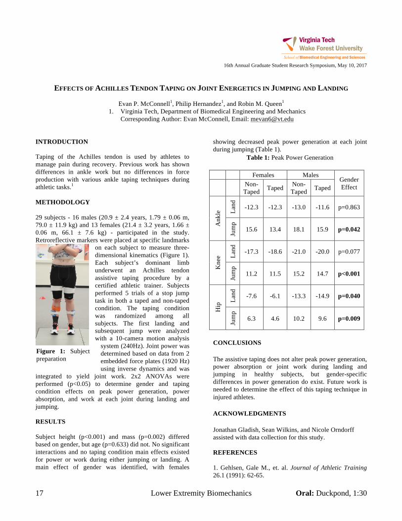
17 Lower Extremity Biomechanics Oral: Duckpond, 1:30
16th Annual Graduate Student Research Symposium, May 10, 2017
EFFECTS OF ACHILLES TENDON TAPING ON JOINT ENERGETICS IN JUMPING AND LANDING
Evan P. McConnell1, Philip Hernandez1, and Robin M. Queen1
1. Virginia Tech, Department of Biomedical Engineering and Mechanics Corresponding Author: Evan McConnell, Email: [email protected]
INTRODUCTION Taping of the Achilles tendon is used by athletes to manage pain during recovery. Previous work has shown differences in ankle work but no differences in force production with various ankle taping techniques during athletic tasks.1 METHODOLOGY 29 subjects - 16 males (20.9 ± 2.4 years, 1.79 ± 0.06 m, 79.0 ± 11.9 kg) and 13 females (21.4 ± 3.2 years, 1.66 ± 0.06 m, 66.1 ± 7.6 kg) - participated in the study. Retroreflective markers were placed at specific landmarks
on each subject to measure three-dimensional kinematics (Figure 1). Each subject’s dominant limb underwent an Achilles tendon assistive taping procedure by a certified athletic trainer. Subjects performed 5 trials of a stop jump task in both a taped and non-taped condition. The taping condition was randomized among all subjects. The first landing and subsequent jump were analyzed with a 10-camera motion analysis
system (240Hz). Joint power was determined based on data from 2 embedded force plates (1920 Hz) using inverse dynamics and was
integrated to yield joint work. 2x2 ANOVAs were performed (p<0.05) to determine gender and taping condition effects on peak power generation, power absorption, and work at each joint during landing and jumping. RESULTS Subject height (p<0.001) and mass (p=0.002) differed based on gender, but age (p=0.633) did not. No significant interactions and no taping condition main effects existed for power or work during either jumping or landing. A main effect of gender was identified, with females
showing decreased peak power generation at each joint during jumping (Table 1).
Table 1: Peak Power Generation
Females Males Gender Effect Non-
Taped Taped Non-Taped Taped
Ank
le La
nd
-12.3 -12.3 -13.0 -11.6 p=0.863
Jum
p 15.6 13.4 18.1 15.9 p=0.042
Kne
e Land
-17.3 -18.6 -21.0 -20.0 p=0.077
Jum
p
11.2 11.5 15.2 14.7 p<0.001
Hip
Land
-7.6 -6.1 -13.3 -14.9 p=0.040
Jum
p
6.3 4.6 10.2 9.6 p=0.009
CONCLUSIONS The assistive taping does not alter peak power generation, power absorption or joint work during landing and jumping in healthy subjects, but gender-specific differences in power generation do exist. Future work is needed to determine the effect of this taping technique in injured athletes. ACKNOWLEDGMENTS Jonathan Gladish, Sean Wilkins, and Nicole Orndorff assisted with data collection for this study. REFERENCES 1. Gehlsen, Gale M., et. al. Journal of Athletic Training 26.1 (1991): 62-65.
Figure 1: Subject preparation
Oral: Smithfield, 1:15 Developing Novel Medical Devices 18
16th Annual Graduate Student Research Symposium, May 10, 2017
USING FMRI DYNAMIC NETWORKS IN A HYPERGRAPH LEARNING MODEL FOR PREDICTING THE
SUCCESS OF LIFESTYLE WEIGHT LOSS INTERVENTIONS IN OBESE OLDER ADULTS
Fatemeh Mokhtari1,2
, Jonathan H. Burdette1, Anthony P. Marsh
1, W. Jack Rejeski
1, Paul J. Laurienti
1,2
1. Laboratory for Complex Brain Networks, Department of Radiology, Wake Forest School of Medicine,
2. Virginia Tech-Wake Forest University School of Biomedical Engineering and Sciences
Winston Salem, NC, USA
INTRODUCTION
Obesity and its adverse health effects is highly prevalent
even among older adults (65+ years), and is uniquely
problematic in this age group, as it is associated with
declines in physical functioning, the main predictor of
disability and loss of independence with aging (1).
The majority of people who follow behavioral treatments
fail to accomplish the pre-established goals. Hence, great
attention has now focused on finding biomarkers to predict
long-term treatment efficiency. In the present study, we
hypothesized that functional magnetic resonance imaging
(fMRI) dynamic networks, as a biomarker, would identify
those overweight and obese older adults who would lose
the largest percentage of body weight in a behaviorally-
based weight loss intervention.
METHODOLOGY
Fifty two obese/overweight older adults participated in a
baseline MR imaging session. Following the completion
of an 18-month lifestyle weight loss intervention, their
weight loss percent was evaluated. A median split of
weight loss (%) was used for identifying participants who
were in the top and bottom halves of percent weight loss,
based on the baseline body weight.
The dynamic functional brain networks were generated
using sliding window correlation technique. The
magnitude of correlations was thresholded to generate
sparse networks with the strong connections retained. The
dimensionality of the networks was reduced using higher-
order singular value decomposition (HOSVD). The
resulting networks were used in a hypergraph learning
model, a multivariate prediction technique (2). Random
subsampling cross validation with 100 permutations was
run to obtain an estimation of average accuracy, with 42
samples in the training and 10 samples in the test subsets.
RESULTS
As evident by Figure 1, prediction performance measures
(mean ± standard deviation) including accuracy,
sensitivity, and specificity were consistently significantly
high (above 0.9) for different sliding window lengths
(range: [61TR, 101TR], with increments of 10TR, where
TR=2s), and different connection density thresholding
values (range: [10%, 30%] with increments of 5%).
Accuracy Sensitivity Specificity
61 71 81 91 101 Total
80
100
80
100
80
100
80
100
61 71 81 91 101 Total
80
100
10
%
Window Size (TR)
15
%2
0%
25
%
30
%
61 71 81 91 101 Total
80
100
80
100
80
100
80
100
61 71 81 91 101 Total
80
100
10
%
Window Size (TR)
15
%2
0%
25
%
30
%
61 71 81 91 101 Total
80
100
80
100
80
100
80
100
61 71 81 91 101 Total
80
100
10
%
Window Size (TR)
15
%2
0%
25
%
30
%
Figure 1: Prediction performance measures
The two brain network components accounting for the
greatest amount of data variability are shown in Figure 2.
Figure 2: (1) high order motor
planning network, 2)
parietoinsular
network. These networks are
involved in
behavioral planning and
executive
functioning.
CONCLUSIONS
The outcomes of this study suggest that functional brain
phenotypes may be useful for tailoring weight loss
treatment to the individual level. Our approach provides
critical groundwork for the growing initiative of
personalized medicine. It will be vital for this work to be
followed by future studies involving larger sample sizes
for different genders and behavioral treatment regimens.
REFERENCES
1. Flegal, K.M. et al. 1999-2010. Jama-Journal of the American Medical Association.
2. Nie, F. et al. 2014. Proceedings of the 20th ACM SIGKDD
international conference on Knowledge discovery and data mining.
19 Developing Novel Medical Devices Oral: Smithfield, 1:45
16th Annual Graduate Student Research Symposium, May 10, 2017
ADVANCED STATISTICAL PROCESS CONTROL TECHNIQUES FOR ANALYSIS OF MEDICAL LINEAR
ACCELERATOR PERFORMANCE
Callistus M. Nguyen1,2
, Alan H. Baydush1, Charles M. Able
3, and Michael T. Munley
1,2
1. Radiation Oncology (Wake Forest School of Medicine, Radiation Oncology)
2. Virginia Tech – Wake Forest School of Biomedical Engineering and Sciences, Biomedical Engineering
3. Radiation Oncology (Florida Cancer Specialists, Radiation Oncology)
Corresponding Author: Callistus M. Nguyen, Email: [email protected]
INTRODUCTION
The focus of this work is to determine the utility and
effectiveness of SPC techniques in order to predict
machine dysfunction. It will also assess the most effective
data set to use SPC techniques that accurately reflects the
behavior of the process. Specific research objectives are
as follows: (1) Evaluate new SPC techniques for
analyzing accelerator system operating parameters and/or
performance; (2) Determine the effectiveness of
Exponentially Weighted Moving Average (EWMA) and
Cumulative Sum (CUSUM) SPC techniques in the
detection of performance dysfunction; (3) Assess and
validate the quality of digital data being produced by
linear accelerators against those produced by quality
assurance instrumentation.
METHODOLOGY
Seven linear accelerators were monitored by delivering
daily robust quality assurance (QA) treatments. Each QA
treatment generated trajectory and text log files that were
used to monitor various operational components and
subsystem performance. Given the large number of
parameters contained within these log files, the focus of
the project was to monitor performance parameters that
were considered to be clinically relevant in radiation
oncology. EWMA and CUSUM control charts were
constructed to analyze those parameters; evaluation
guidelines were also established to detect performance
dysfunction. Each SPC technique was then compared
using statistical analytics to quantitatively determine their
uniqueness and effectiveness. The quality of the digital
data was compared to determine which data set accurately
represents the performance of a linac during clinical
operation.
RESULTS
Advanced SPC techniques showed their effectiveness in
detecting erratic behavior in linac behavior. EWMA and
CUSUM techniques are responsive to small and subtle
shifts; thus, they are more efficient at tracking changes
when processes do not drift rapidly over time. The
incorporation of quantitative metrics supplemented SPC
techniques as indicators to adjust control limits when the
process is trending upwards or downwards. Although
PdM analytics had utility in predicting and preventing
component failure, traditional SPC techniques were not as
effective. Given the power of advanced SPC techniques,
they can become new tools in overall predictive
maintenance of linacs.
CONCLUSIONS
SPC techniques have unique characteristics that separate
them from each other. Each characteristic is specific to
that technique but ultimately they all have one objective:
determine the variation existing within the process.
Depending upon the level of sensitivity, each technique is
then categorized even further, forcing the user to choose a
technique that is most relevant to the level of detection.
There is no generally accepted definition of which type of
SPC technique is most efficient in detecting changes. In
order to quantitatively assess performance, each SPC
technique was subjected to a custom performance analysis
test consisting of three tests: a tabular analysis, an
operating characteristic curve analysis, and Average Run
Length (ARL) analysis (Figure 1). This test was also used
to verify if there were differences between each technique
in detecting changes. In sum, SPC was reliable in
predicting component failure and an effective method in
monitoring and characterizing process performance.
Figure 1: ARL analysis of SPC techniques.
Oral: Smithfield, 1:30 Developing Novel Medical Devices 20
16th Annual Graduate Student Research Symposium, May 10, 2017
DEVELOPMENT AND VALIDATION OF A BRAIN PHANTOM FOR THERAPEUTIC COOLING DEVICES
Ryan D.M. Packett1, Philip J. Brown1, Gautam S. Popli1, and F. Scott Gayzik2
1. Wake Forest University, Department of Biomedical Engineering 2. Wake Forest Baptist Medical Center, Department of Neurology Corresponding Author: F. Scott Gayzik, [email protected]
INTRODUCTION Tissue cooling has been proven as a viable therapy for multiple conditions and injuries, and has been applied to the brain to treat epilepsy and concussions, leading to improved long-term outcomes [1,2]. To facilitate the study of temperature reduction as a function of various cooling methods, a thermal brain phantom was developed and analyzed. METHODOLOGY The phantom is composed of a granular hydrogel through which is circulated 37 degree water, representing blood perfusion. The phantom was tested in a series of cooling trials using a fluid-cooled cooling block during which the perfusion rate was varied across a volume-scaled physiologic range. Results were compared against a validated finite difference (FD) model. The model was then used to calculate steady state cooling at a depth of 5 mm for all flow rates, for both the phantom and a model of the brain. This effort was undertaken to 1. validate the FD model against the phantom results and 2. evaluate how similar the thermal response of the phantom is to that of a perfused brain. RESULTS
Figure 1: Internal phantom temperature versus perfusion
rate, comparison of experimental and mathematical results
The FD phantom model showed good agreement with the empirical phantom results. Furthermore, the empirical phantom agreed with the predicted brain response within 3.5% at physiological flow, suggesting a biofidelic thermal response. CONCLUSIONS We presented a thermal phantom of the human brain, its design and construction and demonstrated flow-mediated sensitivity in the steady state temperature at a depth of 5 mm. Comparison of experimental steady temperatures with a previously-validated finite difference solver yielded good agreement, including an average deviation of 2.0% across experimental perfusion rates between the phantom and the FD model of the phantom experiment, while the phantom-to-FD brain model comparison showed an average deviation of -3.5% for the same measure. The brain phantom presented provides a tunable physical analog that reasonably predicts the temperature drop of perfused brain parenchyma during a surface-based, fluid-driven cooling application, and will be used as a platform for future studies of thermally-mediated therapies applied to the cerebral cortex. ACKNOWLEDGMENTS Pilot funding was provided by Wake Forest Innovations and the Wake Forest Alumni Association. Summer internship support (Megan Fritz) was provided by Wake Forest School of Medicine and by the National Science Foundation (REU Site: Imaging and Mechanics-based Projects on Accidental Cases of Trauma Impact, Award No. 1559700). REFERENCES [1] Nolan, J.P., et al, “Therapeutic hypothermia after cardiac arrest…” Circulation, 2003. 108(1): p. 118-21. [2] Karnatovskaia, L.V. et al, “Therapeutic hypothermia for neuroprotection…” Neurohospitalist, 2014. 4(3): p. 153-63.
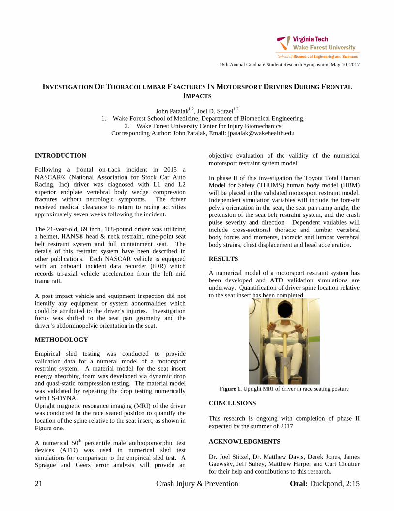
21 Crash Injury & Prevention Oral: Duckpond, 2:15
16th Annual Graduate Student Research Symposium, May 10, 2017
INVESTIGATION OF THORACOLUMBAR FRACTURES IN MOTORSPORT DRIVERS DURING FRONTAL IMPACTS
John Patalak1,2, Joel D. Stitzel1,2
1. Wake Forest School of Medicine, Department of Biomedical Engineering, 2. Wake Forest University Center for Injury Biomechanics
Corresponding Author: John Patalak, Email: [email protected] INTRODUCTION Following a frontal on-track incident in 2015 a NASCAR® (National Association for Stock Car Auto Racing, Inc) driver was diagnosed with L1 and L2 superior endplate vertebral body wedge compression fractures without neurologic symptoms. The driver received medical clearance to return to racing activities approximately seven weeks following the incident. The 21-year-old, 69 inch, 168-pound driver was utilizing a helmet, HANS® head & neck restraint, nine-point seat belt restraint system and full containment seat. The details of this restraint system have been described in other publications. Each NASCAR vehicle is equipped with an onboard incident data recorder (IDR) which records tri-axial vehicle acceleration from the left mid frame rail. A post impact vehicle and equipment inspection did not identify any equipment or system abnormalities which could be attributed to the driver’s injuries. Investigation focus was shifted to the seat pan geometry and the driver’s abdominopelvic orientation in the seat. METHODOLOGY Empirical sled testing was conducted to provide validation data for a numeral model of a motorsport restraint system. A material model for the seat insert energy absorbing foam was developed via dynamic drop and quasi-static compression testing. The material model was validated by repeating the drop testing numerically with LS-DYNA. Upright magnetic resonance imaging (MRI) of the driver was conducted in the race seated position to quantify the location of the spine relative to the seat insert, as shown in Figure one. A numerical 50th percentile male anthropomorphic test devices (ATD) was used in numerical sled test simulations for comparison to the empirical sled test. A Sprague and Geers error analysis will provide an
objective evaluation of the validity of the numerical motorsport restraint system model. In phase II of this investigation the Toyota Total Human Model for Safety (THUMS) human body model (HBM) will be placed in the validated motorsport restraint model. Independent simulation variables will include the fore-aft pelvis orientation in the seat, the seat pan ramp angle, the pretension of the seat belt restraint system, and the crash pulse severity and direction. Dependent variables will include cross-sectional thoracic and lumbar vertebral body forces and moments, thoracic and lumbar vertebral body strains, chest displacement and head acceleration. RESULTS A numerical model of a motorsport restraint system has been developed and ATD validation simulations are underway. Quantification of driver spine location relative to the seat insert has been completed.
Figure 1. Upright MRI of driver in race seating posture
CONCLUSIONS This research is ongoing with completion of phase II expected by the summer of 2017. ACKNOWLEDGMENTS Dr. Joel Stitzel, Dr. Matthew Davis, Derek Jones, James Gaewsky, Jeff Suhey, Matthew Harper and Curt Cloutier for their help and contributions to this research.
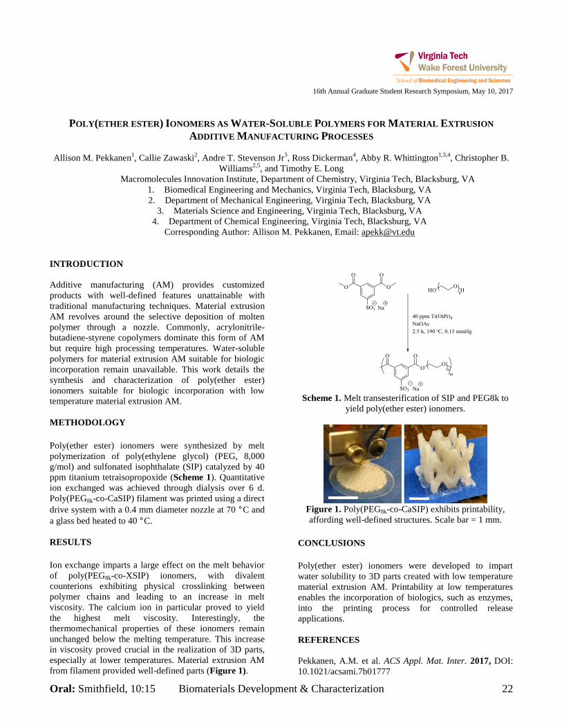
Oral: Smithfield, 10:15 Biomaterials Development & Characterization 22
16th Annual Graduate Student Research Symposium, May 10, 2017
POLY(ETHER ESTER) IONOMERS AS WATER-SOLUBLE POLYMERS FOR MATERIAL EXTRUSION
ADDITIVE MANUFACTURING PROCESSES
Allison M. Pekkanen1, Callie Zawaski
2, Andre T. Stevenson Jr
3, Ross Dickerman
4, Abby R. Whittington
1,3,4, Christopher B.
Williams2,5
, and Timothy E. Long
Macromolecules Innovation Institute, Department of Chemistry, Virginia Tech, Blacksburg, VA
1. Biomedical Engineering and Mechanics, Virginia Tech, Blacksburg, VA
2. Department of Mechanical Engineering, Virginia Tech, Blacksburg, VA
3. Materials Science and Engineering, Virginia Tech, Blacksburg, VA
4. Department of Chemical Engineering, Virginia Tech, Blacksburg, VA
Corresponding Author: Allison M. Pekkanen, Email: [email protected]
INTRODUCTION
Additive manufacturing (AM) provides customized
products with well-defined features unattainable with
traditional manufacturing techniques. Material extrusion
AM revolves around the selective deposition of molten
polymer through a nozzle. Commonly, acrylonitrile-
butadiene-styrene copolymers dominate this form of AM
but require high processing temperatures. Water-soluble
polymers for material extrusion AM suitable for biologic
incorporation remain unavailable. This work details the
synthesis and characterization of poly(ether ester)
ionomers suitable for biologic incorporation with low
temperature material extrusion AM.
METHODOLOGY
Poly(ether ester) ionomers were synthesized by melt
polymerization of poly(ethylene glycol) (PEG, 8,000
g/mol) and sulfonated isophthalate (SIP) catalyzed by 40
ppm titanium tetraisopropoxide (Scheme 1). Quantitative
ion exchanged was achieved through dialysis over 6 d.
Poly(PEG8k-co-CaSIP) filament was printed using a direct
drive system with a 0.4 mm diameter nozzle at 70 °C and
a glass bed heated to 40 °C.
RESULTS
Ion exchange imparts a large effect on the melt behavior
of poly(PEG8k-co-XSIP) ionomers, with divalent
counterions exhibiting physical crosslinking between
polymer chains and leading to an increase in melt
viscosity. The calcium ion in particular proved to yield
the highest melt viscosity. Interestingly, the
thermomechanical properties of these ionomers remain
unchanged below the melting temperature. This increase
in viscosity proved crucial in the realization of 3D parts,
especially at lower temperatures. Material extrusion AM
from filament provided well-defined parts (Figure 1).
Scheme 1. Melt transesterification of SIP and PEG8k to
yield poly(ether ester) ionomers.
Figure 1. Poly(PEG8k-co-CaSIP) exhibits printability,
affording well-defined structures. Scale bar = 1 mm.
CONCLUSIONS
Poly(ether ester) ionomers were developed to impart
water solubility to 3D parts created with low temperature
material extrusion AM. Printability at low temperatures
enables the incorporation of biologics, such as enzymes,
into the printing process for controlled release
applications.
REFERENCES
Pekkanen, A.M. et al. ACS Appl. Mat. Inter. 2017, DOI:
10.1021/acsami.7b01777
23 Biomaterials Development & Characterization Oral: Smithfield, 9:45
16th Annual Graduate Student Research Symposium, May 10, 2017
MOLECULAR ANALYSIS OF HYALURONIC ACID WITH SOLID-STATE NANOPORES
Felipe Rivas1, Osama Zahid1, Paul L. DeAngelis2, Aleksander Skardal1,3,4, Adam R. Hall1,3,4, Elaheh Rahbar1
1. Virginia-Tech Wake Forest School of Biomedical Engineering, Winston Salem, NC 2. Department of Biochemistry and Molecular Biology, University of Oklahoma Health Sciences Center, Oklahoma City, OK
3. Institute for Regenerative Medicine, Wake Forest University School of Medicine Winston Salem, NC 4. Comprehensive Cancer Center, Wake Forest University, School of Biomedical Medicine, Winston Salem, NC
Felipe, Rivas: [email protected]
INTRODUCTION Hyaluronic acid (HA; hyaluronan) has emerged as a powerful indicator for a range of diseases and physiological conditions, including cancer1, osteoarthritis2, and wound healing after injury2. Evidence has also shown that low molecular weight HA triggers pro-inflammatory responses whereas high molecular weight HA is anti-inflammatory2, suggesting the potential importance of HA size as an independent biomarker. However, current methods to quantitate HA size and concentration are costly, time consuming, and rarely distinguish HA by molecular weight. Thus, there is a need for improved HA detection and quantification methods. Herein, we propose a new method to differentiate varying molecular weights of HA based on their electrophoretic movement across a solid-state (SS-) nanopore. METHODOLOGY Using methods described by Yang, et. al3, a single nanopore (5-8 nm diameter) was fabricated on a 30-nm thick silicon nitride membrane supported by a 4.4 mm Si chip. The device was then used as a barrier between two electrically isolated reservoirs. An ionic solution (6M LiCl) was loaded onto both sides of the chip, with one chamber also containing HA. A voltage was applied across the device, generating an electric field gradient capable of translocating HA molecules electrophoretically through the pore. Molecular translocations caused temporary interruptions (“events”) in the trans-membrane ionic current that were measured by a patch-clamp amplifier and recorded using custom software. Event depth, duration, and rate were collected across a voltage range of 100-600 mV. Polydisperse HA was obtained from Sigma Aldrich; monodisperse HA ranging from 50-2,500kDa was provided by Hyalose, LLC. RESULTS Using polydisperse HA, we demonstrated SS-nanopore detection of HA for the first time, yielding a series of
easily resolved current blockades that each marked the passage of a single molecule. Events were observed only towards a positive bias, in agreement with the expected electrophoretic direction of anionic HA. Event rate was found to have a linear dependence on applied voltage, indicating diffusion-limited kinetics, and to vary directly with HA concentration. Subsequent measurements on purified, monodisperse HA yielded well-defined populations of event properties for HA samples 50-2500kDa in size. Specifically, the event charge deficit, (ecd), which is the integrated area of each measured event; was unique for each HA size. Importantly, ecd displayed a power-law dependence on HA molecular weight, showing that the SS-nanopore system can differentiate HA based on molecular weight.
CONCLUSIONS SS-nanopore detection was successfully used to distinguish between different molecular weights of HA based on ecd. The data from this study shows promise in using the SS-nanopore system as a potential quick analytical assay to quantitate HA size and concentration in the clinical setting. ACKNOWLEDGMENTS This project has been supported by Dr. Adam Hall’s and Dr. Elaheh Rahbar’s start-up funds. REFERENCES 1. Lokeshwar, V, B. The Journal of Biological Chemistry. 2001, 276, 11922–11932. 2. Necas, J. Veterinarni Medicina. 2008, 53, 397–411. 3. Yang, J., et al. Nanotechnology. 2011, 22, 285310.
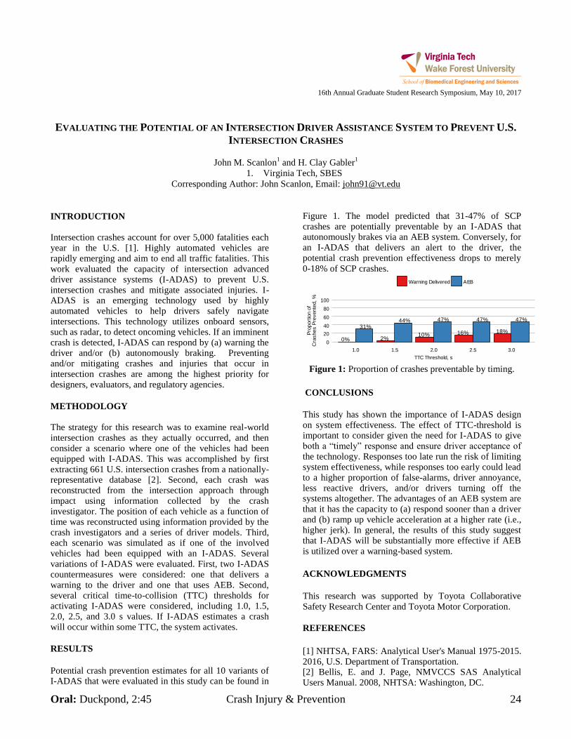
Oral: Duckpond, 2:45 Crash Injury & Prevention 24
16th Annual Graduate Student Research Symposium, May 10, 2017
EVALUATING THE POTENTIAL OF AN INTERSECTION DRIVER ASSISTANCE SYSTEM TO PREVENT U.S.
INTERSECTION CRASHES
John M. Scanlon1 and H. Clay Gabler
1
1. Virginia Tech, SBES
Corresponding Author: John Scanlon, Email: [email protected]
INTRODUCTION
Intersection crashes account for over 5,000 fatalities each year in the U.S. [1]. Highly automated vehicles are rapidly emerging and aim to end all traffic fatalities. This work evaluated the capacity of intersection advanced driver assistance systems (I-ADAS) to prevent U.S. intersection crashes and mitigate associated injuries. I-ADAS is an emerging technology used by highly automated vehicles to help drivers safely navigate intersections. This technology utilizes onboard sensors, such as radar, to detect oncoming vehicles. If an imminent crash is detected, I-ADAS can respond by (a) warning the driver and/or (b) autonomously braking. Preventing and/or mitigating crashes and injuries that occur in intersection crashes are among the highest priority for designers, evaluators, and regulatory agencies.
METHODOLOGY
The strategy for this research was to examine real-world intersection crashes as they actually occurred, and then consider a scenario where one of the vehicles had been equipped with I-ADAS. This was accomplished by first extracting 661 U.S. intersection crashes from a nationally-representative database [2]. Second, each crash was reconstructed from the intersection approach through impact using information collected by the crash investigator. The position of each vehicle as a function of time was reconstructed using information provided by the crash investigators and a series of driver models. Third, each scenario was simulated as if one of the involved vehicles had been equipped with an I-ADAS. Several variations of I-ADAS were evaluated. First, two I-ADAS countermeasures were considered: one that delivers a warning to the driver and one that uses AEB. Second, several critical time-to-collision (TTC) thresholds for activating I-ADAS were considered, including 1.0, 1.5, 2.0, 2.5, and 3.0 s values. If I-ADAS estimates a crash will occur within some TTC, the system activates.
RESULTS
Potential crash prevention estimates for all 10 variants of I-ADAS that were evaluated in this study can be found in
Figure 1. The model predicted that 31-47% of SCP crashes are potentially preventable by an I-ADAS that autonomously brakes via an AEB system. Conversely, for an I-ADAS that delivers an alert to the driver, the potential crash prevention effectiveness drops to merely
0-18% of SCP crashes.
31%
0%
44%
2%
47%
10%
47%
16%
47%
18%
0
20
40
60
80
100
1.0 1.5 2.0 2.5 3.0
TTC Threshold, s
Pro
port
ion o
fC
rashes P
revente
d, %
Warning Delivered AEB
Figure 1: Proportion of crashes preventable by timing.
CONCLUSIONS
This study has shown the importance of I-ADAS design on system effectiveness. The effect of TTC-threshold is important to consider given the need for I-ADAS to give both a “timely” response and ensure driver acceptance of the technology. Responses too late run the risk of limiting system effectiveness, while responses too early could lead to a higher proportion of false-alarms, driver annoyance, less reactive drivers, and/or drivers turning off the systems altogether. The advantages of an AEB system are that it has the capacity to (a) respond sooner than a driver and (b) ramp up vehicle acceleration at a higher rate (i.e., higher jerk). In general, the results of this study suggest that I-ADAS will be substantially more effective if AEB is utilized over a warning-based system.
ACKNOWLEDGMENTS
This research was supported by Toyota Collaborative
Safety Research Center and Toyota Motor Corporation.
REFERENCES
[1] NHTSA, FARS: Analytical User's Manual 1975-2015. 2016, U.S. Department of Transportation. [2] Bellis, E. and J. Page, NMVCCS SAS Analytical Users Manual. 2008, NHTSA: Washington, DC.
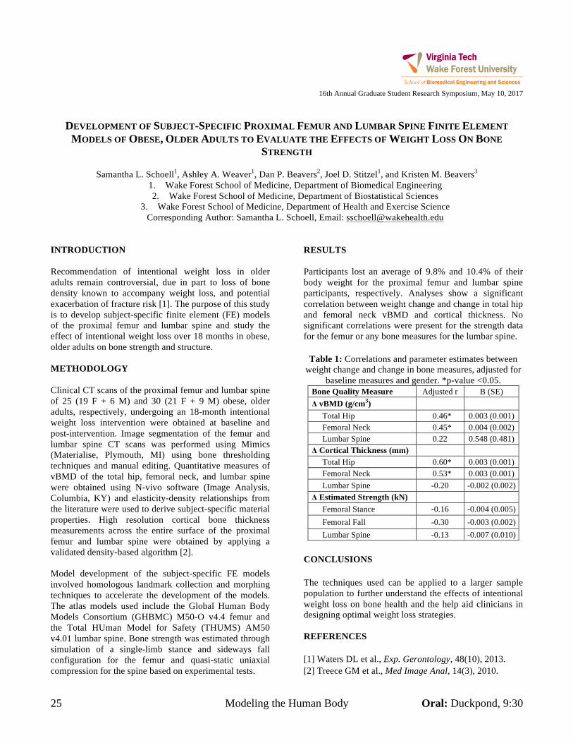
25 Modeling the Human Body Oral: Duckpond, 9:30
16th Annual Graduate Student Research Symposium, May 10, 2017
DEVELOPMENT OF SUBJECT-SPECIFIC PROXIMAL FEMUR AND LUMBAR SPINE FINITE ELEMENT
MODELS OF OBESE, OLDER ADULTS TO EVALUATE THE EFFECTS OF WEIGHT LOSS ON BONE STRENGTH
Samantha L. Schoell1, Ashley A. Weaver1, Dan P. Beavers2, Joel D. Stitzel1, and Kristen M. Beavers3
1. Wake Forest School of Medicine, Department of Biomedical Engineering 2. Wake Forest School of Medicine, Department of Biostatistical Sciences
3. Wake Forest School of Medicine, Department of Health and Exercise Science Corresponding Author: Samantha L. Schoell, Email: [email protected]
INTRODUCTION Recommendation of intentional weight loss in older adults remain controversial, due in part to loss of bone density known to accompany weight loss, and potential exacerbation of fracture risk [1]. The purpose of this study is to develop subject-specific finite element (FE) models of the proximal femur and lumbar spine and study the effect of intentional weight loss over 18 months in obese, older adults on bone strength and structure. METHODOLOGY Clinical CT scans of the proximal femur and lumbar spine of 25 (19 F + 6 M) and 30 (21 F + 9 M) obese, older adults, respectively, undergoing an 18-month intentional weight loss intervention were obtained at baseline and post-intervention. Image segmentation of the femur and lumbar spine CT scans was performed using Mimics (Materialise, Plymouth, MI) using bone thresholding techniques and manual editing. Quantitative measures of vBMD of the total hip, femoral neck, and lumbar spine were obtained using N-vivo software (Image Analysis, Columbia, KY) and elasticity-density relationships from the literature were used to derive subject-specific material properties. High resolution cortical bone thickness measurements across the entire surface of the proximal femur and lumbar spine were obtained by applying a validated density-based algorithm [2]. Model development of the subject-specific FE models involved homologous landmark collection and morphing techniques to accelerate the development of the models. The atlas models used include the Global Human Body Models Consortium (GHBMC) M50-O v4.4 femur and the Total HUman Model for Safety (THUMS) AM50 v4.01 lumbar spine. Bone strength was estimated through simulation of a single-limb stance and sideways fall configuration for the femur and quasi-static uniaxial compression for the spine based on experimental tests.
RESULTS Participants lost an average of 9.8% and 10.4% of their body weight for the proximal femur and lumbar spine participants, respectively. Analyses show a significant correlation between weight change and change in total hip and femoral neck vBMD and cortical thickness. No significant correlations were present for the strength data for the femur or any bone measures for the lumbar spine.
Table 1: Correlations and parameter estimates between weight change and change in bone measures, adjusted for
baseline measures and gender. *p-value <0.05. Bone Quality Measure Adjusted r B (SE) Δ vBMD (g/cm3) Total Hip 0.46* 0.003 (0.001) Femoral Neck 0.45* 0.004 (0.002) Lumbar Spine 0.22 0.548 (0.481) Δ Cortical Thickness (mm) Total Hip 0.60* 0.003 (0.001) Femoral Neck 0.53* 0.003 (0.001) Lumbar Spine -0.20 -0.002 (0.002) Δ Estimated Strength (kN) Femoral Stance -0.16 -0.004 (0.005) Femoral Fall -0.30 -0.003 (0.002) Lumbar Spine -0.13 -0.007 (0.010)
CONCLUSIONS The techniques used can be applied to a larger sample population to further understand the effects of intentional weight loss on bone health and the help aid clinicians in designing optimal weight loss strategies. REFERENCES [1] Waters DL et al., Exp. Gerontology, 48(10), 2013. [2] Treece GM et al., Med Image Anal, 14(3), 2010.
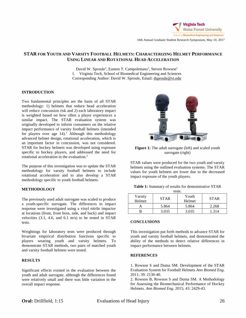
Oral: Drillfield, 1:15 Evaluations of Head Injury 26
16th Annual Graduate Student Research Symposium, May 10, 2017
STAR FOR YOUTH AND VARSITY FOOTBALL HELMETS: CHARACTERIZING HELMET PERFORMANCE
USING LINEAR AND ROTATIONAL HEAD ACCELERATION
David W. Sproule1, Eamon T. Campolettano1, Steven Rowson1
1. Virginia Tech, School of Biomedical Engineering and Sciences Corresponding Author: David W. Sproule, Email: [email protected]
INTRODUCTION Two fundamental principles are the basis of all STAR methodology: 1) helmets that reduce head acceleration will reduce concussion risk and 2) each laboratory impact is weighted based on how often a player experiences a similar impact. The STAR evaluation system was originally developed to inform consumers on the relative impact performance of varsity football helmets (intended for players over age 14).1 Although this methodology advanced helmet design, rotational acceleration, which is an important factor in concussion, was not considered. STAR for hockey helmets was developed using exposure specific to hockey players, and addressed the need for rotational acceleration in the evaluation.2 The purpose of this investigation was to update the STAR methodology for varsity football helmets to include rotational acceleration and to also develop a STAR methodology specific to youth football helmets. METHODOLOGY The previously used adult surrogate was scaled to produce a youth-specific surrogate. The differences in impact response were investigated using a vinyl nitrile impactor at locations (front, front boss, side, and back) and impact velocities (3.1, 4.6, and 6.1 m/s) to be tested in STAR tests. Weightings for laboratory tests were produced through bivariate empirical distribution functions specific to players wearing youth and varsity helmets. To demonstrate STAR methods, two pairs of matched youth and varsity football helmets were tested. RESULTS Significant effects existed in the evaluation between the youth and adult surrogate, although the differences found were relatively small and there was little variation in the overall impact response.
Figure 1: The adult surrogate (left) and scaled youth surrogate (right)
STAR values were produced for the two youth and varsity helmets using the outlined evaluation systems. The STAR values for youth helmets are lower due to the decreased impact exposure of the youth players.
Table 1: Summary of results for demonstrative STAR tests.
Varsity Helmet STAR Youth
Helmet STAR
A 5.864 5.864 2.268 B 3.035 3.035 1.314
CONCLUSIONS This investigation put forth methods to advance STAR for youth and varsity football helmets, and demonstrated the ability of the methods to detect relative differences in impact performance between helmets. REFERENCES 1. Rowson S and Duma SM. Development of the STAR Evaluation System for Football Helmets Ann Biomed Eng. 2011; 39: 2130-40. 2. Rowson B, Rowson S and Duma SM. A Methodology for Assessing the Biomechanical Performance of Hockey Helmets. Ann Biomed Eng. 2015; 43: 2429-43.
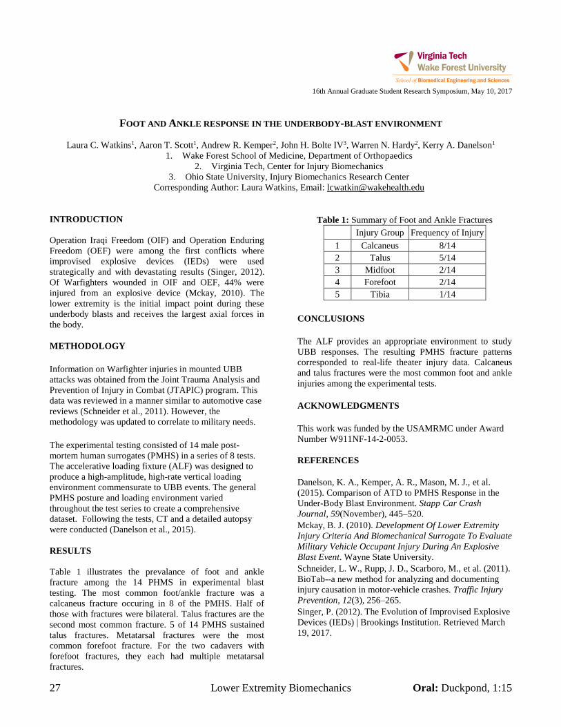
27 Lower Extremity Biomechanics Oral: Duckpond, 1:15
16th Annual Graduate Student Research Symposium, May 10, 2017
FOOT AND ANKLE RESPONSE IN THE UNDERBODY-BLAST ENVIRONMENT
Laura C. Watkins1, Aaron T. Scott1, Andrew R. Kemper2, John H. Bolte IV3, Warren N. Hardy2, Kerry A. Danelson1
1. Wake Forest School of Medicine, Department of Orthopaedics 2. Virginia Tech, Center for Injury Biomechanics
3. Ohio State University, Injury Biomechanics Research Center Corresponding Author: Laura Watkins, Email: [email protected]
INTRODUCTION Operation Iraqi Freedom (OIF) and Operation Enduring Freedom (OEF) were among the first conflicts where improvised explosive devices (IEDs) were used strategically and with devastating results (Singer, 2012). Of Warfighters wounded in OIF and OEF, 44% were injured from an explosive device (Mckay, 2010). The lower extremity is the initial impact point during these underbody blasts and receives the largest axial forces in the body. METHODOLOGY Information on Warfighter injuries in mounted UBB attacks was obtained from the Joint Trauma Analysis and Prevention of Injury in Combat (JTAPIC) program. This data was reviewed in a manner similar to automotive case reviews (Schneider et al., 2011). However, the methodology was updated to correlate to military needs. The experimental testing consisted of 14 male post-mortem human surrogates (PMHS) in a series of 8 tests. The accelerative loading fixture (ALF) was designed to produce a high-amplitude, high-rate vertical loading environment commensurate to UBB events. The general PMHS posture and loading environment varied throughout the test series to create a comprehensive dataset. Following the tests, CT and a detailed autopsy were conducted (Danelson et al., 2015). RESULTS Table 1 illustrates the prevalance of foot and ankle fracture among the 14 PHMS in experimental blast testing. The most common foot/ankle fracture was a calcaneus fracture occuring in 8 of the PMHS. Half of those with fractures were bilateral. Talus fractures are the second most common fracture. 5 of 14 PMHS sustained talus fractures. Metatarsal fractures were the most common forefoot fracture. For the two cadavers with forefoot fractures, they each had multiple metatarsal fractures.
Table 1: Summary of Foot and Ankle Fractures Injury Group Frequency of Injury
1 Calcaneus 8/14 2 Talus 5/14 3 Midfoot 2/14 4 Forefoot 2/14 5 Tibia 1/14
CONCLUSIONS The ALF provides an appropriate environment to study UBB responses. The resulting PMHS fracture patterns corresponded to real-life theater injury data. Calcaneus and talus fractures were the most common foot and ankle injuries among the experimental tests. ACKNOWLEDGMENTS This work was funded by the USAMRMC under Award Number W911NF-14-2-0053. REFERENCES Danelson, K. A., Kemper, A. R., Mason, M. J., et al. (2015). Comparison of ATD to PMHS Response in the Under-Body Blast Environment. Stapp Car Crash Journal, 59(November), 445–520. Mckay, B. J. (2010). Development Of Lower Extremity Injury Criteria And Biomechanical Surrogate To Evaluate Military Vehicle Occupant Injury During An Explosive Blast Event. Wayne State University. Schneider, L. W., Rupp, J. D., Scarboro, M., et al. (2011). BioTab--a new method for analyzing and documenting injury causation in motor-vehicle crashes. Traffic Injury Prevention, 12(3), 256–265. Singer, P. (2012). The Evolution of Improvised Explosive Devices (IEDs) | Brookings Institution. Retrieved March 19, 2017.
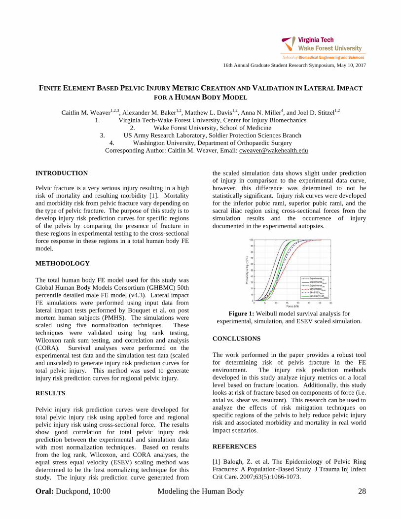
Oral: Duckpond, 10:00 Modeling the Human Body 28
16th Annual Graduate Student Research Symposium, May 10, 2017
FINITE ELEMENT BASED PELVIC INJURY METRIC CREATION AND VALIDATION IN LATERAL IMPACT
FOR A HUMAN BODY MODEL
Caitlin M. Weaver1,2,3, Alexander M. Baker1,2, Matthew L. Davis1,2, Anna N. Miller4, and Joel D. Stitzel1,2
1. Virginia Tech-Wake Forest University, Center for Injury Biomechanics 2. Wake Forest University, School of Medicine
3. US Army Research Laboratory, Soldier Protection Sciences Branch 4. Washington University, Department of Orthopaedic Surgery
Corresponding Author: Caitlin M. Weaver, Email: [email protected] INTRODUCTION Pelvic fracture is a very serious injury resulting in a high risk of mortality and resulting morbidity [1]. Mortality and morbidity risk from pelvic fracture vary depending on the type of pelvic fracture. The purpose of this study is to develop injury risk prediction curves for specific regions of the pelvis by comparing the presence of fracture in these regions in experimental testing to the cross-sectional force response in these regions in a total human body FE model. METHODOLOGY The total human body FE model used for this study was Global Human Body Models Consortium (GHBMC) 50th percentile detailed male FE model (v4.3). Lateral impact FE simulations were performed using input data from lateral impact tests performed by Bouquet et al. on post mortem human subjects (PMHS). The simulations were scaled using five normalization techniques. These techniques were validated using log rank testing, Wilcoxon rank sum testing, and correlation and analysis (CORA). Survival analyses were performed on the experimental test data and the simulation test data (scaled and unscaled) to generate injury risk prediction curves for total pelvic injury. This method was used to generate injury risk prediction curves for regional pelvic injury.
RESULTS Pelvic injury risk prediction curves were developed for total pelvic injury risk using applied force and regional pelvic injury risk using cross-sectional force. The results show good correlation for total pelvic injury risk prediction between the experimental and simulation data with most normalization techniques. Based on results from the log rank, Wilcoxon, and CORA analyses, the equal stress equal velocity (ESEV) scaling method was determined to be the best normalizing technique for this study. The injury risk prediction curve generated from
the scaled simulation data shows slight under prediction of injury in comparison to the experimental data curve, however, this difference was determined to not be statistically significant. Injury risk curves were developed for the inferior pubic rami, superior pubic rami, and the sacral iliac region using cross-sectional forces from the simulation results and the occurrence of injury documented in the experimental autopsies.
Figure 1: Weibull model survival analysis for
experimental, simulation, and ESEV scaled simulation. CONCLUSIONS The work performed in the paper provides a robust tool for determining risk of pelvis fracture in the FE environment. The injury risk prediction methods developed in this study analyze injury metrics on a local level based on fracture location. Additionally, this study looks at risk of fracture based on components of force (i.e. axial vs. shear vs. resultant). This research can be used to analyze the effects of risk mitigation techniques on specific regions of the pelvis to help reduce pelvic injury risk and associated morbidity and mortality in real world impact scenarios. REFERENCES [1] Balogh, Z. et al. The Epidemiology of Pelvic Ring Fractures: A Population-Based Study. J Trauma Inj Infect Crit Care. 2007;63(5):1066-1073.
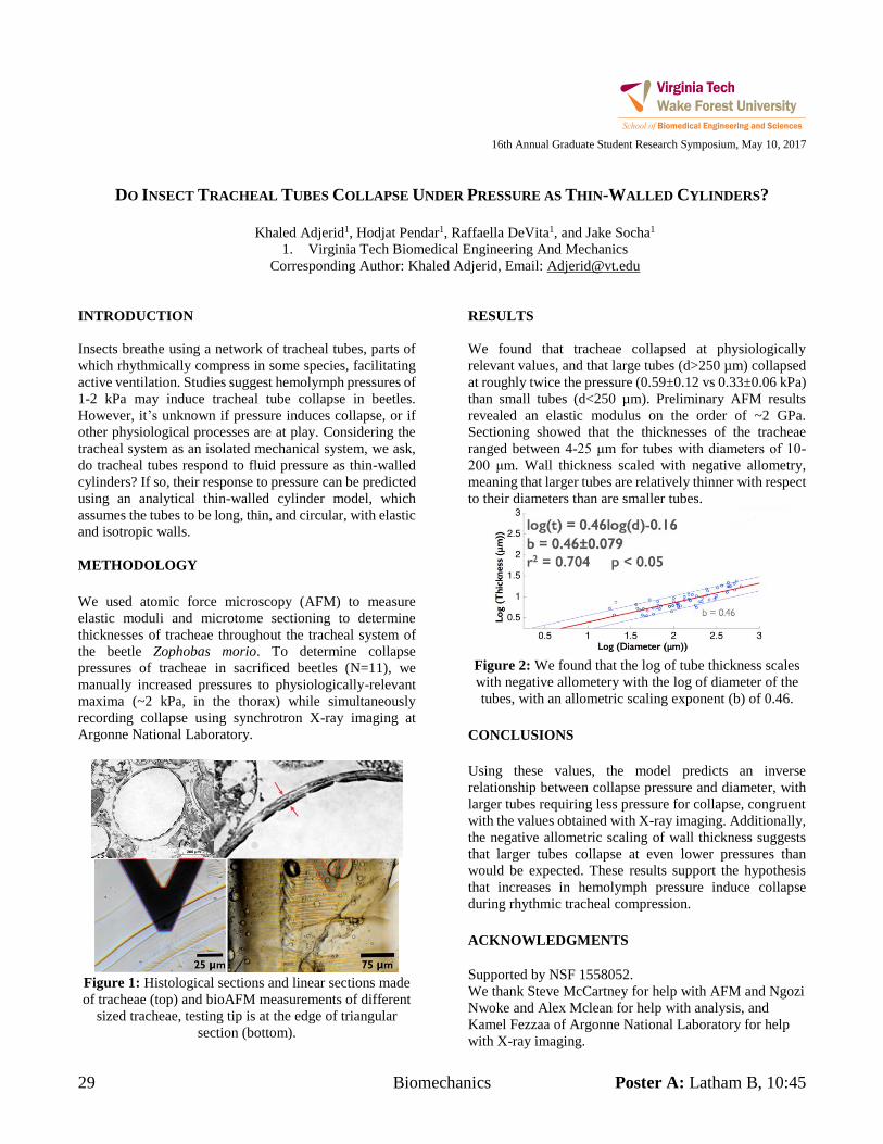
29 Biomechanics Poster A: Latham B, 10:45
16th Annual Graduate Student Research Symposium, May 10, 2017
DO INSECT TRACHEAL TUBES COLLAPSE UNDER PRESSURE AS THIN-WALLED CYLINDERS?
Khaled Adjerid1, Hodjat Pendar1, Raffaella DeVita1, and Jake Socha1
1. Virginia Tech Biomedical Engineering And Mechanics Corresponding Author: Khaled Adjerid, Email: [email protected]
INTRODUCTION Insects breathe using a network of tracheal tubes, parts of which rhythmically compress in some species, facilitating active ventilation. Studies suggest hemolymph pressures of 1-2 kPa may induce tracheal tube collapse in beetles. However, it’s unknown if pressure induces collapse, or if other physiological processes are at play. Considering the tracheal system as an isolated mechanical system, we ask, do tracheal tubes respond to fluid pressure as thin-walled cylinders? If so, their response to pressure can be predicted using an analytical thin-walled cylinder model, which assumes the tubes to be long, thin, and circular, with elastic and isotropic walls. METHODOLOGY We used atomic force microscopy (AFM) to measure elastic moduli and microtome sectioning to determine thicknesses of tracheae throughout the tracheal system of the beetle Zophobas morio. To determine collapse pressures of tracheae in sacrificed beetles (N=11), we manually increased pressures to physiologically-relevant maxima (~2 kPa, in the thorax) while simultaneously recording collapse using synchrotron X-ray imaging at Argonne National Laboratory.
Figure 1: Histological sections and linear sections made of tracheae (top) and bioAFM measurements of different
sized tracheae, testing tip is at the edge of triangular section (bottom).
RESULTS We found that tracheae collapsed at physiologically relevant values, and that large tubes (d>250 µm) collapsed at roughly twice the pressure (0.59±0.12 vs 0.33±0.06 kPa) than small tubes (d<250 µm). Preliminary AFM results revealed an elastic modulus on the order of ~2 GPa. Sectioning showed that the thicknesses of the tracheae ranged between 4-25 μm for tubes with diameters of 10-200 μm. Wall thickness scaled with negative allometry, meaning that larger tubes are relatively thinner with respect to their diameters than are smaller tubes.
Figure 2: We found that the log of tube thickness scales with negative allometery with the log of diameter of the tubes, with an allometric scaling exponent (b) of 0.46.
CONCLUSIONS Using these values, the model predicts an inverse relationship between collapse pressure and diameter, with larger tubes requiring less pressure for collapse, congruent with the values obtained with X-ray imaging. Additionally, the negative allometric scaling of wall thickness suggests that larger tubes collapse at even lower pressures than would be expected. These results support the hypothesis that increases in hemolymph pressure induce collapse during rhythmic tracheal compression. ACKNOWLEDGMENTS Supported by NSF 1558052. We thank Steve McCartney for help with AFM and Ngozi Nwoke and Alex Mclean for help with analysis, and Kamel Fezzaa of Argonne National Laboratory for help with X-ray imaging.
Poster B: Latham B, 11:15 Biomechanics 30
16th Annual Graduate Student Research Symposium, May 10, 2017
VALIDATION OF A DIRECT METHOD FOR MEASURING HEPATIC AND SPLENIC PRESSURES OF POST-
MORTEM HUMAN SURROGATES DURING FULL-SCALE FRONTAL SLED TESTS
Devon L. Albert1, Stephanie M. Beeman1, and Andrew R. Kemper1
1. Virginia Tech, Department of Biomedical Engineering and Mechanics Corresponding Author: Devon L. Albert, Email: [email protected]
INTRODUCTION Although the liver and spleen are the two most frequently injured abdominal organs in motor vehicle collisions1-2, there is a lack of research regarding potential splenic injury metrics. Conversely, previous research has been conducted that correlated internal liver pressure to liver injury risk3-4. Such studies have instrumented the livers of whole-body post-mortem human surrogates (PMHSs) with pressure sensors via an indirect method guided by fluoroscopy. Fluoroscopy is expensive and can be time consuming when routing sensors to a target location through peripheral vasculature. Therefore, a more direct approach is needed to instrument the solid abdominal organs with pressure sensors. The objective of this research was to implement a direct approach for pressure sensor instrumentation of the liver and spleen of PMHSs and validate the measured pressures obtained during full-scale frontal sled tests against previous research involving comparable blunt impacts. METHODOLOGY The abdominal cavities of six fresh, previously frozen PMHSs were accessed, and the hepatic and splenic arteries were located. Both arteries were cut after branching from the celiac trunk, and Millar catheter style pressure sensors were inserted into the open arteries and routed through the vasculature to the liver and spleen. Each PMHS underwent one full-scale frontal sled test under one of two safety restraint conditions: knee bolster or knee bolster airbag. All tests involved a seatbelt and steering wheel airbag. Before each test, the vascular system was pressurized as follows: the hepatic artery to 2 psi, the splenic artery to 2 psi, the carotid artery to 2 psi, and the jugular vein to 0.5 psi. The peak hepatic pressures from the sled tests were compared to those reported by previous studies using a Welch’s ANOVA. RESULTS Hepatic peak pressures ranged from approximately 40 to 76 kPa, while splenic peak pressures ranged from approximately 34 to 105 kPa. The pressure time histories
of each organ were variable with different shapes and peak timing. Variation between tests was likely a result of differences in anthropometry between subjects. Organ location relative to safety restraints, such as the seatbelt and airbag, or other vehicle components, such as the steering wheel, can influence the magnitude and timing of loading experienced from contact with these features. The ANOVA comparing the hepatic pressures from the sled tests to previous studies resulted in a p-value of 0.955. This implied that the pressures from the current study were not significantly different from those reported by previous studies. CONCLUSIONS This study successfully implemented a direct approach for instrumenting the livers and spleens of PMHSs with pressure sensors. Further research should be conducted to develop and validate injury risk curves for the liver and spleen. ACKNOWLEDGMENTS The authors would like to thank the Toyota Motor Corporation for sponsoring this research. REFERENCES 1. A. M. Elhagediab et al., "Patterns of abdominal injury
in frontal automotive crashes," in The 16th International Technical Conference on the Enhanced Safety of Vehicles, 1998.
2. N. Yoganandan, et al., "Patterns of abdominal injuries in frontal and side impacts," in Annual Proc Assoc Adv Automot Med, 2000, pp. 17-36.
3. J. L. Sparks, et al., "Using pressure to predict liver injury risk from blunt impact," Stapp car crash journal, vol. 51, pp. 401-432, 2007.
4. M. A. Kremer, et al., "Pressure-based abdominal injury criteria using isolated liver and full-body post-mortem human subject impact tests," Stapp car crash journal, vol. 55, p. 317, 2011
31 Biomechanics Poster A: Latham B, 10:45
16th Annual Graduate Student Research Symposium, May 10, 2017
EFFECTS OF AUTOMATIC EMERGENCY BRAKING ON OCCUPANT KINEMATICS
Roy Anderson1, and Andrew Kemper, Ph.D. 1
1. Virginia Tech, School of Biomedical Engineering and Sciences Corresponding Author: Roy Anderson, Email: [email protected]
INTRODUCTION Automatic Emergency Brakes (AEB) is a safety mechanism in cars used to reduce speed or completely stop the car before an impact. AEB utilizes radar scanning to detect traffic or objects in front of the car, alerts the driver of the potential hazard, and then automatically decelerates the car if the driver does not apply brakes. AEB’s are highly effective at preventing crashes at
speeds less than 25 mph. While at speeds greater than 55 mph, the AEB will decrease speed, but not prevent the crash, also while decreasing the speed the occupant can be displaced into a non-ideal position, thus increasing the chance of injuries such as fatal rib fractures1. The occupant kinematics, or the occupant’s movement
through the car is highly related to injury. From this understanding, the focus of this project is to research the effects of AEB braking at high speeds on occupant kinematics to assess the chances of occupant injury. METHODOLOGY A total of 6 subjects 3-male and 3-female will be subjected to testing. A total of 36 tests will be conducted. Subjects will be positioned at the center of the test seat with the feet centered on the footplates and hands on the steering column. Their feet will be centered on the footplates with hands on the steering column. Each try will have a standard load limiting driver-side 3 point seat belt position around the test subject and slack will be removed. Dynamic low severity crash pulse tests will be conducted at varying decelerations using a custom mini-sled and test buck accelerated by a pneumatic piston. A Vicon motion analysis system (Vicon Motion Systems, Centennial, CO), consisting of 12- MX-T20 2 megapixel cameras will be used to quantify the 3-dimensional occupant kinematics of the volunteers and the test sled at a sampling rate of 1kHz. Reflective markers will be adhered to each test subject and the test buck. A 3-D reconstruction of the Vicon data will be generated. High speed video will also be captured from the subjects’ lateral aspect at a sampling
rate of 1kHz with the use of a high resolution, high light sensitivity camera.
Figure 1: (Left) Image of custom test buck with pneumatic piston. (Right) anatomical marker placement
A Vicon motion analysis system (Vicon Motion Systems, Centennial, CO), consisting of 12- MX-T20 2 megapixel cameras will be used to quantify the 3-dimensional occupant kinematics of the volunteers and the test sled at a sampling rate of 1kHz. Reflective markers will be adhered to each test subject and the test buck. A 3-D reconstruction of the Vicon data will be generated. High speed video will also be captured from the subjects’
lateral aspect at a sampling rate of 1kHz with the use of a high resolution, high light sensitivity camera. Changes in occupant kinematics, and injury risk between the AEB pulse and constant deceleration pulse will be compared to assess the effects of AEB on occupant kinematics. Statistical analysis will be conducted using Matrix Laboratory and Microsoft Excel (Microsoft Corp, Redmond, WA). Analysis of variance will be conducted between each severity tests (1.0g vs. 1.15g, 1.15 vs. 1.3 g, 1.0 vs. 1.3 g) to analyze the effects of severity on occupant kinematics. . REFERENCES 1. n.d. (2016). Autonomous Emergency braking. Web. ThatCam Research. 2. Ólafsdóttir et al. (2013). Passenger Kinematics and Muscle Responses in Autonomous Braking Events with Standard and Reversible Pre‐tensioned Restraints. IRCOBI Conference 2013 3. Saito et.al. (2016) Evaluation of Frontal Impact Restraint System in Integrated Safety Scenario Using Human Body Model with PID Controlled Active Muscles. IRCOBI Conference 2016. 4. Beeman et.al. (2011) Effects of Bracing on Human kinematics in low speed frontal sled tests., Annals of Biomedical Engineering
Poster B: Latham B, 11:15 Biomedical Imaging 32
16th Annual Graduate Student Research Symposium, May 10, 2017
THE IMPACTS OF PESTICIDE AND NICOTINE ON FUNCTIONAL BRAIN NETWORKS IN LATINO
FARMWORKERS
Mohsen Bahami1, Paul J. Laurienti1,2, Thomas A. Arcury3, and Sean L. Simpson1,4
1. Virginia Tech – Wake Forest School of Biomedical Engineering and Science 2. Department of Radiology, Wake Forest School of Medicine
3. Department of Family and Community Medicine, Wake Forest School of Medicine 4. Department of Biostatistical Sciences, Wake Forest School of Medicine
Corresponding Author: M. Bahrami, Email: [email protected] INTRODUCTION Migrant Latino farmworkers are consistently exposed to pesticides as part of their work. Pesticides that inhibit cholinesterase, such as organophosphates and carbamates, alter acetylcholine levels in the nervous system and are neurotoxic. Long-term exposure to these pesticides may increase the risk of developing neurodegenerative diseases such as Parkinson’s disease. Tobacco farmworkers are not only exposed to pesticides but also experience nicotine exposure through dermal absorption. While it is known that large doses of nicotine are toxic, many studies have indicated that lower, nontoxic doses of nicotine improve cognitive performance. Functional brain network analyses that use rs-fMRI have demonstrated great promise in examining systemic brain changes across health and disease [1]. This study used brain network analyses of rs-fMRI data and a mixed-effects modeling framework [2] to compare brain network connectivity patterns between Latino farmworkers and non-farmworkers. METHODOLOGY 48 Latino farmworkers and 26 non-farmworkers had brain MRI scans and serum cholinesterase measures completed in the same agricultural season as part of a larger study. Functional brain networks were constructed for each subject using their resting state fMRI data. The probability and the strength of a connection were both modeled via a mixed-effects modeling framework. RESULTS Average Clustering Coefficient and Modularity in farmworkers were significantly higher than in non-farmworkers (Figure 1). FC probability and strength differed between farmworkers and non-farmworkers by nodal clustering coefficient and modularity (Table1). Cholinesterase activity was associated with population differences in brain community (not shown here) .
Figure 1. Boxplots for population-level measures of clustering
coefficient and modularity in farmworkers and non-farmworkers.
CONCLUSIONS Farmworkers have more clustered and modular networks than non-farmworkers. Increased modularity could create implications for high-level cognitive tasks. ACKNOWLEDGMENTS This work was supported by NEIHS (grant number: R01 ES008739) and NIH award (K25 EB012236). REFERENCES [1] Bullmore, E. and O. Sporns, Complex brain networks: graph theoretical analysis of structural and functional systems. Nature Reviews Neuroscience, 2009. 10(3): p. 186-198. [2] Simpson, S.L. and P.J. Laurienti, A two-part mixed-effects modeling framework for analyzing whole-brain network data. NeuroImage, 2015. 113: p. 310-319.
Parameter Estimate SE *p-value Farmworker Status (FWS)
-0.039 0.047 0.408
Clustering Coef × FWS
2.087 0.996 0.036
Global Efficiency× FWS
-4.532 2.540 0.074
Degree × FWS 0.033 0.041 0.412 Modularity × FWS -4.548 0.798 <0.0001
Table 1. Summary of Abrasion Test Results
33 Biomedical Imaging Poster A: Latham B, 10:45
16th Annual Graduate Student Research Symposium, May 10, 2017
PROPOSED ANALYSIS OF MEG DELTA POWER FOLLOWING IMPACT EXPOSURE IN YOUTH
Leonardo Bezerra1, Christopher Whitlow2 1. Virginia Tech – Wake Forest School of Biomedical Engineering and Sciences
2. Wake Forest School of Medicine Corresponding Author: Leonardo Bezerra Email: [email protected]
INTRODUCTION Magnetoencephalography, or MEG, is a technique that measures the firing of groups of neurons within the brain through the associated magnetic fields generated from the short electrical bursts. This technique is a direct, noninvasive measure of brain activity with high temporal resolution. MEG can be used to both measure the locations of brain activity, as well as the determination of which frequency bands are involved in a process. The activity of the brain is generally classified based on the firing frequency, with the Gamma band corresponding to the highest and Delta band activity being the lowest. Increased delta band activity within the brain has been demonstrated following concussion. This frequency band is also seen in various other neurological disease states such as; Alzheimer’s disease, Parkinson’s disease, and schizophrenia. In general, increased levels of delta activity are pathologic in awake humans. Recently, work from our lab has shown that there is an increase in delta band activity after only a season of football in high schoolers. Thus, one could assume that even subconcussive impacts could negatively affect the brain. We believe that there is a causal relationship between the level of impact exposure and the level of delta wave power present within the brain. METHODOLOGY Subjects are to be separated into three groups based on their impact exposure. A group consisting of young athletes who play noncontact sports are to be used as a negative control group. Football players who have had concussions as a result of a football impact are to act as a positive control group. Football players who have not had a previous concussion are to be used as an experimental group. We will attempt to match the experimental group in terms of impact exposure as closely as possible to the positive control group up to the point of concussion. Impacts are to be measured through the use of HITS (Head Impact Telemetry System) sensors built into the
helmets of young adults playing football. Analysis of impact of the biomechanical impact data allows the creation of a Risk Weighted cumulative Impact Exposure (RWE) metric to be generated. Subject anatomical MRI scans have been completed for both pre- and postseason. If a patient was diagnosed with a concussion, a third MRI scan was performed. All MEG signals will be beamformed onto the patients corresponding MRI. MRI data will be warped into MNI space to account for individual variations in shape or size. Preseason and postseason resting state MEG scans have been completed. If a subject was diagnosed with a concussion, a third MEG was done at the time of diagnosis. MEG data is to be processed using an in-house pipeline based on SPM software in matlab in order to do a synthetic aperture magnetometry (SAM) analysis. MEG data are to be baseline corrected, bandstop filtered at 60 Hz and its harmonics, downsampled to 100 Hz, and bandpass filtered from 1 to 4 Hz. Head motion and muscle artifacts will be removed. By using the CTF beamformer and SAM, the voxel-wise power of the delta band can be created for each brain scan. The number of abnormal voxels between the pre and post season scans will be used in this study. Abnormal voxels are to be defined as those 2 standard deviations above the mean. Abnormal voxels counts will be generated from taking the difference of the preseason power map from the postseason power map, determining which are 2 standard deviations above the mean, and totaling them. ACKNOWLEDGMENTS We would like to thank the National Science Foundation (NSF) and the National Institute of Health (NIH) for providing funding for this study.
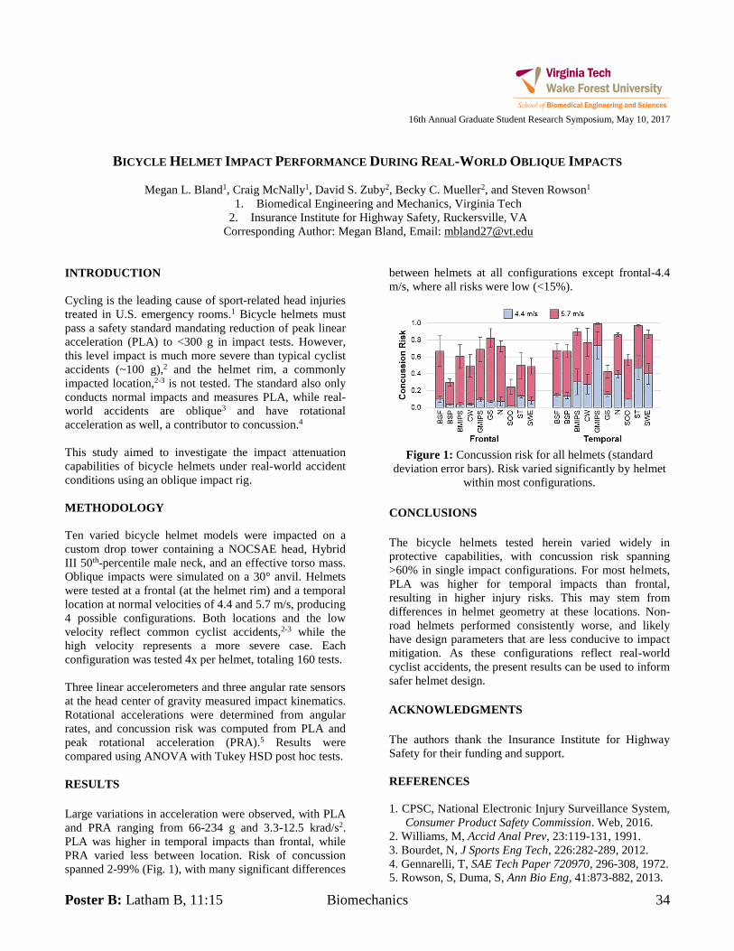
Poster B: Latham B, 11:15 Biomechanics 34
16th Annual Graduate Student Research Symposium, May 10, 2017
BICYCLE HELMET IMPACT PERFORMANCE DURING REAL-WORLD OBLIQUE IMPACTS
Megan L. Bland1, Craig McNally1, David S. Zuby2, Becky C. Mueller2, and Steven Rowson1
1. Biomedical Engineering and Mechanics, Virginia Tech 2. Insurance Institute for Highway Safety, Ruckersville, VA
Corresponding Author: Megan Bland, Email: [email protected] INTRODUCTION Cycling is the leading cause of sport-related head injuries treated in U.S. emergency rooms.1 Bicycle helmets must pass a safety standard mandating reduction of peak linear acceleration (PLA) to <300 g in impact tests. However, this level impact is much more severe than typical cyclist accidents (~100 g),2 and the helmet rim, a commonly impacted location,2-3 is not tested. The standard also only conducts normal impacts and measures PLA, while real-world accidents are oblique3 and have rotational acceleration as well, a contributor to concussion.4
This study aimed to investigate the impact attenuation capabilities of bicycle helmets under real-world accident conditions using an oblique impact rig. METHODOLOGY Ten varied bicycle helmet models were impacted on a custom drop tower containing a NOCSAE head, Hybrid III 50th-percentile male neck, and an effective torso mass. Oblique impacts were simulated on a 30° anvil. Helmets were tested at a frontal (at the helmet rim) and a temporal location at normal velocities of 4.4 and 5.7 m/s, producing 4 possible configurations. Both locations and the low velocity reflect common cyclist accidents,2-3 while the high velocity represents a more severe case. Each configuration was tested 4x per helmet, totaling 160 tests. Three linear accelerometers and three angular rate sensors at the head center of gravity measured impact kinematics. Rotational accelerations were determined from angular rates, and concussion risk was computed from PLA and peak rotational acceleration (PRA).5 Results were compared using ANOVA with Tukey HSD post hoc tests. RESULTS Large variations in acceleration were observed, with PLA and PRA ranging from 66-234 g and 3.3-12.5 krad/s2. PLA was higher in temporal impacts than frontal, while PRA varied less between location. Risk of concussion spanned 2-99% (Fig. 1), with many significant differences
between helmets at all configurations except frontal-4.4 m/s, where all risks were low (<15%).
Figure 1: Concussion risk for all helmets (standard
deviation error bars). Risk varied significantly by helmet within most configurations.
CONCLUSIONS The bicycle helmets tested herein varied widely in protective capabilities, with concussion risk spanning >60% in single impact configurations. For most helmets, PLA was higher for temporal impacts than frontal, resulting in higher injury risks. This may stem from differences in helmet geometry at these locations. Non-road helmets performed consistently worse, and likely have design parameters that are less conducive to impact mitigation. As these configurations reflect real-world cyclist accidents, the present results can be used to inform safer helmet design. ACKNOWLEDGMENTS The authors thank the Insurance Institute for Highway Safety for their funding and support. REFERENCES 1. CPSC, National Electronic Injury Surveillance System,
Consumer Product Safety Commission. Web, 2016. 2. Williams, M, Accid Anal Prev, 23:119-131, 1991. 3. Bourdet, N, J Sports Eng Tech, 226:282-289, 2012. 4. Gennarelli, T, SAE Tech Paper 720970, 296-308, 1972. 5. Rowson, S, Duma, S, Ann Bio Eng, 41:873-882, 2013.
35 Biomaterials Poster A: Latham B, 10:45
16th Annual Graduate Student Research Symposium, May 10, 2017
KERATIN COATED PLASMA ETCHED PARYLENE C FOR NEURAL INTERFACES
Kyle Brown1, Alexis Trent
2, and Mark Van Dyke
3
1. Virginia Tech, Biochemistry and Nanoscience
2. Virginia Tech, Material Science and Engineering
3. Virginia Tech, Biomedical Engineering and Sciences
Corresponding Author: Mark Van Dyke, Email: [email protected]
INTRODUCTION
Implantable neural interfaces such as the Utah Electrical
Array have been used to study and treat neural disorders
by converting neural impulses into electrical signals1.
However as with most percutaneous devices significant
problems have arisen from biocompatibility issues that
cause both a foreign body response from the host and a
multitude of infections at the site of implantation2. To
reduce problems with percutaneous neural implants we
want to coat the parylene C with keratin nanomaterials to
reduce the foreign body response from the host and the
risk of infection.
METHODOLOGY
One cm
2 titanium substrates were coated with Parylene C
and plasma etched at the University of Utah. The
substrates were then coated with 10% 3-
Isocyanatopropyltriethoxysilane (ICPTES) or 5 %
Glycidoxypropyl-trimethoxysilane (GPTES) in a 100%
ethanol or a 95:5 ethanol –water mixture and saturated for
3 hours. The silane mixture was removed and the
substrates were dried for 30 minutes at 110C. The
ICPTES coated substrates were then coated with keratin
(iKTN) and GPTES coated substrates were soaked in
keratose (gKOS) and soaked for 24 hours and rinsed with
water thereafter. The substrates were then sterilized via
UV light for 1 hour. X-ray Photoelectron Spectroscopy
(XPS) and Atomic Force Microscopy (AFM) were used to
validate the surface. Glial cells were seeded onto each
substrate and incubated for 24 hours. The cells were then
stained and imaged for cellular adhesion. Coated titanium
wires were inserted into 96 well plates with 10,000 glial
cells to observe the thickness of cellular encapsulation.
The data was statistically analyzed via ANOVA.
RESULTS
Shown in Figure 1, we have confirmed that an increase of
nitrogen is present on the KTN and KOS substrates and
have observed the topography through AFM (data not
shown).
Nitrogen
Pla
in P
aryl
ene
C
ICPTE
S
GPTE
S
iKTN
gKOS
0
5
10
15NonPlama Treated
Plasma Treated
Substrate
Ele
men
t P
erc
en
tag
e
Figure 1: XPS on Parylene C substrates for Nitrogen.
Figure 2 shows the optical differences between Plain
Titanium (pTi) and iKTN for glial cell encapsulation
thickness at Day 7.
Figure 2: A) pTi and B) iKTN are shown to observe glial
cell encapsulation thickness.
CONCLUSIONS
We have confirmed that keratin nanomaterials are able to
attach to Parylene C. We have observed the glial focal
adhesions (data not shown) and cellular encapsulation
thickness.
REFERENCES
1. Xie, X., et al. Journal of Neural Engineering 2014,
11 (2), 026016.
2. Chang, T. Y, et al. A. Langmuir 2007, 23 (23), 11718–
11725.
A B
Poster B: Latham B, 11:15 Biomechanics 36
16th Annual Graduate Student Research Symposium, May 10, 2017
EVALUATING THE PERFORMANCE OF ALTERNATIVE SHOULDER STABILIZATION METHODS
Kristine Cantin1, Sophia Ulman1, Jang-Ho Park1, Sunwook Kim1 and Maury A. Nussbaum1
1. Virginia Polytechnic Institute and State University Corresponding Author: Kristine Cantin, Email: [email protected]
INTRODUCTION Secondary pain and discomfort cause concern for clinicians when prescribing braces to patients that require shoulder stability, and may result in poor compliance by the patient if prescribed for longer durations1. Shoulder straps are often used in conventional brace design for stability, yet they are potential sources of discomfort. Novel supporting methods used in more recently developed braces could mitigate the above concerns associated with traditional bracing methods while stabilizing the shoulder. By comparing the impacts of different designs on stability, this work was intended to facilitate improved future brace prescriptions. METHODOLOGY A total 10 young adults (M = 24.2±4.9) and 10 older adults (M = 66.9±4.3), gender-balanced in each group, completed this study. A within-subjects design was implemented to test the stability of five arm braces, including four existing braces on the market and one newly developed brace. The study consisted of simulated activities of daily living (ADLs) with stability defined as shoulder immobilization and quantified by muscle activity and movement of the braced arm. Participants were asked to reach and retrieve objects; during these tasks surface electrodes on the descending trapezius and deltoid of the braced arm (Figure 1) measured muscle activity and motion capture tracked displacements between the braced arm and trunk. Raw EMG data were collected at 1000 Hz using a telemetered system (Noraxon DTS EMG TeleMyo) and 3D marker locations were tracked at 100 Hz using a 6-camera system (Nexus MX-T, Vicon).
Figure 1: Surface electrode placement on the braced
shoulder in accordance with SENIAM guidelines.
RESULTS Raw EMG and motion capture data were low-pass filtered, and EMG data were normalized to maximal values. Separate mixed-factor analyses of variance (ANOVA) were used to assess the effects of brace, age, and gender on each of the outcome measures, with brace order as a blocking effect (Table 1, for forward reach).
Table 1: ANOVA results for brace main effect during forward reach (* denotes significance).
Measure F(p)
Mean EMG Signal 11.49 (<0.0001)*
Maximum Forearm Rotation 8.77 (<0.0001)*
Maximum distance of forearm from trunk
21.68 (<0.0001)*
CONCLUSIONS Regardless of additional straps on common braces, there were rarely clinically significant differences (~3mm) in movement when compared to single-waist strapped braces. Muscle activity was frequently lower while wearing a brace without shoulder straps, indicating participants minimally use their “injured” braced shoulder while in the strapless braces. These results reveal that a novel brace design offers a potential solution to shoulder immobilizing without the concern of shoulder straps. ACKNOWLEDGMENTS This research was financially supported by GENEDGE. While contributing to the study design, it had no involvement in data analysis or interpretation. REFERENCES 1. Struijs, P., et al. (2004). Conservative treatment of lateral epicondylitis brace versus physical therapy or a combination of both—a randomized clinical trial. American Journal of Sports Medicine, 32(2), 462-469.
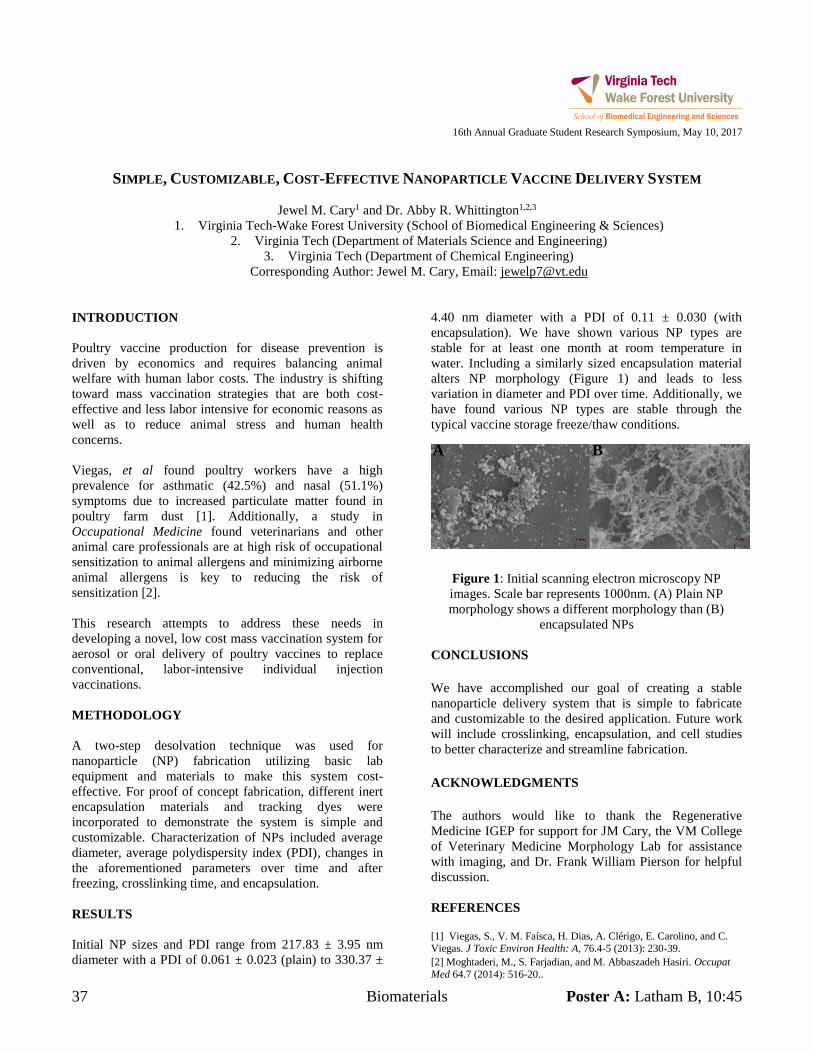
37 Biomaterials Poster A: Latham B, 10:45
16th Annual Graduate Student Research Symposium, May 10, 2017
SIMPLE, CUSTOMIZABLE, COST-EFFECTIVE NANOPARTICLE VACCINE DELIVERY SYSTEM
Jewel M. Cary1 and Dr. Abby R. Whittington1,2,3
1. Virginia Tech-Wake Forest University (School of Biomedical Engineering & Sciences) 2. Virginia Tech (Department of Materials Science and Engineering)
3. Virginia Tech (Department of Chemical Engineering) Corresponding Author: Jewel M. Cary, Email: [email protected]
INTRODUCTION Poultry vaccine production for disease prevention is driven by economics and requires balancing animal welfare with human labor costs. The industry is shifting toward mass vaccination strategies that are both cost-effective and less labor intensive for economic reasons as well as to reduce animal stress and human health concerns. Viegas, et al found poultry workers have a high prevalence for asthmatic (42.5%) and nasal (51.1%) symptoms due to increased particulate matter found in poultry farm dust [1]. Additionally, a study in Occupational Medicine found veterinarians and other animal care professionals are at high risk of occupational sensitization to animal allergens and minimizing airborne animal allergens is key to reducing the risk of sensitization [2]. This research attempts to address these needs in developing a novel, low cost mass vaccination system for aerosol or oral delivery of poultry vaccines to replace conventional, labor-intensive individual injection vaccinations. METHODOLOGY A two-step desolvation technique was used for nanoparticle (NP) fabrication utilizing basic lab equipment and materials to make this system cost-effective. For proof of concept fabrication, different inert encapsulation materials and tracking dyes were incorporated to demonstrate the system is simple and customizable. Characterization of NPs included average diameter, average polydispersity index (PDI), changes in the aforementioned parameters over time and after freezing, crosslinking time, and encapsulation. RESULTS Initial NP sizes and PDI range from 217.83 ± 3.95 nm diameter with a PDI of 0.061 ± 0.023 (plain) to 330.37 ±
4.40 nm diameter with a PDI of 0.11 ± 0.030 (with encapsulation). We have shown various NP types are stable for at least one month at room temperature in water. Including a similarly sized encapsulation material alters NP morphology (Figure 1) and leads to less variation in diameter and PDI over time. Additionally, we have found various NP types are stable through the typical vaccine storage freeze/thaw conditions.
CONCLUSIONS We have accomplished our goal of creating a stable nanoparticle delivery system that is simple to fabricate and customizable to the desired application. Future work will include crosslinking, encapsulation, and cell studies to better characterize and streamline fabrication. ACKNOWLEDGMENTS The authors would like to thank the Regenerative Medicine IGEP for support for JM Cary, the VM College of Veterinary Medicine Morphology Lab for assistance with imaging, and Dr. Frank William Pierson for helpful discussion. REFERENCES [1] Viegas, S., V. M. Faísca, H. Dias, A. Clérigo, E. Carolino, and C. Viegas. J Toxic Environ Health: A, 76.4-5 (2013): 230-39. [2] Moghtaderi, M., S. Farjadian, and M. Abbaszadeh Hasiri. Occupat Med 64.7 (2014): 516-20..
Figure 1: Initial scanning electron microscopy NP images. Scale bar represents 1000nm. (A) Plain NP morphology shows a different morphology than (B)
encapsulated NPs
B A
Poster B: Latham B, 11:15 Biomechanics 38
16th Annual Graduate Student Research Symposium, May 10, 2017
RESPONSE OF FEMALE AND MALE PMHS TO BLAST-INDUCED VERTICAL ACCELERATIVE LOADING
Danielle M. Cristino1, Warren N. Hardy1
1. Virginia Polytechnic Institute and State University, Biomedical Engineering and Mechanics (BEAM) Corresponding Author: Danielle M. Cristino, Email: [email protected]
INTRODUCTION Improvised explosive devices (IEDs) deliver a high energy blast to the underbody of military vehicles, exposing modern Warfighters to considerable risk during ground transport. Biomechanical data are required to develop an operationally relevant anthropomorphic test device (ATD) suited for the assessment of risk associated with the female Warfighter, or to map the injury prediction outputs of a male ATD to the female. The objective of this study is to determine the origin of the differences between female and male impact and injury response, such as sex, morphology, anatomy, or tolerance. METHODOLOGY This study was conducted using the Accelerative Loading Fixture (ALF), which generates floor and seat loading conditions representative of the underbody blast (UBB) environment. The ALF consists of two rigid seats mounted to a platform that is accelerated upward within a superstructure frame. The platform is explosive-driven. A brake system arrests its motion as it passes through the apex of its travel. The floor and seat performance can be modulated independently. Twelve post-mortem human surrogates (PMHS) were tested in pairs, using two different floor conditions. The PMHS tested include 50th percentile males, and 5th and 75th percentile females. The PMHS were instrumented with strain gages, accelerometers, angular rate sensors, and video markers. The data obtained include floor and seat acceleration and speed, generalized kinematics of the distal tibia and femur, sagittal perspective planar segment motion, and PMHS lower extremity damage results. RESULTS The female and male tibia vertical acceleration responses are similar, with the females attaining greater acceleration earlier in the event. The female PMHS tibia (especially 5th percentile) attains greater peak vertical speed compared to males. Also, peak speed for the females is reached earlier in the event compared to males. Similar trends are observed for femur acceleration and speed, with the 5th percentile female reaching greater vertical
acceleration and peak vertical speed earlier in the event. The femur response of the female is notably shorter in duration, but similar in shape to the male response. The female lower extremities initially rotate in the sagittal plane at a higher rate than the males. For the lower-energy floor condition, no female nor male PMHS sustained damage to the lower extremities, with the exception of a minor talus chip observed in a 5th percentile female. For the higher-energy floor condition, differences were seen between 5th and 75th percentile females. In one test, a lighter female sustained a complete separation of the body of the right (outboard) talus, a pilon fracture to the left (inboard) tibia, a segmented bending fracture to the left femur, and bilateral crushed calcanei. In the same test, a large female sustained a crushed right calcaneus, a compression fracture to the left distal tibia, and a complete oblique fracture to the left distal fibula. In another test, a 50th percentile male sustained bilateral crushed calcanei and a spiral wedge fracture of the left femur, while a small female sustained bilateral crushed calcanei and a fracture through the posterior calcaneal articular surface of the left talus. CONCLUSIONS The female and male lower extremity vertical acceleration and speed responses are similar, with the females attaining greater peak acceleration and speed earlier in the event. Damage was sustained by the calcanei, tali, tibiae, and fibulae. ACKNOWLEDGMENTS This work was funded by the USAMRMC. REFERENCES Danelson, K.A., Kemper, A.R., Mason, M.J., Tegtmeyer, M., Swiatkowski, S.A., Bolte, J.H. and Hardy, W.N. (2015) Comparison of ATD to PMHS Response in the Under-Body Blast Environment. Stapp Car Crash J 59: 445-520.
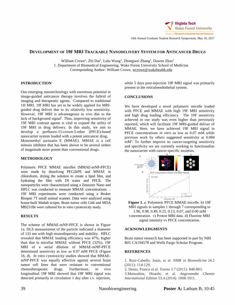
39 Nanobioengineering Poster A: Latham B, 10:45
16th Annual Graduate Student Research Symposium, May 10, 2017
DEVELOPMENT OF 19F MRI TRACKABLE NANODELIVERY SYSTEM FOR ANTICANCER DRUGS
William Crowe1, Zhi Dai1, Lulu Wang1, Zhongwei Zhang1, Dawen Zhao1
1. Department of Biomedical Engineering, Wake Forest University School of Medicine Corresponding Author: William Crowe, [email protected]
INTRODUCTION One emerging nanotechnology with enormous potential in image-guided anticancer therapy involves the hybrid of imaging and therapeutic agents. Compared to traditional 1H MRI, 19f MRI has yet to be widely applied for MRI-guided drug deliver due to its relatively low sensitivity. However, 19F MRI is advantageous in vivo due to the lack of background signal1. Thus, improving sensitivity of 19F MRI contrast agents is vital to expand the utility of 19F MRI in drug delivery. In this study, we aim to develop a perfluoro-15-crown-5-ether (PFCE)-based nanocarrier system loaded with a potent anticancer drug, Monomethyl auristatin E (MMAE). MMAE is a cell mitosis inhibitor that has been shown to be several orders of magnitude more potent than conventional drugs. METHODOLOGY Polymeric PFCE MMAE micelles (MMAE-mNP-PFCE) were made by dissolving PEG2kPE and MMAE in chloroform, drying the solution to create a lipid film, and hydrating the film with DI water and PFCE. The nanoparticles were characterized using a Zetasizer Nano and HPLC was conducted to measure MMAE concentrations. . 19F MRI experiments were conducted using a Bruker Biospin 7T small animal scanner. Data were analyzed using house-built Matlab scripts. Brain tumor cells G44 and MDA MB231Br were cultured for in vitro cytotoxicity study. RESULTS The scheme of MMAE-mNP-PFCE is shown in Figure 1a. DLS measurement of the particle indicated a diameter of 133 nm with high monodispersity and stability. HPLC revealed that MMAE loading efficiency was 47%, higher than that in micellar MMAE without PFCE (32%). 19F MRI of a serial dilution of MMAE-mNP-PFCE determined sensitivity as low as 0.07 mM PFCE (Figure 1b, d). In vitro cytotoxicity studies showed that MMAE-mNP-PFCE was equally effective against several brain tumor cell lines that were resistant to conventional chemotherapeutic drugs. Furthermore, in vivo longitudinal 19F MRI showed that 19F MRI signal was detected primarily in circulation 1 day after i.v. injection,
while 5 days post-injection 19F MRI signal was primarily present in the reticuloendothelial system. CONCLUSIONS We have developed a novel polymeric micelle loaded with PFCE and MMAE with high 19F MRI sensitivity and high drug loading efficiency. The 19F sensitivity achieved in our study was even higher than previously reported, which will facilitate 19F MRI-guided deliver of MMAE. Here, we have achieved 19F MRI signal in PFCE concentrations in vitro as low as 0.07 mM while previous work by others suggested sensitivity at 0.084 mM3. To further improve its cancer-targeting sensitivity and specificity we are currently working to functionalize the nanocarrier with cancer-specific moieties.
Figure 1. a. Polymeric PFCE MMAE micelle. b) 19F MRI signals in samples 1 through 7 corresponding to
1.96, 0.98, 0.49, 0.25, 0.13, 0.07, and 0.00 mM concentration. c) Proton MRI data. d) Fluorine MRI
signal intensity vs PFCE concentration. ACKNOWLEDGMENTS Brain tumor research has been supported in part by NIH R01 CA194578 and Wells Fargo Scholar Program. REFERENCES 1. Ruiz‐Cabello, Jesús, et al. NMR in Biomedicine 24.2 (2011): 114-129. 2. Dosio, Franco et al. Toxins 3.7 (2011): 848-883. 3.Matsushita, Hisashi, et al. Angewandte Chemie International Edition 53.4 (2014): 1008-1011.
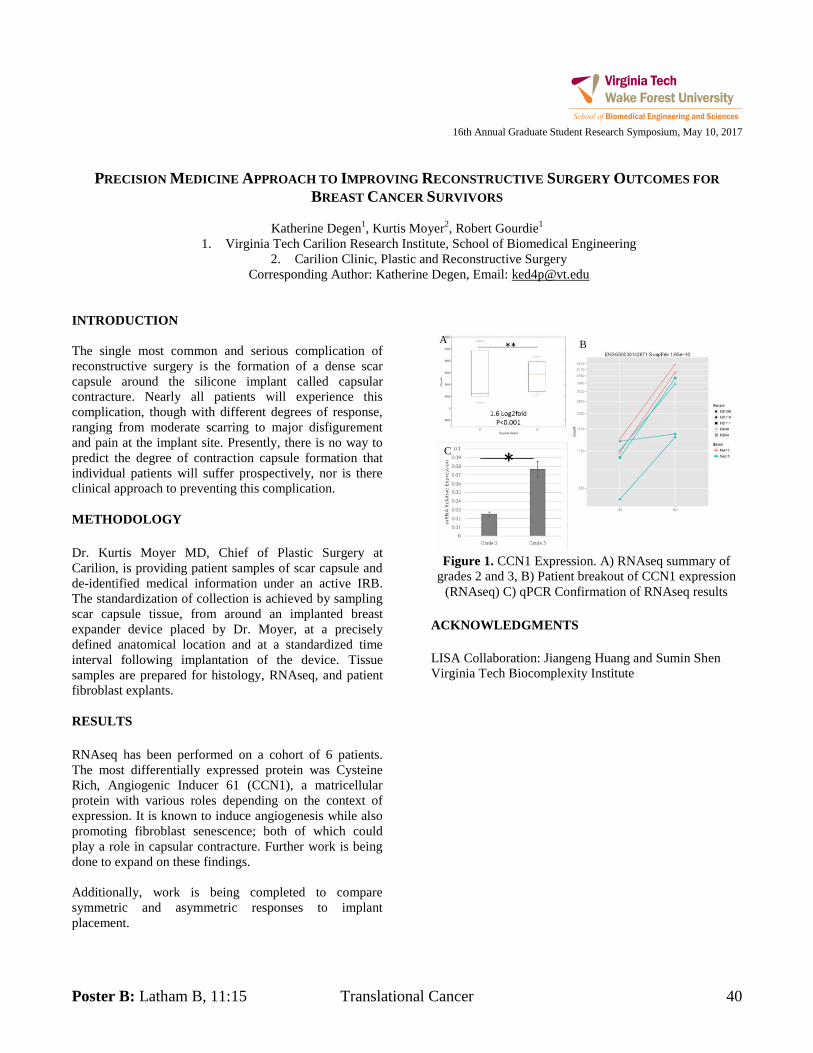
Poster B: Latham B, 11:15 Translational Cancer 40
16th Annual Graduate Student Research Symposium, May 10, 2017
PRECISION MEDICINE APPROACH TO IMPROVING RECONSTRUCTIVE SURGERY OUTCOMES FOR
BREAST CANCER SURVIVORS
Katherine Degen1, Kurtis Moyer
2, Robert Gourdie
1
1. Virginia Tech Carilion Research Institute, School of Biomedical Engineering
2. Carilion Clinic, Plastic and Reconstructive Surgery
Corresponding Author: Katherine Degen, Email: [email protected]
INTRODUCTION
The single most common and serious complication of
reconstructive surgery is the formation of a dense scar
capsule around the silicone implant called capsular
contracture. Nearly all patients will experience this
complication, though with different degrees of response,
ranging from moderate scarring to major disfigurement
and pain at the implant site. Presently, there is no way to
predict the degree of contraction capsule formation that
individual patients will suffer prospectively, nor is there
clinical approach to preventing this complication.
METHODOLOGY
Dr. Kurtis Moyer MD, Chief of Plastic Surgery at
Carilion, is providing patient samples of scar capsule and
de-identified medical information under an active IRB.
The standardization of collection is achieved by sampling
scar capsule tissue, from around an implanted breast
expander device placed by Dr. Moyer, at a precisely
defined anatomical location and at a standardized time
interval following implantation of the device. Tissue
samples are prepared for histology, RNAseq, and patient
fibroblast explants.
RESULTS
RNAseq has been performed on a cohort of 6 patients.
The most differentially expressed protein was Cysteine
Rich, Angiogenic Inducer 61 (CCN1), a matricellular
protein with various roles depending on the context of
expression. It is known to induce angiogenesis while also
promoting fibroblast senescence; both of which could
play a role in capsular contracture. Further work is being
done to expand on these findings.
Additionally, work is being completed to compare
symmetric and asymmetric responses to implant
placement.
Figure 1. CCN1 Expression. A) RNAseq summary of
grades 2 and 3, B) Patient breakout of CCN1 expression
(RNAseq) C) qPCR Confirmation of RNAseq results
ACKNOWLEDGMENTS
LISA Collaboration: Jiangeng Huang and Sumin Shen
Virginia Tech Biocomplexity Institute
*
A B
C
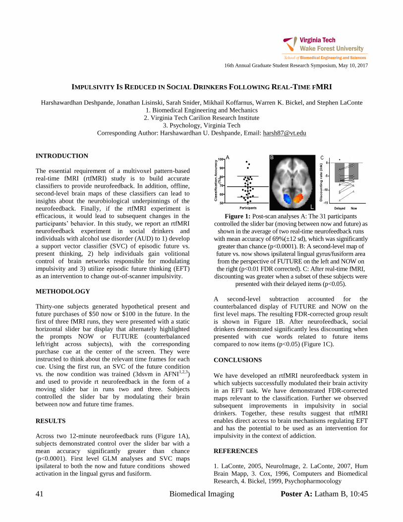
41 Biomedical Imaging Poster A: Latham B, 10:45
16th Annual Graduate Student Research Symposium, May 10, 2017
IMPULSIVITY IS REDUCED IN SOCIAL DRINKERS FOLLOWING REAL-TIME FMRI
Harshawardhan Deshpande, Jonathan Lisinski, Sarah Snider, Mikhail Koffarnus, Warren K. Bickel, and Stephen LaConte
1. Biomedical Engineering and Mechanics
2. Virginia Tech Carilion Research Institute
3. Psychology, Virginia Tech
Corresponding Author: Harshawardhan U. Deshpande, Email: [email protected]
INTRODUCTION
The essential requirement of a multivoxel pattern-based
real-time fMRI (rtfMRI) study is to build accurate
classifiers to provide neurofeedback. In addition, offline,
second-level brain maps of these classifiers can lead to
insights about the neurobiological underpinnings of the
neurofeedback. Finally, if the rtfMRI experiment is
efficacious, it would lead to subsequent changes in the
participants’ behavior. In this study, we report an rtfMRI
neurofeedback experiment in social drinkers and
individuals with alcohol use disorder (AUD) to 1) develop
a support vector classifier (SVC) of episodic future vs.
present thinking, 2) help individuals gain volitional
control of brain networks responsible for modulating
impulsivity and 3) utilize episodic future thinking (EFT)
as an intervention to change out-of-scanner impulsivity.
METHODOLOGY
Thirty-one subjects generated hypothetical present and
future purchases of $50 now or $100 in the future. In the
first of three fMRI runs, they were presented with a static
horizontal slider bar display that alternately highlighted
the prompts NOW or FUTURE (counterbalanced
left/right across subjects), with the corresponding
purchase cue at the center of the screen. They were
instructed to think about the relevant time frames for each
cue. Using the first run, an SVC of the future condition
vs. the now condition was trained (3dsvm in AFNI1,2,3
)
and used to provide rt neurofeedback in the form of a
moving slider bar in runs two and three. Subjects
controlled the slider bar by modulating their brain
between now and future time frames.
RESULTS
Across two 12-minute neurofeedback runs (Figure 1A),
subjects demonstrated control over the slider bar with a
mean accuracy significantly greater than chance (p<0.0001). First level GLM analyses and SVC maps ipsilateral to both the now and future conditions showed
activation in the lingual gyrus and fusiform.
Figure 1: Post-scan analyses A: The 31 participants
controlled the slider bar (moving between now and future) as
shown in the average of two real-time neurofeedback runs
with mean accuracy of 69%(±12 sd), which was significantly
greater than chance (p<0.0001). B: A second-level map of
future vs. now shows ipsilateral lingual gyrus/fusiform area
from the perspective of FUTURE on the left and NOW on
the right (p<0.01 FDR corrected). C: After real-time fMRI,
discounting was greater when a subset of these subjects were
presented with their delayed items (p<0.05).
A second-level subtraction accounted for the
counterbalanced display of FUTURE and NOW on the
first level maps. The resulting FDR-corrected group result
is shown in Figure 1B. After neurofeedback, social
drinkers demonstrated significantly less discounting when
presented with cue words related to future items
compared to now items (p<0.05) (Figure 1C).
CONCLUSIONS
We have developed an rtfMRI neurofeedback system in
which subjects successfully modulated their brain activity
in an EFT task. We have demonstrated FDR-corrected
maps relevant to the classification. Further we observed
subsequent improvements in impulsivity in social
drinkers. Together, these results suggest that rtfMRI
enables direct access to brain mechanisms regulating EFT
and has the potential to be used as an intervention for
impulsivity in the context of addiction.
REFERENCES
1. LaConte, 2005, NeuroImage, 2. LaConte, 2007, Hum
Brain Mapp, 3. Cox, 1996, Computers and Biomedical
Research, 4. Bickel, 1999, Psychopharmocology
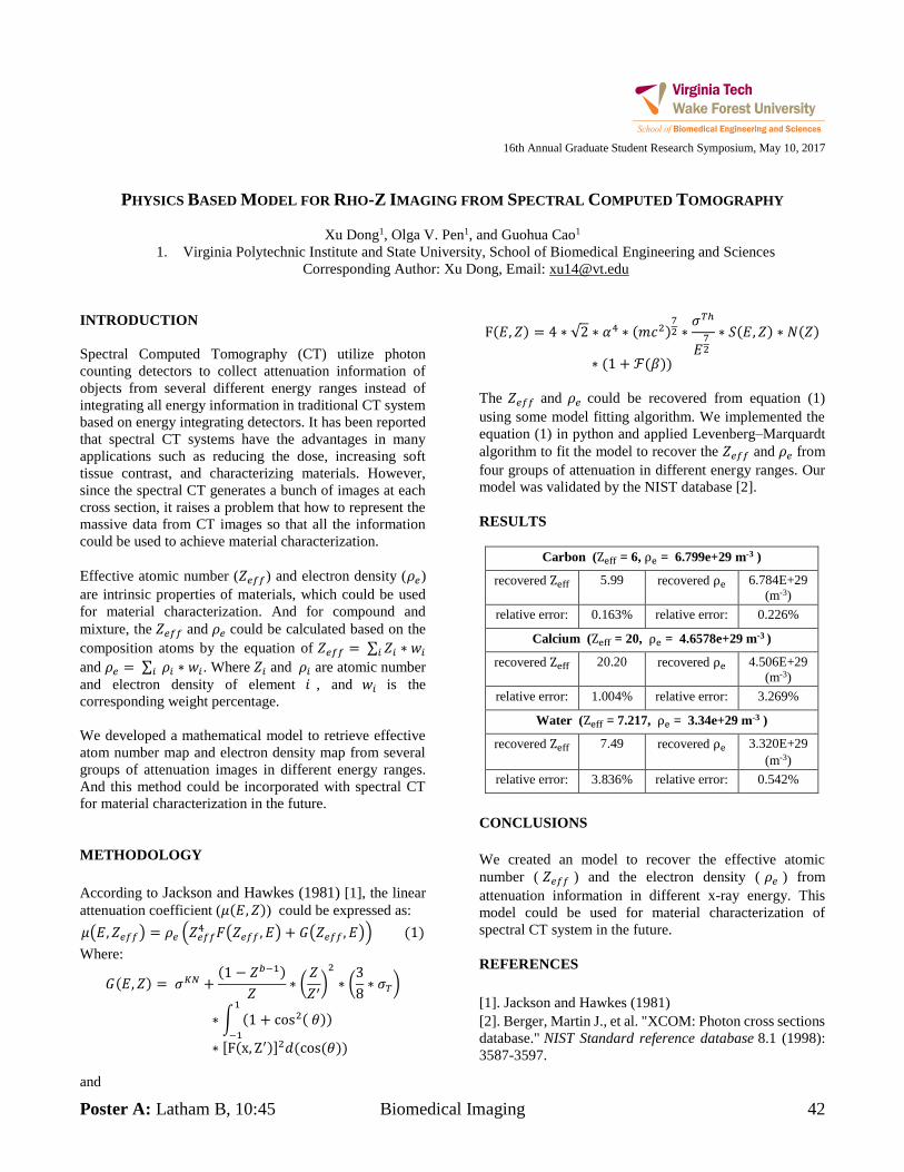
Poster A: Latham B, 10:45 Biomedical Imaging 42
16th Annual Graduate Student Research Symposium, May 10, 2017
PHYSICS BASED MODEL FOR RHO-Z IMAGING FROM SPECTRAL COMPUTED TOMOGRAPHY
Xu Dong1, Olga V. Pen1, and Guohua Cao1
1. Virginia Polytechnic Institute and State University, School of Biomedical Engineering and Sciences Corresponding Author: Xu Dong, Email: [email protected]
INTRODUCTION Spectral Computed Tomography (CT) utilize photon counting detectors to collect attenuation information of objects from several different energy ranges instead of integrating all energy information in traditional CT system based on energy integrating detectors. It has been reported that spectral CT systems have the advantages in many applications such as reducing the dose, increasing soft tissue contrast, and characterizing materials. However, since the spectral CT generates a bunch of images at each cross section, it raises a problem that how to represent the massive data from CT images so that all the information could be used to achieve material characterization. Effective atomic number (𝑍𝑒𝑓𝑓) and electron density (𝜌𝑒) are intrinsic properties of materials, which could be used for material characterization. And for compound and mixture, the 𝑍𝑒𝑓𝑓 and 𝜌𝑒 could be calculated based on the composition atoms by the equation of 𝑍𝑒𝑓𝑓 = ∑ 𝑍𝑖 ∗ 𝑤𝑖𝑖 and 𝜌𝑒 = ∑ 𝜌𝑖 ∗ 𝑤𝑖𝑖 . Where 𝑍𝑖 and 𝜌𝑖 are atomic number and electron density of element 𝑖 , and 𝑤𝑖 is the corresponding weight percentage. We developed a mathematical model to retrieve effective atom number map and electron density map from several groups of attenuation images in different energy ranges. And this method could be incorporated with spectral CT for material characterization in the future. METHODOLOGY According to Jackson and Hawkes (1981) [1], the linear attenuation coefficient (𝜇(𝐸, 𝑍)) could be expressed as: 𝜇(𝐸, 𝑍𝑒𝑓𝑓) = 𝜌𝑒 (𝑍𝑒𝑓𝑓
4 𝐹(𝑍𝑒𝑓𝑓 , 𝐸) + 𝐺(𝑍𝑒𝑓𝑓 , 𝐸)) (1) Where:
𝐺(𝐸, 𝑍) = 𝜎𝐾𝑁 +(1 − 𝑍𝑏−1)
𝑍∗ (
𝑍
𝑍′)
2
∗ (3
8∗ 𝜎𝑇)
∗ ∫ (1 + cos2( 𝜃))1
−1
∗ [F(x, Z′)]2𝑑(cos (𝜃)) and
F(𝐸, 𝑍) = 4 ∗ √2 ∗ 𝛼4 ∗ (𝑚𝑐2)72 ∗
𝜎𝑇ℎ
𝐸72
∗ 𝑆(𝐸, 𝑍) ∗ 𝑁(𝑍)
∗ (1 + ℱ(𝛽)) The 𝑍𝑒𝑓𝑓 and 𝜌𝑒 could be recovered from equation (1) using some model fitting algorithm. We implemented the equation (1) in python and applied Levenberg–Marquardt algorithm to fit the model to recover the 𝑍𝑒𝑓𝑓 and 𝜌𝑒 from four groups of attenuation in different energy ranges. Our model was validated by the NIST database [2]. RESULTS
Carbon (Zeff = 6, ρe = 6.799e+29 m-3 )
recovered Zeff 5.99 recovered ρe 6.784E+29 (m-3)
relative error: 0.163% relative error: 0.226%
Calcium (Zeff = 20, ρe = 4.6578e+29 m-3 )
recovered Zeff 20.20 recovered ρe 4.506E+29 (m-3)
relative error: 1.004% relative error: 3.269%
Water (Zeff = 7.217, ρe = 3.34e+29 m-3 )
recovered Zeff 7.49 recovered ρe 3.320E+29 (m-3)
relative error: 3.836% relative error: 0.542%
CONCLUSIONS We created an model to recover the effective atomic number ( 𝑍𝑒𝑓𝑓 ) and the electron density ( 𝜌𝑒 ) from attenuation information in different x-ray energy. This model could be used for material characterization of spectral CT system in the future. REFERENCES [1]. Jackson and Hawkes (1981) [2]. Berger, Martin J., et al. "XCOM: Photon cross sections database." NIST Standard reference database 8.1 (1998): 3587-3597.
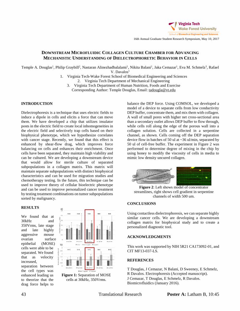
43 Translational Research Poster A: Latham B, 10:45
16th Annual Graduate Student Research Symposium, May 10, 2017
DOWNSTREAM MICROFLUIDIC COLLAGEN CULTURE CHAMBER FOR ADVANCING MECHANISTIC UNDERSTANDING OF DIELECTROPHORETIC BEHAVIOR IN CELLS
Temple A. Douglas1, Philip Graybill2, Nastaran Alinezhadbalalami1, Nikita Balani1, Jaka Cemazar1, Eva M. Schmelz3, Rafael
V. Davalos1 1. Virginia Tech-Wake Forest School of Biomedical Engineering and Sciences
2. Virginia Tech Department of Mechanical Engineering 3. Virginia Tech Department of Human Nutrition, Foods and Exercise
Corresponding Author: Temple Douglas, Email: [email protected] INTRODUCTION Dielectrophoresis is a technique that uses electric fields to induce a dipole in cells and elicits a force that can move them. We have developed a chip that utilizes insulator posts in the electric field to create local inhomogeneities in the electric field and selectively trap cells based on their biophysical phenotype, which we hypothesize correlates with cancer stage. Recently, we found that this effect is enhanced by shear-flow drag, which improves force balancing on cells and enhances their enrichment. Once cells have been separated, they maintain high viability and can be cultured. We are developing a downstream device that would allow for sterile culture of separated subpopulations in a collagen matrix. This matrix will maintain separate subpopulations with distinct biophysical characteristics and can be used for migration studies and chemotherapy testing. In the future, this technique can be used to improve theory of cellular bioelectric phenotype and can be used to improve personalized cancer treatment by testing treatment combinations on tumor subpopulations sorted by malignancy. RESULTS We found that at 30kHz and 350Vrms, late stage and late highly aggressive mouse ovarian surface epithelial (MOSE) cells were able to be separated. We found that as velocity increased, separation between the cell types was enhanced leading us to theorize that the drag force helps to
balance the DEP force. Using COMSOL, we developed a model of a device to separate cells from low conductivity DEP buffer, concentrate them, and mix them with collagen. A wall of small pores with higher net cross-sectional area than a secondary outlet allows DEP buffer to flow through, while cells roll along the edge of the porous wall into a collagen solution. Cells are collected in a serpentine channel, as shown. Cells coming off the DEP separation device flow in batches of 50 ul at ~36 ul/min, separated by 50 ul of cell-free buffer. The experiment in Figure 2 was performed to determine degree of mixing in the chip by using honey to modify the viscosity of cells in media to mimic low density uncured collagen.
CONCLUSIONS Using contactless dielectrophoresis, we can separate highly similar cancer cells. We are developing a downstream collagen matrix for biophysical study and to create a personalized diagnostic tool. ACKNOWLEDGMENTS This work was supported by NIH 5R21 CA173092-01, and CIT MF13-037-LS. REFERENCES T Douglas, J Cemazar, N Balani, D Sweeney, E Schmelz, R Davalos. Electrophoresis (Accepted manuscript). J Cemazar, T Douglas, E Schmelz, R Davalos. Biomicrofluidics (January 2016).
Figure 2: Left shows model of concentrator streamlines, right shows cell gradient in serpentine
channels of width 500 um.
Figure 1: Separation of MOSE cells at 30kHz, 350Vrms.
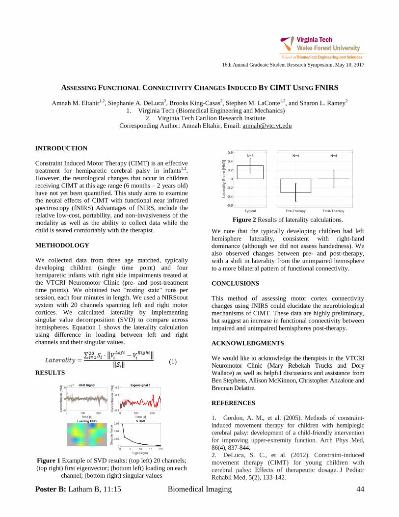
Poster B: Latham B, 11:15 Biomedical Imaging 44
16th Annual Graduate Student Research Symposium, May 10, 2017
ASSESSING FUNCTIONAL CONNECTIVITY CHANGES INDUCED BY CIMT USING FNIRS
Amnah M. Eltahir1,2
, Stephanie A. DeLuca2, Brooks King-Casas
2, Stephen M. LaConte
1,2, and Sharon L. Ramey
2
1. Virginia Tech (Biomedical Engineering and Mechanics)
2. Virginia Tech Carilion Research Institute
Corresponding Author: Amnah Eltahir, Email: [email protected]
INTRODUCTION
Constraint Induced Motor Therapy (CIMT) is an effective
treatment for hemiparetic cerebral palsy in infants1,2
.
However, the neurological changes that occur in children
receiving CIMT at this age range (6 months – 2 years old)
have not yet been quantified. This study aims to examine
the neural effects of CIMT with functional near infrared
spectroscopy (fNIRS) Advantages of fNIRS, include the
relative low-cost, portability, and non-invasiveness of the
modality as well as the ability to collect data while the
child is seated comfortably with the therapist.
METHODOLOGY
We collected data from three age matched, typically
developing children (single time point) and four
hemiparetic infants with right side impairments treated at
the VTCRI Neuromotor Clinic (pre- and post-treatment
time points). We obtained two “resting state” runs per
session, each four minutes in length. We used a NIRScout
system with 20 channels spanning left and right motor
cortices. We calculated laterality by implementing
singular value decomposition (SVD) to compare across
hemispheres. Equation 1 shows the laterality calculation
using difference in loading between left and right
channels and their singular values.
(1)
RESULTS
Figure 1 Example of SVD results: (top left) 20 channels;
(top right) first eigenvector; (bottom left) loading on each
channel; (bottom right) singular values
Figure 2 Results of laterality calculations.
We note that the typically developing children had left
hemisphere laterality, consistent with right-hand
dominance (although we did not assess handedness). We
also observed changes between pre- and post-therapy,
with a shift in laterality from the unimpaired hemisphere
to a more bilateral pattern of functional connectivity.
CONCLUSIONS
This method of assessing motor cortex connectivity
changes using fNIRS could elucidate the neurobiological
mechanisms of CIMT. These data are highly preliminary,
but suggest an increase in functional connectivity between
impaired and unimpaired hemispheres post-therapy.
ACKNOWLEDGMENTS
We would like to acknowledge the therapists in the VTCRI
Neuromotor Clinic (Mary Rebekah Trucks and Dory
Wallace) as well as helpful discussions and assistance from
Ben Stephens, Allison McKinnon, Christopher Anzalone and
Brennan Delattre.
REFERENCES
1. Gordon, A. M., et al. (2005). Methods of constraint-
induced movement therapy for children with hemiplegic
cerebral palsy: development of a child-friendly intervention
for improving upper-extremity function. Arch Phys Med,
86(4), 837-844.
2. DeLuca, S. C., et al. (2012). Constraint-induced
movement therapy (CIMT) for young children with
cerebral palsy: Effects of therapeutic dosage. J Pediatr
Rehabil Med, 5(2), 133-142.
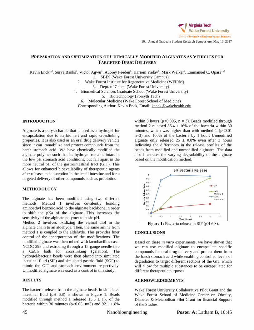
45 Nanobioengineering Poster A: Latham B, 10:45
16th Annual Graduate Student Research Symposium, May 10, 2017
PREPARATION AND OPTIMIZATION OF CHEMICALLY MODIFIED ALGINATES AS VEHICLES FOR
TARGETED DRUG DELIVERY
Kevin Enck1,2
, Surya Banks3, Victor Agwu
4, Aubrey Peeden
5, Hariom Yadav
6, Mark Welker
3, Emmanuel C. Opara
1,2
1. SBES (Wake Forest University Campus)
2. Wake Forest Institute for Regenerative Medicine (WFIRM)
3. Dept. of Chem. (Wake Forest University)
4. Biomedical Sciences Graduate School (Wake Forest University)
5. Biotechnology (Forsyth Tech)
6. Molecular Medicine (Wake Forest School of Medicine)
Corresponding Author: Kevin Enck, Email: [email protected]
INTRODUCTION
Alginate is a polysacharide that is used as a hydrogel for
encapsulation due to its bioinert and rapid crosslinking
properties. It is also used as an oral drug delivery vehicle
since it can immobilize and protect compounds from the
harsh stomach acid. We have chemically modified the
alginate polymer such that its hydrogel remains intact in
the low pH stomach acid conditions, but fall apart in the
more neutral pH of the gastrointestinal tract (GIT). This
allows for enhanced bioavailability of therapeutic agents
after release and absorption in the small intestine and for a
targeted delivery of other compounds such as probiotics
METHODOLOGY
The alginate has been modified using two different
methods. Method 1 involves covalently bonding
aminoethyl benzoic acid to the alginate backbone in order
to shift the pKa of the alginate. This increases the
sensitivity of the alginate polymer to basic pH.
Method 2 involves oxidizing the vicinal diol in the
alginate chain to an aldehyde. Then, the same amine from
method 1 is coupled to the aldehyde. This provides finer
control of the incorporation of the modifications. The
modified alginate was then mixed with lactobacillus casei
NCDC 298 and extruding through a 15-gauge needle into
a CaCl2 bath for crosslinking (gelation). The
hydrogel/bacteria beads were then placed into simulated
intestinal fluid (SIF) and simulated gastric fluid (SGF) to
mimic the GIT and stomach environment respectively.
Unmodified alginate was used as a control in this study.
RESULTS
The bacteria release from the alginate beads in simulated
intestinal fluid (pH 6.8) is shown in Figure 1. Beads
modified through method 1 released 15.5 ± 1% of the
bacteria within 30 minutes (p>0.05, n=3) and 92.1 ± 8%
within 3 hours (p<0.005, n = 3). Beads modified through
method 2 released 86.4 ± 16% of the bacteria within 30
minutes, which was higher than with method 1 (p<0.01
n=3) and 100% of the bacteria by 1 hour. Unmodified
alginate only released 25 ± 0.8% even after 3 hours
indicating the differences in the release profiles of the
beads from modified and unmodified alginates. The data
also illustrates the varying degradability of the alginate
based on the modification method.
Figure 1: Bacteria release in SIF (pH 6.8).
CONCLUSIONS
Based on these in vitro experiments, we have shown that
we can use modified alginate to encapsulate specific
compounds for oral drug delivery and protect them from
the harsh stomach acid while enabling controlled levels of
degradation to target different sections of the GIT which
will allow for multiple substances to be encapsulated for
different therapeutic purposes.
ACKNOWLEDGEMENTS
Wake Forest University Collaborative Pilot Grant and the
Wake Forest School of Medicine Center on Obesity,
Diabetes & Metabolism Pilot Grant for financial Support
of the Studies.
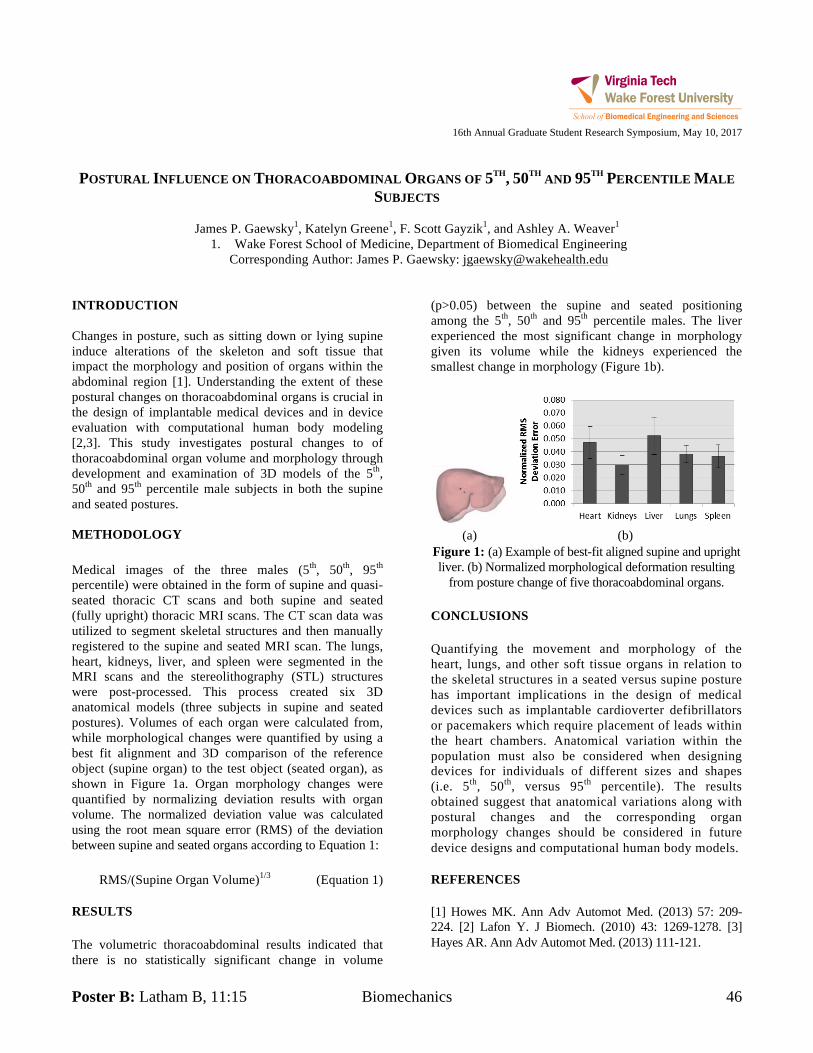
Poster B: Latham B, 11:15 Biomechanics 46
16th Annual Graduate Student Research Symposium, May 10, 2017
POSTURAL INFLUENCE ON THORACOABDOMINAL ORGANS OF 5TH, 50TH AND 95TH PERCENTILE MALE
SUBJECTS
James P. Gaewsky1, Katelyn Greene1, F. Scott Gayzik1, and Ashley A. Weaver1
1. Wake Forest School of Medicine, Department of Biomedical Engineering Corresponding Author: James P. Gaewsky: [email protected]
INTRODUCTION Changes in posture, such as sitting down or lying supine induce alterations of the skeleton and soft tissue that impact the morphology and position of organs within the abdominal region [1]. Understanding the extent of these postural changes on thoracoabdominal organs is crucial in the design of implantable medical devices and in device evaluation with computational human body modeling [2,3]. This study investigates postural changes to of thoracoabdominal organ volume and morphology through development and examination of 3D models of the 5th, 50th and 95th percentile male subjects in both the supine and seated postures. METHODOLOGY Medical images of the three males (5th, 50th, 95th percentile) were obtained in the form of supine and quasi-seated thoracic CT scans and both supine and seated (fully upright) thoracic MRI scans. The CT scan data was utilized to segment skeletal structures and then manually registered to the supine and seated MRI scan. The lungs, heart, kidneys, liver, and spleen were segmented in the MRI scans and the stereolithography (STL) structures were post-processed. This process created six 3D anatomical models (three subjects in supine and seated postures). Volumes of each organ were calculated from, while morphological changes were quantified by using a best fit alignment and 3D comparison of the reference object (supine organ) to the test object (seated organ), as shown in Figure 1a. Organ morphology changes were quantified by normalizing deviation results with organ volume. The normalized deviation value was calculated using the root mean square error (RMS) of the deviation between supine and seated organs according to Equation 1:
RMS/(Supine Organ Volume)1/3 (Equation 1) RESULTS The volumetric thoracoabdominal results indicated that there is no statistically significant change in volume
(p>0.05) between the supine and seated positioning among the 5th, 50th and 95th percentile males. The liver experienced the most significant change in morphology given its volume while the kidneys experienced the smallest change in morphology (Figure 1b).
(a) (b)
Figure 1: (a) Example of best-fit aligned supine and upright liver. (b) Normalized morphological deformation resulting
from posture change of five thoracoabdominal organs. CONCLUSIONS Quantifying the movement and morphology of the heart, lungs, and other soft tissue organs in relation to the skeletal structures in a seated versus supine posture has important implications in the design of medical devices such as implantable cardioverter defibrillators or pacemakers which require placement of leads within the heart chambers. Anatomical variation within the population must also be considered when designing devices for individuals of different sizes and shapes (i.e. 5th, 50th, versus 95th percentile). The results obtained suggest that anatomical variations along with postural changes and the corresponding organ morphology changes should be considered in future device designs and computational human body models. REFERENCES [1] Howes MK. Ann Adv Automot Med. (2013) 57: 209-224. [2] Lafon Y. J Biomech. (2010) 43: 1269-1278. [3] Hayes AR. Ann Adv Automot Med. (2013) 111-121.
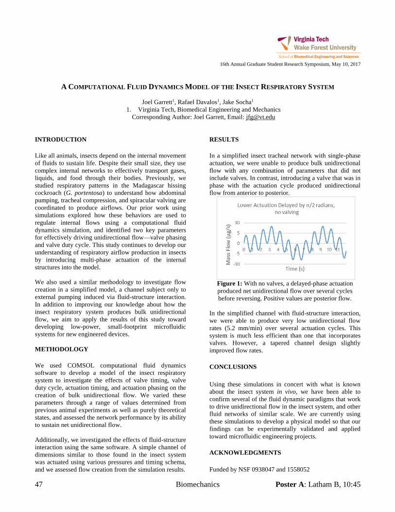
47 Biomechanics Poster A: Latham B, 10:45
16th Annual Graduate Student Research Symposium, May 10, 2017
A COMPUTATIONAL FLUID DYNAMICS MODEL OF THE INSECT RESPIRATORY SYSTEM
Joel Garrett1, Rafael Davalos1, Jake Socha1
1. Virginia Tech, Biomedical Engineering and Mechanics Corresponding Author: Joel Garrett, Email: [email protected]
INTRODUCTION Like all animals, insects depend on the internal movement of fluids to sustain life. Despite their small size, they use complex internal networks to effectively transport gases, liquids, and food through their bodies. Previously, we studied respiratory patterns in the Madagascar hissing cockroach (G. portentosa) to understand how abdominal pumping, tracheal compression, and spiracular valving are coordinated to produce airflows. Our prior work using simulations explored how these behaviors are used to regulate internal flows using a computational fluid dynamics simulation, and identified two key parameters for effectively driving unidirectional flow—valve phasing and valve duty cycle. This study continues to develop our understanding of respiratory airflow production in insects by introducing multi-phase actuation of the internal structures into the model. We also used a similar methodology to investigate flow creation in a simplified model, a channel subject only to external pumping induced via fluid-structure interaction. In addition to improving our knowledge about how the insect respiratory system produces bulk unidirectional flow, we aim to apply the results of this study toward developing low-power, small-footprint microfluidic systems for new engineered devices. METHODOLOGY We used COMSOL computational fluid dynamics software to develop a model of the insect respiratory system to investigate the effects of valve timing, valve duty cycle, actuation timing, and actuation phasing on the creation of bulk unidirectional flow. We varied these parameters through a range of values determined from previous animal experiments as well as purely theoretical states, and assessed the network performance by its ability to sustain net unidirectional flow. Additionally, we investigated the effects of fluid-structure interaction using the same software. A simple channel of dimensions similar to those found in the insect system was actuated using various pressures and timing schema, and we assessed flow creation from the simulation results.
RESULTS In a simplified insect tracheal network with single-phase actuation, we were unable to produce bulk unidirectional flow with any combination of parameters that did not include valves. In contrast, introducing a valve that was in phase with the actuation cycle produced unidirectional flow from anterior to posterior.
Figure 1: With no valves, a delayed-phase actuation produced net unidirectional flow over several cycles before reversing. Positive values are posterior flow.
In the simplified channel with fluid-structure interaction, we were able to produce very low unidirectional flow rates (5.2 mm/min) over several actuation cycles. This system is much less efficient than one that incorporates valves. However, a tapered channel design slightly improved flow rates. CONCLUSIONS Using these simulations in concert with what is known about the insect system in vivo, we have been able to confirm several of the fluid dynamic paradigms that work to drive unidirectional flow in the insect system, and other fluid networks of similar scale. We are currently using these simulations to develop a physical model so that our findings can be experimentally validated and applied toward microfluidic engineering projects. ACKNOWLEDGMENTS Funded by NSF 0938047 and 1558052
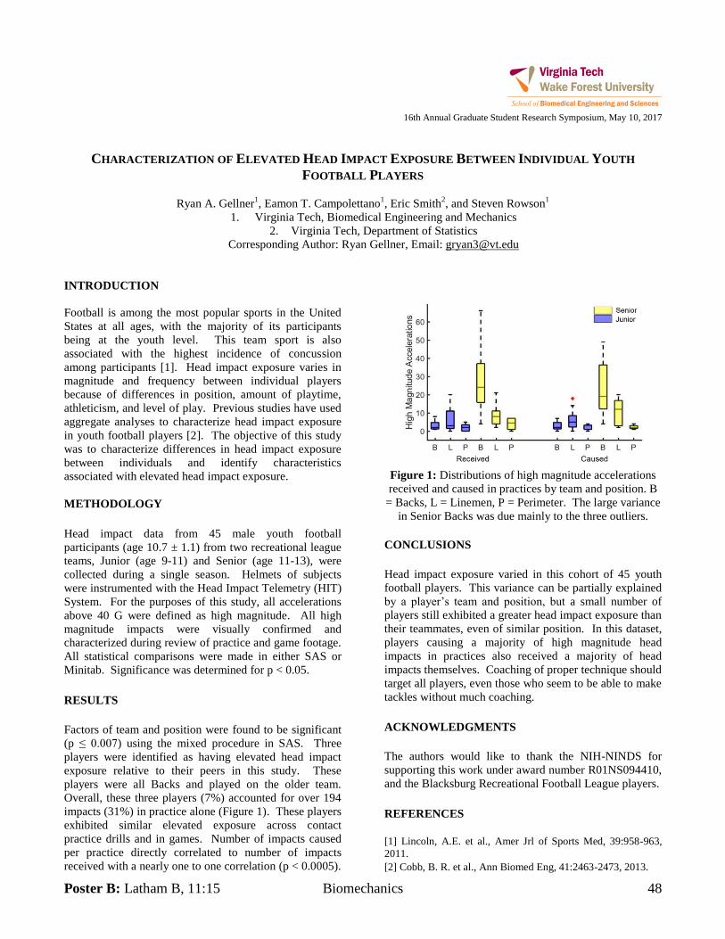
Poster B: Latham B, 11:15 Biomechanics 48
16th Annual Graduate Student Research Symposium, May 10, 2017
CHARACTERIZATION OF ELEVATED HEAD IMPACT EXPOSURE BETWEEN INDIVIDUAL YOUTH
FOOTBALL PLAYERS
Ryan A. Gellner1, Eamon T. Campolettano
1, Eric Smith
2, and Steven Rowson
1
1. Virginia Tech, Biomedical Engineering and Mechanics
2. Virginia Tech, Department of Statistics
Corresponding Author: Ryan Gellner, Email: [email protected]
INTRODUCTION
Football is among the most popular sports in the United
States at all ages, with the majority of its participants
being at the youth level. This team sport is also
associated with the highest incidence of concussion
among participants [1]. Head impact exposure varies in
magnitude and frequency between individual players
because of differences in position, amount of playtime,
athleticism, and level of play. Previous studies have used
aggregate analyses to characterize head impact exposure
in youth football players [2]. The objective of this study
was to characterize differences in head impact exposure
between individuals and identify characteristics
associated with elevated head impact exposure.
METHODOLOGY
Head impact data from 45 male youth football
participants (age 10.7 ± 1.1) from two recreational league
teams, Junior (age 9-11) and Senior (age 11-13), were
collected during a single season. Helmets of subjects
were instrumented with the Head Impact Telemetry (HIT)
System. For the purposes of this study, all accelerations
above 40 G were defined as high magnitude. All high
magnitude impacts were visually confirmed and
characterized during review of practice and game footage.
All statistical comparisons were made in either SAS or
Minitab. Significance was determined for p < 0.05.
RESULTS
Factors of team and position were found to be significant
(p ≤ 0.007) using the mixed procedure in SAS. Three
players were identified as having elevated head impact
exposure relative to their peers in this study. These
players were all Backs and played on the older team.
Overall, these three players (7%) accounted for over 194
impacts (31%) in practice alone (Figure 1). These players
exhibited similar elevated exposure across contact
practice drills and in games. Number of impacts caused
per practice directly correlated to number of impacts
received with a nearly one to one correlation (p < 0.0005).
Figure 1: Distributions of high magnitude accelerations
received and caused in practices by team and position. B
= Backs, L = Linemen, P = Perimeter. The large variance
in Senior Backs was due mainly to the three outliers.
CONCLUSIONS
Head impact exposure varied in this cohort of 45 youth
football players. This variance can be partially explained
by a player’s team and position, but a small number of
players still exhibited a greater head impact exposure than
their teammates, even of similar position. In this dataset,
players causing a majority of high magnitude head
impacts in practices also received a majority of head
impacts themselves. Coaching of proper technique should
target all players, even those who seem to be able to make
tackles without much coaching.
ACKNOWLEDGMENTS
The authors would like to thank the NIH-NINDS for
supporting this work under award number R01NS094410,
and the Blacksburg Recreational Football League players.
REFERENCES
[1] Lincoln, A.E. et al., Amer Jrl of Sports Med, 39:958-963,
2011.
[2] Cobb, B. R. et al., Ann Biomed Eng, 41:2463-2473, 2013.
49 Tissue Engineering Poster A: Latham B, 10:45
16th Annual Graduate Student Research Symposium, May 10, 2017
OPTIMIZATION OF HYDROGEL-BASED BIOINK PRINTABILITY USING RHEOLOGICAL PARAMETERS:
A SYSTEMATIC APPROACH
Gregory J. Gillispie1,2, Teng Gao2, Josh Copus2, James Yoo1,2, Anthony Atala1,2, and Sang Jin Lee1,2
1. Virginia Tech-Wake Forest, School for Biomedical Engineering Sciences 2. Wake Forest University, Wake Forest Institute for Regenerative Medicine
Corresponding Author: Greg Gillispie [email protected] INTRODUCTION Bioprinting is a promising technique in tissue engineering, but the dearth of suitable bioinks is frequently cited as a major limiting factor for the advancement of this technology.1 It is well known that the viscosity of a bioink has a direct impact on its printability. Highly viscous materials maintain their structure better, but also have decreased cell viability due to the high forces which are required for extrusion.1 However, complex viscosity is comprised of two discrete components, storage modulus (G’) and loss modulus (G”)
and their individual contributions to printability are unknown. METHODOLOGY For this study, gelatin and alginate were mixed at various concentrations to obtain hydrogels with a wide range of storage and loss moduli. These hydrogels were then evaluated for the quantitatively defined values of extrudability, structural integrity, and extrusion uniformity. Extrudability was defined by the pressure required to extrude the material and a mathematical model relating the G’ and G” to the required extrusion pressure was derived based on our data. Structural integrity was defined as the ability of the hydrogel to maintain its height as a printed tubular structure. Extrusion uniformity was defined as the straightness and smoothness of the individual lines of extrusion. RESULTS Increasing either the loss or storage modulus, and thereby the viscosity, increased the pressure required to extrude the bioink. Extrudability data was best fit to a first-order interactive model with two independent variables:
Pressure = 26.6 + 0.05G’ + 0.07G” – 0.0000035G’G” While neither loss nor storage moduli alone were excellent predictors of printability, a lower loss tangent (G”/G’) typically correlated with increased structural
integrity while a higher loss tangent correlated with increased extrusion uniformity. Hydrogels with a loss
tangent in the range of 0.3 to 0.6 exhibited an excellent compromise between structural integrity and extrusion uniformity.
Figure 1: Structural stability (left) and extrusion uniformity (right) as loss tangent increases from A-F.
CONCLUSIONS By making a simple measurement of the rheological properties (G’ and G”), the extrudability, structural
integrity, and extrusion uniformity of a bioink can be inferred. Using this approach, a variety of different formulations can be quickly and accurately evaluated for printability. Immediate future work will incorporate cell viability studies, further define printability with additional parameters, and expand the methodology to other hydrogels in order to determine the generalizability of these evaluation techniques. ACKNOWLEDGMENTS Funding provided by Wake Forest Innovations & NIH REFERENCES 1. Chimene, D. et al. (2016). Advanced Bioinks for 3D Printing: A Materials Science Perspective. Ann Biomed Eng, 44(6), 2090-2102.
Poster B: Latham B, 11:15 Biomaterials 50
16th Annual Graduate Student Research Symposium, May 10, 2017
MECHANICAL CHARACTERIZATION OF EMULSIFICATION POLYMERIZED KERATIN MICROPARTICLES
USING VARIED CROSSLINKING AGENTS
Aaron Giuffre’1, Marc Thompson
2, Mark Van Dyke
2
1. Virginia Tech, Electrical and Computer Engineering
2. Virginia Tech, Biomedical Engineering and Mechanics
Corresponding Author: Name, Email: [email protected]
INTRODUCTION
The application of microparticles in tissue engineering are
a well-documented and cost efficient method of
increasing the efficacy of drug delivery in the body.
Keratin as a building block for these materials is
advantageous due to its natural abundance in the body,
decreasing the likelihood of rejection. Emulsion
polymerization with a chemical cross-linker achieves a
fixed, spherical shape, increasing the particle surface area
and increasing the treatment’s loading capacity. The
successful application of loaded microparticles depends
largely on interactions within the body, hence their
mechanical and chemical properties become important
This study seeks to measure the effect of polymer and
cross-linker concentration on size, viscoelastic, and
degradation properties of keratin microparticles.
METHODOLOGY
Keratose (KOS) was extracted from human hair, via an
oxidation reaction. Paracetic acid breaches the hair
cuticle, which is then a followed by Tris base that
removes all protein within the strand. The KOS is purified
by washing samples encapsulated in dialysis tubing with
buffer solution to remove low molecular weight chains.
This sample is then manufactured into a dried network by
concentrating, freezing, and lyophilizing. This material is
made into solutions of varying concentrations either by
using water or phosphate buffer. Microparticles are
created using an oil emulsion process. A nonpolar oil
phase is stirred for a number of hours with a polar phase
of keratin solution along with a cross-linker of varying
concentration. This polymerizes the keratin solution into
spherical shape. A lyophilizing step stabilizes the
microparticle samples. Average particle size, expansion
rates in varying degrees of water and ethanol, gel
rheological properties, and rates of degradation via
enzymes and phosphate buffer were quantified across
microparticles produced with varying polymer and cross-
linker concentrations and pH. Size was quantified using
both scanning electron microscopy (SEM) and light
microscopy. Surface topography was observed via SEM.
Viscoelastic properties of microparticle gels were
measured by a rheometer. Degradation was quantified by
fluorescence read by a plate reader via loaded bovine
serum albumin fluorescein isothiocyanate (BSA-FITC).
RESULTS
Particle size, behavior in solution, and viscoelastic
properties were found to differ across production
variables like pH, polymer concentration, and cross-linker
concentration. This is unexpected and interesting, as the
various crosslinking agents used are very similar in length
and reaction chemistry.
Figure 1: Microparticle sample imaged via SEM
CONCLUSIONS
Manufacturing variables previously considered of minute
difference offer differing outcomes of product. This
expands the ability to fine tune and customize the keratin
microparticle system to various applications.
ACKNOWLEDGMENTS
The Davis Lab, The Nanoscale Fabrication and
Characterization Laboratory (NCFL).
51 Biomechanics Poster A: Latham B, 10:45
16th Annual Graduate Student Research Symposium, May 10, 2017
JUMPING BEHAVIORS INCREASE GAP BRIDGING PERFORMANCE IN THE FLYING SNAKE CHRYSOPELEA
PARADISI
Michelle R. Graham1, Bruce Jayne2, Talia M. Weiss1, and Jake Socha2
1. Virginia Tech, Biomedical Engineering and Mechanics 2. University of Cincinnati, Biology
Corresponding Author: Michelle Graham, Email: [email protected] INTRODUCTION In arboreal habitats, the most direct route between two locations may involve spatial discontinuities. Snakes can navigate these gaps using gap-bridging behaviors, most commonly the cantilever, in which the snake extends itself into the gap, holding its body stiff. Some snakes also use dynamic lunges to increase their performance [1]. The flying snake Chrysopelea paradisi often initiates glides with a J-loop take-off [2], in which the snake forms a hanging curve with the anterior body and rapidly straightens to depart the branch. Such jumping, which is more energetic than lunging, might also be used to bridge gaps. In this experiment, our aim was to investigate how flying snakes use jumping in the context of gap bridging, and whether these behaviors increase the snake’s gap
bridging performance. METHODOLOGY
Figure 1: Snake with infrared (IR) reflective markers to
facilitate motion capture.
Six C. paradisi were marked with IR-reflective markers (Fig. 1) and recorded using a six-camera motion capture system (Vicon Motion Systems Ltd.) as they crossed gaps of increasing size between two branch-like cylinders. Video was also recorded from various perspectives. RESULTS We found that C. paradisi uses both lunging and jumping behaviors (Fig. 2), typically to cross gaps that they cannot span by cantilevering. The largest gaps were crossed exclusively by jumping.
Figure 2: Behavior used depends on gap size
The average factorial increase in crossable gap size when using dynamic behaviors was 2.10.18 times the largest gap size crossable with a cantilever alone (n=4 snakes, 196 trials). CONCLUSIONS C. paradisi do indeed use jumping behaviors in the context of gap bridging, and these behaviors enable them to cross larger gaps than they can by cantilevering. Hence, jumping in Chrysopelea may serve as a mechanism to increase locomotor effectiveness in patchy arboreal environments, and provide a locomotor rationale for jumping independent from gliding. ACKNOWLEDGMENTS Supported partially by NSF 1351322 and the New Horizons Graduate Fellowship Program. Facilities: W. Samuel Easterling Spline reconstructions: Isaac Yeaton. Experimental assistance: Corey Spohn, Adair Maynard. REFERENCES 1] Jayne and Riley, 2007, JEB. 210 (7) [2] Socha, 2006, JEB. 209 (17)
Poster B: Latham B, 11:15 Biomechanics 52
16th Annual Graduate Student Research Symposium, May 10, 2017
FAILED RIB REGION PREDICTION IN A HUMAN BODY MODEL DURING COLLISION EVENTS WITH
PRECRASH BRAKING
Berkan Guleyupoglu1,2
, Jeremy Schap1,2
, Matt Davis1,2
, F. Scott Gayzik1,2
1. Wake Forest University School of Medicine
2. Virginia Tech – Wake Forest Center for Injury Biomechanics
Corresponding Author: F. Scott Gayzik, Email: [email protected]
INTRODUCTION
Road traffic injuries are a leading contributor of DALYs
(Disability Adjusted Life Years) lost [1]. Active safety
systems will play a large role in reducing the likelihood of
occupant injury but, such systems need to function in
concert with passive safety countermeasures,
underscoring the need for a method to evaluate the two
simultaneously. Human body models are ideally situated
to serve as this link. The purpose of this study is to investigate the differences in prediction between
deterministic and probabilistic methods of rib injury
prediction in the computational model.
METHODOLOGY
The GHBMC M50-O model was gravity settled in the
driver position of a generic interior equipped with an
advanced three point belt and airbag. Twenty-four
simulations were conducted with permutations for failure,
pre-crash braking system and crash severity. The
severities used were median (17 kph), severe (34 kph) and
NCAP (56.4 kph). Cases with failure enabled removed rib
cortical bone elements once 1.8% strain was exceeded.
Alternatively, a probabilistic framework found in
literature was used to predict rib failure. Both the
probabilistic and deterministic methods take into
consideration location (Anterior, Lateral, and Posterior).
The deterministic method is based on rubric that defines
rib Failed Regions (FR’s) dependent on a threshold for
contiguous failed elements. The probabilistic method
depends on age-based strain and failure functions.
RESULTS
Kinematics between both methods were similar (peak
∆Xhead = 0-17 mm; ∆Zhead = 1-4 mm; ∆Xthorax = 3-5
mm; ∆Zthorax = 1 mm). Seatbelt forces at the time of
probabilistic FR (PFR) initiation were lower than at
deterministic FR (DFR) initiation. The probabilistic
method for rib fracture predicted more failed regions in
the rib (an analog for fracture) than the deterministic
method in all but 2 cases. The FR patterns between
models are similar however there are differences that arise
due to stress let-off from element elimination that cause
other PFRs to appear where no DFR was predicted.
Figure 1. Failed region prediction counts for
probabilistic (black) and deterministic (orange). CONCLUSIONS
Both methods indicated similar trends with the addition of
precrash braking however there are tradeoffs. The DFR
method was more spatially sensitive and solved faster.
The PFR method consistently predicted higher failed
regions and has greater capabilities in post-processing.
ACKNOWLEDGMENTS
Work was supported by the Global Human Body Models
Consortium, LLC and NHSTA under GHBMC Project
No.: WFU-005. All simulations were run on the DEAC
cluster at Wake Forest University, with support provided
by Drs. David Chin and Timothy Miller.
REFERENCES
[1] Peden M. World Health Organization; 2014
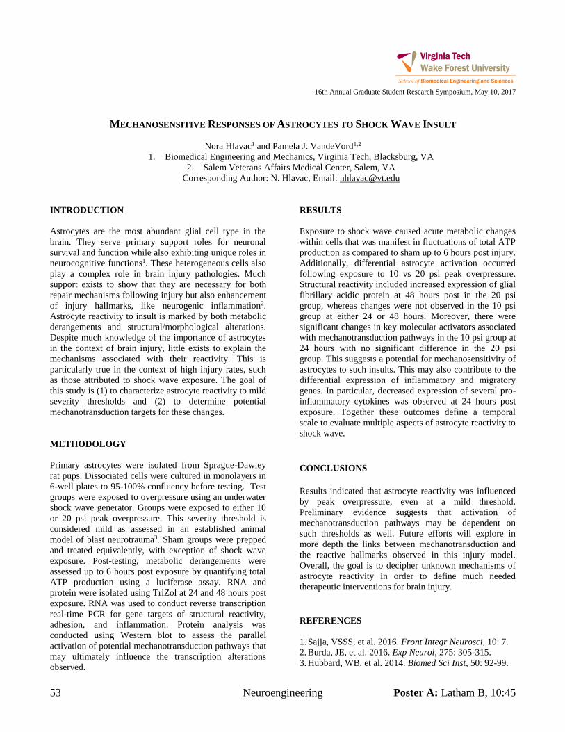
53 Neuroengineering Poster A: Latham B, 10:45
16th Annual Graduate Student Research Symposium, May 10, 2017
MECHANOSENSITIVE RESPONSES OF ASTROCYTES TO SHOCK WAVE INSULT
Nora Hlavac1 and Pamela J. VandeVord1,2
1. Biomedical Engineering and Mechanics, Virginia Tech, Blacksburg, VA 2. Salem Veterans Affairs Medical Center, Salem, VA
Corresponding Author: N. Hlavac, Email: [email protected] INTRODUCTION Astrocytes are the most abundant glial cell type in the brain. They serve primary support roles for neuronal survival and function while also exhibiting unique roles in neurocognitive functions1. These heterogeneous cells also play a complex role in brain injury pathologies. Much support exists to show that they are necessary for both repair mechanisms following injury but also enhancement of injury hallmarks, like neurogenic inflammation2. Astrocyte reactivity to insult is marked by both metabolic derangements and structural/morphological alterations. Despite much knowledge of the importance of astrocytes in the context of brain injury, little exists to explain the mechanisms associated with their reactivity. This is particularly true in the context of high injury rates, such as those attributed to shock wave exposure. The goal of this study is (1) to characterize astrocyte reactivity to mild severity thresholds and (2) to determine potential mechanotransduction targets for these changes. METHODOLOGY Primary astrocytes were isolated from Sprague-Dawley rat pups. Dissociated cells were cultured in monolayers in 6-well plates to 95-100% confluency before testing. Test groups were exposed to overpressure using an underwater shock wave generator. Groups were exposed to either 10 or 20 psi peak overpressure. This severity threshold is considered mild as assessed in an established animal model of blast neurotrauma3. Sham groups were prepped and treated equivalently, with exception of shock wave exposure. Post-testing, metabolic derangements were assessed up to 6 hours post exposure by quantifying total ATP production using a luciferase assay. RNA and protein were isolated using TriZol at 24 and 48 hours post exposure. RNA was used to conduct reverse transcription real-time PCR for gene targets of structural reactivity, adhesion, and inflammation. Protein analysis was conducted using Western blot to assess the parallel activation of potential mechanotransduction pathways that may ultimately influence the transcription alterations observed.
RESULTS Exposure to shock wave caused acute metabolic changes within cells that was manifest in fluctuations of total ATP production as compared to sham up to 6 hours post injury. Additionally, differential astrocyte activation occurred following exposure to 10 vs 20 psi peak overpressure. Structural reactivity included increased expression of glial fibrillary acidic protein at 48 hours post in the 20 psi group, whereas changes were not observed in the 10 psi group at either 24 or 48 hours. Moreover, there were significant changes in key molecular activators associated with mechanotransduction pathways in the 10 psi group at 24 hours with no significant difference in the 20 psi group. This suggests a potential for mechanosensitivity of astrocytes to such insults. This may also contribute to the differential expression of inflammatory and migratory genes. In particular, decreased expression of several pro-inflammatory cytokines was observed at 24 hours post exposure. Together these outcomes define a temporal scale to evaluate multiple aspects of astrocyte reactivity to shock wave. CONCLUSIONS Results indicated that astrocyte reactivity was influenced by peak overpressure, even at a mild threshold. Preliminary evidence suggests that activation of mechanotransduction pathways may be dependent on such thresholds as well. Future efforts will explore in more depth the links between mechanotransduction and the reactive hallmarks observed in this injury model. Overall, the goal is to decipher unknown mechanisms of astrocyte reactivity in order to define much needed therapeutic interventions for brain injury. REFERENCES 1. Sajja, VSSS, et al. 2016. Front Integr Neurosci, 10: 7. 2. Burda, JE, et al. 2016. Exp Neurol, 275: 305-315. 3. Hubbard, WB, et al. 2014. Biomed Sci Inst, 50: 92-99.

Poster B: Latham B, 11:15 Biomechanics 54
Figure 1: Variable Times as a function of walking speed.
16th Annual Graduate Student Research Symposium, May 10, 2017
QUANTIFYING LIMP USING TEMPORAL GAIT VARIABLES
Cherice Hughes-Oliver1, Daniel Schmitt2, Robin Queen1
1. Virginia Tech, Biomedical Engineering and Mechanics 2. Duke University, Department of Evolutionary Anthropology
Corresponding Author: Cherice Hughes-Oliver, Email: [email protected] INTRODUCTION Ankle osteoarthritis is a debilitating disease with patients reporting levels of impairment as high as those associated with end-stage kidney diseases, congestive heart failure, and end-stage hip arthritis [1, 2]. The objective of this study was to investigate a clinically-accessible method of identifying gait asymmetries (“limp”) and other changes
in patient gait associated with ankle osteoarthritis. We predicted that there would be differences in swing and stance time between affected and unaffected limbs. METHODOLOGY This is a secondary analysis of prospectively collected data on a group of 242 (110 men, 132 women) end-stage ankle OA patients. Only subjects with isolated unilateral involvement, no contralateral pain, and no rheumatoid arthritis diagnosis were included. Each subject completed at least four self-selected speed walking trials using an eight-camera motion capture system (Motion Analysis Corporation). Heel-strike and toe-off timings were exported from Visual3D. Stride time, swing time, stance time, and double support time were calculated in Matlab. Each variable was averaged across steps for a single trial and then across trials for each subject, and for each limb. An ANCOVA comparing sex and limb (affected and unaffected) and co-varying for walking speed was run for each variable using JMP Pro 13 (α = 0.05). RESULTS Men had higher stride, swing, and stance times than women (Fig 1), and swing time was higher on the affected limb and stance time was higher on the unaffected limb. This limb difference may function as a compensatory pattern due to ankle pain on the affected limb. Effects of walking speed were seen in stride, stance, and double support times but not in swing time. It is unclear whether the sex-specific effects observed in this study are present in healthy populations or are a result
of sex-specific pain coping compensatory mechanisms. Similarly to our results, Valderrabano et al found no inter-limb differences in stride time [3]. Nuesch, et al observed no decrease in stance time as a percent of stride time [4], which disagrees with our findings. Conversely, Shih et al found decreased stance time and increased swing time on the affected limb, which agrees with our findings [5]. Shih et al also saw an increase in double support time on the affected limb, which our results did not reflect. CONCLUSIONS It is possible that temporal variables, which are much easier to collect than 3D motion capture data, can be used to quantify limp and gait compensation; walking speed could play a large role in this quantification, along with limb-specific differences between swing and stance times. These results demonstrate the value of using these simpler variables to evaluate compensation. REFERENCES 1. Saltzman, C.L., et al. J Bone Joint Surg Am, 2006. 88(11): p. 2366-2372. 2. Glazebrook, M., et al. J Bone Joint Surg Am, 2008. 90(3): p. 499-505. 3. Valderrabano, V., et al. Clin Biomech, 2007. 22(8): p. 894-904. 4. Nuesch, C., et al. Clin Biomech (Bristol, Avon), 2012. 27(6): p. 613-8. 5. Shih, L.-Y., J.-J. Wu, and W.-H. Lo, Foot & Ankle International, 1993. 14(2): p. 97-103.
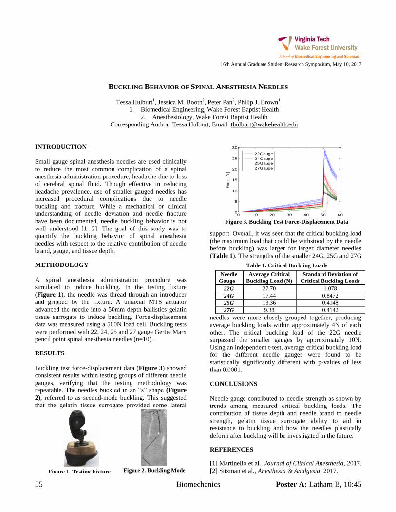
55 Biomechanics Poster A: Latham B, 10:45
Table 1. Critical Buckling Loads
0 10 20 30 40 50 600
5
10
15
20
25
30
Displacement (mm)
Forc
e (N
)
22Gauge
24Gauge
25Gauge
27Gauge
Figure 3. Buckling Test Force-Displacement Data
Figure 2. Buckling Mode Figure 1. Testing Fixture
16th Annual Graduate Student Research Symposium, May 10, 2017
BUCKLING BEHAVIOR OF SPINAL ANESTHESIA NEEDLES
Tessa Hulburt1, Jessica M. Booth
2, Peter Pan
2, Philip J. Brown
1
1. Biomedical Engineering, Wake Forest Baptist Health
2. Anesthesiology, Wake Forest Baptist Health
Corresponding Author: Tessa Hulburt, Email: [email protected]
INTRODUCTION
Small gauge spinal anesthesia needles are used clinically
to reduce the most common complication of a spinal
anesthesia administration procedure, headache due to loss
of cerebral spinal fluid. Though effective in reducing
headache prevalence, use of smaller gauged needles has
increased procedural complications due to needle
buckling and fracture. While a mechanical or clinical
understanding of needle deviation and needle fracture
have been documented, needle buckling behavior is not
well understood [1, 2]. The goal of this study was to
quantify the buckling behavior of spinal anesthesia
needles with respect to the relative contribution of needle
brand, gauge, and tissue depth.
METHODOLOGY
A spinal anesthesia administration procedure was
simulated to induce buckling. In the testing fixture
(Figure 1), the needle was thread through an introducer
and gripped by the fixture. A uniaxial MTS actuator
advanced the needle into a 50mm depth ballistics gelatin
tissue surrogate to induce buckling. Force-displacement
data was measured using a 500N load cell. Buckling tests
were performed with 22, 24, 25 and 27 gauge Gertie Marx
pencil point spinal anesthesia needles (n=10).
RESULTS
Buckling test force-displacement data (Figure 3) showed
consistent results within testing groups of different needle
gauges, verifying that the testing methodology was
repeatable. The needles buckled in an “s” shape (Figure
2), referred to as second-mode buckling. This suggested
that the gelatin tissue surrogate provided some lateral
support. Overall, it was seen that the critical buckling load
(the maximum load that could be withstood by the needle
before buckling) was larger for larger diameter needles
(Table 1). The strengths of the smaller 24G, 25G and 27G
needles were more closely grouped together, producing
average buckling loads within approximately 4N of each
other. The critical buckling load of the 22G needle
surpassed the smaller gauges by approximately 10N.
Using an independent t-test, average critical buckling load
for the different needle gauges were found to be
statistically significantly different with p-values of less
than 0.0001.
CONCLUSIONS
Needle gauge contributed to needle strength as shown by
trends among measured critical buckling loads. The
contribution of tissue depth and needle brand to needle
strength, gelatin tissue surrogate ability to aid in
resistance to buckling and how the needles plastically
deform after buckling will be investigated in the future.
REFERENCES
[1] Martinello et al., Journal of Clinical Anesthesia, 2017.
[2] Sitzman et al., Anesthesia & Analgesia, 2017.
Needle
Gauge
Average Critical
Buckling Load (N)
Standard Deviation of
Critical Buckling Loads
22G 27.70 1.078
24G 17.44 0.8472
25G 13.36 0.4148
27G 9.38 0.4142
Poster B: Latham B, 11:15 Translational Cancer 56
16th Annual Graduate Student Research Symposium, May 10, 2017
FLUID SHEAR STRESS IMPACTS OVARIAN CANCER CELL VIABILITY, SUBCELLULAR ORGANIZATION,
AND INDUCES MALIGNANT PHENOTYPES
Alexandra R. Hyler1, Nicolaas C. Baudoin2, Mark A. Stremler1, and Daniela Cimini2, Rafael V. Davalos1, Eva M. Schmelz1
1. VT-WFU School of Biomedical Engineering, Virginia Tech, Blacksburg, VA 2. Department of Biological Sciences and Biocomplexity Institute, Virginia Tech, Blacksburg, VA
Corresponding Author: Alexandra R. Hyler, Email: [email protected] INTRODUCTION All cells exist in a physiologic environment that is determined by chemical and physical factors; in concert, these factors direct tissue growth, organization and function but also can cause or contribute to diseases such as cancer. Epithelial ovarian cancer (EOC) is the fourth most deadly cancer, with a 5-year survival rate below 30% if diagnosed after the cancer has spread beyond its boundaries.1 Metastatic ovarian cancer cells exfoliated into the peritoneal cavity are exposed to continual fluid shear stress (FSS) due to gastrointestinal and diaphragm movements, abdominal pressure, gravity, and ascites build-up in advanced stages of ovarian cancer. 2
We hypothesized that relatively small values of FSS induced shear stress will differentially affect the various stages of disease. To test this hypothesis, we imposed FSS onto mouse ovarian epithelial (MOSE) cells3 to mimic physiologically relevant fluid movement of the peritoneal cavity.4 METHODOLOGY We estimated the shear stress for our system to be ~1.37-1.99 dyne/cm2 which is physiologically relevant.4 Three stages of disease were investigated: MOSE-E (benign), MOSE-L (slow-developing disease, malignant), MOSE-LTICv(fast-developing disease, malignant). Cells were seeded and placed immediately (-imm-) onto or allowed to adhere (-adh-) before placement onto a Lab Rotator. Cell viability (hemocytometer count), spheroid formation (imaging), cytoskeleton structure (indirect immunofluorescence), nuclear morphology (CREST staining) and chromosome counts (metaphase spreads) were assessed. RESULTS FSS affected cell viability differentially in MOSE cells representing different disease stage. In particular, MOSE-Eadh and MOSE-LTICv-adh cells appeared more resistant to FSS after adherence whereas all stages immediately
exposed to FSS responded with significant cell death. FSS induced spheroid formation in tumorigenic (MOSE-L and MOSE-LTICv) cells. These spheroids were free floating in both adherent and immediate MOSE-L cells, but the MOSE-LTICv-adh spheroids reattached and grew out, a capacity associated with invasiveness. We also observed FSS-induced changes in cytoskeleton organization such as indicated significant actin disorganization, and the appearance of protrusions. In conjunction with cytoskeletal changes, we observed the emergence in all cell lines of multi-lobed nuclei after FSS exposure, which was particularly drastic in benign cells. This result paralleled an observation of an increase in all cell lines of CREST-positive micronuclei suggested that FSS may cause errors in chromosome segregation, so lastly, we investigated chromosome number in MOSE-E cells. Indeed, FSS dramatically increased the fraction of benign MOSE-E cells with near-tetraploid chromosome numbers.
CONCLUSIONS Overall, our results indicate that low magnitudes of continual FSS significantly and differentially affect MOSE cells of various stages of progression. In particular, benign cells that survive under FSS display phenotypic changes (increased multi-lobed nuclei, CREST-positive micronuclei, and chromosome number) that resemble more aggressive stages of disease. Future work will employ a physiologically relevant testing platform to investigate the interaction of biomechanical and molecular interactions in ovarian cancer metastasis. ACKNOWLEDGMENTS This work was supported by NSF IGERT Grant 0966125 and seed funds from the Center for Engineered Health (ICTAS). REFERENCES
1. J. Berntsson, et al. J Ovarian Res, 2014, 7, 26. 2. D.S. Tan, et al. Lancet Oncol, 2006, 7, 925-34. 3. Roberts PC, et al. Neoplasia. Oct 2005;7(10) 944-956 4. Even-Tzur, N., et al. Biophysical J. Sept 2008;95(6) 2998-
3008.
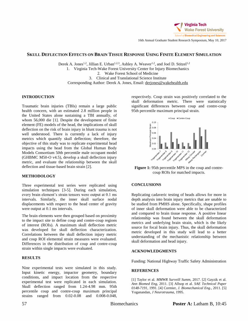
57 Biomechanics Poster A: Latham B, 10:45
16th Annual Graduate Student Research Symposium, May 10, 2017
SKULL DEFLECTION EFFECTS ON BRAIN TISSUE RESPONSE USING FINITE ELEMENT SIMULATION
Derek A. Jones1,2, Jillian E. Urban1,2,3, Ashley A. Weaver1,2, and Joel D. Stitzel1,2
1. Virginia Tech-Wake Forest University Center for Injury Biomechanics 2. Wake Forest School of Medicine
3. Clinical and Translational Science Institute Corresponding Author: Derek A. Jones, Email: [email protected]
INTRODUCTION Traumatic brain injuries (TBIs) remain a large public health concern, with an estimated 2.8 million people in the United States alone sustaining a TBI annually, of whom 56,000 die [1]. Despite the development of finite element (FE) models of the head, the implications of skull deflection on the risk of brain injury in blunt trauma is not well understood. There is currently a lack of injury metrics which quantify skull deflection; therefore, the objective of this study was to replicate experimental head impacts using the head from the Global Human Body Models Consortium 50th percentile male occupant model (GHBMC M50-O v4.5), develop a skull deflection injury metric, and evaluate the relationship between the skull deflection and tissue-based brain strain [2]. METHODOLOGY Three experimental test series were replicated using simulation techniques [3-5]. During each simulation, every brain element’s strain tensors were output at 0.1 ms
intervals. Similarly, the inner skull surface nodal displacements with respect to the head center of gravity were output at 0.1 ms intervals.
The brain elements were then grouped based on proximity to the impact site to define coup and contre-coup regions of interest (ROIs). A maximum skull deflection metric was developed for skull deflection characterization. Correlations between the skull deflection injury metric and coup ROI elemental strain measures were evaluated. Differences in the distribution of coup and contre-coup strain within single impacts were evaluated. RESULTS Nine experimental tests were simulated in this study. Input kinetic energy, impactor geometry, boundary conditions, and impact location from the respective experimental test were replicated in each simulation. Skull deflection ranged from 1.24-4.98 mm. 95th percentile coup and contre-coup maximum principal strains ranged from 0.02-0.08 and 0.008-0.048,
respectively. Coup strain was positively correlated to the skull deformation metric. There were statistically significant differences between coup and contre-coup 95th percentile maximum principal strain.
0.00
0.02
0.04
0.06
0.08
0.10
Max
imum
Prin
cipa
l Stra
in
Coup Contre-Coup
Figure 1: 95th percentile MPS in the coup and contre-
coup ROIs for matched impacts.
CONCLUSIONS Replicating cadaveric testing of heads allows for more in depth analysis into brain injury metrics that are unable to be studied from PMHS alone. Specifically, shape profiles of inner skull deformation were able to be characterized and compared to brain tissue response. A positive linear relationship was found between the skull deformation metrics and underlying brain strain, which is the likely source for focal brain injury. Thus, the skull deformation metric developed in this study will lead to a better understanding of the mechanistic relationship between skull deformation and head injury. ACKNOWLEDGMENTS Funding: National Highway Traffic Safety Administration REFERENCES [1] Taylor et al. MMWR Surveill Summ, 2017. [2] Gayzik et al. Ann Biomed Eng, 2011. [3] Allsop et al. SAE Technical Paper 0148-7191, 1991. [4] Cormier, J. Biomechanical Eng., 2011. [5] Yoganandan, J Neurotrauma, 1995.
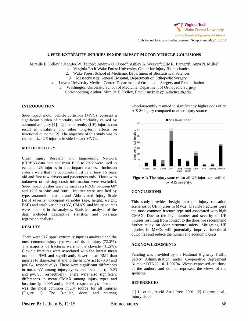
Poster B: Latham B, 11:15 Biomechanics 58
16th Annual Graduate Student Research Symposium, May 10, 2017
UPPER EXTREMITY INJURIES IN SIDE-IMPACT MOTOR VEHICLE COLLISIONS
Mireille E. Kelley1,; Jennifer W. Talton2; Andrew O. Usoro3; Ashley A. Weaver1; Eric R. Barnard4; Anna N. Miller5
1. Virginia Tech-Wake Forest University, Center for Injury Biomechanics 2. Wake Forest School of Medicine, Department of Biostatistical Sciences
3. Massachusetts General Hospital, Department of Orthopedic Surgery 4. Loyola University Medical Center, Department of Orthopedic Surgery and Rehabilitation
5. Washington University School of Medicine, Department of Orthopedic Surgery Corresponding Author: Mireille E. Kelley, Email: mekelley@wakehealth,edu
INTRODUCTION Side-impact motor vehicle collisions (MVC) represent a significant burden of mortality and morbidity caused by automotive injury [1]. Upper extremity (UE) injuries can result in disability and other long-term effects on functional outcome [2]. The objective of this study was to characterize UE injuries in side-impact MVCs. METHODOLOGY Crash Injury Research and Engineering Network (CIREN) data obtained from 1998 to 2012 were used to evaluate UE injuries in side-impact crashes. Inclusion criteria were that the occupants must be at least 16 years old and first row drivers and passengers only. Those with unknown or missing crash information were excluded. Side-impact crashes were defined as a PDOF between 60° and 120° or 240° and 300°. Injuries were stratified by type, anatomic location and Abbreviated Injury Scale (AIS) severity. Occupant variables (age, height, weight, BMI) and crash variables (ΔV, CMAX, and injury source)
were included in the analyses. Statistical analysis of the data included descriptive statistics and bivariate regression analyses. RESULTS There were 917 upper extremity injuries analyzed and the most common injury type was soft tissue injury (72.3%). The majority of fractures were to the clavicle (41.5%). Clavicle fractures were associated with the lowest mean occupant BMI and significantly lower mean BMI than injuries to skin/external and to the hand/wrist (p=0.04 and p=0.04, respectively). There were significant differences in mean ΔV among injury types and locations (p=0.03 and p=0.03, respectively). There were also significant differences in mean CMAX among injury types and locations (p=0.005 and p=0.001, respectively). The door was the most common injury source for all injuries (Figure 1). The B-pillar, door, and steering
wheel/assembly resulted in significantly higher odds of an AIS 2+ injury compared to other injury sources.
0
50
100
150
200
250
300
350
Air bag Belt B-pillar Door Flyingglass
IP/kneebolster
Other Seat Steering Unknown
Num
ber o
f UE
Inju
ries
AIS 1 AIS 2 AIS 3
Figure 1: The injury sources for all UE injuries stratified
by AIS severity. CONCLUSIONS This study provides insight into the injury causation scenarios of UE injuries in MVCs. Clavicle fractures were the most common fracture type and associated with high CMAX. Due to the high number and severity of UE injuries resulting from contact to the door, we recommend further study on door structure safety. Mitigating UE injuries in MVCs will potentially improve functional outcomes and reduce the human and economic costs. ACKNOWLEDGMENTS Funding was provided by the National Highway Traffic Safety Administration under Cooperative Agreement Number DTN22-10-H-00294. Views expressed are those of the authors and do not represent the views of the sponsors. REFERENCES [1] Li et al., Accid Anal Prev. 2001. [2] Conroy et al., Injury. 2007.
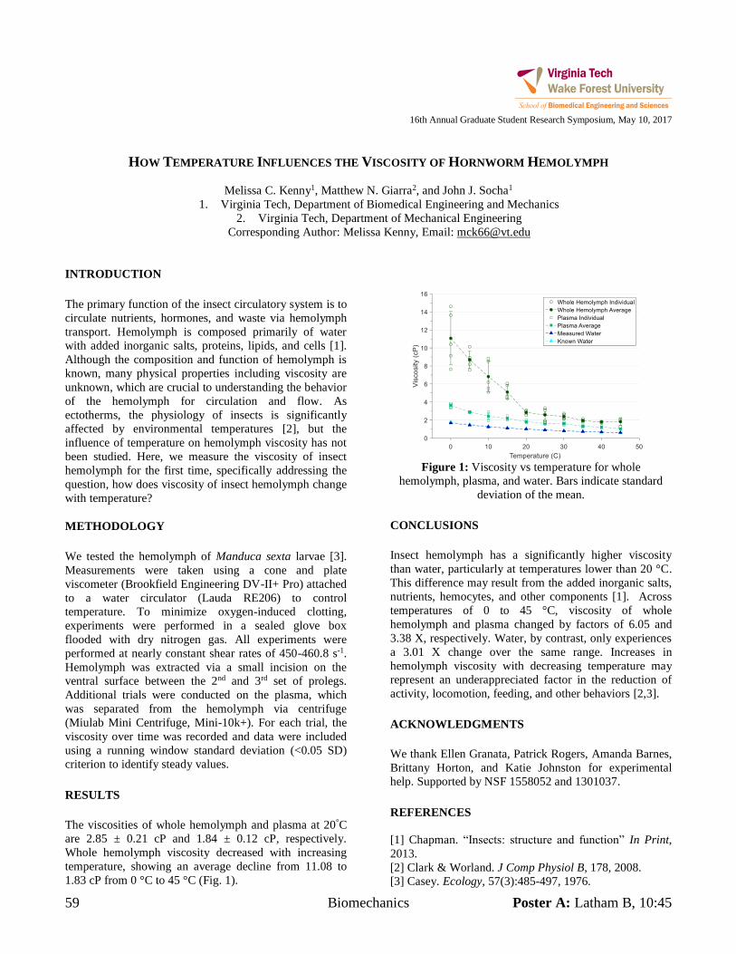
59 Biomechanics Poster A: Latham B, 10:45
16th Annual Graduate Student Research Symposium, May 10, 2017
HOW TEMPERATURE INFLUENCES THE VISCOSITY OF HORNWORM HEMOLYMPH
Melissa C. Kenny1, Matthew N. Giarra2, and John J. Socha1
1. Virginia Tech, Department of Biomedical Engineering and Mechanics 2. Virginia Tech, Department of Mechanical Engineering
Corresponding Author: Melissa Kenny, Email: [email protected] INTRODUCTION The primary function of the insect circulatory system is to circulate nutrients, hormones, and waste via hemolymph transport. Hemolymph is composed primarily of water with added inorganic salts, proteins, lipids, and cells [1]. Although the composition and function of hemolymph is known, many physical properties including viscosity are unknown, which are crucial to understanding the behavior of the hemolymph for circulation and flow. As ectotherms, the physiology of insects is significantly affected by environmental temperatures [2], but the influence of temperature on hemolymph viscosity has not been studied. Here, we measure the viscosity of insect hemolymph for the first time, specifically addressing the question, how does viscosity of insect hemolymph change with temperature? METHODOLOGY We tested the hemolymph of Manduca sexta larvae [3]. Measurements were taken using a cone and plate viscometer (Brookfield Engineering DV-II+ Pro) attached to a water circulator (Lauda RE206) to control temperature. To minimize oxygen-induced clotting, experiments were performed in a sealed glove box flooded with dry nitrogen gas. All experiments were performed at nearly constant shear rates of 450-460.8 s-1. Hemolymph was extracted via a small incision on the ventral surface between the 2nd and 3rd set of prolegs. Additional trials were conducted on the plasma, which was separated from the hemolymph via centrifuge (Miulab Mini Centrifuge, Mini-10k+). For each trial, the viscosity over time was recorded and data were included using a running window standard deviation (<0.05 SD) criterion to identify steady values. RESULTS The viscosities of whole hemolymph and plasma at 20°C are 2.85 ± 0.21 cP and 1.84 ± 0.12 cP, respectively. Whole hemolymph viscosity decreased with increasing temperature, showing an average decline from 11.08 to 1.83 cP from 0 °C to 45 °C (Fig. 1).
Figure 1: Viscosity vs temperature for whole
hemolymph, plasma, and water. Bars indicate standard deviation of the mean.
CONCLUSIONS Insect hemolymph has a significantly higher viscosity than water, particularly at temperatures lower than 20 °C. This difference may result from the added inorganic salts, nutrients, hemocytes, and other components [1]. Across temperatures of 0 to 45 °C, viscosity of whole hemolymph and plasma changed by factors of 6.05 and 3.38 X, respectively. Water, by contrast, only experiences a 3.01 X change over the same range. Increases in hemolymph viscosity with decreasing temperature may represent an underappreciated factor in the reduction of activity, locomotion, feeding, and other behaviors [2,3]. ACKNOWLEDGMENTS We thank Ellen Granata, Patrick Rogers, Amanda Barnes, Brittany Horton, and Katie Johnston for experimental help. Supported by NSF 1558052 and 1301037. REFERENCES [1] Chapman. “Insects: structure and function” In Print, 2013. [2] Clark & Worland. J Comp Physiol B, 178, 2008. [3] Casey. Ecology, 57(3):485-497, 1976.
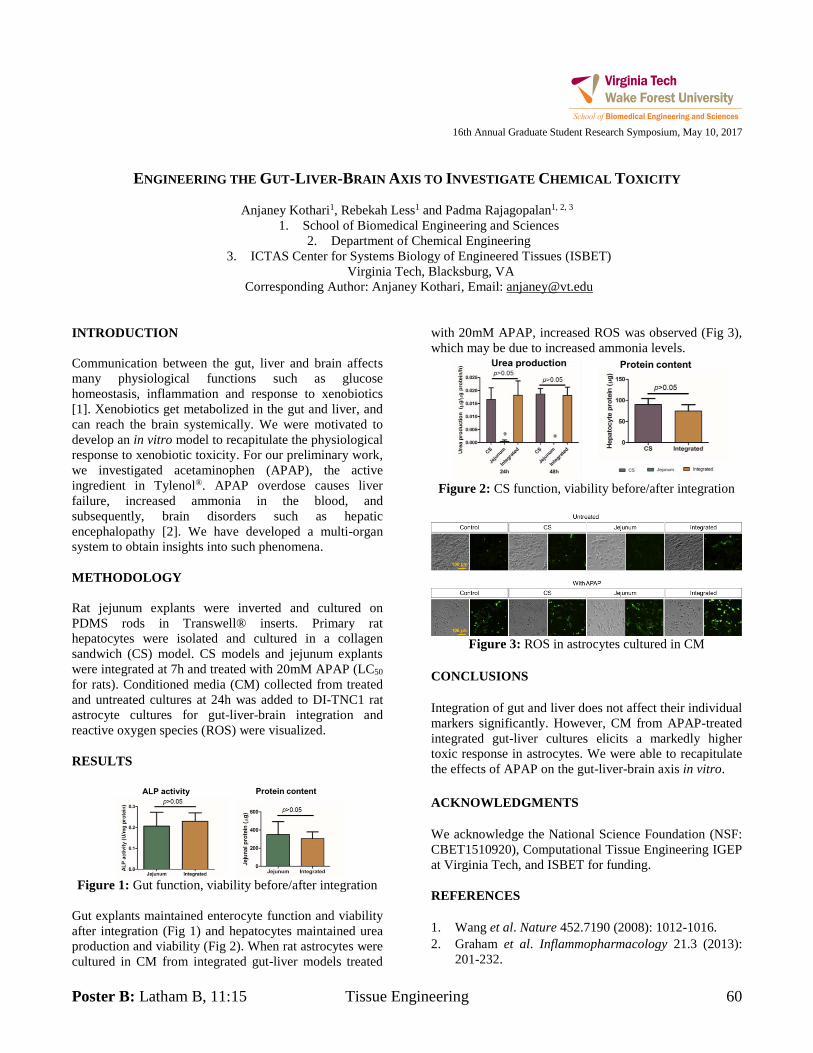
Poster B: Latham B, 11:15 Tissue Engineering 60
16th Annual Graduate Student Research Symposium, May 10, 2017
ENGINEERING THE GUT-LIVER-BRAIN AXIS TO INVESTIGATE CHEMICAL TOXICITY
Anjaney Kothari1, Rebekah Less1 and Padma Rajagopalan1, 2, 3
1. School of Biomedical Engineering and Sciences 2. Department of Chemical Engineering
3. ICTAS Center for Systems Biology of Engineered Tissues (ISBET) Virginia Tech, Blacksburg, VA
Corresponding Author: Anjaney Kothari, Email: [email protected] INTRODUCTION Communication between the gut, liver and brain affects many physiological functions such as glucose homeostasis, inflammation and response to xenobiotics [1]. Xenobiotics get metabolized in the gut and liver, and can reach the brain systemically. We were motivated to develop an in vitro model to recapitulate the physiological response to xenobiotic toxicity. For our preliminary work, we investigated acetaminophen (APAP), the active ingredient in Tylenol®. APAP overdose causes liver failure, increased ammonia in the blood, and subsequently, brain disorders such as hepatic encephalopathy [2]. We have developed a multi-organ system to obtain insights into such phenomena. METHODOLOGY Rat jejunum explants were inverted and cultured on PDMS rods in Transwell® inserts. Primary rat hepatocytes were isolated and cultured in a collagen sandwich (CS) model. CS models and jejunum explants were integrated at 7h and treated with 20mM APAP (LC50 for rats). Conditioned media (CM) collected from treated and untreated cultures at 24h was added to DI-TNC1 rat astrocyte cultures for gut-liver-brain integration and reactive oxygen species (ROS) were visualized. RESULTS
Figure 1: Gut function, viability before/after integration
Gut explants maintained enterocyte function and viability after integration (Fig 1) and hepatocytes maintained urea production and viability (Fig 2). When rat astrocytes were cultured in CM from integrated gut-liver models treated
with 20mM APAP, increased ROS was observed (Fig 3), which may be due to increased ammonia levels.
Figure 2: CS function, viability before/after integration
Figure 3: ROS in astrocytes cultured in CM
CONCLUSIONS Integration of gut and liver does not affect their individual markers significantly. However, CM from APAP-treated integrated gut-liver cultures elicits a markedly higher toxic response in astrocytes. We were able to recapitulate the effects of APAP on the gut-liver-brain axis in vitro. ACKNOWLEDGMENTS We acknowledge the National Science Foundation (NSF: CBET1510920), Computational Tissue Engineering IGEP at Virginia Tech, and ISBET for funding. REFERENCES 1. Wang et al. Nature 452.7190 (2008): 1012-1016. 2. Graham et al. Inflammopharmacology 21.3 (2013):
201-232.
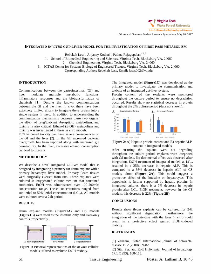
61 Tissue Engineering Poster A: Latham B, 10:45
16th Annual Graduate Student Research Symposium, May 10, 2017
INTEGRATED IN VITRO GUT-LIVER MODEL FOR THE INVESTIGATION OF FIRST PASS METABOLISM
Rebekah Less1, Anjaney Kothari
1, Padma Rajagopalan
1, 2, 3
1. School of Biomedical Engineering and Sciences, Virginia Tech, Blacksburg VA, 24060
2. Chemical Engineering, Virginia Tech, Blacksburg VA, 24060
3. ICTAS Center for Systems Biology of Engineered Tissues, Virginia Tech, Blacksburg VA, 24060
Corresponding Author: Rebekah Less, Email: [email protected]
INTRODUCTION
Communications between the gastrointestinal (GI) and
liver modulate multiple metabolic functions,
inflammatory responses and the biotransformation of
chemicals [1]. Despite the known communications
between the GI and the liver in vivo, there have been
extremely limited efforts to integrate these organs into a
single system in vitro. In addition to understanding the
communication mechanisms between these two organs,
the effect of drug/toxicant absorption, metabolism, and
toxicity is also critical. Ethanol (EtOH) metabolism and
toxicity was investigated in these in vitro models.
EtOH-induced toxicity can have severe consequences on
the GI and the liver [2]. In the GI, increased bacterial
overgrowth has been reported along with increased gut
permeability. In the liver, excessive ethanol consumption
can lead to fibrosis.
METHODOLOGY
We describe a novel integrated GI-liver model that is
designed by integrating a primary rat ileum explant with a
primary hepatocyte liver model. Primary ileum tissues
were surgically excised from rats. These explants were
cultured in oxygenated culture medium that contained
antibiotics. EtOH was administered over 100-200mM
concentration range. These concentrations ranged from
sub-lethal to 50% lethal concentration (LC50). All models
were cultured over a 24h period.
RESULTS
Ileum explant models (Figure1A) and CS models
(Figure1B) were used as the intestine-only and liver-only
controls, respectively.
Figure 1: Pictorial representations of the in vitro cellular
models utilized to evaluate EtOH toxicity.
The Integrated model (Figure1C) was developed as the
primary model to investigate the communication and
toxicity of an integrated gut-liver system.
Protein content of the explants were monitored
throughout the culture period to ensure no degradation
occurred. Results show no statistical decrease in protein
throughout the 24h culture period (data not shown).
Figure 2: A) Hepatic protein content and B) hepatic ALP
content in integrated models
After ensuring the explants were not degrading
throughout the culture period, explants were integrated
with CS models. No detrimental effect was observed after
integration. EtOH treatment of integrated models at LC50
resulted in a 25% decrease in hepatic ALP. This is
compared to a 56% decrease in hepatic ALP of CS
models alone (Figure 2A). This could suggest a
protective effect of the intestine on hepatocytes. This
hypothesis is further supported by hepatic protein. In
integrated cultures, there is a 7% decrease in hepatic
protein after LC50 EtOH treatment, however in the CS
models, this decrease is 22% (Figure 2B).
CONCLUSIONS
Results show ileum explants can be cultured for 24h
without significant degradation. Furthermore, the
integration of the intestine with the liver in vitro could
result in a protective effect against EtOH-induced
toxicity.
REFERENCES
[1] Zeuzem, Stefan. International journal of colorectal
disease 15.2 (2000): 59-82.
[2] Stål, Per, and Rolf Hultcrantz. Journal of hepatology
17.1 (1993): 108-115.
A. B.

Poster B: Latham B, 11:15 Translational Cancer 62
16th Annual Graduate Student Research Symposium, May 10, 2017
IRREVERSIBLE ELECTROPORATION FOR THE ABLATION OF PANCREATIC
MALIGNANCIES: A PATIENT-SPECIFIC METHODOLOGY
Melvin F. Lorenzo BS1, Eduardo L. Latouche MS1, Michael B. Sano PhD2,3, Robert C. G. Martin II MD PhD4, Rafael V. Davalos PhD1
1. Biomedical Engineering and Mechanics, Virginia Polytechnic Institute and State University, Blacksburg, Virginia 2. Radiation Oncology, Stanford University School of Medicine, Stanford, California
3. UNC/NCSU Joint Department of Biomedical Engineering, Chapel Hill, North Carolina 4. Surgery, Division of Surgical Oncology, University of Louisville, Louisville, Kentucky
Corresponding Author: Rafael V. Davalos, Email: [email protected] INTRODUCTION Irreversible electroporation (IRE) is a minimally invasive focal ablation technique used to nonthermally ablate soft tumor tissue [1]. Due to its nonthermal mechanism of ablation, IRE can be safely implemented in unresectable tumors located near critical vasculature [2]. Although effective, widespread use of IRE is hindered by challenges associated with treatment planning protocols, real-time feedback for monitoring lesion size, and the total duration of treatment. METHODOLOGY In order to address some of these issues, our group established a pre-treatment planning methodology for IRE procedures (Figure 1) in locally advanced pancreatic cancer (LAPC), in compliance with HIPAA and hospital IRB approval. Also, a real-time resistance measurement system is proposed to track for a change in resistance of 28 Ω, marking a clinical endpoint for IRE [3]. RESULTS
Figure 1: Patient specific 3D geometry of the pancreas (green), tumor (blue), and vasculature (red). This
geometry was reconstructed from stacked images of the
patient’s anatomy. The electric field distribution after 100 pulses is shown.
Changes in maximum temperature at the midpoint between electrode-pairs is comparable between the experimental procedure (10.3℃) and the proposed model (8.2 ℃). By implementing the proposed resistance measurement system, 210 ± 26.1 (mean ± standard deviation) fewer pulses were delivered per electrode-pair. CONCLUSIONS The proposed physics-based pre-treatment plan through finite element analysis and system for actively monitoring resistance changes can be paired to significantly reduce ablation times and risk of thermal damage during IRE procedures for LAPC. ACKNOWLEDGMENTS Virginia Center for Innovative Technology, Grant number: MF13-034-LS; CureAccelerator (Cures Within Reach), Grant number: PUJFSANY REFERENCES [1] R. V. Davalos, L. M. Mir, and B. Rubinsky,
“Tissue ablation with irreversible
electroporation,” Ann. Biomed. Eng., vol. 33, no. 2, pp. 223–231, 2005.
[2] E. Maor, A. Ivorra, J. Leor, and B. Rubinsky, “The Effect of Irreversible Electroporation on Blood Vessels,” Technol. Cancer Res. Treat., vol. 6, no. 4, pp. 307–312, 2007.
[3] E. L. Latouche, M. B. Sano, M. F. Lorenzo, R. V. Davalos, and R. C. G. Martin, “Irreversible
electroporation for the ablation of pancreatic malignancies: A patient-specific methodology,” J. Surg. Oncol., no. January, pp. 1–7, 2017.
63 Nanobioengineering Poster A: Latham B, 10:45
16th Annual Graduate Student Research Symposium, May 10, 2017
CELL TYPE-SPECIFIC MOUSE BRAIN METHYLOMES PROFILED USING AN ULTRALOW-INPUT
MICROFLUIDIC DEVICE
Sai Ma1, Zhixiong Sun
2, Chen Sun
1, Travis W. Murphy
3, Hehuang Xie
2,4, Chang Lu
2
1. School of Biomedical Engineering and Sciences, Virginia Tech
2. Department of Biological Sciences, Virginia Tech
3. Chemical Engineering, Virginia Tech
4. Biocomplexity Institute, Virginia Tech
Corresponding Author: Chang Lu, Email: [email protected]
INTRODUCTION
Epigenetics refer to any process that alters gene activities
without changing the DNA sequence. Previous studies
have demonstrated that epigenetic regulation, such as
DNA methylation, plays a critical role in many biological
procedures, such as embryo development and learning
process. Genome-wide study of dynamics of DNA
methylation at single-base resolution provides a
comprehensive understanding of methylome confi-
guration. Bisulfite sequencing is regarded as the gold-
standard technology for analyzing DNA methylation. This
technology is based on the different consequences after
cytosine and 5-methycytosine (5-mC) treated with sodium
bisulfite. However, conventional protocols required more
than 300 ng DNA due to the significant sample loss
during bisulfite treatment (up to 96%). This limits its
application for studying precious samples and
understanding the underlying heterogeneity of clinic
samples. To broaden the applications of bisulfite
sequencing, it is necessary to develop ultrasensitive tools
for epigenomic study, particularly DNA methylation
analysis. We reported the first microfluidic DNA
methylation assay at genome-wide scale and single-base
resolution with >3 orders of magnitude higher sensitivity
than conventional protocols.
METHODOLOGY
We demonstrate a microfluidic technology, referred to as
MIcrofluidic Diffusion-based RRBS (MID-RRBS), for
low-input (down to 0.3 ng DNA) assay with high bisulfite
conversion efficiency and high coverage of CpGs. We
used a diffusion-based reagent swapping approach for
multi-step treatment of DNA on the microfluidic
platform. Our protocol preserved substantially more
amplifiable DNA than conventional bisulfite treatment
10, 24-26 while achieving high conversion rate.
RESULTS
We mapped methylomes of a cell line (GM 12878),
neurons and glia isolated from mouse cerebellums. Our
protocol yielded data that were heavy in CpGs of high
coverage (2.0-2.6 million CpGs with ≥1×, 1.3-1.8 million
CpGs with ≥10× coverage with 0.3 to 10 ng starting
DNA, respectively). MID-RRBS technology allowed
differentiation of methylomic landscapes of neurons and
glia from mouse cerebellum, generating insights into cell
type-specific features and their relationships to gene
expressions.
CONCLUSIONS
Our MID-RRBS technology offers capability for low-
input profiling of DNA methylomes on a microfluidic
platform. The diffusion-based reagent exchange method
permits loading/releasing of various small-molecule
reagents without substantial loss in the DNA amount.
Such approach facilitates conducting a complex molecular
biology treatment in a microfluidic device with a simple
structure. In principle, similar approaches can be applied
to WGBS.
ACKNOWLEDGMENTS
We thank C. Luo, J. Lucero, M.M. Behrens, and J.R.
Ecker at Salk Institute for help with the experiments and
discussion of the data; J. Ma at Carnegie Mellon
University for proofreading and comments. This work
was supported by US National Institutes of Health grants
EB017235 (C.L.), HG009256 (C.L.), and NS094574
(H.X.).
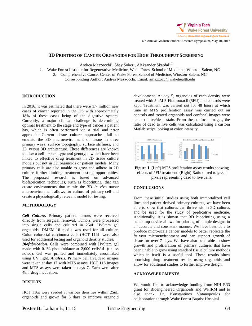
Poster B: Latham B, 11:15 Tissue Engineering 64
16th Annual Graduate Student Research Symposium, May 10, 2017
3D PRINTING OF CANCER ORGANOIDS FOR HIGH THROUGHPUT SCREENING
Andrea Mazzocchi1, Shay Soker
1, Aleksander Skardal
1,2
1. Wake Forest Institute for Regenerative Medicine, Wake Forest School of Medicine, Winston-Salem, NC
2. Comprehensive Cancer Center of Wake Forest School of Medicine, Winston-Salem, NC
Corresponding Author: Andrea Mazzocchi, Email: [email protected]
INTRODUCTION
In 2016, it was estimated that there were 1.7 million new
cases of cancer reported in the US with approximately
18% of these cases being of the digestive system.
Currently, a major clinical challenge is determining
optimal treatment to the stage and type of cancer a patient
has, which is often performed via a trial and error
approach. Current tissue culture approaches fail to
emulate the 3D microenvironment of tissue in three
primary ways: surface topography, surface stiffness, and
2D versus 3D architecture. These differences are known
to alter a cell’s phenotype and genotype which have been
linked to effective drug treatment in 2D tissue culture
models but not in 3D organoids or patient models. Many
primary cells are also unable to grow and adhere in 2D
culture further limiting treatment testing opportunities.
The proposed research is based on advanced
biofabrication techniques, such as bioprinting, that can
create environments that mimic the 3D in vivo tumor
microenvironment allows for culture of primary cell and
create a physiologically relevant model for testing.
METHODOLOGY
Cell Culture. Primary patient tumors were received
directly from surgical removal. Tumors were processed
into single cells and cultured in 25uL HyStem gel
organoids. DMEM-10 media was used for all culture.
Colon colorectal carcinoma cells (HCT 116) were also
used for additional testing and organoid density studies.
Biofabrication. Cells were combined with HyStem gel
made with 0.1% photoinitiator at 2,000 cells/uL (unless
noted). Gel was printed and immediately crosslinked
using UV light. Analysis. Primary cell live/dead images
were taken at day 17 with MTS assays. HCT 116 images
and MTS assays were taken at days 7. Each were after
48hr drug incubation.
RESULTS
HCT 116s were seeded at various densities within 25uL
organoids and grown for 5 days to improve organoid
development. At day 5, organoids of each density were
treated with 5mM 5-Fluorouracil (5FU) and controls were
kept. Treatment was carried out for 48 hours at which
time an MTS proliferation assay was carried out on
controls and treated organoids and confocal images were
taken of live/dead stain. From the confocal images, the
ratio of dead to live cells was calculated using a custom
Matlab script looking at color intensity.
Figure 1. (Left) MTS proliferation assay results showing
effects of 5FU treatment. (Right) Ratio of red to green
pixels representing dead to live cells.
CONCLUSIONS
From these initial studies using both immortalized cell
lines and patient derived primary cultures, we have been
able to show that cultures can thrive within 3D cultures
and be used for the study of predicative medicine.
Additionally, it is shown that 3D bioprinting using a
bench top device allows for printing of simple designs in
an accurate and consistent manner. We have been able to
produce micro-scale cancer models to better replicate the
in vivo microenvironment and can support growth of
tissue for over 7 days. We have also been able to show
growth and proliferation of primary cultures that have
been unable to grow using standard tissue culture methods
which in itself is a useful tool. These results show
promising drug treatment results using organoids and
allow for additional studies to further improve design.
ACKNOWLEDGMENTS
We would like to acknowledge funding from NIH R33
grant for Bioengineered Organoids and WFIRM and to
also thank Dr. Konstantinos Votanopoulos for
collaboration through Wake Forest Baptist Hospital.
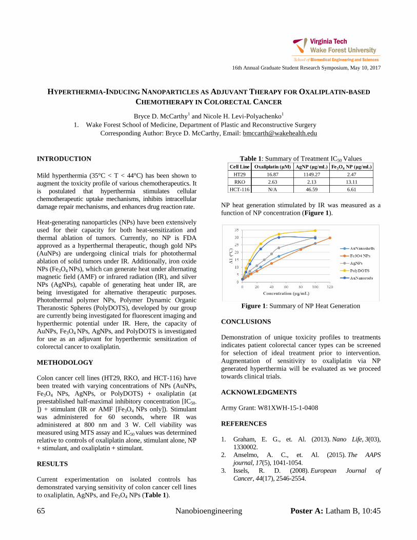
65 Nanobioengineering Poster A: Latham B, 10:45
16th Annual Graduate Student Research Symposium, May 10, 2017
HYPERTHERMIA-INDUCING NANOPARTICLES AS ADJUVANT THERAPY FOR OXALIPLATIN-BASED
CHEMOTHERAPY IN COLORECTAL CANCER
Bryce D. McCarthy1 and Nicole H. Levi-Polyachenko
1
1. Wake Forest School of Medicine, Department of Plastic and Reconstructive Surgery
Corresponding Author: Bryce D. McCarthy, Email: [email protected]
INTRODUCTION
Mild hyperthermia (35°C < T < 44°C) has been shown to
augment the toxicity profile of various chemotherapeutics. It
is postulated that hyperthermia stimulates cellular
chemotherapeutic uptake mechanisms, inhibits intracellular
damage repair mechanisms, and enhances drug reaction rate.
Heat-generating nanoparticles (NPs) have been extensively
used for their capacity for both heat-sensitization and
thermal ablation of tumors. Currently, no NP is FDA
approved as a hyperthermal therapeutic, though gold NPs
(AuNPs) are undergoing clinical trials for photothermal
ablation of solid tumors under IR. Additionally, iron oxide
NPs (Fe3O4 NPs), which can generate heat under alternating
magnetic field (AMF) or infrared radiation (IR), and silver
NPs (AgNPs), capable of generating heat under IR, are
being investigated for alternative therapeutic purposes.
Photothermal polymer NPs, Polymer Dynamic Organic
Theranostic Spheres (PolyDOTS), developed by our group
are currently being investigated for fluorescent imaging and
hyperthermic potential under IR. Here, the capacity of
AuNPs, Fe3O4 NPs, AgNPs, and PolyDOTS is investigated
for use as an adjuvant for hyperthermic sensitization of
colorectal cancer to oxaliplatin.
METHODOLOGY
Colon cancer cell lines (HT29, RKO, and HCT-116) have
been treated with varying concentrations of NPs (AuNPs,
Fe3O4 NPs, AgNPs, or PolyDOTS) + oxaliplatin (at
preestablished half-maximal inhibitory concentration [IC50-
]) + stimulant (IR or AMF [Fe3O4 NPs only]). Stimulant
was administered for 60 seconds, where IR was
administered at 800 nm and 3 W. Cell viability was
measured using MTS assay and IC50 values was determined
relative to controls of oxaliplatin alone, stimulant alone, NP
+ stimulant, and oxaliplatin + stimulant.
RESULTS
Current experimentation on isolated controls has
demonstrated varying sensitivity of colon cancer cell lines
to oxaliplatin, AgNPs, and Fe3O4 NPs (Table 1).
Table 1: Summary of Treatment IC50 Values
NP heat generation stimulated by IR was measured as a
function of NP concentration (Figure 1).
Figure 1: Summary of NP Heat Generation
CONCLUSIONS
Demonstration of unique toxicity profiles to treatments
indicates patient colorectal cancer types can be screened
for selection of ideal treatment prior to intervention.
Augmentation of sensitivity to oxaliplatin via NP
generated hyperthermia will be evaluated as we proceed
towards clinical trials.
ACKNOWLEDGMENTS
Army Grant: W81XWH-15-1-0408
REFERENCES
1. Graham, E. G., et. Al. (2013). Nano Life, 3(03),
1330002.
2. Anselmo, A. C., et. Al. (2015). The AAPS
journal, 17(5), 1041-1054.
3. Issels, R. D. (2008). European Journal of
Cancer, 44(17), 2546-2554.
Cell Line Oxaliplatin (μM) AgNP (μg/mL) Fe3O4 NP (μg/mL)
HT29 16.87 1149.27 2.47
RKO 2.63 2.13 13.11
HCT-116 N/A 46.59 6.61
Poster B: Latham B, 11:15 Biomechanics 66
16th Annual Graduate Student Research Symposium, May 10, 2017
AN INVESTIGATION INTO THE VALIDATION AND FUTURE USE OF FE HYBRID III, THOR, AND GHBMC
M50-OS FOR SPACEFLIGHT CONFIGURATION TESTING
Kyle P. McNamara1,2
, Derek A. Jones1,2
, James P. Gaewsky1,2
, F. Scott Gayzik1,2
, Ashley A. Weaver1,2
, Joel D. Stitzel1,2
1Wake Forest School of Medicine,
2Virginia Tech-Wake Forest University Center for Injury Biomechanics
Corresponding Author: Kyle McNamara, Email: [email protected]
INTRODUCTION
Injury assessment reference values (IARVs) posture post-
mortem human subjects in specific configurations to
determine injury risks to occupants. While historically
used in both automotive and military research, these
testing configurations may not translate directly to
spaceflight. Namely, there are differences in the loading
directions, postures, and restraint systems in spaceflight.
This study begins to addresses these differences by
validating Finite Element (FE) models of
Anthropomorphic Test Devices (ATDs) and FE Human
Body Models (HBMs) against physical dummies and
human volunteers in various loading directions. This step
is crucial for future spaceflight seat design.
METHODOLOGY
Between 1976 and 2013, a combination of Hybrid III,
THOR, and human volunteer tests were conducted using
both the Horizontal Impulse Accelerator (HIA) and
Vertical Deceleration Tower (VDT) at Wright-Patterson
Air Force Base and USAF Armstrong Laboratory [1-3].
These tests formed a matrix for FE validation. All tests
used a 5-point belt restraint system in a flat pan seat with
a vertical back [1]. The 275 tests selected comprised 49
physical test configurations with accelerations in the
frontal (X-), rear (X+), lateral (Y), and vertical (Z+)
directions. The acceleration magnitudes varied from 3-20
G and had pulse duration ranges from 20-110 ms.
Simulations were performed using the Humanetics 50th
percentile male Hybrid III, NHTSA THOR 50th Male,
and the Global Human Body Models Consortium
(GHBMC) 50th male simplified occupant (M50-OS)
models in LS_DYNA [4-6]. All simulations consisted of a
150 ms period of gravitational settling and belt
pretensioning followed by the acceleration pulse taken
from the physical test of interest.
Analysis consisted of both a visual comparison of
kinematics as well as a quantitative analysis. Simulation
signals in the head, neck, thorax, and pelvis were
compared to matched physical signals using the Gehre et
al. method (CORrelation and Analysis, or CORA, size
phase, and shape) [7].
RESULTS
Visual inspection for the 49 test configuration simulations
showed agreement with the physical test cases in regards
to the excursion magnitude and direction of the thorax
and the head. Comparison of the THOR model shows
agreement in neck flexion, translation of the back relative
to the seat back, hand position relative to the knees, and
feet position relative to the chair legs.
CONCLUSIONS
Initial validation of ATD and human body models against
physical test data is essential for building confidence in
future applications of those models. Overall the physical
and simulation results were comparable, ensuring
confidence in FE model performance in the validated
regime. The results of this study are highly applicable to
both government and commercial spaceflight and provide
confidence in FE simulation for future design.
REFERENCES
[1] C. Perry, C. Burneka, and C. Albery, "Biodynamic
Assessment of the THOR-K Manikin," DTIC Document2013.
[2] B. F. Hearon and J. W. Brinkley, Aviation, space, and
environmental medicine, vol. 57, pp. 301-12, Apr 1986.
[3] J. R. Buhrman and C. E. Perry, Aviation, space, and
environmental medicine, vol. 65, pp. 1086-90, Dec 1994.
[4] "H350 Adult Dummy Model LS-Dyna," Humanetics
Innovative Solutions, Inc.2012.
[5] J. B. Panzer, S. Giudice, and J. B. Putnam,
NHTSA/USDOT2015.
[6] D. Schwartz, B. Guleyupoglu, B. Koya, J. D. Stitzel,
and F. S. Gayzik, Traffic injury prevention, vol. 16 Suppl
1, pp. S49-56, 2015.
[7] C. Gehre, H. Gades, and P. Wernicke, in 21st International
Technical Conference on the Enhanced Safety of Vehicles
Conference (ESV), Stuttgart, Germany, June, 2009, pp. 15-18.
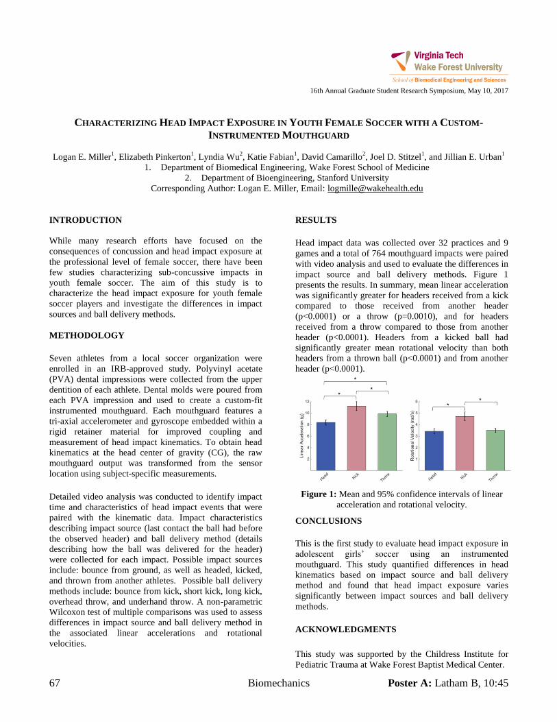
67 Biomechanics Poster A: Latham B, 10:45
16th Annual Graduate Student Research Symposium, May 10, 2017
CHARACTERIZING HEAD IMPACT EXPOSURE IN YOUTH FEMALE SOCCER WITH A CUSTOM-
INSTRUMENTED MOUTHGUARD
Logan E. Miller1, Elizabeth Pinkerton
1, Lyndia Wu
2, Katie Fabian
1, David Camarillo
2, Joel D. Stitzel
1, and Jillian E. Urban
1
1. Department of Biomedical Engineering, Wake Forest School of Medicine
2. Department of Bioengineering, Stanford University
Corresponding Author: Logan E. Miller, Email: [email protected]
INTRODUCTION
While many research efforts have focused on the
consequences of concussion and head impact exposure at
the professional level of female soccer, there have been
few studies characterizing sub-concussive impacts in
youth female soccer. The aim of this study is to
characterize the head impact exposure for youth female
soccer players and investigate the differences in impact
sources and ball delivery methods.
METHODOLOGY
Seven athletes from a local soccer organization were
enrolled in an IRB-approved study. Polyvinyl acetate
(PVA) dental impressions were collected from the upper
dentition of each athlete. Dental molds were poured from
each PVA impression and used to create a custom-fit
instrumented mouthguard. Each mouthguard features a
tri-axial accelerometer and gyroscope embedded within a
rigid retainer material for improved coupling and
measurement of head impact kinematics. To obtain head
kinematics at the head center of gravity (CG), the raw
mouthguard output was transformed from the sensor
location using subject-specific measurements.
Detailed video analysis was conducted to identify impact
time and characteristics of head impact events that were
paired with the kinematic data. Impact characteristics
describing impact source (last contact the ball had before
the observed header) and ball delivery method (details
describing how the ball was delivered for the header)
were collected for each impact. Possible impact sources
include: bounce from ground, as well as headed, kicked,
and thrown from another athletes. Possible ball delivery
methods include: bounce from kick, short kick, long kick,
overhead throw, and underhand throw. A non-parametric
Wilcoxon test of multiple comparisons was used to assess
differences in impact source and ball delivery method in
the associated linear accelerations and rotational
velocities.
RESULTS
Head impact data was collected over 32 practices and 9
games and a total of 764 mouthguard impacts were paired
with video analysis and used to evaluate the differences in
impact source and ball delivery methods. Figure 1
presents the results. In summary, mean linear acceleration
was significantly greater for headers received from a kick
compared to those received from another header
(p<0.0001) or a throw (p=0.0010), and for headers
received from a throw compared to those from another
header (p<0.0001). Headers from a kicked ball had
significantly greater mean rotational velocity than both
headers from a thrown ball (p<0.0001) and from another
header (p<0.0001).
Figure 1: Mean and 95% confidence intervals of linear
acceleration and rotational velocity.
CONCLUSIONS
This is the first study to evaluate head impact exposure in
adolescent girls’ soccer using an instrumented
mouthguard. This study quantified differences in head
kinematics based on impact source and ball delivery
method and found that head impact exposure varies
significantly between impact sources and ball delivery
methods.
ACKNOWLEDGMENTS
This study was supported by the Childress Institute for
Pediatric Trauma at Wake Forest Baptist Medical Center.
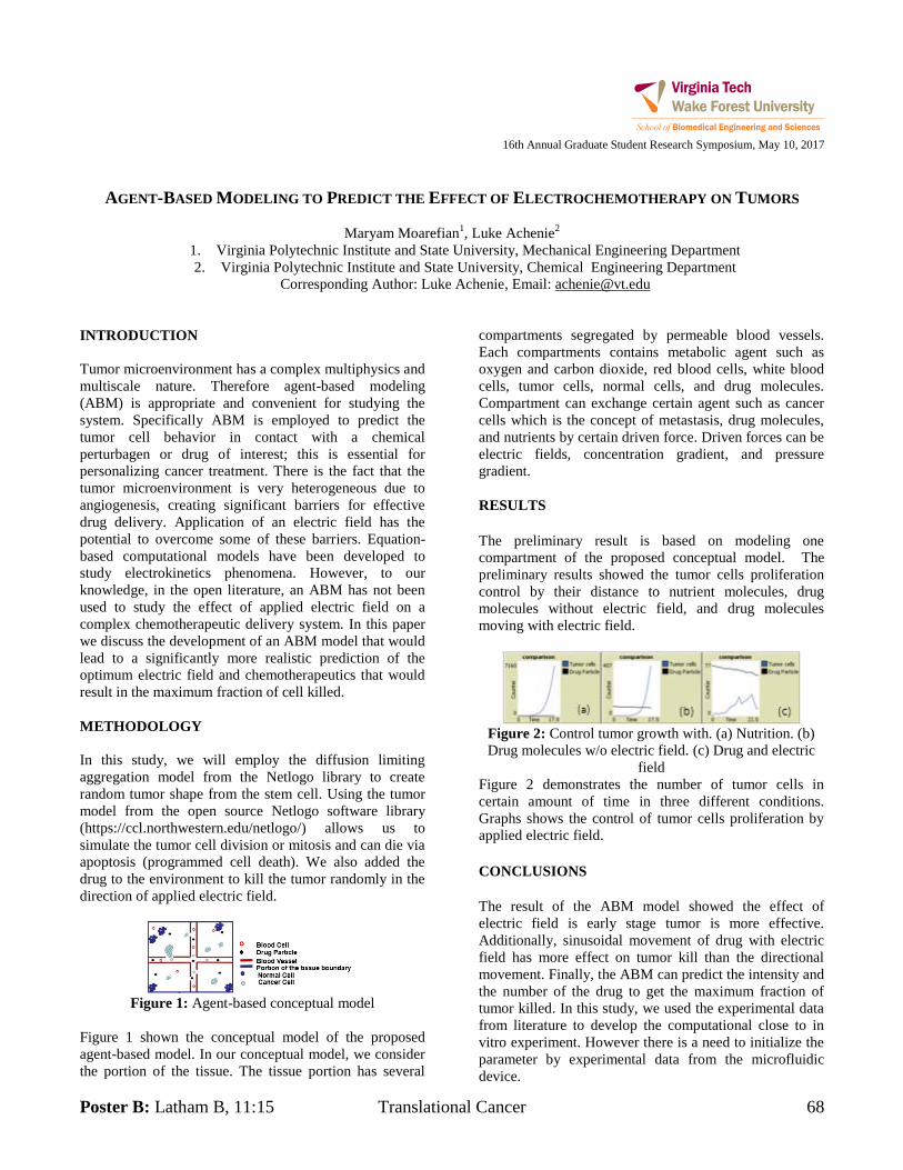
Poster B: Latham B, 11:15 Translational Cancer 68
16th Annual Graduate Student Research Symposium, May 10, 2017
AGENT-BASED MODELING TO PREDICT THE EFFECT OF ELECTROCHEMOTHERAPY ON TUMORS
Maryam Moarefian1, Luke Achenie
2
1. Virginia Polytechnic Institute and State University, Mechanical Engineering Department
2. Virginia Polytechnic Institute and State University, Chemical Engineering Department
Corresponding Author: Luke Achenie, Email: [email protected]
INTRODUCTION
Tumor microenvironment has a complex multiphysics and
multiscale nature. Therefore agent-based modeling
(ABM) is appropriate and convenient for studying the
system. Specifically ABM is employed to predict the
tumor cell behavior in contact with a chemical
perturbagen or drug of interest; this is essential for
personalizing cancer treatment. There is the fact that the
tumor microenvironment is very heterogeneous due to
angiogenesis, creating significant barriers for effective
drug delivery. Application of an electric field has the
potential to overcome some of these barriers. Equation-
based computational models have been developed to
study electrokinetics phenomena. However, to our
knowledge, in the open literature, an ABM has not been
used to study the effect of applied electric field on a
complex chemotherapeutic delivery system. In this paper
we discuss the development of an ABM model that would
lead to a significantly more realistic prediction of the
optimum electric field and chemotherapeutics that would
result in the maximum fraction of cell killed.
METHODOLOGY
In this study, we will employ the diffusion limiting
aggregation model from the Netlogo library to create
random tumor shape from the stem cell. Using the tumor
model from the open source Netlogo software library
(https://ccl.northwestern.edu/netlogo/) allows us to
simulate the tumor cell division or mitosis and can die via
apoptosis (programmed cell death). We also added the
drug to the environment to kill the tumor randomly in the
direction of applied electric field.
Figure 1: Agent-based conceptual model
Figure 1 shown the conceptual model of the proposed
agent-based model. In our conceptual model, we consider
the portion of the tissue. The tissue portion has several
compartments segregated by permeable blood vessels.
Each compartments contains metabolic agent such as
oxygen and carbon dioxide, red blood cells, white blood
cells, tumor cells, normal cells, and drug molecules.
Compartment can exchange certain agent such as cancer
cells which is the concept of metastasis, drug molecules,
and nutrients by certain driven force. Driven forces can be
electric fields, concentration gradient, and pressure
gradient.
RESULTS
The preliminary result is based on modeling one
compartment of the proposed conceptual model. The
preliminary results showed the tumor cells proliferation
control by their distance to nutrient molecules, drug
molecules without electric field, and drug molecules
moving with electric field.
Figure 2: Control tumor growth with. (a) Nutrition. (b)
Drug molecules w/o electric field. (c) Drug and electric
field
Figure 2 demonstrates the number of tumor cells in
certain amount of time in three different conditions.
Graphs shows the control of tumor cells proliferation by
applied electric field.
CONCLUSIONS
The result of the ABM model showed the effect of
electric field is early stage tumor is more effective.
Additionally, sinusoidal movement of drug with electric
field has more effect on tumor kill than the directional
movement. Finally, the ABM can predict the intensity and
the number of the drug to get the maximum fraction of
tumor killed. In this study, we used the experimental data
from literature to develop the computational close to in
vitro experiment. However there is a need to initialize the
parameter by experimental data from the microfluidic
device.
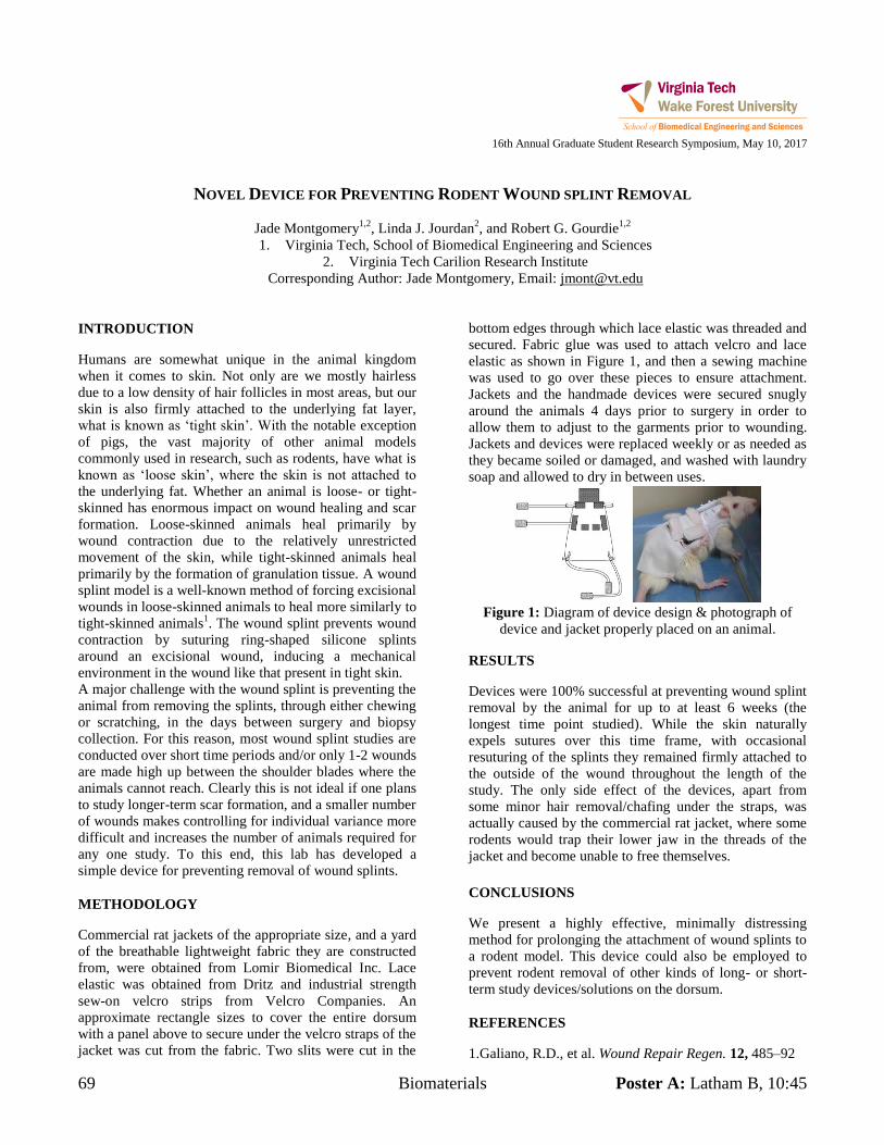
69 Biomaterials Poster A: Latham B, 10:45
16th Annual Graduate Student Research Symposium, May 10, 2017
NOVEL DEVICE FOR PREVENTING RODENT WOUND SPLINT REMOVAL
Jade Montgomery1,2
, Linda J. Jourdan2, and Robert G. Gourdie
1,2
1. Virginia Tech, School of Biomedical Engineering and Sciences
2. Virginia Tech Carilion Research Institute
Corresponding Author: Jade Montgomery, Email: [email protected]
INTRODUCTION
Humans are somewhat unique in the animal kingdom
when it comes to skin. Not only are we mostly hairless
due to a low density of hair follicles in most areas, but our
skin is also firmly attached to the underlying fat layer,
what is known as ‘tight skin’. With the notable exception
of pigs, the vast majority of other animal models
commonly used in research, such as rodents, have what is
known as ‘loose skin’, where the skin is not attached to
the underlying fat. Whether an animal is loose- or tight-
skinned has enormous impact on wound healing and scar
formation. Loose-skinned animals heal primarily by
wound contraction due to the relatively unrestricted
movement of the skin, while tight-skinned animals heal
primarily by the formation of granulation tissue. A wound
splint model is a well-known method of forcing excisional
wounds in loose-skinned animals to heal more similarly to
tight-skinned animals1. The wound splint prevents wound
contraction by suturing ring-shaped silicone splints
around an excisional wound, inducing a mechanical
environment in the wound like that present in tight skin.
A major challenge with the wound splint is preventing the
animal from removing the splints, through either chewing
or scratching, in the days between surgery and biopsy
collection. For this reason, most wound splint studies are
conducted over short time periods and/or only 1-2 wounds
are made high up between the shoulder blades where the
animals cannot reach. Clearly this is not ideal if one plans
to study longer-term scar formation, and a smaller number
of wounds makes controlling for individual variance more
difficult and increases the number of animals required for
any one study. To this end, this lab has developed a
simple device for preventing removal of wound splints.
METHODOLOGY
Commercial rat jackets of the appropriate size, and a yard
of the breathable lightweight fabric they are constructed
from, were obtained from Lomir Biomedical Inc. Lace
elastic was obtained from Dritz and industrial strength
sew-on velcro strips from Velcro Companies. An
approximate rectangle sizes to cover the entire dorsum
with a panel above to secure under the velcro straps of the
jacket was cut from the fabric. Two slits were cut in the
bottom edges through which lace elastic was threaded and
secured. Fabric glue was used to attach velcro and lace
elastic as shown in Figure 1, and then a sewing machine
was used to go over these pieces to ensure attachment.
Jackets and the handmade devices were secured snugly
around the animals 4 days prior to surgery in order to
allow them to adjust to the garments prior to wounding.
Jackets and devices were replaced weekly or as needed as
they became soiled or damaged, and washed with laundry
soap and allowed to dry in between uses.
Figure 1: Diagram of device design & photograph of
device and jacket properly placed on an animal.
RESULTS
Devices were 100% successful at preventing wound splint
removal by the animal for up to at least 6 weeks (the
longest time point studied). While the skin naturally
expels sutures over this time frame, with occasional
resuturing of the splints they remained firmly attached to
the outside of the wound throughout the length of the
study. The only side effect of the devices, apart from
some minor hair removal/chafing under the straps, was
actually caused by the commercial rat jacket, where some
rodents would trap their lower jaw in the threads of the
jacket and become unable to free themselves.
CONCLUSIONS
We present a highly effective, minimally distressing
method for prolonging the attachment of wound splints to
a rodent model. This device could also be employed to
prevent rodent removal of other kinds of long- or short-
term study devices/solutions on the dorsum.
REFERENCES
1.Galiano, R.D., et al. Wound Repair Regen. 12, 485–92
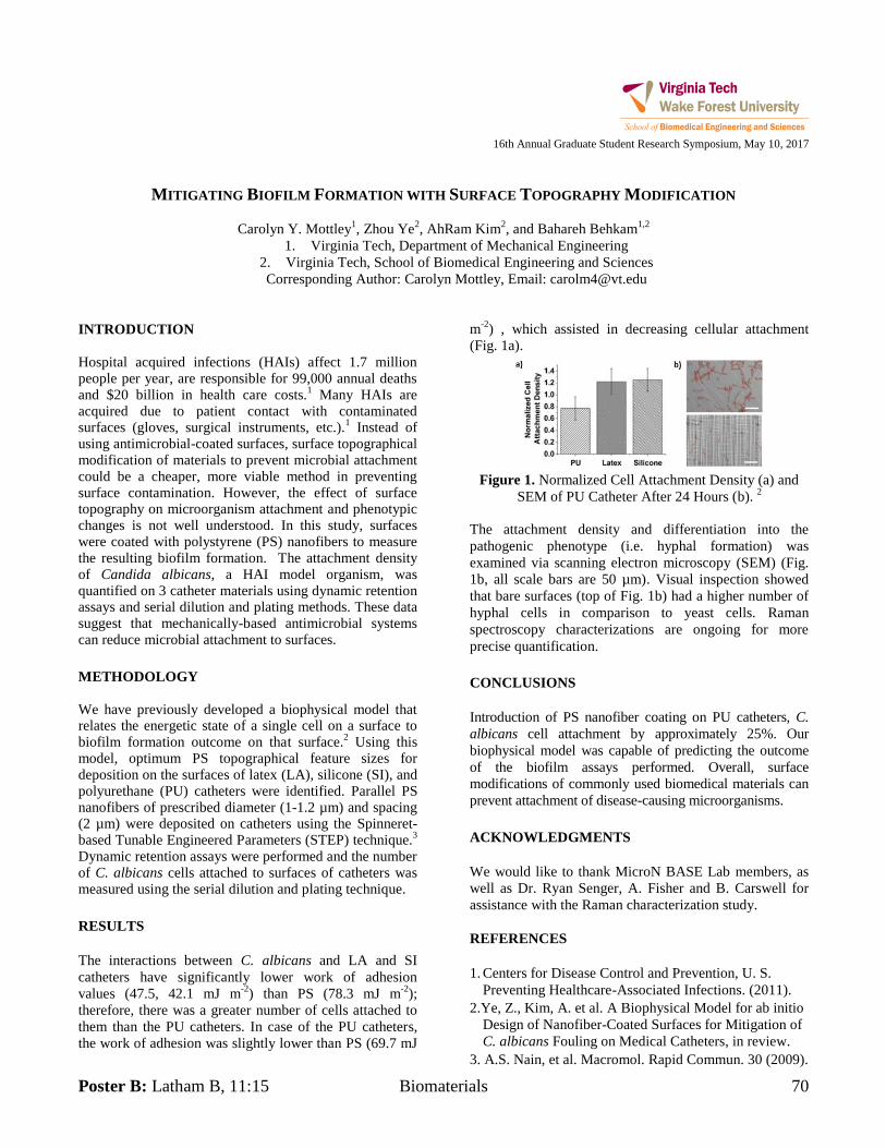
Poster B: Latham B, 11:15 Biomaterials 70
16th Annual Graduate Student Research Symposium, May 10, 2017
MITIGATING BIOFILM FORMATION WITH SURFACE TOPOGRAPHY MODIFICATION
Carolyn Y. Mottley1, Zhou Ye
2, AhRam Kim
2, and Bahareh Behkam
1,2
1. Virginia Tech, Department of Mechanical Engineering
2. Virginia Tech, School of Biomedical Engineering and Sciences
Corresponding Author: Carolyn Mottley, Email: [email protected]
INTRODUCTION
Hospital acquired infections (HAIs) affect 1.7 million people per year, are responsible for 99,000 annual deaths and $20 billion in health care costs.
1 Many HAIs are
acquired due to patient contact with contaminated surfaces (gloves, surgical instruments, etc.).
1 Instead of
using antimicrobial-coated surfaces, surface topographical modification of materials to prevent microbial attachment could be a cheaper, more viable method in preventing surface contamination. However, the effect of surface topography on microorganism attachment and phenotypic changes is not well understood. In this study, surfaces were coated with polystyrene (PS) nanofibers to measure the resulting biofilm formation. The attachment density of Candida albicans, a HAI model organism, was quantified on 3 catheter materials using dynamic retention assays and serial dilution and plating methods. These data suggest that mechanically-based antimicrobial systems can reduce microbial attachment to surfaces.
METHODOLOGY
We have previously developed a biophysical model that relates the energetic state of a single cell on a surface to biofilm formation outcome on that surface.
2 Using this
model, optimum PS topographical feature sizes for deposition on the surfaces of latex (LA), silicone (SI), and polyurethane (PU) catheters were identified. Parallel PS nanofibers of prescribed diameter (1-1.2 µm) and spacing (2 µm) were deposited on catheters using the Spinneret-based Tunable Engineered Parameters (STEP) technique.
3
Dynamic retention assays were performed and the number of C. albicans cells attached to surfaces of catheters was measured using the serial dilution and plating technique.
RESULTS
The interactions between C. albicans and LA and SI
catheters have significantly lower work of adhesion
values (47.5, 42.1 mJ m-2
) than PS (78.3 mJ m-2
);
therefore, there was a greater number of cells attached to
them than the PU catheters. In case of the PU catheters,
the work of adhesion was slightly lower than PS (69.7 mJ
m-2
) , which assisted in decreasing cellular attachment
(Fig. 1a).
Figure 1. Normalized Cell Attachment Density (a) and
SEM of PU Catheter After 24 Hours (b). 2
The attachment density and differentiation into the
pathogenic phenotype (i.e. hyphal formation) was
examined via scanning electron microscopy (SEM) (Fig.
1b, all scale bars are 50 µm). Visual inspection showed
that bare surfaces (top of Fig. 1b) had a higher number of
hyphal cells in comparison to yeast cells. Raman
spectroscopy characterizations are ongoing for more
precise quantification.
CONCLUSIONS
Introduction of PS nanofiber coating on PU catheters, C.
albicans cell attachment by approximately 25%. Our
biophysical model was capable of predicting the outcome
of the biofilm assays performed. Overall, surface
modifications of commonly used biomedical materials can
prevent attachment of disease-causing microorganisms.
ACKNOWLEDGMENTS
We would like to thank MicroN BASE Lab members, as
well as Dr. Ryan Senger, A. Fisher and B. Carswell for
assistance with the Raman characterization study.
REFERENCES
1. Centers for Disease Control and Prevention, U. S.
Preventing Healthcare-Associated Infections. (2011).
2.Ye, Z., Kim, A. et al. A Biophysical Model for ab initio
Design of Nanofiber-Coated Surfaces for Mitigation of
C. albicans Fouling on Medical Catheters, in review.
3. A.S. Nain, et al. Macromol. Rapid Commun. 30 (2009).
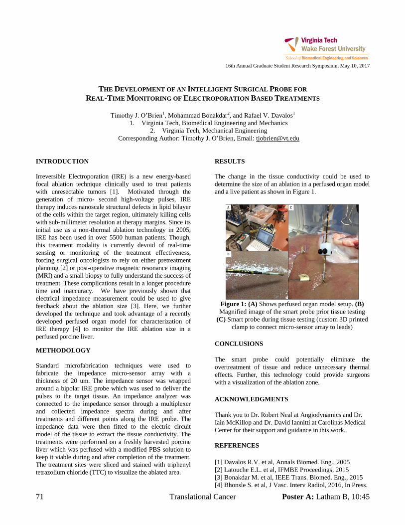
71 Translational Cancer Poster A: Latham B, 10:45
16th Annual Graduate Student Research Symposium, May 10, 2017
THE DEVELOPMENT OF AN INTELLIGENT SURGICAL PROBE FOR
REAL-TIME MONITORING OF ELECTROPORATION BASED TREATMENTS
Timothy J. O’Brien
1, Mohammad Bonakdar
2, and Rafael V. Davalos
1
1. Virginia Tech, Biomedical Engineering and Mechanics
2. Virginia Tech, Mechanical Engineering
Corresponding Author: Timothy J. O’Brien, Email: [email protected]
INTRODUCTION
Irreversible Electroporation (IRE) is a new energy-based
focal ablation technique clinically used to treat patients
with unresectable tumors [1]. Motivated through the
generation of micro- second high-voltage pulses, IRE
therapy induces nanoscale structural defects in lipid bilayer
of the cells within the target region, ultimately killing cells
with sub-millimeter resolution at therapy margins. Since its
initial use as a non-thermal ablation technology in 2005,
IRE has been used in over 5500 human patients. Though,
this treatment modality is currently devoid of real-time
sensing or monitoring of the treatment effectiveness,
forcing surgical oncologists to rely on either pretreatment
planning [2] or post-operative magnetic resonance imaging
(MRI) and a small biopsy to fully understand the success of
treatment. These complications result in a longer procedure
time and inaccuracy. We have previously shown that
electrical impedance measurement could be used to give
feedback about the ablation size [3]. Here, we further
developed the technique and took advantage of a recently
developed perfused organ model for characterization of
IRE therapy [4] to monitor the IRE ablation size in a
perfused porcine liver.
METHODOLOGY
Standard microfabrication techniques were used to
fabricate the impedance micro-sensor array with a
thickness of 20 um. The impedance sensor was wrapped
around a bipolar IRE probe which was used to deliver the
pulses to the target tissue. An impedance analyzer was
connected to the impedance sensor through a multiplexer
and collected impedance spectra during and after
treatments and different points along the IRE probe. The
impedance data were then fitted to the electric circuit
model of the tissue to extract the tissue conductivity. The
treatments were performed on a freshly harvested porcine
liver which was perfused with a modified PBS solution to
keep it viable during and after completion of the treatment.
The treatment sites were sliced and stained with triphenyl
tetrazolium chloride (TTC) to visualize the ablated area.
RESULTS
The change in the tissue conductivity could be used to
determine the size of an ablation in a perfused organ model
and a live patient as shown in Figure 1.
Figure 1: (A) Shows perfused organ model setup. (B)
Magnified image of the smart probe prior tissue testing
(C) Smart probe during tissue testing (custom 3D printed
clamp to connect micro-sensor array to leads)
CONCLUSIONS
The smart probe could potentially eliminate the
overtreatment of tissue and reduce unnecessary thermal
effects. Further, this technology could provide surgeons
with a visualization of the ablation zone.
ACKNOWLEDGMENTS
Thank you to Dr. Robert Neal at Angiodynamics and Dr.
Iain McKillop and Dr. David Iannitti at Carolinas Medical
Center for their support and guidance in this work.
REFERENCES
[1] Davalos R.V. et al, Annals Biomed. Eng., 2005
[2] Latouche E.L. et al, IFMBE Proceedings, 2015
[3] Bonakdar M. et al, IEEE Trans. Biomed. Eng., 2015
[4] Bhonsle S. et al, J Vasc. Interv Radiol, 2016, In Press.
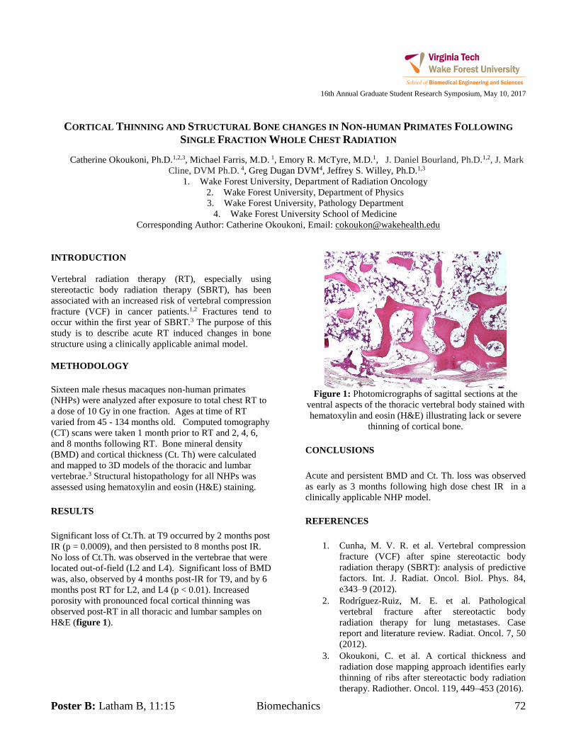
Poster B: Latham B, 11:15 Biomechanics 72
16th Annual Graduate Student Research Symposium, May 10, 2017
CORTICAL THINNING AND STRUCTURAL BONE CHANGES IN NON-HUMAN PRIMATES FOLLOWING
SINGLE FRACTION WHOLE CHEST RADIATION
Catherine Okoukoni, Ph.D.1,2,3, Michael Farris, M.D. 1, Emory R. McTyre, M.D.1, J. Daniel Bourland, Ph.D.1,2, J. Mark Cline, DVM Ph.D. 4, Greg Dugan DVM4, Jeffrey S. Willey, Ph.D.1,3
1. Wake Forest University, Department of Radiation Oncology 2. Wake Forest University, Department of Physics 3. Wake Forest University, Pathology Department
4. Wake Forest University School of Medicine Corresponding Author: Catherine Okoukoni, Email: [email protected]
INTRODUCTION Vertebral radiation therapy (RT), especially using stereotactic body radiation therapy (SBRT), has been associated with an increased risk of vertebral compression fracture (VCF) in cancer patients.1,2 Fractures tend to occur within the first year of SBRT.3 The purpose of this study is to describe acute RT induced changes in bone structure using a clinically applicable animal model. METHODOLOGY Sixteen male rhesus macaques non-human primates (NHPs) were analyzed after exposure to total chest RT to a dose of 10 Gy in one fraction. Ages at time of RT varied from 45 - 134 months old. Computed tomography (CT) scans were taken 1 month prior to RT and 2, 4, 6, and 8 months following RT. Bone mineral density (BMD) and cortical thickness (Ct. Th) were calculated and mapped to 3D models of the thoracic and lumbar vertebrae.3 Structural histopathology for all NHPs was assessed using hematoxylin and eosin (H&E) staining. RESULTS Significant loss of Ct.Th. at T9 occurred by 2 months post IR (p = 0.0009), and then persisted to 8 months post IR. No loss of Ct.Th. was observed in the vertebrae that were located out-of-field (L2 and L4). Significant loss of BMD was, also, observed by 4 months post-IR for T9, and by 6 months post RT for L2, and L4 (p < 0.01). Increased porosity with pronounced focal cortical thinning was observed post-RT in all thoracic and lumbar samples on H&E (figure 1).
Figure 1: Photomicrographs of sagittal sections at the
ventral aspects of the thoracic vertebral body stained with hematoxylin and eosin (H&E) illustrating lack or severe
thinning of cortical bone. CONCLUSIONS Acute and persistent BMD and Ct. Th. loss was observed as early as 3 months following high dose chest IR in a clinically applicable NHP model. REFERENCES
1. Cunha, M. V. R. et al. Vertebral compression fracture (VCF) after spine stereotactic body radiation therapy (SBRT): analysis of predictive factors. Int. J. Radiat. Oncol. Biol. Phys. 84, e343–9 (2012).
2. Rodríguez-Ruiz, M. E. et al. Pathological vertebral fracture after stereotactic body radiation therapy for lung metastases. Case report and literature review. Radiat. Oncol. 7, 50 (2012).
3. Okoukoni, C. et al. A cortical thickness and radiation dose mapping approach identifies early thinning of ribs after stereotactic body radiation therapy. Radiother. Oncol. 119, 449–453 (2016).
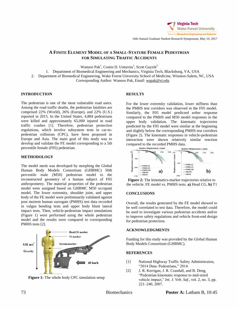
73 Biomechanics Poster A: Latham B, 10:45
16th Annual Graduate Student Research Symposium, May 10, 2017
A FINITE ELEMENT MODEL OF A SMALL-STATURE FEMALE PEDESTRIAN
FOR SIMULATING TRAFFIC ACCIDENTS
Wansoo Pak1, Costin D. Untaroiu
1, Scott Gayzik
2
1. Department of Biomedical Engineering and Mechanics, Virginia Tech, Blacksburg, VA, USA
2. Department of Biomedical Engineering, Wake Forest University School of Medicine, Winston-Salem, NC, USA
Corresponding Author: Wansoo Pak, Email: [email protected]
INTRODUCTION
The pedestrian is one of the most vulnerable road users.
Among the road traffic deaths, the pedestrian fatalities are
comprised 22% (World), 26% (Europe), and 22% (U.S.)
reported in 2015. In the United States, 4,884 pedestrians
were killed and approximately 65,000 injured in road
traffic crashes [1]. Therefore, pedestrian protection
regulations, which involve subsystem tests in car-to-
pedestrian collisions (CPC), have been proposed in
Europe and Asia. The main goal of this study was to
develop and validate the FE model corresponding to a 5th
percentile female (F05) pedestrian.
METHODOLOGY
The model mesh was developed by morphing the Global
Human Body Models Consortium (GHBMC) 50th
percentile male (M50) pedestrian model to the
reconstructed geometry of a human subject of F05
anthropometry. The material properties of the pedestrian
model were assigned based on GHBMC M50 occupant
model. The lower extremity, shoulder joint, and upper
body of the FE model were preliminarily validated against
post mortem human surrogate (PMHS) test data recorded
in valgus bending tests and upper body blunt lateral
impact tests. Then, vehicle-pedestrian impact simulations
(Figure 1) were performed using the whole pedestrian
model and the results were compared to corresponding
PMHS tests [2].
Figure 1: The whole body CPC simulation setup
RESULTS
For the lower extremity validation, lower stiffness than
the PMHS test corridors was observed in the F05 model.
Similarly, the F05 model predicted softer response
compared to the PMHS and M50 model responses in the
upper body validation. The kinematic trajectories
predicted by the F05 model were similar at the beginning
and slightly below the corresponding PMHS test corridors
(Figure 2). The kinematic responses in vehicle-pedestrian
interaction were shown relatively similar reaction
compared to the recorded PMHS data.
Figure 2: The kinematics-marker trajectories relative to
the vehicle: FE model vs. PMHS tests. a) Head CG, b) T1
CONCLUSIONS
Overall, the results generated by the FE model showed to
be well correlated to test data. Therefore, the model could
be used to investigate various pedestrian accidents and/or
to improve safety regulations and vehicle front-end design
for pedestrian protection.
ACKNOWLEDGMENTS
Funding for this study was provided by the Global Human
Body Models Consortium (GHBMC).
REFERENCES
[1] National Highway Traffic Safety Administraion,
“2014 Data: Pedestrians,” 2014.
[2] J. R. Kerrigan, J. R. Crandall, and B. Deng,
“Pedestrian kinematic response to mid-sized
vehicle impact,” Int. J. Veh. Saf., vol. 2, no. 3, pp.
221–240, 2007.
a) b)
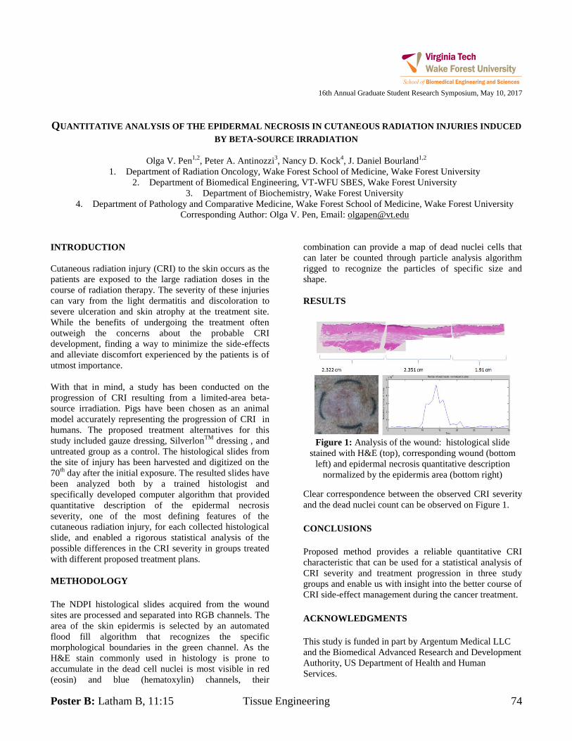
Poster B: Latham B, 11:15 Tissue Engineering 74
16th Annual Graduate Student Research Symposium, May 10, 2017
QUANTITATIVE ANALYSIS OF THE EPIDERMAL NECROSIS IN CUTANEOUS RADIATION INJURIES INDUCED
BY BETA-SOURCE IRRADIATION
Olga V. Pen1,2
, Peter A. Antinozzi3, Nancy D. Kock
4, J. Daniel Bourland
1,2
1. Department of Radiation Oncology, Wake Forest School of Medicine, Wake Forest University
2. Department of Biomedical Engineering, VT-WFU SBES, Wake Forest University
3. Department of Biochemistry, Wake Forest University
4. Department of Pathology and Comparative Medicine, Wake Forest School of Medicine, Wake Forest University
Corresponding Author: Olga V. Pen, Email: [email protected]
INTRODUCTION
Cutaneous radiation injury (CRI) to the skin occurs as the
patients are exposed to the large radiation doses in the
course of radiation therapy. The severity of these injuries
can vary from the light dermatitis and discoloration to
severe ulceration and skin atrophy at the treatment site.
While the benefits of undergoing the treatment often
outweigh the concerns about the probable CRI
development, finding a way to minimize the side-effects
and alleviate discomfort experienced by the patients is of
utmost importance.
With that in mind, a study has been conducted on the
progression of CRI resulting from a limited-area beta-
source irradiation. Pigs have been chosen as an animal
model accurately representing the progression of CRI in
humans. The proposed treatment alternatives for this
study included gauze dressing, SilverlonTM
dressing , and
untreated group as a control. The histological slides from
the site of injury has been harvested and digitized on the
70th
day after the initial exposure. The resulted slides have
been analyzed both by a trained histologist and
specifically developed computer algorithm that provided
quantitative description of the epidermal necrosis
severity, one of the most defining features of the
cutaneous radiation injury, for each collected histological
slide, and enabled a rigorous statistical analysis of the
possible differences in the CRI severity in groups treated
with different proposed treatment plans.
METHODOLOGY
The NDPI histological slides acquired from the wound
sites are processed and separated into RGB channels. The
area of the skin epidermis is selected by an automated
flood fill algorithm that recognizes the specific
morphological boundaries in the green channel. As the
H&E stain commonly used in histology is prone to
accumulate in the dead cell nuclei is most visible in red
(eosin) and blue (hematoxylin) channels, their
combination can provide a map of dead nuclei cells that
can later be counted through particle analysis algorithm
rigged to recognize the particles of specific size and
shape.
RESULTS
Figure 1: Analysis of the wound: histological slide
stained with H&E (top), corresponding wound (bottom
left) and epidermal necrosis quantitative description
normalized by the epidermis area (bottom right)
Clear correspondence between the observed CRI severity
and the dead nuclei count can be observed on Figure 1.
CONCLUSIONS
Proposed method provides a reliable quantitative CRI
characteristic that can be used for a statistical analysis of
CRI severity and treatment progression in three study
groups and enable us with insight into the better course of
CRI side-effect management during the cancer treatment.
ACKNOWLEDGMENTS
This study is funded in part by Argentum Medical LLC
and the Biomedical Advanced Research and Development
Authority, US Department of Health and Human
Services.
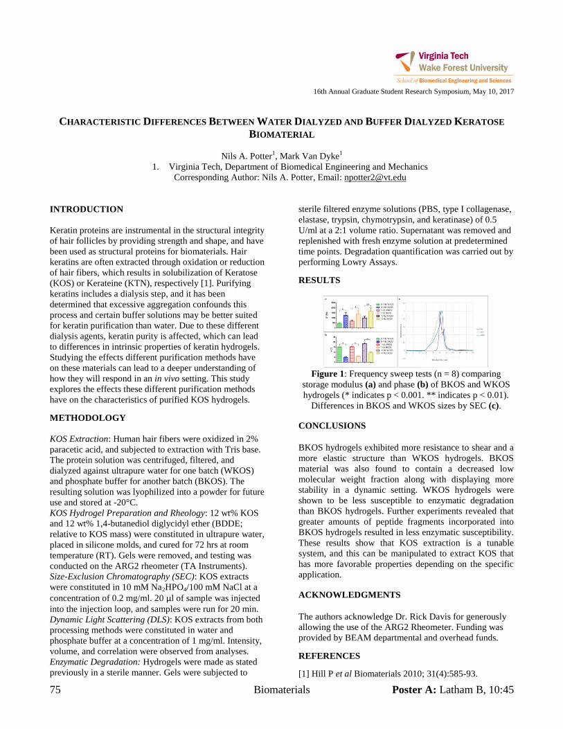
75 Biomaterials Poster A: Latham B, 10:45
16th Annual Graduate Student Research Symposium, May 10, 2017
CHARACTERISTIC DIFFERENCES BETWEEN WATER DIALYZED AND BUFFER DIALYZED KERATOSE
BIOMATERIAL
Nils A. Potter1, Mark Van Dyke
1
1. Virginia Tech, Department of Biomedical Engineering and Mechanics
Corresponding Author: Nils A. Potter, Email: [email protected]
INTRODUCTION
Keratin proteins are instrumental in the structural integrity
of hair follicles by providing strength and shape, and have
been used as structural proteins for biomaterials. Hair
keratins are often extracted through oxidation or reduction
of hair fibers, which results in solubilization of Keratose
(KOS) or Kerateine (KTN), respectively [1]. Purifying
keratins includes a dialysis step, and it has been
determined that excessive aggregation confounds this
process and certain buffer solutions may be better suited
for keratin purification than water. Due to these different
dialysis agents, keratin purity is affected, which can lead
to differences in intrinsic properties of keratin hydrogels.
Studying the effects different purification methods have
on these materials can lead to a deeper understanding of
how they will respond in an in vivo setting. This study
explores the effects these different purification methods
have on the characteristics of purified KOS hydrogels.
METHODOLOGY
KOS Extraction: Human hair fibers were oxidized in 2%
paracetic acid, and subjected to extraction with Tris base.
The protein solution was centrifuged, filtered, and
dialyzed against ultrapure water for one batch (WKOS)
and phosphate buffer for another batch (BKOS). The
resulting solution was lyophilized into a powder for future
use and stored at -20°C.
KOS Hydrogel Preparation and Rheology: 12 wt% KOS
and 12 wt% 1,4-butanediol diglycidyl ether (BDDE;
relative to KOS mass) were constituted in ultrapure water,
placed in silicone molds, and cured for 72 hrs at room
temperature (RT). Gels were removed, and testing was
conducted on the ARG2 rheometer (TA Instruments).
Size-Exclusion Chromatography (SEC): KOS extracts
were constituted in 10 mM Na2HPO4/100 mM NaCl at a
concentration of 0.2 mg/ml. 20 µl of sample was injected
into the injection loop, and samples were run for 20 min.
Dynamic Light Scattering (DLS): KOS extracts from both
processing methods were constituted in water and
phosphate buffer at a concentration of 1 mg/ml. Intensity,
volume, and correlation were observed from analyses.
Enzymatic Degradation: Hydrogels were made as stated
previously in a sterile manner. Gels were subjected to
sterile filtered enzyme solutions (PBS, type I collagenase,
elastase, trypsin, chymotrypsin, and keratinase) of 0.5
U/ml at a 2:1 volume ratio. Supernatant was removed and
replenished with fresh enzyme solution at predetermined
time points. Degradation quantification was carried out by
performing Lowry Assays.
RESULTS
Figure 1: Frequency sweep tests (n = 8) comparing
storage modulus (a) and phase (b) of BKOS and WKOS
hydrogels (* indicates p < 0.001. ** indicates p < 0.01).
Differences in BKOS and WKOS sizes by SEC (c).
CONCLUSIONS
BKOS hydrogels exhibited more resistance to shear and a
more elastic structure than WKOS hydrogels. BKOS
material was also found to contain a decreased low
molecular weight fraction along with displaying more
stability in a dynamic setting. WKOS hydrogels were
shown to be less susceptible to enzymatic degradation
than BKOS hydrogels. Further experiments revealed that
greater amounts of peptide fragments incorporated into
BKOS hydrogels resulted in less enzymatic susceptibility.
These results show that KOS extraction is a tunable
system, and this can be manipulated to extract KOS that
has more favorable properties depending on the specific
application.
ACKNOWLEDGMENTS
The authors acknowledge Dr. Rick Davis for generously
allowing the use of the ARG2 Rheometer. Funding was
provided by BEAM departmental and overhead funds.
REFERENCES
[1] Hill P et al Biomaterials 2010; 31(4):585-93.
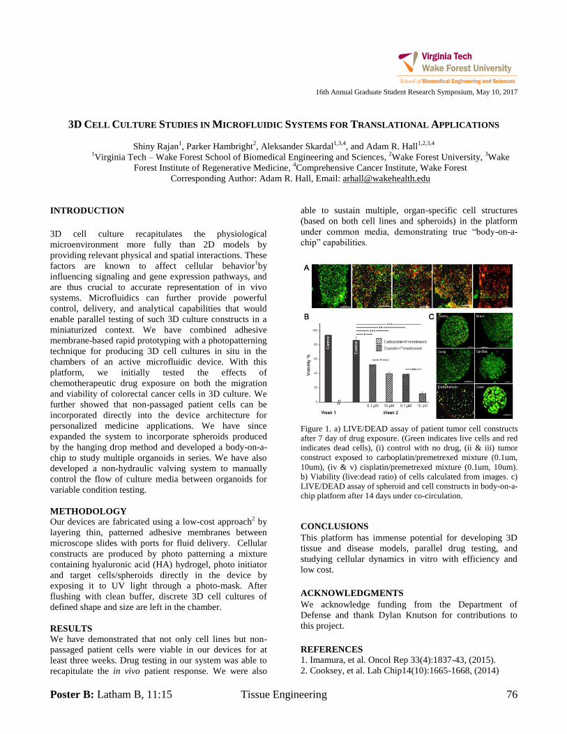
Poster B: Latham B, 11:15 Tissue Engineering 76
16th Annual Graduate Student Research Symposium, May 10, 2017
3D CELL CULTURE STUDIES IN MICROFLUIDIC SYSTEMS FOR TRANSLATIONAL APPLICATIONS
Shiny Rajan1, Parker Hambright
2, Aleksander Skardal
1,3,4, and Adam R. Hall
1,2,3,4
1Virginia Tech – Wake Forest School of Biomedical Engineering and Sciences,
2Wake Forest University,
3Wake
Forest Institute of Regenerative Medicine, 4Comprehensive Cancer Institute, Wake Forest
Corresponding Author: Adam R. Hall, Email: [email protected]
INTRODUCTION
3D cell culture recapitulates the physiological
microenvironment more fully than 2D models by
providing relevant physical and spatial interactions. These
factors are known to affect cellular behavior1by
influencing signaling and gene expression pathways, and
are thus crucial to accurate representation of in vivo
systems. Microfluidics can further provide powerful
control, delivery, and analytical capabilities that would
enable parallel testing of such 3D culture constructs in a
miniaturized context. We have combined adhesive
membrane-based rapid prototyping with a photopatterning
technique for producing 3D cell cultures in situ in the
chambers of an active microfluidic device. With this
platform, we initially tested the effects of
chemotherapeutic drug exposure on both the migration
and viability of colorectal cancer cells in 3D culture. We
further showed that non-passaged patient cells can be
incorporated directly into the device architecture for
personalized medicine applications. We have since
expanded the system to incorporate spheroids produced
by the hanging drop method and developed a body-on-a-
chip to study multiple organoids in series. We have also
developed a non-hydraulic valving system to manually
control the flow of culture media between organoids for
variable condition testing.
METHODOLOGY
Our devices are fabricated using a low-cost approach2 by
layering thin, patterned adhesive membranes between
microscope slides with ports for fluid delivery. Cellular
constructs are produced by photo patterning a mixture
containing hyaluronic acid (HA) hydrogel, photo initiator
and target cells/spheroids directly in the device by
exposing it to UV light through a photo-mask. After
flushing with clean buffer, discrete 3D cell cultures of
defined shape and size are left in the chamber.
RESULTS
We have demonstrated that not only cell lines but non-
passaged patient cells were viable in our devices for at
least three weeks. Drug testing in our system was able to
recapitulate the in vivo patient response. We were also
able to sustain multiple, organ-specific cell structures
(based on both cell lines and spheroids) in the platform
under common media, demonstrating true “body-on-a-
chip” capabilities.
Figure 1. a) LIVE/DEAD assay of patient tumor cell constructs
after 7 day of drug exposure. (Green indicates live cells and red
indicates dead cells), (i) control with no drug, (ii & iii) tumor
construct exposed to carboplatin/premetrexed mixture (0.1um,
10um), (iv & v) cisplatin/premetrexed mixture (0.1um, 10um).
b) Viability (live:dead ratio) of cells calculated from images. c)
LIVE/DEAD assay of spheroid and cell constructs in body-on-a-
chip platform after 14 days under co-circulation.
CONCLUSIONS
This platform has immense potential for developing 3D
tissue and disease models, parallel drug testing, and
studying cellular dynamics in vitro with efficiency and
low cost.
ACKNOWLEDGMENTS
We acknowledge funding from the Department of
Defense and thank Dylan Knutson for contributions to
this project.
REFERENCES
1. Imamura, et al. Oncol Rep 33(4):1837-43, (2015).
2. Cooksey, et al. Lab Chip14(10):1665-1668, (2014)
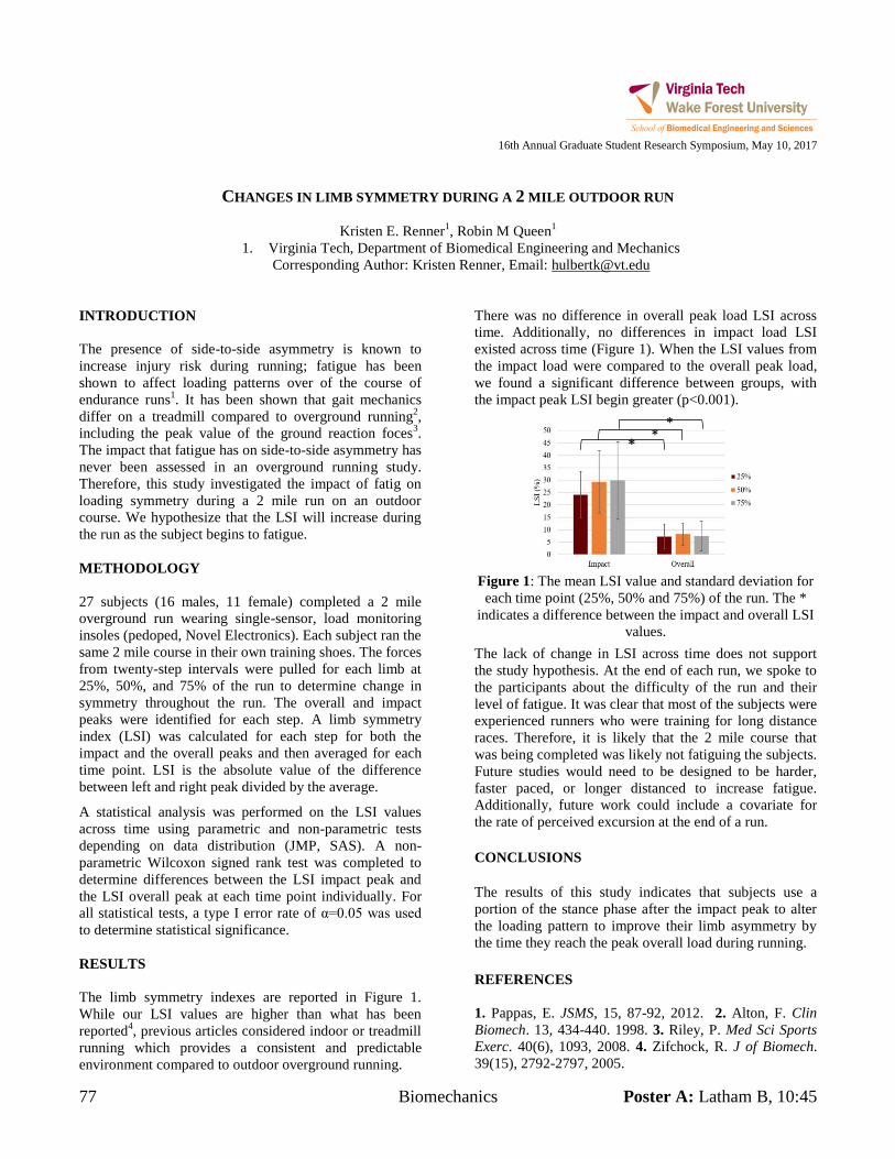
77 Biomechanics Poster A: Latham B, 10:45
16th Annual Graduate Student Research Symposium, May 10, 2017
CHANGES IN LIMB SYMMETRY DURING A 2 MILE OUTDOOR RUN
Kristen E. Renner1, Robin M Queen
1
1. Virginia Tech, Department of Biomedical Engineering and Mechanics
Corresponding Author: Kristen Renner, Email: [email protected]
INTRODUCTION
The presence of side-to-side asymmetry is known to
increase injury risk during running; fatigue has been
shown to affect loading patterns over of the course of
endurance runs1. It has been shown that gait mechanics
differ on a treadmill compared to overground running2,
including the peak value of the ground reaction foces3.
The impact that fatigue has on side-to-side asymmetry has
never been assessed in an overground running study.
Therefore, this study investigated the impact of fatig on
loading symmetry during a 2 mile run on an outdoor
course. We hypothesize that the LSI will increase during
the run as the subject begins to fatigue.
METHODOLOGY
27 subjects (16 males, 11 female) completed a 2 mile
overground run wearing single-sensor, load monitoring
insoles (pedoped, Novel Electronics). Each subject ran the
same 2 mile course in their own training shoes. The forces
from twenty-step intervals were pulled for each limb at
25%, 50%, and 75% of the run to determine change in
symmetry throughout the run. The overall and impact
peaks were identified for each step. A limb symmetry
index (LSI) was calculated for each step for both the
impact and the overall peaks and then averaged for each
time point. LSI is the absolute value of the difference
between left and right peak divided by the average.
A statistical analysis was performed on the LSI values
across time using parametric and non-parametric tests
depending on data distribution (JMP, SAS). A non-
parametric Wilcoxon signed rank test was completed to
determine differences between the LSI impact peak and
the LSI overall peak at each time point individually. For
all statistical tests, a type I error rate of α=0.05 was used
to determine statistical significance.
RESULTS
The limb symmetry indexes are reported in Figure 1.
While our LSI values are higher than what has been
reported4, previous articles considered indoor or treadmill
running which provides a consistent and predictable
environment compared to outdoor overground running.
There was no difference in overall peak load LSI across
time. Additionally, no differences in impact load LSI
existed across time (Figure 1). When the LSI values from
the impact load were compared to the overall peak load,
we found a significant difference between groups, with
the impact peak LSI begin greater (p<0.001).
Figure 1: The mean LSI value and standard deviation for
each time point (25%, 50% and 75%) of the run. The *
indicates a difference between the impact and overall LSI
values.
The lack of change in LSI across time does not support
the study hypothesis. At the end of each run, we spoke to
the participants about the difficulty of the run and their
level of fatigue. It was clear that most of the subjects were
experienced runners who were training for long distance
races. Therefore, it is likely that the 2 mile course that
was being completed was likely not fatiguing the subjects.
Future studies would need to be designed to be harder,
faster paced, or longer distanced to increase fatigue.
Additionally, future work could include a covariate for
the rate of perceived excursion at the end of a run.
CONCLUSIONS
The results of this study indicates that subjects use a
portion of the stance phase after the impact peak to alter
the loading pattern to improve their limb asymmetry by
the time they reach the peak overall load during running.
REFERENCES
1. Pappas, E. JSMS, 15, 87-92, 2012. 2. Alton, F. Clin
Biomech. 13, 434-440. 1998. 3. Riley, P. Med Sci Sports
Exerc. 40(6), 1093, 2008. 4. Zifchock, R. J of Biomech.
39(15), 2792-2797, 2005.
* * *
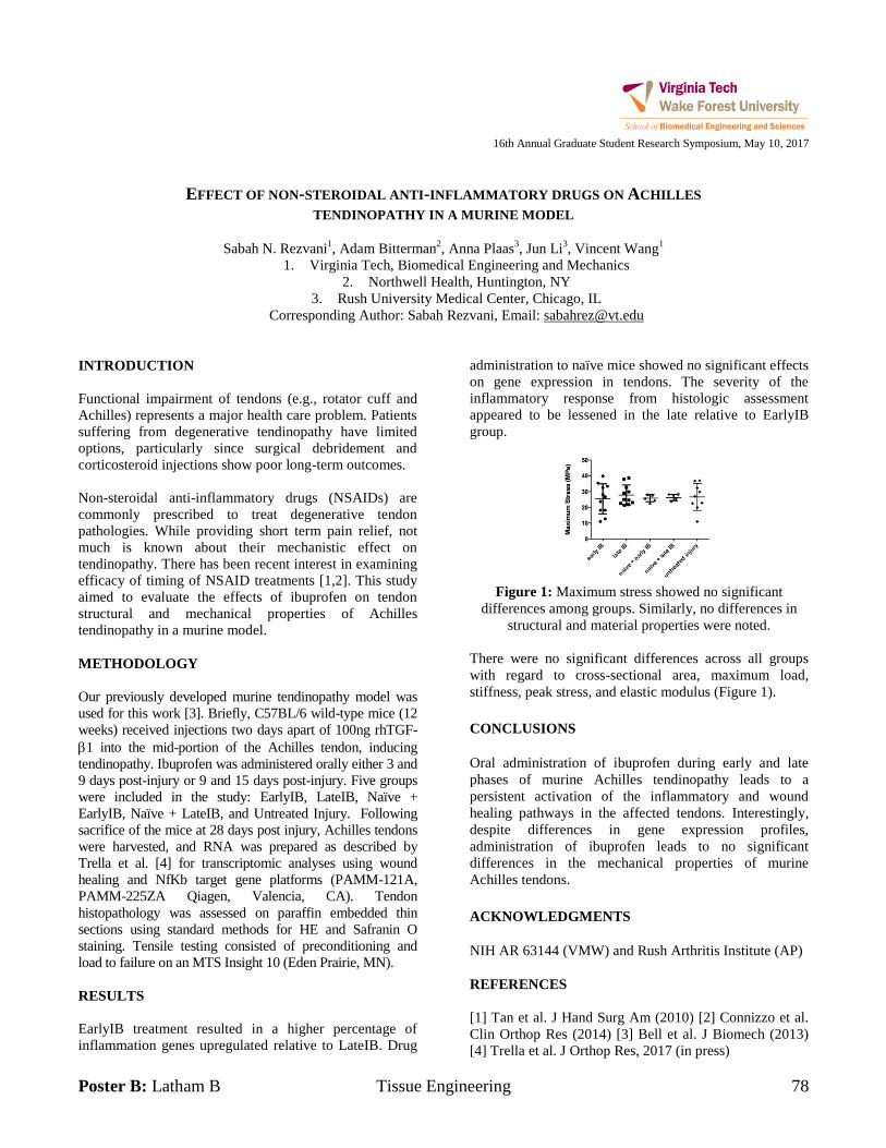
Poster B: Latham B Tissue Engineering 78
16th Annual Graduate Student Research Symposium, May 10, 2017
EFFECT OF NON-STEROIDAL ANTI-INFLAMMATORY DRUGS ON ACHILLES
TENDINOPATHY IN A MURINE MODEL
Sabah N. Rezvani1, Adam Bitterman
2, Anna Plaas
3, Jun Li
3, Vincent Wang
1
1. Virginia Tech, Biomedical Engineering and Mechanics
2. Northwell Health, Huntington, NY
3. Rush University Medical Center, Chicago, IL
Corresponding Author: Sabah Rezvani, Email: [email protected]
INTRODUCTION
Functional impairment of tendons (e.g., rotator cuff and
Achilles) represents a major health care problem. Patients
suffering from degenerative tendinopathy have limited
options, particularly since surgical debridement and
corticosteroid injections show poor long-term outcomes.
Non-steroidal anti-inflammatory drugs (NSAIDs) are
commonly prescribed to treat degenerative tendon
pathologies. While providing short term pain relief, not
much is known about their mechanistic effect on
tendinopathy. There has been recent interest in examining
efficacy of timing of NSAID treatments [1,2]. This study
aimed to evaluate the effects of ibuprofen on tendon
structural and mechanical properties of Achilles
tendinopathy in a murine model.
METHODOLOGY
Our previously developed murine tendinopathy model was
used for this work [3]. Briefly, C57BL/6 wild-type mice (12
weeks) received injections two days apart of 100ng rhTGF-
1 into the mid-portion of the Achilles tendon, inducing
tendinopathy. Ibuprofen was administered orally either 3 and
9 days post-injury or 9 and 15 days post-injury. Five groups
were included in the study: EarlyIB, LateIB, Naïve +
EarlyIB, Naïve + LateIB, and Untreated Injury. Following
sacrifice of the mice at 28 days post injury, Achilles tendons
were harvested, and RNA was prepared as described by
Trella et al. [4] for transcriptomic analyses using wound
healing and NfKb target gene platforms (PAMM-121A,
PAMM-225ZA Qiagen, Valencia, CA). Tendon
histopathology was assessed on paraffin embedded thin
sections using standard methods for HE and Safranin O
staining. Tensile testing consisted of preconditioning and
load to failure on an MTS Insight 10 (Eden Prairie, MN).
RESULTS
EarlyIB treatment resulted in a higher percentage of
inflammation genes upregulated relative to LateIB. Drug
administration to naïve mice showed no significant effects
on gene expression in tendons. The severity of the
inflammatory response from histologic assessment
appeared to be lessened in the late relative to EarlyIB
group.
Figure 1: Maximum stress showed no significant
differences among groups. Similarly, no differences in
structural and material properties were noted.
There were no significant differences across all groups
with regard to cross-sectional area, maximum load,
stiffness, peak stress, and elastic modulus (Figure 1).
CONCLUSIONS
Oral administration of ibuprofen during early and late
phases of murine Achilles tendinopathy leads to a
persistent activation of the inflammatory and wound
healing pathways in the affected tendons. Interestingly,
despite differences in gene expression profiles,
administration of ibuprofen leads to no significant
differences in the mechanical properties of murine
Achilles tendons.
ACKNOWLEDGMENTS
NIH AR 63144 (VMW) and Rush Arthritis Institute (AP)
REFERENCES
[1] Tan et al. J Hand Surg Am (2010) [2] Connizzo et al.
Clin Orthop Res (2014) [3] Bell et al. J Biomech (2013)
[4] Trella et al. J Orthop Res, 2017 (in press)
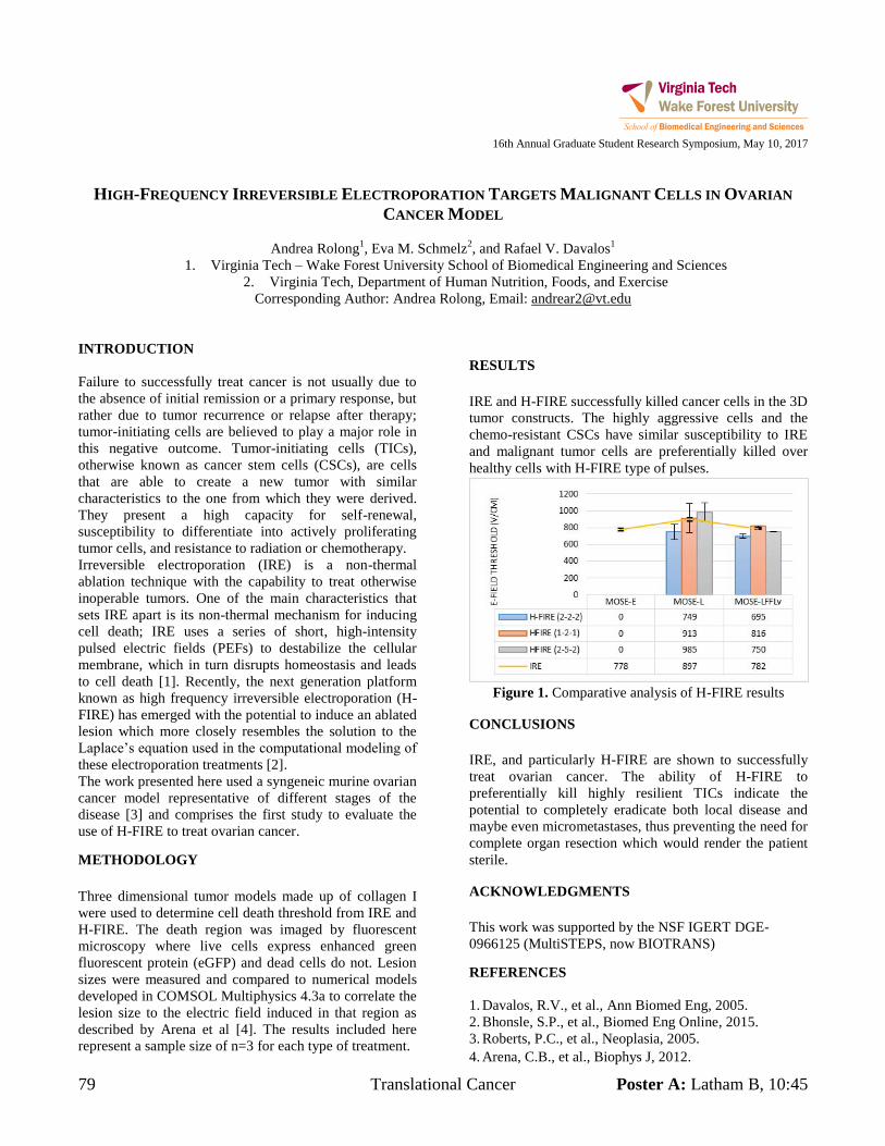
79 Translational Cancer Poster A: Latham B, 10:45
16th Annual Graduate Student Research Symposium, May 10, 2017
HIGH-FREQUENCY IRREVERSIBLE ELECTROPORATION TARGETS MALIGNANT CELLS IN OVARIAN
CANCER MODEL
Andrea Rolong1, Eva M. Schmelz
2, and Rafael V. Davalos
1
1. Virginia Tech – Wake Forest University School of Biomedical Engineering and Sciences
2. Virginia Tech, Department of Human Nutrition, Foods, and Exercise
Corresponding Author: Andrea Rolong, Email: [email protected]
INTRODUCTION
Failure to successfully treat cancer is not usually due to
the absence of initial remission or a primary response, but
rather due to tumor recurrence or relapse after therapy;
tumor-initiating cells are believed to play a major role in
this negative outcome. Tumor-initiating cells (TICs),
otherwise known as cancer stem cells (CSCs), are cells
that are able to create a new tumor with similar
characteristics to the one from which they were derived.
They present a high capacity for self-renewal,
susceptibility to differentiate into actively proliferating
tumor cells, and resistance to radiation or chemotherapy.
Irreversible electroporation (IRE) is a non-thermal
ablation technique with the capability to treat otherwise
inoperable tumors. One of the main characteristics that
sets IRE apart is its non-thermal mechanism for inducing
cell death; IRE uses a series of short, high-intensity
pulsed electric fields (PEFs) to destabilize the cellular
membrane, which in turn disrupts homeostasis and leads
to cell death [1]. Recently, the next generation platform
known as high frequency irreversible electroporation (H-
FIRE) has emerged with the potential to induce an ablated
lesion which more closely resembles the solution to the
Laplace’s equation used in the computational modeling of
these electroporation treatments [2].
The work presented here used a syngeneic murine ovarian
cancer model representative of different stages of the
disease [3] and comprises the first study to evaluate the
use of H-FIRE to treat ovarian cancer.
METHODOLOGY
Three dimensional tumor models made up of collagen I
were used to determine cell death threshold from IRE and
H-FIRE. The death region was imaged by fluorescent
microscopy where live cells express enhanced green
fluorescent protein (eGFP) and dead cells do not. Lesion
sizes were measured and compared to numerical models
developed in COMSOL Multiphysics 4.3a to correlate the
lesion size to the electric field induced in that region as
described by Arena et al [4]. The results included here
represent a sample size of n=3 for each type of treatment.
RESULTS
IRE and H-FIRE successfully killed cancer cells in the 3D
tumor constructs. The highly aggressive cells and the
chemo-resistant CSCs have similar susceptibility to IRE
and malignant tumor cells are preferentially killed over
healthy cells with H-FIRE type of pulses.
Figure 1. Comparative analysis of H-FIRE results
CONCLUSIONS
IRE, and particularly H-FIRE are shown to successfully
treat ovarian cancer. The ability of H-FIRE to
preferentially kill highly resilient TICs indicate the
potential to completely eradicate both local disease and
maybe even micrometastases, thus preventing the need for
complete organ resection which would render the patient
sterile.
ACKNOWLEDGMENTS
This work was supported by the NSF IGERT DGE-
0966125 (MultiSTEPS, now BIOTRANS)
REFERENCES
1. Davalos, R.V., et al., Ann Biomed Eng, 2005.
2. Bhonsle, S.P., et al., Biomed Eng Online, 2015.
3. Roberts, P.C., et al., Neoplasia, 2005.
4. Arena, C.B., et al., Biophys J, 2012.
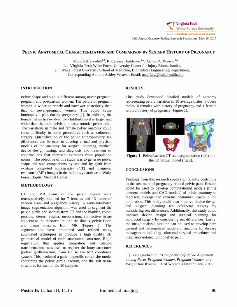
Poster B: Latham B, 11:15 Biomedical Imaging 80
16th Annual Graduate Student Research Symposium, May 10, 2017
PELVIC ANATOMICAL CHARACTERIZATION AND COMPARISON BY SEX AND HISTORY OF PREGNANCY
Mona Saffarzadeh1,2
, R. Caresse Hightower1,2
, Ashley A. Weaver1,2
1. Virginia Tech-Wake Forest University Center for Injury Biomechanics,
2. Wake Forest University School of Medicine, Biomedical Engineering Department,
Corresponding Author: Ashley Weaver, Email: [email protected]
INTRODUCTION
Pelvic shape and size is different among never-pregnant,
pregnant and postpartum women. The pelvis of pregnant
women is wider anteriorly and narrower posteriorly than
that of never-pregnant women. This could cause
lumbopelvic pain during pregnancy [1]. In addition, the
female pelvis has evolved for childbirth so it is larger and
wider than the male pelvis and has a rounder pelvic inlet.
The variations in male and female pelvic anatomy could
cause difficulty in some procedures such as colorectal
surgery. Quantification of the pelvic anthropometry sex
differences can be used to develop virtual and physical
models of the anatomy for surgical planning, medical
device design testing, and diagnosis and treatment of
abnormalities that represent extremes from population
norms. The objective of this study was to generate pelvic
shape and size comparisons by sex and by girth from
existing computed tomography (CT) and magnetic
resonance (MR) images in the radiology database at Wake
Forest Baptist Medical Center.
METHODOLOGY
CT and MR scans of the pelvic region were
retrospectively obtained for 7 females and 13 males of
various sizes and pregnancy history. A semi-automated
image segmentation algorithm was used to segment the
pelvic girdle and sacrum from CT and the bladder, colon,
prostate, uterus, vagina, mesorectum, connective tissue
adjacent to the mesorectum, and the iliacus, pelvic floor,
and psoas muscles from MR (Figure 1). The
segmentations were smoothed and refined using
automated techniques to produce a high quality 3D
geometrical model of each anatomical structure. Rigid
registration that applies translation and rotation
transformations was used to register the bony structures
(pelvic girdle/sacrum) from CT to the MR coordinate
system. This produced a patient-specific composite model
containing the pelvic girdle, sacrum, and the soft tissue
structures for each of the 20 subjects.
RESULTS
This study developed detailed models of anatomy
representing pelvic variation in 10 average males, 3 obese
males, 6 females with history of pregnancy and 1 female
without history of pregnancy (Figure 1).
Figure 1: Pelvic/sacrum CT scan segmentation (left) and
the 3D virtual model (right).
CONCLUSIONS
Findings from this research could significantly contribute
to the treatment of pregnancy-related pelvic pain. Results
could be used to develop computational models (finite
element models and CAD models) of pelvic anatomy to
represent average and extreme anatomical cases in the
population. This study could also improve device design
and surgical planning for colorectal surgery by
considering sex differences. Additionally, this study could
improve device design and surgical planning for
colorectal surgery by considering sex differences. Lastly,
the image analysis pipeline can be used to develop both
general and personalized models of anatomy for disease
management including colorectal surgical procedures and
pregnancy-related lumbopelvic pain.
REFERENCES
[1]. Yamaguchi et al., “Comparison of Pelvic Alignment
among Never-Pregnant Women, Pregnant Women, and
Postpartum Women”, J. of Women’s Health Care, 2016.
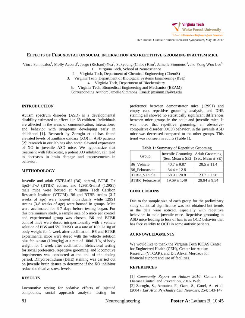
81 Neuroengineering Poster A: Latham B, 10:45
16th Annual Graduate Student Research Symposium, May 10, 2017
EFFECTS OF FEBUXOSTAT ON SOCIAL INTERACTION AND REPETITIVE GROOMING IN AUTISM MICE
Vince Sannicalos1, Molly Accord
2, Jaegu (Richard) Yea
3, Sukyoung (Chloe) Kim
4, Jamelle Simmons
5, and Yong Woo Lee
5
1. Virginia Tech, School of Neuroscience
2. Virginia Tech, Department of Chemical Engineering (ChemE)
3. Virginia Tech, Department of Biological Systems Engineering (BSE)
4. Virginia Tech, Department of Biochemistry
5. Virginia Tech, Biomedical Engineering and Mechanics (BEAM)
Corresponding Author: Jamelle Simmons, Email: [email protected]
INTRODUCTION
Autism spectrum disorder (ASD) is a developmental
disability estimated to effect 1 in 68 children. Individuals
are affected in the areas of communication, interactions,
and behavior with symptoms developing early in
childhood [1]. Research by Zoroglu et al has found
elevated levels of xanthine oxidase (XO) in ASD patients
[2]; research in our lab has also noted elevated expression
of XO in juvenile ASD mice. We hypothesize that
treatment with febuxostat, a potent XO inhibitor, can lead
to decreases in brain damage and improvements in
behavior.
METHODOLOGY
Juvenile and adult C57BL/6J (B6) control, BTBR T+
ltpr3<tf>/J (BTBR) autism, and 129S1/SvImJ (129S1)
male mice were housed at Virginia Tech Carilion
Research Institute (VTCRI). B6 and BTBR strains (3-7
weeks of age) were housed individually while 129S1
strains (3-8 weeks of age) were housed in groups. Mice
were acclimated for 3-7 days before testing began. For
this preliminary study, a sample size of 5 mice per control
and experimental group was chosen. B6 and BTBR
control mice were dosed intraperitoneally with a vehicle
solution of PBS and 5% DMSO at a rate of 100uL/10g of
body weight for 1 week after acclimation. B6 and BTBR
experimental mice were dosed with the vehicle solution
plus febuxostat (10mg/kg) at a rate of 100uL/10g of body
weight for 1 week after acclimation. Behavioral testing
for social preference, repetitive grooming, and locomotive
impairments was conducted at the end of the dosing
period. Dihydroethidium (DHE) staining was carried out
on juvenile brain tissues to determine if the XO inhibitor
reduced oxidative stress levels.
RESULTS
Locomotive testing for sedative effects of injected
compounds, social approach analysis testing for
preference between demonstrator mice (129S1) and
empty cup, repetitive grooming analysis, and DHE
staining all showed no statistically significant differences
between mice groups in the adult and juvenile mice. It
was noted that repetitive grooming, an obsessive-
compulsive disorder (OCD) behavior, in the juvenile ASD
mice was decreased compared to the other groups. This
trend was not seen in adults (Table 1).
Table 1: Summary of Repetitive Grooming
Group Juvenile Grooming
(Sec, Mean ± SE)
Adult Grooming
(Sec, Mean ± SE)
B6_Vehicle 40.7 ± 9.87 28.5 ± 11.4
B6_Febuxostat 34.4 ± 12.8 ----
BTBR_Vehicle 58.9 ± 20.8 23.7 ± 2.56
BTBR_Febuxostat 19.69 ± 1.49 29.94 ± 9.54
CONCLUSIONS
Due to the sample size of each group for the preliminary
study statistical significance was not obtained but trends
in the data were noticed, especially with repetitive
behaviors in male juvenile mice. Repetitive grooming in
ASD mice leading to loss of hair is an OCD behavior that
has face validity to OCD in some autistic patients.
ACKNOWLEDGMENTS
We would like to thank the Virginia Tech ICTAS Center
for Engineered Health (CEH), Center for Autism
Research (VTCAR), and Dr. Alexei Morozov for
financial support and use of facilities.
REFERENCES
[1] Community Report on Autism 2016. Centers for
Disease Control and Prevention, 2016. Web.
[2] Zoroglu, S., Armutcu, F., Ozen, S., Gurel, A., et al.
(2004). Eur Arch Psychiatry Clin Neurosci, 254: 143-147.
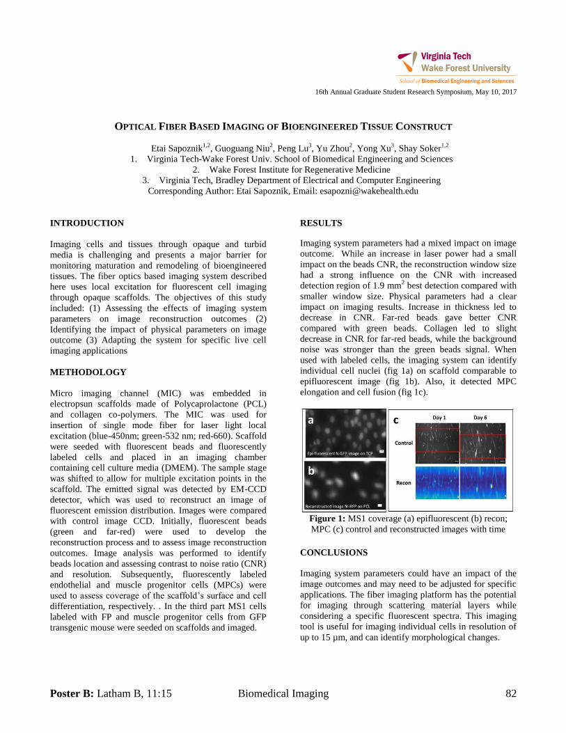
Poster B: Latham B, 11:15 Biomedical Imaging 82
16th Annual Graduate Student Research Symposium, May 10, 2017
OPTICAL FIBER BASED IMAGING OF BIOENGINEERED TISSUE CONSTRUCT
Etai Sapoznik1,2
, Guoguang Niu2, Peng Lu
3, Yu Zhou
2, Yong Xu
3, Shay Soker
1,2
1. Virginia Tech-Wake Forest Univ. School of Biomedical Engineering and Sciences
2. Wake Forest Institute for Regenerative Medicine
3. Virginia Tech, Bradley Department of Electrical and Computer Engineering
Corresponding Author: Etai Sapoznik, Email: [email protected]
INTRODUCTION
Imaging cells and tissues through opaque and turbid
media is challenging and presents a major barrier for
monitoring maturation and remodeling of bioengineered
tissues. The fiber optics based imaging system described
here uses local excitation for fluorescent cell imaging
through opaque scaffolds. The objectives of this study
included: (1) Assessing the effects of imaging system
parameters on image reconstruction outcomes (2)
Identifying the impact of physical parameters on image
outcome (3) Adapting the system for specific live cell
imaging applications
METHODOLOGY
Micro imaging channel (MIC) was embedded in
electropsun scaffolds made of Polycaprolactone (PCL)
and collagen co-polymers. The MIC was used for
insertion of single mode fiber for laser light local
excitation (blue-450nm; green-532 nm; red-660). Scaffold
were seeded with fluorescent beads and fluorescently
labeled cells and placed in an imaging chamber
containing cell culture media (DMEM). The sample stage
was shifted to allow for multiple excitation points in the
scaffold. The emitted signal was detected by EM-CCD
detector, which was used to reconstruct an image of
fluorescent emission distribution. Images were compared
with control image CCD. Initially, fluorescent beads
(green and far-red) were used to develop the
reconstruction process and to assess image reconstruction
outcomes. Image analysis was performed to identify
beads location and assessing contrast to noise ratio (CNR)
and resolution. Subsequently, fluorescently labeled
endothelial and muscle progenitor cells (MPCs) were
used to assess coverage of the scaffold’s surface and cell
differentiation, respectively. . In the third part MS1 cells
labeled with FP and muscle progenitor cells from GFP
transgenic mouse were seeded on scaffolds and imaged.
RESULTS
Imaging system parameters had a mixed impact on image
outcome. While an increase in laser power had a small
impact on the beads CNR, the reconstruction window size
had a strong influence on the CNR with increased
detection region of 1.9 mm2 best detection compared with
smaller window size. Physical parameters had a clear
impact on imaging results. Increase in thickness led to
decrease in CNR. Far-red beads gave better CNR
compared with green beads. Collagen led to slight
decrease in CNR for far-red beads, while the background
noise was stronger than the green beads signal. When
used with labeled cells, the imaging system can identify
individual cell nuclei (fig 1a) on scaffold comparable to
epifluorescent image (fig 1b). Also, it detected MPC
elongation and cell fusion (fig 1c).
Figure 1: MS1 coverage (a) epifluorescent (b) recon;
MPC (c) control and reconstructed images with time
CONCLUSIONS
Imaging system parameters could have an impact of the
image outcomes and may need to be adjusted for specific
applications. The fiber imaging platform has the potential
for imaging through scattering material layers while
considering a specific fluorescent spectra. This imaging
tool is useful for imaging individual cells in resolution of
up to 15 µm, and can identify morphological changes.
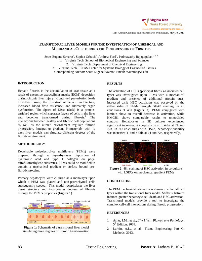
83 Tissue Engineering Poster A: Latham B, 10:45
16th Annual Graduate Student Research Symposium, May 10, 2017
TRANSITIONAL LIVER MODELS FOR THE INVESTIGATION OF CHEMICAL AND
MECHANICAL CUES DURING THE PROGRESSION OF FIBROSIS
Scott-Eugene Saverot1, Sophia Orbach
2, Andrew Ford
2, Padmavathy Rajagopalan
1, 2, 3
1. Virginia Tech, School of Biomedical Engineering and Sciences
2. Virginia Tech, Department of Chemical Engineering
3. Virginia Tech, ICTAS Center for Systems Biology of Engineered Tissues
Corresponding Author: Scott-Eugene Saverot, Email: [email protected]
INTRODUCTION
Hepatic fibrosis is the accumulation of scar tissue as a
result of excessive extracellular matrix (ECM) deposition
during chronic liver injury.1 Continued perturbation leads
to stiffer tissues, the distortion of hepatic architecture,
increased blood flow resistance, and ultimately organ
dysfunction. The Space of Disse (SoD) is a protein-
enriched region which separates layers of cells in the liver
and becomes transformed during fibrosis.1 The
interactions between healthy and fibrotic cell populations
as well as the altered environment regulate fibrotic
progression. Integrating gradient biomaterials with in
vitro liver models can simulate different degrees of the
fibrotic environment.
METHODOLOGY
Detachable polyelectrolyte multilayers (PEMs) were
prepared through a layer-by-layer deposition of
hyaluronic acid and type 1 collagen on poly-
tetrafluoroethylene substrates. PEMs could be modified to
contain a mechanical gradient or surface bound pro-
fibrotic proteins.
Primary hepatocytes were cultured as a monolayer upon
which a PEM was placed and non-parenchymal cells
subsequently seeded.2 This model recapitulates the liver
tissue structure and incorporates degrees of fibrosis
through the PEM’s properties (Figure 1).
Figure 1: Schematic of a transitional liver model
simulating three degrees of fibrotic transformation.
RESULTS
The activation of HSCs (principal fibrosis-associated cell
type) was investigated upon PEMs with a mechanical
gradient and presence of additional protein cues.
Increased early HSC activation was observed on the
stiffer sides of PEMs through GFAP staining, in all
conditions at 48h (Figure 2). PEMs conjugated with
laminin show an overall decrease in activation, while
HMGB1 shows comparable results to unmodified
controls. Hepatocytes in 3D cultures experienced
significant increases in apoptosis on stiff sides at 24 and
72h. In 3D co-cultures with HSCs, hepatocyte viability
was increased 4- and 3-fold at 24 and 72h, respectively.
Figure 2: 48h staining of HSC activation in co-culture
with LSECs on mechanical gradient PEMs
CONCLUSIONS
The PEM mechanical gradient was shown to affect all cell
types within the transitional liver model. Stiffer substrates
induced greater hepatocyte cell death and HSC activation.
Transitional models provide a tool to investigate the
complex cell-cell interactions during fibrotic progression.
REFERENCES
1. Arias, I.M., et al., The Liver: Biology and Pathology,
5th
Edition, 2009.
2. Larkin, A.L., et al., Tissue Engineering Part C:
Methods, 2013.
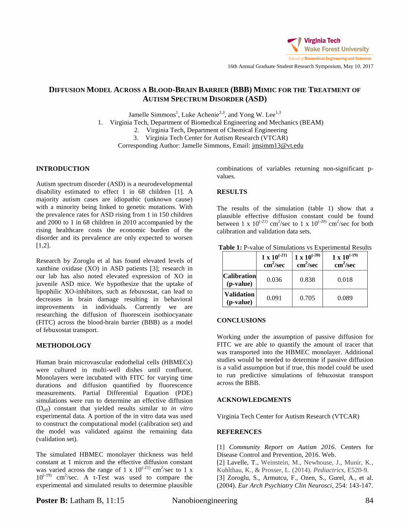
Poster B: Latham B, 11:15 Nanobioengineering 84
16th Annual Graduate Student Research Symposium, May 10, 2017
DIFFUSION MODEL ACROSS A BLOOD-BRAIN BARRIER (BBB) MIMIC FOR THE TREATMENT OF
AUTISM SPECTRUM DISORDER (ASD)
Jamelle Simmons1, Luke Achenie
2,3, and Yong W. Lee
1,3
1. Virginia Tech, Department of Biomedical Engineering and Mechanics (BEAM)
2. Virginia Tech, Department of Chemical Engineering
3. Virginia Tech Center for Autism Research (VTCAR)
Corresponding Author: Jamelle Simmons, Email: [email protected]
INTRODUCTION
Autism spectrum disorder (ASD) is a neurodevelopmental
disability estimated to effect 1 in 68 children [1]. A
majority autism cases are idiopathic (unknown cause)
with a minority being linked to genetic mutations. With
the prevalence rates for ASD rising from 1 in 150 children
and 2000 to 1 in 68 children in 2010 accompanied by the
rising healthcare costs the economic burden of the
disorder and its prevalence are only expected to worsen
[1,2].
Research by Zoroglu et al has found elevated levels of
xanthine oxidase (XO) in ASD patients [3]; research in
our lab has also noted elevated expression of XO in
juvenile ASD mice. We hypothesize that the uptake of
lipophilic XO-inhibitors, such as febuxostat, can lead to
decreases in brain damage resulting in behavioral
improvements in individuals. Currently we are
researching the diffusion of fluorescein isothiocyanate
(FITC) across the blood-brain barrier (BBB) as a model
of febuxostat transport.
METHODOLOGY
Human brain microvascular endothelial cells (HBMECs)
were cultured in multi-well dishes until confluent.
Monolayers were incubated with FITC for varying time
durations and diffusion quantified by fluorescence
measurements. Partial Differential Equation (PDE)
simulations were run to determine an effective diffusion
(Deff) constant that yielded results similar to in vitro
experimental data. A portion of the in vitro data was used
to construct the computational model (calibration set) and
the model was validated against the remaining data
(validation set).
The simulated HBMEC monolayer thickness was held
constant at 1 micron and the effective diffusion constant
was varied across the range of 1 x 10(-21)
cm2/sec to 1 x
10(-19)
cm2/sec. A t-Test was used to compare the
experimental and simulated results to determine plausible
combinations of variables returning non-significant p-
values.
RESULTS
The results of the simulation (table 1) show that a
plausible effective diffusion constant could be found
between 1 x 10(-21)
cm2/sec to 1 x 10
(-20) cm
2/sec for both
calibration and validation data sets.
Table 1: P-value of Simulations vs Experimental Results
1 x 10
(-21)
cm2/sec
1 x 10(-20)
cm2/sec
1 x 10(-19)
cm2/sec
Calibration
(p-value) 0.036 0.838 0.018
Validation
(p-value) 0.091 0.705 0.089
CONCLUSIONS
Working under the assumption of passive diffusion for
FITC we are able to quantify the amount of tracer that
was transported into the HBMEC monolayer. Additional
studies would be needed to determine if passive diffusion
is a valid assumption but if true, this model could be used
to run predictive simulations of febuxostat transport
across the BBB.
ACKNOWLEDGMENTS
Virginia Tech Center for Autism Research (VTCAR)
REFERENCES
[1] Community Report on Autism 2016. Centers for
Disease Control and Prevention, 2016. Web.
[2] Lavelle, T., Weinstein, M., Newhouse, J., Munir, K.,
Kuhlthau, K., & Prosser, L. (2014). Pediactrics, E520-9.
[3] Zoroglu, S., Armutcu, F., Ozen, S., Gurel, A., et al.
(2004). Eur Arch Psychiatry Clin Neurosci, 254: 143-147.
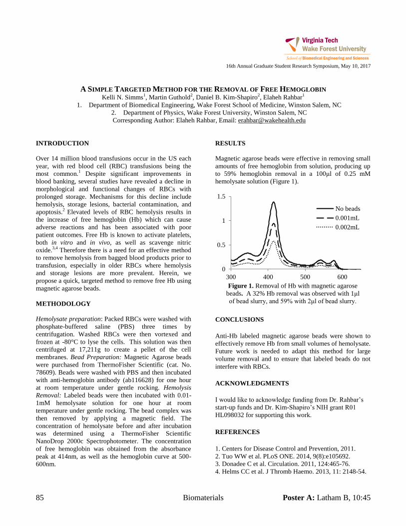
85 Biomaterials Poster A: Latham B, 10:45
16th Annual Graduate Student Research Symposium, May 10, 2017
A SIMPLE TARGETED METHOD FOR THE REMOVAL OF FREE HEMOGLOBIN
Kelli N. Simms1, Martin Guthold
2, Daniel B. Kim-Shapiro
2, Elaheh Rahbar
1
1. Department of Biomedical Engineering, Wake Forest School of Medicine, Winston Salem, NC
2. Department of Physics, Wake Forest University, Winston Salem, NC
Corresponding Author: Elaheh Rahbar, Email: [email protected]
INTRODUCTION
Over 14 million blood transfusions occur in the US each
year, with red blood cell (RBC) transfusions being the
most common.1 Despite significant improvements in
blood banking, several studies have revealed a decline in
morphological and functional changes of RBCs with
prolonged storage. Mechanisms for this decline include
hemolysis, storage lesions, bacterial contamination, and
apoptosis.2
Elevated levels of RBC hemolysis results in
the increase of free hemoglobin (Hb) which can cause
adverse reactions and has been associated with poor
patient outcomes. Free Hb is known to activate platelets,
both in vitro and in vivo, as well as scavenge nitric
oxide.3,4
Therefore there is a need for an effective method
to remove hemolysis from bagged blood products prior to
transfusion, especially in older RBCs where hemolysis
and storage lesions are more prevalent. Herein, we
propose a quick, targeted method to remove free Hb using
magnetic agarose beads.
METHODOLOGY
Hemolysate preparation: Packed RBCs were washed with
phosphate-buffered saline (PBS) three times by
centrifugation. Washed RBCs were then vortexed and
frozen at -80°C to lyse the cells. This solution was then
centrifuged at 17,211g to create a pellet of the cell
membranes. Bead Preparation: Magnetic Agarose beads
were purchased from ThermoFisher Scientific (cat. No.
78609). Beads were washed with PBS and then incubated
with anti-hemoglobin antibody (ab116628) for one hour
at room temperature under gentle rocking. Hemolysis
Removal: Labeled beads were then incubated with 0.01-
1mM hemolysate solution for one hour at room
temperature under gentle rocking. The bead complex was
then removed by applying a magnetic field. The
concentration of hemolysate before and after incubation
was determined using a ThermoFisher Scientific
NanoDrop 2000c Spectrophotometer. The concentration
of free hemoglobin was obtained from the absorbance
peak at 414nm, as well as the hemoglobin curve at 500-
600nm.
RESULTS
Magnetic agarose beads were effective in removing small
amounts of free hemoglobin from solution, producing up
to 59% hemoglobin removal in a 100μl of 0.25 mM
hemolysate solution (Figure 1).
CONCLUSIONS
Anti-Hb labeled magnetic agarose beads were shown to
effectively remove Hb from small volumes of hemolysate.
Future work is needed to adapt this method for large
volume removal and to ensure that labeled beads do not
interfere with RBCs.
ACKNOWLEDGMENTS
I would like to acknowledge funding from Dr. Rahbar’s
start-up funds and Dr. Kim-Shapiro’s NIH grant R01
HL098032 for supporting this work.
REFERENCES
1. Centers for Disease Control and Prevention, 2011.
2. Tuo WW et al. PLoS ONE. 2014, 9(8):e105692.
3. Donadee C et al. Circulation. 2011, 124:465-76.
4. Helms CC et al. J Thromb Haemo. 2013, 11: 2148-54.
0
0.5
1
1.5
300 400 500 600
No beads
0.001mL
0.002mL
Figure 1. Removal of Hb with magnetic agarose
beads. A 32% Hb removal was observed with 1μl
of bead slurry, and 59% with 2μl of bead slurry.
Poster B: Latham B, 11:15 Biomedical Imaging 86
16th Annual Graduate Student Research Symposium, May 10, 2017
USING VIDEOTAPED MOTION CODING TO IMPROVE UNDERSTAND OF F-NIRS SIGNAL CHANGES
Ben Stephens1, Amnah Eltahir
2, Stephen LaConte
3, and Stephanie DeLuca
3
1. Virginia Tech Carilion School of Medicine
2. Virginia Tech, Biomedical Engineering
3. Virginia Tech Carilion Research Insitute
Corresponding Author: Ben Stephens, VTCSOM Email: [email protected]
INTRODUCTION
Functional near-infrared spectroscopy (f-NIRS) is a non-
invasive neuroimaging technique that has application in
the pediatric population due to its safety and ease of use.
However, questions remain about the technology with
regard to its validity and reliability in accurately
measuring neural signals in the presence of motion
(targeted and non-targeted). Video coding of motion
events during scanning may provide new insights into f-
NIRS signals and help interrogate usable signals.
METHODOLOGY
Six children (mean age 14 months) participated in
videotaped f-NIRS sessions that included the collection of
resting-state and motor-task data. Video sessions were
coded for upper-extremity movements that were
translated into a time-in-motion outcome measure. These
were then compared to corresponding f-NIRS signals
across 5-second time blocks with a goal of identifying
segments of high and low signal variance.
RESULTS
Correlations between upper extremity time in motion and
signal variance for corresponding 5 second blocks ranged
from 0.0898 to 0.258 (p<0.01). F-NIRS signal responses
in high-motion blocks appeared to result in decreased
signal quality, while f-NIRS signal response in low-
motion blocks were marked by appropriate hemodynamic
response and negative correlation between oxy- and
deoxyhemoglobin signals.
CONCLUSIONS
Video coding behavior offers new possibilities for
interpreting f-NIRS signals during f-NIRS recordings
with infants. Further study is required to analyze the
validity of our findings and explore other motion events,
both targeted and non-targeted, on f-NIRS signal
characteristics.
ACKNOWLEDGMENTS
VTCRI Neuromotor Research Clinic
The LaConte Lab Team at VTCRI
REFERENCES
Cristia, A., Dupoux, E., Hakune, Y., Lloyd-Fox, S.,
Schuetze, M., Kivits, J., & Minagawa-Kawai, Y. (2013).
An online database of infant functional near infrared
specstroscopy studies: a community-augmented
systematic review. PloS one 8(3), e58906.
Goodwin, J.R, Cannaday, A.E., Palmeri, H.G., Di
Constanzo, A., Emberson, L.L., Aslin, R.N., & Berger,
A.J. (2016). Methodology for high-yield acquisition of
functional near-infrared spectroscopy data from alert,
upright infants. Neurophotonics, 3(3), 031415-031415.
Nishiyori, R. (2016). fNIRS: An emergent method to
document functional cortical activity during infant
movements. Frontiers in psychology, 7.
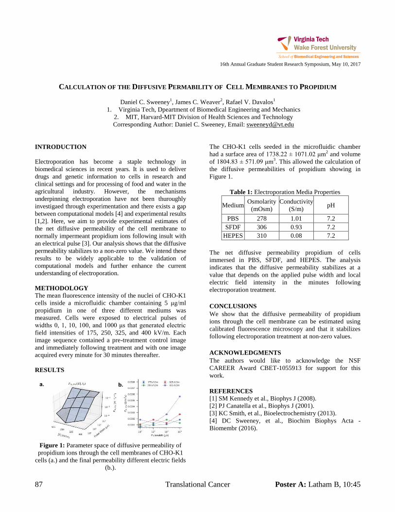
87 Translational Cancer Poster A: Latham B, 10:45
16th Annual Graduate Student Research Symposium, May 10, 2017
CALCULATION OF THE DIFFUSIVE PERMABILITY OF CELL MEMBRANES TO PROPIDIUM
Daniel C. Sweeney1, James C. Weaver
2, Rafael V. Davalos
1
1. Virginia Tech, Dpeartment of Biomedical Engineering and Mechanics
2. MIT, Harvard-MIT Division of Health Sciences and Technology
Corresponding Author: Daniel C. Sweeney, Email: [email protected]
INTRODUCTION
Electroporation has become a staple technology in
biomedical sciences in recent years. It is used to deliver
drugs and genetic information to cells in research and
clinical settings and for processing of food and water in the
agricultural industry. However, the mechanisms
underpinning electroporation have not been thuroughly
investigaed through experimentation and there exists a gap
between computational models [4] and experimental results
[1,2]. Here, we aim to provide experimental estimates of
the net diffusive permeability of the cell membrane to
normally impermeant propidium ions following insult with
an electrical pulse [3]. Our analysis shows that the diffusive
permeability stabilizes to a non-zero value. We intend these
results to be widely applicable to the validation of
computational models and further enhance the current
understanding of electroporation.
METHODOLOGY
The mean fluorescence intensity of the nuclei of CHO-K1
cells inside a microfluidic chamber containing 5 μg/ml
propidium in one of three different mediums was
measured. Cells were exposed to electrical pulses of
widths 0, 1, 10, 100, and 1000 μs that generated electric
field intensities of 175, 250, 325, and 400 kV/m. Each
image sequence contained a pre-treatment control image
and immediately following treatment and with one image
acquired every minute for 30 minutes thereafter.
RESULTS
Figure 1: Parameter space of diffusive permeability of
propidium ions through the cell membranes of CHO-K1
cells (a.) and the final permeability different electric fields
(b.).
The CHO-K1 cells seeded in the microfluidic chamber
had a surface area of 1738.22 ± 1071.02 μm2 and volume
of 1804.83 ± 571.09 μm3. This allowed the calculation of
the diffusive permeabilities of propidium showing in
Figure 1.
Table 1: Electroporation Media Properties
Medium Osmolarity
(mOsm)
Conductivity
(S/m) pH
PBS 278 1.01 7.2
SFDF 306 0.93 7.2
HEPES 310 0.08 7.2
The net diffusive permeability propidium of cells
immersed in PBS, SFDF, and HEPES. The analysis
indicates that the diffusive permeability stabilizes at a
value that depends on the applied pulse width and local
electric field intensity in the minutes following
electroporation treatment.
CONCLUSIONS
We show that the diffusive permeability of propidium
ions through the cell membrane can be estimated using
calibrated fluorescence microscopy and that it stabilizes
following electroporation treatment at non-zero values.
ACKNOWLEDGMENTS
The authors would like to acknowledge the NSF
CAREER Award CBET-1055913 for support for this
work.
REFERENCES
[1] SM Kennedy et al., Biophys J (2008).
[2] PJ Canatella et al., Biophys J (2001).
[3] KC Smith, et al., Bioelectrochemistry (2013).
[4] DC Sweeney, et al., Biochim Biophys Acta -
Biomembr (2016).
Poster B: Latham B, 11:15 Biomechanics 88
16th Annual Graduate Student Research Symposium, May 10, 2017
REAR IMPACT CRASHES IN THE UNITED STATES: HOW DANGEROUS ARE THEY?
Whitney M. Tatem1 and H. Clay Gabler
1
1. Virginia Tech, Department of Biomedical Engineering and Mechanics
Corresponding Author: Whitney Tatem, Email: [email protected]
INTRODUCTION
One of the most common crash modes on roadways
world-wide are rear end collisions. The National Highway
Traffic Safety Administration (NHTSA) has reported that
approximately 2 million rear end crashes occurred in the
U.S. in 2014 (NHTSA 2016). Despite this large number
of crash occurrences, rear impact crashes are generally
viewed as a benign crash mode with property damage
only and at most minor injuries. However, in 2015, there
were almost 1000 fatalities in cars and light trucks that
were struck in the rear. The question becomes, if rear
impacts are indeed a benign crash mode, what is leading
to these fatalities?
The objective of this study is to investigate the
characteristics of serious-to-fatal rear impact crashes and
to identify the underlying crash features that may lead to
serious or fatal injury for occupants seated in rear-
impacted vehicles.
METHODOLOGY
The analysis examined fatal rear crashes extracted from
the Fatality Analysis Reporting System (FARS) 2011-
2015 and serious-to-fatally injured occupants in the
National Automotive Sampling System/Crashworthiness
Data System (NASS/CDS) data from case years 2000-
2015. The FARS analysis first investigated all crashes in
which a passenger vehicle was rear struck. A follow-on
analysis was then conducted limiting the dataset to model
year 2000 and greater passenger vehicles to focus the
analysis on vehicles which were more likely to have been
designed for the new Federal Motor Vehicle Safety
Standard (FMVSS) 301, Fuel System Integrity - a
structural integrity test in the rear impact crash mode.
To better understand the nature of the fatalities studied in
FARS, detailed case reviews were conducted of all fatally
to seriously injured rear-struck occupants in the
NASS/CDS data. Our analysis examined seriously injured
occupants, as our hypothesis was that the factors which
led to fatal injury might also have been present at lower
injury severity crashes in which the occupant survived the
impact.
RESULTS
There appear to be three mechanisms leading to serious-
to-fatal injury: (1) catastrophic collapse of the occupant
compartment, (2) impacts with hard vehicle interior
components, e.g. the B-Pillar, and (3) thoracic loading
from the seat, especially on older occupants. All three
injury mechanisms present themselves in each injury
severity category, both fatal, severe, and serious. There
appears to be a continuum where compartment collapse is
most often associated with fatal injuries, direct head
contact produces predominantly severe injuries, and
thoracic loading appears more related to serious injuries.
This study is the first step in a research effort to explore
the factors that lead to a higher injury risk in rear impact
crashes. The next steps will be to expand this study to a
more comprehensive analysis of rear structural response
and seat performance to gain a fuller understanding of the
injury mechanics in rear crashes, and to determine the
priority for countermeasure development.
CONCLUSIONS
This study has investigated the factors associated with
serious-to-fatal rear crashes. Despite the perception that
rear crashes are benign, over 1000 rear-struck occupants
were fatally injured in 2015. Compartmental collapse
appears to be a major risk factor in rear impact crashes,
particularly for occupants seated in the rear seats. The
vast majority of fatal crashes appeared to involve
compartmental collapse although other injury
mechanisms such as direct head contact to rigid interior
vehicle structures and thoracic loading from the seat
should not be disregarded.
REFERENCES
NHTSA. Traffic Safety Facts 2014: A Compilation of
Motor Vehicle Crash Data From the Fatality Analysis
Reporting System and the General Estimation
System.Washington, DC: NHTSA, U.S. Department of
Transportation; 2016. Report No. DOT HS 812 261
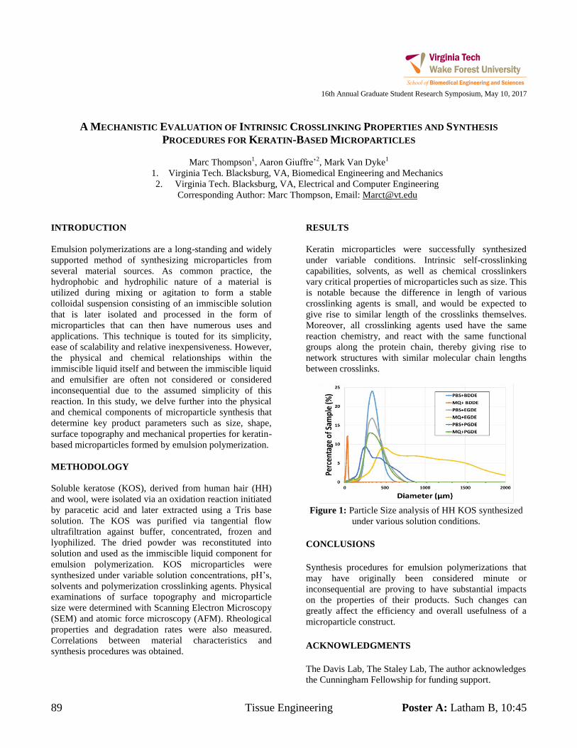
89 Tissue Engineering Poster A: Latham B, 10:45
16th Annual Graduate Student Research Symposium, May 10, 2017
A MECHANISTIC EVALUATION OF INTRINSIC CROSSLINKING PROPERTIES AND SYNTHESIS
PROCEDURES FOR KERATIN-BASED MICROPARTICLES
Marc Thompson1, Aaron Giuffre’
2, Mark Van Dyke
1
1. Virginia Tech. Blacksburg, VA, Biomedical Engineering and Mechanics
2. Virginia Tech. Blacksburg, VA, Electrical and Computer Engineering
Corresponding Author: Marc Thompson, Email: [email protected]
INTRODUCTION
Emulsion polymerizations are a long-standing and widely
supported method of synthesizing microparticles from
several material sources. As common practice, the
hydrophobic and hydrophilic nature of a material is
utilized during mixing or agitation to form a stable
colloidal suspension consisting of an immiscible solution
that is later isolated and processed in the form of
microparticles that can then have numerous uses and
applications. This technique is touted for its simplicity,
ease of scalability and relative inexpensiveness. However,
the physical and chemical relationships within the
immiscible liquid itself and between the immiscible liquid
and emulsifier are often not considered or considered
inconsequential due to the assumed simplicity of this
reaction. In this study, we delve further into the physical
and chemical components of microparticle synthesis that
determine key product parameters such as size, shape,
surface topography and mechanical properties for keratin-
based microparticles formed by emulsion polymerization.
METHODOLOGY
Soluble keratose (KOS), derived from human hair (HH)
and wool, were isolated via an oxidation reaction initiated
by paracetic acid and later extracted using a Tris base
solution. The KOS was purified via tangential flow
ultrafiltration against buffer, concentrated, frozen and
lyophilized. The dried powder was reconstituted into
solution and used as the immiscible liquid component for
emulsion polymerization. KOS microparticles were
synthesized under variable solution concentrations, pH’s,
solvents and polymerization crosslinking agents. Physical
examinations of surface topography and microparticle
size were determined with Scanning Electron Microscopy
(SEM) and atomic force microscopy (AFM). Rheological
properties and degradation rates were also measured.
Correlations between material characteristics and
synthesis procedures was obtained.
RESULTS
Keratin microparticles were successfully synthesized
under variable conditions. Intrinsic self-crosslinking
capabilities, solvents, as well as chemical crosslinkers
vary critical properties of microparticles such as size. This
is notable because the difference in length of various
crosslinking agents is small, and would be expected to
give rise to similar length of the crosslinks themselves.
Moreover, all crosslinking agents used have the same
reaction chemistry, and react with the same functional
groups along the protein chain, thereby giving rise to
network structures with similar molecular chain lengths
between crosslinks.
Figure 1: Particle Size analysis of HH KOS synthesized
under various solution conditions.
CONCLUSIONS
Synthesis procedures for emulsion polymerizations that
may have originally been considered minute or
inconsequential are proving to have substantial impacts
on the properties of their products. Such changes can
greatly affect the efficiency and overall usefulness of a
microparticle construct.
ACKNOWLEDGMENTS
The Davis Lab, The Staley Lab, The author acknowledges
the Cunningham Fellowship for funding support.
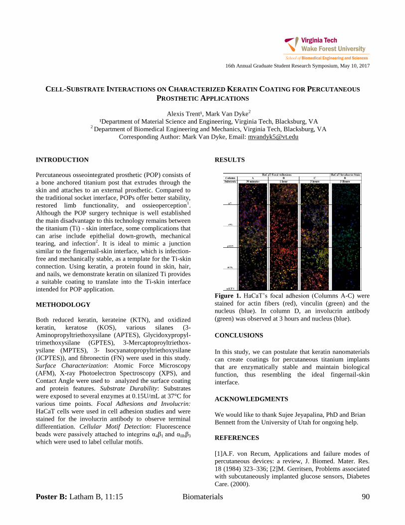
Poster B: Latham B, 11:15 Biomaterials 90
16th Annual Graduate Student Research Symposium, May 10, 2017
CELL-SUBSTRATE INTERACTIONS ON CHARACTERIZED KERATIN COATING FOR PERCUTANEOUS
PROSTHETIC APPLICATIONS
Alexis Trent¹, Mark Van Dyke2
¹Department of Material Science and Engineering, Virginia Tech, Blacksburg, VA 2 Department of Biomedical Engineering and Mechanics, Virginia Tech, Blacksburg, VA
Corresponding Author: Mark Van Dyke, Email: [email protected]
INTRODUCTION
Percutaneous osseointegrated prosthetic (POP) consists of
a bone anchored titanium post that extrudes through the
skin and attaches to an external prosthetic. Compared to
the traditional socket interface, POPs offer better stability,
restored limb functionality, and ossieoperception1.
Although the POP surgery technique is well established
the main disadvantage to this technology remains between
the titanium (Ti) - skin interface, some complications that
can arise include epithelial down-growth, mechanical
tearing, and infection2. It is ideal to mimic a junction
similar to the fingernail-skin interface, which is infection-
free and mechanically stable, as a template for the Ti-skin
connection. Using keratin, a protein found in skin, hair,
and nails, we demonstrate keratin on silanized Ti provides
a suitable coating to translate into the Ti-skin interface
intended for POP application.
METHODOLOGY
Both reduced keratin, kerateine (KTN), and oxidized
keratin, keratose (KOS), various silanes (3-
Aminopropyltriethoxysilane (APTES), Glycidoxypropyl-
trimethoxysilane (GPTES), 3-Mercaptoproyltriethox-
ysilane (MPTES), 3- Isocyanatopropyltriethoxysilane
(ICPTES)), and fibronectin (FN) were used in this study.
Surface Characterization: Atomic Force Microscopy
(AFM), X-ray Photoelectron Spectroscopy (XPS), and
Contact Angle were used to analyzed the surface coating
and protein features. Substrate Durability: Substrates
were exposed to several enzymes at 0.15U/mL at 37°C for
various time points. Focal Adhesions and Involucrin:
HaCaT cells were used in cell adhesion studies and were
stained for the involucrin antibody to observe terminal
differentiation. Cellular Motif Detection: Fluorescence
beads were passively attached to integrins α4β1 and αIIbβ3
which were used to label cellular motifs.
RESULTS
Figure 1. HaCaT’s focal adhesion (Columns A-C) were
stained for actin fibers (red), vinculin (green) and the
nucleus (blue). In column D, an involucrin antibody
(green) was observed at 3 hours and nucleus (blue).
CONCLUSIONS
In this study, we can postulate that keratin nanomaterials
can create coatings for percutaneous titanium implants
that are enzymatically stable and maintain biological
function, thus resembling the ideal fingernail-skin
interface.
ACKNOWLEDGMENTS
We would like to thank Sujee Jeyapalina, PhD and Brian
Bennett from the University of Utah for ongoing help.
REFERENCES
[1]A.F. von Recum, Applications and failure modes of
percutaneous devices: a review, J. Biomed. Mater. Res.
18 (1984) 323–336; [2]M. Gerritsen, Problems associated
with subcutaneously implanted glucose sensors, Diabetes
Care. (2000).
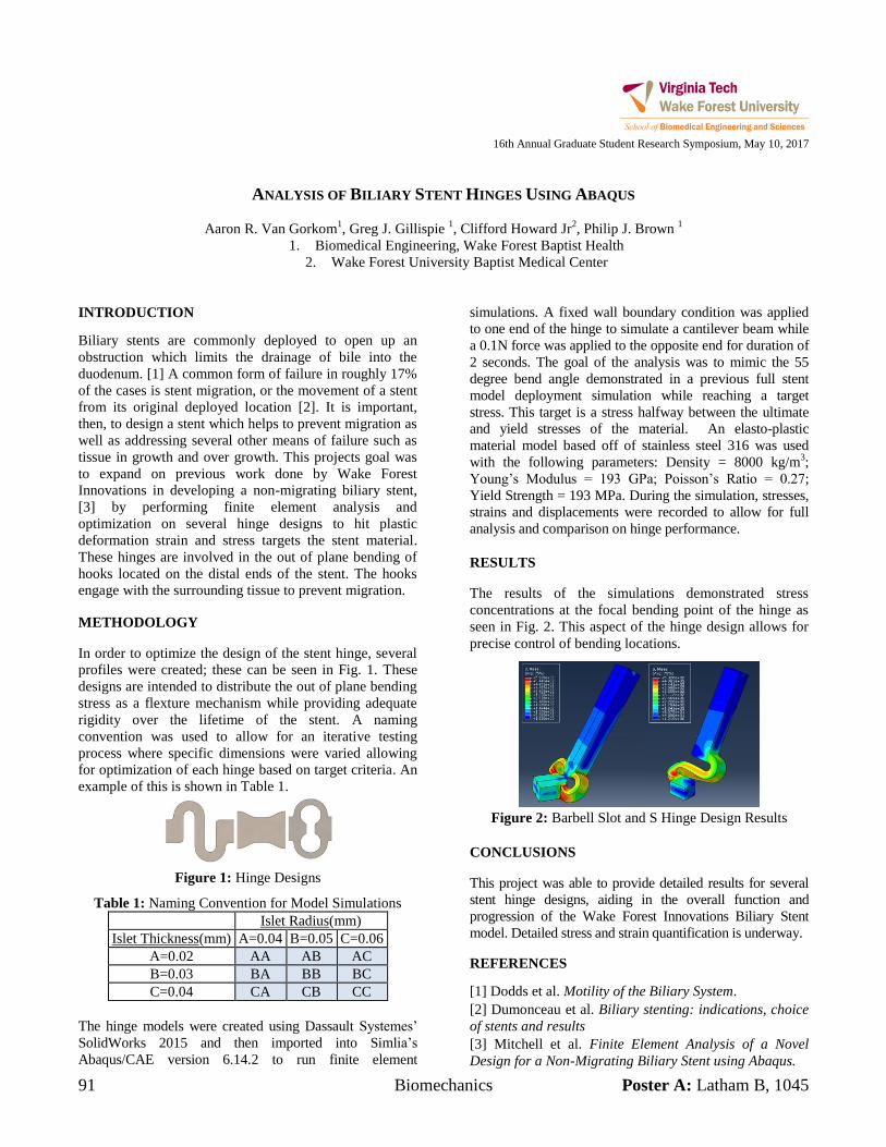
91 Biomechanics Poster A: Latham B, 1045
16th Annual Graduate Student Research Symposium, May 10, 2017
ANALYSIS OF BILIARY STENT HINGES USING ABAQUS
Aaron R. Van Gorkom1, Greg J. Gillispie
1, Clifford Howard Jr
2, Philip J. Brown
1
1. Biomedical Engineering, Wake Forest Baptist Health
2. Wake Forest University Baptist Medical Center
INTRODUCTION
Biliary stents are commonly deployed to open up an
obstruction which limits the drainage of bile into the
duodenum. [1] A common form of failure in roughly 17%
of the cases is stent migration, or the movement of a stent
from its original deployed location [2]. It is important,
then, to design a stent which helps to prevent migration as
well as addressing several other means of failure such as
tissue in growth and over growth. This projects goal was
to expand on previous work done by Wake Forest
Innovations in developing a non-migrating biliary stent,
[3] by performing finite element analysis and
optimization on several hinge designs to hit plastic
deformation strain and stress targets the stent material.
These hinges are involved in the out of plane bending of
hooks located on the distal ends of the stent. The hooks
engage with the surrounding tissue to prevent migration.
METHODOLOGY
In order to optimize the design of the stent hinge, several
profiles were created; these can be seen in Fig. 1. These
designs are intended to distribute the out of plane bending
stress as a flexture mechanism while providing adequate
rigidity over the lifetime of the stent. A naming
convention was used to allow for an iterative testing
process where specific dimensions were varied allowing
for optimization of each hinge based on target criteria. An
example of this is shown in Table 1.
Figure 1: Hinge Designs
Table 1: Naming Convention for Model Simulations
Islet Radius(mm)
Islet Thickness(mm) A=0.04 B=0.05 C=0.06
A=0.02 AA AB AC
B=0.03 BA BB BC
C=0.04 CA CB CC
The hinge models were created using Dassault Systemes’
SolidWorks 2015 and then imported into Simlia’s
Abaqus/CAE version 6.14.2 to run finite element
simulations. A fixed wall boundary condition was applied
to one end of the hinge to simulate a cantilever beam while
a 0.1N force was applied to the opposite end for duration of
2 seconds. The goal of the analysis was to mimic the 55
degree bend angle demonstrated in a previous full stent
model deployment simulation while reaching a target
stress. This target is a stress halfway between the ultimate
and yield stresses of the material. An elasto-plastic
material model based off of stainless steel 316 was used
with the following parameters: Density = 8000 kg/m3;
Young’s Modulus = 193 GPa; Poisson’s Ratio = 0.27;
Yield Strength = 193 MPa. During the simulation, stresses,
strains and displacements were recorded to allow for full
analysis and comparison on hinge performance.
RESULTS
The results of the simulations demonstrated stress
concentrations at the focal bending point of the hinge as
seen in Fig. 2. This aspect of the hinge design allows for
precise control of bending locations.
Figure 2: Barbell Slot and S Hinge Design Results
CONCLUSIONS
This project was able to provide detailed results for several
stent hinge designs, aiding in the overall function and
progression of the Wake Forest Innovations Biliary Stent
model. Detailed stress and strain quantification is underway.
REFERENCES
[1] Dodds et al. Motility of the Biliary System.
[2] Dumonceau et al. Biliary stenting: indications, choice
of stents and results
[3] Mitchell et al. Finite Element Analysis of a Novel
Design for a Non-Migrating Biliary Stent using Abaqus.
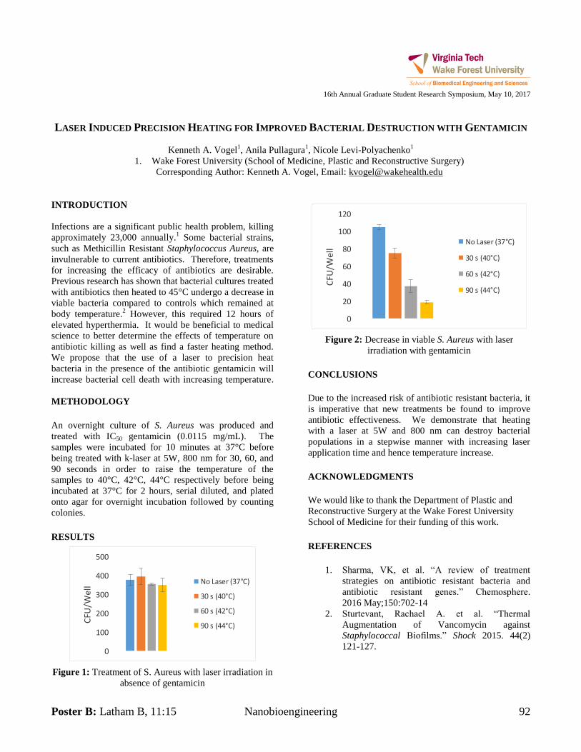
Poster B: Latham B, 11:15 Nanobioengineering 92
16th Annual Graduate Student Research Symposium, May 10, 2017
LASER INDUCED PRECISION HEATING FOR IMPROVED BACTERIAL DESTRUCTION WITH GENTAMICIN
Kenneth A. Vogel1, Anila Pullagura
1, Nicole Levi-Polyachenko
1
1. Wake Forest University (School of Medicine, Plastic and Reconstructive Surgery)
Corresponding Author: Kenneth A. Vogel, Email: [email protected]
INTRODUCTION
Infections are a significant public health problem, killing
approximately 23,000 annually.1 Some bacterial strains,
such as Methicillin Resistant Staphylococcus Aureus, are
invulnerable to current antibiotics. Therefore, treatments
for increasing the efficacy of antibiotics are desirable.
Previous research has shown that bacterial cultures treated
with antibiotics then heated to 45°C undergo a decrease in
viable bacteria compared to controls which remained at
body temperature.2 However, this required 12 hours of
elevated hyperthermia. It would be beneficial to medical
science to better determine the effects of temperature on
antibiotic killing as well as find a faster heating method.
We propose that the use of a laser to precision heat
bacteria in the presence of the antibiotic gentamicin will
increase bacterial cell death with increasing temperature.
METHODOLOGY
An overnight culture of S. Aureus was produced and
treated with IC50 gentamicin (0.0115 mg/mL). The
samples were incubated for 10 minutes at 37°C before
being treated with k-laser at 5W, 800 nm for 30, 60, and
90 seconds in order to raise the temperature of the
samples to 40°C, 42°C, 44°C respectively before being
incubated at 37°C for 2 hours, serial diluted, and plated
onto agar for overnight incubation followed by counting
colonies.
RESULTS
0
100
200
300
400
500
CFU
/We
ll
No Laser (37°C)
30 s (40°C)
60 s (42°C)
90 s (44°C)
Figure 1: Treatment of S. Aureus with laser irradiation in
absence of gentamicin
0
20
40
60
80
100
120
CFU
/Wel
l
No Laser (37°C)
30 s (40°C)
60 s (42°C)
90 s (44°C)
Figure 2: Decrease in viable S. Aureus with laser
irradiation with gentamicin
CONCLUSIONS
Due to the increased risk of antibiotic resistant bacteria, it
is imperative that new treatments be found to improve
antibiotic effectiveness. We demonstrate that heating
with a laser at 5W and 800 nm can destroy bacterial
populations in a stepwise manner with increasing laser
application time and hence temperature increase.
ACKNOWLEDGMENTS
We would like to thank the Department of Plastic and
Reconstructive Surgery at the Wake Forest University
School of Medicine for their funding of this work.
REFERENCES
1. Sharma, VK, et al. “A review of treatment
strategies on antibiotic resistant bacteria and
antibiotic resistant genes.” Chemosphere.
2016 May;150:702-14
2. Sturtevant, Rachael A. et al. “Thermal
Augmentation of Vancomycin against
Staphylococcal Biofilms.” Shock 2015. 44(2)
121-127.
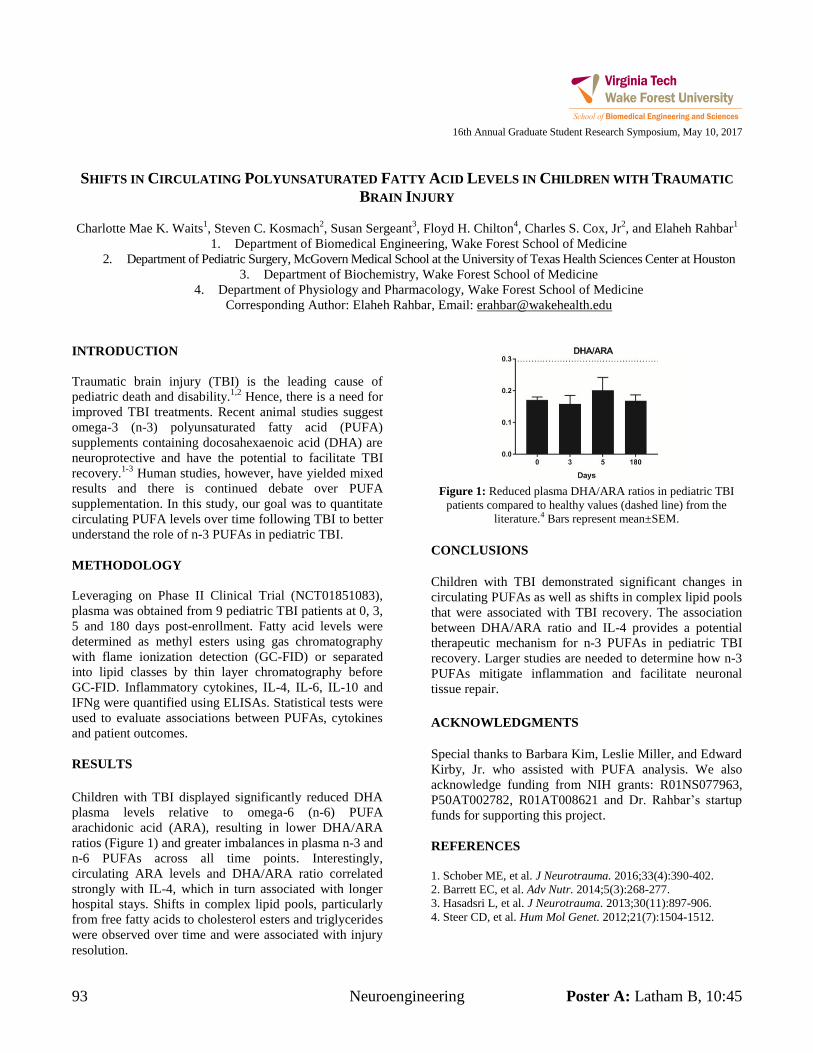
93 Neuroengineering Poster A: Latham B, 10:45
16th Annual Graduate Student Research Symposium, May 10, 2017
SHIFTS IN CIRCULATING POLYUNSATURATED FATTY ACID LEVELS IN CHILDREN WITH TRAUMATIC
BRAIN INJURY
Charlotte Mae K. Waits1, Steven C. Kosmach
2, Susan Sergeant
3, Floyd H. Chilton
4, Charles S. Cox, Jr
2, and Elaheh Rahbar
1
1. Department of Biomedical Engineering, Wake Forest School of Medicine
2. Department of Pediatric Surgery, McGovern Medical School at the University of Texas Health Sciences Center at Houston
3. Department of Biochemistry, Wake Forest School of Medicine
4. Department of Physiology and Pharmacology, Wake Forest School of Medicine
Corresponding Author: Elaheh Rahbar, Email: [email protected]
INTRODUCTION
Traumatic brain injury (TBI) is the leading cause of
pediatric death and disability.1,2
Hence, there is a need for
improved TBI treatments. Recent animal studies suggest
omega-3 (n-3) polyunsaturated fatty acid (PUFA)
supplements containing docosahexaenoic acid (DHA) are
neuroprotective and have the potential to facilitate TBI
recovery.1-3
Human studies, however, have yielded mixed
results and there is continued debate over PUFA
supplementation. In this study, our goal was to quantitate
circulating PUFA levels over time following TBI to better
understand the role of n-3 PUFAs in pediatric TBI.
METHODOLOGY
Leveraging on Phase II Clinical Trial (NCT01851083),
plasma was obtained from 9 pediatric TBI patients at 0, 3,
5 and 180 days post-enrollment. Fatty acid levels were
determined as methyl esters using gas chromatography
with flame ionization detection (GC-FID) or separated
into lipid classes by thin layer chromatography before
GC-FID. Inflammatory cytokines, IL-4, IL-6, IL-10 and
IFNg were quantified using ELISAs. Statistical tests were
used to evaluate associations between PUFAs, cytokines
and patient outcomes.
RESULTS
Children with TBI displayed significantly reduced DHA
plasma levels relative to omega-6 (n-6) PUFA
arachidonic acid (ARA), resulting in lower DHA/ARA
ratios (Figure 1) and greater imbalances in plasma n-3 and
n-6 PUFAs across all time points. Interestingly,
circulating ARA levels and DHA/ARA ratio correlated
strongly with IL-4, which in turn associated with longer
hospital stays. Shifts in complex lipid pools, particularly
from free fatty acids to cholesterol esters and triglycerides
were observed over time and were associated with injury
resolution.
Figure 1: Reduced plasma DHA/ARA ratios in pediatric TBI
patients compared to healthy values (dashed line) from the
literature.4 Bars represent mean±SEM.
CONCLUSIONS
Children with TBI demonstrated significant changes in
circulating PUFAs as well as shifts in complex lipid pools
that were associated with TBI recovery. The association
between DHA/ARA ratio and IL-4 provides a potential
therapeutic mechanism for n-3 PUFAs in pediatric TBI
recovery. Larger studies are needed to determine how n-3
PUFAs mitigate inflammation and facilitate neuronal
tissue repair.
ACKNOWLEDGMENTS
Special thanks to Barbara Kim, Leslie Miller, and Edward
Kirby, Jr. who assisted with PUFA analysis. We also
acknowledge funding from NIH grants: R01NS077963,
P50AT002782, R01AT008621 and Dr. Rahbar’s startup
funds for supporting this project.
REFERENCES
1. Schober ME, et al. J Neurotrauma. 2016;33(4):390-402.
2. Barrett EC, et al. Adv Nutr. 2014;5(3):268-277.
3. Hasadsri L, et al. J Neurotrauma. 2013;30(11):897-906.
4. Steer CD, et al. Hum Mol Genet. 2012;21(7):1504-1512.
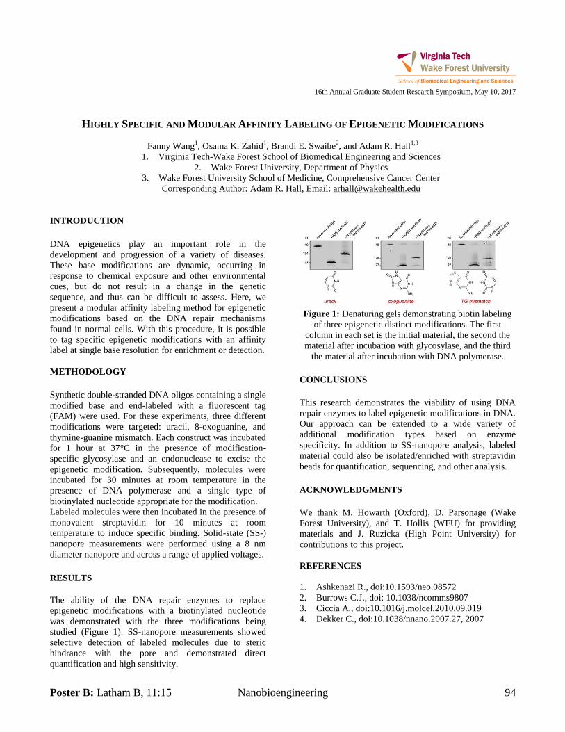
Poster B: Latham B, 11:15 Nanobioengineering 94
16th Annual Graduate Student Research Symposium, May 10, 2017
HIGHLY SPECIFIC AND MODULAR AFFINITY LABELING OF EPIGENETIC MODIFICATIONS
Fanny Wang1, Osama K. Zahid
1, Brandi E. Swaibe
2, and Adam R. Hall
1,3
1. Virginia Tech-Wake Forest School of Biomedical Engineering and Sciences
2. Wake Forest University, Department of Physics
3. Wake Forest University School of Medicine, Comprehensive Cancer Center
Corresponding Author: Adam R. Hall, Email: [email protected]
INTRODUCTION
DNA epigenetics play an important role in the
development and progression of a variety of diseases.
These base modifications are dynamic, occurring in
response to chemical exposure and other environmental
cues, but do not result in a change in the genetic
sequence, and thus can be difficult to assess. Here, we
present a modular affinity labeling method for epigenetic
modifications based on the DNA repair mechanisms
found in normal cells. With this procedure, it is possible
to tag specific epigenetic modifications with an affinity
label at single base resolution for enrichment or detection.
METHODOLOGY
Synthetic double-stranded DNA oligos containing a single
modified base and end-labeled with a fluorescent tag
(FAM) were used. For these experiments, three different
modifications were targeted: uracil, 8-oxoguanine, and
thymine-guanine mismatch. Each construct was incubated
for 1 hour at 37°C in the presence of modification-
specific glycosylase and an endonuclease to excise the
epigenetic modification. Subsequently, molecules were
incubated for 30 minutes at room temperature in the
presence of DNA polymerase and a single type of
biotinylated nucleotide appropriate for the modification.
Labeled molecules were then incubated in the presence of
monovalent streptavidin for 10 minutes at room
temperature to induce specific binding. Solid-state (SS-)
nanopore measurements were performed using a 8 nm
diameter nanopore and across a range of applied voltages.
RESULTS
The ability of the DNA repair enzymes to replace
epigenetic modifications with a biotinylated nucleotide
was demonstrated with the three modifications being
studied (Figure 1). SS-nanopore measurements showed
selective detection of labeled molecules due to steric
hindrance with the pore and demonstrated direct
quantification and high sensitivity.
Figure 1: Denaturing gels demonstrating biotin labeling
of three epigenetic distinct modifications. The first
column in each set is the initial material, the second the
material after incubation with glycosylase, and the third
the material after incubation with DNA polymerase.
CONCLUSIONS
This research demonstrates the viability of using DNA
repair enzymes to label epigenetic modifications in DNA.
Our approach can be extended to a wide variety of
additional modification types based on enzyme
specificity. In addition to SS-nanopore analysis, labeled
material could also be isolated/enriched with streptavidin
beads for quantification, sequencing, and other analysis.
ACKNOWLEDGMENTS
We thank M. Howarth (Oxford), D. Parsonage (Wake
Forest University), and T. Hollis (WFU) for providing
materials and J. Ruzicka (High Point University) for
contributions to this project.
REFERENCES
1. Ashkenazi R., doi:10.1593/neo.08572
2. Burrows C.J., doi: 10.1038/ncomms9807
3. Ciccia A., doi:10.1016/j.molcel.2010.09.019
4. Dekker C., doi:10.1038/nnano.2007.27, 2007
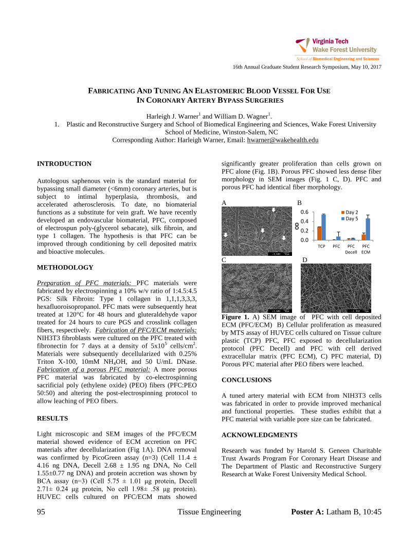
95 Tissue Engineering Poster A: Latham B, 10:45
16th Annual Graduate Student Research Symposium, May 10, 2017
FABRICATING AND TUNING AN ELASTOMERIC BLOOD VESSEL FOR USE
IN CORONARY ARTERY BYPASS SURGERIES
Harleigh J. Warner1 and William D. Wagner
1.
1. Plastic and Reconstructive Surgery and School of Biomedical Engineering and Sciences, Wake Forest University
School of Medicine, Winston-Salem, NC
Corresponding Author: Harleigh Warner, Email: [email protected]
INTRODUCTION
Autologous saphenous vein is the standard material for
bypassing small diameter (<6mm) coronary arteries, but is
subject to intimal hyperplasia, thrombosis, and
accelerated atherosclerosis. To date, no biomaterial
functions as a substitute for vein graft. We have recently
developed an endovascular biomaterial, PFC, composed
of electrospun poly-(glycerol sebacate), silk fibroin, and
type 1 collagen. The hypothesis is that PFC can be
improved through conditioning by cell deposited matrix
and bioactive molecules.
METHODOLOGY
Preparation of PFC materials: PFC materials were
fabricated by electrospinning a 10% w/v ratio of 1:4.5:4.5
PGS: Silk Fibroin: Type 1 collagen in 1,1,1,3,3,3,
hexafluoroisopropanol. PFC mats were subsequently heat
treated at 120°C for 48 hours and gluteraldehyde vapor
treated for 24 hours to cure PGS and crosslink collagen
fibers, respectively. Fabrication of PFC/ECM materials:
NIH3T3 fibroblasts were cultured on the PFC treated with
fibronectin for 7 days at a density of 5x10⁵ cells/cm
2.
Materials were subsequently decellularized with 0.25%
Triton X-100, 10mM NH4OH, and 50 U/mL DNase.
Fabrication of a porous PFC material: A more porous
PFC material was fabricated by co-electrospinning
sacrificial poly (ethylene oxide) (PEO) fibers (PFC:PEO
50:50) and altering the post-electrospinning protocol to
allow leaching of PEO fibers.
RESULTS
Light microscopic and SEM images of the PFC/ECM
material showed evidence of ECM accretion on PFC
materials after decellularization (Fig 1A). DNA removal
was confirmed by PicoGreen assay (n=3) (Cell 11.4 ±
4.16 ng DNA, Decell 2.68 ± 1.95 ng DNA, No Cell
1.55±0.77 ng DNA) and protein accretion was shown by
BCA assay (n=3) (Cell 5.75 ± 1.01 μg protein, Decell
2.71± 0.24 μg protein, No cell 1.98± .58 μg protein).
HUVEC cells cultured on PFC/ECM mats showed
significantly greater proliferation than cells grown on
PFC alone (Fig. 1B). Porous PFC showed less dense fiber
morphology in SEM images (Fig. 1 C, D). PFC and
porous PFC had identical fiber morphology.
A B
C D
Figure 1. A) SEM image of PFC with cell deposited
ECM (PFC/ECM) B) Cellular proliferation as measured
by MTS assay of HUVEC cells cultured on Tissue culture
plastic (TCP) PFC, PFC exposed to decellularization
protocol (PFC Decell) and PFC with cell derived
extracellular matrix (PFC ECM), C) PFC material, D)
Porous PFC material after PEO fibers were leached.
CONCLUSIONS
A tuned artery material with ECM from NIH3T3 cells
was fabricated in order to provide improved mechanical
and functional properties. These studies exhibit that a
PFC material with variable pore size can be fabricated.
ACKNOWLEDGMENTS
Research was funded by Harold S. Geneen Charitable
Trust Awards Program For Coronary Heart Disease and
The Department of Plastic and Reconstructive Surgery
Research at Wake Forest University Medical School.
0.0
0.2
0.4
0.6
TCP PFC PFCDecell
PFCECM
OD
Day 2Day 5
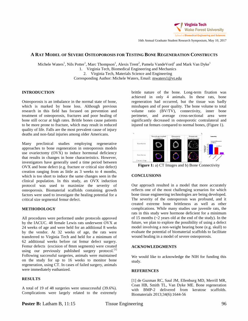
Poster B: Latham B, 11:15 Tissue Engineering 96
16th Annual Graduate Student Research Symposium, May 10, 2017
A RAT MODEL OF SEVERE OSTEOPOROSIS FOR TESTING BONE REGENERATION CONSTRUCTS
Michele Waters1, Nils Potter
1, Marc Thompson
1, Alexis Trent
2, Pamela VandeVord
1 and Mark Van Dyke
1 1. Virginia Tech, Biomedical Engineering and Mechanics
2. Virginia Tech, Materials Science and Engineering
Corresponding Author: Michele Waters, Email: [email protected]
INTRODUCTION
Osteoporosis is an imbalance in the normal state of bone,
which is marked by bone loss. Although previous
research in this field has focused on prevention and
treatment of osteoporosis, fractures and poor healing of
bone still occur at high rates. Brittle bones cause patients
to be more prone to fracture, which may result in reduced
quality of life. Falls are the most prevalent cause of injury
deaths and non-fatal injuries among older Americans.
Many preclinical studies employing regenerative
approaches to bone regeneration in osteoporosis models
use ovariectomy (OVX) to induce hormonal deficiency
that results in changes in bone characteristics. However,
investigators have generally used a time period between
OVX and bone defect (e.g. fracture or critical size defect)
creation ranging from as little as 3 weeks to 4 months,
which is too short to induce the same changes seen in the
clinical population. In this study, an OVX induction
protocol was used to maximize the severity of
osteoporosis. Biomaterial scaffolds containing growth
factors were used to investigate the healing potential for a
critical size segmental femur defect.
METHODOLOGY
All procedures were performed under protocols approved
by the IACUC. 48 female Lewis rats underwent OVX at
24 weeks of age and were held for an additional 8 weeks
by the vendor. At 32 weeks of age, the rats were
transferred to Virginia Tech and held for a minimum of
62 additional weeks before rat femur defect surgery.
Femur defects (excision of 8mm segments) were created
using our previously published surgery protocol.[1]
Following successful surgeries, animals were maintained
on the study for up to 16 weeks to monitor bone
regeneration, using CT. In cases of failed surgery, animals
were immediately euthanized.
RESULTS
A total of 19 of 48 surgeries were unsuccessful (39.6%).
Complications were largely related to the extremely
brittle nature of the bone. Long-term fixation was
achieved in only 4 animals. In these rats, bone
regeneration had occurred, but the tissue was badly
misshapen and of poor quality. The bone volume to total
volume ratio (BV/TV), connectivity, inner bone
perimeter, and average cross-sectional area were
significantly decreased in osteoporotic contralateral and
injured rat femurs compared to normal bones, (Figure 1).
Figure 1: a) CT Images and b) Bone Connectivity
CONCLUSIONS
Our approach resulted in a model that more accurately
reflects one of the most challenging scenarios for which
bone tissue engineering technologies are being developed.
The severity of the osteoporosis was profound, and it
created extreme bone brittleness as well as other
complications. While many studies use juvenile rats, the
rats in this study were hormone deficient for a minimum
of 15 months (~2 years old at the end of the study). In the
future, we plan to explore the possibility of using a defect
model involving a non-weight bearing bone (e.g. skull) to
evaluate the potential of biomaterial scaffolds to facilitate
wound healing in a model of severe osteoporosis.
ACKNOWLEDGMENTS
We would like to acknowledge the NIH for funding this
study.
REFERENCES
[1] de Guzman RC, Saul JM, Ellenburg MD, Merrill MR,
Coan HB, Smith TL, Van Dyke ME. Bone regeneration
with BMP-2 delivered from keratose scaffolds.
Biomaterials 2013;34(6):1644-56
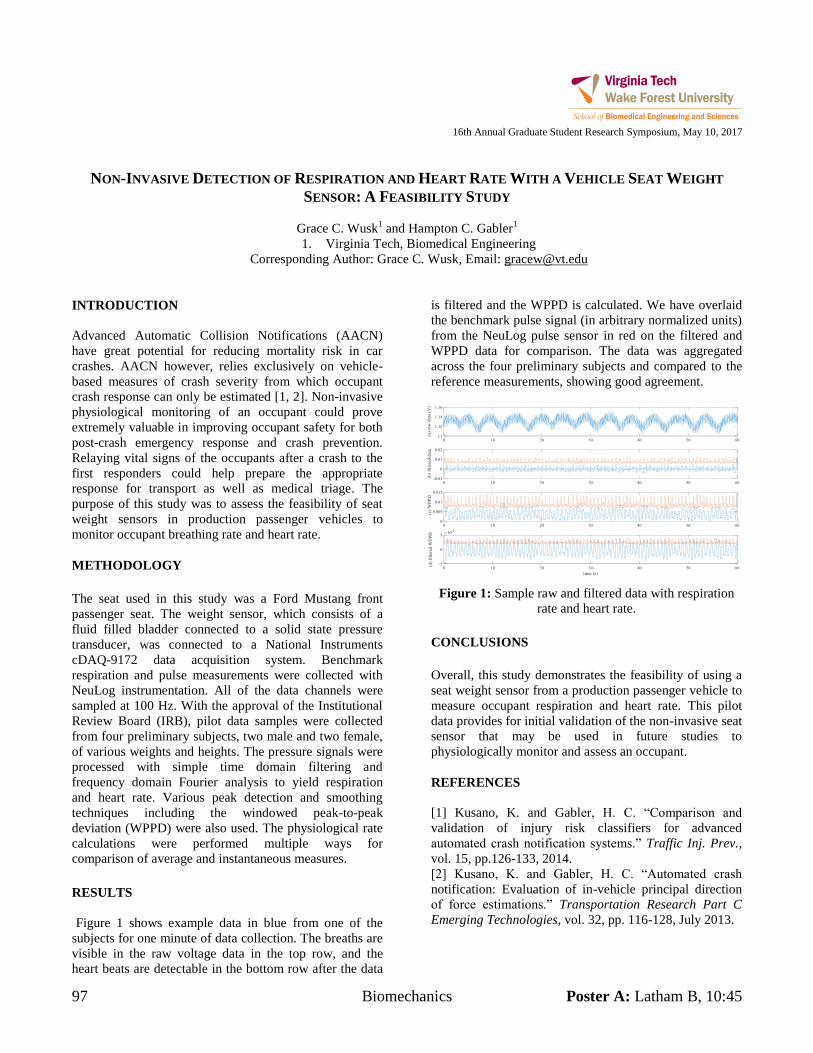
97 Biomechanics Poster A: Latham B, 10:45
16th Annual Graduate Student Research Symposium, May 10, 2017
NON-INVASIVE DETECTION OF RESPIRATION AND HEART RATE WITH A VEHICLE SEAT WEIGHT
SENSOR: A FEASIBILITY STUDY
Grace C. Wusk1 and Hampton C. Gabler
1
1. Virginia Tech, Biomedical Engineering
Corresponding Author: Grace C. Wusk, Email: [email protected]
INTRODUCTION
Advanced Automatic Collision Notifications (AACN)
have great potential for reducing mortality risk in car
crashes. AACN however, relies exclusively on vehicle-
based measures of crash severity from which occupant
crash response can only be estimated [1, 2]. Non-invasive
physiological monitoring of an occupant could prove
extremely valuable in improving occupant safety for both
post-crash emergency response and crash prevention.
Relaying vital signs of the occupants after a crash to the
first responders could help prepare the appropriate
response for transport as well as medical triage. The
purpose of this study was to assess the feasibility of seat
weight sensors in production passenger vehicles to
monitor occupant breathing rate and heart rate.
METHODOLOGY
The seat used in this study was a Ford Mustang front
passenger seat. The weight sensor, which consists of a
fluid filled bladder connected to a solid state pressure
transducer, was connected to a National Instruments
cDAQ-9172 data acquisition system. Benchmark
respiration and pulse measurements were collected with
NeuLog instrumentation. All of the data channels were
sampled at 100 Hz. With the approval of the Institutional
Review Board (IRB), pilot data samples were collected
from four preliminary subjects, two male and two female,
of various weights and heights. The pressure signals were
processed with simple time domain filtering and
frequency domain Fourier analysis to yield respiration
and heart rate. Various peak detection and smoothing
techniques including the windowed peak-to-peak
deviation (WPPD) were also used. The physiological rate
calculations were performed multiple ways for
comparison of average and instantaneous measures.
RESULTS
Figure 1 shows example data in blue from one of the
subjects for one minute of data collection. The breaths are
visible in the raw voltage data in the top row, and the
heart beats are detectable in the bottom row after the data
is filtered and the WPPD is calculated. We have overlaid
the benchmark pulse signal (in arbitrary normalized units)
from the NeuLog pulse sensor in red on the filtered and
WPPD data for comparison. The data was aggregated
across the four preliminary subjects and compared to the
reference measurements, showing good agreement.
Figure 1: Sample raw and filtered data with respiration
rate and heart rate.
CONCLUSIONS
Overall, this study demonstrates the feasibility of using a
seat weight sensor from a production passenger vehicle to
measure occupant respiration and heart rate. This pilot
data provides for initial validation of the non-invasive seat
sensor that may be used in future studies to
physiologically monitor and assess an occupant.
REFERENCES
[1] Kusano, K. and Gabler, H. C. “Comparison and
validation of injury risk classifiers for advanced
automated crash notification systems.” Traffic Inj. Prev.,
vol. 15, pp.126-133, 2014.
[2] Kusano, K. and Gabler, H. C. “Automated crash
notification: Evaluation of in-vehicle principal direction
of force estimations.” Transportation Research Part C
Emerging Technologies, vol. 32, pp. 116-128, July 2013.
Poster B: Latham B, 11:15 Translational Cancer 98
16th Annual Graduate Student Research Symposium, May 10, 2017
INVESTIGATION OF MLL3 COMPLEX IN DNA REPAIR MECHANISM IN LUNG ADENOCARCINOMA
Ted G. Xiao1, Wei Zhang
2
1. Wake Forest School of Medicine, Virginia Tech-Wake Forest School of Biomedical Engineering and Sciences
2. Wake Forest School of Medicine, Department of Cancer Biology
Corresponding Author: Ted Xiao, Email: [email protected]
INTRODUCTION
Increasing efforts have been put forth in exploring post-
transcriptional and translational modifications. Histone
modification is in the center of this increased effort.
MLL3 is a chromatin modifier that facilitates the
deposition of histone 3 lysine 4 mono- and dimethylation
(H3K4me1/me2). MLL3/4 assembles as a complex,
leading to modifications to histone methylation.
A recent study demonstrates a direct connection between
Forkhead box protein A1 (FOXA1) and MLL31. The
results indicate that FOXA1 transcription factor recruits
MLL3 to mono-methylate H3K4 sites at the enhancer. In
another recent study, FOXA1 is reported to associate
DNA repair complexes and DNA demethylation2.
There is a study in Arabidopsis that showed histone
acetyltransferase regulating DNA demethylation3. Given
the frequent occurrences of MLL3 mutation in lung
adenocarcinoma patients4, we hypothesize that histone
methyltransferase protein MLL3/4 plays a crucial role in
FOXA1-associated DNA repair.
METHODOLOGY
Lung Cancer Cell Lines
Three (NCI-H1975, NCI-H522, HCC827) lines have no
MLL3/4 mutations. One line (NCI-H2342) has MLL3
mutation and the other (NCI-H2135) has MLL4 mutation.
The non-mutated lines require RPMI-1640 medium and the
mutant lines require DMEM-F12 medium with supplements.
All cell lines are cultured in standard cell culture conditions.
Cisplatin Treatment and Protein Extraction
Add cisplatin (5mM) to medium at 1:50. Mix the solution
and apply to cell lines. Incubate the cells at 37C for 30-45
minutes. Wash with iced PBS. Make sure to dish is as dry
as possible. Transfer the cell culture dishes onto ice. Add
cell lysis solution and wait 30 minutes at 4C. Scrape off
the lysate and transfer into a microfuge tube. Sonicate at
40% amplitude. Spin in centrifuge at 4C and 15,000 rpm.
Isolate the protein lysate. Store at -80C.
Western Blot
Heat for 5 minutes at 100C and load protein lysates into
pre-cast gel (40μg). Run gel electrophoresis at 60V for 20
minutes and turn up the voltage to 120V for 40-60
minutes. Transfer gel into transfer cassette with PVDF
membrane. Place cassette into transfer box and turn
voltage to 20V at 4C overnight. Use 5% milk for blocking
at 4C on a shaker at 20rpm for >1hr. Add primary
antibody and place on shaker at 20rpm at 4C overnight.
Add corresponding secondary antibody for 1hr. Expose
with Supersignal Chemiluminiscent Substrate.
Transfection
Seed cells onto 6-well culture dish at 60-80% confluency.
Dilute Lipofectamine RNAiMAX Reagent in Opti-MEM
Medium. Dilute siRNA in Opti-MEM Medium. Mix the
two solutions at 1:1 ratio. Incubate at room temperature
for 5 minutes. Add siRNA-lipid complex to cells.
Incubate cells for 1-3 days at 37C before examination.
RESULTS
The expected result based on literatures is a positive
correlation between MLL3/4 expression and DNA repair
activation. If true, the western blot results should show
DNA repair protein expression based on the presence of
MLL3/4. If MLL3/4 are knocked down via siRNA, DNA
repair protein expression is expected to be decreased
compared to control.
ACKNOWLEDGMENTS
The author would like to thank the members of the Zhang
Lab for their help.
REFERENCES
1. Jozwik, K., Chernukhin, I., Serandour, A. (2016). Cell
Reports 17, 2715-2723.
2. Zhang, Y., Zhang, D., Li, Q. (2016). Nature Genetics
48, 1003-1013.
3. Qian, W., Miki, D., Zhang, H. (2012). Science 336, 1445-1448.
4. TCGA data analysis performed in Zhang Lab.
99 Biomechanics Poster A: Latham B, 10:45
16th Annual Graduate Student Research Symposium, May 10, 2017
DEVELOPMENT AND VALIDATION OF A GÖTTINGEN MINIATURE PIG FINITE ELEMENT MODEL TO
INVESTIGATE INJURY SCALING TECHNIQUES
Keegan M. Yates1, Costin D. Untaroiu
1
1. Virginia Tech, Center for Injury Biomechanics
Corresponding Author: Keegan Yates, Email: [email protected]
INTRODUCTION
Every year, approximately 1.7 million people in the
United States receive a traumatic brain injury (TBI). In
order to better prevent or mitigate these injuries, several
kinematic injury risk metrics have been developed. The
development of these metrics is dependent on animal
studies because many types of brain injury can only be
seen in living tissue. This presents a challenge, as any
animal data must be scaled to apply to a human brain.
Current methods rely on scaling based solely on the
masses of the brains. Utilizing finite element (FE)
analysis, impacts can be simulated on an animal model,
and injury can be correlated with mesh based factors,
which can then be used as a target to direct an optimized
scaling method.
METHODOLOGY
Prior to this study, Göttingen mini-pigs were given mild
TBIs in an impact device, and the motion of markers
implanted in the interior of the brain was recorded via
biplanar x-ray [1]. An FE model of a pig head was created
by meshing MRI (brain) and CT (skull) scans of a pig.
The pig impacts were recreated with the FE model, and
the in vivo marker data was used to validate the model.
Next, scaling was investigated with the pig model and an
established human brain FE model (GHBMC). First, two
mesh based injury metrics were established. The first was
the cumulative strain damage measure (CSDM), which
was a volume fraction of elements receiving a strain over
2% throughout the duration of the impact. The second
was a volume fraction of elements experiencing a
pressure over 10 kPa. Next, mass based kinematic scaling
[2] was applied to obtain a baseline scaled impact to the
human model. Finally, the scaling factors for linear and
angular motion were adjusted such that the injury metrics
in the scaled human impact matched the pig impact.
RESULTS
It was seen that the baseline mass based scaling greatly
over-predicted the strain to the human brain, while
pressure was slightly below or equal to what was
predicted. Through adjusting the scaling factors, the
injury metrics in the scaled human impact were made
much closer to the pig impact (Figure 1).
CONCLUSIONS
The results have shown that mass based scaling alone can
either over or under-predict certain injury types. While
mesh based injury metrics have not been well established
for this type of mild TBI, this study has shown as a proof
of concept that an FE method can be used to refine injury
scaling. This scaling can be tailored to specific injury
types or loading types, and therefore it has the possibility
of improving prediction of injury in humans.
ACKNOWLEDGMENTS
This research was sponsored by the Takata Corporation.
The experimental data used to construct the pig model
were obtained by Elizabeth Fievisohn.
REFERENCES
[1] E. M. Fievisohn et al., “Evaluation of Impact-
Induced Traumatic Brain Injury in the Göttingen Minipig
Using Two Input Modes,” Traffic Inj. Prev., vol. 15, no.
sup1, pp. S81–S87, 2014.
[2] A. Petitjean et al., “Normalization and scaling
for human response corridors and development of injury
risk curves,” in Accidental Injury, Springer, 2015, pp.
769–792.
Figure 1. Strain (a) and pressure (b) response of
scaled human model compared to the pig model.
a b
Poster B: Latham B, 11:15 Biomechanics 100
16th Annual Graduate Student Research Symposium, May 10, 2017
NUMERICAL INVESTIGATION OF LOWER EXTREMITY INJURIES IN FRONTAL CRASH RECONSTRUCTION
Xin Ye1,2
, James Gaewsky1,2
, Derek Jones1,2
, Logan Miller1,2
, Mireille Kelly1,2
, Jeff Suhey1,2
, Bharath Koya1,2
,Ashley
Weaver1,2
and Joel Stitzel1,2
1. Wake Forest University School of Medicine
Virginia Tech-Wake Forest University Center for Injury Biomechanics
Corresponding Author: Xin Ye, Email: [email protected]
INTRODUCTION
Lower extremity injuries are the most frequent
Abbreviated Injury Scale (AIS) 2 injury for drivers in
frontal crashes [1]. The objective was to reconstruct
eleven real-world motor vehicle crashes (two with AIS 2+
distal lower extremity injury and nine without lower
extremity injury) to analyze the vehicle parameters and
driver attributes that affect injury risk.
METHODOLOGY
Eleven frontal crashes were reconstructed with a finite
element simplified vehicle model (SVM) using a semi-
automated optimization method. The SVM was tuned to
each corresponding vehicle and the Total HUman Model
for Safety (THUMS) v4.01 was scaled and positioned in a
baseline configuration to mimic the documented pre-crash
driver posture. The event data recorder crash pulse was
applied as the boundary condition for each case.
Additionally, for each of the eleven cases, 120 simulations
to quantify the uncertainty and response variation were
performed varying the following parameters using a Latin
Hypercube design of experiments (DOE): seat track
position, seatback angle, steering column angle, steering
column position, and D-ring height. Injury metrics
implemented within THUMS were calculated from the
femur, tibia, and ankle, and cross-compared among the
eleven baseline cases using tibia index and multiple injury
risk functions. Kinetic and kinematic data from the 120
simulation DOE were analyzed and fit to regression models
to examine causal relationships between occupant
positioning and lower extremity injury risks.
RESULTS
Of the eleven real-world crashes, both cases with lower
extremity injuries resulted in elevated tibia axial forces and
resultant bending moments, compared to the nine cases
without lower extremity injury. The average tibia index of
the two cases with distal lower extremity injury (left: 1.79;
right: 1.19) was higher than the nine cases without lower
extremity injury (left: 1.16, p=0.024; right: 0.82, p= 0.024).
An increased risk of AIS 2+ tibia shaft (33.6%), distal tibia
(20.0%), and ankle (14.5%) fracture was also observed for
the injured compared to the non-injured cases. Rearward
seat track position, reclined seat back angle and reduced
seat height were correlated with elevated tibia axial force
and increased tibia index, imposing additional lower
extremity injury risk (Figure 1).
Figure 1: Linear regression of lower left tibia index
versus seat track position for Camry DOE. Red circle
indicated the baseline case.
CONCLUSIONS
This study provides a computational framework for assessing
lower extremity injuries, and elucidates the effect of pre-
crash driver posture on lower extremity injury risk while
accounting for vehicle parameters and driver attributes.
Results from the study aid in the evaluation of real-world
injury data, the understanding of factors contributing to
injury risk and the prevention of lower extremity injuries.
ACKNOWLEDGMENTS
Funding for this project was provided by Toyota’s
Collaborative Safety Research Center.
REFERENCES
[1]Ye X. et al., Accident Analysis& Prevention. 2015, 83:37-46.
101 Cardiovascular Engineering Poster A: Latham B, 10:45
Figure 1: Pre-sorting, brightfield and red-
fluorescence (left) and after-sorting, brightfield and
red-fluorescence (right) (10X).
Figure 2: Primitive vessel formation in a collagen
matrix (left, 20X) in basic 2D co-culture (Right, 10X)
16th Annual Graduate Student Research Symposium, May 10, 2017
PERICYTE CELL LINE ISOLATION, VALIDATION AND APPLICATIONS
Huaning Zhao1, John C. Chappell
1,2
1. School of Biomedical Engineering and Science
2. Center for Heart and Regenerative Medicine, Virginia Tech Carilion Research Institute
Corresponding Author: John C. Chappell, Email: [email protected]
INTRODUCTION
Pericytes, also known as perivascular or mural cells,
envelope the outer surface of vascular endothelial cells
and play a key role in microvessel homeostasis. Pericyte
functionality in healthy and pathological conditions has
recently gained significant attention, as numerous studies
have found that pericyte dysfunction or loss of pericytes
indicate the beginning of vascular diseases, such as in
diabetic retinopathy and neurodegenerative conditions.
However, isolation and validation of a pericyte cell line
has remained a challenge since no specific genetic marker
exists. In my research, I have isolated an enriched
population of pericytes from mouse embryos. I validated
this cell line using several expression and functional
assays, and applied these cells in experiments of basic
vessel development. I found that these cells displayed
similar functions as endogenous pericytes, and they
appear to uniquely contribute to vessel development.
METHODOLOGY
A pericyte cell line was isolated from NG2:DsRed
transgenic mouse embryos at embryonic day 13.5 (E13.5),
using Fluorescence Activated Cell Sorting (FACS). These
cells were cultured in Pericyte Media for expansion, and
stored in liquid nitrogen at passage 2 (p2). As a primary
cell line, this cell line was validated to maintain normal
proliferation and morphological features until p10. In
subsequent experiments, we used cells between p3-6.
RESULTS
From images taken before and after FACS, we found that
both the density of cells with DsRed fluorescence and the
intensity of the DsRed signal were higher after FACS (Fig
1). From quantitative analysis of cell density, the
concentration of pericytes was found to increase almost
three fold after FACS.
In validation experiments, I compared the morphology
and gene expression of these cells with a related but
distinct cell type, mouse embryonic fibroblasts (MEFs).
Several lines of evidence suggest that these cell lines are
indeed distinct cell types. In addition, I applied these
exogenous pericytes to vessels forming in differentiated
mouse embryoid bodies, mouse embryonic back skin
(E13.5) and compared them with endogenous pericytes.
Our results suggest that our pericyte cell line displays
similar morphology and function features in embryoid
body-derived blood vessel development as endogenous
pericytes. They also readily engage developing blood
vessels and are morphologically similar to endogenous
pericytes on a tissue level (embryonic back skin E13.5). I
cultured these cells with Human Umbilical Vein
Endothelial Cells (HUVECs) and found unique aspects to
their contribution to primitive vessel development (Fig 2).
CONCLUSIONS
This pericyte cell line has been validated, and can be used
to better understand embryonic vessel development, as
well as pericyte dysfunction in different pathology-related
culture conditions (e.g. hypoxia, hyperglycemia, etc.).
ACKNOWLEDGMENTS
Members of the Chappell Lab, and my mentor, Dr. John
Chappell.


