Embed Size (px)
Citation preview
SYNTHESIS, SPECTROSCOPIC ANALYSES AND
BIOLOGICAL EVALUATION OF SILVER AND
GOLD BASED PRO-NANOMEDICINES DERIVED
FROM FLUOROQUINOLONES
Ph.D Thesis
By
SHUJAAT ALI KHAN
INSTITUTE OF CHEMICAL SCIENCES
UNIVERSITY OF PESHAWAR,
PAKISTAN
January 2017
SYNTHESIS, SPECTROSCOPIC ANALYSES AND
BIOLOGICAL EVALUATION OF SILVER AND
GOLD BASED PRO-NANOMEDICINES DERIVED
FROM FLUOROQUINOLONES
By
SHUJAAT ALI KHAN
DISSERTATION
SUBMITTED TO THE UNIVERSITY OF PESHAWAR IN
PARTIAL FULFILLMENT OF THE REQUIREMENTS
FOR THE DEGREE OF DOCTOR OF PHILOSOPHY
IN CHEMISTRY
INSTITUTE OF CHEMICAL SCIENCES
UNIVERSITY OF PESHAWAR,
PAKISTAN
January 2017
INSTITUTE OF CHEMICAL SCIENCES
UNIVERSITY OF PESHAWAR
PAKISTAN
It is recommended that this dissertation prepared by Mr. Shujaat Ali Khan entitled
“Synthesis, Spectroscopic Analyses and Biological Evaluation of Silver and Gold
Based Pro-nanomedicines derived from Fluoroquinolones” be accepted as
fulfilling this part of the requirements for the degree of “Doctor of Philosophy in
Chemistry”.
________________________ ________________________
SUPERVISOR CO-SUPERVISOR
Meritorious Prof. Dr. Muhammad Nisar Prof. Dr. Ghias Uddin
Institute of Chemical Sciences, Institute of Chemical Sciences,
University of Peshawar University of Peshawar
________________________ ________________________ INTERNAL EVALUATOR/ EXAMINER Meritorious Prof. Dr. Jasmin Shah Prof. Dr. Muhammad Arfan Director,
HEJ, Research Institute of Chemistry Institute of Chemical Sciences,
ICCBS, university of Karachi University of Peshawar
________________________
EXAMINER
Prof. Dr. Hamidullah Shah
Dean, Faculty of Nutrition Sciences,
Agriculture University, Peshawar
________________________
INTERNAL EXAMINER
Prof. Dr. Muhammad Ishaq
Institute of Chemical Sciences,
University of Peshawar
Author’s Declaration
I Mr. Shujaat Ali Khan hereby state that my Ph.D thesis titled “Synthesis,
Spectroscopic Analyses and Biological Evaluation of Silver and Gold Based Pro-
nanomedicines derived from Fluoroquinolones” is my own work and has not been
submitted to previously by me for taking any degree from this University of Peshawar
or anywhere else in the country/world.
At any time if my statement is found to be incorrect even after my graduation the
University has the right to withdraw my Ph.D degree.
________________
Shujaat Ali Khan
Date: / /2018

DEDICATION
TO
MY LOVING PARENTS
Whose encouragement towards knowledge served me
As beacon of light
And to my younger
Brother and Sister
For their sweet feelings who always
Pray for my bright future and success
Table of Contents
S. No. Title Page No.
Acknowledgments...................................................................................................... i
List of Figures ........................................................................................................... iii
List of Tables ............................................................................................................. ix
List of Schemes .......................................................................................................... x
List of Abbreviations ................................................................................................ xi
Summary ................................................................................................................... xiii
Chapter 1 General introduction
1.1 History of quinolones .................................................................................... 1
1.2 Quinolone nucleus......................................................................................... 1
1.3 Classification ................................................................................................. 2
1.3.1 First generation ............................................................................................. 2
1.3.2 Second generation ......................................................................................... 2
1.3.3 Third generation ............................................................................................ 3
1.3.4 Fourth generation .......................................................................................... 4
1.4 Mechanism of action of quinolones and fluoroquinolones ...................... 5
1.5 Medicinal plants ..................................................................................... 6
1.6 Phytochemistry and bioactivities of Rhododendron arboretum ................... 7
1.6.1 Plant introduction .......................................................................................... 7
1.7 Literature review of genus Rhododendron .................................................... 8
1.7.1 Chemical constituents of the genus Rhododendron ................................................. 8
1.7.2 Medicinal and pharmacological properties ....................................................... 9
1.8 Phytochemistry and bioactivities of Eulophia dabia .................................... 9
1.8.1 Plants introduction ........................................................................................ 9
1.9 Literature review of genus Eulophia ....................................................... 10
1.9.1 Chemical constituents of the genus Eulophia ......................................... 10
1.9.2 Medicinal and Pharmacological Properties ............................................. 10
1.10 Phytochemistry and bioactivities of Kigelia pinnata ........................................ 11
1.10.1 Plant introduction .............................................................................. 11
1.11 Literature review of genus Kigelia .................................................................... 11
1.11.1 Plant introduction .............................................................................. 11
1.11.2 Medicinal and pharmacological properties....................................... 12
1.12 Phytochemistry and bioactivities of Desmodium elegans ................................ 12
1.12.1 Plant introduction .................................................................... 12
1.13 Literature review of genus Desmodium ............................................................. 13
1.13.1 Chemical constituents of the genus Desmodium .............................. 13
1.13.2 Medicinal and pharmacological properties....................................... 14
1.14 Biological evaluation ......................................................................................... 14
1.14.1 Urease inhibition activity ................................................................... 14
1.14.2 Leishmanicidal studies ....................................................................... 15
1.14.3 Antioxidant assay ............................................................................... 15
1.14.4 Antibacterial assay ............................................................................. 16
1.14.5 Antifungal activity ............................................................................. 16
Chapter 2 Introduction to Nanotechnology
2.1 Nanotechnology ..................................................................................... 18
2.2 Noble metal NPs .................................................................................... 18
2.3 Synthesis ................................................................................................ 19
2.4 Characterization techniques .................................................................... 22
2.5 Applications ........................................................................................... 22
2.6 Properties ............................................................................................... 25
2.6.1 Optical properties ................................................................................... 25
2.6.2 Biomedical properties ............................................................................ 25
2.6.3 Magnetic properties ................................................................................ 26
2.6.4 Electronic properties .............................................................................. 26
2.6.5 Energy properties ................................................................................... 26
2.7 Aims and objectives of the present research ................................................. 26
2.8 Review on topical advancements in noble metal NPs (28) ........................... 27
2.8.1 Review on Ag NPs ................................................................................. 27
2.8.2 Review on Au NPs ................................................................................. 31
Chapter 3 Results and Discussion
3.1 Results and discussion ............................................................................ 36
3.1.1 Synthesis of Ag-Mox and Au-Mox NPs capped with
moxifloxacin ................................................................................................. 36
3.1.2 Characterization of moxifloxacin-capped Ag/Au NPs ................................. 38

3.1.3 AFM, SEM and EDX spectroscopy analyses ............................................... 40
3.1.4 Stability check of Ag and Au NPs ................................................................ 41
3.2 Synthesis of ciprofloxacin-capped metallic NPs .......................................... 43
3.2.1 FTIR and AFM studies ................................................................................. 45
3.2.2 EDX and SEM studies .................................................................................. 47
3.2.3 Stability check of Ag and Au NPs ................................................................ 48
3.3 Synthesis of sparfloxacin mediated Ag and Au NPs .................................... 51
3.3.1 UV visible spectroscopic analysis ................................................................. 51
3.3.2 AFM, SEM and EDX analyses ..................................................................... 52
3.3.3 Fourier transform infrared spectroscopy (FTIR) .......................................... 53
3.3.4 Stability check of Ag and Au NPs ................................................................ 54
3.4 Synthesis of gemifloxacin-capped noble metal NPs ..................................... 59
3.4.1 UV visible spectroscopy for synthesis of Ag/Au NPs .................................. 59
3.4.2 AFM, SEM and EDX spectroscopy analysis ................................................ 60
3.4.3 FTIR analysis ................................................................................................ 62
3.4.4 Stability check of Ag and Au NPs ................................................................ 63
3.5 Biological evaluation of fluoroquinolones-capped Ag/Au NPs ................... 67
3.5.1 Urease study .................................................................................................. 67
3.5.2 Leishmanicidal activity ................................................................................. 69
3.5.3 Antioxidant activity....................................................................................... 71
3.5.4 Antibacterial activity ..................................................................................... 73
3.5.5 Antifungal activity ........................................................................................ 74
3.6 Green synthesis of noble metal NPs by using flower extract of
Rhododendron arboretum .............................................................................
76
3.6.1 Visual inspection and UV visible spectroscopy analysis .............................. 76
3.6.2 FTIR spectroscopy analysis .......................................................................... 78
3.6.3 Stability of the biosynthesized Ag/Au NPs ................................................... 79
3.6.4 AFM, SEM and EDX analysis ...................................................................... 82
3.7 Green synthesis of metallic NPs by using Kigelia pinnata ........................... 84
3.7.1 UV Visible spectroscopy .............................................................................. 84
3.7.2 FTIR analysis ................................................................................................ 85
3.7.3 Stability check of the synthesized Ag/Au NPs ............................................. 86
3.7.4 AFM and SEM analysis ................................................................................ 88
3.8 Green phytosynthesis of noble metal NPs using Eulophia dabia
extract as reducing and stabilizing agent ......................................................
89
3.8.1 Visual inspection and UV visible spectroscopy analysis .............................. 89
3.8.2 FTIR spectroscopy analysis .......................................................................... 89
3.8.3 Stability check of Ag and Au NPs ................................................................ 90
3.8.4 AFM and EDX analysis ................................................................................ 91
3.9 Synthesis of metallic NPs by using Desmodium elegans ............................. 93
3.9.1 UV visible spectroscopy ............................................................................... 93
3.9.2 FTIR Analysis ............................................................................................... 95
3.9.3 Stability check of synthesized metallic NPs ................................................. 96
3.9.4 SEM and EDX analysis................................................................................. 98
3.10 Biological evaluation of selected medicinal plants and their
NPs ................................................................................................................
100
3.10.1 Urease inhibition assay ................................................................. 100
3.10.2 Leishmanicidal activity ................................................................. 102
3.10.3 Antioxidant activity ....................................................................... 104
3.10.4 Antibacterial activity ..................................................................... 106
3.10.5 Antifungal activity ........................................................................ 107
Chapter 4 Experimental
4.1 General experimental procedures .................................................................. 109
4.1.1 Collection of fluoroquinolones and plants material ...................................... 109
4.1.2 Preparation of stock solution......................................................................... 110
4.1.3 Synthesis of Ag and Au NPs capped with fluoroquinolones ........................ 110
4.1.4 Green synthesis of metallic NPs by using selected medicinal
plants .............................................................................................................
111
4.2 Biological evaluation .................................................................................... 112
4.2.1 Protocol for urease assay and inhibition ............................................................ 112
4.2.2 Procedure for leishmanicidal activity (in vitro)................................................. 112
4.2.3 Antioxidant activity ............................................................................................ 113
4.2.4 Bacterial strains assortment and preservation ................................................... 114
4.2.5 Antibacterial activity .......................................................................................... 114
4.2.6 Antifungal activity .............................................................................................. 114
Conclusion 116
References 118
List of Publications 137
Biography 138
i
Acknowledgments
In the name of Almighty Allah (the most Beneficent, the most Merciful) who had
given me courage to accomplish this task. It would not have been possible without
His will and support. All respect for His Holy Prophet (SAW), who enabled us to
recognize our creator.
Pursuing a Ph.D. project is both painful and pleasant experience. It is just like
climbing a peak, step by step, accompanied with bitterness, hardships, frustration,
encouragement and trust. When I found myself at the top enjoying the beautiful
scenario, I realized that it was, in fact, teamwork that got me there. Though it will not
be enough to express my appreciation in simple words to all those people who helped
me, I would still like to give my many thanks to all these people.
First of all, I would like to give a bundle of thanks to my honorable supervisor, Dr.
Muhammad Nisar (meritorious professor), who accepted me as his Ph.D. student
without any reluctance. Furthermore, he patiently supervised me and always guided
me in the right way. I have learnt a lot from him, without his help, I could not have
completed my dissertation successfully.
It is difficult to express in words what I feel and what is in my heart for my respected
Co-supervisor Prof. Dr. Ghias Uddin and Assoc. Prof. Dr. Muhammad Raza Shah,
H.E.J. Research Institute of Chemistry, University of Karachi, because the work
presented in this dissertation would have never been completed without their keen
interest, precious attention, and continuous encouragement. By observing their
personality, hard work and devotion, I learnt about the key to success.
My deep appreciation and sincere thanks are due to all my teachers in I.C.S.,
especially Prof. Dr. Jasmin Shah (Director, ICS), Prof. Dr. Yousaf Iqbal, Prof. Dr.
Imtiaz Ahmad, Dr. Adnan Khan, Dr. Rasool Khan and Dr. Salman Zafar for their kind
help whenever I needed.
I would like to express my heartiest gratitude and regards to my mother, father,
younger brother, sister and my late grandfather (May Allah keep his soul in rest and
peace) for their love, prayers and encouragement throughout my studies. I would like
to express my appreciation and thanks to my colleagues Dr. Ajmal Khan, Ayaz Khan,
ii
Qamar Altaf Jaffery, Rahmanullah, Sher Ayaz, Hanif ur Rahman, Muhmmad Atif
Khan and Mansor Khan for providing a pleasant and helping atmosphere during my
studies.
I am thankful to Mr. Masood Jan and Mr. Zulfiqar (Lab Assistants) for their help in
every step during my bench work at the Institute of Chemical Sciences, University of
Peshawar.
I am also thankful to Higher Education Commission (HEC) of Pakistan for providing
necessary grant for access to spectroscopic facilities.
Last but not the least, I would like to thank all those who assisted and guided me in
the completion of this study.
Shujaat Ali Khan

iii
List of Figures
Figure No. Title Page No.
Fig. 1.1 Mechanism of action of fluoroquinolones..................................................... 5
Fig. 1.2 Photograph of Rhododendron arboreum flowers .......................................... 8
Fig. 1.3 Photograph of Eulophia dabia ...................................................................... 10
Fig. 1.4 Photograph of Kigelia pinnata .......................................................................... 11
Fig. 1.5 Photograph of Desmodium elegans .................................................................. 13
Fig. 2.1 Diagram for silver and gold nanomaterials ................................................... 18
Fig. 2.2 Illustrative chemical reduction diagrams for noble metal NPs
synthesis ........................................................................................................
20
Fig. 2.3 Optical inspection of the colloidal solution of noble metal NPs ................... 25
Fig. 3.1 Optical inspection of Ag-Mox NPs ............................................................... 37
Fig. 3.2 Optical inspection of Au-Mox NPs ............................................................... 37
Fig. 3.3 Optimized UV visible spectra of Ag-Mox NPs............................................. 38
Fig. 3.4 Optimized UV visible spectra of Au-Mox NPs............................................. 38
Fig. 3.5 FTIR spectra of Mox, Ag and Au NPs .......................................................... 39
Fig. 3.6 AFM images of Ag NPs ................................................................................ 40
Fig. 3.7 AFM images of Au NPs ................................................................................ 41
Fig. 3.8 SEM image of Ag-Mox ................................................................................. 41
Fig. 3.9 SEM image of Au-Mox ................................................................................. 41
Fig. 3.10 The EDX spectrum for Ag NPs ..................................................................... 41
Fig. 3.11 The EDX spectrum for Au NPs ..................................................................... 41
Fig. 3.12 Effect of brine on Ag-Mox ............................................................................ 42
Fig. 3.13 Effect of brine on Au-Mox ............................................................................ 42
Fig. 3.14 Effect of pH on stability of Ag NPs .............................................................. 43
Fig. 3.15 Effect of pH on stability of Au NPs .............................................................. 43
iv
Fig. 3.16 Optimized UV visible spectra of Ag NPs (Inset: Optical inspection
of Ag NPs) .....................................................................................................
44
Fig. 3.17 Optimized UV visible spectra of Au-Cip NPs (Inset: Au-Cip NPs
color) .............................................................................................................
44
Fig. 3.18 FTIR spectra of Ag-Cip NPs ......................................................................... 46
Fig. 3.19 FTIR spectra of Au-Cip NPs ......................................................................... 46
Fig. 3.20 AFM images of Ag NPs ................................................................................ 47
Fig. 3.21 AFM images of Au NPs ................................................................................ 47
Fig. 3.22 EDX spectrum of Ag-Cip NPs ...................................................................... 48
Fig. 3.23 EDX spectrum of Au-Cip NPs ...................................................................... 48
Fig. 3.24 SEM image of Ag-Cip NPs ........................................................................... 48
Fig. 3.25 SEM image of Au-Cip NPs ........................................................................... 48
Fig. 3.26 Effect of brine on the stability of Ag NPs ..................................................... 49
Fig. 3.27 Effect of brine on the stability of Au NPs ..................................................... 49
Fig. 3.28 pH effect on the stability of Ag-Cip NPs ...................................................... 50
Fig. 3.29 pH effect on the stability of Au-Cip NPs ..................................................... 50
Fig. 3.30 Temperature effect on the stability of Ag and Au-Cip NPs (Inset:
effect of temperature on Ag and Au-Cip NPs colors ....................................
50
Fig. 3.31 UV visible spectra of Ag NPs (Inset: color of Ag NPs) ................................ 51
Fig. 3.32 UV visible spectra of Au NPs (Inset: color of Au NPs) ................................ 52
Fig. 3.33 AFM image of Ag-Sp NPs ............................................................................ 52
Fig. 3.34 AFM image of Au-Sp NPs ............................................................................ 52
Fig. 3.35 SEM image of Ag NPs .................................................................................. 53
Fig. 3.36 SEM image of Au NPs .................................................................................. 53
Fig. 3.37 EDX spectrum of Ag NPs ............................................................................. 53
Fig. 3.38 EDX Spectrum of Au NPs............................................................................. 53
Fig. 3.39 FTIR spectra of Ag and Au-Sp NPs .............................................................. 54
Fig. 3.40 Effect of brine on the stability of Ag-Sp NPs ............................................... 55
v
Fig. 3.41 Effect of brine on the stability of Au-Sp NPs ............................................... 55
Fig. 3.42 pH effect on the stability of Ag-Sp NPs ........................................................ 56
Fig. 3.43 pH effect on the stability of Au-Sp NPs ........................................................ 56
Fig. 3.44 Heat effect on the stability of Ag-Sp NPs ..................................................... 57
Fig. 3.45 Temperature effect on the stability of Au-Sp NPs ........................................ 57
Fig. 3.46 Reaction time effect on the stability of Ag-Sp NPs ...................................... 58
Fig. 3.47 Reaction time effect on the stability of Au-Sp NPs ...................................... 58
Fig. 3.48 Molecular structure of gemifloxacin ............................................................. 59
Fig. 3.49 Optimized UV visible spectra of Ag NPs (Inset: Color of Ag NPs) ............. 60
Fig. 3.50 Optimized UV visible spectra of Au NPs. (Inset: Colors of Au NPs) .......... 60
Fig. 3.51 AFM images of Ag NPs ................................................................................ 61
Fig. 3.52 AFM images of Au NPs ................................................................................ 61
Fig. 3.53 SEM image of Ag NPs .................................................................................. 61
Fig. 3.54 SEM image of Au NPs .................................................................................. 61
Fig. 3.55 EDX spectrum of Ag NPs ............................................................................. 62
Fig. 3.56 EDX spectrum of Au NPs ............................................................................. 62
Fig. 3.57 FTIR spectra of gemifloxacin mesylate and its noble metal (Ag/Au)
NPs ................................................................................................................
62
Fig. 3.58 Temperature effect on the stability of Ag NPs .............................................. 63
Fig. 3.59 Effect of temperature on the stability of Au NPs .......................................... 63
Fig. 3.60 Salt effect on the stability of Ag NPs ............................................................ 64
Fig. 3.61 Effect of salt on the stability of Au NPs ........................................................ 64
Fig. 3.62 Effect of pH on the stability of Ag NPs ........................................................ 65
Fig. 3.63 Effect of pH on the stability of Au NPs (Insets: Effect of pH on the
colors of Ag/Au NPs) ....................................................................................
65
Fig. 3.64 Effect of reaction time on the stability of Ag NPs ........................................ 66
Fig. 3.65 Effect of reaction time on the stability of Au NPs ........................................ 66
vi
Fig. 3.66 Graphical representation of urease activities of fluoroquinolones-
capped noble metal NPs ................................................................................
69
Fig. 3.67 Antioxidant assay of fluoroquinolnes-capped Ag/Au NPs ........................... 71
Fig. 3.68 Antioxidant assay of fluoroquinolnes-capped Ag/Au NPs ........................... 72
Fig. 3.69 Optimized UV visible spectral data of Ag NPs at reaction ratio of
5:1 ..................................................................................................................
77
Fig. 3.70 UV visible spectral data of Au NPs at optimized reaction ratio of
10:1 ................................................................................................................
77
Fig. 3.71 FTIR analysis for RAFE and Ag NPs ........................................................... 78
Fig. 3.72 FTIR spectra of RAFE and Au NPs .............................................................. 79
Fig. 3.73 Effect of salt (NaCl) on stability of Ag NPs ................................................. 79
Fig. 3.74 Effect of pH on stability of Ag NPs .............................................................. 79
Fig. 3.75 Effect of salt (NaCl) on stability of Au NPs ................................................. 80
Fig. 3.76 Effect of pH on stability of Au NPs .............................................................. 80
Fig. 3.77 Effect of brine (1 M NaCl) on the color of Ag NPs ...................................... 80
Fig. 3.78 Effect of pH on the color of Ag NPs ............................................................. 81
Fig. 3.79 Effect of salt (1 M NaCl) on the color of Au NPs ......................................... 81
Fig. 3.80 Effect of pH on the color of Au NPs ............................................................. 81
Fig. 3.81 Atomic force microscope spectrum of RAFE stabilized Ag NPs ................. 82
Fig. 3.82 AFM spectrum of RAFE-capped Au NPs ..................................................... 82
Fig. 3.83 SEM image of Ag NPs .................................................................................. 83
Fig. 3.84 SEM image of Au NPs .................................................................................. 83
Fig. 3.85 EDX spectrum of Ag NPs ............................................................................. 84
Fig. 3.86 EDX spectrum of Au NPs ............................................................................. 84
Fig. 3.87 Optimized UV visible spectra of Ag NPs ..................................................... 85
Fig. 3.88 Optimized UV visible spectra of Au NPs ..................................................... 85
Fig. 3.89 FTIR spectra of K. pinnata and its Ag/ Au NPs ............................................ 86
Fig. 3.90 Effect of salt (NaCl) on stability of Ag NPs ................................................. 86
vii
Fig. 3.91 Salt (NaCl) effect on stability of Au NPs ...................................................... 87
Fig. 3.92 Effect of pH on stability of Ag NPs .............................................................. 87
Fig. 3.93 Effect of pH on stability of Au NPs .............................................................. 87
Fig. 3.94 AFM images of Ag NPs ................................................................................ 88
Fig. 3.95 AFM images of Au NPs ................................................................................ 88
Fig. 3.96 SEM image of Ag NPs .................................................................................. 88
Fig. 3.97 SEM image of Au NPs .................................................................................. 88
Fig. 3.98 UV visible spectra of Ag-NPs ....................................................................... 89
Fig. 3.99 UV visible spectra of Au NPs ....................................................................... 89
Fig. 3.100 FTIR spectrum of E. dabia ............................................................................ 90
Fig. 3.101 FTIR spectra of Ag and Au NPs ................................................................... 91
Fig. 3.102 Effect of salt (1 M NaCl) on stability of Ag NPs .......................................... 91
Fig. 3.103 Effect of salt (1 M NaCl) on stability of Au NPs .......................................... 91
Fig. 3.104 Effect of pH on stability of Ag NPs .............................................................. 92
Fig. 3.105 Effect of pH on stability of Au NPs .............................................................. 92
Fig. 3.106 AFM image of Ag NPs .................................................................................. 92
Fig. 3.107 AFM image of Au NPs .................................................................................. 92
Fig. 3.108 EDX spectrum of Ag-ED .............................................................................. 93
Fig. 3.109 EDX spectrum of Au-ED .............................................................................. 93
Fig. 3.110 UV visible data of Ag NPs reduced with TEA ............................................. 94
Fig. 3.111 Color of the Ag NPs reduced with TEA........................................................ 94
Fig. 3.112 UV visible data of Au NPs reduced with TEA ............................................. 95
Fig. 3.113 Color of Au NPs reduced with TEA ............................................................. 95
Fig. 3.114 FTIR spectra of D. elegans and its Ag/Au NPs reduced with TEA .............. 96
Fig. 3.115 Effect of temperature on the stability of Ag NPs .......................................... 96
Fig. 3.116 Salt effect on the stability of Ag NPs ............................................................ 97
Fig. 3.117 Effect of NaCl salt concentration on the color of Ag NPs ............................ 97
viii
Fig. 3.118 Effect of pH on the stability of Ag NPs ........................................................ 98
Fig. 3.119 Effect of pH on the color of Ag NPs ............................................................. 98
Fig. 3.120 SEM image of Ag NPs .................................................................................. 99
Fig. 3.121 SEM image of Au NPs .................................................................................. 99
Fig. 3.122 The EDX spectrum for Ag NPs ..................................................................... 100
Fig. 3.123 The EDX spectrum for Au NPs ..................................................................... 100
Fig. 3.124 Urease inhibitory activity of selected medicinal plants and their
metallic NPs ............................................................................................................
102
Fig. 3.125 Antioxidant assay of selected medicinal plants-capped Ag/Au NPs ............ 105
Fig. 3.126 Antioxidant assay of selected medicinal plants-capped Ag/Au NPs ............ 106
ix
List of Tables
Table No. Title Page No.
Table 3.1 Urease enzyme inhibition studies of fluoroquinolones and
fluoroquinolones-capped Ag and Au NPs .....................................................
68
Table 3.2 In vitro efficacy of fluoroquinolones and their metallic NPs against
promastigotes of L. tropica ...........................................................................
70
Table 3.3 Antioxidant activity of fluoroquinolones and fluoroquinolones-
capped Ag/Au NPs ........................................................................................
71
Table 3.4 Antioxidant activity of fluoroquinolones-capped Ag and Au NPs ............... 72
Table 3.5 Antibacterial activities of fluoroquinolones and fluoroquinolones-
capped Ag and Au NPs .................................................................................
74
Table 3.6 Antifungal activities of fluoroquinolones and fluoroquinolones-
capped Ag and Au NPs .................................................................................
75
Table 3.7 Urease enzyme inhibition studies of selected medicinal plants and
plants mediated Ag and Au NPs....................................................................
101
Table 3.8 In vitro efficacy of selected medicinal plants and their metallic NPs
against promastigotes of L. tropica ...............................................................
103
Table 3.9 Antioxidant activity of selected medicinal plants and their Ag/Au
stabilized NPs ................................................................................................
104
Table 3.10 Antioxidant activity of selected medicinal plants Ag/Au stabilized
NPs ................................................................................................................
105
Table 3.11 Antibacterial activities of selected medicinal plants and their Ag/Au
stabilized nanoparticles .................................................................................
107
Table 3.12 Antifungal activities of selected medicinal plants and their stabilized
Ag/Au NPs ....................................................................................................
108
x
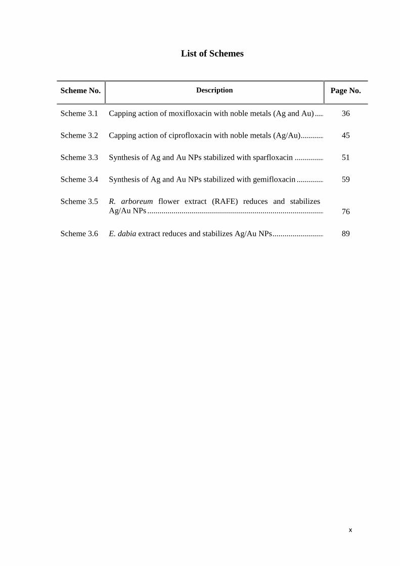
List of Schemes
Scheme No. Description Page No.
Scheme 3.1 Capping action of moxifloxacin with noble metals (Ag and Au) ................. 36
Scheme 3.2 Capping action of ciprofloxacin with noble metals (Ag/Au) ........................ 45
Scheme 3.3 Synthesis of Ag and Au NPs stabilized with sparfloxacin ........................... 51
Scheme 3.4 Synthesis of Ag and Au NPs stabilized with gemifloxacin .......................... 59
Scheme 3.5 R. arboreum flower extract (RAFE) reduces and stabilizes
Ag/Au NPs ....................................................................................................
76
Scheme 3.6 E. dabia extract reduces and stabilizes Ag/Au NPs ...................................... 89
xi
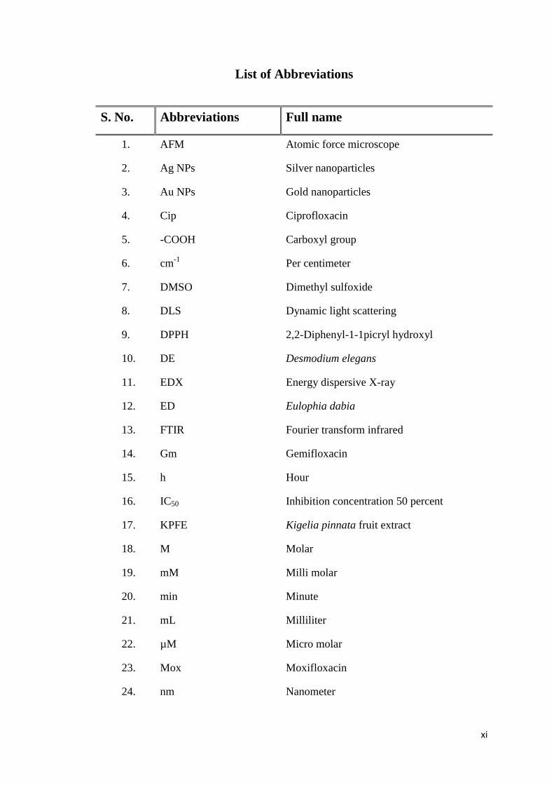
List of Abbreviations
S. No. Abbreviations Full name
1. AFM Atomic force microscope
2. Ag NPs Silver nanoparticles
3. Au NPs Gold nanoparticles
4. Cip Ciprofloxacin
5. -COOH Carboxyl group
6. cm-1
Per centimeter
7. DMSO Dimethyl sulfoxide
8. DLS Dynamic light scattering
9. DPPH 2,2-Diphenyl-1-1picryl hydroxyl
10. DE Desmodium elegans
11. EDX Energy dispersive X-ray
12. ED Eulophia dabia
13. FTIR Fourier transform infrared
14. Gm Gemifloxacin
15. h Hour
16. IC50 Inhibition concentration 50 percent
17. KPFE Kigelia pinnata fruit extract
18. M Molar
19. mM Milli molar
20. min Minute
21. mL Milliliter
22. µM Micro molar
23. Mox Moxifloxacin
24. nm Nanometer
xii
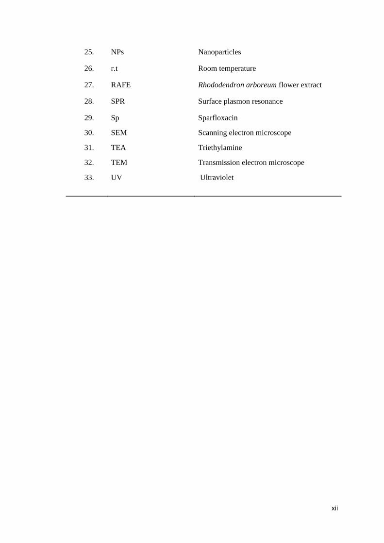
25. NPs Nanoparticles
26. r.t Room temperature
27. RAFE Rhododendron arboreum flower extract
28. SPR Surface plasmon resonance
29.
30.
31.
32.
33.
Sp
SEM
TEA
TEM
UV
Sparfloxacin
Scanning electron microscope
Triethylamine
Transmission electron microscope
Ultraviolet
xiii
SUMMARY
The work presented in this dissertation comprises of synthesis, spectroscopic analyses
and biological evaluation of silver and gold based pronanomedicines derived from
fluoroquinolones. Among others, it includes convenient and time saving production of
noble metals (Ag/Au) nanoparticles (NPs) capped with fluoroquinolone antibiotics
(moxifloxacin (Mox), ciprofloxacin (Cip), sparfloxacin (Sp) and gemifloxacin (Gm).
Different reducing agents such as triethylamine, hydroquinone and sodium
borohydride were employed to transform Ag/Au salts into feasible capping agents.
Among them, sodium borohydride relatively gave better results. As for we understand
and based on FTIR data, the NH moiety of fluoroquinolones were mainly responsible
for the capping of Ag/Au nanoparticles.
In order to manifest alternate green method, Ag and Au NPs were also produced by
using selected medicinal plants; Rhododendron arboreum (RA), Kigelia pinnata (KP)
and Eulophia dabia (ED) as reducing and stabilizing agent, while triethylamine was
used to synthesize NPs of the extract of Desmodium elegans (DE).
The structural framework and size morphology of synthesized NPs were
characterized by using advanced analytical techniques such as atomic force
microscope (AFM), UV visible, fourier transform infrared spectroscope (FTIR),
energy dispersive X-ray (EDX) and scanning electron microscope (SEM).
To find alternate to wide spread resistive strains of pathogenic microbes; new
antimicrobial agents are needed to treat the patients infected with such resistive
pathogenic microbes. The locally synthesized pronanomedicines derived of
fluoroquinolones were evaluated for biological properties namely urease inhibition,
xiv
leishmanicidal, antimicrobial and antioxidant activities. Interestingly and as for our
expectations, these NPs enhanced biological and pharmacological activities.
The synthesized pronanomedicines and the capping ligands were independently
screened for jack bean urease enzyme inhibition potential. Mostly, the Ag-Mox NPs
exhibited higher level of enzyme inhibition activity of 93% at 0.2 mg/mL and IC50
value of 0.66 ± 0.042 μg/mL concentration, while the ligand; Mox revealed weak
inhibition with IC50 value of 183.25 ± 2.06 μg/mL. On the other hand, the Au-Mox
NPs remained inactive as compared to the parent ligand (Mox) having IC50 = 183.25 ±
2.06 μg/mL. These results reflect that after conjugation of Mox with Ag, the activity
of moxifloxacin was significantly increased about 250 times. However, the urease
inhibition activity of the Au conjugated counterpart of moxifloxacin decreased
significantly.
The synthesized metallic nano-conjugates (Ag-Cip and Au-Cip NPs) and the parent
ligand, ciprofloxacin were also tested for jack bean urease enzyme inhibition
potential. Ag-Cip pronanomedicine exhibited better urease enzyme inhibition
indicating 96 % at 0.2 mg/ mL (IC50 = 1.181 ± 0.02 μg/mL) concentration. On the
other hand, Au-Cip NPs showed comparatively weaker urease inhibition (90 % at 0.2
mg/mL concentration) with IC50 = 52.55±2.3 μg/mL. As anticipated, the parent ligand
ciprofloxacin revealed weaker inhibition to the values of 75 % at 0.2 mg/mL and IC50
= 82.95 ±1.62 μg/mL concentrations.
Furthermore, leishmanicidal, antimicrobial and antioxidant activities were tested for
both synthesized pronanomedicines and all the parent ligands under discussion but
they revealed good to moderate activities.
The selected plants namely R. arboreum, K. pinnata, E. dabia and D. elegans and
their metallic NPs were also screened for jack beans urease enzyme, leishmanicidal,
xv
antimicrobial and antioxidant activities which exhibited promising activities, while D.
elegans-capped NPs showed moderate activities.
Convincingly, the synthesized pronanomedicines were monodispersed and revealed
stability to some extent by changing pH, concentration of table salt and temperature.
The silver based pronanomedicines were anticipated to be good candidate for urease
inhibition and leishmanicidal potentials.
Chapter-1 Introduction
1
1.1 History of quinolones
The prolific development of the quinolones initiated in 1962, when George Lesher
and coworkers accidentally discover nalidixic acid as a derivative of the synthesis of
the antimalarial drug chloroquine [1]. This discovery led the expansion of quinolone
compounds, particularly the innovative quinolones in medical use at the current time.
Other discoveries followed, but exclusively a few were of substantial importance
because they supplied us with a fuller apprehension of the mechanisms of activity of
the quinolones. The capability to change the quinolone nucleus to enhance
effectiveness and the spectrum of bactericidal activity. Furthermore, the opportunity
to extend the eradication half-life and to improve the pharmacodynamics and
pharmacokinetic properties of quinolones and understanding of the significance of the
structure-activity relationships (SARs) of the quinolones, with respect to their
comparative susceptibilities to the progress of bacterial resistance and their efficacy
for causing adverse effects in treated patient [2].
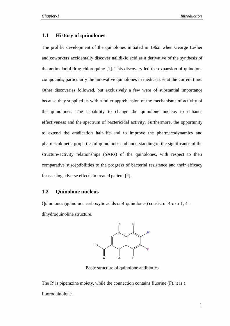
1.2 Quinolone nucleus
Quinolones (quinolone carboxylic acids or 4-quinolones) consist of 4-oxo-1, 4-
dihydroquinoline structure.
Basic structure of quinolone antibiotics
The R' is piperazine moiety, while the connection contains fluorine (F), it is a
fluoroquinolone.
Chapter-1 Introduction
2
1.3 Classification
The quinolones can be separated into different generations on the basis of bactericidal
spectrum. The first generation members are more confined antibacterial spectrum than
the advanced ones. The only universal standard employed is the grouping of the non-
fluorinated drugs found within the heading of 'first generation'. However, there is no
standard to determine which drug belongs to which generation.
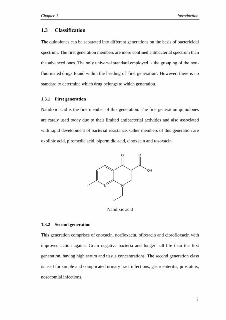
1.3.1 First generation
Nalidixic acid is the first member of this generation. The first generation quinolones
are rarely used today due to their limited antibacterial activities and also associated
with rapid development of bacterial resistance. Other members of this generation are
oxolinic acid, piromedic acid, pipermidic acid, cinoxacin and rosoxacin.
Nalidixic acid
1.3.2 Second generation
This generation comprises of enoxacin, norfloxacin, ofloxacin and ciprofloxacin with
improved action against Gram negative bacteria and longer half-life than the first
generation, having high serum and tissue concentrations. The second generation class
is used for simple and complicated urinary tract infections, gastroenteritis, prostatitis,
nosocomial infections.
Chapter-1 Introduction
3
Ciprofloxacin
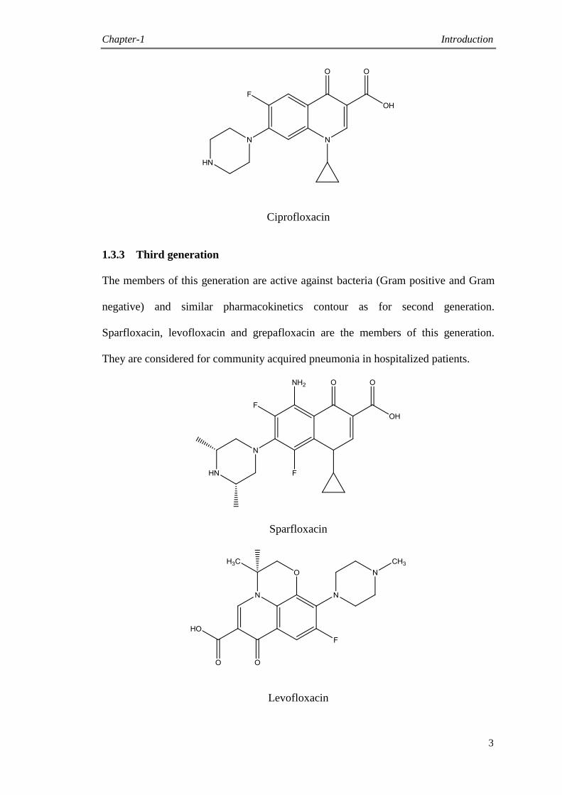
1.3.3 Third generation
The members of this generation are active against bacteria (Gram positive and Gram
negative) and similar pharmacokinetics contour as for second generation.
Sparfloxacin, levofloxacin and grepafloxacin are the members of this generation.
They are considered for community acquired pneumonia in hospitalized patients.
Sparfloxacin
Levofloxacin
Chapter-1 Introduction
4
Grepafloxacin
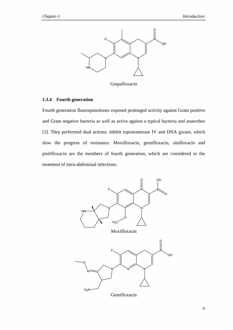
1.3.4 Fourth generation
Fourth generation fluoroquinolones exposed prolonged activity against Gram positive
and Gram negative bacteria as well as active against a typical bacteria and anaerobes
[3]. They performed dual actions; inhibit topoisomerase IV and DNA gyrase, which
slow the progress of resistance. Moxifloxacin, gemifloxacin, sitafloxacin and
prulifloxacin are the members of fourth generation, which are considered in the
treatment of intra-abdominal infections.
Moxifloxacin
Gemifloxacin
Chapter-1 Introduction
5
Sitafloxacin
Prulifloxacin
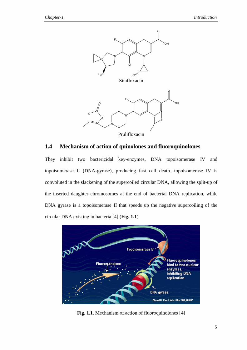
1.4 Mechanism of action of quinolones and fluoroquinolones
They inhibit two bactericidal key-enzymes, DNA topoisomerase IV and
topoisomerase II (DNA-gyrase), producing fast cell death. topoisomerase IV is
convoluted in the slackening of the supercoiled circular DNA, allowing the split-up of
the inserted daughter chromosomes at the end of bacterial DNA replication, while
DNA gyrase is a topoisomerase II that speeds up the negative supercoiling of the
circular DNA existing in bacteria [4] (Fig. 1.1).
Fig. 1.1. Mechanism of action of fluoroquinolones [4]
Chapter-1 Introduction
6
1.5 Medicinal plants
Medicinal plant is the best source of herbal medicines/drugs. The connection between
humanity and plants is as old as human evolution. In current decades, medicinal
plants are the chief source of medications for the world‟s population. The human has
an expedition for sound health, long life and everlasting beauty. Through such
creative expeditions, more or less founded on his ideas and practical experiences man
empowered to discriminate facts from imaginary fictions. Up to great extent natural
medicines initiated as story, conveyed to the new age group as a traditional
medication and advanced with time.
The medicine‟s history is an explanation of man‟s attempts to share with human
ailment from the crude efforts of preliterate man to the current multifaceted array of
areas in cures. The Chinese, Babylonian, Egyptian and Indian societies, pursued by
Greek, Roman, Arabic and Persian, all established their own representative materia
medica [5, 6]. New medication traces its ancestry to the Greeks. The Greece
medication was brought over by the Romans and then by the Arabs, after its
development with Chinese and Indian medicine, it was followed by Europe. The
Muslim elites familiarized it in India and merged with it the Ayurvedic medicine. This
combination is now termed as Eastern medicine or Unani medicine [7, 8].
The first ideas about the therapeutic purpose of plants are drawn in the Ayurveda
(2500-600 BC) and Rigveda (4500-1600 BC). Charaka scrutinized about 50 groups of
herbs, while Sushruta explained 760 herbs in 37 groups. Buddhist era developed the
importance of medicinal plants and contributed a significant consideration to harvest
these medicinal plants in a systematic way [9].
Chapter-1 Introduction
7
A natural product has distinctive biosynthetic gateways. The comprehensive studies
of the synthetic protocols are managed by expert professionals. Various techniques
have been recognized by the use of isotopically labeled precursors of the biogenetic
substances along with certain biotic combination.
Photosynthesis performs a key role in biogenesis in green plants, photosynthetic
bacteria and algae [10]. The carbon frameworks of biological products are
accumulated by specific arrangements of enzyme catalyzed reactions. These
interconnected metabolic arrangements form the foundation for a biosynthetic
grouping [11].
Primary metabolites like polysaccharides, nucleic acids and proteins are the basis of
all living things. The entire chains of procedure by which plants prepares and blow up
these materials in order to stay alive, set up the primary metabolic process [12].
Secondary metabolites has an important role in the existence of one species over the
other but they are not indispensable for their subsistence; so they are called secondary
metabolites [12]. Materials from secondary metabolism have a tendency to overlap
with the natural compounds of organic chemistry, like pigments, alkaloids, terpenes,
steroids, phenols, oligosaccharides, coumarins and antibiotics.
Secondary metabolites play a critical part in the natural selection of distinct plant
classes during the evolutionary process and in the interface of plants with the
surrounding.
1.6 Phytochemistry and bioactivities of Rhododendron arboreum
1.6.1 Plant introduction
The genus Rhododendron comprises of 1000 species which are distributed all over the
world mostly concentrated in China, Malaysia, Pakistan, India and Nepal [13].
Chapter-1 Introduction
8
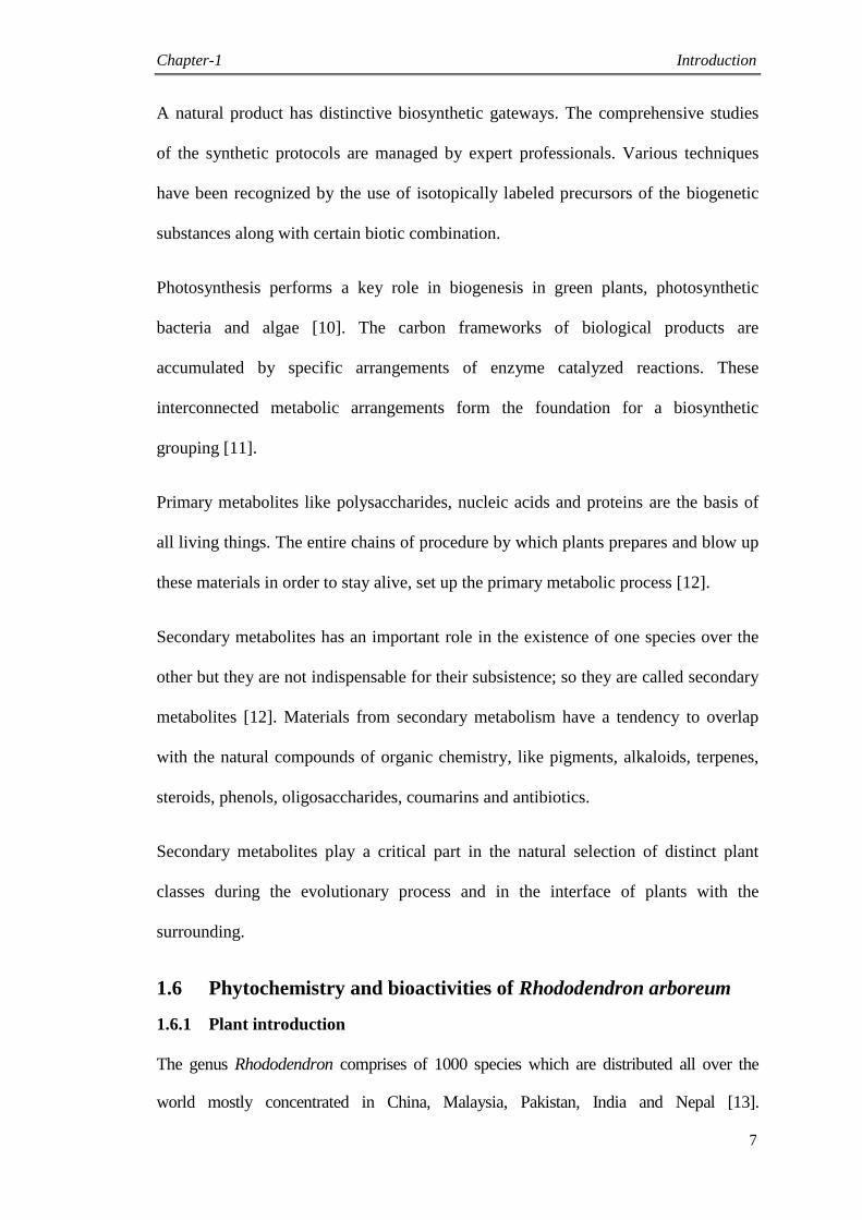
Rhododendron arboreum is an evergreen plant with bright red flowers (Fig. 1.2). The name
„Rhododendron‟ is originated from the Greek term „Rhodo‟ means rose and „dendron‟
means tree, arising in the high altitudes from 1500 m to 6000 m in Himalaya and Nilgiri in
South India. It is the national flower of Nepal, locally identified as Lali Guras or „rose tree‟
in English. R. arboreum belongs to family Ericaceae. The blooming season is from March-
April/June-September bears deep red or crimson to pale pinkish flowers. The aesthetic
flowers owe its spiritual importance; it is considered holy and put up in temples for
ornamenting purposes. A stamp was dispensed by the Indian postal department to honor
this flower [14].
Fig. 1.2. Photograph of Rhododendron arboreum flowers
1.7 Literature review of genus Rhododendron
1.7.1 Chemical constituents of the genus Rhododendron
The petroleum ether extract of the bark revealed the presence of triterpenoid taraxerol
(C30H50O) and ursolic acid acetate (C32H50O4), while the ether extract of the bark revealed
the identity of betulinic acid (C30H48O3). The acetone extract of the bark indicated the
leuco-pelargonidin (C15H14O6) [15]. The green foliage are described to consist of glucoside,
ericolin (arbutin) (C12H16O7), ursolic acid (C30H48O4), α-amyrin (C30H50O), epifriedelinol
(C30H52O), campanulin (C26H34ClN3O3S), quercetin (C15H10O7) and hyperoside

Chapter-1 Introduction
9
(C21H20O12) [16]. R. arboreum var. nilagiricum indicated the presence of hyperoside (3-D-
galactoside of quercetin) (C21H20O12), epifriedelinol (C30H52O), a triterpenoid (C30H48O7S)
and ursolic acid (C30H48O4) [17]. From the flower of this species, quercetin-3-rhamnoside
has been isolated [18]. Biologically active phenolic compounds i.e. coumaric acid
(C9H8O3), quercetin (C15H10O7) and rutin (C27H30O16) also have been isolated from the
flowers of R. arboreum [19].
1.7.2 Medicinal and pharmacological properties
Conventionally, dehydrated flowers deep-fried with vegetable oil are observed very much
effective in checking dysentery and squeeze for the cure of psychological disorders.
Flowers have the potential for cholinergic assay and anti-inflammatory. “Ashoka Aristha”
Ayurvedic preparation comprising R. arboreum has oestrogenic, oxytocic, and
prostaglandin synthetase inhibiting activities. The phytochemistry of dried foliage has been
cited in Homeopathic Materia Medica, to be beneficial in rheumatism and gout [20]. The
fresh corolla is acid sweet in taste and is given when fish bones stuck in the gullet [15]. The
flowers are also used for preparing native wine to avoid high altitude illness in the
Darjeeling hills of the eastern Himalayas. The fresh leaves are smeared on the forehead to
get rid of headache [21].
1.8 Phytochemistry and bioactivities of Eulophia dabia
1.8.1 Plant introduction
Eulophia dabia belongs to family Orchidaceae. It is the leading family amongst the
monocotyledons comprising 600 to 800 genera. Orchids consist of epiphytic, saprophytic
and terrestrial forms. The Eulophia genus covers permanent terrestrial orchids with fleshy
tubers (Fig. 1.3).
The genus of Eulophia contains around 230 species and distributed worldwide in
tropical as well as temperate climate. In Pakistan it is present in district Swat and
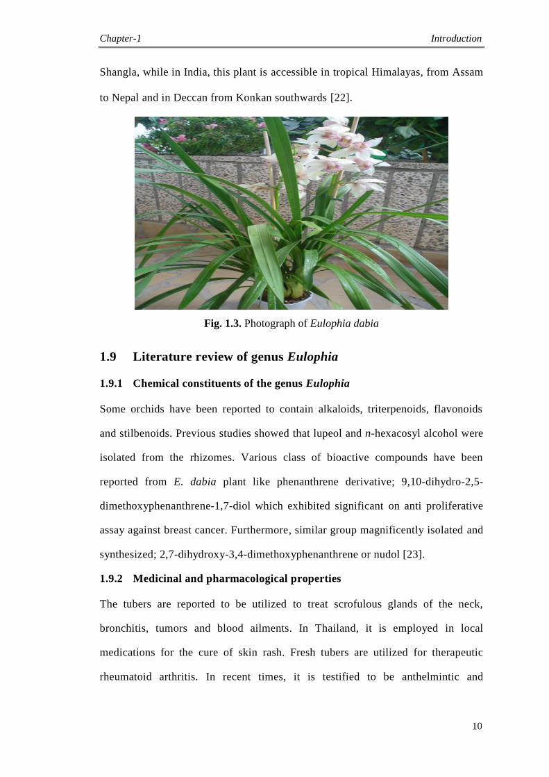
Chapter-1 Introduction
10
Shangla, while in India, this plant is accessible in tropical Himalayas, from Assam
to Nepal and in Deccan from Konkan southwards [22].
Fig. 1.3. Photograph of Eulophia dabia
1.9 Literature review of genus Eulophia
1.9.1 Chemical constituents of the genus Eulophia
Some orchids have been reported to contain alkaloids, triterpenoids, flavonoids
and stilbenoids. Previous studies showed that lupeol and n-hexacosyl alcohol were
isolated from the rhizomes. Various class of bioactive compounds have been
reported from E. dabia plant like phenanthrene derivative; 9,10-dihydro-2,5-
dimethoxyphenanthrene-1,7-diol which exhibited significant on anti proliferative
assay against breast cancer. Furthermore, similar group magnificently isolated and
synthesized; 2,7-dihydroxy-3,4-dimethoxyphenanthrene or nudol [23].
1.9.2 Medicinal and pharmacological properties
The tubers are reported to be utilized to treat scrofulous glands of the neck,
bronchitis, tumors and blood ailments. In Thailand, it is employed in local
medications for the cure of skin rash. Fresh tubers are utilized for therapeutic
rheumatoid arthritis. In recent times, it is testified to be anthelmintic and
Chapter-1 Introduction
11
demulcent. Tubers also called as „Salep‟ is used as an aphrodisiac drug. Moreover,
E. dabia tubers are employed to cure piles, acidity and stomach illnesses [24].
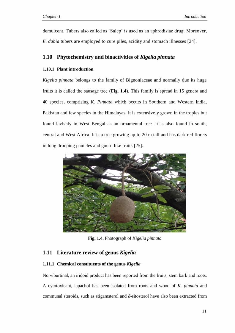
1.10 Phytochemistry and bioactivities of Kigelia pinnata
1.10.1 Plant introduction
Kigelia pinnata belongs to the family of Bignoniaceae and normally due its huge
fruits it is called the sausage tree (Fig. 1.4). This family is spread in 15 genera and
40 species, comprising K. Pinnata which occurs in Southern and Western India,
Pakistan and few species in the Himalayas. It is extensively grown in the tropics but
found lavishly in West Bengal as an ornamental tree. It is also found in south,
central and West Africa. It is a tree growing up to 20 m tall and has dark red florets
in long drooping panicles and gourd like fruits [25].
Fig. 1.4. Photograph of Kigelia pinnata
1.11 Literature review of genus Kigelia
1.11.1 Chemical constituents of the genus Kigelia
Norviburtinal, an iridoid product has been reported from the fruits, stem bark and roots.
A cytotoxicant, lapachol has been isolated from roots and wood of K. pinnata and
communal steroids, such as stigamsterol and β-sitosterol have also been extracted from
Chapter-1 Introduction
12
the root and bark of K. pinnata. γ -Sitosterol was stated to be existing in the pod of K.
pinnata. The flavonol quercitin and four flavonones i.e. luteolin, 6-OH luteolin,
luteolin-7-glucoside and 6-OH luteolin-7-glucoside were isolated from the foliage and
fruit of K. pinnata. Up till now, only a few phytochemical exertions on K. pinnata
flower were reported. Cyanidin glycoside, cyanidin-3-rutinoside and anthoxanthine
constituents were isolated from the flowers of K. pinnata [26].
1.11.2 Medicinal and pharmacological properties
K. pinnata, as a medicinal plant has a long history, utilized by African and several rural
countries. It is employed as an interesting use on abscesses and wounds, for the cure of
sexually transmitted diseases and skin sicknesses such as psoriasis, boils and acne.
Inside, the plant also utilize as medication for tape worm, ring worm, post partum
hemorrhage, dysentery, diabetes, pneumonia, toothaches and malaria. K. pinnata fruits
are applied as gauze for injuries and sores, hemorrhoids, for rheumatism as a cleansing,
to upsurge milk in lactating mothers and for skin firming possessions. Diverse portions
of the plant, as well as the fruits, are utilized either in a residue form or as ethanolic or
aqueous drinks, which are drunk or smeared to the pretentious body region. The
medicinal assets associated with K. pinnata are owing to the existence of various
subordinate metabolites, comprising iridoids, furonaphthoquinones, naphthoquinones,
meroterpenoids naphthoquinones, coumarin derivatives, lignans, sterols, flavonoids,
furanones, and volatile constituents [27].
1.12 Phytochemistry and bioactivities of Desmodium elegans
1.12.1 Plant introduction
Desmodium elegans is the member of genus Desmodium and family Fabaceae with 650
genera and 18000 species, generally comprising of shrubs or herbs (Fig. 1.5). Flowers
are light purple and organized in terminal panicles from August to September. Pod is

Chapter-1 Introduction
13
sessile and spread via seeds from September to December. The Desmodium genus is
scattered in temperate and tropical zones of the biosphere excluding New Zealand and
Europe. The D. elegans plant is mainly distributed in Pakistan, Kashmir, India, Nepal
and Bhutan [28].
Fig. 1.5. Photograph of Desmodium elegans
1.13 Literature review of genus Desmodium
1.13.1 Chemical constituents of the genus Desmodium
Up till now, 40 alkaloids, 13 steroids, 14 terpenoids, 81 flavonoids, 2 glycosides, 8
phenylpropanoids, 10 phenols and numerous volatile compounds have been isolated
from Desmodium species. Phytochemical analysis revealed that alkaloids and
flavonoids are the main metabolites in this genus. Overall 40 alkaloids were extracted
from Desmodium species and identified mostly as amide, indole, pyrrolidine,
phenylethylamine and alkylamine alkaloids. The principal types of flavonoids present
in Desmodium plants were flavanonols, flavones, flavan-3-ols, flavonols, 7, 8-prenyl-
lactone and flavonoids, whereas isoflavonoids contain isoflavones, isoflavanones,
pterocarpans and coumaronochromone. In addition, alkaloids and flavonoids, a variety
Chapter-1 Introduction
14
of terpenoids, phenols, steroids, glycosides, phenylpropanoids and fixed oils have also
been described from the Desmodium species [29].
1.13.2 Medicinal and pharmacological properties
Usually, Desmodium plants have been employed to therapy numerous illnesses such
as constipation, jaundice, asthma, fever, paralysis, edema, cold, cough and convulsion
[30]. D. elegans has many applications in common medication and many portions of
the plant have been stated to be utilized for various motives, for instance roots were
used as tonic, diuretic and carminative and the pulverized foliage was smeared on
incisions for soothing wounds [31].
1.14 Biological evaluation
Preliminary bioactivity shows an important feat in the drug discovery project. It offers a
platform for bioassays and comforts in the assortment of leads like drugs for evaluation
of pharmacological assessments. Positively, novel or improved therapeutic agents retain
through the preliminary bioactivity with a suitable safety outline. Biological assays are
the best skill of finding valuable and precious substituents present in medicinal plants
and their nanomaterials. Various biological activities have been carried out for noble
metal nanoparticles capped with fluoroquinolones and selected medicinal plants to
explore the hidden potentials. These bioactivities included urease enzyme inhibition,
antimicrobial, antioxidant and leishmanicidal.
1.14.1 Urease inhibition activity
Metal ions exist in the active sites of metal containing proteins i.e. ureases enzyme and
hemocyanin, lactase, ascorbate oxidase and tyrosinase [32]. Urease is a Ni (nickel)
encompassing enzyme and known to speed up the hydrolysis of urea into NH3 and CO2
(urea amidohydrolase). It lets an entity to utilize urea as nitrogen source [33]. Besides,
Chapter-1 Introduction
15
Urease enzyme is one of the highest sources of pathogenesis persuaded by Helicobacter
pylori, consequently allow them to stick at low pH of the gastrointestinal. It acts
significantly in the pathogenesis of intestinal and peptic ulcers [34].
1.14.2 Leishmanicidal studies
Leishmaniasis is the most dreadful of parasitic sicknesses. Except Australia, it exist in
all regions [35]. It is typically zoonotic, but also occurs in an anthroponotic mode of
transmission in some parts of Asia and Europe. WHO reported that almost 350 million
people in the biosphere are at risk of acquiring leishmaniasis [36].
Symptomatologically, leishmaniasis exists in mucocutaneous (MCL), cutaneous (CL),
visceral (VL) and diffuse (DCL) forms [37], [38]. Leishmania tropica and L. major
grounds the cutaneous form of the sickness, which is a stigmatizing and spoiling disease
[39]. If remains untreated, it becomes visceral infection, which is very lethal.
Approximately 500,000 humans losses, have been caused by L. chagasi/infantum in
Latin America and Southern Europe and by L. donovani in Pakistan, India, Middle East
and Africa [40]. In most endemic countries, the systemic pentavalent antimonials still
remain the recommended drug for treatment, but these have disagreeable and severe
side effect i.e. renal, neural, and cardiac toxicity [41]. Handling of VL or CL with
antimonials is even more problematic in human immunodeficiency virus; (HIV)-
infected individuals and is allied with frequent deteriorations because these drugs
require healthy immune system for optimal anti-parasitic activity. Finally, the treatment
of leishmaniasis is still intricate, with a partial therapeutic arsenal, toxic drugs and
resistance cases [42].
1.14.3 Antioxidant assay
Antioxidants can defend cells from the destruction produced by unstable molecules
identified as free radicals. Antioxidants interact with these unstable molecules (free
radicals) and may inhibit the deterioration of cells. Some of the examples of
antioxidants consist of vitamins C, E, A, lycopene, β-carotene and so forth [43]. An
Chapter-1 Introduction
16
antioxidant is an entity capable of slowing or preventing the oxidation of further
substances. Oxidation is a process that transfers electrons from a molecule to an
oxidizing agent. It can originate unstable molecules (free radicals), which produce chain
reactions that deteriorate cells. Antioxidants discontinue these chain reactions by taking
away free radicals and stop further oxidation by being oxidized themselves.
Antioxidants may be reducing agents such as polyphenols, ascorbic acid or thiols. For
several years chemists have acknowledged that free radicals cause oxidation which can
be controlled or stopped by a variety of antioxidants [44].
1.14.4 Antibacterial assay
Universally, the fast spread of infective diseases is due to various causes like failure of
available drug therapies by microbial resistance, poor health care systems and
population growth. In 20th
century, the death rate due to infections has increased [45].
Major worldwide public health problem is due to antimicrobial resistance.
Around one half of all victims are due to infectious diseases in nations of the humid
region. In developed countries, serious infectious casualties due to antimicrobial
resistance also take place where there is best understanding about microbes and their
control [46].
1.14.5 Antifungal activity
The prevalence of infections triggered by fungi also amplified terrifically in the last two
decades and it is expected to continue in future [47]. The increase ratio of fungal
infection is due to the population growth of immune-suppressed persons [48]. Among
the fungi, Candida causes severe fatal fungal infections. It is found on the mucus
membrane. When there is unscrupulous infection in the buccal cavity of the individual,
Candida becomes pathogenic particularly in the subject with immune-deficiencies [49].
Chapter-1 Introduction
17
Failures of available antifungal treatments are due to the adverse drug reactions, fungal
resistance and toxicity. Severe hepatic disorders, gastrointestinal and endochrinologial
are caused by antifungal azoles [50]. The exposure frequency of medicine and
dimensions of fungal population are significant aspects which contribute towards the
fungal resistance. There is key influence by HIV patients in the field of fungal
resistance [51]. Therefore, it is required to search innovative drugs for infections caused
by the resistance strains and better health care.
Chapter-2 Introduction to nanotechnology
18
2.1 Nanotechnology
Nanotech may be defined as the technology manipulating matter with nano scale
extending from 1 to 100 nm in one dimension. Nanotechnology is a broad term
covering all fields of science, such as organic chemistry, molecular biology, surface
science and microfabrication. Nanomaterials reveal different properties based on their
nano size and shape [52].
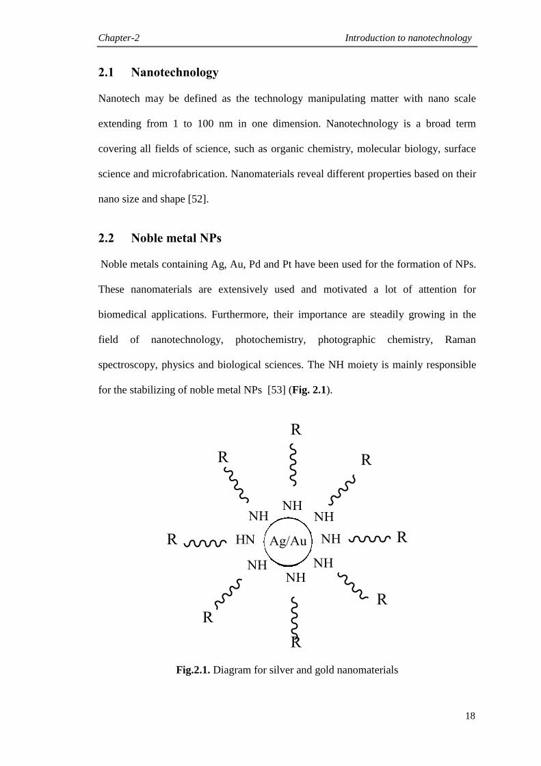
2.2 Noble metal NPs
Noble metals containing Ag, Au, Pd and Pt have been used for the formation of NPs.
These nanomaterials are extensively used and motivated a lot of attention for
biomedical applications. Furthermore, their importance are steadily growing in the
field of nanotechnology, photochemistry, photographic chemistry, Raman
spectroscopy, physics and biological sciences. The NH moiety is mainly responsible
for the stabilizing of noble metal NPs [53] (Fig. 2.1).
Fig.2.1. Diagram for silver and gold nanomaterials
Chapter-2 Introduction to nanotechnology
19
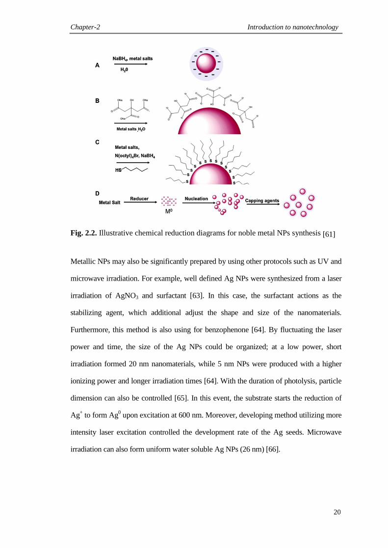
2.3 Synthesis
Synthesis of noble metal NPs has burst in the preceding eras. The best prevalent practices
are chemical reduction, physical techniques and biological processes. The physical
parameters of NPs comprise composition, shape and size. The capability to overcome of
these properties through slight modifications has led to a main effort in research finding
of NPs moreover, amplified the prospective for applications inside the area of catalysis,
microchip technology, diagnostics and therapeutics. The most generalized scheme for the
formation of Ag and Au NPs is chemical reduction method. In this technique the salts of
noble metal is reduced in the presence of a reductant [54, 55]. The initial renowned report
of the solution phase production of Ag and Au was in 1857, when Michael Faraday
reduced HAuCl4 with phosphorous in an aqueous solution [56]. Turkevitch et al., in 1951
developed the citrate reduction method. This synthesis of citrate capped Au nano scale
materials was founded on a single phase reduction of Au chloride by sodium citrate and
formed nanomaterials around 20 nm in size [57]. Frens suggested varying the ratio
between trisodium citrate and tetrachloroaurate and this technique is still hired. Following
this scheme of merely changing reaction parameters for instance, ratios [58], solution pH
[59] and solvent [60] has indorsed for controlling of the noble metal NPs sizes [61]. In the
last few decades, several groups have concentrated on fabricating monodispersed
nanomaterials by finding out possible NPs development tools in imperative to check the
size distribution. Natan et al., was a poineer for the exploration of seed development of
Au NPs employing alterations on the Frens synthesis [62]. Bastus et al., have effectively
prepared uniform citrate capped NPs via kinetically measured seed development [61]
(Fig. 2.2).
Chapter-2 Introduction to nanotechnology
20
Fig. 2.2. Illustrative chemical reduction diagrams for noble metal NPs synthesis [61]
Metallic NPs may also be significantly prepared by using other protocols such as UV and
microwave irradiation. For example, well defined Ag NPs were synthesized from a laser
irradiation of AgNO3 and surfactant [63]. In this case, the surfactant actions as the
stabilizing agent, which additional adjust the shape and size of the nanomaterials.
Furthermore, this method is also using for benzophenone [64]. By fluctuating the laser
power and time, the size of the Ag NPs could be organized; at a low power, short
irradiation formed 20 nm nanomaterials, while 5 nm NPs were produced with a higher
ionizing power and longer irradiation times [64]. With the duration of photolysis, particle
dimension can also be controlled [65]. In this event, the substrate starts the reduction of
Ag+ to form Ag
0 upon excitation at 600 nm. Moreover, developing method utilizing more
intensity laser excitation controlled the development rate of the Ag seeds. Microwave
irradiation can also form uniform water soluble Ag NPs (26 nm) [66].
Chapter-2 Introduction to nanotechnology
21
The fast nucleation due to microwave radiation is crucial to the even size dispersal of the
NPs. Suzuki suggested a novel method to formulate monodispersed Ag NPs reaching
from 10 to 80 nm [67]. This sophisticated technique uses a blend of laser treatments and
seeding.
Various reductants have been reported for instance, hydroxylamine [62, 68], ascorbic acid
[69, 70] and biogenic approaches which employ an iodide mediated reduction [71]. In the
light of above mentioned synthesis, several efforts have been forwarded to illuminate
biotic processes to synthesize NPs. Plant fabricated noble metal NPs production has
developed impetus due to eco friendliness and ease [72]. Synthesis with plant extracts as
well as iodide mediated reductions of Au salt has been reported. Zingiber officinale plant
can create NPs extending from 5-15 nm in diameter. The plant acts as a reductant as well
as a capping agent and the biological importance are confirmed through physiological
reliability [73]. For exploring the green synthesis, microbes has also developed as a
substitute to chemical reduction. Photosynthetic bacteria [74], prokaryotic bacteria [75-
77], eukaryotic fungus [78, 79] and medicinal plant extracts [80-83] all have been
examined for the reduction of metal ions to yield nanomaterials. Numerous biological
procedures have a slow reaction rate and a wide distribution in particle size [84]. Though,
a current publication by Darroudi investigated the key role of sodium hydroxide as an
accelerator to produce Ag NPs [85].
In summary, optimizing noble metal NPs formation is a productive zone of research.
Monitoring shape, size and distribution is a sophisticated and laborious process. These
reactions are governed by various parameters for example, reaction rate, reactant
concentration, reduction potential, solubility, heat and time. All of the variables are
basically tangled. More study is firm to be in current area for increased tenability.
Chapter-2 Introduction to nanotechnology
22
2.4 Characterization techniques
The synthesized NPs are characterized with various advanced techniques to confirm their
morphology and size. These techniques are:
Electron microscopy including, Transmission Electron Microscope (TEM) and
Scanning Electron Microscope (SEM)
Atomic Force Microscopy (AFM)
Fourier Transform Infrared Spectroscopy (FTIR)
X-ray Photoelectron Spectroscopy (XPS)
Dynamic Light Scattering (DLS)
Aerosol Particle Mass Analyzer (APM)
Ultraviolet visible Spectroscopy (UV vis)
Nanoparticle Tracking Analysis (NTA)
Condensation Particle Counter (CPC)
X-ray Diffraction (XRD)
Differential Mobility Analyzer (DMA)
Nanoparticle Surface Area Monitor (NSAM)
2.5 Applications
Noble metal nanotechnology is a flourishing arena with massive prospective for real
world and clinical applications. To understand this potential, it is essential to engineer
and design Ag and Au NPs that can be targeted to tissues of interest, also to create
particular and anticipated results. Particularly NPs with a metallic core due to their
encouraging safety profile in human beings are used in the preclinical analysis for
diagnostic, therapy and imaging. Consequently, the sizes of the NPs employed in the
arena of bio nanotechnology ranges from 2 to 500 nm. The atomic and molecular
Chapter-2 Introduction to nanotechnology
23
scales show really new things due to their advanced molecular design and small
configuration can be exactly produced with a high grade of flexibility. This couture is
mainly due to self assembly of the nanomaterials by charge compatibility and non
covalent interfaces. These NPs, have demonstrated to be the most resourceful and
commonly used constituents with wide applications like delivery vectors [86],
imaging [87], inhibitors [88], and sensors [89]. Therefore, these contrived nano-
conjugates assist as exclusive many dimensional frameworks that differ from their
mass complement [90].
The cellular uptake of inorganic NPs is a space of penetrating exploration. However,
Ag, Au and Pt NPs are noble metals; their mechanism of intracellular internalization is
not essentially similar or well understood. Geiser et al., reported blood cells to explain
intracellular uptake of Au NPs [91]. Their findings sustenance a diffusive mechanism of
entry since, Au NPs were originated in the cytosol free from membrane encapsulation.
Comparatively, it has been revealed that uptake of cellular in Au NPs is due to
micropinocytosis [92, 93] which was deep-rooted by other researchers. Similarly,
macrophages simply take on Ag NPs, which were confine to vacuoles [94]. In a
comparable investigation by Yen et al., indicated that Ag/Au NPs were restricted in
cytoplasmic vesicles of the macrophages [95]. On the other hand, the researchers more
ventured that the protein corona development prejudiced cellular uptake of Au NPs as
compared to Ag NPs, hence confusing the internalization process [95, 96].
The role of Ag NPs as bactericidal agents have been well recognized. Antiviral
possessions of Ag NPs biogenically designed are more energetic than chemically
formed Ag NPs [97]. Metallic NPs have also been designated as a possible HIV
defensive tonic [98, 99]. In different investigations, it is confirmed that Ag NPs
prohibited the virus from binding to the host cells in vitro [100, 101]. Furthermore, It was
Chapter-2 Introduction to nanotechnology
24
indicated that Ag acts straight on the virus as an antiviral agent by binding to the
glycoprotein [102] and binding in turn stops the CD4 dependent virion binding which
successfully declines HIV-1‟s infection [103]. These noble metal NPs also have been
active virucidal against herpes simplex virus [104], flu [105], and lung syncytial virus
[106].
Angiogenesis plays an important role in numerous ailments like rheumatoid arthritis,
cancer and macular degeneration [107-109]. Normally, angiogenesis is firmly controlled
between various anti-angiogenic and pro-angiogenic growth factors [110]. But the
angiogenic switch is turned on when the balance is disturbed under pathological
conditions. This incident encourages extremely abnormal blood vessels which becomes
hyper permeable to plasma proteins. Several anti-angiogenic mediators are being
currently used in the hospitals, but most of them have been premeditated to only prevent
[110] mediated signaling [111]. Moreover, further reports have shown acute
poisonousness of these conservative materials containing thrombosis, fatal hemorrhage
and hypertension [107, 108].
Some other common therapy for cancer patients is the utilization of radiation. Even
though this technique is operative for preventing the propagation rate of malignant
neoplastic disease cells, it can be aggressive and side effects are legion and normal tissue
is much injured. Noble metal NPs may provide a benefit in this arena by manipulating
their brilliant optical properties, wave length tunability and surface resonance. For
instance, upon X-ray treatment, Au NPs can create cellular apoptosis through the
generation of unstable molecules [112]. This cure method has amplified the killing of
tumor cells without hurting the neighboring normal tissue [113]. X-ray radiation of mice
inoculated with Au NPs at 250 kV produced a fourfold reduction in tumor and improved
endurance of the animals. Similarly, the inherent radioactive properties of Au-198 and
Au-199 NPs marks them the best candidates for radiotherapy [114].
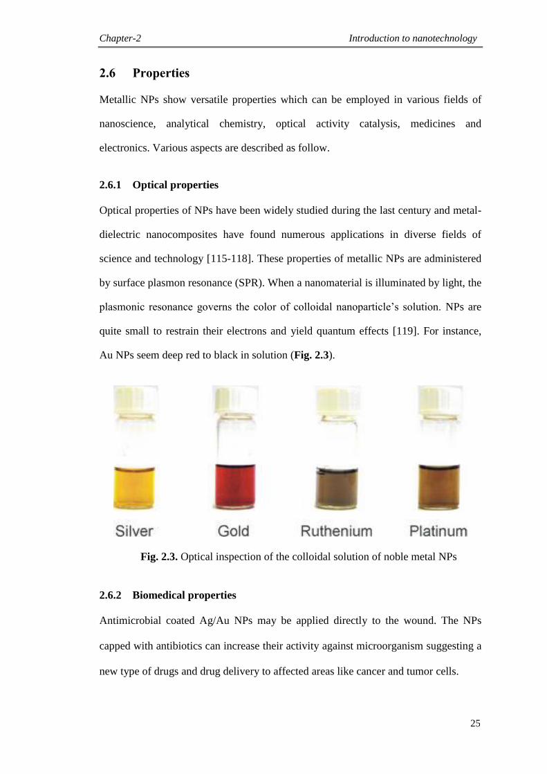
Chapter-2 Introduction to nanotechnology
25
2.6 Properties
Metallic NPs show versatile properties which can be employed in various fields of
nanoscience, analytical chemistry, optical activity catalysis, medicines and
electronics. Various aspects are described as follow.
2.6.1 Optical properties
Optical properties of NPs have been widely studied during the last century and metal-
dielectric nanocomposites have found numerous applications in diverse fields of
science and technology [115-118]. These properties of metallic NPs are administered
by surface plasmon resonance (SPR). When a nanomaterial is illuminated by light, the
plasmonic resonance governs the color of colloidal nanoparticle‟s solution. NPs are
quite small to restrain their electrons and yield quantum effects [119]. For instance,
Au NPs seem deep red to black in solution (Fig. 2.3).
Fig. 2.3. Optical inspection of the colloidal solution of noble metal NPs
2.6.2 Biomedical properties
Antimicrobial coated Ag/Au NPs may be applied directly to the wound. The NPs
capped with antibiotics can increase their activity against microorganism suggesting a
new type of drugs and drug delivery to affected areas like cancer and tumor cells.
Chapter-2 Introduction to nanotechnology
26
2.6.3 Magnetic properties
Magnetic NPs can be prepared; their magnetic behaviors may be used in diverse
fields, for instance, they may amend the contrast and detail of MRI images. To show
how magnetic NPs morphology and the resultant features are entangled, we can
employ a particular presentation to key out the factors that refrain vital magnetic
features. In bio sensing, for instance, NPs with greater capacity of magnetization are
preferred because they offer higher efficiency and sensitivity [120].
2.6.4 Electronic properties
Nano scale materials are much appropriate for the manufacturing of extraordinary
performance delicate metallic NPs, which are not only distribute ingredients with high
conductivity, but slicker parts for electronic engineering such as laptops and cell
phones etc. Nano electronics may be used to design digital displays which are brighter
in color and cheap [121].
2.6.5 Energy properties
Metallic NPs can be used in energy batteries with long long-term and have more
energy density. They have affinity for storing of efficient fuel cell and hydrogen gas
might be manufactured by expending the property of electro catalytic agent for such
strategies. They make the engine more efficient and economic when metal NPs are
employed as catalysts, [122].
2.7 Aims and objectives of the present research
The main aims and objectives of the present research activity are to:
synthesize objectively Ag and Au NPs of commercially available
fluoroquinolones (FQs) belonging to 2nd
, 3rd
and 4th
generations.
Chapter-2 Introduction to nanotechnology
27
develop and employ green phytosynthetic process of preparing noble metal
NPs mediated by selected medicinal plants (R. arboreum, K. pinnata, E. dabia
and D. elegans) as efficient reductants and stabilizers
provide access to eco-friendly synthetic protocol that can ensure high atom
economy and reasonable selectivity for producing NPs of relatively uniform
sizes and shapes.
isolate Ag and Au NPs derived from commercial fluoroquinolones and
designate plant extracts through selective mode of capping approach.
develop nanomaterial based drugs
evaluate locally synthesized pro-nanomedicines for biological activities
namely antimicrobial, enzyme inhibition, leishmanicidal and antioxidant.
2.8 Reviews on topical advancements in noble metal NPs
2.8.1 Review on Ag NPs
Ghosh et al., prepared Ag NPs by a single pot synthesis method of the antimicrobial
drug; ciprofloxacin. Numerous procedures were employed to illustrate the NPs bound
and free states of ciprofloxacin. In drug delivery, the time dependent release of the
prescription molecules from the NPs displays its importance [123].
Rodríguez León et al., described the formation of Ag NPs from AgNO3 solutions of
Rumex hymenosepalus extracts, which is rich in antioxidant molecules and used as
reductant. The nanomaterials were characterized by UV visible spectroscopy and
transmission electron microscopy, as a function of the ratio of Ag ions to reducing agent
molecules. The NPs were in the size of 2 to 40 nm. The HRTEM and FTIR analysis
revealed hexagonal and face-centered cubic crystal configurations were obtained [124].
Abboud et al., reported the application of Allium cepa in the preparation of Ag NPs
under microwave irradiation. The effect of several reaction factors such as microwave
Chapter-2 Introduction to nanotechnology
28
irradiation time and microwave irradiation power was analyzed. Furthermore, these
nanomaterials were exhibited potent antibacterial activity against two dissimilar strains
of bacteria Escherichia coli and Staphylococcus aureus [125].
Gopinath et al., synthesized Ag NPs by facile, reliable and green synthetic route using
the Pterocarpus santalinus aqueous extract, which comprises glycosides, glycerides,
steroids, phenols, saponins, triterpenoids, tannins and flavonoids to be accountable for
bio-reduction during the formation of sphere-shaped Ag NPs. FTIR study was
conducted to investigate the molecules involved in the synthesis of Ag NPs and
inveterate the Ag NPs by XRD. The synthesized Ag NPs presented significant
antibacterial assay against Gram positive and Gram negative bacterial strains [126].
Lavanya et al., described the formation of Ag NPs of Paederia foetida L. leaf extract
and showed significant antibacterial activity against various bacterial species [127].
Thirunavoukkarasu et al., employed the formation of Ag NPs by leaf extract of
Desmodium gangeticum. Robust formation of stable NPs was perceived on disclosure
of the aqueous leaf extract with solution of AgNO3. UV visible peak of the aqueous
medium at 450 nm conforming to the plasmon absorbance of Ag NPs. These
biosynthesized NPs were considered to be very toxic against pathogenic bacteria E.
coli, therefore, suggesting implication of the current study in fabrication of
biomedical products [128].
Kumar et al., fabricated Ag NPs using leaf extract of Sacha inchi (SI) as a nontoxic
reductant with particle size from 4 to 25 nm. Infrared measurement was conceded to
assume the possible phytochemicals accountable for capping of Ag NPs [129].
Olenin et al., described the development of Janus nanoparticles in the chemical
modification of the surface of Ag colloids. This is attained via sorption of
nanomaterials on the surface of chemically modified silica grafted with amine [130].
Chapter-2 Introduction to nanotechnology
29
Elias et al., reported the base of in situ assortment using a reaction kinetics factor, a
novel and straight forward technique for evaluating the synthesis of catalytically
dynamic Ag-PVP NPs. The catalytic properties with respect to the reduction of five
nitro aromatic compounds having various substituents at the para position were
determined [131].
Singh et al., studied the bactericidal activity of Ag NPs produced from Tinospora
cordifolia stem were analyzed against multidrug resistant strains of Pseudomonas
aeruginosa extracted from burn patients. Ag NPs possessed significant antibacterial
assay, which makes them an intoxicating source of bactericidal agent [132].
Sun et al., developed simple, environmental friendly and cost-effective process to
produce Ag NPs of tea leaf extract. They have also studied the effects of the tea extract
dosage, reaction time and reaction temperature on the formation of Ag NPs. These NPs
showed low antibacterial activity against E. coli [133].
Elsupikhe et al., reported a green sono-chemical process for preparing Ag NPs in
various concentrations of Kappa carrageenan. Ultrasonic irradiation was operated as a
green reducing agent and the Kappa carrageenan was employed as a natural
ecofriendly stabilizer [134].
Padalia et al., synthesized Ag NPs using of Tagetes erecta flower broth as reducing
agent by ecofriendly and a simple route. The Ag ions when uncovered to flower broth,
were reduced and give rise to green synthesis of Ag NPs. The antimicrobial
evaluation of Ag NPs with antibiotics was enhanced than antibiotics alone against
Gram negative bacteria and the tested fungal strains [135].
Ibrahim reported an ecofriendly, cost efficient, rapid and easy protocol for the
production of Ag NPs with banana peel extract (BPE) as a reductant and capping
Chapter-2 Introduction to nanotechnology
30
agent. The different factor affecting Ag reduction was investigated. The Ag NPs
showed effective antibacterial activity against representative pathogens of bacteria
and yeast [136].
Yakout and Mostafa established the preparation of Ag NPs by green method. It was
perceived that use of starch makes expedient technique for the formation of Ag NPs.
The synthesized green Ag NPs revealed a potential antibacterial activity [137].
Ahmed and Ikram reported a simple, cost effective bio-reduction on the principle of
“green biosynthesis” of Ag NPs by Terminalia arjuna extract. Biological evaluation
of Ag NPs were also done against Gram negative (E. coli) and Gram positive (S.
aureus) bacteria for their future applications in nanomedicines especially for the
treatment of wounds [138].
Verma et al., studied an ecofriendly, economic, and simple photo catalytic green
method for the facile synthesis of Ag NPs. An aqueous leaf extract of marine fern,
Salvinia molesta, was employed as a bio-reductant and stabilizing agent. The
synthesized Ag NPs were originated to be an active bactericidal mediator against
Gram negative and Gram positive bacteria [139].
Kumar et al., established low cost and an ecofriendly approach for the green creation
of spherical and stable Ag NPs of Erigeron bonariensis extract, which act as reducing
and capping agent. The Ag NPs revealed catalytic activity towards degradation of
Acridine orange without of involvement of any harmful reductant [140].
Govindarajan et al., investigated green synthesis of Ag NPs employing an economy,
Anisomeles indica leaf extract by reduction of Ag ions from AgNO3 solution. The
acute toxicity of synthesized Ag NPs and leaf extract of A. indica was appraised
Chapter-2 Introduction to nanotechnology
31
against larvae of the malaria vector Anopheles subpictus, the dengue vector Aedes
albopictus and the Japanese encephalitis vector Culex tritaeniorhynchus. Both Ag NPs
and the A. indica leaf extract exhibited dose dependent larvicidal effect against all tested
mosquito species [141].
Balakumaran et al., reported the synthesis of both Ag and Au NPs using soil fungi. The
Aspergillus terreus has produced extremely stable NPs. The myco derived Ag NPs
presented greater antimicrobial assay as compare to the standard antibiotic,
streptomycin [142].
Saha et al., synthesized the Ag NPs by distinctive grouping of tyrosine and a natural
polymer (starch) using ultrasound assisted green practice. A comprehensive mechanistic
study on the reactive oxygen species (ROS) mediated filaricidal (against Setaria cervi)
and mosquitocidal (against second and fourth instar larvae of Culex quin-quefasciatus)
activities of Ag NPs has been made for the first time [143].
2.8.2 Review on Au NPs
Naveena and Prakash synthesized Au NPs of red marine algae, Gracilaria corticata
aqueous extract as a reductant. The synthesized NPs were assessed against bacterial
pathogens Gram negative Escherichia coli, Enterobacter aerogenes and Gram positive
Staphylococcus aureus, Enterococcus faecalis and also considered for its antioxidant
assay by DPPH free radical scavenging activity. The antibacterial and antioxidant
activities of the Au NPs exhibited significant assays in contrast with the standards [144].
Li et al., has employed carboxylato pillar arene, a new water soluble macrocyclic
synthetic receptor, as a stabilizing ligand for in situ synthesis of Au NPs.
Supramolecular self-assembly of carboxylato pillar arene-modified Au NPs arbitrated
Chapter-2 Introduction to nanotechnology
32
by appropriate guest molecules was also examined, signifying that the novel hybrid
entity is convenient for recognition and sensing of the herbicide [145].
Rajeshkumar et al., reported the synthesis of Au NPs by using marine brown algae
Turbinaria conoides. Finally the bactericidal assay of Au NPs was conducted; it
revealed streptococcus having the maximum inhibition and medium range of inhibition
was examined against bacillus subtilis and klebsiella pneumoniae [146].
Joh et al., studied in cell culture tests and in an animal model of glioblastoma multiforme
(GBM) in which radiation therapy (RT) is supplemented by PEGylated-Au NPs. Au NPs
considerably enlarged cellular DNA destruction caused by ionizing radiation in human
GBM-derived cell lines and occasioned in reduced clonogenic survival. Follow up in
vitro experiments deep-rooted that the arrangement of Au NPs and RT resulted in
significantly increased DNA mutilation in brain derived endothelial cells [147].
Orza et al., described that temozolomide loaded Au nanomaterials were effective in
reducing chemo-resistance and abolish 82.7% of cancer stem cells compared with a
42% damage rate of temozolomide alone [148].
Zare et al., utilized amino acids as a reductant and stabilizing Au NPs. The Au NPs
were synthesized with a reduction solution comprising Au ions with Au chloride and
functionalized by phenylalanine, glutamic acid and tryptophan. The therapeutic uses of
proteins and amino acids may be utilized as a useful process owing to the strong
interface of peripheral amine groups with NPs [149].
Wang et al., stated the facile synthesis of Au NPs-alginate composite spheres. The
fabricated spheres are potential for numerous applications, such as bactericide, drug
carriers and micro sensors [150].
Chapter-2 Introduction to nanotechnology
33
Pooja et al., revealed the importance of Xanthan gum (XG) in the formation of Au NPs.
XG was used as reductant and capping agent. The improved NPs were also explored as
drug delivery carrier for doxorubicin hydrochloride [151].
Goldstein et al., reported that the charging artifacts associated to non conductive biotic
sample can be magnificently eradicated by employing the uncoated biological specimen
on a conductive substrate. By developing the cells on glass pre coated with a chromium
layer, they observed the uptake of 10 nm Au NPs inside uncoated and unstained
keratinocytes cells and macrophages [152].
Kim et al., described the light-induced synthesis of numerous Au NPs and demonstrated
their considerable use as effective photo thermal heating constituents and practical
heterogeneous catalysts under the treatment of a solar based light after being loaded
onto a paper based substrate [153].
Dharmatti et al., synthesized biogenic Au nano triangles (GNTs) of Azadirachta indica
leaf extract and its application in competent drug delivery of doxorubicin (DOX)
(anticancer drug) [154].
Schroder et al., reported the application of a new catalyst for cycloisomerizations. The
new catalyst scheme comprises Au NPs reinforced on Aluminum-Santa Barbara
amorphous no.15 (Al-SBA15). The catalyst is very selective and ecofriendly.
Cycloisomerization reaction yields excellent important to the synthesis of two new
classes of six and seven membered heterocycles [155].
Ding et al., synthesized Au core-induced poly pyrrole nano hybrids (Au-PPyNHs)
through in situ oxidation polymerization of pyrrole molecules and was immobilized
onto glassy carbon electrode and applied to construct dopamine (DA) sensor. They
Chapter-2 Introduction to nanotechnology
34
found that the fabricated sensor with Au-PPyNH-Au nano hybrids is highly specific
probe or sensing DA [156].
Li et al., successfully synthesized Au NPs-decorated poly (o-phenylenediamine)
(PoPD@Au) hollow microspheres. Furthermore, PoPD@Au hollow microspheres were
immobilized onto the surface of a glassy carbon electrode and applied to construct a
sensor [157].
Ding et al., reported the enhancement of innovative protein Au hybrid nanocubes
(PGHNs), which were accumulated by Au nano-clusters, tryptophan and bovine serum
albumin as building blocks. The PGHNs can assist as a novel kind of dual purpose
gadget; a nano-carrier in drug delivery studies and a blue emitting cell marker in bio
imaging exploration [158].
Kidonakis and Stratakis reported the Au-catalyzed hydrosilylation of allenes using
recyclable Au NPs as catalyst. The hydrosilane addition takes place on the more
substituted double bond of terminal allenes in a highly regioselective manner. The
observed regioselectivity/reactivity modes are attributed to steric and electronic factors
[159].
Sathishkumar et al., synthesized Au NPs using the aqueous fruit extract of Couroupita
guianensis Aubl. (CGFE) as a potential bio-reductant. DLS and EDX findings were
confirmed that the produced CG-Au NPs were stable having negative electric charge
without accumulation [160].
Dhamecha et al., summarized a distinctive green process for the preparation of Au
NPs by modest treatment of Au salts with Nepenthes khasiana extract. Fast synthesis
Chapter-2 Introduction to nanotechnology
35
of biocompatible Au NPs possessed exclusive physical and chemical characteristics
which function as a benefit for its purpose in numerous biomedical uses [161].
Paul et al., reported a green method for the formation of Ag/Au nanomaterials
employing dried biomass of Parkia roxburghii leaf. The antibacterial assay of the
created NPs was studied on Gram negative bacteria Escherichia coli and Gram
positive bacteria Staphylococcus aureus and both Ag/Au NPs exhibited slightly
higher efficacy on S. aureus than on E. coli [162].
Yan et al., reported a simplistic, green and single pot synthesis of biomolecule capped
Au NPs with higher catalytic potential. Cellulose nanocrystal (CNC)-capped Au NPs
were arranged by warming the aqueous mixture of Au chloride. The resulting CNC-
supported Au NPs displayed catalytic activities for the reduction of 4-nitrophenol by
NaBH4 [163].
Karthik et al., described the green synthesis of Au NPs by leaves extract of Cerasus
serrulata. The prepared Au NPs revealed greater bactericidal assay against Gram
negative (Escherichia coli) than Gram positive (Staphylococcus aureus) bacteria
[164].
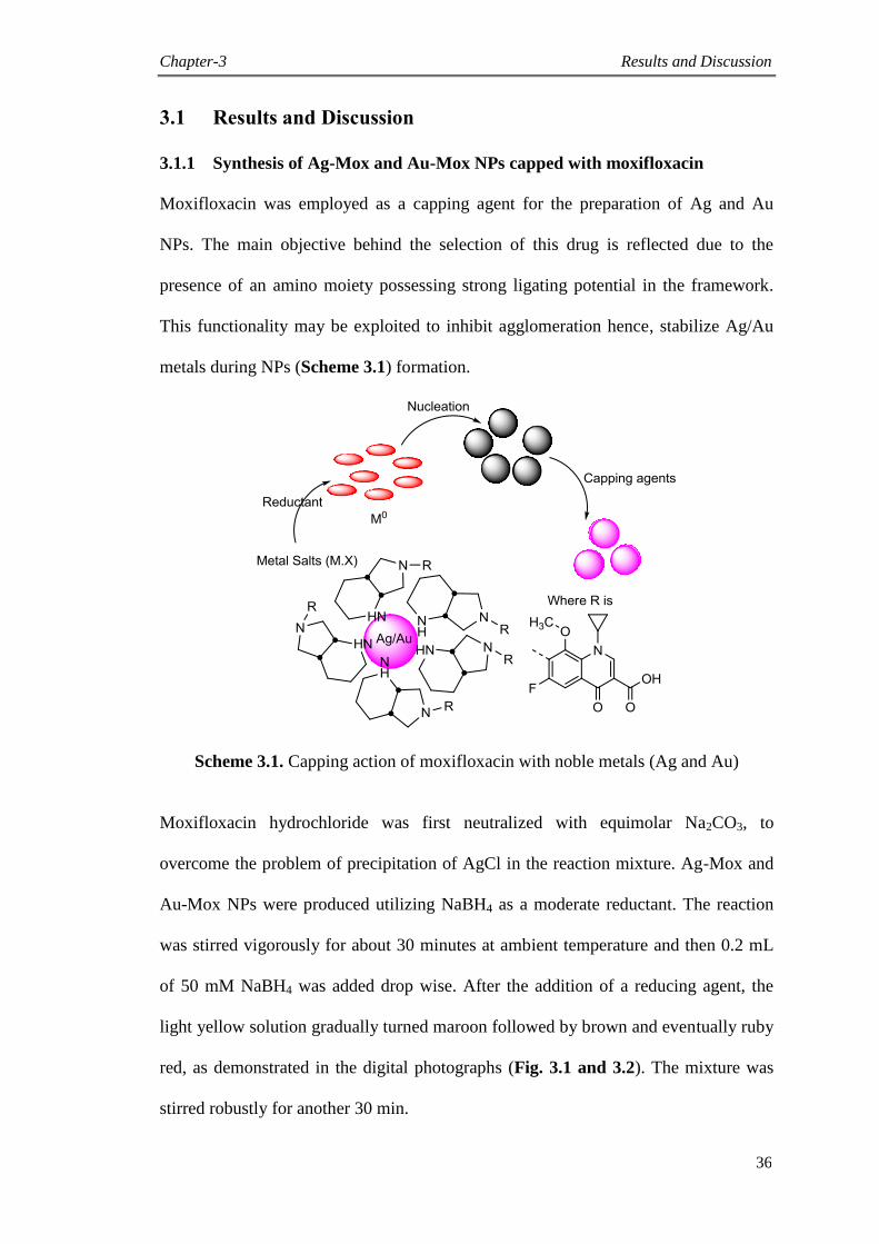
Chapter-3 Results and Discussion
36
3.1 Results and Discussion
3.1.1 Synthesis of Ag-Mox and Au-Mox NPs capped with moxifloxacin
Moxifloxacin was employed as a capping agent for the preparation of Ag and Au
NPs. The main objective behind the selection of this drug is reflected due to the
presence of an amino moiety possessing strong ligating potential in the framework.
This functionality may be exploited to inhibit agglomeration hence, stabilize Ag/Au
metals during NPs (Scheme 3.1) formation.
Scheme 3.1. Capping action of moxifloxacin with noble metals (Ag and Au)
Moxifloxacin hydrochloride was first neutralized with equimolar Na2CO3, to
overcome the problem of precipitation of AgCl in the reaction mixture. Ag-Mox and
Au-Mox NPs were produced utilizing NaBH4 as a moderate reductant. The reaction
was stirred vigorously for about 30 minutes at ambient temperature and then 0.2 mL
of 50 mM NaBH4 was added drop wise. After the addition of a reducing agent, the
light yellow solution gradually turned maroon followed by brown and eventually ruby
red, as demonstrated in the digital photographs (Fig. 3.1 and 3.2). The mixture was
stirred robustly for another 30 min.
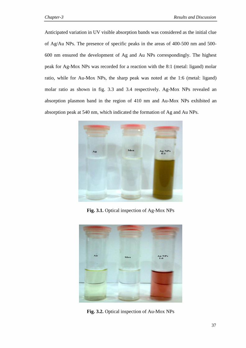
Chapter-3 Results and Discussion
37
Anticipated variation in UV visible absorption bands was considered as the initial clue
of Ag/Au NPs. The presence of specific peaks in the areas of 400-500 nm and 500-
600 nm ensured the development of Ag and Au NPs correspondingly. The highest
peak for Ag-Mox NPs was recorded for a reaction with the 8:1 (metal: ligand) molar
ratio, while for Au-Mox NPs, the sharp peak was noted at the 1:6 (metal: ligand)
molar ratio as shown in fig. 3.3 and 3.4 respectively. Ag-Mox NPs revealed an
absorption plasmon band in the region of 410 nm and Au-Mox NPs exhibited an
absorption peak at 540 nm, which indicated the formation of Ag and Au NPs.
Fig. 3.1. Optical inspection of Ag-Mox NPs
Fig. 3.2. Optical inspection of Au-Mox NPs
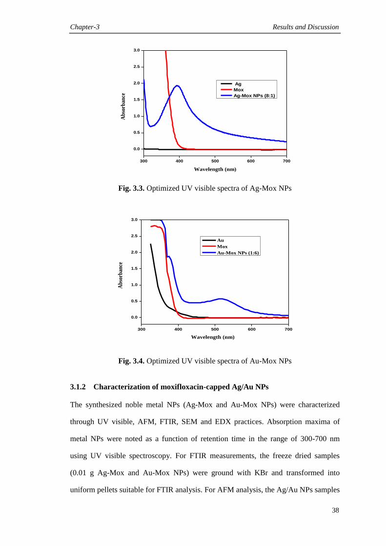
Chapter-3 Results and Discussion
38
300 400 500 600 700
0.0
0.5
1.0
1.5
2.0
2.5
3.0
Ag
Mox
Ag-Mox NPs (8:1)
Ab
sorb
ance
Wavelength (nm)
Fig. 3.3. Optimized UV visible spectra of Ag-Mox NPs
300 400 500 600 700
0.0
0.5
1.0
1.5
2.0
2.5
3.0
Ab
sorb
ance
Wavelength (nm)
Au
Mox
Au-Mox NPs (1:6)
Fig. 3.4. Optimized UV visible spectra of Au-Mox NPs
3.1.2 Characterization of moxifloxacin-capped Ag/Au NPs
The synthesized noble metal NPs (Ag-Mox and Au-Mox NPs) were characterized
through UV visible, AFM, FTIR, SEM and EDX practices. Absorption maxima of
metal NPs were noted as a function of retention time in the range of 300-700 nm
using UV visible spectroscopy. For FTIR measurements, the freeze dried samples
(0.01 g Ag-Mox and Au-Mox NPs) were ground with KBr and transformed into
uniform pellets suitable for FTIR analysis. For AFM analysis, the Ag/Au NPs samples
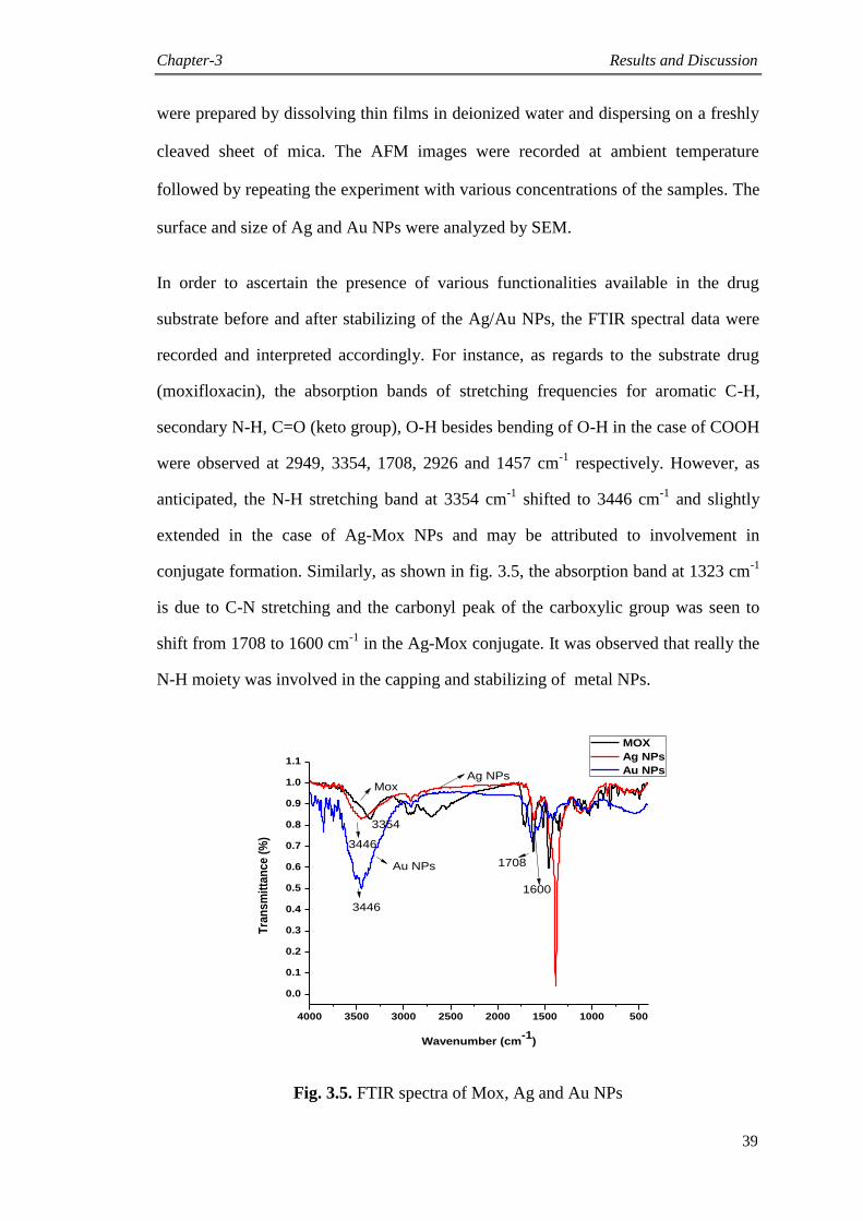
Chapter-3 Results and Discussion
39
were prepared by dissolving thin films in deionized water and dispersing on a freshly
cleaved sheet of mica. The AFM images were recorded at ambient temperature
followed by repeating the experiment with various concentrations of the samples. The
surface and size of Ag and Au NPs were analyzed by SEM.
In order to ascertain the presence of various functionalities available in the drug
substrate before and after stabilizing of the Ag/Au NPs, the FTIR spectral data were
recorded and interpreted accordingly. For instance, as regards to the substrate drug
(moxifloxacin), the absorption bands of stretching frequencies for aromatic C-H,
secondary N-H, C=O (keto group), O-H besides bending of O-H in the case of COOH
were observed at 2949, 3354, 1708, 2926 and 1457 cm-1
respectively. However, as
anticipated, the N-H stretching band at 3354 cm-1
shifted to 3446 cm-1
and slightly
extended in the case of Ag-Mox NPs and may be attributed to involvement in
conjugate formation. Similarly, as shown in fig. 3.5, the absorption band at 1323 cm-1
is due to C-N stretching and the carbonyl peak of the carboxylic group was seen to
shift from 1708 to 1600 cm-1
in the Ag-Mox conjugate. It was observed that really the
N-H moiety was involved in the capping and stabilizing of metal NPs.
4000 3500 3000 2500 2000 1500 1000 500
0.0
0.1
0.2
0.3
0.4
0.5
0.6
0.7
0.8
0.9
1.0
1.1
Tra
ns
mit
tan
ce
(%
)
Wavenumber (cm-1
)
MOX
Ag NPs
Au NPs
Mox
Au NPs
Ag NPs
3354
3446
1708
1600
3446
Fig. 3.5. FTIR spectra of Mox, Ag and Au NPs
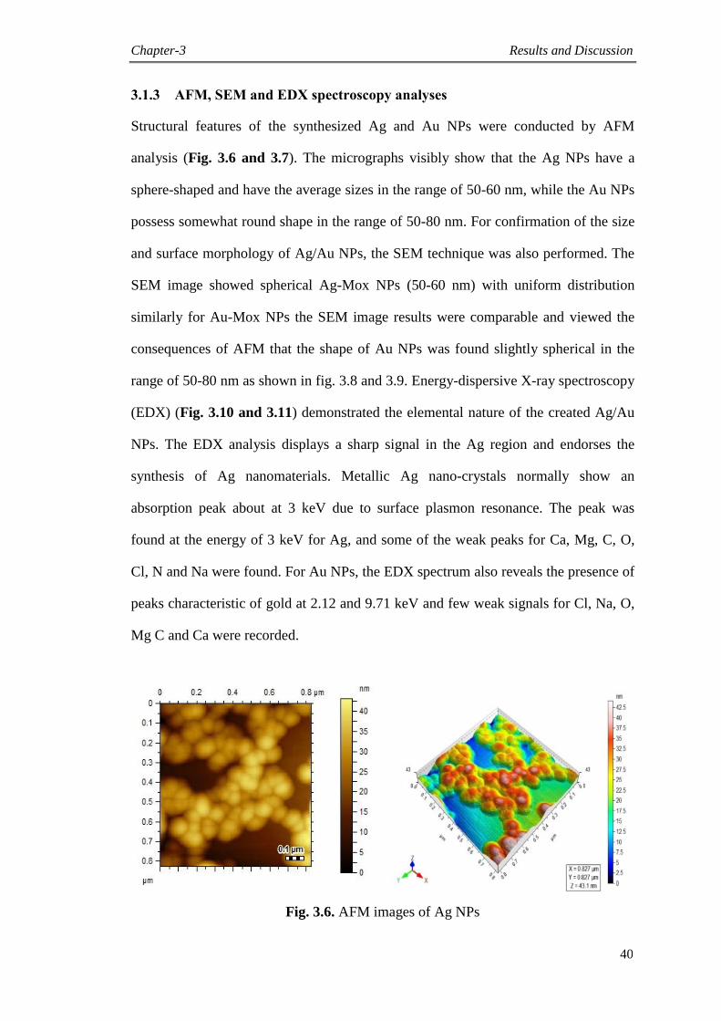
Chapter-3 Results and Discussion
40
3.1.3 AFM, SEM and EDX spectroscopy analyses
Structural features of the synthesized Ag and Au NPs were conducted by AFM
analysis (Fig. 3.6 and 3.7). The micrographs visibly show that the Ag NPs have a
sphere-shaped and have the average sizes in the range of 50-60 nm, while the Au NPs
possess somewhat round shape in the range of 50-80 nm. For confirmation of the size
and surface morphology of Ag/Au NPs, the SEM technique was also performed. The
SEM image showed spherical Ag-Mox NPs (50-60 nm) with uniform distribution
similarly for Au-Mox NPs the SEM image results were comparable and viewed the
consequences of AFM that the shape of Au NPs was found slightly spherical in the
range of 50-80 nm as shown in fig. 3.8 and 3.9. Energy-dispersive X-ray spectroscopy
(EDX) (Fig. 3.10 and 3.11) demonstrated the elemental nature of the created Ag/Au
NPs. The EDX analysis displays a sharp signal in the Ag region and endorses the
synthesis of Ag nanomaterials. Metallic Ag nano-crystals normally show an
absorption peak about at 3 keV due to surface plasmon resonance. The peak was
found at the energy of 3 keV for Ag, and some of the weak peaks for Ca, Mg, C, O,
Cl, N and Na were found. For Au NPs, the EDX spectrum also reveals the presence of
peaks characteristic of gold at 2.12 and 9.71 keV and few weak signals for Cl, Na, O,
Mg C and Ca were recorded.
Fig. 3.6. AFM images of Ag NPs
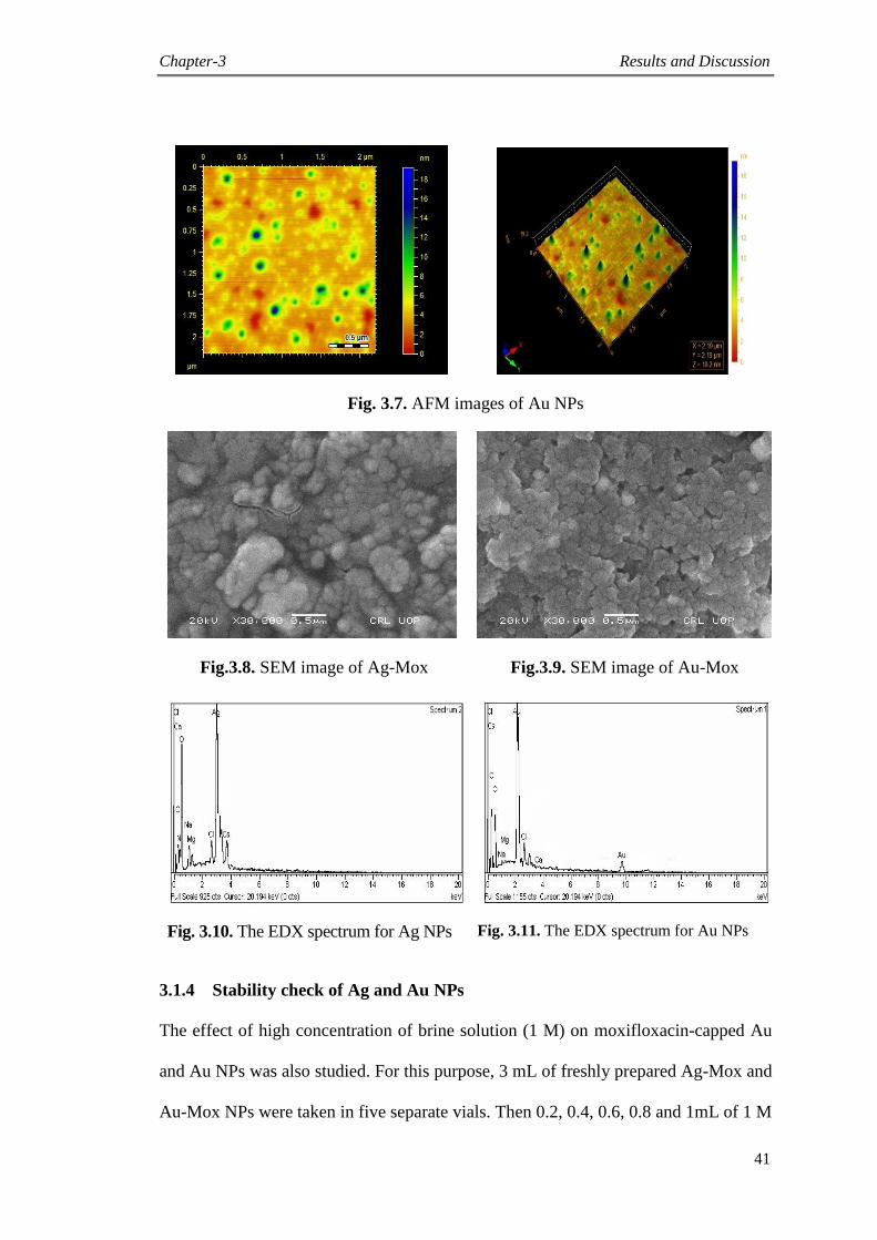
Chapter-3 Results and Discussion
41
Fig. 3.7. AFM images of Au NPs
Fig.3.8. SEM image of Ag-Mox Fig.3.9. SEM image of Au-Mox
Fig. 3.10. The EDX spectrum for Ag NPs Fig. 3.11. The EDX spectrum for Au NPs
3.1.4 Stability check of Ag and Au NPs
The effect of high concentration of brine solution (1 M) on moxifloxacin-capped Au
and Au NPs was also studied. For this purpose, 3 mL of freshly prepared Ag-Mox and
Au-Mox NPs were taken in five separate vials. Then 0.2, 0.4, 0.6, 0.8 and 1mL of 1 M

Chapter-3 Results and Discussion
42
NaCl solution were added to these vials. The resulting solutions were shaken well and
then kept at room temperature for 24 h. UV visible spectra were recorded for Ag-Mox
and Au-Mox NPs. The results showed that higher concentration of brine decreased the
λ max. This rapid decrease in absorbance of Ag/Au NPs containing NaCl may be
attributed to the aggregation effect promoted by Cl-1
ions. From these clarifications it
was concluded that at a higher concentration of sodium chloride, however,
aggregation turned out to be dominant. As for long term stability, Ag-Mox NPs and
Au-Mox NPs are much more stable in neat water than those in brine solution as
shown in fig. 3.12 and 3.13 respectively. In addition, the stability of synthesized Ag-
Mox and Au-Mox NPs against pH variations ranging from 2 to 13 was also examined.
Freshly prepared nano-conjugates of Ag-Mox and Au-Mox (3 mL) were taken in six
separate vials. The pH of Ag-Mox and Au-Mox NPs was measured and found to be
4.7. The pH of Ag and Au NPs in the range 6-13 was monitored by employing 1 M
NaOH solution. Also, the pH of Ag-Mox and Au-Mox NPs ranging from 2 to 3 was
adjusted with 1 M HCl. The UV visible spectra of the resultant solutions were noted
after 24 h. Ag NPs were stable in the pH range of 4-7 and completely unstable in
highly acidic medium at pH 2-3 and basic medium 8-13 (Fig. 3.14), while the Au NPs
showed stability in basic medium (pH = 8-9) and less stable at pH 12-13 (Fig. 3.15).
The synthesized Ag/Au NPs were also found to be stable up to 60 oC.
300 400 500 600 700
0.0
0.3
0.6
0.9
1.2
1.5
1.8
2.1
2.4
Ab
so
rba
nc
e
Wavelength (nm)
AgNPs
0.2mL Brine
0.4mL Brine
0.6mL Brine
0.8mL Brine
1 mL Brine
300 400 500 600 700
0.0
0.5
1.0
1.5
2.0
2.5
Ab
so
rba
nc
e
Wavelength (nm)
AuNPs
0.2 mL Brine
0.4 mL Brine
0.6 mL Brine
0.8 mL Brine
1 mL Brine
Fig. 3.12. Effect of brine on Ag-Mox Fig. 3.13. Effect of brine on Au-Mox
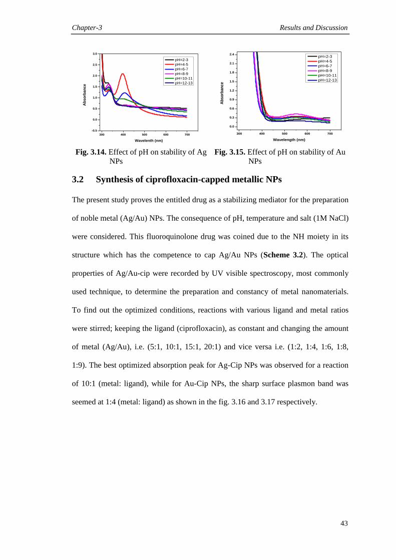
Chapter-3 Results and Discussion
43
300 400 500 600 700
-0.5
0.0
0.5
1.0
1.5
2.0
2.5
3.0
Ab
so
rba
nc
e
Wavelenth (nm)
pH=2-3
pH=4-5
pH=6-7
pH=8-9
pH=10-11
pH=12-13
300 400 500 600 700
0.0
0.3
0.6
0.9
1.2
1.5
1.8
2.1
2.4
Ab
so
rba
nc
e
Wavelength (nm)
pH=2-3
pH=4-5
pH=6-7
pH=8-9
pH=10-11
pH=12-13
Fig. 3.14. Effect of pH on stability of Ag
NPs
Fig. 3.15. Effect of pH on stability of Au
NPs
3.2 Synthesis of ciprofloxacin-capped metallic NPs
The present study proves the entitled drug as a stabilizing mediator for the preparation
of noble metal (Ag/Au) NPs. The consequence of pH, temperature and salt (1M NaCl)
were considered. This fluoroquinolone drug was coined due to the NH moiety in its
structure which has the competence to cap Ag/Au NPs (Scheme 3.2). The optical
properties of Ag/Au-cip were recorded by UV visible spectroscopy, most commonly
used technique, to determine the preparation and constancy of metal nanomaterials.
To find out the optimized conditions, reactions with various ligand and metal ratios
were stirred; keeping the ligand (ciprofloxacin), as constant and changing the amount
of metal (Ag/Au), i.e. (5:1, 10:1, 15:1, 20:1) and vice versa i.e. (1:2, 1:4, 1:6, 1:8,
1:9). The best optimized absorption peak for Ag-Cip NPs was observed for a reaction
of 10:1 (metal: ligand), while for Au-Cip NPs, the sharp surface plasmon band was
seemed at 1:4 (metal: ligand) as shown in the fig. 3.16 and 3.17 respectively.
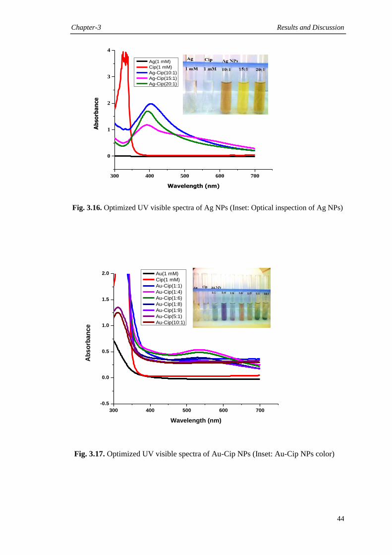
Chapter-3 Results and Discussion
44
300 400 500 600 700
0
1
2
3
4
Ab
sorb
an
ce
Wavelength (nm)
Ag(1 mM)
Cip(1 mM)
Ag-Cip(10:1)
Ag-Cip(15:1)
Ag-Cip(20:1)
300 400 500 600 700
-0.5
0.0
0.5
1.0
1.5
2.0
Ab
so
rba
nc
e
Wavelength (nm)
Au(1 mM)
Cip(1 mM)
Au-Cip(1:1)
Au-Cip(1:4)
Au-Cip(1:6)
Au-Cip(1:8)
Au-Cip(1:9)
Au-Cip(5:1)
Au-Cip(10:1)
Fig. 3.17. Optimized UV visible spectra of Au-Cip NPs (Inset: Au-Cip NPs color)
Fig. 3.16. Optimized UV visible spectra of Ag NPs (Inset: Optical inspection of Ag NPs)
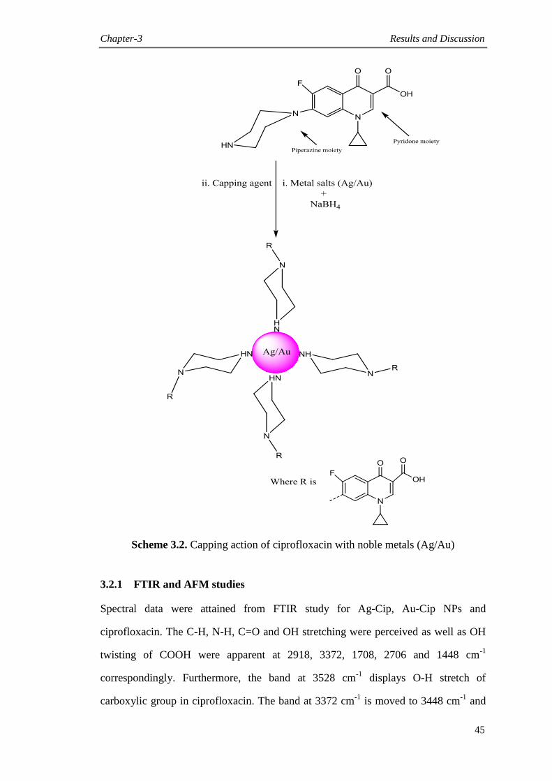
Chapter-3 Results and Discussion
45
Scheme 3.2. Capping action of ciprofloxacin with noble metals (Ag/Au)
3.2.1 FTIR and AFM studies
Spectral data were attained from FTIR study for Ag-Cip, Au-Cip NPs and
ciprofloxacin. The C-H, N-H, C=O and OH stretching were perceived as well as OH
twisting of COOH were apparent at 2918, 3372, 1708, 2706 and 1448 cm-1
correspondingly. Furthermore, the band at 3528 cm-1
displays O-H stretch of
carboxylic group in ciprofloxacin. The band at 3372 cm-1
is moved to 3448 cm-1
and
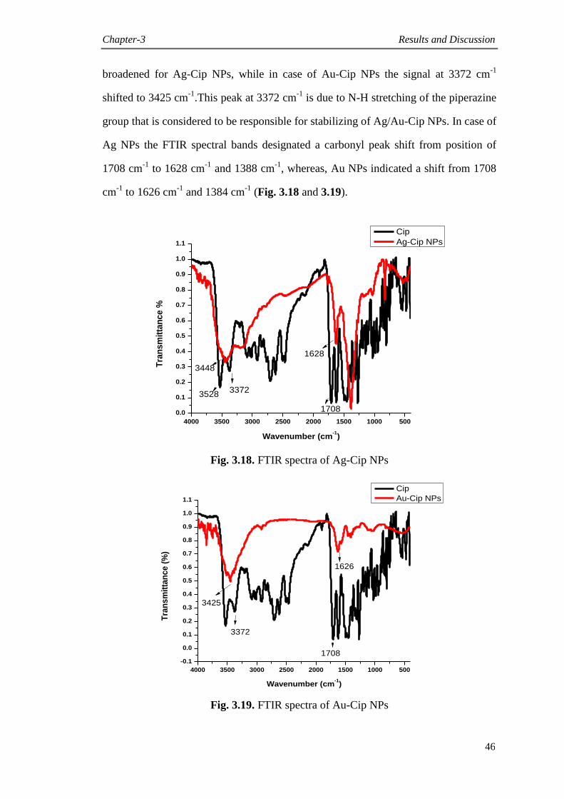
Chapter-3 Results and Discussion
46
broadened for Ag-Cip NPs, while in case of Au-Cip NPs the signal at 3372 cm-1
shifted to 3425 cm-1
.This peak at 3372 cm-1
is due to N-H stretching of the piperazine
group that is considered to be responsible for stabilizing of Ag/Au-Cip NPs. In case of
Ag NPs the FTIR spectral bands designated a carbonyl peak shift from position of
1708 cm-1
to 1628 cm-1
and 1388 cm-1
, whereas, Au NPs indicated a shift from 1708
cm-1
to 1626 cm-1
and 1384 cm-1
(Fig. 3.18 and 3.19).
4000 3500 3000 2500 2000 1500 1000 500
0.0
0.1
0.2
0.3
0.4
0.5
0.6
0.7
0.8
0.9
1.0
1.1
Tra
ns
mit
tan
ce
%
Wavenumber (cm-1)
Cip
Ag-Cip NPs
3528
3448
3372
1708
1628
Fig. 3.18. FTIR spectra of Ag-Cip NPs
4000 3500 3000 2500 2000 1500 1000 500
-0.1
0.0
0.1
0.2
0.3
0.4
0.5
0.6
0.7
0.8
0.9
1.0
1.1
Tra
ns
mit
tan
ce
(%
)
Wavenumber (cm-1)
Cip
Au-Cip NPs
1626
1708
3372
3425
Fig. 3.19. FTIR spectra of Au-Cip NPs

Chapter-3 Results and Discussion
47
The structural topographies of the ciprofloxacin-stabilized Ag and Au NPs was achieved
by AFM (Fig. 3.20 and 3.21) and specifies that the nano-conjugates of Ag-Cip NPs are
sphere-shaped with size in the range of 40-50 nm, while the Au-Cip NPs also have round
shape with sizes from 60-85 nm (Fig. 3.21).
Fig. 3.20. AFM images of Ag NPs
Fig. 3.21. AFM images of Au NPs
3.2.2 EDX and SEM studies
Energy-dispersive X-ray spectroscopy (EDX) verified the chemical make-up of metal
NPs by ciprofloxacin as capping and stabilizing agent. The presence of signal in region of
Ag in EDX study verifies the production of Ag NPs. Metallic Ag typically shows a visual
absorption peak at closely 3 keV due to surface plasmon resonance (SPR). Au-Cip NPs
designate the presence of typical peaks of Au at 2.12 and 9.71 keV as shown in fig. 3.22
and 3.23.
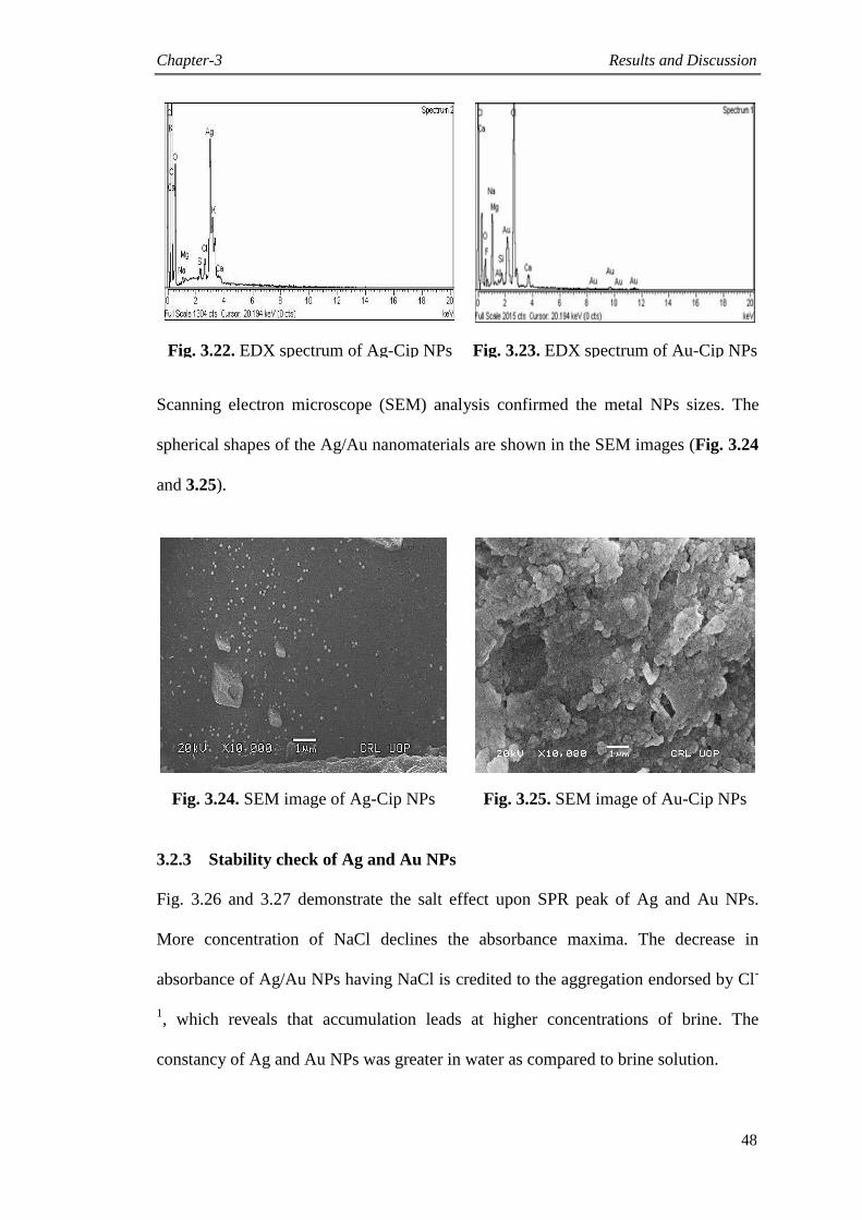
Chapter-3 Results and Discussion
48
Fig. 3.22. EDX spectrum of Ag-Cip NPs Fig. 3.23. EDX spectrum of Au-Cip NPs
Scanning electron microscope (SEM) analysis confirmed the metal NPs sizes. The
spherical shapes of the Ag/Au nanomaterials are shown in the SEM images (Fig. 3.24
and 3.25).
Fig. 3.24. SEM image of Ag-Cip NPs Fig. 3.25. SEM image of Au-Cip NPs
3.2.3 Stability check of Ag and Au NPs
Fig. 3.26 and 3.27 demonstrate the salt effect upon SPR peak of Ag and Au NPs.
More concentration of NaCl declines the absorbance maxima. The decrease in
absorbance of Ag/Au NPs having NaCl is credited to the aggregation endorsed by Cl-
1, which reveals that accumulation leads at higher concentrations of brine. The
constancy of Ag and Au NPs was greater in water as compared to brine solution.
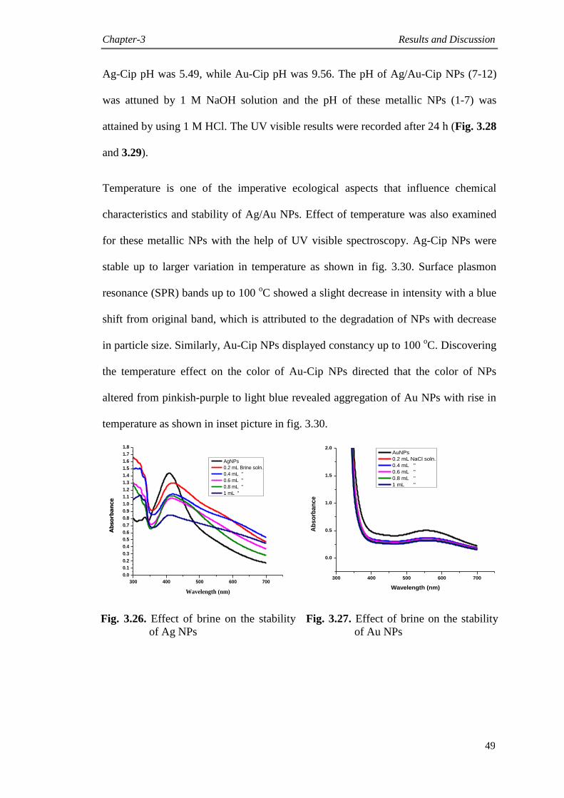
Chapter-3 Results and Discussion
49
Ag-Cip pH was 5.49, while Au-Cip pH was 9.56. The pH of Ag/Au-Cip NPs (7-12)
was attuned by 1 M NaOH solution and the pH of these metallic NPs (1-7) was
attained by using 1 M HCl. The UV visible results were recorded after 24 h (Fig. 3.28
and 3.29).
Temperature is one of the imperative ecological aspects that influence chemical
characteristics and stability of Ag/Au NPs. Effect of temperature was also examined
for these metallic NPs with the help of UV visible spectroscopy. Ag-Cip NPs were
stable up to larger variation in temperature as shown in fig. 3.30. Surface plasmon
resonance (SPR) bands up to 100 oC showed a slight decrease in intensity with a blue
shift from original band, which is attributed to the degradation of NPs with decrease
in particle size. Similarly, Au-Cip NPs displayed constancy up to 100 oC. Discovering
the temperature effect on the color of Au-Cip NPs directed that the color of NPs
altered from pinkish-purple to light blue revealed aggregation of Au NPs with rise in
temperature as shown in inset picture in fig. 3.30.
300 400 500 600 700
0.0
0.1
0.2
0.3
0.4
0.5
0.6
0.7
0.8
0.9
1.0
1.1
1.2
1.3
1.4
1.5
1.6
1.7
1.8
Ab
so
rba
nce
Wavelength (nm)
AgNPs
0.2 mL Brine soln.
0.4 mL "
0.6 mL "
0.8 mL "
1 mL "
300 400 500 600 700
0.0
0.5
1.0
1.5
2.0
Ab
so
rba
nc
e
Wavelength (nm)
AuNPs
0.2 mL NaCl soln.
0.4 mL "
0.6 mL "
0.8 mL "
1 mL "
Fig. 3.26. Effect of brine on the stability
of Ag NPs
Fig. 3.27. Effect of brine on the stability
of Au NPs
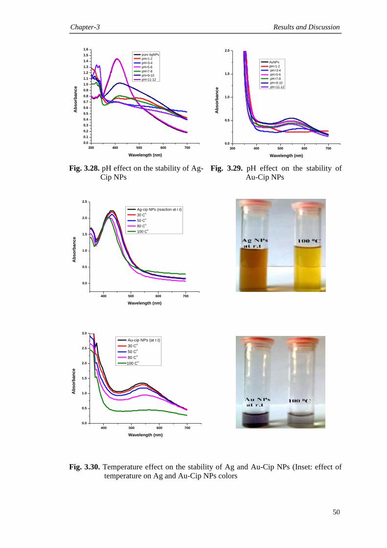
Chapter-3 Results and Discussion
50
300 400 500 600 700
0.0
0.1
0.2
0.3
0.4
0.5
0.6
0.7
0.8
0.9
1.0
1.1
1.2
1.3
1.4
1.5
1.6
Ab
so
rba
nc
e
Wavelength (nm)
pure AgNPs
pH=1-2
pH=3-4
pH=5-6
pH=7-8
pH=9-10
pH=11-12
300 400 500 600 700
0.0
0.5
1.0
1.5
2.0
Ab
so
rba
nc
e
Wavelength (nm)
AuNPs
pH=1-2
pH=3-4
pH=5-6
pH=7-8
pH=9-10
pH=11-12
Fig. 3.28. pH effect on the stability of Ag-
Cip NPs
Fig. 3.29. pH effect on the stability of
Au-Cip NPs
400 500 600 700
0.0
0.5
1.0
1.5
2.0
2.5
Ab
so
rba
nc
e
Wavelength (nm)
Ag-cip NPs (reaction at r.t)
30 Co
50 Co
80 Co
100 Co
400 500 600 700
0.0
0.5
1.0
1.5
2.0
2.5
3.0
Ab
so
rba
nc
e
Wavelength (nm)
Au-cip NPs (at r.t)
30 Co
50 Co
80 Co
100 Co
Fig. 3.30. Temperature effect on the stability of Ag and Au-Cip NPs (Inset: effect of
temperature on Ag and Au-Cip NPs colors

Chapter-3 Results and Discussion
51
3.3 Synthesis of sparfloxacin mediated Ag and Au NPs
Sparfloxacin is a fluoroquinolone antibiotic having capability of capping Ag and Au
NPs as shown in scheme 3.3.
Scheme 3.3: Synthesis of Ag and Au NPs stabilized with sparfloxacin
3.3.1 UV visible spectroscopic analysis
Maximum absorption of metallic NPs was noted as a function of retention time in the
range of 300 to 700 nm. Variations in UV visible absorption bands were reflected as
the preliminary clue for the synthesis of Ag/Au NPs. The presence of specific peaks
in the areas of 400-500 nm and 500-600 nm certified the synthesis of Ag and Au NPs
correspondingly. The sharp peak for Ag-Sp NPs was perceived for a 10:1 (metal:
drug) molar ratio (Fig. 3.31), while for Au-Sp NPs, the maximum peak was at 1:2
(metal: drug) molar ratio as shown in the fig. 3.32.
300 400 500 600 700
0.0
0.5
1.0
1.5
2.0
2.5
3.0
Ab
so
rba
nc
e
Wavelength (nm)
Ag
Sp
Ag-sp (10:1)
Fig. 3.31. UV visible spectra of Ag NPs (Inset: color of Ag NPs)
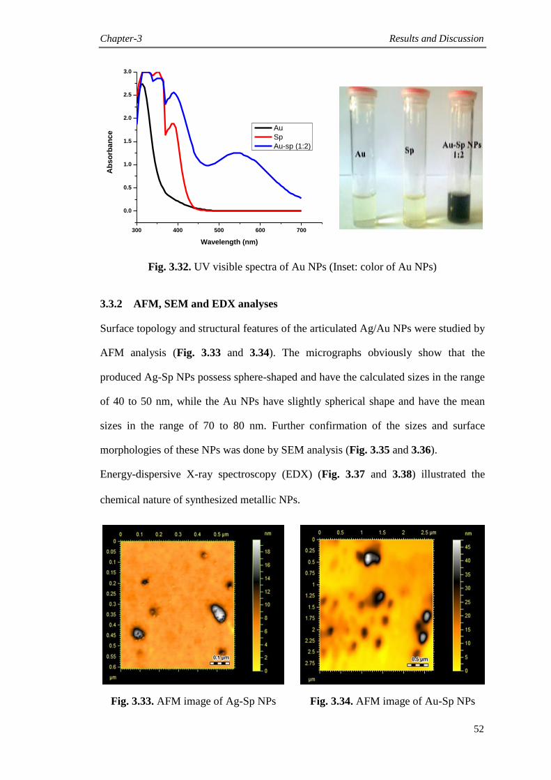
Chapter-3 Results and Discussion
52
300 400 500 600 700
0.0
0.5
1.0
1.5
2.0
2.5
3.0
Ab
so
rba
nc
e
Wavelength (nm)
Au
Sp
Au-sp (1:2)
Fig. 3.32. UV visible spectra of Au NPs (Inset: color of Au NPs)
3.3.2 AFM, SEM and EDX analyses
Surface topology and structural features of the articulated Ag/Au NPs were studied by
AFM analysis (Fig. 3.33 and 3.34). The micrographs obviously show that the
produced Ag-Sp NPs possess sphere-shaped and have the calculated sizes in the range
of 40 to 50 nm, while the Au NPs have slightly spherical shape and have the mean
sizes in the range of 70 to 80 nm. Further confirmation of the sizes and surface
morphologies of these NPs was done by SEM analysis (Fig. 3.35 and 3.36).
Energy-dispersive X-ray spectroscopy (EDX) (Fig. 3.37 and 3.38) illustrated the
chemical nature of synthesized metallic NPs.
Fig. 3.33. AFM image of Ag-Sp NPs Fig. 3.34. AFM image of Au-Sp NPs
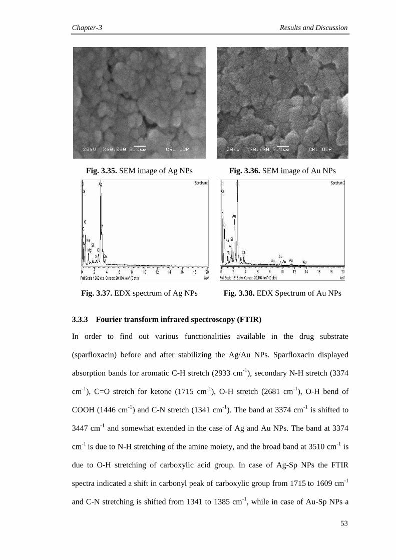
Chapter-3 Results and Discussion
53
Fig. 3.35. SEM image of Ag NPs Fig. 3.36. SEM image of Au NPs
Fig. 3.37. EDX spectrum of Ag NPs Fig. 3.38. EDX Spectrum of Au NPs
3.3.3 Fourier transform infrared spectroscopy (FTIR)
In order to find out various functionalities available in the drug substrate
(sparfloxacin) before and after stabilizing the Ag/Au NPs. Sparfloxacin displayed
absorption bands for aromatic C-H stretch (2933 cm-1
), secondary N-H stretch (3374
cm-1
), C=O stretch for ketone (1715 cm-1
), O-H stretch (2681 cm-1
), O-H bend of
COOH (1446 cm-1
) and C-N stretch (1341 cm-1
). The band at 3374 cm-1
is shifted to
3447 cm-1
and somewhat extended in the case of Ag and Au NPs. The band at 3374
cm-1
is due to N-H stretching of the amine moiety, and the broad band at 3510 cm-1
is
due to O-H stretching of carboxylic acid group. In case of Ag-Sp NPs the FTIR
spectra indicated a shift in carbonyl peak of carboxylic group from 1715 to 1609 cm
-1
and C-N stretching is shifted from 1341 to 1385 cm-1
, while in case of Au-Sp NPs a
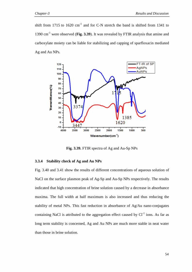
Chapter-3 Results and Discussion
54
shift from 1715 to 1620 cm
-1 and for C-N stretch the band is shifted from 1341 to
1390 cm-1
were observed (Fig. 3.39). It was revealed by FTIR analysis that amine and
carboxylate moiety can be liable for stabilizing and capping of sparfloxacin mediated
Ag and Au NPs.
Fig. 3.39. FTIR spectra of Ag and Au-Sp NPs
3.3.4 Stability check of Ag and Au NPs
Fig. 3.40 and 3.41 show the results of different concentrations of aqueous solution of
NaCl on the surface plasmon peak of Ag-Sp and Au-Sp NPs respectively. The results
indicated that high concentration of brine solution caused by a decrease in absorbance
maxima. The full width at half maximum is also increased and thus reducing the
stability of metal NPs. This fast reduction in absorbance of Ag/Au nano-conjugates
containing NaCl is attributed to the aggregation effect caused by Cl-1
ions. As far as
long term stability is concerned, Ag and Au NPs are much more stable in neat water
than those in brine solution.

Chapter-3 Results and Discussion
55
300 400 500 600 700
0.0
0.5
1.0
1.5
2.0
2.5
Ab
so
rba
nc
e
Wavelength (nm)
Ag-sp (salt effect)
0.2 mL (Brine)
0.4 mL
0.6 mL
0.8 mL
1 mL
Fig. 3.40. Effect of brine on the stability of Ag-Sp NPs
300 400 500 600 700
0.5
1.0
1.5
2.0
2.5
3.0
Ab
so
rba
nc
e
Wavelength (nm)
AuNPs (Sp)
0.2 mL (Brine)
0.4 mL
0.6 mL
0.8 mL
1 mL
Fig. 3.41. Effect of brine on the stability of Au-Sp NPs
The effect of pH on the stability of Ag and Au-Sp NPs was also studied. pH of 3 mL
solutions of the freshly prepared NPs were found to be 4.49 (Ag-Sp NPs) and 9.66
(Au-Sp NPs). pH of 1-7 was adjusted by using 1M HCl solution, while that of 7-12
was done with 1M NaOH solution. The UV visible bands of the resultant solutions
were verified after 24 hours (Fig. 3.42 and 3.43).

Chapter-3 Results and Discussion
56
300 400 500 600 700
0.0
0.5
1.0
1.5
2.0
B
A
pH=2-3
pH=4-5 (pure Ag-sp)
pH=6-7
pH=8-9
pH=10-11
pH=12-13
Fig. 3.42. pH effect on the stability of Ag-Sp NPs
300 400 500 600 700
0.0
0.5
1.0
1.5
2.0
2.5
3.0
Ab
so
rba
nc
e
Wavelength (nm)
Au NPs (Sp)
pH=1-2
pH=3-4
pH=5-6
pH=7-8
pH=9-10
pH=11-12
Fig. 3.43. pH effect on the stability of Au-Sp NPs
Temperature is an important ecological factor that distresses the permanency and
chemical features of Ag and Au NPs. Ag-Sp NPs were stable at broad range of
temperatures, as shown in fig. 3.44. Surface plasmon resonance bands up to 100 oC
indicated a reduction in strength with a blue shift from the original band, which is
recognized to the dreadful conditions of NPs, with lessening in NPs size.
Furthermore, Au-Sp NPs revealed less stability by increasing heat up to 100 oC (Fig.
3.45).
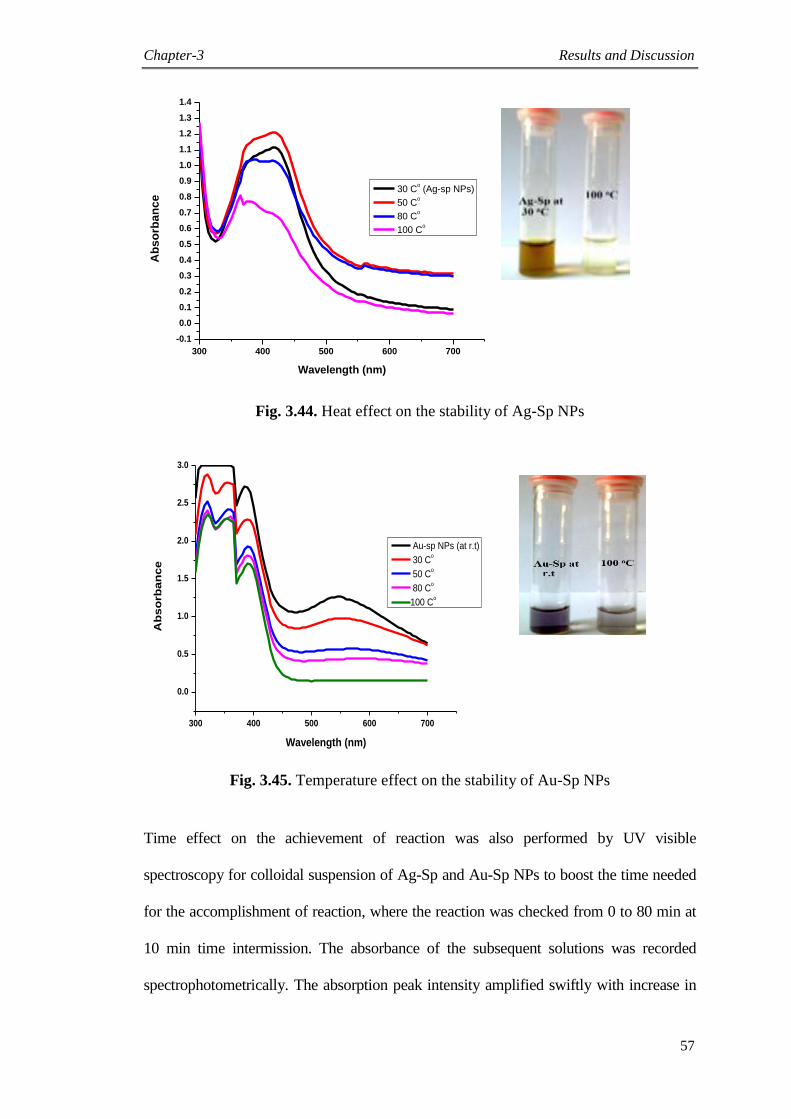
Chapter-3 Results and Discussion
57
300 400 500 600 700
-0.1
0.0
0.1
0.2
0.3
0.4
0.5
0.6
0.7
0.8
0.9
1.0
1.1
1.2
1.3
1.4
Ab
so
rba
nc
e
Wavelength (nm)
30 Co (Ag-sp NPs)
50 Co
80 Co
100 Co
Fig. 3.44. Heat effect on the stability of Ag-Sp NPs
300 400 500 600 700
0.0
0.5
1.0
1.5
2.0
2.5
3.0
Ab
so
rba
nc
e
Wavelength (nm)
Au-sp NPs (at r.t)
30 Co
50 Co
80 Co
100 Co
Fig. 3.45. Temperature effect on the stability of Au-Sp NPs
Time effect on the achievement of reaction was also performed by UV visible
spectroscopy for colloidal suspension of Ag-Sp and Au-Sp NPs to boost the time needed
for the accomplishment of reaction, where the reaction was checked from 0 to 80 min at
10 min time intermission. The absorbance of the subsequent solutions was recorded
spectrophotometrically. The absorption peak intensity amplified swiftly with increase in
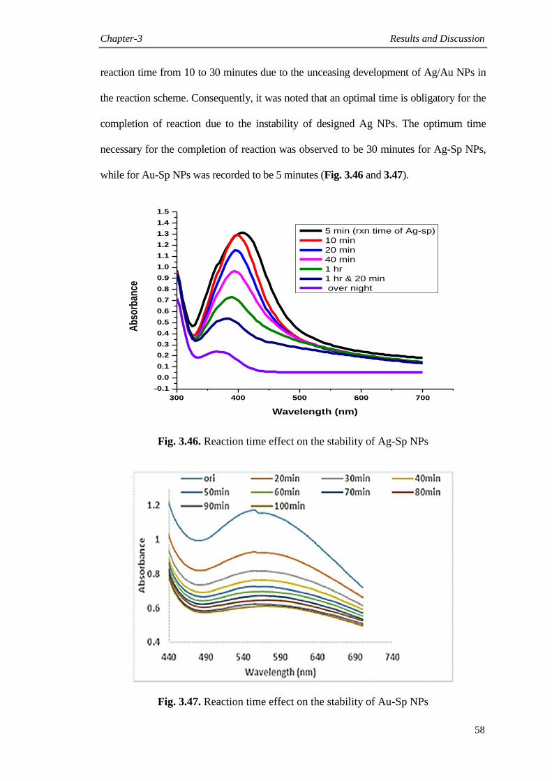
Chapter-3 Results and Discussion
58
reaction time from 10 to 30 minutes due to the unceasing development of Ag/Au NPs in
the reaction scheme. Consequently, it was noted that an optimal time is obligatory for the
completion of reaction due to the instability of designed Ag NPs. The optimum time
necessary for the completion of reaction was observed to be 30 minutes for Ag-Sp NPs,
while for Au-Sp NPs was recorded to be 5 minutes (Fig. 3.46 and 3.47).
300 400 500 600 700
-0.1
0.0
0.1
0.2
0.3
0.4
0.5
0.6
0.7
0.8
0.9
1.0
1.1
1.2
1.3
1.4
1.5
Ab
sorb
ance
Wavelength (nm)
5 min (rxn time of Ag-sp)
10 min
20 min
40 min
1 hr
1 hr & 20 min
over night
Fig. 3.46. Reaction time effect on the stability of Ag-Sp NPs
Fig. 3.47. Reaction time effect on the stability of Au-Sp NPs
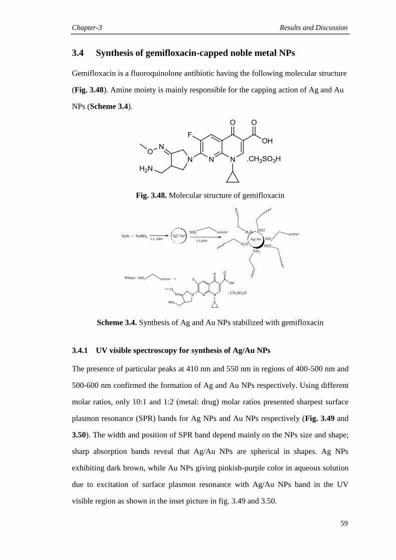
Chapter-3 Results and Discussion
59
3.4 Synthesis of gemifloxacin-capped noble metal NPs
Gemifloxacin is a fluoroquinolone antibiotic having the following molecular structure
(Fig. 3.48). Amine moiety is mainly responsible for the capping action of Ag and Au
NPs (Scheme 3.4).
Fig. 3.48. Molecular structure of gemifloxacin
Scheme 3.4. Synthesis of Ag and Au NPs stabilized with gemifloxacin
3.4.1 UV visible spectroscopy for synthesis of Ag/Au NPs
The presence of particular peaks at 410 nm and 550 nm in regions of 400-500 nm and
500-600 nm confirmed the formation of Ag and Au NPs respectively. Using different
molar ratios, only 10:1 and 1:2 (metal: drug) molar ratios presented sharpest surface
plasmon resonance (SPR) bands for Ag NPs and Au NPs respectively (Fig. 3.49 and
3.50). The width and position of SPR band depend mainly on the NPs size and shape;
sharp absorption bands reveal that Ag/Au NPs are spherical in shapes. Ag NPs
exhibiting dark brown, while Au NPs giving pinkish-purple color in aqueous solution
due to excitation of surface plasmon resonance with Ag/Au NPs band in the UV
visible region as shown in the inset picture in fig. 3.49 and 3.50.

Chapter-3 Results and Discussion
60
Fig. 3.49. Optimized UV visible spectra of Ag NPs (Inset: Color of Ag NPs)
Fig. 3.50. Optimized UV visible spectra of Au NPs. (Inset: Colors of Au NPs)
3.4.2 AFM, SEM and EDX spectroscopy analysis
Structural features were determined by using atomic force microscopy and scanning
electron microscopy analysis. AFM micrographs showed that both Ag NPs and Au
NPs are spherical in shapes and have sizes of about 40 nm and 70 nm respectively
(Fig. 3.51 and 3.52).
0
0.5
1
1.5
2
2.5
3
300 400 500 600 700
Ab
sorb
ance
wave length (nm)
Ag
Gm
1:01
5:01
10:01
-0.5
0
0.5
1
1.5
2
2.5
3
3.5
250 350 450 550 650 750
Ab
sorb
ance
Wavelength (nm)
Au
Gm
1:02
1:03
1:04
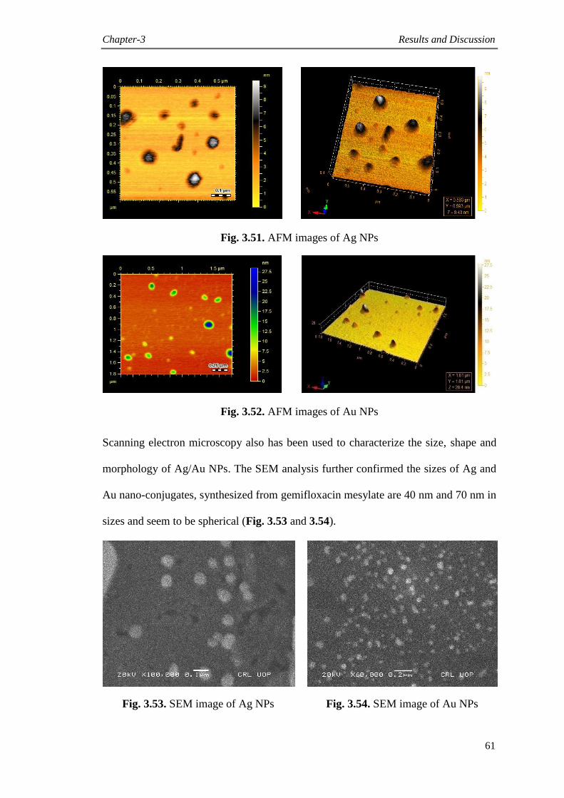
Chapter-3 Results and Discussion
61
Fig. 3.51. AFM images of Ag NPs
Fig. 3.52. AFM images of Au NPs
Scanning electron microscopy also has been used to characterize the size, shape and
morphology of Ag/Au NPs. The SEM analysis further confirmed the sizes of Ag and
Au nano-conjugates, synthesized from gemifloxacin mesylate are 40 nm and 70 nm in
sizes and seem to be spherical (Fig. 3.53 and 3.54).
Fig. 3.53. SEM image of Ag NPs Fig. 3.54. SEM image of Au NPs
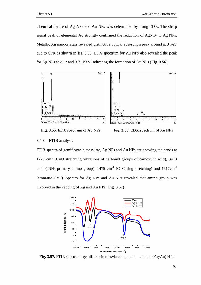
Chapter-3 Results and Discussion
62
Chemical nature of Ag NPs and Au NPs was determined by using EDX. The sharp
signal peak of elemental Ag strongly confirmed the reduction of AgNO3 to Ag NPs.
Metallic Ag nanocrystals revealed distinctive optical absorption peak around at 3 keV
due to SPR as shown in fig. 3.55. EDX spectrum for Au NPs also revealed the peak
for Ag NPs at 2.12 and 9.71 KeV indicating the formation of Au NPs (Fig. 3.56).
Fig. 3.55. EDX spectrum of Ag NPs Fig. 3.56. EDX spectrum of Au NPs
3.4.3 FTIR analysis
FTIR spectra of gemifloxacin mesylate, Ag NPs and Au NPs are showing the bands at
1725 cm-1
(C=O stretching vibrations of carbonyl groups of carboxylic acid), 3410
cm-1
(-NH2 primary amino group), 1475 cm-1
(C=C ring stretching) and 1617cm-1
(aromatic C=C). Spectra for Ag NPs and Au NPs revealed that amino group was
involved in the capping of Ag and Au NPs (Fig. 3.57).
4000 3500 3000 2500 2000 1500 1000 500
0
20
40
60
80
100
120
140
Tra
nsm
itta
nce
(%
)
Wavenumber (cm-1)
Gm
Ag NPs
Au NPs
3410
1725
Fig. 3.57. FTIR spectra of gemifloxacin mesylate and its noble metal (Ag/Au) NPs

Chapter-3 Results and Discussion
63
3.4.4 Stability check of Ag and Au NPs
Temperature effect was investigated for Ag and Au NPs with the help of UV visible
spectroscopy. Ag NPs were stable up to large changes in temperature, SPR bands up
to 100 oC presented a slight decrease in intensity of the SPR band with a blue shift
from original band showing slight degradation of Ag NPs, also color of the heated
solution (up to 100 o
C) was not much different from original solution‟s color (Fig.
3.58), while in case of Au NPs, SPR bands decreased in intensity, broadened and
slight red shift in absorption wavelength was happened (Fig. 3.59).
Fig. 3.58. Temperature effect on the stability of Ag NPs
Fig. 3.59. Effect of temperature on the stability of Au NPs
0
0.5
1
1.5
2
2.5
368 418 468 518 568 618
Ab
sorb
ance
Wavelength (nm)
20ᵒ C
30° C
50° C
80° C
100° C
-0.1
0.1
0.3
0.5
0.7
0.9
1.1
1.3
1.5
420 520 620
Ab
sorb
ance
Wavelength (nm)
20ᵒC
30°C
50°C
80°C
100°C
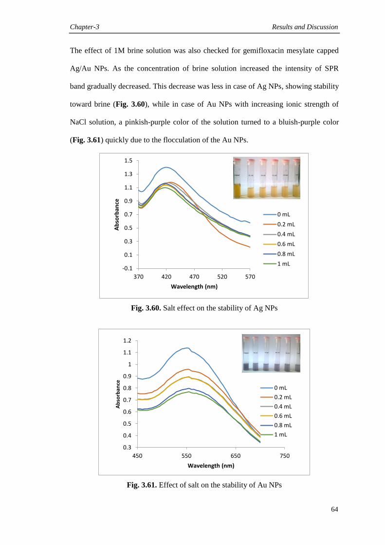
Chapter-3 Results and Discussion
64
The effect of 1M brine solution was also checked for gemifloxacin mesylate capped
Ag/Au NPs. As the concentration of brine solution increased the intensity of SPR
band gradually decreased. This decrease was less in case of Ag NPs, showing stability
toward brine (Fig. 3.60), while in case of Au NPs with increasing ionic strength of
NaCl solution, a pinkish-purple color of the solution turned to a bluish-purple color
(Fig. 3.61) quickly due to the flocculation of the Au NPs.
Fig. 3.60. Salt effect on the stability of Ag NPs
Fig. 3.61. Effect of salt on the stability of Au NPs
-0.1
0.1
0.3
0.5
0.7
0.9
1.1
1.3
1.5
370 420 470 520 570
Ab
sorb
ance
Wavelength (nm)
0 mL
0.2 mL
0.4 mL
0.6 mL
0.8 mL
1 mL
0.3
0.4
0.5
0.6
0.7
0.8
0.9
1
1.1
1.2
450 550 650 750
Ab
sorb
ance
Wavelength (nm)
0 mL
0.2 mL
0.4 mL
0.6 mL
0.8 mL
1 mL
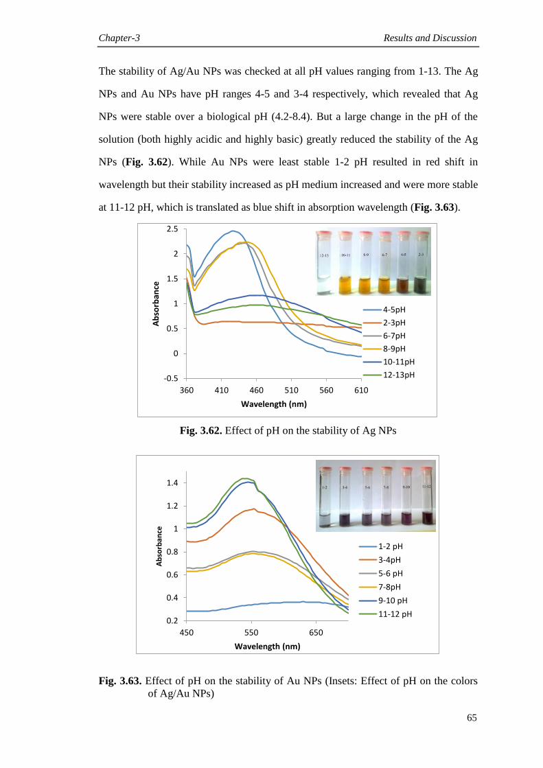
Chapter-3 Results and Discussion
65
The stability of Ag/Au NPs was checked at all pH values ranging from 1-13. The Ag
NPs and Au NPs have pH ranges 4-5 and 3-4 respectively, which revealed that Ag
NPs were stable over a biological pH (4.2-8.4). But a large change in the pH of the
solution (both highly acidic and highly basic) greatly reduced the stability of the Ag
NPs (Fig. 3.62). While Au NPs were least stable 1-2 pH resulted in red shift in
wavelength but their stability increased as pH medium increased and were more stable
at 11-12 pH, which is translated as blue shift in absorption wavelength (Fig. 3.63).
Fig. 3.62. Effect of pH on the stability of Ag NPs
Fig. 3.63. Effect of pH on the stability of Au NPs (Insets: Effect of pH on the colors
of Ag/Au NPs)
-0.5
0
0.5
1
1.5
2
2.5
360 410 460 510 560 610
Ab
sorb
ance
Wavelength (nm)
4-5pH
2-3pH
6-7pH
8-9pH
10-11pH
12-13pH
0.2
0.4
0.6
0.8
1
1.2
1.4
450 550 650
Ab
sorb
ance
Wavelength (nm)
1-2 pH
3-4pH
5-6 pH
7-8pH
9-10 pH
11-12 pH
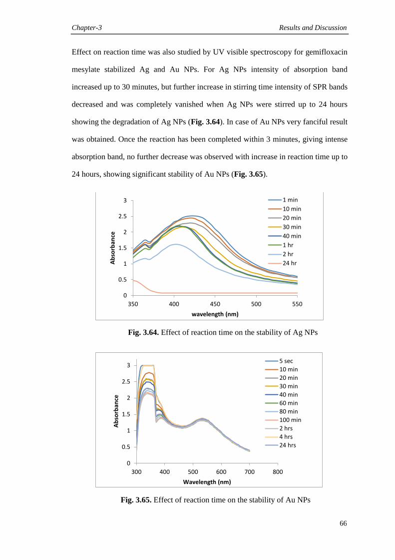
Chapter-3 Results and Discussion
66
Effect on reaction time was also studied by UV visible spectroscopy for gemifloxacin
mesylate stabilized Ag and Au NPs. For Ag NPs intensity of absorption band
increased up to 30 minutes, but further increase in stirring time intensity of SPR bands
decreased and was completely vanished when Ag NPs were stirred up to 24 hours
showing the degradation of Ag NPs (Fig. 3.64). In case of Au NPs very fanciful result
was obtained. Once the reaction has been completed within 3 minutes, giving intense
absorption band, no further decrease was observed with increase in reaction time up to
24 hours, showing significant stability of Au NPs (Fig. 3.65).
Fig. 3.64. Effect of reaction time on the stability of Ag NPs
Fig. 3.65. Effect of reaction time on the stability of Au NPs
0
0.5
1
1.5
2
2.5
3
350 400 450 500 550
Ab
sorb
ance
wavelength (nm)
1 min
10 min
20 min
30 min
40 min
1 hr
2 hr
24 hr
0
0.5
1
1.5
2
2.5
3
300 400 500 600 700 800
Ab
sorb
ance
Wavelength (nm)
5 sec
10 min
20 min
30 min
40 min
60 min
80 min
100 min
2 hrs
4 hrs
24 hrs

Chapter-3 Results and Discussion
67
3.5 Biological evaluation of fluoroquinolones-capped Ag/Au NPs
Different biological activities were evaluated to express the hidden potentials of Ag
and Au nano-conjugates. Some activities revealed significant results, while the
outcomes of other activities were moderate. All activities are described in below
section.
3.5.1 Urease study
The NPs along with the capping ligands (fluoroquinolones) were independently screened
for jack bean urease enzymes inhibition potentials. Mostly the Ag nano-conjugates
showed significant urease inhibition activity. The Ag-Mox NPs exhibited significantly
higher levels of enzyme inhibition activity of 93% at 0.2 mg/mL and IC50 value of 0.66 ±
0.042 μg/mL, while the ligand (Moxifloxacin) revealed weak inhibition with IC50 value of
183.25 ± 2.06 μg/mL. The Au-Mox was found inactive as compared to the parent
compound (Mox) having IC50 = 183.25 ± 2.06 μg/mL. The results deduced that after
conjugation of moxifloxacin with Ag, the activity of moxifloxacin was significantly
increased even more than 250 times. Interestingly, after conjugation with Au the activity
of moxifloxacin was significantly decreased, but conjugation of Mox to Ag had a robust
inhibition effect in comparison to Mox (Table 3.1).
The metal nano-conjugates (Ag-Cip and Au-Cip) along with parent drug, ciprofloxacin
(Cip) were also subjected for urease enzymes inhibition studies to search the inhibition
potential. After screening of Cip, Ag-Cip and Au-Cip against urease enzyme, the Ag-
Cip was found significantly active with % inhibition of 96 at 0.2 mg/mL. It revealed
significant activity with IC50 value of 1.181 ± 0.02 μg/mL. The Au-Cip also exhibited
good urease inhibitory activity with IC50 value of 52.55±2.3 μg/mL and % inhibition 90
at 0.2 mg/mL comparatively ciprofloxacin, which presented less activity with %
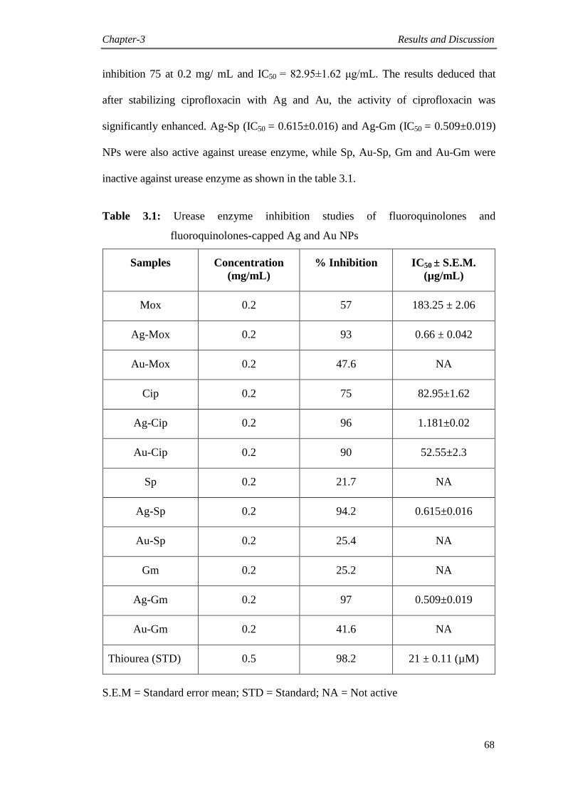
Chapter-3 Results and Discussion
68
inhibition 75 at 0.2 mg/ mL and IC50 = 82.95±1.62 μg/mL. The results deduced that
after stabilizing ciprofloxacin with Ag and Au, the activity of ciprofloxacin was
significantly enhanced. Ag-Sp (IC50 = 0.615±0.016) and Ag-Gm (IC50 = 0.509±0.019)
NPs were also active against urease enzyme, while Sp, Au-Sp, Gm and Au-Gm were
inactive against urease enzyme as shown in the table 3.1.
Table 3.1: Urease enzyme inhibition studies of fluoroquinolones and
fluoroquinolones-capped Ag and Au NPs
Samples Concentration
(mg/mL)
% Inhibition IC50 ± S.E.M.
(µg/mL)
Mox 0.2 57 183.25 ± 2.06
Ag-Mox 0.2 93 0.66 ± 0.042
Au-Mox 0.2 47.6 NA
Cip 0.2 75 82.95±1.62
Ag-Cip 0.2 96 1.181±0.02
Au-Cip 0.2 90 52.55±2.3
Sp 0.2 21.7 NA
Ag-Sp 0.2 94.2 0.615±0.016
Au-Sp 0.2 25.4 NA
Gm 0.2 25.2 NA
Ag-Gm 0.2 97 0.509±0.019
Au-Gm 0.2 41.6 NA
Thiourea (STD) 0.5 98.2 21 ± 0.11 (µM)
S.E.M = Standard error mean; STD = Standard; NA = Not active
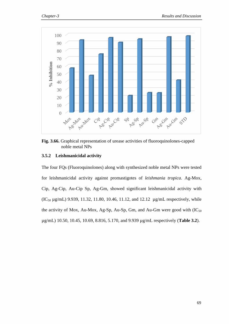
Chapter-3 Results and Discussion
69
Fig. 3.66. Graphical representation of urease activities of fluoroquinolones-capped
noble metal NPs
3.5.2 Leishmanicidal activity
The four FQs (Fluoroquinolones) along with synthesized noble metal NPs were tested
for leishmanicidal activity against promastigotes of leishmania tropica. Ag-Mox,
Cip, Ag-Cip, Au-Cip Sp, Ag-Gm, showed significant leishmanicidal activity with
(IC50 µg/mL) 9.939, 11.32, 11.80, 10.46, 11.12, and 12.12 µg/mL respectively, while
the activity of Mox, Au-Mox, Ag-Sp, Au-Sp, Gm, and Au-Gm were good with (IC50
µg/mL) 10.50, 10.45, 10.69, 8.816, 5.170, and 9.939 µg/mL respectively (Table 3.2).
0
10
20
30
40
50
60
70
80
90
100
% I
nh
ibit
ion
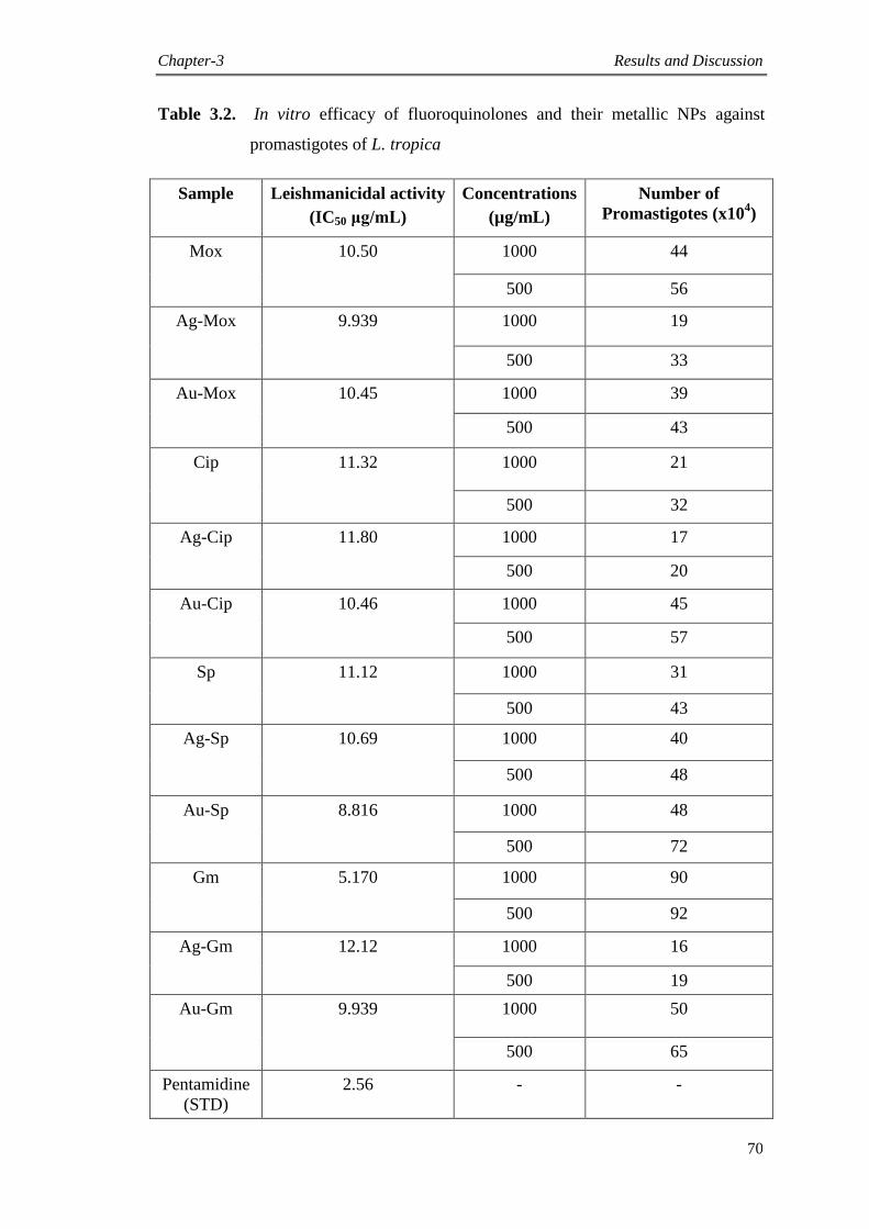
Chapter-3 Results and Discussion
70
Table 3.2. In vitro efficacy of fluoroquinolones and their metallic NPs against
promastigotes of L. tropica
Sample Leishmanicidal activity
(IC50 μg/mL)
Concentrations
(µg/mL)
Number of
Promastigotes (x104)
Mox 10.50 1000 44
500 56
Ag-Mox 9.939 1000 19
500 33
Au-Mox 10.45 1000 39
500 43
Cip 11.32 1000 21
500 32
Ag-Cip 11.80 1000 17
500 20
Au-Cip 10.46 1000 45
500 57
Sp 11.12 1000 31
500 43
Ag-Sp 10.69 1000 40
500 48
Au-Sp 8.816 1000 48
500 72
Gm 5.170 1000 90
500 92
Ag-Gm 12.12 1000 16
500 19
Au-Gm 9.939 1000 50
500 65
Pentamidine
(STD)
2.56 - -
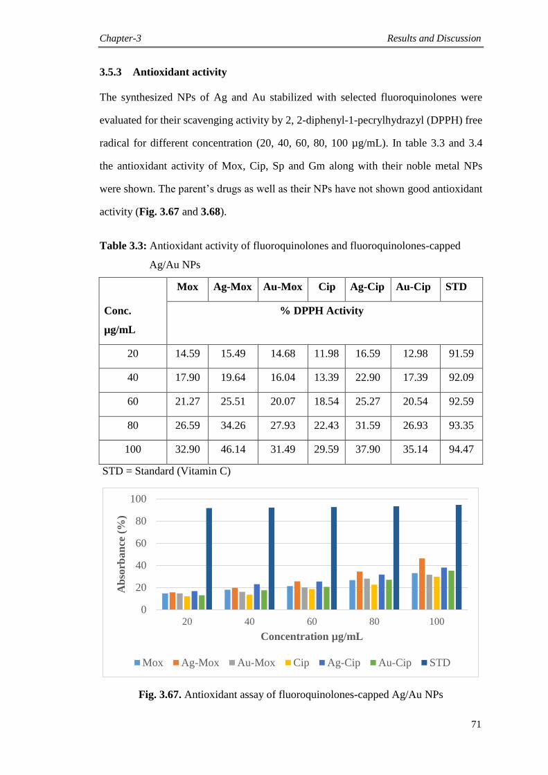
Chapter-3 Results and Discussion
71
3.5.3 Antioxidant activity
The synthesized NPs of Ag and Au stabilized with selected fluoroquinolones were
evaluated for their scavenging activity by 2, 2-diphenyl-1-pecrylhydrazyl (DPPH) free
radical for different concentration (20, 40, 60, 80, 100 µg/mL). In table 3.3 and 3.4
the antioxidant activity of Mox, Cip, Sp and Gm along with their noble metal NPs
were shown. The parent‟s drugs as well as their NPs have not shown good antioxidant
activity (Fig. 3.67 and 3.68).
Table 3.3: Antioxidant activity of fluoroquinolones and fluoroquinolones-capped
Ag/Au NPs
Mox Ag-Mox Au-Mox Cip Ag-Cip Au-Cip STD
Conc.
µg/mL
% DPPH Activity
20 14.59 15.49 14.68 11.98 16.59 12.98 91.59
40 17.90 19.64 16.04 13.39 22.90 17.39 92.09
60 21.27 25.51 20.07 18.54 25.27 20.54 92.59
80 26.59 34.26 27.93 22.43 31.59 26.93 93.35
100 32.90 46.14 31.49 29.59 37.90 35.14 94.47
STD = Standard (Vitamin C)
Fig. 3.67. Antioxidant assay of fluoroquinolones-capped Ag/Au NPs
0
20
40
60
80
100
20 40 60 80 100
Ab
sorb
an
ce (
%)
Concentration µg/mL
Mox Ag-Mox Au-Mox Cip Ag-Cip Au-Cip STD
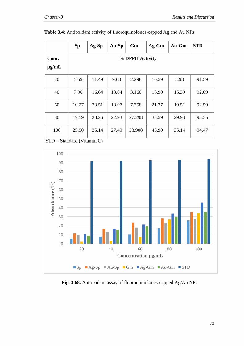
Chapter-3 Results and Discussion
72
Table 3.4: Antioxidant activity of fluoroquinolones-capped Ag and Au NPs
Sp Ag-Sp Au-Sp Gm Ag-Gm Au-Gm STD
Conc.
µg/mL
% DPPH Activity
20 5.59 11.49 9.68 2.298 10.59 8.98 91.59
40 7.90 16.64 13.04 3.160 16.90 15.39 92.09
60 10.27 23.51 18.07 7.758 21.27 19.51 92.59
80 17.59 28.26 22.93 27.298 33.59 29.93 93.35
100 25.90 35.14 27.49 33.908 45.90 35.14 94.47
STD = Standard (Vitamin C)
Fig. 3.68. Antioxidant assay of fluoroquinolones-capped Ag/Au NPs
0
10
20
30
40
50
60
70
80
90
100
20 40 60 80 100
Ab
sorb
an
ce (
%)
Concentration µg/mL
Sp Ag-Sp Au-Sp Gm Ag-Gm Au-Gm STD
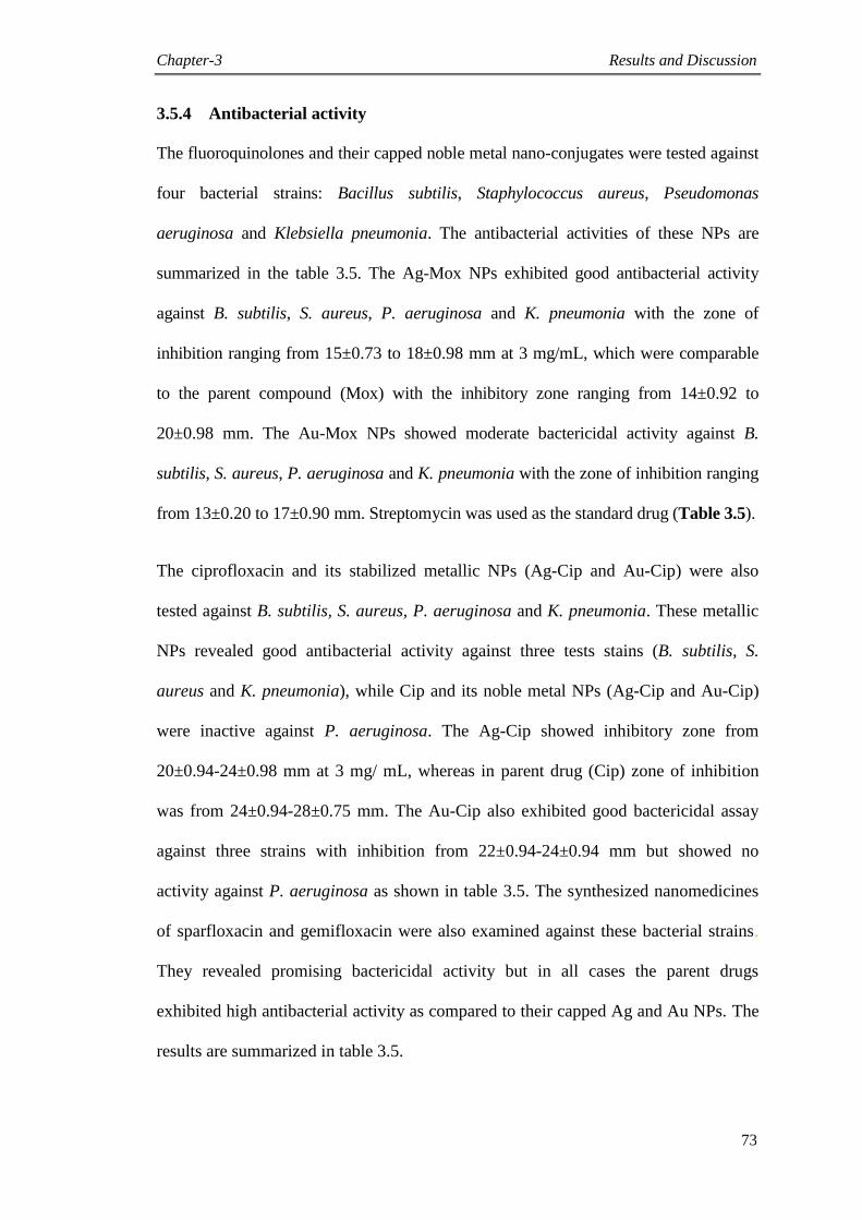
Chapter-3 Results and Discussion
73
3.5.4 Antibacterial activity
The fluoroquinolones and their capped noble metal nano-conjugates were tested against
four bacterial strains: Bacillus subtilis, Staphylococcus aureus, Pseudomonas
aeruginosa and Klebsiella pneumonia. The antibacterial activities of these NPs are
summarized in the table 3.5. The Ag-Mox NPs exhibited good antibacterial activity
against B. subtilis, S. aureus, P. aeruginosa and K. pneumonia with the zone of
inhibition ranging from 15±0.73 to 18±0.98 mm at 3 mg/mL, which were comparable
to the parent compound (Mox) with the inhibitory zone ranging from 14±0.92 to
20±0.98 mm. The Au-Mox NPs showed moderate bactericidal activity against B.
subtilis, S. aureus, P. aeruginosa and K. pneumonia with the zone of inhibition ranging
from 13±0.20 to 17±0.90 mm. Streptomycin was used as the standard drug (Table 3.5).
The ciprofloxacin and its stabilized metallic NPs (Ag-Cip and Au-Cip) were also
tested against B. subtilis, S. aureus, P. aeruginosa and K. pneumonia. These metallic
NPs revealed good antibacterial activity against three tests stains (B. subtilis, S.
aureus and K. pneumonia), while Cip and its noble metal NPs (Ag-Cip and Au-Cip)
were inactive against P. aeruginosa. The Ag-Cip showed inhibitory zone from
20±0.94-24±0.98 mm at 3 mg/ mL, whereas in parent drug (Cip) zone of inhibition
was from 24±0.94-28±0.75 mm. The Au-Cip also exhibited good bactericidal assay
against three strains with inhibition from 22±0.94-24±0.94 mm but showed no
activity against P. aeruginosa as shown in table 3.5. The synthesized nanomedicines
of sparfloxacin and gemifloxacin were also examined against these bacterial strains.
They revealed promising bactericidal activity but in all cases the parent drugs
exhibited high antibacterial activity as compared to their capped Ag and Au NPs. The
results are summarized in table 3.5.
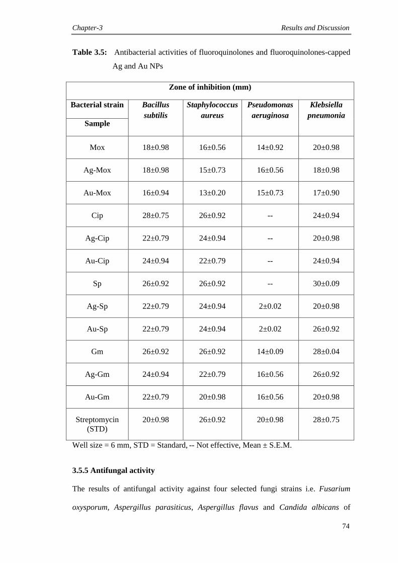
Chapter-3 Results and Discussion
74
Table 3.5: Antibacterial activities of fluoroquinolones and fluoroquinolones-capped
Ag and Au NPs
Zone of inhibition (mm)
Bacterial strain Bacillus
subtilis
Staphylococcus
aureus
Pseudomonas
aeruginosa
Klebsiella
pneumonia Sample
Mox 18±0.98 16±0.56 14±0.92 20±0.98
Ag-Mox 18±0.98 15±0.73 16±0.56 18±0.98
Au-Mox 16±0.94 13±0.20 15±0.73 17±0.90
Cip 28±0.75 26±0.92 -- 24±0.94
Ag-Cip 22±0.79 24±0.94 -- 20±0.98
Au-Cip 24±0.94 22±0.79 -- 24±0.94
Sp 26±0.92 26±0.92 -- 30±0.09
Ag-Sp 22±0.79 24±0.94 2±0.02 20±0.98
Au-Sp 22±0.79 24±0.94 2±0.02 26±0.92
Gm 26±0.92 26±0.92 14±0.09 28±0.04
Ag-Gm 24±0.94 22±0.79 16±0.56 26±0.92
Au-Gm 22±0.79 20±0.98 16±0.56 20±0.98
Streptomycin
(STD)
20±0.98 26±0.92 20±0.98 28±0.75
Well size = 6 mm, STD = Standard, -- Not effective, Mean ± S.E.M.
3.5.5 Antifungal activity
The results of antifungal activity against four selected fungi strains i.e. Fusarium
oxysporum, Aspergillus parasiticus, Aspergillus flavus and Candida albicans of
Chapter-3 Results and Discussion
75
fluoroquinolones and their nano-conjugates have been presented in table 3.6. The
results obtained give us information about the antifungal potential of pure drugs and
their capped NPs. They revealed moderate antifungal activity against A. parasiticus,
A. flavus and C. albicans while the parent drugs and their Ag/Au nano-conjugates
showed low antifungal activity against F. oxysporum. The antifungal activity was not
enhanced after capping with Ag and Au metal (Table 3.6).
Table 3.6: Antifungal activities of fluoroquinolones and fluoroquinolones-capped
Ag and Au NPs
Antifungal activities (% inhibition)
Sample Fusarium
oxysporum
Aspergillus
parasiticus
Aspergillus
flavus
Candida
albicans
Mox 30 63 70 55
Ag-Mox 22 60 68 50
Au-Mox 28 65 60 52
Cip 10 70 80 55
Ag-Cip 10 68 77 52
Au-Cip 12 66 70 50
Sp 22 80 75 45
Ag-Sp 22 73 70 40
Au-Sp 20 70 66 42
Gm 10 63 73 40
Ag-Gm 10 60 70 35
Au-Gm 9 55 65 40
Miconazole
(STD)
100 100 100 100
-- Not effective
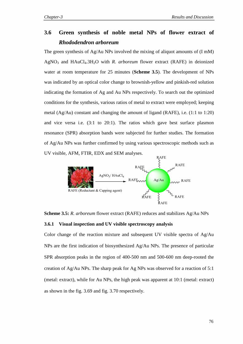
Chapter-3 Results and Discussion
76
3.6 Green synthesis of noble metal NPs of flower extract of
Rhododendron arboreum
The green synthesis of Ag/Au NPs involved the mixing of aliquot amounts of (I mM)
AgNO3 and HAuCl4.3H2O with R. arboreum flower extract (RAFE) in deionized
water at room temperature for 25 minutes (Scheme 3.5). The development of NPs
was indicated by an optical color change to brownish-yellow and pinkish-red solution
indicating the formation of Ag and Au NPs respectively. To search out the optimized
conditions for the synthesis, various ratios of metal to extract were employed; keeping
metal (Ag/Au) constant and changing the amount of ligand (RAFE), i.e. (1:1 to 1:20)
and vice versa i.e. (3:1 to 20:1). The ratios which gave best surface plasmon
resonance (SPR) absorption bands were subjected for further studies. The formation
of Ag/Au NPs was further confirmed by using various spectroscopic methods such as
UV visible, AFM, FTIR, EDX and SEM analyses.
Scheme 3.5: R. arboreum flower extract (RAFE) reduces and stabilizes Ag/Au NPs
3.6.1 Visual inspection and UV visible spectroscopy analysis
Color change of the reaction mixture and subsequent UV visible spectra of Ag/Au
NPs are the first indication of biosynthesized Ag/Au NPs. The presence of particular
SPR absorption peaks in the region of 400-500 nm and 500-600 nm deep-rooted the
creation of Ag/Au NPs. The sharp peak for Ag NPs was observed for a reaction of 5:1
(metal: extract), while for Au NPs, the high peak was apparent at 10:1 (metal: extract)
as shown in the fig. 3.69 and fig. 3.70 respectively.

Chapter-3 Results and Discussion
77
300 400 500 600 700
-0.5
0.0
0.5
1.0
1.5
2.0
2.5
3.0
Abs
orba
nce
Wavelength (nm)
Ag
RAFE
Ag:ligand=1:1
Ag:ligand=1:5
Ag:ligand=1:10
Ag:ligand=3:1
Ag:ligand=5:1
Ag:ligand=7:1
Ag:ligand=10:1
Fig. 3.69. Optimized UV visible spectral data of Ag NPs at reaction ratio of 5:1
300 400 500 600 700
-0.5
0.0
0.5
1.0
1.5
2.0
2.5
3.0
Ab
so
rba
nce
Wavelength (nm)
Au
RAFE
Au:ligand=1:1
Au:ligand=1:10
Au:ligand=5:1
Au:ligand=10:1
Au:ligand=15:1
Au:ligand=20:1
Fig. 3.70. UV visible spectral data of Au NPs at optimized reaction ratio of 10:1

Chapter-3 Results and Discussion
78
3.6.2 FTIR spectroscopy analysis
The spectra were obtained from FTIR studies at 4000 to 400 cm-1
for RAFE, Ag-
RAFE NPs and Au-RAFE NPs. The FTIR analyses were taken to ascertain the
conceivable biomolecules accountable for capping and stabilization of the Ag/Au NPs
formed by RAFE (Fig. 3.71 and 3.72). The bands from 1790 to 740 cm-1
reflected the
biochemical compositions, mainly the moieties of polyphenols, carbohydrates, lipids
and proteins in RAFE.
4000 3500 3000 2500 2000 1500 1000 500
0.0
0.1
0.2
0.3
0.4
0.5
0.6
0.7
0.8
0.9
1.0
1.1
Tra
nsm
ittan
ce (%
)
Wavenumber (cm-1
)
RAFE
Ag NPs
3398
1730
3432 1629
Fig. 3.71. FTIR analysis for RAFE and Ag NPs
The spectral data revealed a broader band for the extract at 3398 cm-1
area which is
due to the occurrence of -OH groups. Likewise, a sharp peak was observed at 1730
cm-1
due the existence of -C=O. In the instance of Ag NPs, a large shift in the
absorbance band with decreased band intensity was observed from 3398 to 3432 cm−1
and 1730 to 1629 cm−1
, suggesting the binding of Ag ions with -OH and -COO-
groups of the RAFE. There was a prominent change in the FTIR spectrum of Ag NPs
as compared to RAFE. The intensity of the peak at 1629 cm-1
has been reduced when
Ag+ is reduced to Ag
0. In case of Au NPs, the band at 3398 cm
-1 is shifted to 3410 cm
-
1 and also a shift in carbonyl peak of C=O from 1730 cm
-1 to 1633 cm
-1 and 1402 cm
-1
was observed (Fig. 3.72).
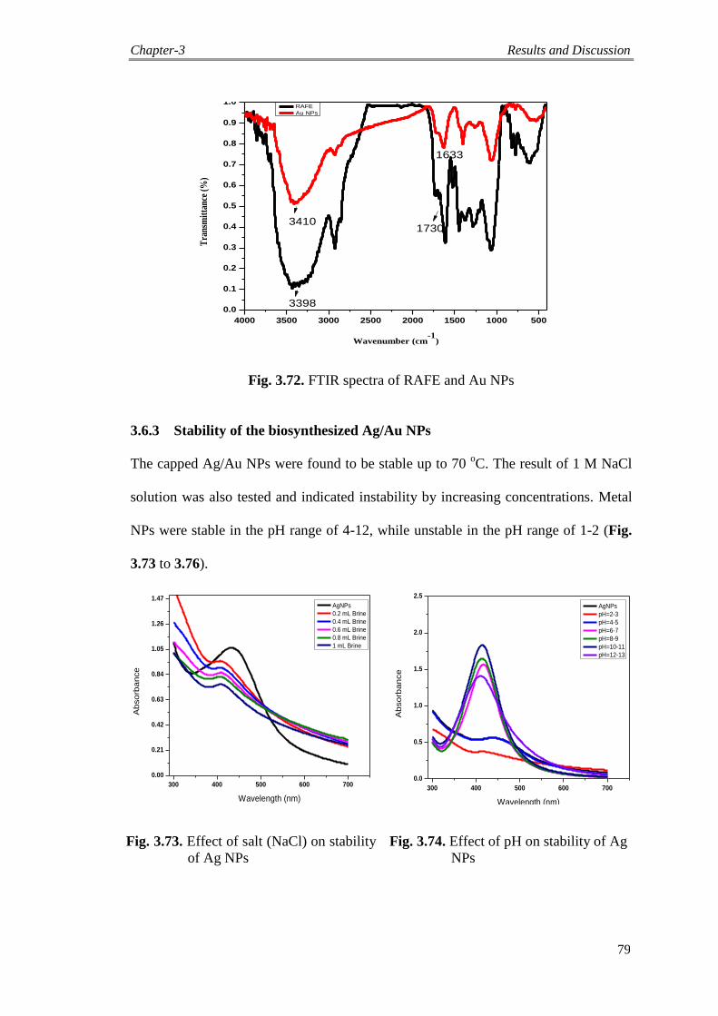
Chapter-3 Results and Discussion
79
4000 3500 3000 2500 2000 1500 1000 500
0.0
0.1
0.2
0.3
0.4
0.5
0.6
0.7
0.8
0.9
1.0
Tra
nsm
itta
nce
(%
)
Wavenumber (cm-1
)
RAFE
Au NPs
3398
34101730
1633
Fig. 3.72. FTIR spectra of RAFE and Au NPs
3.6.3 Stability of the biosynthesized Ag/Au NPs
The capped Ag/Au NPs were found to be stable up to 70 oC. The result of 1 M NaCl
solution was also tested and indicated instability by increasing concentrations. Metal
NPs were stable in the pH range of 4-12, while unstable in the pH range of 1-2 (Fig.
3.73 to 3.76).
300 400 500 600 700
0.00
0.21
0.42
0.63
0.84
1.05
1.26
1.47
Ab
so
rba
nce
Wavelength (nm)
AgNPs
0.2 mL Brine
0.4 mL Brine
0.6 mL Brine
0.8 mL Brine
1 mL Brine
300 400 500 600 700
0.0
0.5
1.0
1.5
2.0
2.5
Ab
so
rba
nce
Wavelength (nm)
AgNPs
pH=2-3
pH=4-5
pH=6-7
pH=8-9
pH=10-11
pH=12-13
Fig. 3.73. Effect of salt (NaCl) on stability
of Ag NPs
Fig. 3.74. Effect of pH on stability of Ag
NPs
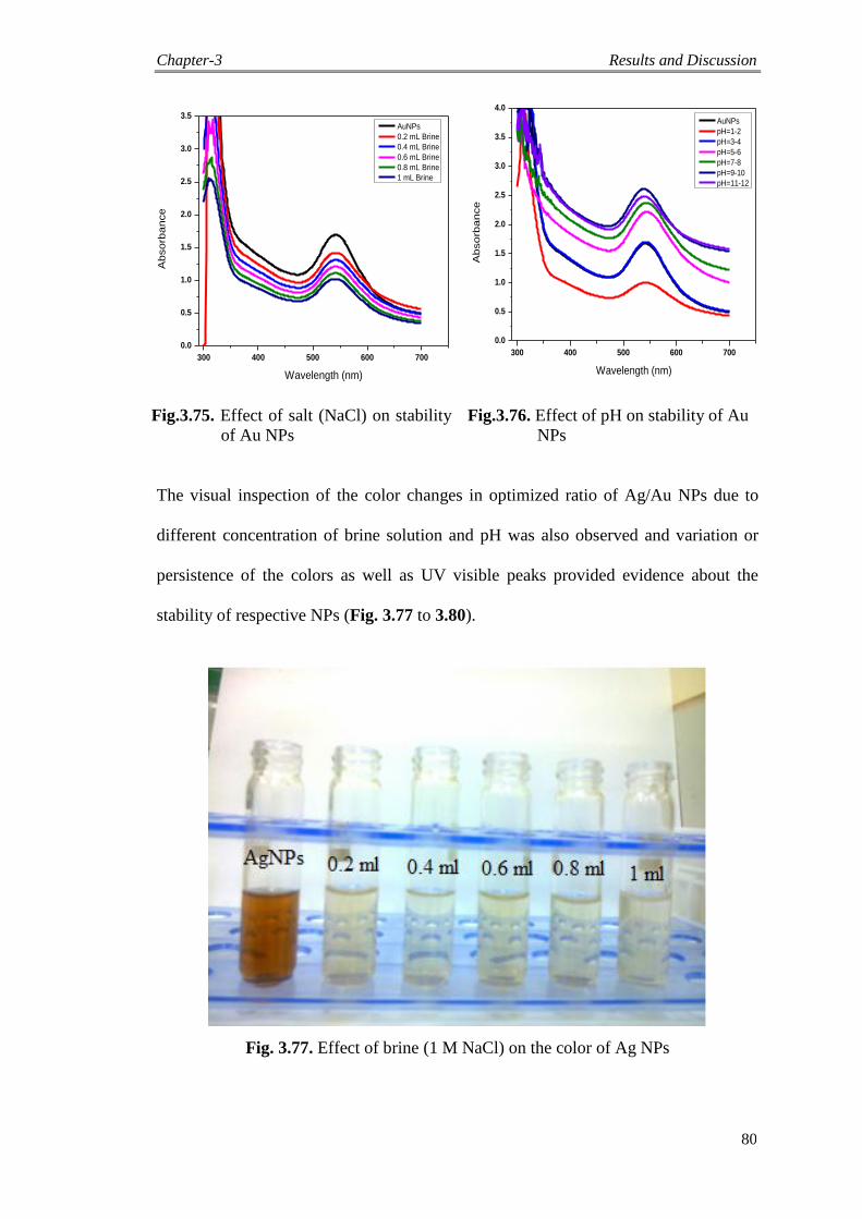
Chapter-3 Results and Discussion
80
300 400 500 600 700
0.0
0.5
1.0
1.5
2.0
2.5
3.0
3.5
Ab
so
rba
nce
Wavelength (nm)
AuNPs
0.2 mL Brine
0.4 mL Brine
0.6 mL Brine
0.8 mL Brine
1 mL Brine
300 400 500 600 700
0.0
0.5
1.0
1.5
2.0
2.5
3.0
3.5
4.0
Ab
so
rba
nce
Wavelength (nm)
AuNPs
pH=1-2
pH=3-4
pH=5-6
pH=7-8
pH=9-10
pH=11-12
Fig.3.75. Effect of salt (NaCl) on stability
of Au NPs
Fig.3.76. Effect of pH on stability of Au
NPs
The visual inspection of the color changes in optimized ratio of Ag/Au NPs due to
different concentration of brine solution and pH was also observed and variation or
persistence of the colors as well as UV visible peaks provided evidence about the
stability of respective NPs (Fig. 3.77 to 3.80).
Fig. 3.77. Effect of brine (1 M NaCl) on the color of Ag NPs
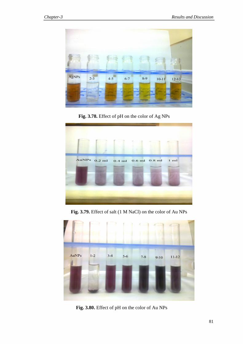
Chapter-3 Results and Discussion
81
Fig. 3.78. Effect of pH on the color of Ag NPs
Fig. 3.79. Effect of salt (1 M NaCl) on the color of Au NPs
Fig. 3.80. Effect of pH on the color of Au NPs

Chapter-3 Results and Discussion
82
3.6.4 AFM, SEM and EDX analysis
The nature and size of the Ag/Au NPs were further confirmed by AFM. Most of the
NPs were monodispersed and spherical in shape (Fig. 3.81 and 3.82) and the majority
of the Ag/Au NPs were less than 40 nm in size. Furthermore, the sizes of both Ag/Au
NPs were also confirmed by SEM images (Fig. 3.83 and 3.84).
Fig. 3.81. Atomic force microscope spectrum of RAFE stabilized Ag NPs
Fig. 3.82. AFM spectrum of RAFE-capped Au NPs
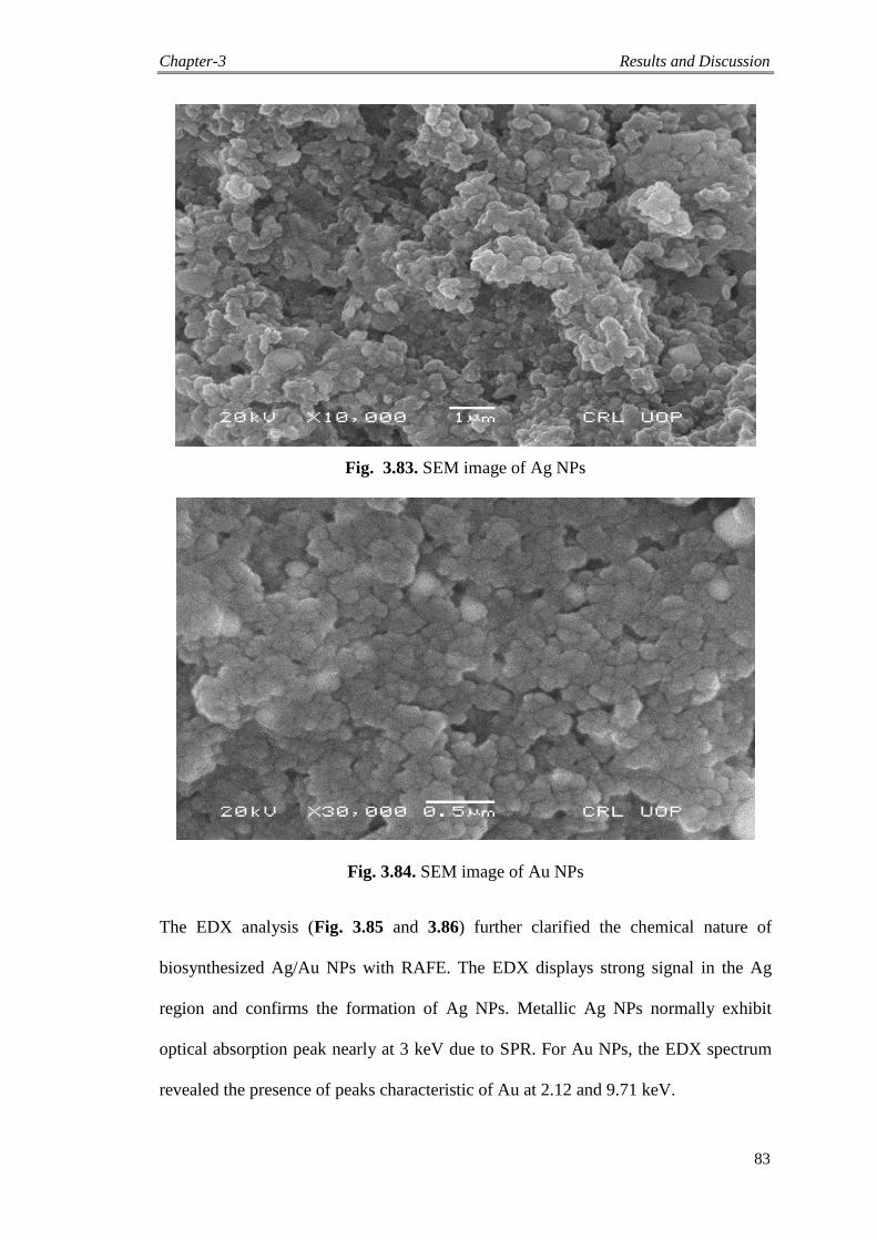
Chapter-3 Results and Discussion
83
Fig. 3.83. SEM image of Ag NPs
Fig. 3.84. SEM image of Au NPs
The EDX analysis (Fig. 3.85 and 3.86) further clarified the chemical nature of
biosynthesized Ag/Au NPs with RAFE. The EDX displays strong signal in the Ag
region and confirms the formation of Ag NPs. Metallic Ag NPs normally exhibit
optical absorption peak nearly at 3 keV due to SPR. For Au NPs, the EDX spectrum
revealed the presence of peaks characteristic of Au at 2.12 and 9.71 keV.

Chapter-3 Results and Discussion
84
Fig.3.85. EDX spectrum of Ag NPs
Fig. 3.86. EDX spectrum of Au NPs
3.7 Green synthesis of metallic NPs of Kigelia pinnata extract
K. pinnata fruits were used as both reductant and capping agent for the green
biosynthesis of Ag and Au NPs.
3.7.1 UV Visible spectroscopy
The colorless mixture turned into brownish-yellow and pinkish-red solution after 30
minutes, indicating the biotransformation of ionic Ag and Au reduced to Ag/Au NPs,
as a result of the surface plasmon resonance phenomenon (SPR). Ag NPs showed
absorption peak at 430 nm (Fig. 3.87), while Au NPs exhibited at 560 nm (Fig. 3.88).
The color change ensued as of the active molecules present in the fruits extract that
owing to the excitation of SPR effect. The sharp peak for Ag NPs was observed for a
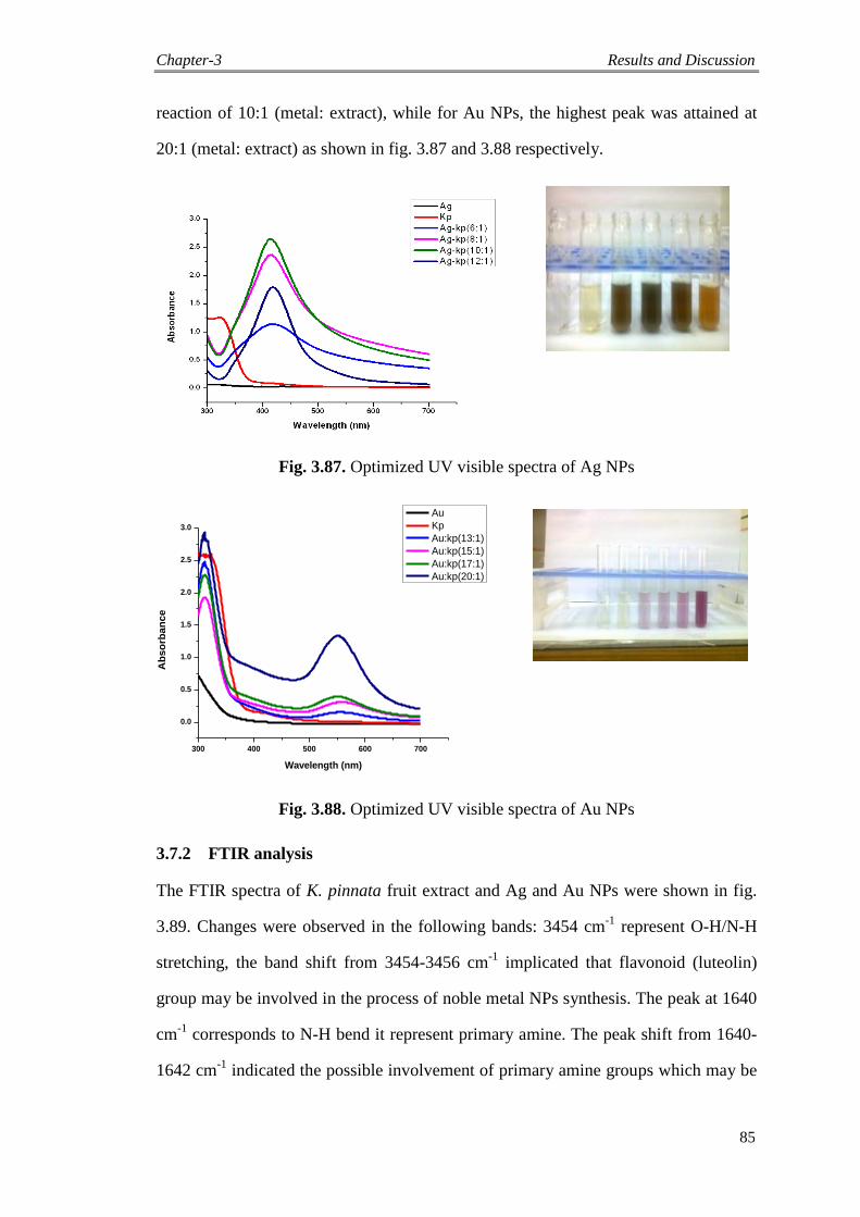
Chapter-3 Results and Discussion
85
reaction of 10:1 (metal: extract), while for Au NPs, the highest peak was attained at
20:1 (metal: extract) as shown in fig. 3.87 and 3.88 respectively.
Fig. 3.87. Optimized UV visible spectra of Ag NPs
300 400 500 600 700
0.0
0.5
1.0
1.5
2.0
2.5
3.0
Ab
so
rba
nc
e
Wavelength (nm)
Au
Kp
Au:kp(13:1)
Au:kp(15:1)
Au:kp(17:1)
Au:kp(20:1)
Fig. 3.88. Optimized UV visible spectra of Au NPs
3.7.2 FTIR analysis
The FTIR spectra of K. pinnata fruit extract and Ag and Au NPs were shown in fig.
3.89. Changes were observed in the following bands: 3454 cm-1
represent O-H/N-H
stretching, the band shift from 3454-3456 cm-1
implicated that flavonoid (luteolin)
group may be involved in the process of noble metal NPs synthesis. The peak at 1640
cm-1
corresponds to N-H bend it represent primary amine. The peak shift from 1640-
1642 cm-1
indicated the possible involvement of primary amine groups which may be
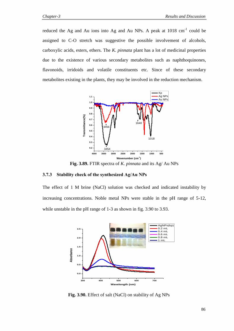
Chapter-3 Results and Discussion
86
reduced the Ag and Au ions into Ag and Au NPs. A peak at 1018 cm-1
could be
assigned to C-O stretch was suggestive the possible involvement of alcohols,
carboxylic acids, esters, ethers. The K. pinnata plant has a lot of medicinal properties
due to the existence of various secondary metabolites such as naphthoquinones,
flavonoids, irridoids and volatile constituents etc. Since of these secondary
metabolites existing in the plants, they may be involved in the reduction mechanism.
4000 3500 3000 2500 2000 1500 1000 500
0.2
0.3
0.4
0.5
0.6
0.7
0.8
0.9
1.0
1.1
Tra
ns
mit
tan
ce
(%)
Wavenumber (cm-1)
Kp
Ag NPs
Au NPs
3454
3456
1640
1018
Fig. 3.89. FTIR spectra of K. pinnata and its Ag/ Au NPs
3.7.3 Stability check of the synthesized Ag/Au NPs
The effect of 1 M brine (NaCl) solution was checked and indicated instability by
increasing concentrations. Noble metal NPs were stable in the pH range of 5-12,
while unstable in the pH range of 1-3 as shown in fig. 3.90 to 3.93.
Fig. 3.90. Effect of salt (NaCl) on stability of Ag NPs
300 400 500 600 700
0.0
0.5
1.0
1.5
2.0
2.5
Abs
orba
nce
Wavelength (nm)
AgNPs(kp)
0.2 mL
0.4 mL
0.6 mL
0.8 mL
1 mL
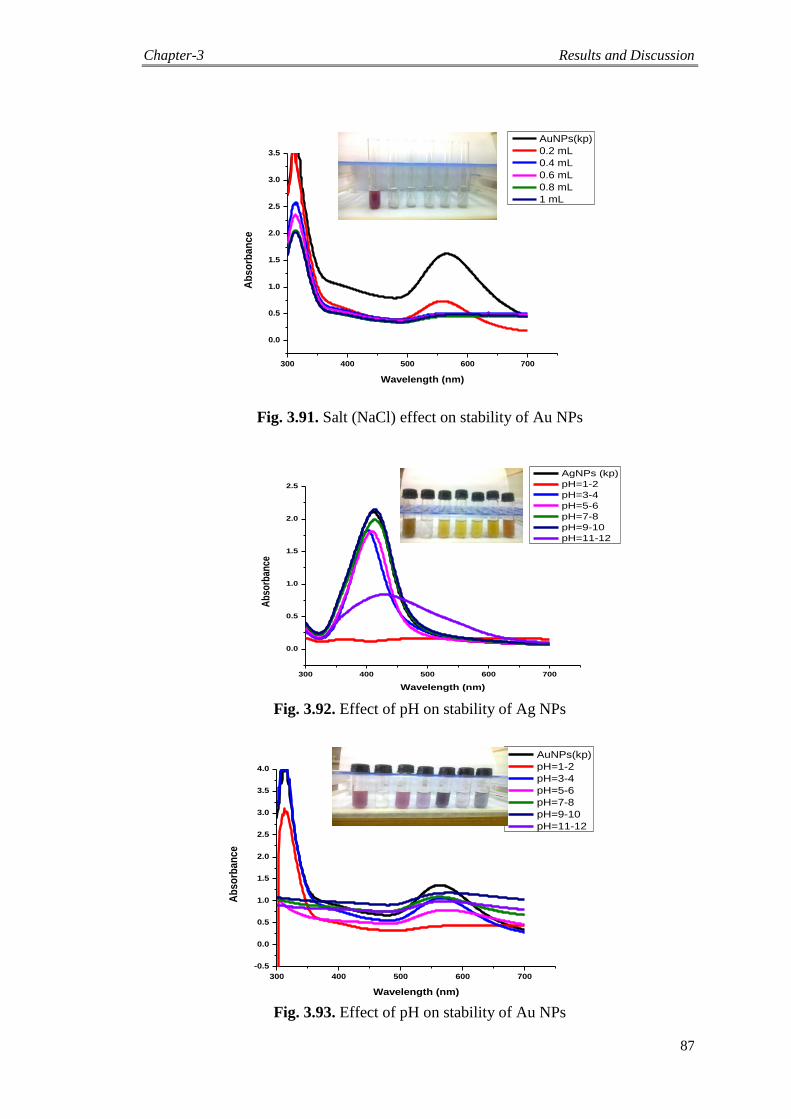
Chapter-3 Results and Discussion
87
Fig. 3.91. Salt (NaCl) effect on stability of Au NPs
Fig. 3.92. Effect of pH on stability of Ag NPs
Fig. 3.93. Effect of pH on stability of Au NPs
300 400 500 600 700
0.0
0.5
1.0
1.5
2.0
2.5
Ab
sorb
ance
Wavelength (nm)
AgNPs (kp)
pH=1-2
pH=3-4
pH=5-6
pH=7-8
pH=9-10
pH=11-12
300 400 500 600 700
-0.5
0.0
0.5
1.0
1.5
2.0
2.5
3.0
3.5
4.0
Ab
so
rba
nc
e
Wavelength (nm)
AuNPs(kp)
pH=1-2
pH=3-4
pH=5-6
pH=7-8
pH=9-10
pH=11-12
300 400 500 600 700
0.0
0.5
1.0
1.5
2.0
2.5
3.0
3.5
Ab
so
rba
nc
e
Wavelength (nm)
AuNPs(kp)
0.2 mL
0.4 mL
0.6 mL
0.8 mL
1 mL
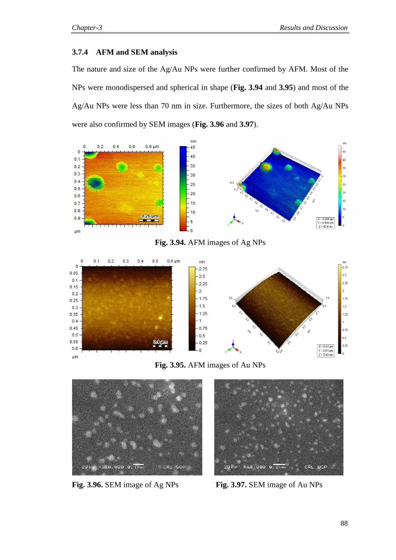
Chapter-3 Results and Discussion
88
3.7.4 AFM and SEM analysis
The nature and size of the Ag/Au NPs were further confirmed by AFM. Most of the
NPs were monodispersed and spherical in shape (Fig. 3.94 and 3.95) and most of the
Ag/Au NPs were less than 70 nm in size. Furthermore, the sizes of both Ag/Au NPs
were also confirmed by SEM images (Fig. 3.96 and 3.97).
Fig. 3.94. AFM images of Ag NPs
Fig. 3.95. AFM images of Au NPs
Fig. 3.96. SEM image of Ag NPs Fig. 3.97. SEM image of Au NPs
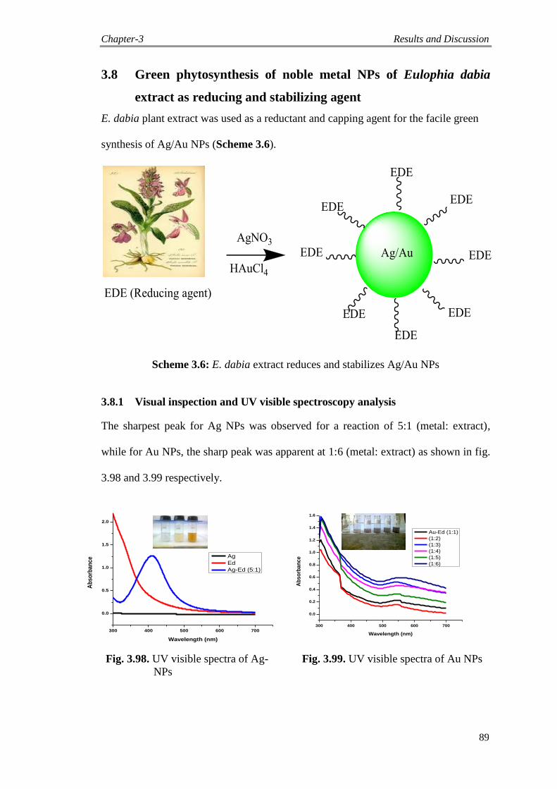
Chapter-3 Results and Discussion
89
3.8 Green phytosynthesis of noble metal NPs of Eulophia dabia
extract as reducing and stabilizing agent
E. dabia plant extract was used as a reductant and capping agent for the facile green
synthesis of Ag/Au NPs (Scheme 3.6).
Scheme 3.6: E. dabia extract reduces and stabilizes Ag/Au NPs
3.8.1 Visual inspection and UV visible spectroscopy analysis
The sharpest peak for Ag NPs was observed for a reaction of 5:1 (metal: extract),
while for Au NPs, the sharp peak was apparent at 1:6 (metal: extract) as shown in fig.
3.98 and 3.99 respectively.
Fig. 3.98. UV visible spectra of Ag-
NPs
Fig. 3.99. UV visible spectra of Au NPs
300 400 500 600 700
0.0
0.5
1.0
1.5
2.0
Ab
so
rba
nc
e
Wavelength (nm)
Ag
Ed
Ag-Ed (5:1)
300 400 500 600 700
0.0
0.2
0.4
0.6
0.8
1.0
1.2
1.4
1.6
Ab
so
rba
nc
e
Wavelength (nm)
Au-Ed (1:1)
(1:2)
(1:3)
(1:4)
(1:5)
(1:6)
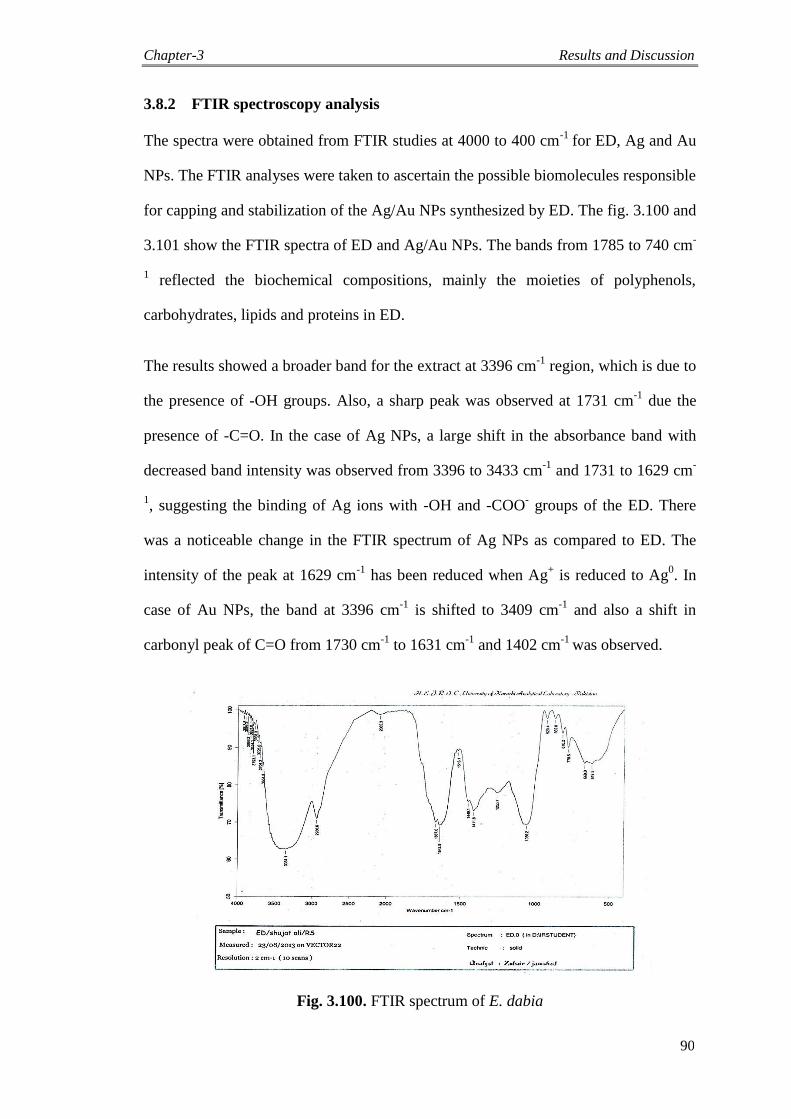
Chapter-3 Results and Discussion
90
3.8.2 FTIR spectroscopy analysis
The spectra were obtained from FTIR studies at 4000 to 400 cm-1
for ED, Ag and Au
NPs. The FTIR analyses were taken to ascertain the possible biomolecules responsible
for capping and stabilization of the Ag/Au NPs synthesized by ED. The fig. 3.100 and
3.101 show the FTIR spectra of ED and Ag/Au NPs. The bands from 1785 to 740 cm-
1 reflected the biochemical compositions, mainly the moieties of polyphenols,
carbohydrates, lipids and proteins in ED.
The results showed a broader band for the extract at 3396 cm-1
region, which is due to
the presence of -OH groups. Also, a sharp peak was observed at 1731 cm-1
due the
presence of -C=O. In the case of Ag NPs, a large shift in the absorbance band with
decreased band intensity was observed from 3396 to 3433 cm-1
and 1731 to 1629 cm-
1, suggesting the binding of Ag ions with -OH and -COO
- groups of the ED. There
was a noticeable change in the FTIR spectrum of Ag NPs as compared to ED. The
intensity of the peak at 1629 cm-1
has been reduced when Ag+ is reduced to Ag
0. In
case of Au NPs, the band at 3396 cm-1
is shifted to 3409 cm-1
and also a shift in
carbonyl peak of C=O from 1730 cm-1
to 1631 cm-1
and 1402 cm-1
was observed.
Fig. 3.100. FTIR spectrum of E. dabia
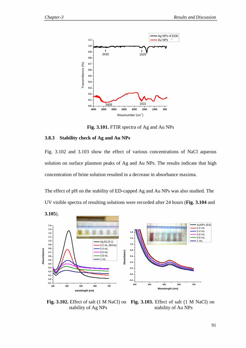
Chapter-3 Results and Discussion
91
4000 3500 3000 2500 2000 1500 1000 500
0.0
0.1
0.2
0.3
0.4
0.5
0.6
0.7
0.8
0.9
1.0
1.1
Tra
nsm
itta
nce (
%)
Wavenumber (cm-1)
Ag NPs of EDE
Au NPs "
3433
3409 1631
1629
Fig. 3.101. FTIR spectra of Ag and Au NPs
3.8.3 Stability check of Ag and Au NPs
Fig. 3.102 and 3.103 show the effect of various concentrations of NaCl aqueous
solution on surface plasmon peaks of Ag and Au NPs. The results indicate that high
concentration of brine solution resulted in a decrease in absorbance maxima.
The effect of pH on the stability of ED-capped Ag and Au NPs was also studied. The
UV visible spectra of resulting solutions were recorded after 24 hours (Fig. 3.104 and
3.105).
Fig. 3.102. Effect of salt (1 M NaCl) on
stability of Ag NPs
Fig. 3.103. Effect of salt (1 M NaCl) on
stability of Au NPs
300 400 500 600 700
-0.1
0.0
0.1
0.2
0.3
0.4
0.5
0.6
0.7
0.8
0.9
1.0
1.1
1.2
1.3
1.4
Ab
so
rba
nc
e
wavelength (nm)
Ag-Ed (5:1)
0.2 mL (Brine)
0.4 mL
0.6 mL
0.8 mL
1 mL
300 400 500 600 700
-0.2
0.0
0.2
0.4
0.6
0.8
1.0
1.2
1.4
Ab
so
rba
nc
e
Wavelength (nm)
AuNPs (Ed)
0.2 mL
0.4 mL
0.6 mL
0.8 mL
1 mL
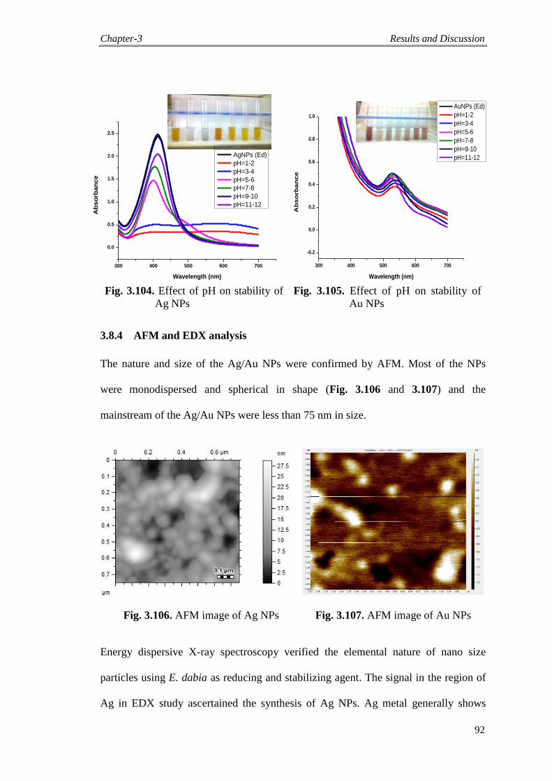
Chapter-3 Results and Discussion
92
Fig. 3.104. Effect of pH on stability of
Ag NPs
Fig. 3.105. Effect of pH on stability of
Au NPs
3.8.4 AFM and EDX analysis
The nature and size of the Ag/Au NPs were confirmed by AFM. Most of the NPs
were monodispersed and spherical in shape (Fig. 3.106 and 3.107) and the
mainstream of the Ag/Au NPs were less than 75 nm in size.
Fig. 3.106. AFM image of Ag NPs Fig. 3.107. AFM image of Au NPs
Energy dispersive X-ray spectroscopy verified the elemental nature of nano size
particles using E. dabia as reducing and stabilizing agent. The signal in the region of
Ag in EDX study ascertained the synthesis of Ag NPs. Ag metal generally shows
300 400 500 600 700
0.0
0.5
1.0
1.5
2.0
2.5
Ab
so
rba
nc
e
Wavelength (nm)
AgNPs (Ed)
pH=1-2
pH=3-4
pH=5-6
pH=7-8
pH=9-10
pH=11-12
300 400 500 600 700
-0.2
0.0
0.2
0.4
0.6
0.8
1.0
Ab
so
rba
nc
e
Wavelength (nm)
AuNPs (Ed)
pH=1-2
pH=3-4
pH=5-6
pH=7-8
pH=9-10
pH=11-12
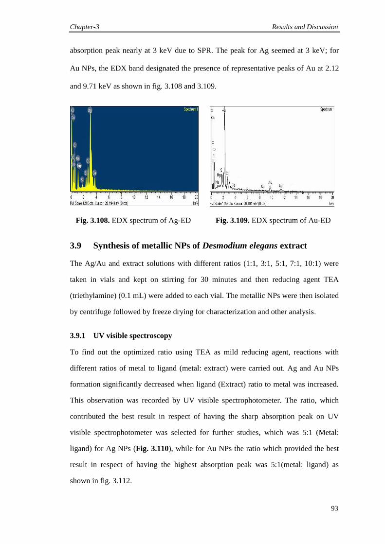
Chapter-3 Results and Discussion
93
absorption peak nearly at 3 keV due to SPR. The peak for Ag seemed at 3 keV; for
Au NPs, the EDX band designated the presence of representative peaks of Au at 2.12
and 9.71 keV as shown in fig. 3.108 and 3.109.
Fig. 3.108. EDX spectrum of Ag-ED Fig. 3.109. EDX spectrum of Au-ED
3.9 Synthesis of metallic NPs of Desmodium elegans extract
The Ag/Au and extract solutions with different ratios (1:1, 3:1, 5:1, 7:1, 10:1) were
taken in vials and kept on stirring for 30 minutes and then reducing agent TEA
(triethylamine) (0.1 mL) were added to each vial. The metallic NPs were then isolated
by centrifuge followed by freeze drying for characterization and other analysis.
3.9.1 UV visible spectroscopy
To find out the optimized ratio using TEA as mild reducing agent, reactions with
different ratios of metal to ligand (metal: extract) were carried out. Ag and Au NPs
formation significantly decreased when ligand (Extract) ratio to metal was increased.
This observation was recorded by UV visible spectrophotometer. The ratio, which
contributed the best result in respect of having the sharp absorption peak on UV
visible spectrophotometer was selected for further studies, which was 5:1 (Metal:
ligand) for Ag NPs (Fig. 3.110), while for Au NPs the ratio which provided the best
result in respect of having the highest absorption peak was 5:1(metal: ligand) as
shown in fig. 3.112.
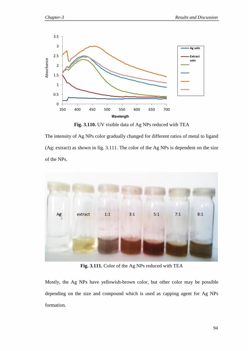
Chapter-3 Results and Discussion
94
0
0.5
1
1.5
2
2.5
3
3.5
350 400 450 500 550 600 650 700
Ag soln
Extractsoln
Fig. 3.110. UV visible data of Ag NPs reduced with TEA
The intensity of Ag NPs color gradually changed for different ratios of metal to ligand
(Ag: extract) as shown in fig. 3.111. The color of the Ag NPs is dependent on the size
of the NPs.
Ag extract 1:1 3:1 5:1 7:1 8:1
Fig. 3.111. Color of the Ag NPs reduced with TEA
Mostly, the Ag NPs have yellowish-brown color, but other color may be possible
depending on the size and compound which is used as capping agent for Ag NPs
formation.
Ab
sorb
ance
Wavelength
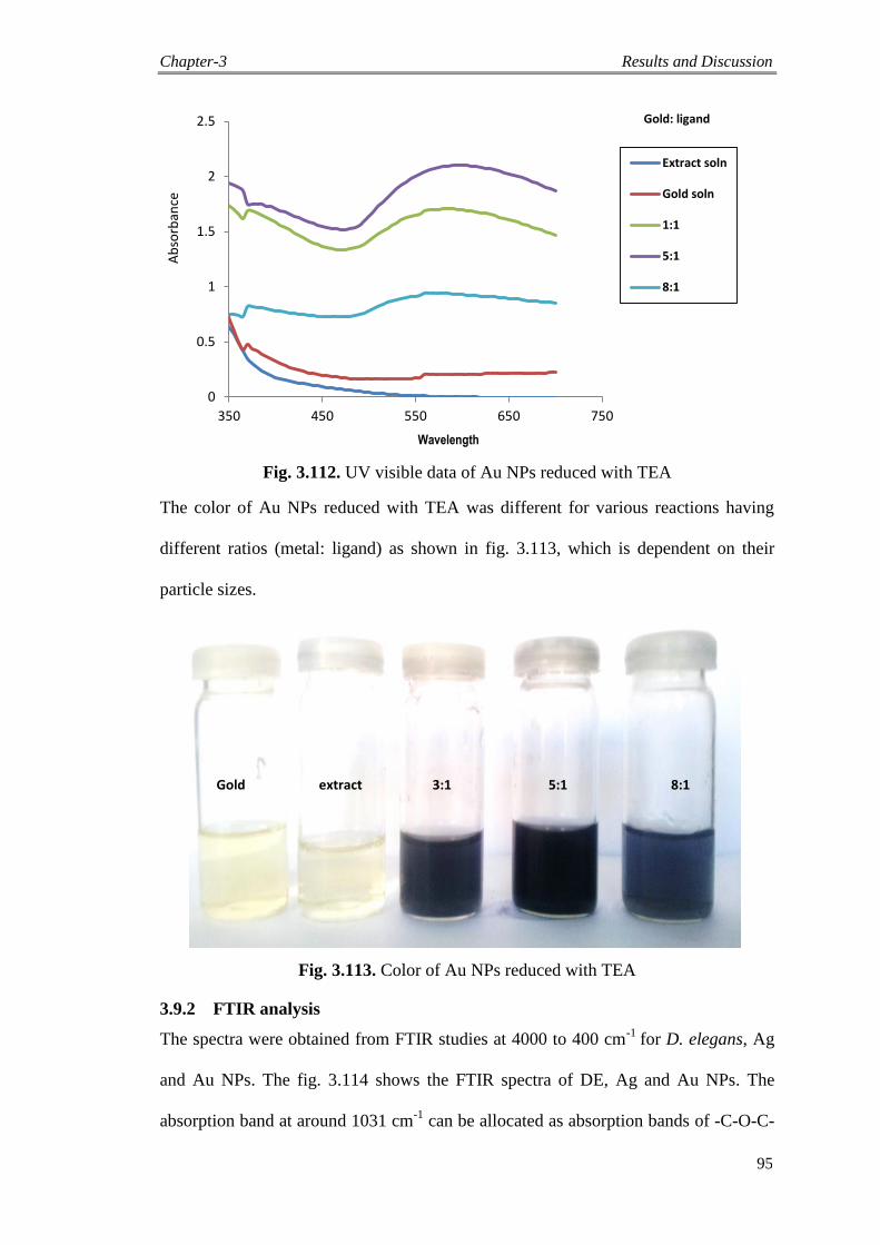
Chapter-3 Results and Discussion
95
Fig. 3.112. UV visible data of Au NPs reduced with TEA
The color of Au NPs reduced with TEA was different for various reactions having
different ratios (metal: ligand) as shown in fig. 3.113, which is dependent on their
particle sizes.
Gold extract 3:1 5:1 8:1
Fig. 3.113. Color of Au NPs reduced with TEA
3.9.2 FTIR analysis
The spectra were obtained from FTIR studies at 4000 to 400 cm-1
for D. elegans, Ag
and Au NPs. The fig. 3.114 shows the FTIR spectra of DE, Ag and Au NPs. The
absorption band at around 1031 cm-1
can be allocated as absorption bands of -C-O-C-
0
0.5
1
1.5
2
2.5
350 450 550 650 750
Extract soln
Gold soln
1:1
5:1
8:1
Wavelength
Gold: ligand
Ab
sorb
ance
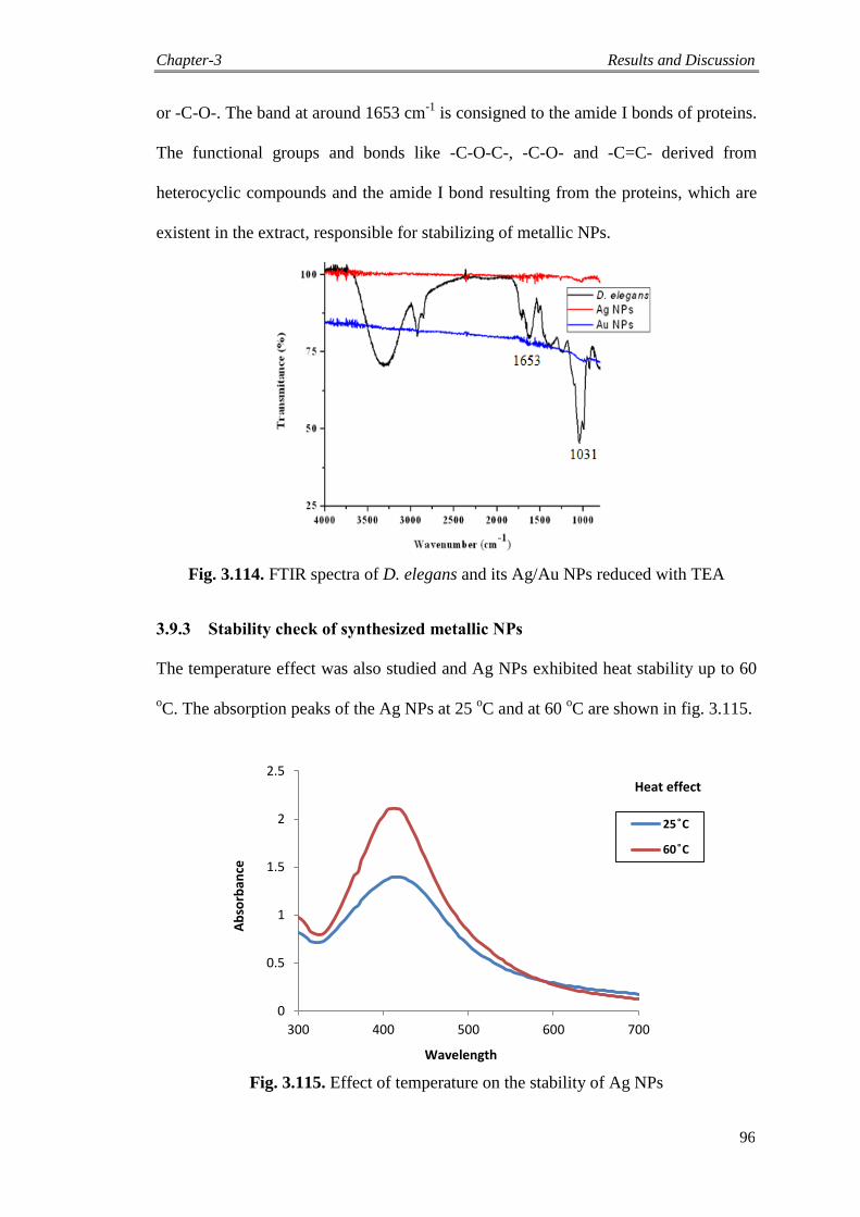
Chapter-3 Results and Discussion
96
or -C-O-. The band at around 1653 cm-1
is consigned to the amide I bonds of proteins.
The functional groups and bonds like -C-O-C-, -C-O- and -C=C- derived from
heterocyclic compounds and the amide I bond resulting from the proteins, which are
existent in the extract, responsible for stabilizing of metallic NPs.
Fig. 3.114. FTIR spectra of D. elegans and its Ag/Au NPs reduced with TEA
3.9.3 Stability check of synthesized metallic NPs
The temperature effect was also studied and Ag NPs exhibited heat stability up to 60
oC. The absorption peaks of the Ag NPs at 25
oC and at 60
oC are shown in fig. 3.115.
Fig. 3.115. Effect of temperature on the stability of Ag NPs
0
0.5
1
1.5
2
2.5
300 400 500 600 700
25 ͦC
60 ͦC
Wavelength
Heat effect
Ab
sorb
ance
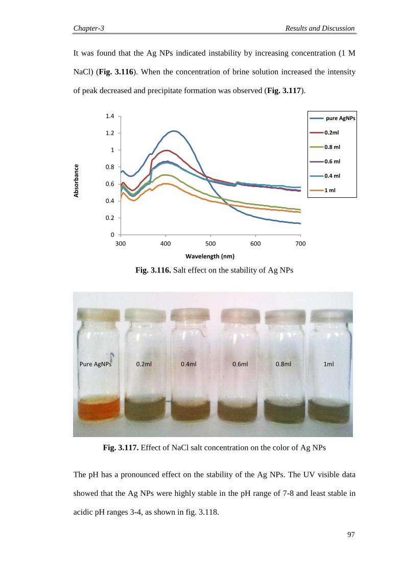
Chapter-3 Results and Discussion
97
It was found that the Ag NPs indicated instability by increasing concentration (1 M
NaCl) (Fig. 3.116). When the concentration of brine solution increased the intensity
of peak decreased and precipitate formation was observed (Fig. 3.117).
Fig. 3.116. Salt effect on the stability of Ag NPs
Pure AgNPs 0.2ml 0.4ml 0.6ml 0.8ml 1ml
Fig. 3.117. Effect of NaCl salt concentration on the color of Ag NPs
The pH has a pronounced effect on the stability of the Ag NPs. The UV visible data
showed that the Ag NPs were highly stable in the pH range of 7-8 and least stable in
acidic pH ranges 3-4, as shown in fig. 3.118.
0
0.2
0.4
0.6
0.8
1
1.2
1.4
300 400 500 600 700
pure AgNPs
0.2ml
0.8 ml
0.6 ml
0.4 ml
1 ml
Wavelength (nm)
Ab
sorb
ance
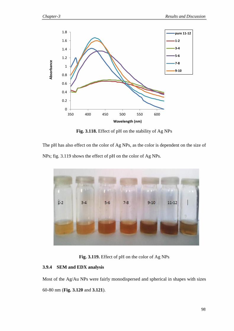
Chapter-3 Results and Discussion
98
Wavelength (nm)
Fig. 3.118. Effect of pH on the stability of Ag NPs
The pH has also effect on the color of Ag NPs, as the color is dependent on the size of
NPs; fig. 3.119 shows the effect of pH on the color of Ag NPs.
Fig. 3.119. Effect of pH on the color of Ag NPs
3.9.4 SEM and EDX analysis
Most of the Ag/Au NPs were fairly monodispersed and spherical in shapes with sizes
60-80 nm (Fig. 3.120 and 3.121).
0
0.2
0.4
0.6
0.8
1
1.2
1.4
1.6
1.8
350 400 450 500 550 600
pure 11-12
1-2
3-4
5-6
7-8
9-10
Ab
sorb
ance
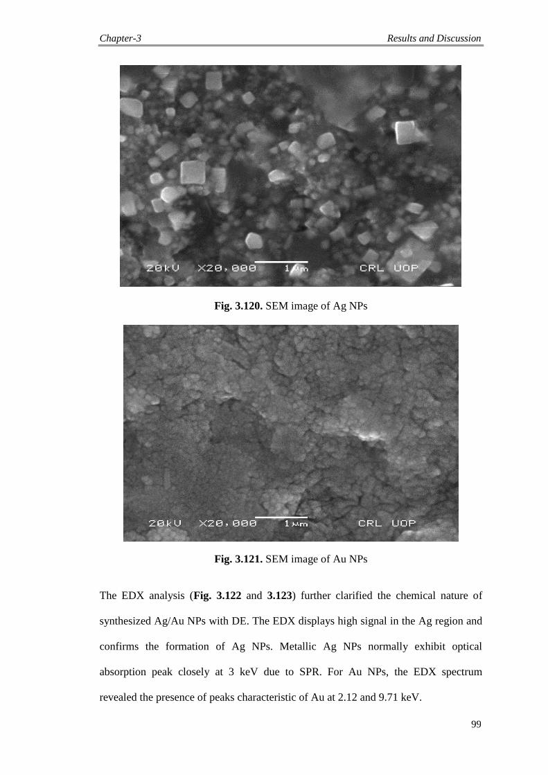
Chapter-3 Results and Discussion
99
Fig. 3.120. SEM image of Ag NPs
Fig. 3.121. SEM image of Au NPs
The EDX analysis (Fig. 3.122 and 3.123) further clarified the chemical nature of
synthesized Ag/Au NPs with DE. The EDX displays high signal in the Ag region and
confirms the formation of Ag NPs. Metallic Ag NPs normally exhibit optical
absorption peak closely at 3 keV due to SPR. For Au NPs, the EDX spectrum
revealed the presence of peaks characteristic of Au at 2.12 and 9.71 keV.
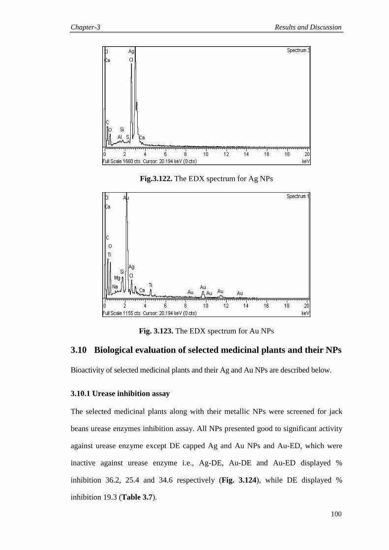
Chapter-3 Results and Discussion
100
Fig.3.122. The EDX spectrum for Ag NPs
Fig. 3.123. The EDX spectrum for Au NPs
3.10 Biological evaluation of selected medicinal plants and their NPs
Bioactivity of selected medicinal plants and their Ag and Au NPs are described below.
3.10.1 Urease inhibition assay
The selected medicinal plants along with their metallic NPs were screened for jack
beans urease enzymes inhibition assay. All NPs presented good to significant activity
against urease enzyme except DE capped Ag and Au NPs and Au-ED, which were
inactive against urease enzyme i.e., Ag-DE, Au-DE and Au-ED displayed %
inhibition 36.2, 25.4 and 34.6 respectively (Fig. 3.124), while DE displayed %
inhibition 19.3 (Table 3.7).
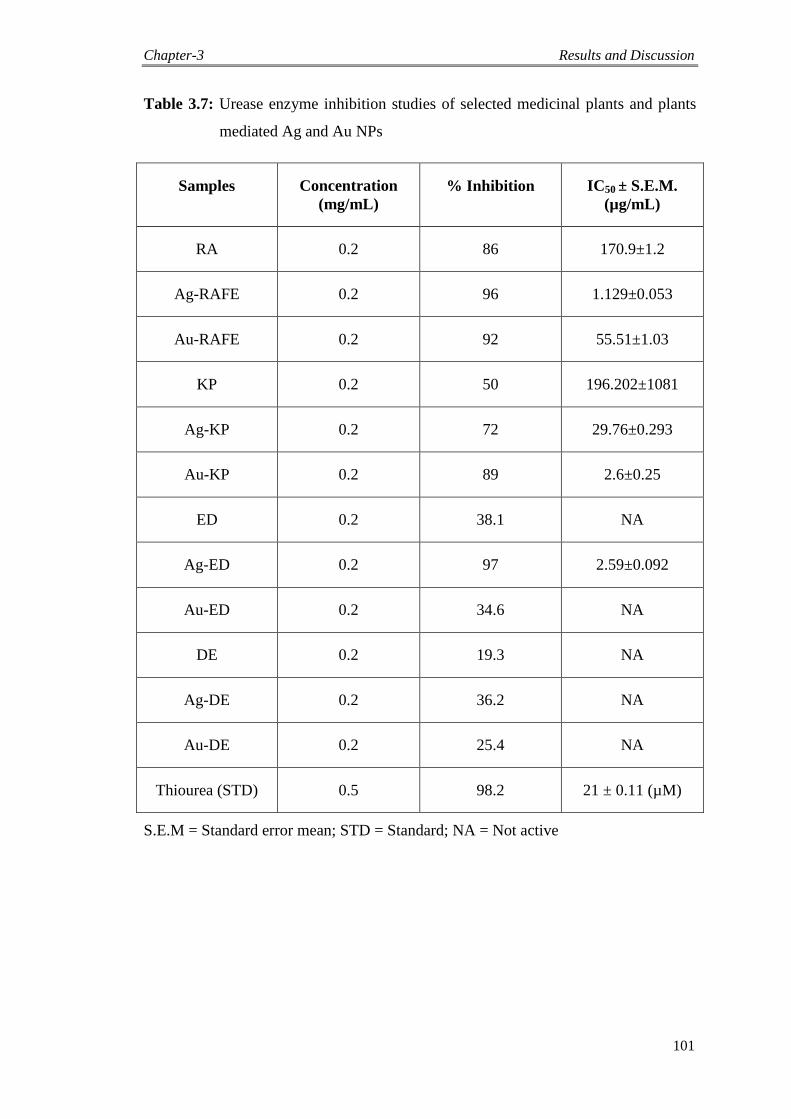
Chapter-3 Results and Discussion
101
Table 3.7: Urease enzyme inhibition studies of selected medicinal plants and plants
mediated Ag and Au NPs
Samples Concentration
(mg/mL)
% Inhibition IC50 ± S.E.M.
(µg/mL)
RA 0.2 86 170.9±1.2
Ag-RAFE 0.2 96 1.129±0.053
Au-RAFE 0.2 92 55.51±1.03
KP 0.2 50 196.202±1081
Ag-KP 0.2 72 29.76±0.293
Au-KP 0.2 89 2.6±0.25
ED 0.2 38.1 NA
Ag-ED 0.2 97 2.59±0.092
Au-ED 0.2 34.6 NA
DE 0.2 19.3 NA
Ag-DE 0.2 36.2 NA
Au-DE 0.2 25.4 NA
Thiourea (STD) 0.5 98.2 21 ± 0.11 (µM)
S.E.M = Standard error mean; STD = Standard; NA = Not active
Chapter-3 Results and Discussion
102
Fig. 3.124. Urease inhibitory activity of selected medicinal plants and their metallic NPs
3.10.2 Leishmanicidal activity
The antileishmanial activity of selected medicinal plants and their Ag and Au NPs
was also tested against Leishmania tropica as a modal parasite. The efficacy of all the
tested NPs was studied for 96 h. Ag-RAFE (IC50 µg/mL = 9.45) and Ag-ED (IC50
µg/mL = 7.20) showed significant activity, while the other noble metal NPs displayed
good activities as shown in the table 3.8.
0
10
20
30
40
50
60
70
80
90
100
% I
nh
ibit
ion
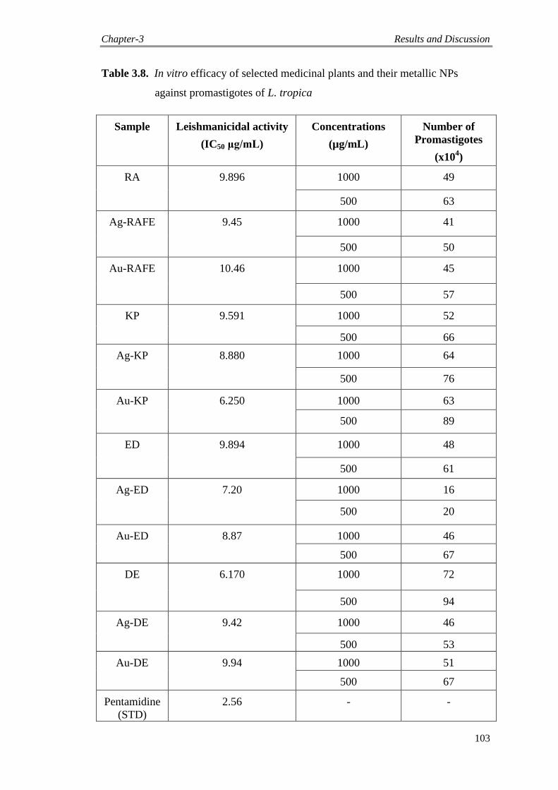
Chapter-3 Results and Discussion
103
Table 3.8. In vitro efficacy of selected medicinal plants and their metallic NPs
against promastigotes of L. tropica
Sample Leishmanicidal activity
(IC50 μg/mL)
Concentrations
(µg/mL)
Number of
Promastigotes
(x104)
RA 9.896 1000 49
500 63
Ag-RAFE 9.45 1000 41
500 50
Au-RAFE 10.46 1000 45
500 57
KP 9.591 1000 52
500 66
Ag-KP 8.880 1000 64
500 76
Au-KP 6.250 1000 63
500 89
ED 9.894 1000 48
500 61
Ag-ED 7.20 1000 16
500 20
Au-ED 8.87 1000 46
500 67
DE 6.170 1000 72
500 94
Ag-DE 9.42 1000 46
500 53
Au-DE 9.94 1000 51
500 67
Pentamidine
(STD)
2.56 - -
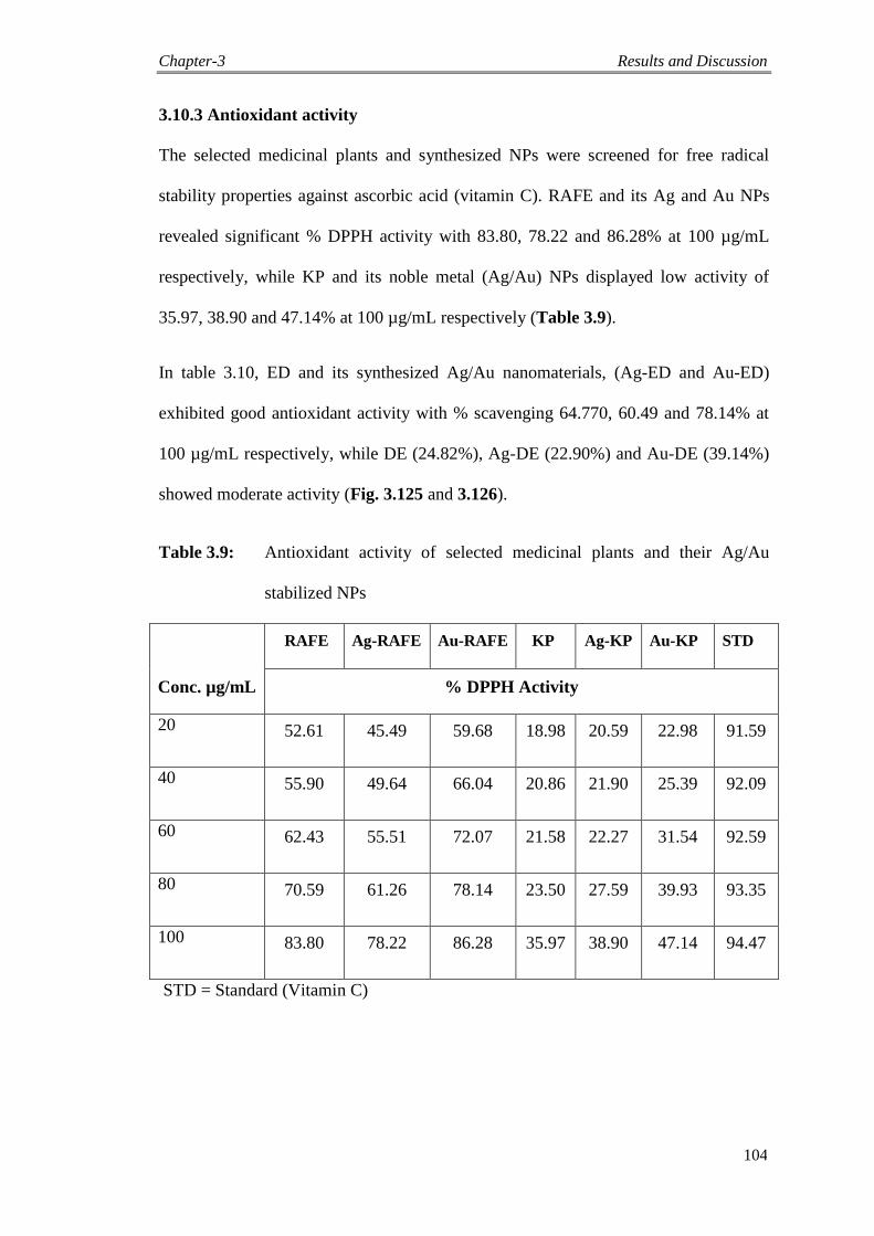
Chapter-3 Results and Discussion
104
3.10.3 Antioxidant activity
The selected medicinal plants and synthesized NPs were screened for free radical
stability properties against ascorbic acid (vitamin C). RAFE and its Ag and Au NPs
revealed significant % DPPH activity with 83.80, 78.22 and 86.28% at 100 µg/mL
respectively, while KP and its noble metal (Ag/Au) NPs displayed low activity of
35.97, 38.90 and 47.14% at 100 µg/mL respectively (Table 3.9).
In table 3.10, ED and its synthesized Ag/Au nanomaterials, (Ag-ED and Au-ED)
exhibited good antioxidant activity with % scavenging 64.770, 60.49 and 78.14% at
100 µg/mL respectively, while DE (24.82%), Ag-DE (22.90%) and Au-DE (39.14%)
showed moderate activity (Fig. 3.125 and 3.126).
Table 3.9: Antioxidant activity of selected medicinal plants and their Ag/Au
stabilized NPs
RAFE Ag-RAFE Au-RAFE KP Ag-KP Au-KP STD
Conc. µg/mL % DPPH Activity
20 52.61 45.49 59.68 18.98 20.59 22.98 91.59
40 55.90 49.64 66.04 20.86 21.90 25.39 92.09
60 62.43 55.51 72.07 21.58 22.27 31.54 92.59
80 70.59 61.26 78.14 23.50 27.59 39.93 93.35
100 83.80 78.22 86.28 35.97 38.90 47.14 94.47
STD = Standard (Vitamin C)

Chapter-3 Results and Discussion
105
Fig. 3.125. Antioxidant assay of selected medicinal plants-capped Ag/Au NPs
Table 3.10: Antioxidant activity of selected medicinal plants Ag/Au stabilized
NPs
ED Ag-ED Au-ED DE Ag-DE Au-DE STD
Conc.
µg/mL
% DPPH Activity
20 27.241 33.681 65.490 10.79 10.59 15.98 91.59
40 29.160 38.040 68.641 20.50 21.90 23.39 92.09
60 39.597 44.072 72.510 20.86 20.27 25.54 92.59
80 47.908 49.93 73.26 22.54 21.59 31.93 93.35
100 64.770 60.49 78.14 24.82 22.90 39.14 94.47
STD = Standard (Vitamin C)
0
10
20
30
40
50
60
70
80
90
100
20 40 60 80 100
Ab
sorb
an
ce (
%)
Concentration µg/mL
RAFE
Ag-RAFE
Au-RAFE
KP
Ag-KP
Au-KP
STD
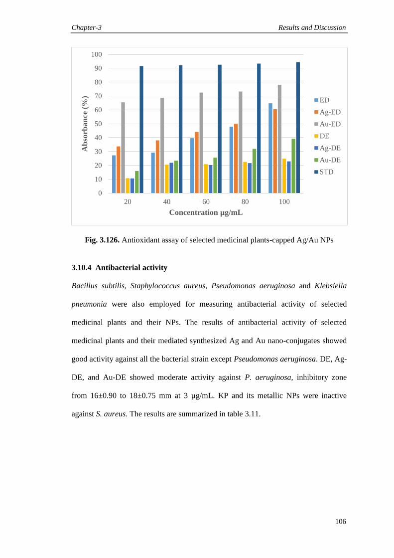
Chapter-3 Results and Discussion
106
Fig. 3.126. Antioxidant assay of selected medicinal plants-capped Ag/Au NPs
3.10.4 Antibacterial activity
Bacillus subtilis, Staphylococcus aureus, Pseudomonas aeruginosa and Klebsiella
pneumonia were also employed for measuring antibacterial activity of selected
medicinal plants and their NPs. The results of antibacterial activity of selected
medicinal plants and their mediated synthesized Ag and Au nano-conjugates showed
good activity against all the bacterial strain except Pseudomonas aeruginosa. DE, Ag-
DE, and Au-DE showed moderate activity against P. aeruginosa, inhibitory zone
from 16±0.90 to 18±0.75 mm at 3 µg/mL. KP and its metallic NPs were inactive
against S. aureus. The results are summarized in table 3.11.
0
10
20
30
40
50
60
70
80
90
100
20 40 60 80 100
Ab
sorb
an
ce (
%)
Concentration µg/mL
ED
Ag-ED
Au-ED
DE
Ag-DE
Au-DE
STD
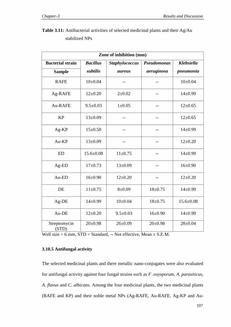
Chapter-3 Results and Discussion
107
Table 3.11: Antibacterial activities of selected medicinal plants and their Ag/Au
stabilized NPs
Zone of inhibition (mm)
Bacterial strain Bacillus
subtilis
Staphylococcus
aureus
Pseudomonas
aeruginosa
Klebsiella
pneumonia Sample
RAFE 10±0.04 -- -- 10±0.04
Ag-RAFE 12±0.20 2±0.02 -- 14±0.99
Au-RAFE 9.5±0.03 1±0.05 -- 12±0.65
KP 13±0.09 -- -- 12±0.65
Ag-KP 15±0.50 -- -- 14±0.99
Au-KP 13±0.09 -- -- 12±0.20
ED 15.6±0.08 11±0.75 -- 14±0.99
Ag-ED 17±0.73 13±0.09 -- 16±0.90
Au-ED 16±0.90 12±0.20 -- 12±0.20
DE 11±0.75 8±0.09 18±0.75 14±0.99
Ag-DE 14±0.99 10±0.04 18±0.75 15.6±0.08
Au-DE 12±0.20 9.5±0.03 16±0.90 14±0.99
Streptomycin
(STD)
20±0.98 26±0.09 20±0.98 28±0.04
Well size = 6 mm, STD = Standard, -- Not effective, Mean ± S.E.M.
3.10.5 Antifungal activity
The selected medicinal plants and there metallic nano-conjugates were also evaluated
for antifungal activity against four fungal strains such as F .oxysporum, A. parasiticus,
A. flavus and C. albicans. Among the four medicinal plants, the two medicinal plants
(RAFE and KP) and their noble metal NPs (Ag-RAFE, Au-RAFE, Ag-KP and Au-
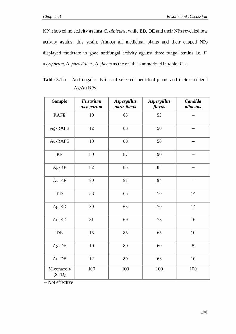
Chapter-3 Results and Discussion
108
KP) showed no activity against C. albicans, while ED, DE and their NPs revealed low
activity against this strain. Almost all medicinal plants and their capped NPs
displayed moderate to good antifungal activity against three fungal strains i.e. F.
oxysporum, A. parasiticus, A. flavus as the results summarized in table 3.12.
Table 3.12: Antifungal activities of selected medicinal plants and their stabilized
Ag/Au NPs
Sample Fusarium
oxysporum
Aspergillus
parasiticus
Aspergillus
flavus
Candida
albicans
RAFE 10 85 52 --
Ag-RAFE 12 88 50 --
Au-RAFE 10 80 50 --
KP 80 87 90 --
Ag-KP 82 85 88 --
Au-KP 80 81 84 --
ED 83 65 70 14
Ag-ED 80 65 70 14
Au-ED 81 69 73 16
DE 15 85 65 10
Ag-DE 10 80 60 8
Au-DE 12 80 63 10
Miconazole
(STD)
100 100 100 100
-- Not effective
Chapter-4 Experimental
109
4.1 General experimental procedures
A digital pH meter model 510 (Oakton, Eutech) equipped with a glass working
electrode and a reference Ag/AgCl electrode was hired for pH measurements. UV
visible spectra were recorded using a Shimadzu UV-240, Hitachi U-3200
spectrophotometer with a path length of 1 cm. Fourier transform infrared (FTIR)
spectra were recorded using a Shimadzu IR-460 spectrophotometer. The shape and
size of nanomedicines were examined using atomic force microscope (AFM),
Multimode, Nanoscope IIIa, Veeco, (California, USA) in the tapping mode and
furthermore, confirmed by scanning electron microscope (SEM) with energy
dispersive X-ray (EDX), (JSM 591 JEOL, Japan).
4.1.1 Collection of fluoroquinolones and plants material
Commercial fluoroquinolones along with tablets (moxifloxacin, ciprofloxacin,
sparfloxacin and gemifloxacin) for parallel study were purchased from local
pharmaceutical market Peshawar and selected medicinal plants (Rhododendron
arboreum, Kigelia pinnata, Eulophia dabia and Desmodium elegans) were collected
from designate area of Pakistan. In February 2011, R. arboreum plant was collected
from Seran Valley, Khyber Pakhtunkhwa, Pakistan. A voucher specimen no.
7212/Bot has been deposited in the National Herbarium of Peshawar University,
Pakistan, while the fruits of K. pinnata were collected from University of Sargodha,
Punjab, Pakistan and identified by an experienced taxonomist Dr. Abdur Rashid,
Department of Botany, University of Peshawar, Pakistan. The E. dabia plant material
was collected from Khwazakhela, Swat, Khyber Pakhtunkhwa, Pakistan, during April
2011 and identified by taxonomist Dr. Hassan Sher, University of Swat, Pakistan. D.
elegans plant was collected during the summer season from Gallyat area, Khyber
Pakhtunkhwa, Pakistan, which was identified by Dr. Abdur Rashid, Department of
Chapter-4 Experimental
110
Botany, University of Peshawar, Pakistan. A reference voucher (726/Bot.) was
deposited in the National Herbarium of Peshawar University, Pakistan.
Silver nitrate (AgNO3) was purchased from Sigma-Aldrich, while hydrogen
tetrachloroaurate (III) trihydrate (HAuCl4 3H2O), sodium carbonate (Na2CO3),
sodium chloride (NaCl), sodium hydroxide (NaOH) and hydrochloric acid (HCl) were
purchased from Merck. Sodium borohydride (NaBH4) was obtained from Wako Pure
Chemical Industries Ltd. Deionized water was used throughout the reactions for the
synthesis of Ag and Au NPs.
4.1.2 Preparation of stock solution
Fresh solutions of metal salts (1 mM AgNO3 and 1 mM HAuCl4) and ligand (1 mM
fluoroquinolones) were prepared in deionized water, while crude extracts of selected
medicinal plants (0.5 g) were dissolved in distilled water (100 mL). The stock
solutions were stored in refrigerator for further studies. Fresh stock solutions (1 M) of
NaCl, HCl and NaOH were also prepared for checking salt and pH stability of Ag/Au
NPs.
4.1.3 Synthesis of Ag and Au NPs capped with fluoroquinolones
Fluoroquinolones were synthesized using NaBH4 as a moderate reductant. Reactions
were carried out by mixing different volumes of fluoroquinolones and Ag/Au salts
solutions. The reaction mixture was stirred vigorously for about 30 minutes at
ambient temperature and then 0.2 mL of 50 mM NaBH4 was added drop wise. A
gradual change in the color of reaction mixture was used as a clue to the formation of
the desired products i.e. fluoroquinolones-capped Ag and Au NPs. After the addition
of a reducing agent, the light yellow solution gradually turned maroon followed by
brown and eventually ruby red, depending on the molar ratio of metals to ligands.
Chapter-4 Experimental
111
Optimization of reaction condition was achieved by varying the molar ratio of metals
to ligands (fluoroquinolones), which resulted in the appearance of the highest
absorption peak in the proposed UV visible region. The residual metal salts and drugs
were removed by centrifugation at 10,000 rpm and the supernatants were freeze dried,
which contained fluoroquinolones-capped Ag and Au NPs. The synthesis of Ag and
Au nano-conjugates was further confirmed by using FTIR, AFM, EDX and SEM.
Bioactivity of these Ag and Au NPs having suitable size and shape were performed
and evaluated accordingly.
4.1.4 Green synthesis of metallic NPs by using selected medicinal plants
The selected medicinal plants (R. arboretum, K. pinnata, E. dabia and D. elegans)
were washed with distill water and air dried under shade. The plants materials were
then ground to obtain fine powder. Shade dried samples (20 g) were kept in conical
flasks and 100 mL of methanol/water solution (70:30 v/v) were added to each conical
flask. The flasks were placed for 10 days and the visual appearances of the liquids
were monitored. The extracts were filtered and utilized as reducing and stabilizing
agents in the synthesis of noble metal NPs.
The green synthesis of metal NPs involved the mixing of aliquot amounts of (I mM)
AgNO3 and HAuCl4.3H2O with selected medicinal plants in deionized water at room
temperature for 30 minutes. The formation of NPs was indicated by an optical color
change to brownish-yellow and pinkish-red solutions indicating the formation of Ag
and Au NPs respectively. To search out the optimized conditions for the synthesis,
different ratios of metals to extracts were used; keeping metal (Ag/Au) constant and
varying the amount of ligands (R. arboretum, K. pinnata, E. dabia and D. elegans).
The ratios which contributed best SPR absorption bands were subjected for further
studies. The metallic NPs were then isolated by centrifugation followed by freeze
drying for characterization and other analyses.
Chapter-4 Experimental
112
4.2 Biological evaluation
Fluoroquinolones and selected medicinal plants along with their Ag/Au NPs were
subjected for various biological activities to evaluate and explore their therapeutic and
medicinal importance. These activities included urease enzyme inhibition,
leishmanicidal, antioxidant and antimicrobial.
4.2.1 Protocol for urease assay and inhibition
Reaction mixtures consisting Jack bean (Canavalia ensiformis) urease (25 μL), 100 mM
of urea, 55 μL of buffer at pH 6.8 and 5 μL of diverse concentrations of test samples
(0.5 to 0.00625 mM) were hatched in 96-well plates at temperature of 30°C for 15
minutes. In kinetics studies, different concentrations of test compounds and substrates
were employed. Subsequently, 45 μL phenol reagents (1% w/v phenol and 0.005% w/v
sodium nitroprussside), and 70 μL of alkali (0.5% w/v NaOH and 0.1% w/v NaOCl)
were added to discrete well. Afterward 50 minutes, the cumulative absorbance at 630
nm was recorded in triplicate in a microplate reader (SpectraMax M2, Molecular
Devices, CA, USA). The making of ammonia (NH3) was measured by using
indophenol method with a standard (thiourea) [165]. Eventually the results were
administered by software SoftMax Pro (Molecular Devices, CA, USA), MS-Excel and
Ez-fit programs. For calculating percent inhibition, the following formula was used:
% Inhibition = 100-(OD test /OD control) ×100.
4.2.2 Procedure for leishmanicidal activity (in vitro)
For this bioassay, Leishmania tropica promastigotes were cultured at 22-25°C in
RPMI-1640 (Sigma) as testified earlier [35]. The medium was complemented with 10%
temperature deactivated (56°C for 30 min) fetal bovine serum (FBS). The
promastigotes culture at logarithmic phase was sedimented at 800 rpm, calculated with
Chapter-4 Experimental
113
the help of advanced Neubaver chamber under the microscope and in the same
condition, washed with saline three times. The last density of 2x10⁶ cells/mL was
attained by diluting the parasites with new culture medium. In 96-well micro titer plate,
180 mL of medium was subjected in the main row, while 100 mL of medium was put in
others wells. The substances to be tested were dissolved to an ultimate concentration of
1.0 mg in 0.1 mL of PBS (Phosphate Buffered Saline, pH 7.4 comprising 0.5% CH3OH,
0.5 % DMSO). One row for standard drugs (Pentamidine and ampicilline to a final
concentration of 1.0 mg/mL was added separately as positive control), while one was
used for control (DMSO) which received medium. Dish was incubated at 22-24°C for
72 h. Data are the replicates of three diverse experiments. The 50% inhibitory
concentrations (IC50) were designed by EZ-Fit 5.03 Perrella Scientific Software.
4.2.3 Antioxidant activity
The antioxidant assay was performed by the DPPH radical scavenging method,
according to previous literature reported and standard protocol [166]. The electron
donation abilities of the Ag/Au NPs and standards were evaluated from the varying of
the purple colored methanol solution of 2, 2-diphenyl-1-picrylhydrazyl (DPPH).
Sample solution of 3 mL (drugs/medicinal plants and noble metal NPs) was variegated
with 1 mL of the 1 mM solution of the DPPH solution in methanol, different
concentrations, i.e. (10-100 µg/mL) for precursors and NPs have been organized, while
control has only methanol and DPPH. The mixed solutions have shaken vigorously and
kept for 30 minutes in the dark and then absorbance was recorded at exactly 517 nm.
The decrease of the 2, 2-diphenyl-1-picrylhydrazyl solution absorbance displays an
increase in the free radical scavenging assay. DPPH as percent radical scavenging
activities (% RSA) was determined as below.
% DPPH = (OD control - OD sample) / OD control X 100
Chapter-4 Experimental
114
Where, OD control is the absorbance of the blank sample and OD sample is the
absorbance of samples.
4.2.4 Bacterial strains assortment and preservation
Two selected strains of Gram positive bacteria (Bacillus subtilis and Staphylococcus
aureus) and two of Gram negative bacteria (Pseudomonas aeruginosa and Klebsiella
pneumonia) were obtained from the stock culture of Phytopharmaceutical and
Neutraceutical Research Laboratory (PNRL); Institute of Chemical Sciences,
University of Peshawar, Peshawar, Pakistan and stored in Mueller Hinton agar at low
temperature (4 0C) prior to subculture.
4.2.5 Antibacterial activity
The bactericidal assay was executed by well diffusion process with Mueller Hinton
agar. The culture was developed in triplicates at 37 °C for 72 h. The broth standards
(0.6 mL) of the species were located in a sterilized Petri dish and then 20 mL of the
sterilized molten Mueller Hinton Broth (MHB) was employed. Streptomycin (2
mg/mL) was used as a standard. Inoculation was accomplished for 1 h to make possible
the diffusion of the antibacterial mediator into the medium. Incubation for 24 h at 37 °C
and the breadths of the zone of inhibition of bacterial development was calculated in the
plate in millimeter (mm). The activities were repeated in triplicate.
4.2.6 Antifungal activity
Agar tube dilution technique was used to carry out the antifungal activity of precursors
and their NPs as reported procedure [167]. Sterile Sabouraud‟s dextrose agar media was
prepared and 5 mL of media was put in each screw capped test tubes and autoclaved at
121 °C for 15 min. Each sample (24 mg) was dissolved in DMSO (1 mL) in effendorfs.
66.6 μl of solution was put in test tubes in triplicates and kept at slanted position then
Chapter-4 Experimental
115
four fungal strains of 4 mm diameter were inoculated. All the test tubes were kept
upright in incubator at 27-29 °C for 3-7 days and were daily checked. On the 7th
day
measurement was taken with scale and percentage growth inhibition was calculated
according to formula;
100*)(
)(100%
mmcontrolingrwothlinear
mmsampletestingrowthlinearInhibition
Conclusion
116
Conclusion
Modern synthesis focuses on the discovery and the formation of selective noble
metal nanomaterials. Employing sodium borohydride (NaBH4) as a mild
reductant, a convenient protocol to produce Ag and Au NPs capped with
fluoroquinolones was developed.
The other reducing agents such as triethylamine and quinol were also tested but
failed to show anticipated results.
The spectroscopic study revealed that the amine moiety and carboxylate group of
the substrate FQs are responsible for promoting capping of these Ag and Au
nano-conjugates.
The energy-dispersive X-ray (EDX) analysis demonstrated the inorganic
composition of the synthesized Ag/Au NPs. The synthesized pro-nanomedicines
of noble metals showed stability to some extent by changing pH, concentration of
table salt and temperature.
Furthermore, the fluoroquinolone-capped noble metal NPs showed inhibition
against the urease enzyme and also exhibited good antimicrobial and
leishmanicidal activity while displayed low activity against % DPPH scavenging.
The selected medicinal plants mediated biosynthesis of Ag/Au NPs approach
is a facile, green and economical process which yields highly stable Ag/Au
NPs. These medicinal plants were used for the first time for the synthesis of
Ag/Au nano-conjugates by bio-reduction of aqueous Ag+/Au
+ ions. The noble
metal NPs of R. arboretum, K. pinnata, and E. dabia were synthesized through
green selective mode of capping approach. They acted as reductants and
efficient stabilizers, while for the synthesis of D. elegans capped metallic NPs,
trimethylamine was used as reducing agent. It was concluded that this plant
has no active phytochemicals to reduce Ag and Au.
Conclusion
117
Stability of these metal capped NPs was ascertained by optimizing the
experimental parameters such as the effect of salt (1M NaCl), pH and
temperature. Both noble metals NPs showed good pH stability.
Preliminary bioassay screening plays a very vital role in the drug development
program. It provides an opportunity for bioactivities and thus helps in the
selection of leads like compounds for secondary screening of detailed
pharmacological evaluations. Certainly, new or modified therapeutic agents
always possess through the preliminary bioassay system with an acceptable
safety profile. Biological assays are the best skill of finding valuable and
precious substituents present in medicinal plants and their nanomaterials.
Various biological activities have been carried out for noble metal NPs
capped with fluoroquinolones and selected medicinal plants to explore the
hidden potentials. These bioactivities included leishmanicidal, antioxidant,
urease enzyme inhibition and antimicrobial. Ag-Mox, Cip, Ag-Cip, Au-Cip
Sp, Ag-Gm, showed significant leishmanicidal activity with (IC50 µg/mL)
9.939, 11.32, 11.80, 10.46, 11.12, and 12.12 µg/mL respectively, while the
activity of Mox, Au-Mox, Ag-Sp, Au-Sp, Gm, and Au-Gm were good with
(IC50 µg/mL) 10.50, 10.45, 10.69, 8.816, 5.170, and 9.939 µg/mL respectively.
Ag-RAFE (IC50 µg/mL = 9.45) and Ag-ED (IC50 µg/mL = 7.20) also showed
potential leishmanicidal activity against Leishmania tropica promastigotes.
Conclusively, different sized Ag and Au NPs with multi-functions were
synthesized using different capping agents.
References
118
References
[1] S. Norris, G. Mandell, The quinolones: history and overview, The quinolones
1-22 (1988).
[2] V. T. Andriole, The quinolones: past, present, and future, Clin. Infect. Dis. 41,
113-119 (2005).
[3] V. Uivarosi, Metal complexes of quinolone antibiotics and their applications:
an update, Molecules 18, 11153-11197 (2013).
[4] D. C. Hooper, Mode of action of fluoroquinolones, Drugs 58, 6-10 (1999).
[5] M. Hamburger, K. Hostettmann, Phytochemistry 30, 3864-3874 (1991).
[6] M. Hamburger, K. Hostettmann, 7. Bioactivity in plants: the link between
phytochemistry and medicine, Phytochemistry 30, 3864-3874 (1991).
[7] D. D. Buchmann, Herbal Medicine, P. B. Germercy Publishing Company,
New York, 1980, pp. 31-36.
[8] Proceeding of the First International Conference on Islamic Medicine, Ed. Dr.
Abdul Rahman Abdullah A. Awadi, National Council for Culture, Arts and
Letters, Kuwait, 1981, pp. 700-789.
[9] R. N. Chopra, S. L. Nayar, I. C. Chopra, Glossary of Indian Medicinal Plants,
CSIR Publication, New Delhi, 1956, pp. 66-67.
[10] M. Calvin, J. A. Basshan, The Photosynthesis of Carbon Compounds,
Benjamin, New York, 1962, pp. 1-127.
[11] E. Haslam, Comprehensive Organic Chemistry, Ed., E. D. Barton and W. D.
Ollis, Pergamon Press, New York, 1979, pp. 587-632.
[12] P. Maintto, “Biosynthesis of Natural Products”, Ellis Howard Limited,
Chichester, 1981, pp. 267-279.
[13] M. Nisar, S. Ali, M. Qaisar, Preliminary phytochemical screening of flowers,
leaves, bark, stem and roots of Rhododendron arboretum, Middle East J. Sci.
Res. 10, 472-476 (2011).
References
119
[14] P. Srivastava, Rhododendron arboreum: An overview, J. Appl. Pharm. Sci. 2,
158-162 (2012).
[15] V. Hariharan, S. Rangaswami, Chemical investigation of the bark of R.
arboreum Sm, Science 35, 390-391 (1966).
[16] C. Orwa, A. Mutua, R. Kindt, R. Jamnadass, A. Simons, Agroforestree
database: a tree reference and selection guide version 4.0. 2009.
[17] S. Rangaswami, K. Sambamurthy, Chemical examination of the leaves of
Rhododendron nilagiricum Zenk, Proceedings of the Indian Academy of
Sciences, Section A, Indian Academy of Sciences, 1959, pp. 366-373.
[18] S. Rangaswami, K. Sambamurthy, Crystalline chemical components of the
flowers of Rhododendron nilagiricum Zenk, Proceedings of the Indian
Academy of Sciences-Section A, Springer, 1960, pp. 322-327.
[19] A. Swaroop, A. P. Gupta, A. K. Sinha, Simultaneous Determination of
Quercetin, Rutin and Coumaric Acid in Flowers of Rhododendron arboreum
by HPTLC1, Chromatographia 62, 649-652 (2005).
[20] P. K. Sonar, R. Singh, S. Khan, S. K. Saraf, Isolation, characterization and
activity of the flowers of Rhododendron arboreum (Ericaceae), J. Chem. 9,
631-636 (2012).
[21] S. Kiruba, M. Mahesh, S. R. Nisha, Z. Miller Paul, S. Jeeva, Phytochemical
analysis of the flower extracts of Rhododendron arboreum Sm. ssp.
nilagiricum (Zenker) Tagg, Asian Pac. J. Trop. Biomed. 1, 284-286 (2011).
[22] S. Thomas, A preliminary checklist of the genus Eulophia, Lindleyana 13,
170-202 (1998).
[23] M. Nisar, S. A. Khan, I. Ali, GC-MS Analysis and Pharmacological Potential
of Fixed Oil of Eluphia dabia, Middle East J. Sci. Res. 14, 375-380 (2013).
References
120
[24] V. Shriram, V. Kumar, P. K. Kishor, S. B. Suryawanshi, A. K. Upadhyay, M.
K. Bhat, Cytotoxic activity of 9, 10-dihydro-2, 5-dimethoxyphenanthrene-1, 7-
diol from Eulophia nuda against human cancer cells, J. Ethnopharmacol. 128,
251-253 (2010).
[25] R. Kumar, T. Kumar, V. Kamboj, H. Chander, Pharmacological evaluation of
ethanolic extract of Kigelia pinnata fruit against ethylene glycol induced
urolithiasis in rats, Asian J. Plant Sci. and Res. 2, 63-72 (2012).
[26] M. William Carey, N. V. Rao, B. R. Kumar, G. K. Mohan, Anti-inflammatory
and analgesic activities of methanolic extract of Kigelia pinnata DC flower, J.
Ethnopharmacol. 130, 179-182 (2010).
[27] H. Y. Chenia, Anti-quorum sensing potential of crude Kigelia africana fruit
extracts, Sensors 13, 2802-2817 (2013).
[28] N. Manandhar, Plants and people of Nepal timber press. Oregon, 2002, pp.
527-599.
[29] X. Ma, C. Zheng, C. Hu, K. Rahman, L. Qin, The genus Desmodium
(Fabaceae-traditional uses in Chinese medicine, phytochemistry and
pharmacology, J. Ethnopharmacol. 138, 314-332 (2011).
[30] S. Irshad, J. Singh, P. Kakkar, S. Mehrotra, Molecular characterization of
Desmodium species-An important ingredient of „Dashmoola‟by RAPD
analysis, Fitoterapia 80, 115-118 (2009).
[31] M. Kumar, Y. Paul, V. Anand, An ethnobotanical study of medicinal plants
used by the locals in Kishtwar, Jammu and Kashmir, India, Ethnobot. Leaflets
2009, 5-8 (2009).
[32] D. E. Wilcox, Binuclear metallohydrolases, Chem. Rev. 96, 2435-2458 (1996).
[33] H. Mobley, L.-T. Hu, P. Foxall, Helicobacter pylori urease: properties and role
in pathogenesis, Scand. J. Gastroenterol. 26, 39-46 (1991).
References
121
[34] H. Mobley, M. D. Island, R. P. Hausinger, Molecular biology of microbial
ureases, Microbiol. Rev. 59, 451-480 (1995).
[35] K. M. Khan, M. Rasheed, Z. Ullah, S. Hayat, F. Kaukab, M. I. Choudhary, S.
Perveen, Synthesis and in vitro leishmanicidal activity of some hydrazides and
their analogues, Bioorg. Med. Chem. 11, 1381-1387 (2003).
[36] C. M. Lezama-Dávila, A. P. Isaac-Márquez, G. Kapadia, K. Owens, S.
Oghumu, S. Beverley, A. R. Satoskar, Leishmanicidal activity of two
naphthoquinones against L. donovani, Biol. Pharm. Bull. 35, 1761-1764
(2012).
[37] K. Stuart, R. Brun, S. Croft, A. Fairlamb, R. E. Gürtler, J. McKerrow, S. Reed,
R. Tarleton, Kinetoplastids: related protozoan pathogens, different diseases,
The J. Clin. Invest. 118, 1301-1310 (2008).
[38] V. J. Ram, M. Nath, Progress in chemotherapy of Leishmaniasis, Curr. Med.
Chem. 3, 303-316 (1996).
[39] U. González, M. Pinart, L. Reveiz, M. Rengifo-Pardo, J. Tweed, A. Macaya, J.
Alvar, Designing and reporting clinical trials on treatments for cutaneous
leishmaniasis, Clin. Infect. Dis. 51, 409-419 (2010).
[40] N. Prasad, B. C. Ghiya, R. A. Bumb, H. Kaushal, A. A. Saboskar, C. M.
Lezama-Davila, P. Salotra, A. R. Satoskar, Heat, Oriental sore, and HIV,
Lancet 377, 610-10 (2011).
[41] J. Poupelin, G. Saint‐Ruf, O. Foussard‐Blanpin, G. Narcisse, Uchida‐1 2 3 4 0
20 40 60 80 100 Time (min) Yield (%) Ernouf G, Lacroix R, Eur. J. Med.
Chem. 13, 61-67 (1978).
[42] M. J. Teixeira, Y. M. de Almeida, J. R. Viana, J. G. Holanda Filha, T. P.
Rodrigues, J. R. C. Prata, I. C. B. Coêlho, V. S. Rao, M. M. Pompeu, In vitro
and in vivo Leishmanicidal activity of 2‐hydroxy‐3‐(3‐methyl‐2‐butenyl)‐1, 4‐
naphthoquinone (lapachol), Phytother. Res. 15, 44-48 (2001).
References
122
[43] H. Sies, Oxidative stress: oxidants and antioxidants, Exp. Physiol. 82, 291-295
(1997).
[44] G. Bjelakovic, D. Nikolova, L. L. Gluud, R. G. Simonetti, C. Gluud, Mortality
in randomized trials of antioxidant supplements for primary and secondary
prevention: systematic review and meta-analysis, Jama. 297, 842-857 (2007).
[45] S. Armstrong, P. Tricklebank, A. Lake, S. Frean, P. Lees, Pharmacokinetics of
carprofen enantiomers in equine plasma and synovial fluid–a comparison with
ketoprofen, J. Vet. Pharmacol. Ther. 22, 196-201 (1999).
[46] M. Iwu, A. R. Duncan, C. O. Okunji, New antimicrobials of plant origin,
Perspectives on new crops and new uses. ASHS Press, Alex. 457-462 (1999).
[47] J. Perlroth, B. Choi, B. Spellberg, Nosocomial fungal infections:
epidemiology, diagnosis, and treatment, Med. Mycol. 45, 321-346 (2007).
[48] N. I. Paphitou, L. Ostrosky-Zeichner, V. L. Paetznick, J. R. Rodriguez, E.
Chen, J. H. Rex, In vitro antifungal susceptibilities of Trichosporon species,
Antimicrob. Agents Chemother. 46, 1144-1146 (2002).
[49] T. Hodgson, C. Rachanis, Oral fungal and bacterial infections in HIV‐infected
individuals: an overview in Africa, Oral Dis. 8, 80-87 (2002).
[50] A. J. Ullmann, J. H. Lipton, D. H. Vesole, P. Chandrasekar, A. Langston, S. R.
Tarantolo, H. Greinix, W. Morais de Azevedo, V. Reddy, N. Boparai,
Posaconazole or fluconazole for prophylaxis in severe graft-versus-host
disease, N. Engl. J. Med. 356, 335-347 (2007).
[51] D. Sanglard, F. C. Odds, Resistance of Candida species to antifungal agents:
molecular mechanisms and clinical consequences, Lancet Infect. Dis. 2, 73-85
(2002).
[52] E. Drexler, Reprint. Engines of Creation. The Coming Era of Nanotechnology,
New York: Anchor Books. Original edition, NY: Anchor Books, 1986, pp.
302-312.

References
123
[53] M. Nisar, S. A. Khan, M. R. Shah, A. Khan, U. Farooq, G. Uddin, B. Ahmad,
Moxifloxacin-capped noble metal nanoparticles as potential urease inhibitors,
New J. Chem. 39, 8080-8086 (2015).
[54] D. A. Giljohann, D. S. Seferos, W. L. Daniel, M. D. Massich, P. C. Patel, C.
A. Mirkin, Gold nanoparticles for biology and medicine, Angew. Chem. Int.
Ed. 49, 3280-3294 (2010).
[55] M. A. Hayat, Colloidal gold: principles, methods, and applications, Elsevier,
New Yark, 2012, pp. 503-536.
[56] M. Faraday, The Bakerian lecture: experimental relations of gold (and other
metals) to light, Philos. Trans. R. Soc. Lond. 147, 145-181 (1857).
[57] J. Turkevich, P. C. Stevenson, J. Hillier, A study of the nucleation and growth
processes in the synthesis of colloidal gold, Disc. Faraday Soc. 11, 55-75
(1951).
[58] G. Frens, Controlled nucleation for the regulation of the particle size in
monodisperse gold suspensions, Nature 241, 20-22 (1973).
[59] X. Ji, X. Song, J. Li, Y. Bai, W. Yang, X. Peng, Size control of gold
nanocrystals in citrate reduction: the third role of citrate, J. Am. Chem. Soc.
129, 13939-13948 (2007).
[60] I. Ojea-Jiménez, F. M. Romero, N. G. Bastús, V. Puntes, Small gold
nanoparticles synthesized with sodium citrate and heavy water: insights into
the reaction mechanism, J. Phys. Chem. C 114, 1800-1804 (2010).
[61] N. G. Bastús, J. Comenge, V. Puntes, Kinetically controlled seeded growth
synthesis of citrate-stabilized gold nanoparticles of up to 200 nm: size
focusing versus Ostwald ripening, Langmuir 27, 11098-11105 (2011).
References
124
[62] K. R. Brown, D. G. Walter, M. J. Natan, Seeding of colloidal Au nanoparticle
solutions. 2. Improved control of particle size and shape, Chem. Mater. 12,
306-313 (2000).
[63] J.-P. Abid, A. Wark, P.-F. Brevet, H. Girault, Preparation of silver
nanoparticles in solution from a silver salt by laser irradiation, Chem.
Commun. 7, 792-793 (2002).
[64] S. Eustis, G. Krylova, A. Eremenko, N. Smirnova, A. W. Schill, M. El-Sayed,
Growth and fragmentation of silver nanoparticles in their synthesis with a fs
laser and CW light by photo-sensitization with benzophenone, Photochem.
Photobiol. Sci. 4, 154-159 (2005).
[65] P. Sudeep, P. V. Kamat, Photosensitized growth of silver nanoparticles under
visible light irradiation: a mechanistic investigation, Chem. mater. 17, 5404-
5410 (2005).
[66] B. Hu, S.-B. Wang, K. Wang, M. Zhang, S.-H. Yu, Microwave-assisted rapid
facile “green” synthesis of uniform silver nanoparticles: self-assembly into
multilayered films and their optical properties, J. Phys. Chem. C 112, 11169-
11174 (2008).
[67] A. Pyatenko, M. Yamaguchi, M. Suzuki, Synthesis of spherical silver
nanoparticles with controllable sizes in aqueous solutions, J. Phys. Chem. C
111, 7910-7917 (2007).
[68] K. R. Brown, M. J. Natan, Hydroxylamine seeding of colloidal Au
nanoparticles in solution and on surfaces, Langmuir 14, 726-728 (1998).
[69] N. R. Jana, L. Gearheart, C. J. Murphy, Seeding growth for size control of 5-
40 nm diameter gold nanoparticles, Langmuir 17, 6782-6786 (2001).
[70] J. Rodríguez-Fernández, J. Pérez-Juste, F. J. García de Abajo, L. M. Liz-
Marzán, Seeded growth of submicron Au colloids with quadrupole plasmon
resonance modes, Langmuir 22, 7007-7010 (2006).
References
125
[71] A. K. Das, C. R. Raj, Iodide-Mediated Reduction of AuCl4 and a New Green
Route for the Synthesis of Single Crystalline Au Nanostructures with
Pronounced Electrocatalytic Activity, J. Phys. Chem. C 115, 21041-21046
(2011).
[72] S. Iravani, Green synthesis of metal nanoparticles using plants, Green Chem.
13, 2638-2650 (2011).
[73] K. P. Kumar, W. Paul, C. P. Sharma, Green synthesis of gold nanoparticles
with Zingiber officinale extract: characterization and blood compatibility,
Process Biochem. 46, 2007-2013 (2011).
[74] Z.-B. Yi, Hong-Juan Bai, Bin-Sheng Yang, Chun-Jing Chai, Guan-E. Yang,
Wan-Li Jia, World J. Microbiol. Biotechnol. 27, 2723-2728 (2011).
[75] N. Mokhtari, S. Daneshpajouh, S. Seyedbagheri, R. Atashdehghan, K. Abdi,
S. Sarkar, S. Minaian, H. R. Shahverdi, A. R. Shahverdi, Biological synthesis
of very small silver nanoparticles by culture supernatant of Klebsiella
pneumonia: The effects of visible-light irradiation and the liquid mixing
process, Mater. Res. Bull. 44, 1415-1421 (2009).
[76] A. Nanda, M. Saravanan, Biosynthesis of silver nanoparticles from
Staphylococcus aureus and its antimicrobial activity against MRSA and
MRSE, Nanomed. Nanotech. Biol. Med. 5, 452-456 (2009).
[77] A. R. Shahverdi, S. Minaeian, H. R. Shahverdi, H. Jamalifar, A.-A. Nohi,
Rapid synthesis of silver nanoparticles using culture supernatants of
Enterobacteria: a novel biological approach, Process Biochem. 42, 919-923
(2007).
[78] S. A. Kumar, M. K. Abyaneh, S. Gosavi, S. K. Kulkarni, R. Pasricha, A.
Ahmad, M. Khan, Nitrate reductase-mediated synthesis of silver nanoparticles
from AgNO3, Biotechnol. Lett. 29, 439-445 (2007).
References
126
[79] P. Mukherjee, A. Ahmad, D. Mandal, S. Senapati, S. R. Sainkar, M. I. Khan,
R. Ramani, R. Parischa, P. Ajayakumar, M. Alam, Bioreduction of AuCl4 ions
by the fungus, Verticillium sp. and surface trapping of the gold nanoparticles
formed, Angew. Chem. Int. Ed. 40, 3585-3588 (2001).
[80] D. Philip, Biosynthesis of Au, Ag and Au-Ag nanoparticles using edible
mushroom extract, Spectrochim. Acta A Mol. Biomol. Spectrosc. 73, 374-381
(2009).
[81] D. Philip, Mangifera Indica leaf-assisted biosynthesis of well-dispersed silver
nanoparticles, Spectrochim. Acta A Mol. Biomol. Spectrosc. 78, 327-331
(2011).
[82] H. Bar, D. K. Bhui, G. P. Sahoo, P. Sarkar, S. P. De, A. Misra, Green
synthesis of silver nanoparticles using latex of Jatropha curcas, Colloids surf.,
A Physicochem. Eng. Asp. 339, 134-139 (2009).
[83] S. P. Dubey, M. Lahtinen, H. Särkkä, M. Sillanpää, Bioprospective of Sorbus
aucuparia leaf extract in development of silver and gold nanocolloids, Colloids
Surf., B Biointerfaces 80, 26-33 (2010).
[84] V. K. Sharma, R. A. Yngard, Y. Lin, Silver nanoparticles: green synthesis and
their antimicrobial activities, Adv. Colloid Interface Sci. 145, 83-96 (2009).
[85] M. Darroudi, M. B. Ahmad, A. H. Abdullah, N. A. Ibrahim, K. Shameli,
Effect of accelerator in green synthesis of silver nanoparticles, Int. J. Mol. Sci.
11, 3898-3905 (2010).
[86] A. G. Tkachenko, H. Xie, D. Coleman, W. Glomm, J. Ryan, M. F. Anderson,
S. Franzen, D. L. Feldheim, Multifunctional gold nanoparticle-peptide
complexes for nuclear targeting, J. Am. Chem. Soc. 125, 4700-4701 (2003).
[87] M. Bruchez, M. Moronne, P. Gin, S. Weiss, A. P. Alivisatos, Semiconductor
nanocrystals as fluorescent biological labels, Science 281, 2013-2016 (1998).
References
127
[88] R. Hong, N. O. Fischer, A. Verma, C. M. Goodman, T. Emrick, V. M. Rotello,
Control of protein structure and function through surface recognition by
tailored nanoparticle scaffolds, J. Am. Chem. Soc. 126, 739-743 (2004).
[89] U. H. Bunz, V. M. Rotello, Gold nanoparticle–fluorophore complexes:
sensitive and discerning “noses” for biosystems sensing, Angew. Chem. Int.
Ed. 49, 3268-3279 (2010).
[90] P. S. Ghosh, A. Verma, V. M. Rotello, Binding and templation of nanoparticle
receptors to peptide α-helices through surface recognition, Chem. Commun.
27, 2796-2798 (2007).
[91] M. Geiser, B. Rothen-Rutishauser, N. Kapp, S. Schürch, W. Kreyling, H.
Schulz, M. Semmler, V. I. Hof, J. Heyder, P. Gehr, Ultrafine particles cross
cellular membranes by nonphagocytic mechanisms in lungs and in cultured
cells, Environ Health Perspect. 113, 1555-1560 (2005).
[92] M. Bartneck, H. A. Keul, S. Singh, K. Czaja, J. Bornemann, M. Bockstaller,
M. Moeller, G. Zwadlo-Klarwasser, J. Groll, Rapid uptake of gold nanorods
by primary human blood phagocytes and immunomodulatory effects of
surface chemistry, ACS Nano 4, 3073-3086 (2010).
[93] R. Shukla, V. Bansal, M. Chaudhary, A. Basu, R. R. Bhonde, M. Sastry,
Biocompatibility of gold nanoparticles and their endocytotic fate inside the
cellular compartment: a microscopic overview, Langmuir 21, 10644-10654
(2005).
[94] C. Carlson, S. M. Hussain, A. M. Schrand, L. K. Braydich-Stolle, K. L. Hess,
R. L. Jones, J. J. Schlager, Unique cellular interaction of silver nanoparticles:
size-dependent generation of reactive oxygen species, J. Phys. Chem. B 112,
13608-13619 (2008).
References
128
[95] H. J. Yen, S. h. Hsu, C. L. Tsai, Cytotoxicity and immunological response of
gold and silver nanoparticles of different sizes, Small 5, 1553-1561 (2009).
[96] D. Walczyk, F. B. Bombelli, M. P. Monopoli, I. Lynch, K. A. Dawson, What
the cell “sees” in bionanoscience, J. Am. Chem. Soc. 132, 5761-5768 (2010).
[97] B. De Gusseme, L. Sintubin, L. Baert, E. Thibo, T. Hennebel, G. Vermeulen,
M. Uyttendaele, W. Verstraete, N. Boon, Biogenic silver for disinfection of
water contaminated with viruses, Appl. Environ. Microbiol. 76, 1082-1087
(2010).
[98] M.-C. Bowman, T. E. Ballard, C. J. Ackerson, D. L. Feldheim, D. M.
Margolis, C. Melander, Inhibition of HIV fusion with multivalent gold
nanoparticles, J. Am. Chem. Soc. 130, 6896-6897 (2008).
[99] R. Mallipeddi, L. C. Rohan, Nanoparticle-based vaginal drug delivery systems
for HIV prevention, Expert opin. drug deliv. 7, 37-48 (2010).
[100] H. H. Lara, L. Ixtepan-Turrent, E. N. Garza-Treviño, C. Rodriguez-Padilla,
Research PVP-coated silver nanoparticles block the transmission of cell-free
and cell-associated HIV-1 in human cervical culture, J. Nanobiotechnol. 8, 1
(2010).
[101] J. L. Elechiguerra, J. L. Burt, J. R. Morones, A. Camacho-Bragado, X. Gao, H.
H. Lara, M. J. Yacaman, Interaction of silver nanoparticles with HIV-1, J.
Nanobiotechnol. 3, 1-10 (2005).
[102] U. Taylor, S. Klein, S. Petersen, W. Kues, S. Barcikowski, D. Rath,
Nonendosomal cellular uptake of ligand‐free, positively charged gold
nanoparticles, Cytometry A 77, 439-446 (2010).
[103] H. H. Lara, N. V. Ayala-Nuñez, L. Ixtepan-Turrent, C. Rodriguez-Padilla,
Mode of antiviral action of silver nanoparticles against HIV-1, J.
Nanobiotechnol. 8, 1-8 (2010).
References
129
[104] D. Baram-Pinto, S. Shukla, N. Perkas, A. Gedanken, R. Sarid, Inhibition of
herpes simplex virus type 1 infection by silver nanoparticles capped with
mercaptoethane sulfonate, Bioconjugate chem. 20, 1497-1502 (2009).
[105] I. Papp, C. Sieben, K. Ludwig, M. Roskamp, C. Böttcher, S. Schlecht, A.
Herrmann, R. Haag, Inhibition of Influenza Virus Infection by Multivalent
Sialic‐Acid‐Functionalized Gold Nanoparticles, Small 6, 2900-2906 (2010).
[106] L. Sun, A. K. Singh, K. Vig, S. R. Pillai, S. R. Singh, Silver nanoparticles
inhibit replication of respiratory syncytial virus, J. Biomed. Nanotechnol. 4,
149-158 (2008).
[107] N. Ferrara, R. S. Kerbel, Angiogenesis as a therapeutic target, Nature 438,
967-974 (2005).
[108] N. Ferrara, R. D. Mass, C. Campa, R. Kim, Targeting VEGF-A to treat cancer
and age-related macular degeneration, Annu. Rev. Med. 58, 491-504 (2007).
[109] H. F. Dvorak, Vascular permeability factor/vascular endothelial growth factor:
a critical cytokine in tumor angiogenesis and a potential target for diagnosis
and therapy, J. Clin. Oncol. 20, 4368-4380 (2002).
[110] F. Tosetti, R. Benelli, A. Albini, The angiogenic switch in solid tumors:
clinical implications, Suppl. Tumori 1, 9-11 (2001).
[111] T. A. Yap, C. P. Carden, S. B. Kaye, Beyond chemotherapy: targeted therapies
in ovarian cancer, Nat. Rev. Cancer 9, 167-181 (2009).
[112] J. Hainfeld, D. Slatkin, T. Focella, H. Smilowitz, Gold nanoparticles: a new X-
ray contrast agent, Brit. J. Radiol. 79, 248-253 (2014).
[113] T. Kong, J. Zeng, X. Wang, X. Yang, J. Yang, S. McQuarrie, A. McEwan, W.
Roa, J. Chen, J. Z. Xing, Enhancement of Radiation Cytotoxicity in Breast‐
Cancer Cells by Localized Attachment of Gold Nanoparticles, Small 4, 1537-
1543 (2008).
References
130
[114] K. Katti, R. Kannan, K. Katti, V. Kattumori, R. Pandrapraganda, V. Rahing,
C. Cutler, E. Boote, S. Casteel, C. Smith, Hybrid gold nanoparticles in
molecular imaging and radiotherapy, Czech. J. Phys. 56, 23-34 (2006).
[115] S. Kawata, V. Shalaev, Tip Enhancement, Advances in Nano-Optics and
Nano-Photonics, Elsevier Science 320, 14 (2007).
[116] P. G. Kik, M. L. Brongersma, Surface plasmon nanophotonics, Springer Ser.
Opt. Sci. 131, 1-6 (2007).
[117] J. Tominaga, D. P. Tsai, Optical nanotechnologies: The manipulation of
surface and local plasmons, Springer Science and Business Media, 2003,
pp.183-210.
[118] M. Righini, C. Girard, R. Quidant, Light-induced manipulation with surface
plasmons, J. Opt. A Pure Appl. Opt. 10, 235-238 (2008).
[119] Y. L. Hewakuruppu, L. A. Dombrovsky, C. Chen, V. Timchenko, X. Jiang, S.
Baek, R. A. Taylor, Plasmonic “pump–probe” method to study semi-
transparent nanofluids, Appl. Optics 52, 6041-6050 (2013).
[120] M. Colombo, S. Carregal-Romero, M. F. Casula, L. Gutiérrez, M. P. Morales,
I. B. Böhm, J. T. Heverhagen, D. Prosperi, W. J. Parak, Biological
applications of magnetic nanoparticles, Chem. Soc. Rev. 41, 4306-4334
(2012).
[121] S. Chaturvedi, P. N. Dave, Emerging applications of nanoscience, Materials
Science Forum, Trans Tech Publ, 2014, pp. 25-32.
[122] J. Liu, S. Z. Qiao, J. S. Chen, X. W. D. Lou, X. Xing, G. Q. M. Lu, Yolk/shell
nanoparticles: new platforms for nanoreactors, drug delivery and lithium-ion
batteries, Chem. Commun. 47, 12578-12591 (2011).
[123] S. Ghosh, U. Anand, S. Mukherjee, Investigating the evolution of drug
mediated silver nanoparticles, Analyst 138, 4270-4274 (2013).
References
131
[124] E. Rodríguez-León, R. Iñiguez-Palomares, R. E. Navarro, R. Herrera-Urbina,
J. Tánori, C. Iñiguez-Palomares, A. Maldonado, Synthesis of silver
nanoparticles using reducing agents obtained from natural sources (Rumex
hymenosepalus extracts), Nanoscale Res. lett. 8, 1-9 (2013).
[125] Y. Abboud, A. Eddahbi, A. El Bouari, H. Aitenneite, K. Brouzi, J. Mouslim,
Microwave-assisted approach for rapid and green phytosynthesis of silver
nanoparticles using aqueous onion (Allium cepa) extract and their antibacterial
activity, J. Nanostructure Chem. 3, 1-7 (2013).
[126] K. Gopinath, S. Gowri, A. Arumugam, Phytosynthesis of silver nanoparticles
using Pterocarpus santalinus leaf extract and their antibacterial properties, J.
Nanostructure Chem. 3 1-7 (2013).
[127] M. Lavanya, S. V. Veenavardhini, G. H. Gim, M. N. Kathiravan, S. W. Kim,
Synthesis, characterization and evaluation of antimicrobial efficacy of silver
nanoparticles using Paederia foetida L. leaf extract, J. Biol. Sci. 2, 28-34
(2013).
[128] M. Thirunavoukkarasu, U. Balaji, S. Behera, P. Panda, B. Mishra,
Biosynthesis of silver nanoparticle from leaf extract of Desmodium
gangeticum (L.) DC. and its biomedical potential, Spectrochim. Acta A Mol.
Biomol. Spectrosc. 116, 424-427 (2013).
[129] B. Kumar, K. Smita, L. Cumbal, A. Debut, Synthesis of silver nanoparticles
using Sacha inchi (Plukenetia volubilis L.) leaf extracts, Saudi J. Biol. Sci. 21,
605-609 (2014).
[130] A. Y. Olenin, T. Nizamov, G. Lisichkin, Chemical modification of the
surfaces of silver nanoparticles: Synthesis of Janus particles, Nanotechnol.
Russia 9, 467-473 (2014).
References
132
[131] W. C. Elias, R. Eising, T. b. R. Silva, B. L. Albuquerque, E. Martendal, L.
Meier, J. B. Domingos, Screening the Formation of Silver Nanoparticles
Using a New Reaction Kinetics Multivariate Analysis and Assessing Their
Catalytic Activity in the Reduction of Nitroaromatic Compounds, J. Phys.
Chem. C 118, 12962-12971 (2014).
[132] K. Singh, M. Panghal, S. Kadyan, U. Chaudhary, J. P. Yadav, Antibacterial
activity of synthesized silver nanoparticles from Tinospora cordifolia against
multi drug resistant strains of Pseudomonas aeruginosa isolated from burn
patients, J. Nanomed. Nanotechnol. 5, 1-6 (2014).
[133] Q. Sun, X. Cai, J. Li, M. Zheng, Z. Chen, C.-P. Yu, Green synthesis of silver
nanoparticles using tea leaf extract and evaluation of their stability and
antibacterial activity, Colloids Surf., A Physicochem. Eng. Asp. 444, 226-231
(2014).
[134] R. F. Elsupikhe, K. Shameli, M. B. Ahmad, N. A. Ibrahim, N. Zainudin, Green
sonochemical synthesis of silver nanoparticles at varying concentrations of κ-
carrageenan, Nanoscale Res. Lett. 10, 1-8 (2015).
[135] H. Padalia, P. Moteriya, S. Chanda, Green synthesis of silver nanoparticles
from marigold flower and its synergistic antimicrobial potential, Arabian J.
Chem. 8, 732-741 (2015).
[136] H. M. Ibrahim, Green synthesis and characterization of silver nanoparticles
using banana peel extract and their antimicrobial activity against
representative microorganisms, J. Radiat. Res. Appl. Sci. 8, 265-275 (2015).
[137] S. M. Yakout, A. A. Mostafa, A novel green synthesis of silver nanoparticles
using soluble starch and its antibacterial activity, Int. J. Clin. Exp. Med. 8,
3538-3544 (2015).
References
133
[138] S. Ahmed, S. Ikram, Silver nanoparticles: one pot green synthesis using
Terminalia arjuna extract for biological application, J. Nanomed. Nanotechnol.
6, 1-6 (2015).
[139] D. K. Verma, S. H. Hasan, R. M. Banik, Photo-catalyzed and phyto-mediated
rapid green synthesis of silver nanoparticles using herbal extract of Salvinia
molesta and its antimicrobial efficacy, J. Photochem. Photobiol., B 155, 51-59
(2016).
[140] V. Kumar, D. K. Singh, S. Mohan, S. H. Hasan, Photo-induced biosynthesis of
silver nanoparticles using aqueous extract of Erigeron bonariensis and its
catalytic activity against Acridine Orange, J. Photochem. Photobiol., B 155,
39-50 (2016).
[141] M. Govindarajan, M. Rajeswary, K. Veerakumar, U. Muthukumaran, S. Hoti,
G. Benelli, Green synthesis and characterization of silver nanoparticles
fabricated using Anisomeles indica: Mosquitocidal potential against malaria,
dengue and Japanese encephalitis vectors, Exp. Parasitol. 161, 40-47 (2016).
[142] M. Balakumaran, R. Ramachandran, P. Balashanmugam, D. Mukeshkumar, P.
Kalaichelvan, Mycosynthesis of silver and gold nanoparticles: optimization,
characterization and antimicrobial activity against human pathogens,
Microbiol. Res. 182, 8-20 (2016).
[143] S. K. Saha, P. Roy, P. Saini, M. K. Mondal, P. Chowdhury, S. P. Sinha Babu,
Carbohydrate polymer inspired silver nanoparticles for filaricidal and
mosquitocidal activities: A comprehensive view, Carbohydr. Polym. 137, 390-
401 (2016).
[144] B. E. Naveena, S. Prakash, Biological synthesis of gold nanoparticles using
marine algae Gracilaria corticata and its application as a potent antimicrobial
and antioxidant agent, Asian J. Pharm. Clin. Res. 6, 179-182 (2013).
References
134
[145] H. Li, D.-X. Chen, Y.-L. Sun, Y. B. Zheng, L.-L. Tan, P. S. Weiss, Y.-W.
Yang, Viologen-mediated assembly of and sensing with carboxylatopillar [5]
arene-modified gold nanoparticles, J. Am. Chem. Soc. 135, 1570-1576 (2013).
[146] S. Rajeshkumar, C. Malarkodi, M. Vanaja, G. Gnanajobitha, K. Paulkumar, C.
Kannan, G. Annadurai, Antibacterial activity of algae mediated synthesis of
gold nanoparticles from Turbinaria conoides, Der. Pharma. Chemica 5, 224-
229 (2013).
[147] D. Y. Joh, L. Sun, M. Stangl, A. Al Zaki, S. Murty, P. P. Santoiemma, J. J.
Davis, B. C. Baumann, M. Alonso-Basanta, D. Bhang, Selective targeting of
brain tumors with gold nanoparticle-induced radiosensitization, PloS one 8, 1-
10 (2013).
[148] A. Orza, O. Soritau, C. Tomuleasa, L. Olenic, A. Florea, O. Pana, I. Bratu, E.
Pall, S. Florian, D. Casciano, Reversing chemoresistance of malignant glioma
stem cells using gold nanoparticles, Int. J. Nanomedicine 8, 689-702 (2013).
[149] D. Zare, K. Khoshnevisan, M. Barkhi, H. V. Tahami, Fabrication of capped
gold nanoparticles by using various amino acids, J. Exp. Nanosci. 9, 957-965
(2014).
[150] C.-Y. Wang, K.-S. Huang, C.-H. Yang, W.-R. Chang, A. M. Grumezescu, A
facile synthesis of gold nanoparticles-alginate composite spheres, source: Int.
J. Latest Res. Sci. Technol. 3, 139-143 (2014).
[151] D. Pooja, S. Panyaram, H. Kulhari, S. S. Rachamalla, R. Sistla, Xanthan gum
stabilized gold nanoparticles: characterization, biocompatibility, stability and
cytotoxicity, Carbohydr. Polym. 110, 1-9 (2014).
[152] A. Goldstein, Y. Soroka, M. FRUŠIĆ‐ZLOTKIN, I. Popov, R. Kohen, High
resolution SEM imaging of gold nanoparticles in cells and tissues, J. Microsc.
256, 237-247 (2014).
References
135
[153] J.-H. Kim, K. M. Twaddle, J. Hu, H. Byun, Sunlight-induced synthesis of
various gold nanoparticles and their heterogeneous catalytic properties on a
paper-based substrate, ACS Appl. Mater. Interfaces 6, 11514-11522 (2014).
[154] R. Dharmatti, C. Phadke, A. Mewada, M. Thakur, S. Pandey, M. Sharon,
Biogenic gold nano-triangles: Cargos for anticancer drug delivery, Mater. Sci.
Eng., C 44, 92-98 (2014).
[155] F. Schröder, M. Ojeda, N. Erdmann, J. Jacobs, R. Luque, T. Noël, L. Van
Meervelt, J. Van der Eycken, E. V. Van der Eycken, Supported gold
nanoparticles as efficient and reusable heterogeneous catalyst for
cycloisomerization reactions, Green Chem. 17, 3314-3318 (2015).
[156] J. Ding, K. Zhang, W. Xu, Z. Su, Self-assembly of gold nanoparticles on gold
core-induced polypyrrole nanohybrids for electrochemical sensor of
dopamine, Nano 10, 1550115 (2015).
[157] W. Li, D. Li, H. Xiao, B. He, Facile preparation of gold nanoparticles-
decorated poly (o-phenylenediamine) hollow microspheres and their
application for the detection of dopamine, High Perform. Polym. 28, 993-1002
(2015).
[158] H. Ding, D. Yang, C. Zhao, Z. Song, P. Liu, Y. Wang, Z. Chen, J. Shen,
Protein–Gold Hybrid Nanocubes for Cell Imaging and Drug Delivery, ACS
Appl. Mater. Interfaces 7, 4713-4719 (2015).
[159] M. Kidonakis, M. Stratakis, Ligandless Regioselective Hydrosilylation of
Allenes Catalyzed by Gold Nanoparticles, Org. lett. 17, 4538-4541 (2015).
[160] G. Sathishkumar, P. K. Jha, V. Vignesh, C. Rajkuberan, M. Jeyaraj, M.
Selvakumar, R. Jha, S. Sivaramakrishnan, Cannonball fruit (Couroupita
guianensis, Aubl.) extract mediated synthesis of gold nanoparticles and
evaluation of its antioxidant activity, J. Mol. Liq. 215, 229-236 (2016).
References
136
[161] D. Dhamecha, S. Jalalpure, K. Jadhav, Nepenthes khasiana mediated synthesis
of stabilized gold nanoparticles: Characterization and biocompatibility studies,
J. Photochem. Photobiol., B 154, 108-117 (2016).
[162] B. Paul, B. Bhuyan, D. D. Purkayastha, S. S. Dhar, Photocatalytic and
antibacterial activities of gold and silver nanoparticles synthesized using
biomass of Parkia roxburghii leaf, J. Photochem. Photobiol., B 154, 1-7
(2016).
[163] W. Yan, C. Chen, L. Wang, D. Zhang, A.-J. Li, Z. Yao, L.-Y. Shi, Facile and
green synthesis of cellulose nanocrystal-supported gold nanoparticles with
superior catalytic activity, Carbohydr. Polym. 140, 66-73 (2016).
[164] R. Karthik, S.-M. Chen, A. Elangovan, P. Muthukrishnan, R. Shanmugam, B.-
S. Lou, Phyto mediated biogenic synthesis of gold nanoparticles using Cerasus
serrulata and its utility in detecting hydrazine, Microbial activity and DFT
studies, J. Colloid Interface Sci. 468, 163-175 (2016).
[165] I. Khan, S. Ali, S. Hameed, N. H. Rama, M. T. Hussain, A. Wadood, R.
Uddin, Z. Ul-Haq, A. Khan, S. Ali, Synthesis, antioxidant activities and urease
inhibition of some new 1, 2, 4-triazole and 1, 3, 4-thiadiazole derivatives, Eur.
J. Med. Chem. 45, 5200-5207 (2010).
[166] E.-S. S. Abdel-Hameed, Total phenolic contents and free radical scavenging
activity of certain Egyptian Ficus species leaf samples, Food Chem. 114,
1271-1277 (2009).
[167] Z. F. Rizvi, R. Mukhtar, M. F. Chaudhary, M. Zia, Antibacterial and
antifungal activities of Lawsonia inermis, Lantana camara and Swertia
angustifolia, Pak. J. Bot. 45, 275-278 (2013).
List of Publications
137
List of Publications
1. Nisar, M., Khan, S. A., Qayum, M., Khan, A., Farooq, U., Jaafar, H. Z., &
Ali, R. (2016). Robust Synthesis of Ciprofloxacin-Capped Metallic
Nanoparticles and Their Urease Inhibitory Assay. Molecules, 21(4), 411.
2. Nisar, M., Khan, S. A., Shah, M. R., Khan, A., Farooq, U., Uddin, G., &
Ahmad, B. (2015). Moxifloxacin-capped noble metal nanoparticles as
potential urease inhibitors. New Journal of Chemistry, 39(10), 8080-8086.
3. Nisar, M., Khan, S. A., & Ali, I. (2013). GC-MS Analysis and
Pharmacological Potential of Fixed Oil of Eluphia dabia. Middle-East Journal
of Scientific Research, 14(3), 375-380.

Biograpy
138
BIOGRAPY
The author Mr. Shujaat Ali Khan was born on Feb. 20, 1987 in
village Mashkomai, Tehsil Khwazakhela, District Swat and did
his Secondry School Certificate (matric) from Govt. High school
Chamtali, Swat in 2003, subsequently he passed his Higher
Secondary Examination (HSSC) from Govt. Degree College Matta, Swat. He got
admission in M.Sc. (2008-2009) in the field of Organic Chemistry from Institute of
Chemical Sciences, University of Peshawar. Then he completed B.Ed program (2010)
from University of Peshawar. He enrolled in M. Phil leading to Ph.D program at
Institute of Chemical Sciences, University of Peshawar session, 2010-11 under the
supervision of Prof. Dr. Muhammad Nisar. His Ph.D research work was based on
“Synthesis, Spectroscopic Analyses and Biological Evaluation of Silver and Gold
Based Pro-nanomedicines derived from Fluoroquinolones”. During the research
period, the author participated various national and international conferences.


