Embed Size (px)
Citation preview
Methods xxx (2013) xxx–xxx
Contents lists available at ScienceDirect
Methods
journal homepage: www.elsevier .com/locate /ymeth
PTP-central: A comprehensive resource of protein tyrosine phosphatasesin eukaryotic genomes
1046-2023/$ - see front matter � 2013 Elsevier Inc. All rights reserved.http://dx.doi.org/10.1016/j.ymeth.2013.07.031
⇑ Corresponding authors. Addresses: Goodman Cancer Research Center, McGillUniversity, 1160 Pine Avenue, Montreal H3A 1A3, QC, Canada. Fax: +1 514 398 6769(M.L. Tremblay), Bioinformatics and Genomics Laboratory, World Premier Interna-tional (WPI), Immunology Frontier Research Center (IFReC), Osaka University, 3-1Yamadaoka, Suita, 565-0871 Osaka, Japan. Fax: +81 6 6879 4272 (D. Miranda-Saavedra).
E-mail addresses: [email protected] (M.L. Tremblay), [email protected] (D. Miranda-Saavedra).
1 These authors contributed equally to this work.
Please cite this article in press as: T. Hatzihristidis et al., Methods (2013), http://dx.doi.org/10.1016/j.ymeth.2013.07.031
Teri Hatzihristidis a,b,1, Shaq Liu c,1, Leszek Pryszcz d, Andrew P. Hutchins c, Toni Gabaldón d,Michel L. Tremblay a,b,⇑, Diego Miranda-Saavedra c,⇑a Goodman Cancer Research Center, McGill University, 1160 Pine Avenue, Montreal H3A 1A3, QC, Canadab Department of Biochemistry, McGill University, Montreal, QC, Canadac Bioinformatics and Genomics Laboratory, World Premier International (WPI), Immunology Frontier Research Center (IFReC), Osaka University, 3-1 Yamadaoka, Suita, 565-0871Osaka, Japand Centre for Genomic Regulation (CRG), UPF, Dr. Aiguader, 88, 08003 Barcelona, Spain
a r t i c l e i n f o
Article history:Available online xxxx
Keywords:Tyrosine phosphataseTyrosine phosphorylationDatabaseHMMSequence analysisSignal transduction
a b s t r a c t
Reversible tyrosine phosphorylation is a fundamental signaling mechanism controlling a diversity of cel-lular processes. Whereas protein tyrosine kinases have long been implicated in many diseases, aberrantprotein tyrosine phosphatase (PTP) activity is also increasingly being associated with a wide spectrum ofconditions. PTPs are now regarded as key regulators of biochemical processes instead of simple ‘‘off’’switches operating in tyrosine kinase signaling pathways. Despite the central importance that PTPs playin the cell’s biochemistry, the tyrosine phosphatomes of most species remain uncharted. Here we presenta highly sensitive and specific sequence-based method for the automatic classification of PTPs. As proof ofprinciple we re-annotated the human tyrosine phosphatome, and discovered four new PTP genes that hadnot been reported before. Our method and the predicted tyrosine phosphatomes of 65 eukaryotic gen-omes are accessible online through the user-friendly PTP-central resource (http://www.PTP-cen-tral.org/), where users can also submit their own sequences for prediction. PTP-central is acomprehensive and continually developing resource that currently integrates the predicted tyrosinephosphatomes with structural data and genetic association disease studies, as well as homology relation-ships. PTP-central thus fills an important void for the systematic study of PTPs, both in model organismsand from an evolutionary perspective.
� 2013 Elsevier Inc. All rights reserved.
1. Introduction serine/threonine protein kinase (‘Phosphorylase Kinase’) was re-
The reversible phosphorylation of proteins as carried out byprotein kinases and phosphatases is one of the most widespreadmechanisms for controlling cellular functions [1]: cells can quicklyrespond to intracellular and extracellular cues by altering thephosphorylation status of target proteins with the effect of increas-ing or decreasing their biological activity, modifying their sub-cel-lular localisation, or affecting protein stability and protein–proteininteractions [2]. Reversible protein phosphorylation is thus a sim-ple and flexible regulatory system that has been positively selectedfor in evolution as a general mechanism of cellular control. The first
ported by Fischer and Krebs in1955 [3,4]. It took another 25 yearsto realize that v-Src (encoded by the Rous sarcoma virus) was aprotein kinase [5] that phosphorylates tyrosine residues (a PTK[6]). On the other hand, the first serine/threonine protein phospha-tases were discovered during the late 1970s and early 1980s [7],and the first tyrosine-specific phosphatase (PTP1B) in 1988 [8].
Tyrosine phosphorylation in metazoans is a fundamental sig-naling mechanism controlling a plethora of processes ranging fromdevelopment to cellular shape and motility, transcriptional regula-tion, and proliferation vs. differentiation decisions. Not surpris-ingly, the abnormal regulation of tyrosine phosphorylation ontarget proteins is responsible for a wide spectrum of human condi-tions, including diabetes, obesity, cancer and inflammatory dis-eases. Many diseases have been associated with PTKs as well asprotein tyrosine phosphatase (PTP) over-expression and deficien-cies [9]. Historically, research on PTKs has advanced at a faster ratethan investigations into PTPs. Not only were PTKs identified nearlya decade earlier than PTPs, but also the intrinsic difficulties ofinvestigating the ‘‘disappearance’’ of a phosphate moiety as op-posed to the appearance of the radioactive phosphate represented

2 T. Hatzihristidis et al. / Methods xxx (2013) xxx–xxx
a major burden for the PTP field. Yet the use of both embryonicgene targeting models, and the knockdown technologies of siRNAand shRNA have validated the importance of PTP activities in agreat number of signaling pathways. Moreover, major advanceshave been made with the development of substrate trapping tech-niques where specific mutations of the PTP catalytic domain allowfor the purification, detection and identification of their physiolog-ical substrates.
Whereas genome-wide catalogs of protein kinases have beenavailable since the dawn of the genomics era (e.g. the S. cerevisiaekinome was published in 1997 [10]), the systematic identificationof tyrosine phosphatomes has lagged behind. Previous work byAndersen et al. on classical (tyrosine-specific) PTPs has providedfundamental insights into the structure of the tyrosine phospha-tase catalytic domain, a �280 amino acid region containing 10 dis-crete and highly conserved motifs [11]. This was followed by thecharacterization of the tyrosine-specific PTPs in humans [12], anda three-way comparison including human, fly and worm PTPs[13]. These efforts were later extended by a thorough descriptionof the entire human phosphatome (including tyrosine-specificand non tyrosine-specific PTPs) [14], and a recent review of thefunctions of human PTPs alongside the development of the PTPfield from its origins [15]. Publicly available resources for PTPs in-clude the ‘PTP Database’ (http://ptp.cshl.edu/) and PhosphoregDB(http://phosphoreg.imb.uq.edu.au/home.shtml). The PTP Databaseprovides access to tyrosine-specific PTP sequences, multiple se-quence alignments, phylogenetic trees, structures and links to hu-man diseases. Although a fundamental resource for the PTP field,the sequences stored in the PTP Database are derived fromcomputational analyses done nearly 10 years ago, with the furtherlimitation that it is neither a continually updated or interactivedatabase, nor is it linked to modern databases, but instead consistsof downloadable files mostly in the form of text files, PDFs andExcel tables [13]. PhosphoregDB, on the other hand, is limited toproviding information on mouse and human protein kinases andphosphatases, including protein sequences and interacting part-ners, some pathway information, tissue-specific distribution(drawn from the mouse GNF expression atlas), and a systematicsubcellular localization screen done in HeLa cells [16]. The HumanPhosphatase (HuPho) web portal was recently launched with afocus on human phosphatases only, including serine, threonineand tyrosine phosphatases, as a well as hydrolases [17], and infor-mation is provided on expression profiles, substrates, interactionsand structures. However, since HuPho focuses on more than justPTPs, their proper identification or classification is often sacrificed(for instance, EyA phosphatases are categorized only as haloaciddehalogenases and not PTPs). Finally, an important resource thatfocuses on human phosphatases is the ‘human DEPhOsphorylationDatabase’ (DEPOD), where the authors, among other importantanalyses, integrate substrate information with cellular localization,coexpression data and pathway information to uncover importantenzyme-substrate relationships [18].
Despite the central importance that PTPs play in the regulationof cellular biochemistry and the recent advances in high-through-put sequencing, the tyrosine phosphatomes of most genomes re-main uncharacterized. We present a highly sensitive and specificsequence analysis method for the automatic classification of PTPs.Our method (‘Y-Phosphatomer’) relies on a collection of publiclyavailable protein domain models to represent the diversity of thePTP sequence universe. Upon evaluation Y-Phosphatomer gaveperfect coverage and class-level classification rates on character-ized PTPs, suggesting its broad utility for the genome-wide charac-terization of tyrosine phosphatomes. We scanned 65 distincteukaryotic genomes and describe their PTP complements and theevolutionary distribution of the various PTP classes. Finally wepresent PTP-central, a comprehensive resource of tyrosine
Please cite this article in press as: T. Hatzihristidis et al., Methods (2013), http
phosphatomes in multiple genomes, where users can: (i) accessour PTP sequence predictions and perform multiple sequence andphylogenetic analyses online; (ii) analyze the orthology and paral-ogy relationships of PTP sequences across 65 eukaryotic genomes;(iii) access an up-to-date database of PTP structures as well as a cu-rated set of genetic studies where PTPs have been implicated; and(iv) also submit their own sequences for prediction.
2. Material and methods
2.1. Current classification of protein tyrosine phosphatases (PTPs)
The PTP superfamily has been divided into 4 distinct classesthat differ both in their catalytic mechanisms and phosphatase cat-alytic domain sequences [14]. Class I are cysteine-based PTPsincluding the classical tyrosine-specific phosphatases (both recep-tor and cytoplasmic), and the dual-specificity phosphatases (DSPs,or VH1-like). DSPs represent the most promiscuous group of PTPsin terms of substrate specificity, with some members dephospho-rylating mRNAs while other enzymes dephosphorylate lipids. ClassII PTPs are a small but evolutionarily conserved class of PTPs withonly one member in human (ACP1); they are also found in bacteriaand are structurally related to bacterial arsenate reductases. ClassIII PTPs, like the Class I and II members, are also cysteine-based en-zymes displaying specificity towards phosphotyrosine and phos-phothreonine residues. The human enzymes (CDC25A, CDC25Band CDC25C) control cell cycle progression by dephosphorylatingcyclin-dependent kinases. Despite sharing a cysteine-based cata-lytic mechanism, Class I, II, and III PTPs are believed to haveevolved independently. A fourth class of PTPs displays an asparticacid–based catalytic mechanism with dependence on a cation, andis represented by the developmentally important EyA (‘EyesAbsent’) genes, of which only 1 member is found in the fruit flyversus 4 genes in mouse and human.
2.2. Systematic prediction of PTPs
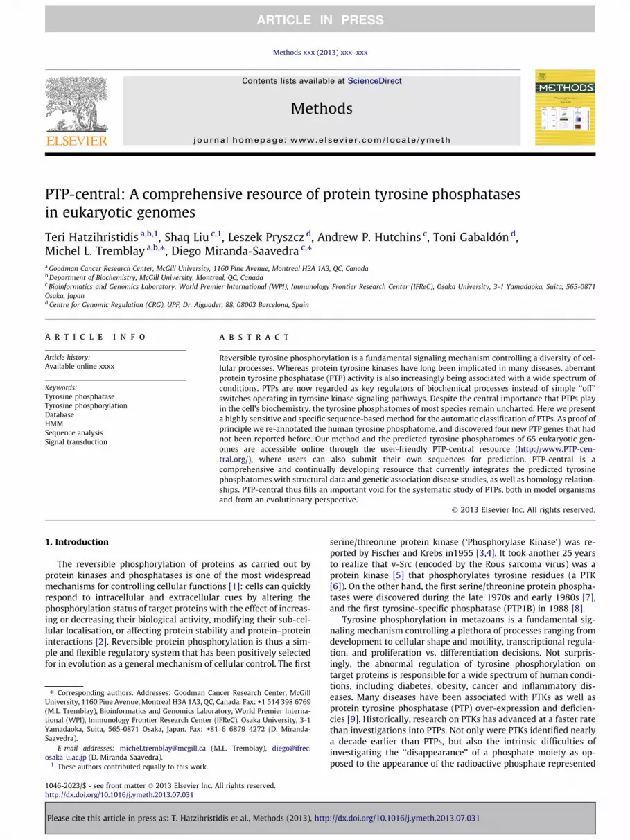
We present a general method for the automatic prediction andclass-level classification of eukaryotic PTPs called Y-Phosphatomer.Y-Phosphatomer relies on a specific combination of publicly avail-able protein domain models (mostly in the form of profile hiddenMarkov models, or HMMs) diagnostic for the various PTP families.Briefly, we built Y-Phosphatomer by characterizing the specificcombination of protein domain signatures from the smallest num-ber of protein domain databases that allows the identification andclassification of a curated set of human PTPs into their correct clas-ses, without cross-hitting other classes (Fig. 1).
2.2.1. Working data sets and identification of protein domain modelsspecific to PTPs
In 2004 Alonso et al. reported the PTP complement of the hu-man genome following comprehensive searches in public dat-abases for genes encoding PTPs and PTP-like genes [14]. A totalof 107 human genes were found to encode PTPs belonging to 4 dis-tinct classes: Class I (receptor PTPs, cytoplasmic PTPs and dual-specificity phosphatases), Class II (low molecular weight phospha-tases), Class III (CDC25), and aspartic acid (Asp)-based phospha-tases (EyA homologues). We mapped these 107 human genesonto the Ensembl database (release 67), leading to the removalof MTMR15 and PTPRVP (a pseudogene, originally annotated asPTPRV). We also expanded gene PTPN20 into 3 distinct genes(PTPN20A, PTPN20B and PTPN20C), and merged DUSP13A andDUSP13B into the DUSP13 gene. This brings the updated list of hu-man PTPs to 105 protein-coding genes, including 20 receptor PTPs,
://dx.doi.org/10.1016/j.ymeth.2013.07.031

Dataset of human PTPs
InterProScan
1
Evaluation of Y-Phosphatomer Library
6
2
3
5
Y-Phosphatomer Library
4
Genome-wide annotation of PTPs
7
PTP
Cdc25
DSP
EYAEYA
PTPs
LMWP
Fig. 1. Flow diagram of the Y-Phosphatomer method for the automatic classification of PTPs. The human set of PTPs was analyzed with a local installation of InterProScan runwith default parameters (steps 1 and 2). This analysis determined that the PTPs from the four classes could be unequivocally distinguished by a specific combination ofprotein domain models, mostly in the form of HMMs (step 3 and 4). We took advantage of this property to build the Y-Phosphatomer library consisting of only 14 proteinmodels (step 5). The Y-Phosphatomer library was evaluated on two data sets and uniformly reported perfect coverage, and a mis-classification rate of zero on the PTP classlevel (step 6). Therefore, Y-Phosphatomer is a robust library that can be applied to the genome-wide annotation of PTPs (step 7).
T. Hatzihristidis et al. / Methods xxx (2013) xxx–xxx 3
19 non-receptor PTPs, 58 DSPs, 1 LMWP, 3 CDC25 homologues and4 EyA homologues (Supplementary Table 1).
The proteins encoded by the 105 human PTP genes were re-trieved from the Ensembl database and analyzed with a localinstallation of InterProScan (release 30.0) run with default param-eters [19]. This was done to identify protein domain models diag-nostic for the enzymes in each PTP family. InterProScan is thefunctional implementation of the InterPro database, a comprehen-sive and integrated resource of protein domains, regions and activesites that is widely employed in the automatic annotation of pro-teins in genome sequencing projects [19]. The overall strategy ofthe method is summarized in Fig. 1. Briefly, first we identified acombination of 14 protein domain models (mostly HMMs) that al-lows the identification of the human PTP data set and its discrim-ination into classes (Supplementary Table 2). We found that it waspossible to combine a reduced set of protein domain models fromthe Pfam [20], PRINTS [21], SMART [22], SUPERFAMILY [23] andTIGRFAMs [24] databases to identify all human PTPs with perfectcoverage. An advantage of InterPro is that it combines variousmember databases, each covering a portion of the total proteinspace. Therefore, by combining a set of protein domain modelsfrom a variety of sources we take advantage of the individualstrengths of InterPro’s member databases. The performance of thisspecific collection of 14 models (the ‘Y-Phosphatomer’ library) wassubsequently evaluated on two distinct data sets, and then appliedto annotate the tyrosine phosphatomes of 65 eukaryotic genomes.
2.2.2. Evaluation of Y-phosphatomerFirst we tested the Y-Phosphatomer library for its ability to
identify and classify a collection of PTP sequences from the ‘PTPDatabase’ (http://ptp.cshl.edu/), an important resource of tyro-sine-specific PTPs. Specifically, we analyzed the larger of the datasets, which includes both vertebrate and non-vertebrate tyrosine-specific PTP sequences derived from an original PSI-BLAST search,
Please cite this article in press as: T. Hatzihristidis et al., Methods (2013), http
followed by expert inspection, dated August 2004 [13]. This collec-tion of sequences comprises a set of 601 PTP domains from 61 spe-cies and 5 phyla. The protein identifiers of these sequences mappedto 6 distinct databases (DDBJ, EMBL, GenBank, PIR, RefSeq andSWISS-PROT). To perform a uniform updating of this 2004 dataset, all identifiers were mapped onto the UniProt database, produc-ing a non-redundant list of 383 UniProt proteins. This set of pro-teins was run through Y-Phosphatomer, and in 100% of the casesthe sequences were identified as tyrosine-specific PTPs. This result,although promising (coverage and correct classification rate of100%) is limited to tyrosine-specific PTPs, the subject of study ofthe PTP Database.
In a second exercise we tested the Y-Phosphatomer library on ahigh-quality set of proteins from the UniProt database, the largestpublic resource of protein sequences. Specifically we selected thoseUniProt proteins (release of 16 May, 2012) that were annotated ashaving PTP catalytic activity according to the UniProt-GO database[25]. The proteins had to be annotated with any of the followingexperimental codes: IDA (‘Inferred from Direct Assay’), IPI(‘Inferred from Physical Interaction’), IMP (‘Inferred from MutantPhenotype’), IGI (‘Inferred from Genetic Interaction’) and EXP(‘Inferred from Experiment’), plus any of the Gene OntologyMolecular Function identifiers listed in Supplementary Table 3.This selection resulted in a list of 124 proteins from a diversity ofspecies including vertebrates (human, mouse, rat, zebrafish),invertebrates (fruit fly), fungi (budding and fission yeasts, Candidaalbicans), the model organisms Arabidopsis thaliana andDictyostelium discoideum, and several bacteria. When applyingour classification criteria, 28/124 proteins were not classified asprotein tyrosine phosphatases, but a close inspection showed thatthese proteins had been incorrectly annotated as PTPs, or arefragments of real PTPs. Inspection of the 96 remaining proteinsthat were classified as PTPs showed that all were correctly as-signed to their annotated classes. The evaluation test on this set
://dx.doi.org/10.1016/j.ymeth.2013.07.031

4 T. Hatzihristidis et al. / Methods xxx (2013) xxx–xxx
of UniProt proteins, as well as on the sequences of the PTP Data-base, suggest that Y-Phosphatomer can retrieve PTPs and classifythem into their correct classes with perfect coverage and classifica-tion rates (Table 1).
2.3. PTP-central: contents and sequence data sources
The Y-Phosphatomer library was used to scan the predictedprotein sets of 65 completely sequenced and published eukaryoticgenomes. These genomes may be classified into four of the fiveeukaryotic supergroups (unikonts, excavates, plants and chromal-veolates) following the classification scheme of Keeling and co-workers [26]. Supplementary Table 4 lists the scientific and com-mon names of the 65 species, plus the databases from which thepredicted proteins were downloaded (including version and/ordata set release date), as well as their predicted tyrosine phosphat-omes split by PTP class. PTP-central is stored as a MySQL relationaldatabase (http://www.mysql.com/) and the server is implementedas a set of Perl and Python scripts running under Apache (http://www.apache.org/). PTP-central is freely available at http://www.PTP-central.org.
2.3.1. Genetic association studiesPositive associations between PTPs and genetic studies were
obtained from the Genetic Association Database (GAD) of the NIH[27]. The GAD is a comprehensive, standardized and curated collec-tion of published genetic association studies of many common dis-ease types. The primary focus of GAD is the archiving andsummarization of information from complex genetic diseases(both from candidate gene studies and genome-wide associationstudies) rather than rare Mendelian disorders. The GAD releaseanalysed (rel. 27 October 2012) features 130,653 entries from56,397 distinct publications, and which are broadly classified into19 distinct disease classes, including cancer, immunological, infec-tious, reproductive, metabolic, cardiovascular, neurological, hema-tological, developmental and mitochondrial diseases.
2.3.2. Three-dimensional structures of PTPsPTP structures were retrieved from the Protein Data Bank
(http://www.rcsb.org/, 6 November 2012), a curated and anno-tated repository of experimentally determined structures of pro-teins, nucleic acids and complex assemblies [28]. Theidentification of PTP structures was done by a combination of se-quence-based and specific keyword searches. The initial set of re-sults was manually inspected to remove false-positive hits,resulting in a compilation of 339 structures. The vast majority ofthe structures in PTP-central comprise PTP catalytic domains,although a few are of accessory PTP domains that play importantroles in PTP function.
2.3.3. Orthology and paralogy relationshipsOrthologs and paralogs of PTPs were fetched from MetaPhOrs
[29] (http://orthology.phylomedb.org/, as of February 2013).
Table 1The Y-Phosphatomer classification of experimentally characterized PTP sequencesretrieved from the UniProt database.
PTP Class Group Proteins Group-level classification accuracyon the data set covered (%)
Class I PTP 66 66 (100%)DSP 13 13 (100%)
Class II LMWP 3 3 (100%)Class III CDC25 0 N/AAsp-based PTPs EYA 14 14 (100%)Total 96 96 (100%)
Please cite this article in press as: T. Hatzihristidis et al., Methods (2013), http
MetaPhOrs is a public repository of orthologs and paralogs derivedfrom phylogenetic trees deposited in five databases (PhylomeDB[30], Ensembl [31], EggNOG [32], Hogenom [33] and TreeFAM[34]). MetaPhOrs contains predictions for over 13 million proteinsfrom 1963 genomes (representing �1.4 billion homologous rela-tionships), and is therefore the most comprehensive database ofhomology relationships currently available. Most importantly,MetaPhOrs also provides quality metrics for each prediction, themost important of which is the Consistency Score (CS). The CS rep-resents the ratio between the number of trees informing about aspecific orthology relationship for a pair of proteins and the totalnumber of trees. The CS ranges from 0 (no trees predicting orthol-ogy) to 1 (all trees predicting orthology), so the closer the CS valueto 1, the more robust the prediction is. We applied a CS cutoff valueof 0.5, meaning that at least one-half of all trees available for a gi-ven pair of proteins do predict an orthology relationship. Any pairof proteins with a CS < 0.5 is annotated as paralogous. MetaPhorsalso describes the type of orthologous relationships (one-to-one,one-to-many or many-to-many) and lists eventual co-orthologs.In total, we describe 79,355 orthologous relationships and640,099 paralogous relationships among the PTPs predicted inthe 65 eukaryotic genomes.
3. Results and discussion
3.1. Y-Phosphatomer: a novel method for the automatic classificationof PTPs
We have developed a sequence-based classification methodcalled Y-Phosphatomer to identify PTP sequences and automati-cally classify them into specific classes (Fig. 1). Y-Phosphatomerworks by combining 14 specific protein domain models (mostlyHMMs) diagnostic for the various classes of human PTP sequences,and drawn from five protein domain databases (Pfam, PRINTS,SMART, SUPERFAMILY and TIGRFAMs). The protein models charac-teristic of each PTP class do not overlap with those of other classes,thus preventing in practice the cross-annotation of sequences tomore than one PTP class. When evaluated, Y-Phosphatomer wasable to retrieve all tyrosine-specific PTP sequences (n = 383) fromthe expertly annotated PTP Database (http://ptp.cshl.edu/) andclassify them automatically as tyrosine-specific PTPs. In a secondexercise using the experimentally validated and manually anno-tated set of PTP sequences from the UniProt database, Y-Phospha-tomer managed to retrieve all real, full-length PTP sequences andassign them to their correct families, reporting a coverage of100% and a mis-classification rate of zero on the class level. TheUniProt data set was limited in size (n = 124) albeit phylogeneti-cally diverse. Y-Phosphatomer relies on the recommended ‘‘trustedcut-offs’’ of the distinct protein models as provided in InterProScan,which are thought to report relevant matches [19]. Other than this,no test for false positives was implemented, as no gold standardcurrently exists for the correct classification of PTPs.
Andersen and colleagues previously reported a set of guidelinesfor the computational identification of tyrosine-specific PTPs usingweb-based resources, including BLAST, PSI-BLAST and keywordsearches [13]. Despite the usefulness of this approach, carefully se-lected collections of protein models describing specific proteinfamilies have been shown to be more sensitive and specific thangeneral tools for the database search and automatic classificationof protein sequences, as we have previously shown for protein ki-nases [35,36] and ubiquitinating and deubiquitinating enzymes[37]. The protein models combined into the Y-Phosphatomerlibrary were specifically built to represent well-studied PTP classesand families, and as a result Y-Phosphatomer managed to retrieveall experimentally validated PTPs from organisms representing a
://dx.doi.org/10.1016/j.ymeth.2013.07.031

T. Hatzihristidis et al. / Methods xxx (2013) xxx–xxx 5
diversity of phylogenetic lineages. Thus the high sensitivity andspecificity of our tool enables the genome-wide identificationand class-level classification of PTPs in a reproducible and auto-matic manner.
3.2. Reannotation of the human tyrosine phosphatome
Y-Phosphatomer was used to scan the predicted proteins of thehuman genome for PTPs (Ensembl release 67) and found 4 newgenes encoding PTPs that were not described in the Alonso et al.paper of the human tyrosine phosphatome [14]. These includethe DSPs DUPD1 (ENSG00000188716), PTPMT1 (ENSG00000110536),TNS3 (ENSG00000136205) and the tyrosine-specific phosphataseKIAA1274 (PALD1) (ENSG00000107719). Although these geneswere not reported as part of the original human tyrosine phospha-tome [14], all four have been subsequently discovered and studiedindependently in recent years. For example, DUPD1 was initiallyidentified as a new phosphatase binding to the intracellular domainof the short isoform of the prolactin receptor and is thought todephosphorylate MAPK, a protein kinase essential for normal follic-ular development [38]. PTPMT1 was originally found to localizeexclusively to the inner membrane of mitochondria, and its knock-down in a pancreatic cell line was shown to alter the mitochondria’sphosphoprotein profile as well as to up-regulate insulin secretionand ATP production [39,40]. Besides being a potential drug targetin type II diabetes, PTPMT1 has recently been found to have an
Amoebozoa
Metazoans (Placozoa)
EXCAVATES
PLANTS
CHROMALVEOLATES
UNIKONTS
Fungi
Metazoans (True tissues)
Choanozoa
Euglenozoa
Metamonada
Streptophytes
Green algae
Red algae
Alveolates
Apicomplexa
PTP DSP CDC25 LMWP EyA
Absent
Present
C
A
Fig. 2. Overview of the contents of PTP-central. (A) In the 65 eukaryotic genomes analyzefollowed by dual-specificity phosphatases (�43%). LMWP/CDC25/EyA class phosphatascorrelation exists between a species’ tyrosine phosphatome and the total number of prophosphatomes remain relatively constant despite large increases in genome sizes. (C) Oneukaryotic supergroups surveyed. (D) PTP-central contains a manually curated dataphosphatases from crystallographic studies.
Please cite this article in press as: T. Hatzihristidis et al., Methods (2013), http
essential role in the biogenesis of cardiolipin (a phospholipid ofthe inner mitochondrial membrane required for mitochondrialmetabolism), and possibly in cardiolipin deficiency diseases too[41,42]. Tensin-3 (TNS3) is a negative regulator of cell migrationthat was initially identified in the epidermal growth factor signalingcascade [43]. The genetic deletion of Tensin-3 showed that the geneis essential for the correct development and function of the smallintestine, lung and bone [44]. Recently, the Src tyrosine kinasewas found to phosphorylate the SH2 domain of Tensin-3 with theeffect of promoting the gene’s oncogenic function [45]. Finally, theimportant regulatory phosphatase KIAA1274 (also called PALD1)was initially identified as a negative regulator of insulin signalingin a genome-wide functional screen [46], and its mouse ortholog(Paladin) was identified in another screen for angiogenesis regula-tors. Paladin is dynamically expressed throughout the vasculature,mainly in endothelial cells in embryonic stages, and in arterialsmooth muscle cells in the adult [47]. In summary, the updatedcount of the human tyrosine phosphatome includes 109 PTP genes(20 receptor PTPs, 20 non-receptor PTPs, 61 DSPs, 1 LMWP, 3 CDC25homologues, and 4 EyA homologues, Supplementary Table 1)encoding 537 distinct PTP protein sequences (Supplementary Ta-ble 4). Although these four new PTP genes were characterised byother researchers following the original publication of the humantyrosine phosphatome [14], their identification proves the useful-ness of Y-Phosphatomer for the genome-wide characterization ofphosphatomes.
Species Structures (% of total)
H. sapiens
M. musculus
R. norvegicus
D. melanogaster
G. gallus
C. intestinalis
A. thaliana
L. major
T. brucei
Total
301 (89%)
17 (5%)
6 (<2%)
6 (<2%)
1 (0.3%)
1 (0.3%)
1 (0.3%)
3 (<1%)
3 (<1%)
339 (100%)
Total
B
D
0 20000 40000 60000 80000 100000
010
020
030
040
050
0
Total number of peptides in genome
Enz
ymes
H. sapiensUnikonts
Excavates
Plants
Chromalveolates
d, nearly 50% of the 4605 PTP sequences are tyrosine-specific phosphatases, closelyes comprise less than 3% each of the entire dataset. (B) For most species a linearteins encoded in the genome. Plants are the exception, where the sizes of tyrosinely tyrosine-specific and dual-specificity phosphatases are universally present in allset of 339 structures, the vast majority of which are human tyrosine-specific
://dx.doi.org/10.1016/j.ymeth.2013.07.031

Fig. 3. Snapshot of the PTP-central web interface. PTP-central features a user-friendly and flexible interface that allows the user to search (i) the predicted PTPs of 65eukaryotic genomes (in any combination of PTP class and species), (ii) the genetic diseases associated with human PTPs as reported in the Genetic Association Database (GAD)of the NIH, (iii) a curated set of PTP structures drawn from the PDB, and (iv) the homology relationships among the PTPs of all 65 genomes. Additionally, the user can submitprotein sequences for scanning with the Y-Phosphatomer library.
6 T. Hatzihristidis et al. / Methods xxx (2013) xxx–xxx
Please cite this article in press as: T. Hatzihristidis et al., Methods (2013), http://dx.doi.org/10.1016/j.ymeth.2013.07.031

Table 2Disease types associated with 26 distinct human PTP genes as reported inthe Genetic Association Database (GAD) of the NIH. The most frequent typeof disease associated with human PTPs is immunological, and many PTPsare associated with more than one disease type.
Type of disease Representation among PTPs witha positive disease association
Immune 52.9%Metabolic 13.8%Cancer 6.5%Developmental 5.8%Cardiovascular
NeurologicalPsychiatricChem. DependencyInfectionReproductionHematologicalVisionAgingRenal
All below 3%
T. Hatzihristidis et al. / Methods xxx (2013) xxx–xxx 7
3.3. The PTP complements of 65 eukaryotic genomes
The perfect coverage and classification rates reported in thetests on the Y-Phosphatomer library indicate that the accurateidentification of tyrosine phosphatomes from genomic data setsis feasible. We scanned 65 eukaryotic genomes for PTPs, includingspecies belonging to four of the five eukaryotic supergroups [26].These are unikonts (33 species), excavates (6 species), plants (10species) and chromalveolates (16 species), which collectively dis-play a vast range of genome sizes and environmental adaptations,including parasites of great importance such as Trypanosoma andLeishmania species. If we accept the classification as presentedhere, our analysis predicted a total of 4605 PTP sequences in 65eukaryotic genomes classified into the PTP, DSP, LMWP, CDC25and EyA classes. The genomes analyzed harbor somewhere be-tween 1 PTP enzyme, as is the case of the intracellular parasiteEncaphalitozoon cuniculi (which also has an extremely economicalkinome [48]), to 537 PTPs (human). Given the comparatively largenumber of unikont genomes, it is not surprising that these speciesdominate the database in terms of PTP sequences (3916 or 85%),followed by plants (339 or 7.4%), chromalveolates (183 or 4.0%)and excavates (167 or 3.6%). Irrespective of the phylogenetic originof the sequences, PTP-central is mostly populated by tyrosine-spe-cific PTPs (2258 or 49%) and DSPs (1998 or 43.4%), followed byEyAs (139 or 3.0%), CDC25s (119 or 2.6%) and LMWPs (91 or2.0%) (Fig. 2A). For most species a linear correlation exists betweenthe number of proteins encoded by a genome and its tyrosinephosphatome. However, this correlation does not appear to holdtrue for the plant genomes whose predicted tyrosine phosphatomesizes remain nearly identical despite large increases in genomesizes (Fig. 2B). PTPs and DSPs are the only enzyme classes thatare universally present in all eukaryotic supergroups. CDC25 en-zymes are reported in all eukaryotic supergroups (but not in allgenomes), and are particularly prominent in mammals maybe be-cause of their complex mechanisms of cell cycle regulation. CDC25enzymes appear to be absent from most excavates, plants andchromalveolates, but not all, suggesting alternative cell cycle regu-lation strategies (for instance, as previously reported in the Api-complexa [49,50]). Likewise, LMWP enzymes are most prominentin mammalian genomes and discreetly distributed among all othergenomes from all four eukaryotic supergroups, with notable lossesas in the Apicomplexa, none of which appear to harbor any LMWPs.Finally, Eye Absent (EyA) homologs are only predicted in true-tis-sue metazoans and streptophyte plants (1–3 members per gen-ome), and are absent from all other species surveyed (Fig. 2C andSupplementary Table 4).
3.4. PTP-central: contents and functionality
Our method and predictions are accessible online via the PTP-central resource (http://www.PTP-central.org/). PTP-central har-bors the precomputed tyrosine phosphatomes of the 65 eukaryoticgenomes (n = 4605 proteins) (Fig. 2A, 2B and 2C, and Supplemen-tary Table 4), as well as the PTP structures retrieved from thePDB (Fig. 2D), the majority of which are tyrosine-specific phospha-tases of human origin; 138 distinct positive disease associationsdrawn from the Genetic Association Database (GAD) [27], and allthe homology relationships detected among PTPs across the 65eukaryotes.
The PTP-central website is a user-friendly and comprehensiveresource, featuring essential background information on PTPs andtheir functions, PTP-related diseases, and the various structuralclasses as well as the implications for inhibitor design. The data-base is particularly easy to navigate, as illustrated in the exampleprovided in Fig. 3: at the top are displayed the distinct searchoptions of the database (‘‘PHOSPHATOMES’’, ‘‘DISEASES’’, ‘‘PTP
Please cite this article in press as: T. Hatzihristidis et al., Methods (2013), http
STRUCTURES’’ and ‘‘PTP HOMOLOGS’’), as well as a ‘‘PEPTIDESCAN’’ interface where users can submit their protein sequencesfor prediction. As an exercise, let’s say that we wish to investigatethe dual-specificity phosphatases that are common to both humanand the Placozoan Trichoplax adhaerens (likely the simplest extantmulticellular metazoan). Ultimately this comparison should beinformative about the DSPs that are present in both organismsand which therefore are likely to represent an early core of animaldual-specificity phosphatases. In the ‘‘SEARCH DB - > PHOSPHATO-MES’’ interface we select the DSPs present in human and the placo-zoan (Fig. 3A), resulting in the tabulated output shown in Fig. 3B:besides the peptide and gene IDs, their classification into one ofthe PTP classes, gene names and description, the database gener-ates a protein architecture mini-plot (which can be enlarged byclicking on it) that provides a quick way of visually inspecting var-ious protein architectures. The gene names include those providedin the original Ensembl annotation, as well as all the aliases avail-able in the Entrez Gene database [51]. The inclusion of gene aliasesmakes the keyword-based search for PTPs far more comprehensiveand flexible (Fig. 3A). Finally, for human genes PTP-central pro-vides a link to the corresponding entry in the OMIM (Online Men-delian Inheritance in Man) database, the main repository of geneticinformation on Mendelian genetic disorders. OMIM providesextensive expert-based historical annotation on each gene’s identi-fication, functions and involvement in specific diseases [52].Although the style of the entries in the OMIM database is rathernarrative, the entries are typically of very high quality and signifi-cance. The user is given the option to download the datasets, eitherin the ‘‘text-only’’ version (fully annotated protein sequences inFasta format), or also including the protein architecture plots, aswell as in a ‘‘tab-separated’’ format (for uploading to Excel). The‘‘Start Jalview’’ button launches a Java applet of the multiple se-quence analysis tool Jalview [53]. Jalview allows the user to editand color the sequences by conservation, protein secondary struc-tural properties, or amino acid chemical characteristics and per-form on-the-fly calculations of phylogenetic trees (Neighbor-Joining and average distance). The full Jalview application can belaunched (‘‘File- > View in Full Application’’) to access various mul-tiple alignment algorithms (MAFFT, Muscle, ClustalW, T-Coffee andProbcons), secondary structure prediction methods (Jnet), and dis-play structures with Jmol. Additionally, multiple alignments in Jal-view can be exported to TOPALi v2 [54] via a synchronizedinterface where more sophisticated phylogenetic methods arereadily available.
The ‘‘SEARCH DB - > DISEASES’’ interface features the positivedisease associations with human PTPs that are found in the
://dx.doi.org/10.1016/j.ymeth.2013.07.031

Table 3The 26 human PTP genes and their associated diseases as reported in the Genetic Association Database (GAD) of the NIH and available through PTP-central.
Gene PTP Class Number of diseases Disease types
PTPN1 PTP 11 Cardiovascular/immune/MetabolicPTPN2 PTP 10 ImmunePTPN9 PTP 1 DevelopmentalPTPN11 PTP 14 Cancer/cardiovascular/developmental/hematological/immune/metabolic/otherPTPN12 PTP 1 VisionPTPN22 PTP 54 Immune/infection/metabolicPTPRC PTP 7 Immune/infection/otherPTPRD PTP 6 Chem. Dependency/metabolic/neurological/psychiatricPTPRE PTP 1 OtherPTPRF PTP 1 MetabolicPTPRG PTP 2 Cardiovascular/psychiatricPTPRJ PTP 1 CancerPTPRK PTP 1 ImmunePTPRT PTP 1 Chem. DependencyTNS1 PTP 1 CardiovascularDUSP6 DSP 1 AgingDUSP12 DSP 1 MetabolicDUSP13 DSP 1 RenalMTM1 DSP 1 OtherMTMR2 DSP 1 OtherMTMR3 DSP 1 ImmuneMTMR11 DSP 1 DevelopmentalPTEN DSP 7 Cancer/Chem. Dependency/metabolicACP1 LMWP 10 Developmental/Immune/metabolic/psychiatric/reproductionEYA1 EYA 1 NeurologicalEYA4 EYA 1 Other
8 T. Hatzihristidis et al. / Methods xxx (2013) xxx–xxx
Genetic Association Database (GAD) [27]. We found 138 positiveassociations between 26 PTP genes (of the PTP and DSP classes)and 14 different types of disease, which suggests that PTP genesare often associated with more than one disease type. The mostfrequent type of disease is immunological (>50%), followed bymetabolic disorders (�14%) (Table 2). PTPN22 is by far the PTPgene associated with the largest number of conditions (54 dis-eases of the immune, infectious and metabolic types), followedby PTPN11 (14 diseases) and PTPN1 (11 diseases) (Table 3).PTP-central provides links between each PTP gene and its associ-ated diseases to the original publications and the GAD, OMIM andEnsembl databases.
4. Conclusions
The main conclusions of our work are:
(i) We developed a highly sensitive and specific classificationmethod (‘Y-Phosphatomer’) for the automatic classificationof protein sequences into the various PTP classes. Ourmethod relies on a specific collection of protein domainmodels drawn from five distinct InterPro member databases.Upon evaluation, Y-Phosphatomer reported perfect coverageand classification rates on curated sets of PTPs.
(ii) As proof of principle we re-annotated the human tyrosinephosphatome, and discovered four new PTP genes that hadnot been reported in an early and important publication ofthe human PTP repertoire [14].
(iii) Y-Phosphatomer was used to scan 65 eukaryotic genomesand we report that the sizes of tyrosine phosphatomes varyextensively, and that only tyrosine-specific and dual-speci-ficity phosphatases are universally found in all eukaryoticsupergroups.
(iv) Our method Y-Phosphatomer and the predicted tyrosinephosphatomes are accessible online through the PTP-centralresource (http://www.PTP-central.org/), which also inte-grates PTP structural information from the Protein DataBank, as well as genetic association studies from the Genetic
Please cite this article in press as: T. Hatzihristidis et al., Methods (2013), http
Association Database (GAD) and pre-calculated homologyrelationships among the 65 tyrosine phosphatomes.
(v) PTP-central is a comprehensive and continually developingresource that integrates a diversity of data sets essentialfor the study of PTPs, both in model organisms as well asfrom an evolutionary perspective. Future plans for PTP-cen-tral include the integration of ‘‘omics’’ data sets from variousmodel organisms, the addition of new genome-wide associ-ation studies as well as disease-specific data, and the imple-mentation of algorithms for investigating the contents of theresource from multiple angles.
Acknowledgements
This work was supported by the Japan Society for the Promotionof Science (JSPS) through the WPI-IFReC Research Program and aKakenhi grant; the Kishimoto Foundation; the ETHZ-JST Japa-nese-Swiss Cooperative Program (to DMS); and a Jeanne andJean-Louis Lévesque Chair in Cancer Research and a Canadian Can-cer Society Research Institute operating grant #700922 (to MLT).
We would like to thank Ms. Mineko Tanimoto for secretarialsupport.
Appendix A. Supplementary data
Supplementary data associated with this article can be found, inthe online version, at http://dx.doi.org/10.1016/j.ymeth.2013.07.031.
References
[1] P. Cohen, Nat. Cell Biol. 4 (2002) E127–E130.[2] P. Cohen, Trends Biochem. Sci. 25 (2000) 596–601.[3] E.H. Fischer, E.G. Krebs, J. Biol. Chem. 216 (1955) 121–132.[4] E.G. Krebs, E.H. Fischer, Biochim. Biophys. Acta 20 (1956) 150–157.[5] M.S. Collett, R.L. Erikson, Proc. Natl. Acad. Sci. USA 75 (1978) 2021–2024.[6] T. Hunter, B.M. Sefton, Proc. Natl. Acad. Sci. USA 77 (1980) 1311–1315.[7] T.S. Ingebritsen, P. Cohen, Eur. J. Biochem. 132 (1983) 255–261.
://dx.doi.org/10.1016/j.ymeth.2013.07.031

T. Hatzihristidis et al. / Methods xxx (2013) xxx–xxx 9
[8] N.K. Tonks, C.D. Diltz, E.H. Fischer, J. Biol. Chem. 263 (1988) 6722–6730.[9] W.J. Hendriks, A. Elson, S. Harroch, R. Pulido, A. Stoker, J. den Hertog, FEBS J 280
(2012) 708–730.[10] T. Hunter, G.D. Plowman, Trends Biochem. Sci. 22 (1997) 18–22.[11] J.N. Andersen, O.H. Mortensen, G.H. Peters, P.G. Drake, L.F. Iversen, O.H. Olsen,
P.G. Jansen, H.S. Andersen, N.K. Tonks, N.P. Moller, Mol. Cell. Biol. 21 (2001)7117–7136.
[12] J.N. Andersen, P.G. Jansen, S.M. Echwald, O.H. Mortensen, T. Fukada, R. DelVecchio, N.K. Tonks, N.P. Moller, FASEB J. 18 (2004) 8–30.
[13] J.N. Andersen, R.L. Del Vecchio, N. Kannan, J. Gergel, A.F. Neuwald, N.K. Tonks,Methods 35 (2005) 90–114.
[14] A. Alonso, J. Sasin, N. Bottini, I. Friedberg, A. Osterman, A. Godzik, T. Hunter, J.Dixon, T. Mustelin, Cell 117 (2004) 699–711.
[15] N.K. Tonks, FEBS J 280 (2012) 346–378.[16] A.R. Forrest, D.F. Taylor, J.L. Fink, M.M. Gongora, C. Flegg, R.D. Teasdale, H.
Suzuki, M. Kanamori, C. Kai, Y. Hayashizaki, S.M. Grimmond, BMCBioinformatics 7 (2006) 82.
[17] S. Liberti, F. Sacco, A. Calderone, L. Perfetto, M. Iannuccelli, S. Panni, E.Santonico, A. Palma, A.P. Nardozza, L. Castagnoli, G. Cesareni, FEBS J. 280(2013) 379–387.
[18] X. Li, M. Wilmanns, J. Thornton, M. Kohn, Sci. Signal. 6 (2013) 10.[19] E.M. Zdobnov, R. Apweiler, Bioinformatics 17 (2001) 847–848.[20] M. Punta, P.C. Coggill, R.Y. Eberhardt, J. Mistry, J. Tate, C. Boursnell, N. Pang, K.
Forslund, G. Ceric, J. Clements, A. Heger, L. Holm, E.L. Sonnhammer, S.R. Eddy,A. Bateman, R.D. Finn, Nucleic Acids Res. 40 (2012) D290–301.
[21] T.K. Attwood, P. Bradley, D.R. Flower, A. Gaulton, N. Maudling, A.L. Mitchell, G.Moulton, A. Nordle, K. Paine, P. Taylor, A. Uddin, C. Zygouri, Nucleic Acids Res.31 (2003) 400–402.
[22] I. Letunic, T. Doerks, P. Bork, Nucleic Acids Res. 40 (2012) D302–305.[23] D. Wilson, R. Pethica, Y. Zhou, C. Talbot, C. Vogel, M. Madera, C. Chothia, J.
Gough, Nucleic Acids Res. 37 (2009) D380–D386.[24] S. Hunter, P. Jones, A. Mitchell, R. Apweiler, T.K. Attwood, A. Bateman, T.
Bernard, D. Binns, P. Bork, S. Burge, E. de Castro, P. Coggill, M. Corbett, U. Das, L.Daugherty, L. Duquenne, R.D. Finn, M. Fraser, J. Gough, D. Haft, N. Hulo, D.Kahn, E. Kelly, I. Letunic, D. Lonsdale, R. Lopez, M. Madera, J. Maslen, C.McAnulla, J. McDowall, C. McMenamin, H. Mi, P. Mutowo-Muellenet, N.Mulder, D. Natale, C. Orengo, S. Pesseat, M. Punta, A.F. Quinn, C. Rivoire, A.Sangrador-Vegas, J.D. Selengut, C.J. Sigrist, M. Scheremetjew, J. Tate, M.Thimmajanarthanan, P.D. Thomas, C.H. Wu, C. Yeats, S.Y. Yong, Nucleic AcidsRes. 40 (2012) D306–D312.
[25] E.C. Dimmer, R.P. Huntley, Y. Alam-Faruque, T. Sawford, C. O’Donovan, M.J.Martin, B. Bely, P. Browne, W. Mun Chan, R. Eberhardt, M. Gardner, K. Laiho, D.Legge, M. Magrane, K. Pichler, D. Poggioli, H. Sehra, A. Auchincloss, K. Axelsen,M.C. Blatter, E. Boutet, S. Braconi-Quintaje, L. Breuza, A. Bridge, E. Coudert, A.Estreicher, L. Famiglietti, S. Ferro-Rojas, M. Feuermann, A. Gos, N. Gruaz-Gumowski, U. Hinz, C. Hulo, J. James, S. Jimenez, F. Jungo, G. Keller, P.Lemercier, D. Lieberherr, P. Masson, M. Moinat, I. Pedruzzi, S. Poux, C. Rivoire,B. Roechert, M. Schneider, A. Stutz, S. Sundaram, M. Tognolli, L. Bougueleret, G.Argoud-Puy, I. Cusin, P. Duek-Roggli, I. Xenarios, R. Apweiler, Nucleic AcidsRes. 40 (2012) D565–D570.
[26] P.J. Keeling, G. Burger, D.G. Durnford, B.F. Lang, R.W. Lee, R.E. Pearlman, A.J.Roger, M.W. Gray, Trends Ecol. Evol. 20 (2005) 670–676.
[27] K.G. Becker, K.C. Barnes, T.J. Bright, S.A. Wang, Nat. Genet. 36 (2004) 431–432.[28] P.W. Rose, C. Bi, W.F. Bluhm, C.H. Christie, D. Dimitropoulos, S. Dutta, R.K.
Green, D.S. Goodsell, A. Prlic, M. Quesada, G.B. Quinn, A.G. Ramos, J.D.Westbrook, J. Young, C. Zardecki, H.M. Berman, P.E. Bourne, Nucleic Acids Res.41 (2013) D475–D482.
[29] L.P. Pryszcz, J. Huerta-Cepas, T. Gabaldon, Nucleic Acids Res. 39 (2011) e32.
Please cite this article in press as: T. Hatzihristidis et al., Methods (2013), http
[30] J. Huerta-Cepas, S. Capella-Gutierrez, L.P. Pryszcz, I. Denisov, D. Kormes, M.Marcet-Houben, T. Gabaldon, Nucleic Acids Res. 39 (2011) D556–D560.
[31] P. Flicek, M.R. Amode, D. Barrell, K. Beal, S. Brent, D. Carvalho-Silva, P. Clapham,G. Coates, S. Fairley, S. Fitzgerald, L. Gil, L. Gordon, M. Hendrix, T. Hourlier, N.Johnson, A.K. Kahari, D. Keefe, S. Keenan, R. Kinsella, M. Komorowska, G.Koscielny, E. Kulesha, P. Larsson, I. Longden, W. McLaren, M. Muffato, B.Overduin, M. Pignatelli, B. Pritchard, H.S. Riat, G.R. Ritchie, M. Ruffier, M.Schuster, D. Sobral, Y.A. Tang, K. Taylor, S. Trevanion, J. Vandrovcova, S. White,M. Wilson, S.P. Wilder, B.L. Aken, E. Birney, F. Cunningham, I. Dunham, R.Durbin, X.M. Fernandez-Suarez, J. Harrow, J. Herrero, T.J. Hubbard, A. Parker, G.Proctor, G. Spudich, J. Vogel, A. Yates, A. Zadissa, S.M. Searle, Nucleic Acids Res.40 (2012) D84–D90.
[32] S. Powell, D. Szklarczyk, K. Trachana, A. Roth, M. Kuhn, J. Muller, R. Arnold, T.Rattei, I. Letunic, T. Doerks, L.J. Jensen, C. von Mering, P. Bork, Nucleic AcidsRes. 40 (2012) D284–D289.
[33] S. Penel, A.M. Arigon, J.F. Dufayard, A.S. Sertier, V. Daubin, L. Duret, M. Gouy, G.Perriere, BMC Bioinformatics 10 (Suppl. 6) (2009) S3.
[34] J. Ruan, H. Li, Z. Chen, A. Coghlan, L.J. Coin, Y. Guo, J.K. Heriche, Y. Hu, K.Kristiansen, R. Li, T. Liu, A. Moses, J. Qin, S. Vang, A.J. Vilella, A. Ureta-Vidal, L.Bolund, J. Wang, R. Durbin, Nucleic Acids Res. 36 (2008) D735–D740.
[35] D. Miranda-Saavedra, G.J. Barton, Proteins 68 (2007) 893–914.[36] D.M. Martin, D. Miranda-Saavedra, G.J. Barton, Nucleic Acids Res. 37 (2009)
D244–D250.[37] A.P. Hutchins, S. Liu, D. Diez, D. Miranda-Saavedra, Mol. Biol. Evol. 30 (2013)
1172–1187.[38] Y.S. Devi, A.M. Seibold, A. Shehu, E. Maizels, J. Halperin, J. Le, N. Binart, L. Bao,
G. Gibori, J. Biol. Chem. 286 (2011) 7609–7618.[39] D.J. Pagliarini, S.E. Wiley, M.E. Kimple, J.R. Dixon, P. Kelly, C.A. Worby, P.J.
Casey, J.E. Dixon, Mol. Cell 19 (2005) 197–207.[40] Y. Boisclair, M.L. Tremblay, Mol. Cell 19 (2005) 291–292.[41] J. Zhang, Z. Guan, A.N. Murphy, S.E. Wiley, G.A. Perkins, C.A. Worby, J.L. Engel,
P. Heacock, O.K. Nguyen, J.H. Wang, C.R. Raetz, W. Dowhan, J.E. Dixon, CellMetab. 13 (2011) 690–700.
[42] K. El-Kouhen, M.L. Tremblay, Cell Metab. 13 (2011) 615–617.[43] Y. Cui, Y.C. Liao, S.H. Lo, Mol. Cancer Res. 2 (2004) 225–232.[44] M.K. Chiang, Y.C. Liao, Y. Kuwabara, S.H. Lo, Dev. Biol. 279 (2005) 368–377.[45] X. Qian, G. Li, W.C. Vass, A. Papageorge, R.C. Walker, L. Asnaghi, P.J. Steinbach,
G. Tosato, K. Hunter, D.R. Lowy, Cancer Cell 16 (2009) 246–258.[46] S.M. Huang, M.K. Hancock, J.L. Pitman, A.P. Orth, N. Gekakis, PLoS One 4 (2009)
e6871.[47] E. Wallgard, A. Nitzsche, J. Larsson, X. Guo, L.C. Dieterich, A. Dimberg, T.
Olofsson, F.C. Ponten, T. Makinen, M. Kalen, M. Hellstrom, Dev. Dyn. 241(2012) 770–786.
[48] D. Miranda-Saavedra, M.J. Stark, J.C. Packer, C.P. Vivares, C. Doerig, G.J. Barton,BMC Genomics 8 (2007) 309.
[49] L. Reininger, J.M. Wilkes, H. Bourgade, D. Miranda-Saavedra, C. Doerig, Mol.Microbiol. 79 (2011) 205–221.
[50] D. Miranda-Saavedra, T. Gabaldon, G.J. Barton, G. Langsley, C. Doerig, MicrobesInfect. 14 (2012) 796–810.
[51] D. Maglott, J. Ostell, K.D. Pruitt, T. Tatusova, Nucleic Acids Res. 39 (2011) D52–D57.
[52] A. Hamosh, A.F. Scott, J. Amberger, C. Bocchini, D. Valle, V.A. McKusick, NucleicAcids Res. 30 (2002) 52–55.
[53] A.M. Waterhouse, J.B. Procter, D.M. Martin, M. Clamp, G.J. Barton,Bioinformatics 25 (2009) 1189–1191.
[54] I. Milne, D. Lindner, M. Bayer, D. Husmeier, G. McGuire, D.F. Marshall, F.Wright, Bioinformatics 25 (2009) 126–127.
://dx.doi.org/10.1016/j.ymeth.2013.07.031


