Embed Size (px)
Citation preview
ELM—the database of eukaryotic linear motifsHolger Dinkel1, Sushama Michael1, Robert J. Weatheritt1, Norman E. Davey1,
Kim Van Roey1, Brigitte Altenberg1, Grischa Toedt1, Bora Uyar1, Markus Seiler1,
Aidan Budd1, Lisa Jodicke1, Marcel A. Dammert1, Christian Schroeter1, Maria Hammer1,
Tobias Schmidt1, Peter Jehl1, Caroline McGuigan1, Magdalena Dymecka2,
Claudia Chica3, Katja Luck4, Allegra Via5, Andrew Chatr-aryamontri6, Niall Haslam7,
Gleb Grebnev7, Richard J. Edwards8, Michel O. Steinmetz9, Heike Meiselbach10,
Francesca Diella1,11 and Toby J. Gibson1,*
1Structural and Computational Biology, European Molecular Biology Laboratory, Heidelberg, Germany,2Laboratory of Bioinformatics and Systems Biology, M. Sklodowska-Curie Cancer Center and Institute ofOncology, WK Roentgena 5, 02-781 Warsaw, Poland, 3Genoscope (CEA – Institut de Genomique), 2 rueGaston Cremieux CP5706, 91057 Evry, 4Group Oncoproteins, Unite CNRS-UDS UMR 7242, Institut deRecherche de l’Ecole de Biotechnologie de Strasbourg, 1, Bd Sebastien Brant, BP 10413, 67412 Illkirch –Cedex, France, 5Biocomputing Group, Department of Physics, Sapienza University of Rome, P.le Aldo Moro 5,Rome, Italy, 6School of Biological Sciences, University of Edinburgh, Mayfield Road, Edinburgh EH9 3JR, UK,7School of Medicine and Medical Science, University College, Dublin, Ireland, 8Centre for Biological Sciences,Institute for Life Sciences, University of Southampton, UK, 9Biomolecular Research, Paul Scherrer Institut,CH-5232 Villigen PSI, Switzerland, 10Bioinformatik, Institut fur Biochemie, Friedrich-Alexander-Universitat,Fahrstraße 17, 91054 Erlangen-Nurnberg and 11Molecular Health GmbH Belfortstr. 2, 69115 Heidelberg,Germany
Received September 13, 2011; Revised and Accepted October 27, 2011
ABSTRACT
Linear motifs are short, evolutionarily plastic com-ponents of regulatory proteins and providelow-affinity interaction interfaces. These compactmodules play central roles in mediating everyaspect of the regulatory functionality of the cell.They are particularly prominent in mediating cellsignaling, controlling protein turnover and directingprotein localization. Given their importance, ourunderstanding of motifs is surprisingly limited,largely as a result of the difficulty of discovery,both experimentally and computationally. TheEukaryotic Linear Motif (ELM) resource at http://elm.eu.org provides the biological community witha comprehensive database of known experimentallyvalidated motifs, and an exploratory tool to discoverputative linear motifs in user-submitted proteinsequences. The current update of the ELMdatabase comprises 1800 annotated motif instancesrepresenting 170 distinct functional classes,including approximately 500 novel instances and
24 novel classes. Several older motif class entrieshave been also revisited, improving annotation andadding novel instances. Furthermore, addition offull-text search capabilities, an enhanced interfaceand simplified batch download has improved theoverall accessibility of the ELM data. The motifdiscovery portion of the ELM resource has addedconservation, and structural attributes have beenincorporated to aid users to discriminate biologic-ally relevant motifs from stochastically occurringnon-functional instances.
INTRODUCTION
Short linear motifs (SLiMs, LMs or MiniMotifs) areregulatory protein modules characterized by theircompact interaction interfaces (the affinity and specificitydetermining residues are usually encoded between 3 and11 contiguous amino acids (1)) and their enrichment innatively unstructured, or disordered, regions of proteins(2). As a result of limited intermolecular contacts withtheir interaction partners, SLiMs bind with relatively
*To whom correspondence should be addressed. Tel: +49 (0) 6221 3878398; Fax: +49 (0) 6221 387517; Email: [email protected]
D242–D251 Nucleic Acids Research, 2012, Vol. 40, Database issue Published online 21 November 2011doi:10.1093/nar/gkr1064
� The Author(s) 2011. Published by Oxford University Press.This is an Open Access article distributed under the terms of the Creative Commons Attribution Non-Commercial License (http://creativecommons.org/licenses/by-nc/3.0), which permits unrestricted non-commercial use, distribution, and reproduction in any medium, provided the original work is properly cited.
low affinity (in the low-micromolar range), an advan-tageous attribute for use as transient, conditional andtunable interactions necessary for many regulatoryprocesses. Due to the limited number of mutationsnecessary for the genesis of a novel motif, SLiMs areamenable to convergent evolution, functioning as adriver of network evolution by adding novel interactioninterfaces, and thereby new functionality, to proteins. Thisevolutionary plasticity facilitates the rapid proliferationwithin a proteome, and as a result, motif use is ubiquitousin higher eukaryotes.
SLiMs play an important role for many regulatoryprocesses such as signal transduction, protein traffickingand post-translational modification (3,4). Their import-ance to the correct functionality of the cell is also reflectedby the outcome of motif deregulation. For example,point mutations in SLiMs have been shown to leadsevere pathologies such as ‘Noonan-like syndrome’ (5),‘Liddle’s syndrome’ (6) or ‘Retinitis pigmentosa’ (7).Furthermore, mimicry of linear motifs by viruses tohijack their hosts’ existing cellular machinery plays animportant role in many viral life cycles (8). However,despite their obvious importance to eukaryotic cell regu-lation, our understanding of SLiM biology is relativelylimited, and it has been suggested that, to date, we haveonly discovered a small portion of the human motifs (9).
Several resources are devoted to the annotation and/ordetection of SLiMs [Prosite (10), MiniMotifMiner (11)and Scansite (12)]. Here, we report on the 2012 statusof the Eukaryotic Linear Motif database.
THE ELM RESOURCE
The ELM initiative (http://elm.eu.org) has focused ongathering, storing and providing information aboutshort linear motifs since 2003. It was established as thefirst manually annotated collection of SLiM classes andas a tool for discovering linear motif instances in proteins(13). As it was mainly focused on the eukaryoticsequences, it was termed the Eukaryotic Linear Motifresource, usually shortened to ELM. The ELM resourceconsists of two applications: the ELM database of curatedmotif classes and instances, and the motif detectionpipeline to detect putative SLiM instances in querysequences. In the ELM database, SLiMs are annotatedas ‘ELM classes’, divided into four ‘types’: cleavage
sites (CLV), ligand binding sites (LIG), sites of post-translational modification (MOD) and subcellular target-ing sites (TRG) (Table 1). Currently, the ELM databasecontains 170 linear motif classes with more than 1800motif instances linked to more than 1500 literaturereferences (Table 1). Each class is described by a regularexpression capturing the key specificity and affinitydetermining amino acid residues. A regular expression isa computer-readable term for sequence annotation and isused by the ELM motif detection pipeline to scan proteinsfor putative instances of annotated ELM classes. Thesearch form for sequence input is shown in Figure 1,while the results page showing the putative and annotatedinstances is illustrated in Figure 2.The ELM resource is powered by a PostgreSQL
relational database for data storage and a PYTHONweb framework for data retrieval/visualization. Themain tables within the database contain informationabout ELM classes, ELM instances, sequences, references,taxonomy and links to other databases [the databasestructure is described in greater detail in (14)].
New ELM classes
Since the last release (14), 24 new ELM classes have beenadded to the ELM database (Table 1) and severalmore have been updated. One of the newly annotatedmotif classes is the AGC kinase docking motif(LIG_AGCK_PIF), consisting of three distinct classes.It is present in the non-catalytic C-terminal tail of AGCkinases that constitute a family of serine/threonine kinasesconsisting of 60 members that regulate critical processes,including cell growth and survival. Deregulation of theseenzymes is a causative factor in different diseases such ascancer and diabetes. The motif interacts with the PDK1Interacting Fragment (PIF) pocket in the kinase domainof AGC kinases. It mediates intramolecular binding tothe PIF pocket, serving as a cis-activating moduletogether with other regulatory sequences in the C-tail.Interestingly, in some kinases the motif also acts as aPDK1 docking site that trans-activates PDK1, whichitself lacks the regulatory C-tail, by interacting with thePDK1 PIF pocket. PDK1 in turn will phosphorylate andactivate the docked kinase. Other novel classes (Table 2)include phosphodegrons, which are important mediatorsof phosphorylation-dependent protein destruction, andthe LYPxL motif, which is involved in endosomal
Table 1. Summary of data stored in the ELM databasea
Number of functionalsite entries
ELM motifclasses
ELM motifinstances
Links to PDBstructures
GO terms Pubmed links
Totals 115 170 1840 195 340 1561
By category LIG 111 Human 1004MOD 30 Mouse 160 Biological process 173 From ELM motif 787TRG 21 Rat 102CLV 8 Fly 67 Cell compartment 74 From instance 1071
Yeast 90Other 417 Molecular function 93
aAs of October 2011.
Nucleic Acids Research, 2012, Vol. 40, Database issue D243
sorting of membrane proteins but is also implicated inretrovirus budding.
New ELM instances
Annotated ELM instances serve as representativeexamples of the respective ELM class. They are also
invaluable for the computational analysis and classifica-tion of motifs (15). Therefore, special emphasis has beenput on the curation of more than 500 novel ELM instances(in 40 different classes) by scanning and annotating morethan 400 articles. The number of protein databank (PDB)entries annotated have been increased to 195 (Table 1),meaning that for �10% of all instances there is a 3D
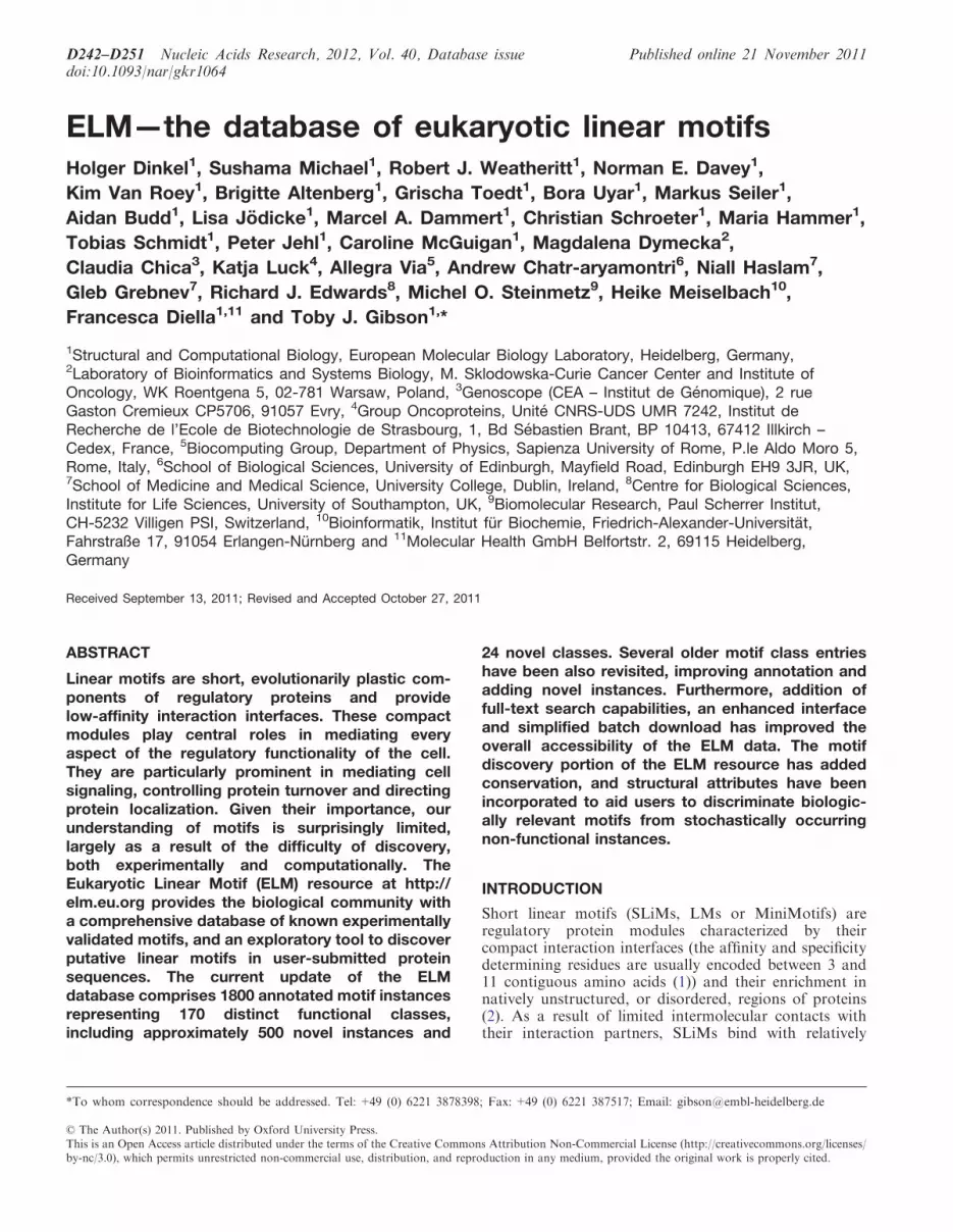
Figure 1. ELM start page. The user can submit a query sequence to the motif detection pipeline either as UniProt accession number or in FASTAformat. Filtering criteria such as taxonomic range or cellular compartment should be activated to limit the resulting list of SLiM instances.
D244 Nucleic Acids Research, 2012, Vol. 40, Database issue
protein structure annotated, giving more detailed informa-tion about the biological context of the respective motif.
NEW FEATURES
The ELM website at http://elm.eu.org can be used in twoways: first, as a front-end to explore the ELM database ofcurated ELM classes and instances, and second, to run themotif detection pipeline to detect putative SLiM instancesin query sequences. Both interfaces have been improvedwith the most notable changes listed below.
User interface
The database user interface, having been stable for manyyears, has been overhauled and replaced by a novelinterface introducing several new features (Figure 1).Up-to-date web technologies have been used to improvethe general user experience: the PYTHON frameworkDJANGO (http://www.djangoproject.com) dynamicallycreates and serves all HTML pages, while JavaScriptwas used to make the whole site more interactive andthus improve the user experience. In particular, theELM detail pages (Figure 3), which hold the most
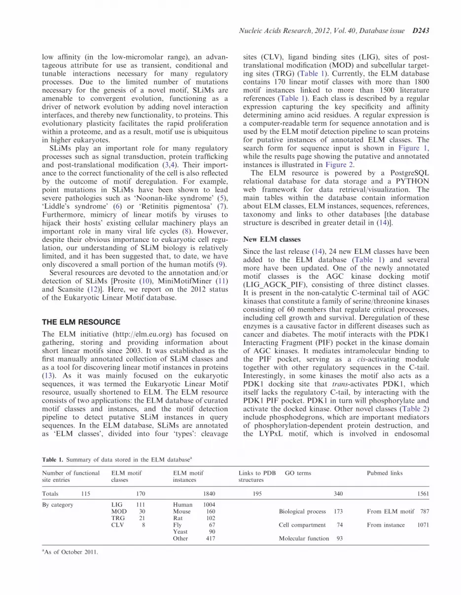
Figure 2. ELM motif detection pipeline output page. The top legend explains the different colors/symbols used. The graphical output of ELMconcentrates the output of multiple sequence classification algorithms; phosphorylation sites from Phospho.ELM, protein domains detected bySMART/Pfam, disorder predictions by GlobPlot and IUPred and secondary structure (18). The lower part contains the annotated and putativeELM instances for the given protein sequence (Epsin1, UniProt accession Q9Y6I3). The background is colored according to the structuralinformation available. Each box represents one ELM instance, the color of which indicates the likelihood that this instance is functional: greyinstances are buried within structured regions, while shades of blue represent instances outside of structured regions and hint on sequenceconservation, with pale blue representing weak sequence conservation and dark blue indicating strong sequence conservation. Red ellipses orboxes mark instances that are annotated in the query sequence or a homologous sequence, respectively.
Nucleic Acids Research, 2012, Vol. 40, Database issue D245

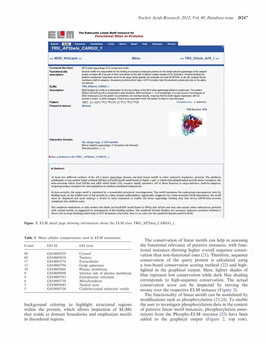
important information about each ELM class includingreferences, regular expression, taxonomic distributionand gene ontology terms (Table 3), have been updatedby annotating the protein domain interacting with the re-spective motif. Where available, a 3D model of represen-tative protein databank structures of linear motifinteractions was added to the ELM detail page (Figure3, top right).To cope with the increasing amount of annotated
classes as well as instances, a novel query interface wasintroduced to assist the user in finding information ofinterest. The ELM browser (Figure 4) now featuresa search interface for free text search. In addition, thesearch results can also be filtered and reordered usingbuttons (Figure 4, left side) and table headers, respective-ly, and be downloaded as tab-separated values (TSV).Further, improvements to the ELM database include
revising the experimental methods used for annotationby using a standardized methods vocabulary [in syncwith PSI-MI ontology (16,17)].A candidate page has been introduced to display novel
ELM classes that have not yet been annotated in detail orare currently undergoing annotation. We invite research-ers to send us their feedback and expert opinion on theseclasses and to contribute novel motif classes that will beadded to the candidate page and ultimately be turned intofull ELM classes (Figure 5). Minimum requirements are at
least one literature reference as well as a short descrip-tion. In addition, a draft regular expression or a 3Dstructure showing the relevant interaction would alsobe helpful. Currently, the number of possible ELMclasses on this candidate list (awaiting further annota-tion) exceeds the number of completely annotatedclasses, indicating the great demand for furtherannotation.
Graphical representation of sequence search
The ELM motif detection pipeline scans protein sequencesfor matches to the regular expressions of annotated ELMclasses (Figure 2). The query output combines theseputative instances with information from the database(annotated ELM instances) as well as predictions fromdifferent algorithms/filters. The ELM resource employs astructural filter (18) to highlight and mask secondarystructure elements, as well as SMART (19) to detectprotein domains. Furthermore, an additional disorderprediction algorithm (IUPred) (20) has been included topredict ordered/disordered regions within the protein.IUPred uses a cutoff of 0.5 to classify a sequence regionas either structured or disordered, with values above thisthreshold corresponding to disorder, highlighted in greenbackground and lower values indicating structuredregions, displayed in red background in the outputgraph. Disorder and domain information is combined by
Table 2. List of novel ELM classesa
Identifier Description
LIG_Actin_WH2_1 Motifs, present in proteins in several repeats, which mediate binding to the hydrophobic cleft created bysubdomains 1 and 3 of G-actinLIG_Actin_WH2_2
LIG_Actin_RPEL_3
LIG_AGCK_PIF_1 The AGCK docking motif mediates intramolecular interactions to the PDK1 Interacting Fragment (PIF) pocket,serving as a cis-activating moduleLIG_AGCK_PIF_2
LIG_AGCK_PIF_3
LIG_BIR_II_1 IAP-binding motifs are found in pro-apoptotic proteins and function in the abrogation of caspase inhibition byinhibitor of apoptosis proteins in apoptotic cellsLIG_BIR_III_1
LIG_BIR_III_2LIG_BIR_III_3LIG_BIR_III_4
LIG_eIF4E_1 Motif binding to the dorsal surface of eIF4ELIG_eIF4E_2
LIG_EVH1_3 A proline-rich motif binding to EVH1/WH1 domains of WASP and N-WASP proteins
LIG_HCF-1_HBM_1 The DHxY Host Cell Factor-1 binding motif interacts with the N-terminal kelch propeller domain of the cellcycle regulator HCF-1
LIG_Integrin_isoDGR_1 Present in proteins of extracellular matrix which upon deamidation forms biologically active isoDGR motif whichbinds to various members of integrin family
LIG_LYPXL_L_2 The LYPxL motif binds the V-domain of Alix, a protein involved in endosomal sortingLIG_LYPXL_S_1
LIG_PAM2_1 Peptide ligand motif that directly interacts with the MLLE/PABC domain found in poly(A) binding proteins andHYD E3 ubiquitin ligases
LIG_PIKK_1 Motif located in the C terminus of Nbs1 and its homologous interacting with PIKK family members
LIG_Rb_pABgroove_1 The LxxLFD motif binds in a deep groove between pocket A and pocket B of the Retinoblastoma protein
LIG_SCF_FBW7_1 The TPxxS phospho-dependent degron binds the FBW7 F box proteins of the SCF (Skp1-Cullin-Fbox) complexLIG_SCF_FBW7_2
LIG_SPAK-OSR1_1 SPAK/OSR1 kinase binding motif acts as a docking site which aids the interaction with their binding partnersincluding the upstream activators and the phosphorylated substrates
aAs of October 2011.
D246 Nucleic Acids Research, 2012, Vol. 40, Database issue
background coloring to highlight structured regionswithin the protein, which allows inspection of SLiMsthat reside at domain boundaries and emphasizes motifsin disordered regions.
The conservation of linear motifs can help in assessingthe functional relevance of putative instances, with func-tional instances showing higher overall sequence conser-vation than non-functional ones (21). Therefore, sequenceconservation of the query protein is calculated usinga tree-based conservation scoring method (22) and high-lighted in the graphical output. Here, lighter shades ofblue represent low conservation while dark blue shadingcorresponds to high-sequence conservation. The actualconservation score can be inspected by moving themouse over the respective ELM instance (Figure 2).The functionality of linear motifs can be modulated by
modifications such as phosphorylation (23,24). To enablethe user to investigate phosphorylation data in the contextof putative linear motif instances, phosphorylation anno-tations from the Phospho.ELM resource (25) have beenadded to the graphical output (Figure 2, top row).
Figure 3. ELM detail page showing information about the ELM class TRG_AP2beta_CARGO_1.
Table 3. Main cellular compartments used in ELM annotation
Count GO Id GO term
98 GO:0005829 Cytosol69 GO:0005634 Nucleus17 GO:0005576 Extracellular12 GO:0005794 Golgi apparatus10 GO:0005886 Plasma membrane9 GO:0009898 Internal side of plasma membrane9 GO:0005783 Endoplasmic reticulum6 GO:0005739 Mitochondrion5 GO:0005643 Nuclear pore5 GO:0045334 Clathrin-coated endocytic vesicle
Nucleic Acids Research, 2012, Vol. 40, Database issue D247
The phosphorylated residues are highlighted in differentcolors (serine: green, threonine: blue, tyrosine: red); eachphosphorylation site is linked to a page showing detailedinformation about the respective modification site fromthe manually curated data set of the Phospho.ELMresource.
VIRAL INSTANCES
The importance of the short linear motifs in virus–hostinteractions makes the ELM resource an important toolfor the viral research community. For example, Cruz et al.(26) analyzed a protein phosphatase 1 (PP1) docking motifin ‘protein 7’ of transmissible gastroenteritis virus usingthe ELM class LIG_PP1. This conserved sequence motifmediates binding to the PP1 catalytic subunit, a key
regulator of the cellular antiviral defense mechanisms,and is also found in other viral proteomes, suggestingthat it might be a recurring strategy to counteract thehosts’ defense against RNA viruses by dephosphorylatingeukaryotic translation initiation factor 2a and ultimatelyribonuclease L.
To reflect our increasing awareness of viral motifs (8),special focus has been attributed to the annotation of viralinstances in the ELM database: in the latest release, morethan 200 novel ELM instances found in 84 different viraltaxons have been added. The notion of viruses abusingexisting SLiMs in their hosts is demonstrated by viral in-stances being annotated alongside instances in their hosts’proteins. For example, the ELM class LIG_PDZ_Class_1contains 12 instances in human proteins but has recentlybeen expanded with 5 instances from 5 different humanpathogenic virus proteins.
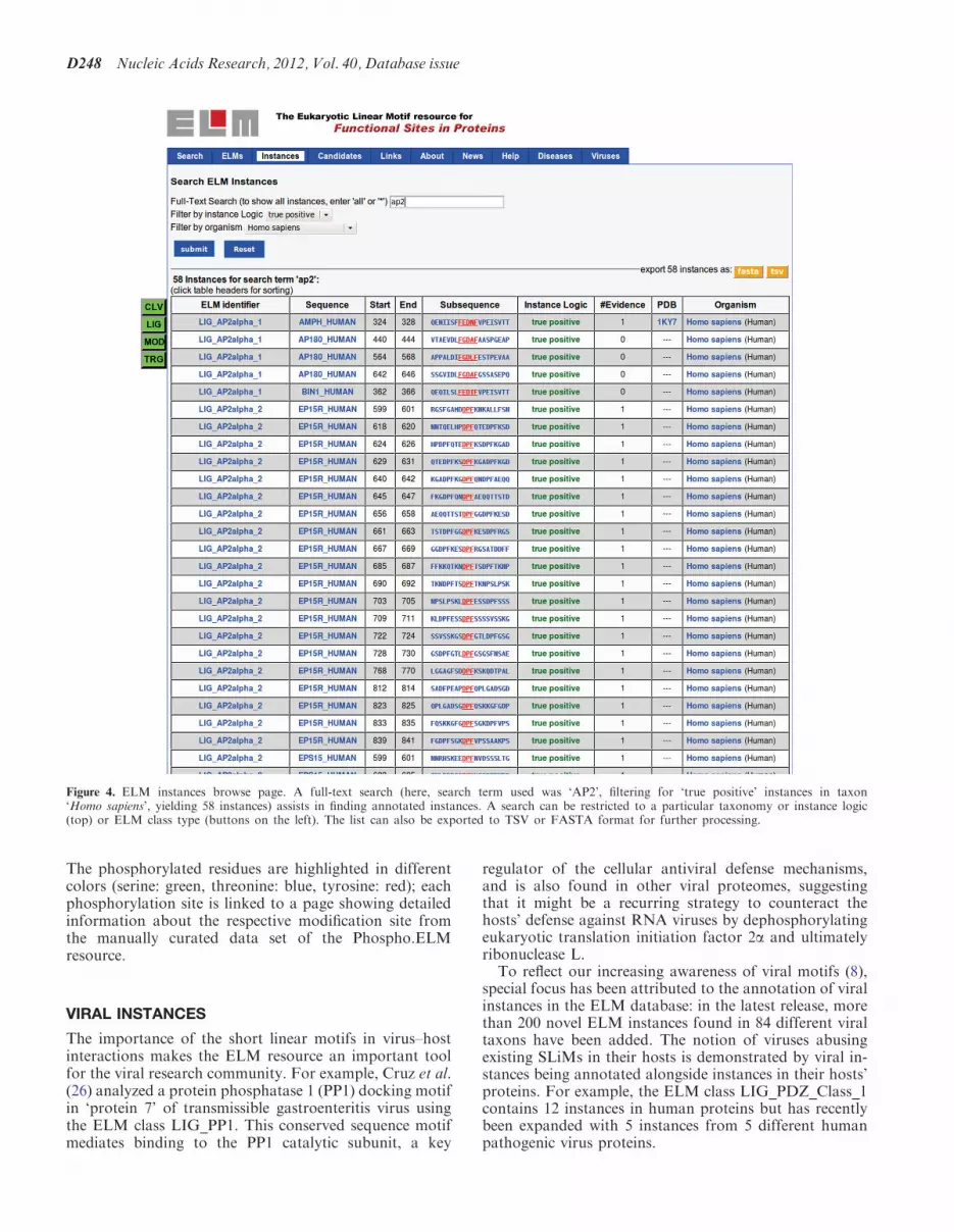
Figure 4. ELM instances browse page. A full-text search (here, search term used was ‘AP2’, filtering for ‘true positive’ instances in taxon‘Homo sapiens’, yielding 58 instances) assists in finding annotated instances. A search can be restricted to a particular taxonomy or instance logic(top) or ELM class type (buttons on the left). The list can also be exported to TSV or FASTA format for further processing.
D248 Nucleic Acids Research, 2012, Vol. 40, Database issue
LINEAR MOTIFS AND DISEASES
The importance of SLiMs is further corroborated by theoccurrence of pathologies that are caused by mutationsthat either mutate existing linear motifs or create novellinear motifs (of undesired function) (27). Examplesinclude ‘Usher’s syndrome’ (28), ‘Liddle’s Syndrome’ (6)or ‘Golabi-Ito-Hall Syndrome’ (29). The developmentaldisorder ‘Noonan Syndrome’ can be caused by mutationsin Raf-1 that abrogate the interaction with 14-3-3 proteinsmediated by corresponding SLiMs and thereby deregulatethe Raf-1 kinase activity (30) (the Raf-1 protein sequencefeatures two LIG_14-3-3_1 binding sites that areannotated at 256-261 and 618-623 in the ELM resource).A related disease, ‘Noonan-like Syndrome’, is caused byan S to G mutation at position 2 of the SHOC2 protein,creating a novel myristoylation site (annotated as ELMclass MOD_NMyristoyl). This irreversible modificationresults in aberrant targeting of SHOC2 to the plasmamembrane and impaired translocation to the nucleusupon growth factor stimulation (5). More informationabout the implication of short linear motifs on diseasesis collected at http://elm.eu.org/infos/diseases.html.
APPLICATION OF THE ELM RESOURCE
By providing a high-quality, manually curated data set oflinear motif classes with experimentally validated SLiMinstances, the ELM database has proven to be invaluableto the community: small-scale (single protein) analyzesbenefit from the detailed annotation of each ELM classin attributing novel features to proteins of interest.By using in vitro and in vivo studies, von Nandelstadhet al. (31) could validate a PDZ class III motif, detectedby ELM at the carboxy terminus of myotilin and theFATZ (calsarcin/myozenin) families. This evolutionarilyconserved carboxy-terminal motif mediates binding toPDZ domains of ZASP/Cypher and other Enigmafamily members (ALP, CLP-36 and RIL) and disruption
of these interactions results in myofibrillar myopathies(32). Additionally, ELM annotations can contribute tohigh-throughput screenings (33) as well as developmentof novel algorithms (34–36), methods (37) and databases(38). Furthermore, the highly curated data of the ELMresource are used as a benchmarking data set to evaluatethe accuracy of prediction algorithms (21,39,40).For any suchanalysis, the user shouldbe aware thatmany
matches to ELM regular expressions are false positives.Before conducting experiments based on ELM results, itis strongly advisable to check if a motif match is conserved,exposed in a cell compartment in which the motif is knownto be functional. The ELM resource applies several filtersto provide the user with such information that shouldideally also be supported by the experimental evidence.
SUMMARY
The importance of SLiMs is highlighted by the growingnumber of instances with relevance to diseases or viruses.Yet, despite their importance and abundance, our under-standing of linear motifs is still limited. This is mainlyowing to the fact that they are still quite difficult topredict computationally and to investigate experimentally(3,41,42). By better understanding the biology of linearmotifs, we hope to increase our insight into diseases andviruses (and vice versa). The ELM resource tries to aid theresearcher in the search for putative SLiM instances byproviding a feature-rich toolset for sequence analysis.Consequently, with the aforementioned additions andchanges, we hope that the ELM resource continues to bea valuable asset to the community.
ACKNOWLEDGEMENTS
The authors would like to thank the users of the ELMresource as well as all colleagues, contributors andannotators of the ELM resource.
FUNDING
EMBL international PhD program (to R.J.W.); EMBLInterdisciplinary PostDoc fellowship (EIPOD toN.E.D.); NGFN framework by the Federal GovernmentDepartment of Education and Science [FKZ01GS0862(DiGtoP) to M.S. and M.H.]; European Community’sSeventh Framework Programme FP7/2009 (SysCilia)(241955 to G.T.) and (SyBoSS) (242129 to K.V.R.);Polish Ministry of Science and Higher Education withinIuventus Plus project (IP2010-0483-70 to M.D.);Biotechnology and Biological Sciences Research Council(BB/F010486/1 to A.C.); Region Alsace and CollegeDoctoral Europeen (to K.L.); Science FoundationIreland (08/IN.1/B1864 to G.G.); BBSRC NewInvestigator Award (BB/I006230/1 to R.J.E.); GermanResearch Foundation (SFB796 Project A2 to H.M.);grants from the Swiss National Science Foundation (toM.O.S.). Funding for open access charge: EMBL.
Conflict of interest statement. None declared.
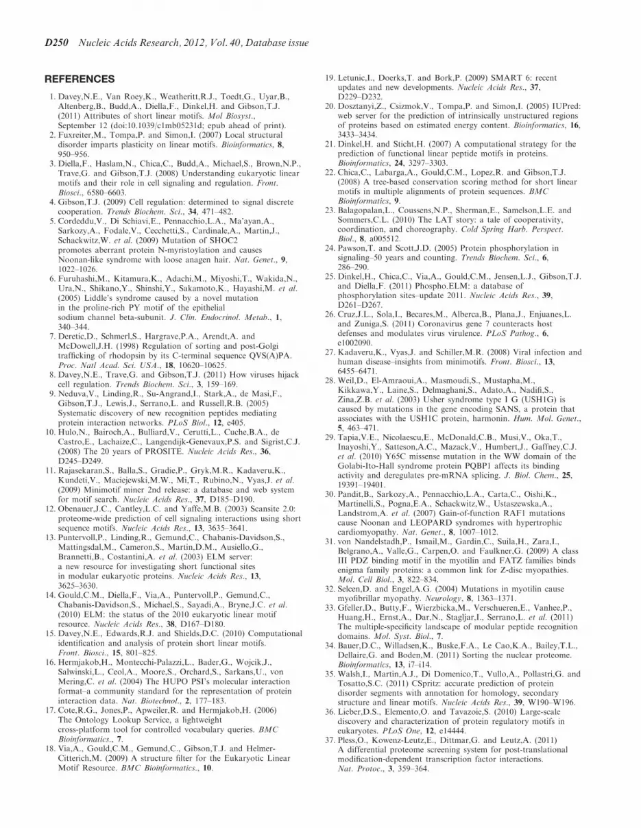
Figure 5. Schema of the ELM resource and data life cycle. AnnotatedELM classes, and instances thereof, can be searched by database query.Via sequence search by the motif detection pipeline, annotated ELMclasses yield putative instances in query sequences. By adding experi-mental evidence and references, these putative instances become candi-date instances for annotation, and, with further curation, ultimatelybecome fully annotated instances.
Nucleic Acids Research, 2012, Vol. 40, Database issue D249
REFERENCES
1. Davey,N.E., Van Roey,K., Weatheritt,R.J., Toedt,G., Uyar,B.,Altenberg,B., Budd,A., Diella,F., Dinkel,H. and Gibson,T.J.(2011) Attributes of short linear motifs. Mol Biosyst.,September 12 (doi:10.1039/c1mb05231d; epub ahead of print).
2. Fuxreiter,M., Tompa,P. and Simon,I. (2007) Local structuraldisorder imparts plasticity on linear motifs. Bioinformatics, 8,950–956.
3. Diella,F., Haslam,N., Chica,C., Budd,A., Michael,S., Brown,N.P.,Trave,G. and Gibson,T.J. (2008) Understanding eukaryotic linearmotifs and their role in cell signaling and regulation. Front.Biosci., 6580–6603.
4. Gibson,T.J. (2009) Cell regulation: determined to signal discretecooperation. Trends Biochem. Sci., 34, 471–482.
5. Cordeddu,V., Di Schiavi,E., Pennacchio,L.A., Ma’ayan,A.,Sarkozy,A., Fodale,V., Cecchetti,S., Cardinale,A., Martin,J.,Schackwitz,W. et al. (2009) Mutation of SHOC2promotes aberrant protein N-myristoylation and causesNoonan-like syndrome with loose anagen hair. Nat. Genet., 9,1022–1026.
6. Furuhashi,M., Kitamura,K., Adachi,M., Miyoshi,T., Wakida,N.,Ura,N., Shikano,Y., Shinshi,Y., Sakamoto,K., Hayashi,M. et al.(2005) Liddle’s syndrome caused by a novel mutationin the proline-rich PY motif of the epithelialsodium channel beta-subunit. J. Clin. Endocrinol. Metab., 1,340–344.
7. Deretic,D., Schmerl,S., Hargrave,P.A., Arendt,A. andMcDowell,J.H. (1998) Regulation of sorting and post-Golgitrafficking of rhodopsin by its C-terminal sequence QVS(A)PA.Proc. Natl Acad. Sci. USA., 18, 10620–10625.
8. Davey,N.E., Trave,G. and Gibson,T.J. (2011) How viruses hijackcell regulation. Trends Biochem. Sci., 3, 159–169.
9. Neduva,V., Linding,R., Su-Angrand,I., Stark,A., de Masi,F.,Gibson,T.J., Lewis,J., Serrano,L. and Russell,R.B. (2005)Systematic discovery of new recognition peptides mediatingprotein interaction networks. PLoS Biol., 12, e405.
10. Hulo,N., Bairoch,A., Bulliard,V., Cerutti,L., Cuche,B.A., deCastro,E., Lachaize,C., Langendijk-Genevaux,P.S. and Sigrist,C.J.(2008) The 20 years of PROSITE. Nucleic Acids Res., 36,D245–D249.
11. Rajasekaran,S., Balla,S., Gradie,P., Gryk,M.R., Kadaveru,K.,Kundeti,V., Maciejewski,M.W., Mi,T., Rubino,N., Vyas,J. et al.(2009) Minimotif miner 2nd release: a database and web systemfor motif search. Nucleic Acids Res., 37, D185–D190.
12. Obenauer,J.C., Cantley,L.C. and Yaffe,M.B. (2003) Scansite 2.0:proteome-wide prediction of cell signaling interactions using shortsequence motifs. Nucleic Acids Res., 13, 3635–3641.
13. Puntervoll,P., Linding,R., Gemund,C., Chabanis-Davidson,S.,Mattingsdal,M., Cameron,S., Martin,D.M., Ausiello,G.,Brannetti,B., Costantini,A. et al. (2003) ELM server:a new resource for investigating short functional sitesin modular eukaryotic proteins. Nucleic Acids Res., 13,3625–3630.
14. Gould,C.M., Diella,F., Via,A., Puntervoll,P., Gemund,C.,Chabanis-Davidson,S., Michael,S., Sayadi,A., Bryne,J.C. et al.(2010) ELM: the status of the 2010 eukaryotic linear motifresource. Nucleic Acids Res., 38, D167–D180.
15. Davey,N.E., Edwards,R.J. and Shields,D.C. (2010) Computationalidentification and analysis of protein short linear motifs.Front. Biosci., 15, 801–825.
16. Hermjakob,H., Montecchi-Palazzi,L., Bader,G., Wojcik,J.,Salwinski,L., Ceol,A., Moore,S., Orchard,S., Sarkans,U., vonMering,C. et al. (2004) The HUPO PSI’s molecular interactionformat–a community standard for the representation of proteininteraction data. Nat. Biotechnol., 2, 177–183.
17. Cote,R.G., Jones,P., Apweiler,R. and Hermjakob,H. (2006)The Ontology Lookup Service, a lightweightcross-platform tool for controlled vocabulary queries. BMCBioinformatics., 7.
18. Via,A., Gould,C.M., Gemund,C., Gibson,T.J. and Helmer-Citterich,M. (2009) A structure filter for the Eukaryotic LinearMotif Resource. BMC Bioinformatics., 10.
19. Letunic,I., Doerks,T. and Bork,P. (2009) SMART 6: recentupdates and new developments. Nucleic Acids Res., 37,D229–D232.
20. Dosztanyi,Z., Csizmok,V., Tompa,P. and Simon,I. (2005) IUPred:web server for the prediction of intrinsically unstructured regionsof proteins based on estimated energy content. Bioinformatics, 16,3433–3434.
21. Dinkel,H. and Sticht,H. (2007) A computational strategy for theprediction of functional linear peptide motifs in proteins.Bioinformatics, 24, 3297–3303.
22. Chica,C., Labarga,A., Gould,C.M., Lopez,R. and Gibson,T.J.(2008) A tree-based conservation scoring method for short linearmotifs in multiple alignments of protein sequences. BMCBioinformatics, 9.
23. Balagopalan,L., Coussens,N.P., Sherman,E., Samelson,L.E. andSommers,C.L. (2010) The LAT story: a tale of cooperativity,coordination, and choreography. Cold Spring Harb. Perspect.Biol., 8, a005512.
24. Pawson,T. and Scott,J.D. (2005) Protein phosphorylation insignaling–50 years and counting. Trends Biochem. Sci., 6,286–290.
25. Dinkel,H., Chica,C., Via,A., Gould,C.M., Jensen,L.J., Gibson,T.J.and Diella,F. (2011) Phospho.ELM: a database ofphosphorylation sites–update 2011. Nucleic Acids Res., 39,D261–D267.
26. Cruz,J.L., Sola,I., Becares,M., Alberca,B., Plana,J., Enjuanes,L.and Zuniga,S. (2011) Coronavirus gene 7 counteracts hostdefenses and modulates virus virulence. PLoS Pathog., 6,e1002090.
27. Kadaveru,K., Vyas,J. and Schiller,M.R. (2008) Viral infection andhuman disease–insights from minimotifs. Front. Biosci., 13,6455–6471.
28. Weil,D., El-Amraoui,A., Masmoudi,S., Mustapha,M.,Kikkawa,Y., Laine,S., Delmaghani,S., Adato,A., Nadifi,S.,Zina,Z.B. et al. (2003) Usher syndrome type I G (USH1G) iscaused by mutations in the gene encoding SANS, a protein thatassociates with the USH1C protein, harmonin. Hum. Mol. Genet.,5, 463–471.
29. Tapia,V.E., Nicolaescu,E., McDonald,C.B., Musi,V., Oka,T.,Inayoshi,Y., Satteson,A.C., Mazack,V., Humbert,J., Gaffney,C.J.et al. (2010) Y65C missense mutation in the WW domain of theGolabi-Ito-Hall syndrome protein PQBP1 affects its bindingactivity and deregulates pre-mRNA splicing. J. Biol. Chem., 25,19391–19401.
30. Pandit,B., Sarkozy,A., Pennacchio,L.A., Carta,C., Oishi,K.,Martinelli,S., Pogna,E.A., Schackwitz,W., Ustaszewska,A.,Landstrom,A. et al. (2007) Gain-of-function RAF1 mutationscause Noonan and LEOPARD syndromes with hypertrophiccardiomyopathy. Nat. Genet., 8, 1007–1012.
31. von Nandelstadh,P., Ismail,M., Gardin,C., Suila,H., Zara,I.,Belgrano,A., Valle,G., Carpen,O. and Faulkner,G. (2009) A classIII PDZ binding motif in the myotilin and FATZ families bindsenigma family proteins: a common link for Z-disc myopathies.Mol. Cell Biol., 3, 822–834.
32. Selcen,D. and Engel,A.G. (2004) Mutations in myotilin causemyofibrillar myopathy. Neurology, 8, 1363–1371.
33. Gfeller,D., Butty,F., Wierzbicka,M., Verschueren,E., Vanhee,P.,Huang,H., Ernst,A., Dar,N., Stagljar,I., Serrano,L. et al. (2011)The multiple-specificity landscape of modular peptide recognitiondomains. Mol. Syst. Biol., 7.
34. Bauer,D.C., Willadsen,K., Buske,F.A., Le Cao,K.A., Bailey,T.L.,Dellaire,G. and Boden,M. (2011) Sorting the nuclear proteome.Bioinformatics, 13, i7–i14.
35. Walsh,I., Martin,A.J., Di Domenico,T., Vullo,A., Pollastri,G. andTosatto,S.C. (2011) CSpritz: accurate prediction of proteindisorder segments with annotation for homology, secondarystructure and linear motifs. Nucleic Acids Res., 39, W190–W196.
36. Lieber,D.S., Elemento,O. and Tavazoie,S. (2010) Large-scalediscovery and characterization of protein regulatory motifs ineukaryotes. PLoS One, 12, e14444.
37. Pless,O., Kowenz-Leutz,E., Dittmar,G. and Leutz,A. (2011)A differential proteome screening system for post-translationalmodification-dependent transcription factor interactions.Nat. Protoc., 3, 359–364.
D250 Nucleic Acids Research, 2012, Vol. 40, Database issue

38. Goel,R., Muthusamy,B., Pandey,A. and Prasad,T.S. (2011)Human protein reference database and human proteinpedia asdiscovery resources for molecular biotechnology. Mol. Biotechnol.,1, 87–95.
39. Edwards,R.J., Davey,N.E. and Shields,D.C. (2007) SLiMFinder:a probabilistic method for identifying over-represented,convergently evolved, short linear motifs in proteins. PLoS One,10, e967.
40. Edwards,R.J., Davey,N.E. and Shields,D.C. (2008) CompariMotif:quick and easy comparisons of sequence motifs. Bioinformatics,10, 1307–1309.
41. Perkins,J.R., Diboun,I., Dessailly,B.H., Lees,J.G. and Orengo,C.(2010) Transient protein-protein interactions: structural,functional, and network properties. Structure, 10, 1233–1243.
42. Edwards,R.J., Davey,N.E., Brien,K.O. and Shields,D.C. (2011)Interactome-wide prediction of short, disordered proteininteraction motifs in humans. Mol. Biosyst., August 30(doi:10.1039/c1mb05212h; epub ahead of print).
Nucleic Acids Research, 2012, Vol. 40, Database issue D251


