Embed Size (px)
Citation preview
Current Biology 17, 293–303, February 20, 2007 ª2007 Elsevier Ltd All rights reserved DOI 10.1016/j.cub.2007.01.068
ArticleMultiple Protein PhosphatasesAre Required for Mitosis in Drosophila
Feng Chen,1,* Vincent Archambault,1 Ashok Kar,1
Pietro Lio’,2 Pier Paolo D’Avino,1 Rita Sinka,1,5
Kathryn Lilley,3 Ernest D. Laue,3 Peter Deak,1,6
Luisa Capalbo,1 and David M. Glover1,4,*1Cancer Research United KingdomCell Cycle Genetics Research GroupDepartment of GeneticsUniversity of CambridgeDowning StreetCambridge CB2 3EHUnited Kingdom2The Computer LaboratoryWilliam Gates Building15 JJ Thomson AvenueCambridge CB3 0FDUnited Kingdom3Department of BiochemistryUniversity of CambridgeTennis Court RoadCambridge CB2 3EHUnited Kingdom4Cyclacel LimitedBabraham Science ParkBabraham Hall, BabrahamCambridge CB2 4ATUnited Kingdom
Summary
Background: Approximately one-third of the Drosophilakinome has been ascribed some cell-cycle function.However, little is known about which of its 117 proteinphosphatases (PPs) or subunits have counteractingroles.Results: We investigated mitotic roles of PPs throughsystematic RNAi. We found that G2-M progression re-quires Puckered, the JNK MAP-kinase inhibitory phos-phatase and PP2C in addition to string (Cdc25). Strongmitotic arrest and chromosome congression failureoccurred after Pp1-87B downregulation. Chromosomealignment and segregation defects also occurred afterknockdown of PP1-Flapwing, not previously thoughtto have a mitotic role. Reduction of several nonreceptortyrosine phosphatases produced spindle and chromo-some behavior defects, and for corkscrew, prematurechromatid separation. RNAi of the dual-specificityphosphatase, Myotubularin, or the related Sbf ‘‘anti-phosphatase’’ resulted in aberrant mitotic chromosomebehavior. Finally, for PP2A, knockdown of the catalytic
*Correspondence: [email protected] (D.M.G.), fc273@
cam.ac.uk (F.C.)5 Present address: MRC Laboratory of Molecular Biology, Hills
Road, Cambridge CB2 2QH, UK.6 Present address: Institute of Biochemistry, Biological Research
Center, Temesvari krt. 62, Szeged H-6726, Hungary.
or A subunits led to bipolar monoastral spindles, knock-down of the Twins B subunit led to bridged and laggingchromosomes, and knockdown of the B0 Widerborstsubunit led to scattering of all mitotic chromosomes.Widerborst was associated with MEI-S332 (Shugoshin)and required for its kinetochore localization.Conclusions: We identify cell-cycle roles for 22 of 117Drosophila PPs. Involvement of several PPs in G2 sug-gests multiple points for its regulation. Major mitoticroles are played by PP1 with tyrosine PPs and Myotubu-larin-related PPs having significant roles in regulatingchromosome behavior. Finally, depending upon itsregulatory subunits, PP2A regulates spindle bipolarity,kinetochore function, and progression into anaphase.Discovery of several novel cell-cycle PPs identifiesa need for further studies of protein dephosphorylation.
Introduction
Together with cell-cycle-dependent transcription andubiquitin-mediated protein degradation, reversible pro-tein phosphorylation provides the cell with a capacityto coordinate progression through the cell divisioncycle. Levels of cellular protein phosphorylation arecontrolled both by protein kinases (PKs) and proteinphosphatases (PPs). The significance of PKs in cell-cycle progression has long been appreciated. In meta-zoans, the major phases of the cell cycle are set bya family of cyclin-dependent kinases (Cdks). Functionswithin each of these phases are also regulated by otherspecific protein kinases. Thus for example, Cdk1 andits cyclin B partner are required for entry into mitosisand to set the mitotic state by phosphorylating histoneH1 and chromatin-associated proteins to facilitate chro-mosome condensation, lamins to promote nuclear-envelope breakdown, and numerous other componentsof the mitotic apparatus [1]. In the mitotic cell, the Poloand Aurora-like kinases augment Cdk1 functions to reg-ulate centrosome maturation and separation, microtu-bule nucleation and dynamics, proper chromosomeattachment to the spindles, sister-chromatid separa-tion, and progression through anaphase into cytokinesis[2, 3]. Protein phosphorylation is also used in the surveil-lance checkpoints to ensure that the cell has fulfilled thenecessary requirements for proceeding to the next cell-cycle stage. Thus, the Chk1 and Chk2 kinases monitordefects in DNA replication and DNA damage before per-mitting mitotic entry, and the BubR1 kinase regulatesthe correct attachment and alignment of kinetochoreson the mitotic spindle before permitting anaphase[4, 5]. Therefore, it is logical to assume that PPs mustplay equally important reciprocal roles in the control ofcell division as PKs. PPs are typically classified into threestructurally distinct superfamilies: the PPP and PPM su-perfamilies that encode protein serine/threonine proteinphosphatases and the protein tyrosine phosphatase(PTP) superfamily comprising both tyrosine-specificand dual-specificity phosphatases (DSP) (Figure S1 in
Current Biology294
the Supplemental Data available online) [6]. However, incontrast to the extensive studies of cell-cycle PKs, littleis known of their antagonistic PPs. In Drosophila forinstance, only a few members of the PPP and PTP havebeen described for their cell-cycle roles, but none ofthe PPM family has been implicated in this process.Genetic evidence in Drosophila has indicated that PP1and PP2A (PPP) regulate spindle organization and sis-ter-chromatid segregation [7–9]; PP4 (PPP) has beenimplicated in centrosome maturation [10, 11]; and theDSP Cdc25/String triggers mitosis [12]. Moreover, it isbecoming clear that the diverse functions of a smallnumber of PPP catalytic subunits are accomplishedthrough interactions with a wide variety of regulatorysubunits [6], and thus reflects the need for further dissec-tion of their cell-cycle roles by study of their regulatorysubunits.
In an attempt to define a complete list of PPs and theirregulatory subunits required for cell-cycle progression,we used Drosophila melanogaster as a model systembecause of the following advantages. The annotatedsequence of the Drosophila genome allows us to identifyall of the predicted PPs. Moreover, the low level ofgenetic redundancy in this organism coupled with thevery high efficiency of RNA interference (RNAi) in S2cells facilitates the detection of functional requirementsfor the assayed gene products [13, 14]. Finally, the highdegree of evolutionary conservation of cell-cycle func-tions in metazoans indicates that our study would pro-vide a significant contribution to the understandingand treatment of proliferative diseases in man.
We surveyed cell-cycle functions of the 117 PPs ortheir regulatory subunits in the Drosophila genome bya combination of three different assays: flow cytometry,mitotic-index measurement, and quantitative assess-ment of mitotic defects. By using this combination ofindependent assays, we identified all the previouslyknown Drosophila cell-cycle PPs identified by classicalgenetics and thus validated our approach. We reportnovel cell-cycle functions for PP5 and PP6 (PP2A-like)of the PPP superfamily and for PP2C of the PPM super-family. In addition, we revealed cell-cycle roles for 11PTPs, consistent with the fact that tyrosine phosphory-lation plays a critical role in the control of a wide arrayof signaling pathways that are involved in the regulationof cell proliferation [15].
Results and Discussion
Global View of Protein Phosphatases Required
for Cell-Cycle ProgressionWe used RNAi to silence 117 known and predicted Dro-sophila genes encoding PPs or their regulatory subunitsin S2 cells. This was followed by flow cytometry for mon-itoring changes in DNA content and cell size, and mi-totic-index analysis for identifying the proportion of cellsin mitosis. We found that downregulation of 29 genes ledto a cell-cycle phenotype. Two recent reports havedemonstrated the possibility of sequence-dependentoff-target effects associated with long dsRNA inDrosophila [16, 17]. In order to minimize the number offalse positives that can potentially arise from suchoff-target effects, we re-examined the flow-cytometryprofiles and mitotic indices by using a second set of
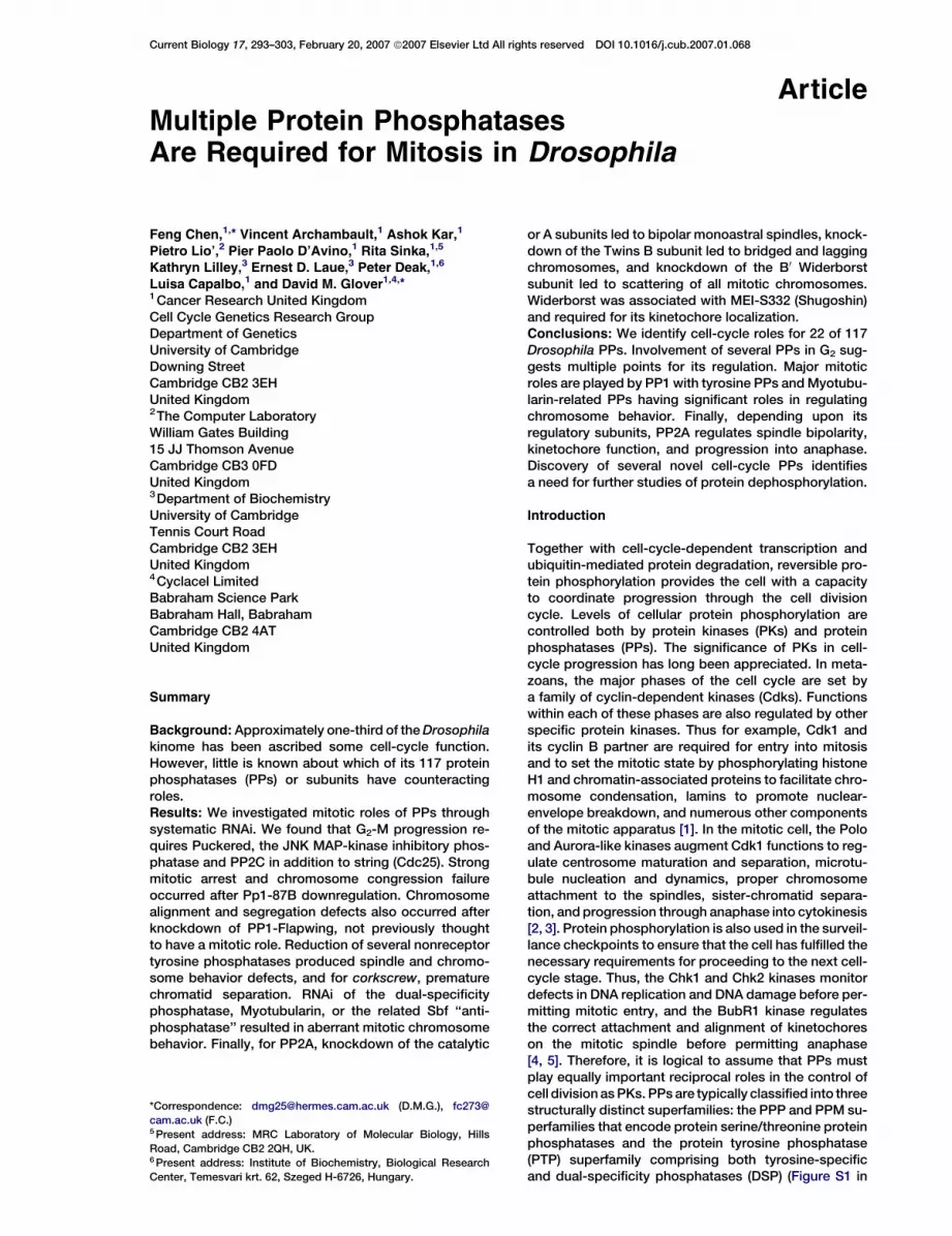
dsRNAs targeting a distinct region of each positive can-didate identified in our first screen. As shown in TableS1, we confirmed the phenotypic effects for the majorityof the positive candidates, although not for seven, whichwere therefore discarded from further analysis. In onecase (sbf), however, we observed similar flow-cytometryprofiles but not mitotic indices when two dsRNAs wereused. Thus in summary, depletion of 18 gene productsaltered the proportions of cells at different cell-cyclestages (in some cases associated with aneuploidy, celldeath, and increased cell size) as revealed by flowcytometry (Figure 1), and 11 showed abnormal mitoticindices (Table 1; see also Experimental Procedures). Intotal, 22 genes (19% of the genes tested) possessedcell-cycle functions by these criteria. We cannot be cer-tain to have identified all PP genes required for cell-cycleprogression in our survey because we did not, and inmost cases cannot, confirm that RNAi did indeed reducelevels of protein products as a result of the limitationof antibodies available. However, this appears notto be a major problem in cell-based RNAi screens inDrosophila [17].
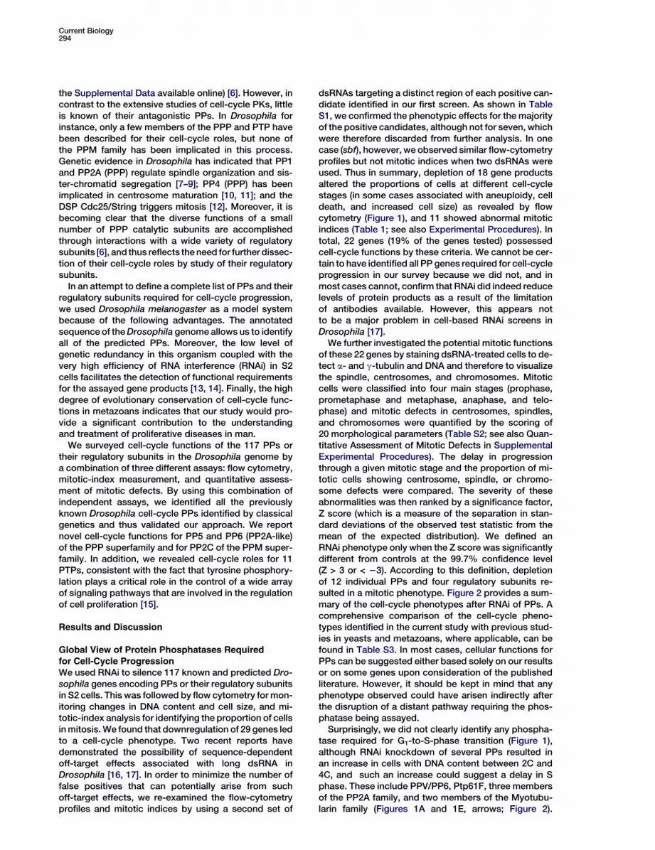
We further investigated the potential mitotic functionsof these 22 genes by staining dsRNA-treated cells to de-tect a- and g-tubulin and DNA and therefore to visualizethe spindle, centrosomes, and chromosomes. Mitoticcells were classified into four main stages (prophase,prometaphase and metaphase, anaphase, and telo-phase) and mitotic defects in centrosomes, spindles,and chromosomes were quantified by the scoring of20 morphological parameters (Table S2; see also Quan-titative Assessment of Mitotic Defects in SupplementalExperimental Procedures). The delay in progressionthrough a given mitotic stage and the proportion of mi-totic cells showing centrosome, spindle, or chromo-some defects were compared. The severity of theseabnormalities was then ranked by a significance factor,Z score (which is a measure of the separation in stan-dard deviations of the observed test statistic from themean of the expected distribution). We defined anRNAi phenotype only when the Z score was significantlydifferent from controls at the 99.7% confidence level(Z > 3 or < 23). According to this definition, depletionof 12 individual PPs and four regulatory subunits re-sulted in a mitotic phenotype. Figure 2 provides a sum-mary of the cell-cycle phenotypes after RNAi of PPs. Acomprehensive comparison of the cell-cycle pheno-types identified in the current study with previous stud-ies in yeasts and metazoans, where applicable, can befound in Table S3. In most cases, cellular functions forPPs can be suggested either based solely on our resultsor on some genes upon consideration of the publishedliterature. However, it should be kept in mind that anyphenotype observed could have arisen indirectly afterthe disruption of a distant pathway requiring the phos-phatase being assayed.
Surprisingly, we did not clearly identify any phospha-tase required for G1-to-S-phase transition (Figure 1),although RNAi knockdown of several PPs resulted inan increase in cells with DNA content between 2C and4C, and such an increase could suggest a delay in Sphase. These include PPV/PP6, Ptp61F, three membersof the PP2A family, and two members of the Myotubu-larin family (Figures 1A and 1E, arrows; Figure 2).
Protein Phosphatases in Mitotic Progression295
Figure 1. Proportion of Cells at Different Cell-Cycle Stages after RNAi of PPs
Changes in the proportion of cells with subdiploid (cell death), 2C (G1 phase), 4C (G2/M phase), and intermediate DNA content between 2C and 4C
(S phase or aneuploidy) and in cell size were identified by flow cytometry after downregulation of 18 PPs. Defects fall into five broad clusters
(A–E). Each panel shows a control cell histogram (black, cells transfected with dsRNA for green fluorescent protein [GFP]) and an example of
an experimental histogram (red) after RNAi for a representative protein phosphatase (the specific protein phosphatase illustrated is indicated
in red lettering; the asterisk indicates a weak RNAi phenotype for PpD3). The forward light scatter (FSC) profile reflects cell size. Phenotypes
are clustered as follows:
(A) RNAi leading to an increase incellswith intermediate DNA content (indicated by arrows) associated withan increase in 4C cells (left) or not (right).
(B) RNAi leading to an increase in 4C cells associated with an increase in cell size. This group has been subdivided according to the extent of
accumulation of 4C cells (left versus right).
(C) RNAi leading to an increase in 4C cells without affecting cell size.
(D) RNAi leading to an increase in subdiploid cells (indicated by arrowhead).
(E) RNAi leading to an increase in 4C cells and in cells intermediate between 2C and 4C (indicated by arrows), and in cell size associated with an
increase in subdiploid cells (left, indicated by arrowhead) or not (right).
However, because it is difficult to distinguish S phasedelay from aneuploidy solely based on flow-cytometryprofiles, further investigation is required so that the po-tential S phase roles of these PPs can be defined. On theother hand, downregulation of many PPs led to a pro-longed G2 phase or mitotic defects, indicating a strongrequirement for PPs in regulating progression into andthrough mitosis.
Mitotic Entry
In the cell cycle, G2 is a pivotal phase at which mitoticentry may be delayed until DNA is correctly replicatedand repaired and until an appropriate cell size isreached. The G2-M transition requires removal of theinhibitory phosphorylation of Cyclin-dependent kinase1 (CDK1) by the tyrosine phosphatase, Cdc25 (Stringin Drosophila somatic cells and Cdc25C in vertebrates),whose activity is opposed by the Wee1 and Myt1 proteinkinases. The regulation of this inhibitory phosphoryla-tion has been thought to be essential in governing mi-totic entry, and its regulators (Cdc25, Wee1, and Myt1)are tightly modulated by a wide range of pathwayssuch as MAP-kinase and DNA replication and repaircheckpoint kinase pathways [18].
Cells accumulating with a 4C DNA content canrepresent G2 phase cells, mitotic cells, or binucleatedG1 cells that have failed cytokinesis. However, defective
Table 1. RNAi of PPs Leading to Abnormal Mitotic Indices
K-S Value
Gene Name First Screen Second Screen
Pp1-87B 1.00000 1.00000
flw 0.77612 0.85341
mts 1.00000 1.00000
Pp2A-29B 1.00000 1.00000
wdb 0.71416 0.71334
Pp4-19C 0.53333 0.78596
PPP4R2r 0.44167 0.83728
PPV 0.88095 1.00000
CG9311 0.56429 0.62955
csw 0.69523 0.70126
ssh 0.44318 0.65575
Mitotic indices were analyzed with the Kolmogorov-Smirnov (K-S)
test (see Supplemental Experimental Procedures). Candidate genes
whose downregulation showed significantly different distributions
from GFP RNAi controls at the 0.01 level in both screens involving
distinct dsRNAs are listed. The corresponding average K-S values
are also shown. The average x-fold change in mitotic index after
RNAi of these genes can be found in Figure 2.
Current Biology296
Figure 2. Cell-Cycle Functions of PPs Identified in Current Study Grouped by Families
**Indicates novel cell-cycle regulators.
*Indicates novel Drosophila cell-cycle regulators whose counterparts in other organisms have been implicated in the cell-cycle control (see Table
S3 for details).
BIndicates an increase in cell size.
yIncreases (+) or decreases (2) in cell death or the proportion of cells in a given phase of the cell cycle are shown when flow-cytometric data
are significantly different from control values.
zAverage x-fold change in the mitotic index is shown when mitotic-index data are significantly different from control values.
xDefects in mitotic progression and morphology were quantified and ranked by Z score (see Supplemental Experimental Procedures). A Z score
of three or more standard deviations from the mean was used as a threshold for selecting positive candidates. ‘‘Centrosome Def,’’ ‘‘Spindle Def,’’
and ‘‘Chromosome Def’’ refers to the percentage of mitotic cells showing defects in centrosome number or positioning, spindle assembly, and
chromosome condensation, alignment, or segregation, respectively.
cytokinesis often also results in an increase in polyploidcells with DNA content of 8C arising from re-entry ofthe binucleated cells into the cell cycle. Consistentwith its mutant phenotype [12], string/Cdc25 RNAi ledto a 50% increase in 4C cells with no increase in themitotic index or 8C cells, suggesting a pronounced G2 ar-rest (Figures 1B and 2). These cells showed an increasein size in anticipation of a delayed mitotic entry, reminis-cent of the cdc25 mutant phenotype in fission yeast [19].
Entry into mitosis can be delayed by activation ofstress-response pathways [20, 21]. Two classes ofstress-activated protein kinases (SAPK) have been iden-tified: c-JUN amino-terminal kinases (JNKs) and p38MAPKs. They are each returned to their inactivated statethrough dephosphorylation by MAPK phosphatases(MKP). RNAi knockdown of Puckered (Puc), the singlephosphatase known to inhibit Drosophila JNK MAP-kinase [22–24], also led to a severe accumulation (33%)
Protein Phosphatases in Mitotic Progression297
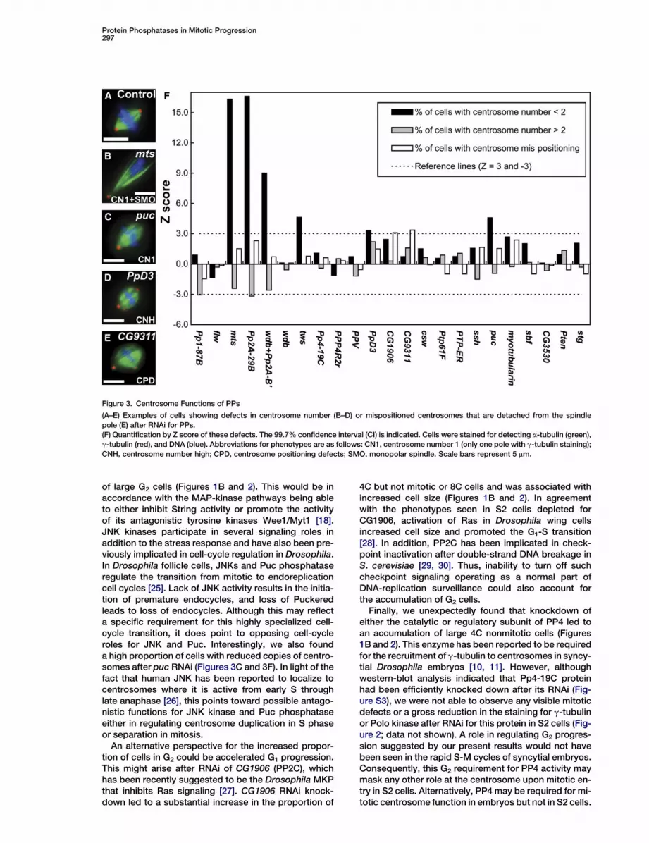
Figure 3. Centrosome Functions of PPs
(A–E) Examples of cells showing defects in centrosome number (B–D) or mispositioned centrosomes that are detached from the spindle
pole (E) after RNAi for PPs.
(F) Quantification by Z score of these defects. The 99.7% confidence interval (CI) is indicated. Cells were stained for detecting a-tubulin (green),
g-tubulin (red), and DNA (blue). Abbreviations for phenotypes are as follows: CN1, centrosome number 1 (only one pole with g-tubulin staining);
CNH, centrosome number high; CPD, centrosome positioning defects; SMO, monopolar spindle. Scale bars represent 5 mm.
of large G2 cells (Figures 1B and 2). This would be inaccordance with the MAP-kinase pathways being ableto either inhibit String activity or promote the activityof its antagonistic tyrosine kinases Wee1/Myt1 [18].JNK kinases participate in several signaling roles inaddition to the stress response and have also been pre-viously implicated in cell-cycle regulation in Drosophila.In Drosophila follicle cells, JNKs and Puc phosphataseregulate the transition from mitotic to endoreplicationcell cycles [25]. Lack of JNK activity results in the initia-tion of premature endocycles, and loss of Puckeredleads to loss of endocycles. Although this may reflecta specific requirement for this highly specialized cell-cycle transition, it does point to opposing cell-cycleroles for JNK and Puc. Interestingly, we also founda high proportion of cells with reduced copies of centro-somes after puc RNAi (Figures 3C and 3F). In light of thefact that human JNK has been reported to localize tocentrosomes where it is active from early S throughlate anaphase [26], this points toward possible antago-nistic functions for JNK kinase and Puc phosphataseeither in regulating centrosome duplication in S phaseor separation in mitosis.
An alternative perspective for the increased propor-tion of cells in G2 could be accelerated G1 progression.This might arise after RNAi of CG1906 (PP2C), whichhas been recently suggested to be the Drosophila MKPthat inhibits Ras signaling [27]. CG1906 RNAi knock-down led to a substantial increase in the proportion of
4C but not mitotic or 8C cells and was associated withincreased cell size (Figures 1B and 2). In agreementwith the phenotypes seen in S2 cells depleted forCG1906, activation of Ras in Drosophila wing cellsincreased cell size and promoted the G1-S transition[28]. In addition, PP2C has been implicated in check-point inactivation after double-strand DNA breakage inS. cerevisiae [29, 30]. Thus, inability to turn off suchcheckpoint signaling operating as a normal part ofDNA-replication surveillance could also account forthe accumulation of G2 cells.
Finally, we unexpectedly found that knockdown ofeither the catalytic or regulatory subunit of PP4 led toan accumulation of large 4C nonmitotic cells (Figures1B and 2). This enzyme has been reported to be requiredfor the recruitment of g-tubulin to centrosomes in syncy-tial Drosophila embryos [10, 11]. However, althoughwestern-blot analysis indicated that Pp4-19C proteinhad been efficiently knocked down after its RNAi (Fig-ure S3), we were not able to observe any visible mitoticdefects or a gross reduction in the staining for g-tubulinor Polo kinase after RNAi for this protein in S2 cells (Fig-ure 2; data not shown). A role in regulating G2 progres-sion suggested by our present results would not havebeen seen in the rapid S-M cycles of syncytial embryos.Consequently, this G2 requirement for PP4 activity maymask any other role at the centrosome upon mitotic en-try in S2 cells. Alternatively, PP4 may be required for mi-totic centrosome function in embryos but not in S2 cells.
Current Biology298
Centrosome Duplication, Maturation, and SeparationThe centrosome is the major microtubule-organizingcenter (MTOC) and thus plays a critical role during mito-sis where it contributes to spindle bipolarity, spindlepositioning, chromosome segregation, and cytokinesis.Centrosomal roles for PPs could be expected to coun-teract the large number of PKs that regulate centrosomefunctions [31, 32]. RNAi in Drosophila S2 cells has previ-ously revealed the respective centrosomal roles of thePolo and SAK/Plk4 protein kinases. However, a quantita-tive approach has proved to be important in such exper-iments because some 20%–30% of control mitotic S2cells show centrosomal defects (see also [33]).
Aberration of centrosome number was found afterdownregulation of PpD3/PP5. Its RNAi resulted in notonly an increase in cells with reduced centrosome num-bers but also a marginal increase (Z = 2.2) in the numberof cells with extra centrosomes (Figures 3D and 3F) andthus was possibly indicative of a role in centrosomeseparation in mitosis.
The localization of proteins at the centrosome hasbeen taken as an indicator of their potential function.PTP-BL, a human phosphatase closely related to flyCG9311, has been reported to be highly enriched atcentrosomes during early mitosis [34]. We found thatwhen CG9311 was downregulated, cells had misposi-tioned centrosomes detached from spindle poles,suggesting the possibility of a conserved relationshipbetween the localization and function of this enzyme(Figures 3E and 3F).
Mitotic Functions of PP1 IsoformsEvidence from both yeast and animal cells suggeststhat PP1 phosphatases oppose both the Aurora A- andB-type kinases [35]. This would implicate PP1 in regulat-ing a range of processes including the establishment ofthe spindle, microtubule-kinetochore interactions, themitotic phosphorylation of histone H3 and its centro-meric counterpart, CENP-A (CID in Drosophila), andcytokinesis.
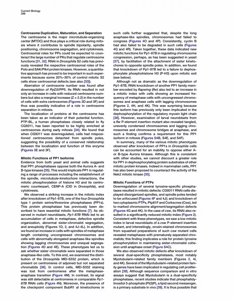
We observed a striking increase in the mitotic indexafter knockdown of Pp1-87B, one of the four Drosophilatype 1 protein serine/threonine phosphatases (PP1s).This protein phosphatase has previously been de-scribed to have essential mitotic functions [7]. As ob-served in mutant neuroblasts, Pp1-87B RNAi led to anaccumulation of cells in metaphase, defective spindleorganization, abnormal sister-chromatid segregation,and aneuploidy (Figures 1D, 2, and 4J–4L). In addition,we found an increase in cells with spindles of metaphaselength containing uncongressed chromosomes andcells with abnormally elongated anaphase-like spindlesshowing lagging chromosomes and unequal segrega-tion (Figures 4D and 4E). These phenotypes led us toask whether sister chromatids were separated in thoseanaphase-like cells. To this end, we examined the distri-bution of the Drosophila MEI-S332 protein, which ispresent on centromeres of adjoined but not separatedchromatids [36]. In control cells, MEI-S332 stainingwas lost from centromeres after the metaphase-anaphase transition (Figure 4M). In contrast, its signalwas still detectable at centromeres in comparable Pp1-87B RNAi cells (Figure 4N). Moreover, the presence ofthe checkpoint component BubR1 at kinetochores in
such cells further suggested that, despite the longanaphase-like spindles, chromosomes had failed tocongress (Figures 4O and 4P). Consistently, cyclin Bhad also failed to be degraded in such cells (Figures4Q and 4R). Taken together, these data indicated newmitotic functions for Pp1-87B in regulating chromosomecongression, perhaps, as has been suggested in yeast[37], by facilitation of the attachment of sister kineto-chores to opposite spindle poles. In addition, we foundthat knockdown of Pp1-87B led to a failure to dephos-phorylate phosphohistone H3 (P-H3) upon mitotic exit(see below).
Although not as dramatic as the downregulation ofPp1-87B, RNAi knockdown of another PP1 family mem-ber encoded by flapwing (flw) also led to an increase ina mitotic index with cells showing an increased fre-quency of metaphase cells with uncongressed chromo-somes and anaphase cells with lagging chromosomes(Figures 2, 4H, and 4K). This was surprising becausethis isoform has previously only been implicated in thedephosphorylation of the regulatory myosin light chain[38]. However, examination of larval neuroblasts froma flw P-element insertion mutant also revealed tangled,unevenly condensed chromosomes and lagging chro-mosomes and chromosome bridges at anaphase, andsuch a finding confirms a requirement for this PP1isoform in mitosis (Figures S4B, S4E, and S4F).
In summary, many of the various mitotic phenotypesobserved after knockdown of PP1s in Drosophila cellscan be accounted for an inability to oppose either A-or B-type Aurora kinases. Although this is consistentwith other studies, we cannot discount a greater rolefor PP1 in dephosphorylating protein substrates of othermitotic protein kinases. Indeed in mammalian cells, PP1has also been proposed to counteract the activity of theNek2 mitotic kinase [35].
Mitotic Functions of PTPsDownregulation of several tyrosine-specific phospha-tases resulted in mitotic defects: CG9311 RNAi cells dis-played disorganized spindles, and spindle poles tendedto be unfocused (Figures 4F and 4J); and knockdown oftwo cytoplasmic PTPs, Ptp61F and Corkscrew (Csw), ledto marked chromosome alignment/segregation defects(Figures 4G and 4K). In the case of csw, its RNAi also re-sulted in a significantly reduced mitotic index (Figure 2).Consistent with these phenotypes, we saw a low mitoticindex in larval neuroblasts of a csw P-element insertionmutant, and interestingly, orcein-stained chromosomesfrom squashed preparations of such csw mutant cellsrevealed metaphases with prematurely separated chro-matids; this finding implicates a role for protein tyrosinephosphorylation in maintaining sister-chromatid cohe-sion until anaphase onset (Figure S4C).
We also observed mitotic defects after knockdown ofseveral dual-specificity phosphatases, most notablyMyotubularin-related family members (Figures 2, 4I,and 4K). Several of the Myotubularin-related protein fam-ily genes have been implicated in regulating cell prolifer-ation [39]. Although sequence comparison and in vitroassays suggest that Myotubularin is a dual-specificityphosphatase, recent studies indicate that phosphatidy-linositol 3-phosphate (PI(3)P), a lipid second messenger,is a primary substrate in vivo [39]. It is thus possible that
Protein Phosphatases in Mitotic Progression299
Figure 4. Mitotic Defects after RNAi for PPs
(A–I) Examples of cells showing defects in spindle assembly or chromosome behavior after RNAi for PPs. Cells were stained for detecting
a-tubulin (green), g-tubulin (red), and DNA (blue). Abbreviations for phenotypes are as follows: AS, abnormal spindle; SBR, branched spindle;
CRSD, chromosome segregation defect; CRLC, lagging chromosomes; UD, unevenly divided DNA.
(J) Quantification by Z score of the percentage of mitotic cells showing spindle defects.
(K) Quantification by Z score of the percentage of mitotic cells showing chromosome defects.
(L) Quantification by Z score of the proportion of prometaphase and metaphase cells relative to the number of total mitotic cells. Indicated
are 99.7% confidence intervals (CI).
(M–R) Pp1-87B RNAi led to defects in chromosome congression. Cells were stained for detecting a-tubulin (green), DNA (blue), and indicated
proteins (red). In control cells, MEI-S332 (M) and BubR1 (O) dissociated from the kinetochores, and cyclin B (Q) was degraded after anaphase
onset. In comparable Pp1-87B RNAi cells, however, MEI-S332 (N) and BubR1 (P) remained associated with the centromere or kinetochore
moving toward the spindle poles and cyclin B (R) was not degraded. Scale bars represent 5 mm.
Myotubularin-related proteins may regulate the cell divi-sion cycle via lipid-mediated signaling pathways. Nota-bly, RNAi of SET domain binding factor (sbf), whichencodes a Myotubularin-related but catalytically inac-tive ‘‘antiphosphatase,’’ also resulted in defects in segre-gation and lagging chromosomes at anaphase (Figures 2and 4K). As has been suggested in mammalian cells, thisprotein may function by binding to and protecting sub-strates from dephosphorylation by Myotubularin-relatedphosphatases [40]. Taken together, these findings pointtoward cooperation between Myotubularin-relatedproteins in the control of cell-cycle progression.
PP2A Displays Multiple Mitotic RolesThe Catalytic and A Subunits
PP2A is a heterotrimeric serine/threonine phosphatasecomposed of invariant catalytic (C) and structural (A)
subunits together with one member of a family of B reg-ulatory subunits thought to direct the AC core to differ-ent substrates [41]. The Drosophila gene for the catalyticsubunit of type 2A protein serine/threonine phosphatase(PP2A) is known as microtubule star (mts) becausemutant embryos show multiple individual centrosomeswith disorganized astral arrays of microtubules [8]. Inagreement with this mutant phenotype, we found thatS2 cells depleted for Mts (PP2A-C) displayed aberrantelongated arrays of microtubules with a high proportion(5- to 10-fold increase over the control) of bipolar mono-astral spindles or monopolar spindles emanating froma single centrosomal mass (Figure 3B). This phenotypeis also consistent with the observations in Xenopusegg extracts where mitotic microtubules grow longerand bipolar spindles can not be assembled after inhibi-tion of PP2A by low concentrations of okadaic acid (OA)
Current Biology300
[42]. We speculate that the monopolar spindle pheno-type after mts dsRNA treatment is a consequence ofthe spindle collapse rather than a failure in centrosomeduplication or separation because most of the RNAi-treated cells showed well-separated centrosomesduring prophase (data not shown). In support of thisview, spindle bipolarity can be rescued by restorationof microtubule dynamics in OA-treated Xenopus eggextracts [42].
In Drosophila, as in many other eukaryotes, mitosis-specific phosphorylation of histone H3 requires AuroraB activity, but the identity of the opposing phosphataseremains unclear [43, 44]. Because P-H3 (Ser 10) levelswere used for monitoring the mitotic index in our analy-sis, it is possible that a high mitotic index observed afterRNAi for PPs may also reflect a defect in dephosphory-lating P-H3 in the absence of PPs upon mitotic exit. Wetherefore examined the phosphorylation state of thishistone after RNAi for PPs that displayed a significantincrease in the mitotic index in our screen. The immu-nostaining of control cells showed that P-H3 signalsbegan to decrease at early telophase and thendisappeared completely at late telophase (FiguresS5A–S5C). After RNAi knockdown of Mts (PP2A-C) orPp1-87B, however, the majority of mitotic cells werearrested at prometaphase (Figure 4L), but we couldoccasionally found late telophase figures showing anabnormal accumulation of P-H3 on decondensedchromosomes (Figures S5D and S5E). To better assessthe effect of depletion of these two PPs on P-H3 de-phosphorylation, we inactivated the spindle-assemblycheckpoint by simultaneously knocking down BubR1.We found that this rescued the prometaphase arrest ofcells simultaneously depleted for Mts or Pp1-87B andallowed us to study telophase cells (Figure S5F). P-H3was present in the majority of such telophase cellscompared to control cells (Figure S5G), indicating thatboth PPs are required for P-H3 dephosphorylation.These results are in accordance with previous studiesshowing that reduction of PP1 activity can partiallysuppress defects in the mitotic histone H3 phosphoryla-tion in yeast and C. elegans [45].
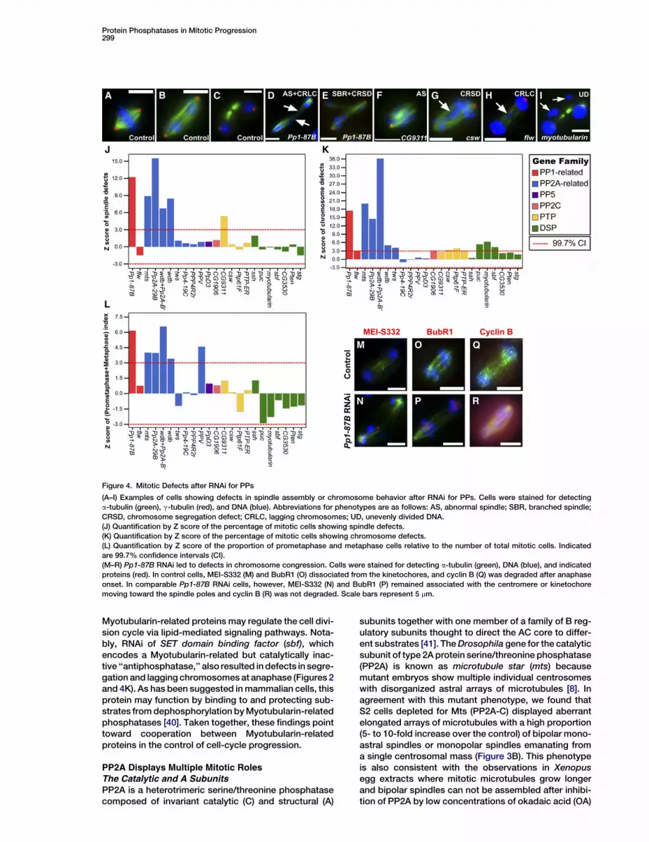
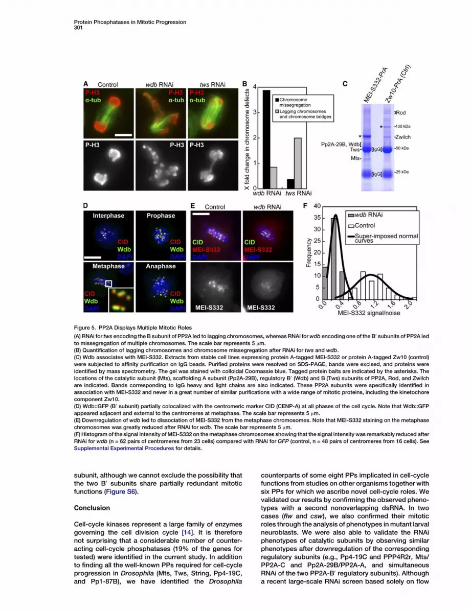
Downregulation of Pp2A-29B, the structural Asubunit, revealed almost identical aberrant phenotypesto those observed after mts (PP2A-C) RNAi (Figure 2and Figure S6). Consistently, knockdown of Pp2A-29B(PP2A-A) led to a reduction of the protein level of Mts(PP2A-C) (Figure S6A; see also [46, 47]).The B SubunitsThe Drosophila genome contains 4 B-type PP2A regula-tory subunits, twins/tws/aar (B sub-type), widerborst/wdb (B0 sub-type), Pp2A-B0 (B0 sub-type), and Pp2A-B00 (B00 sub-type), but mitotic defects have so far onlybeen reported for mutants of tws. Consistent with thephenotype of tws mutants [9], we observed that RNAifor this gene led to an increased proportion of anaphasefigures showing lagging chromosomes and chromo-some bridges (Figures 5A and 5B).
In metazoans, the B0 regulatory subunits of PP2A haveevolved into two related subclasses with conservedcentral regions and diverged amino and carboxy termini.The protein encoded by widerborst (wdb) is moreclosely related to the human a and 3 subunits (79%–80% identity) than to the b, g, or d subunits (69%–75%
identity). Whereas RNAi for tws led to lagging chromo-somes, wdb RNAi led to dramatic scattering of chromo-somes throughout the spindle (Figures 5A and 5B). Weconsidered whether this dramatic effect of wdb RNAion chromosome segregation reflected any particularsubcellular localization of this regulatory subunit. Tothis end, we expressed a GFP-tagged Wdb in S2 cells(Figure 5D). During interphase and prophase, Wdb::GFPpartially colocalized with the centromeric marker CID(CENP-A). After spindle formation, Wdb::GFP was foundadjacent and external to the centromeres. Although lesspronounced, this distribution remained during chromo-some segregation at anaphase. Because MEI-S332(Drosophila Shugoshin) is a dynamic centromericmarker [36], we examined its distribution in wdb RNAicells. In control cells, MEI-S332 localized in a band be-tween each pair of the centromeres at metaphase(Figure 5E). After downregulation of wdb, however, wefound greatly reduced MEI-S332 staining on the meta-phase chromosome (Figures 5E and 5F). In contrast,depletion of MEI-S332 by RNAi did not affect the normallocalization of the Wdb B0 PP2A subunit (Figure S7).Thus, we conclude that the Wdb B0 subunit is requiredfor correct localization of MEI-S332 but not vice versa.We next asked whether the two proteins existed in thesame complex. To address this, we expressed a ProteinA (PrA)-tagged form of MEI-S332 in S2 cells to purify po-tential protein complexes and identify its componentsby mass spectrometry. As shown in Figure 5C, we iden-tified the catalytic C (Mts), the structural A (PP2A-29B),and the regulatory B0 (Wdb) and B (Tws) subunits ofPP2A associated with MEI-S332. Three recent studiesalso identified PP2A complexed to the B0 subunit boundto Shugoshin (Sgo) in human and yeast cells, where theyare thought to protect centromeric cohesin subunitsfrom phosphorylation that would promote prematuresister-chromatid separation [48–50]. As with the arche-typal family member, Drosophila MEI-S332, the Shu-goshins function primarily to protect sister chromatidsfrom separation in the first meiotic division but are alsopresent in mitotic divisions [51]. Consistent with ourobservations in Drosophila S2 cells, Tang et al. [50]and Kitajima et al. [48]. both found that depletion ofPP2A in human cells led to premature dissociation ofShugoshin 1 (Sgo1) from the kinetochore and loss of mi-totic centromere cohesion. The finding of Shugoshincomplexed to PP2A/B0 in yeast and human, and now inDrosophila, points to a highly evolutionally conservedrole for this particular PP2A heterotrimer in regulatingsister-chromatid cohesion. Interestingly, we also recov-ered Tws B regulatory subunit associated with MEI-S332. How this subunit of PP2A might function withMEI-S332 should be the subject of future investigations.
Only a moderatedly elevated mitotic index (by approx-imately 10%) was observed after downregulation ofthe second Drosophila B0 regulatory subunit (Pp2A-B0/B56-1) (Figure S6). However, when this second B0 sub-unit was simultaneously knocked down with Wdb, thisled to similar phenotypes seen in Mts (PP2A-C) orPp2A-29B (PP2A-A)-depleted cells (Figure 2 and Fig-ure S6). Western-blot analysis showed that the Mts(PP2A-C) level decreased after simultaneous knock-down of both B0 subunits, suggesting that this pheno-type could be partially due to the loss of PP2A catalytic
Protein Phosphatases in Mitotic Progression301
Figure 5. PP2A Displays Multiple Mitotic Roles
(A) RNAi for tws encoding the B subunit of PP2A led to lagging chromosomes, whereas RNAi for wdb encoding one of the B0 subunits of PP2A led
to missegregation of multiple chromosomes. The scale bar represents 5 mm.
(B) Quantification of lagging chromosomes and chromosome missegregation after RNAi for tws and wdb.
(C) Wdb associates with MEI-S332. Extracts from stable cell lines expressing protein A-tagged MEI-S332 or protein A-tagged Zw10 (control)
were subjected to affinity purification on IgG beads. Purified proteins were resolved on SDS-PAGE, bands were excised, and proteins were
identified by mass spectrometry. The gel was stained with colloidal Coomassie blue. Tagged protein baits are indicated by the asterisks. The
locations of the catalytic subunit (Mts), scaffolding A subunit (Pp2A-29B), regulatory B0 (Wdb) and B (Tws) subunits of PP2A, Rod, and Zwilch
are indicated. Bands corresponding to IgG heavy and light chains are also indicated. These PP2A subunits were specifically identified in
association with MEI-S332 and never in a great number of similar purifications with a wide range of mitotic proteins, including the kinetochore
component Zw10.
(D) Wdb::GFP (B0 subunit) partially colocalized with the centromeric marker CID (CENP-A) at all phases of the cell cycle. Note that Wdb::GFP
appeared adjacent and external to the centromeres at metaphase. The scale bar represents 5 mm.
(E) Downregulation of wdb led to dissociation of MEI-S332 from the metaphase chromosomes. Note that MEI-S332 staining on the metaphase
chromosomes was greatly reduced after RNAi for wdb. The scale bar represents 5 mm.
(F) Histogram of the signal intensity of MEI-S332 on the metaphase chromosomes showing that the signal intensity was remarkably reduced after
RNAi for wdb (n = 62 pairs of centromeres from 23 cells) compared with RNAi for GFP (control, n = 48 pairs of centromeres from 16 cells). See
Supplemental Experimental Procedures for details.
subunit, although we cannot exclude the possibility thatthe two B0 subunits share partially redundant mitoticfunctions (Figure S6).
Conclusion
Cell-cycle kinases represent a large family of enzymesgoverning the cell division cycle [14]. It is thereforenot surprising that a considerable number of counter-acting cell-cycle phosphatases (19% of the genes fortested) were identified in the current study. In additionto finding all the well-known PPs required for cell-cycleprogression in Drosophila (Mts, Tws, String, Pp4-19C,and Pp1-87B), we have identified the Drosophila
counterparts of some eight PPs implicated in cell-cyclefunctions from studies on other organisms together withsix PPs for which we ascribe novel cell-cycle roles. Wevalidated our results by confirming the observed pheno-types with a second nonoverlapping dsRNA. In twocases (flw and csw), we also confirmed their mitoticroles through the analysis of phenotypes in mutant larvalneuroblasts. We were also able to validate the RNAiphenotypes of catalytic subunits by observing similarphenotypes after downregulation of the correspondingregulatory subunits (e.g., Pp4-19C and PPP4R2r, Mts/PP2A-C and Pp2A-29B/PP2A-A, and simultaneousRNAi of the two PP2A-B0 regulatory subunits). Althougha recent large-scale RNAi screen based solely on flow
Current Biology302
cytometry in Drosophila S2 cells identified many regula-tors of the cell cycle, cell size, and cell death (488) [52],this study showed a very low degree of overlap withour analysis (only six, Table S1), reflecting the need formore sensitive small-scale screens that can examinethe functional requirements of assayed proteins ingreater detail. Our results have provided novel insightsinto the cell-cycle functions of the Drosophila PPs, andit is likely that, in many cases, these functions havebeen conserved in other metazoans including humans.Our study should guide future work aimed at elucidatingthe significance and mechanisms of the balanced activ-ities of PKs and PPs in regulating the cell division cycle.The challenge ahead will be to match up the functions ofthe PPs we have identified with their correspondingcounteracting PKs and to identify their common keysubstrates.
Experimental Procedures
RNAi
Drosophila S2 cells were cultured and transfected with 20 mg of
dsRNA and 20 ml of Transfast (Promega) in six-well plates as de-
scribed [53]. The cells were incubated for 96 hr in the presence of
dsRNA to allow for the depletion of targeted gene products. (See
Supplemental Experimental Procedures for dsRNA production.)
Flow Cytometry
Flow cytometry was carried out as described [14]. Results were
analyzed with Summit (Dako Cytommation) and Multicycle (Phoenix
Flow Systems). At least two independent experiments were per-
formed for each dsRNA.
Immunofluorescence Microscopy and Western blotting
Cells were fixed, permeabilized, and stained as described [14].
Mitotic-index measurement and quantitative assessment of mitotic
defects are described in Supplemental Experimental Procedures.
Standard procedures were used for western blotting. (See Supple-
mental Experimental Procedures for details of antibodies used in
immunofluorescence and western blotting.)
Expression Vectors and Stable Cell Lines
A plasmid expressing an N-terminal EGFP-tagged version of Wdb
was generated with Gateway (Invitrogen) technology as described
[54]. For protein-complex purification, Gateway plasmids, pAc5-
TEV/PrA, and pMT-TEV/PrA were created to allow the expression
of fusion proteins containing a C-terminal Protein A tag. (See Sup-
plemental Experimental Procedures for details). Stable cell lines
were generated as described [54].
Protein A Affinity Purifications and Mass Spectrometry
Protein A fusion proteins were affinity purified with rabbit IgG-
conjugated DynaBeads (Invitrogen). Protein complexes were
resolved by SDS-PAGE on a Novex Tris-Glycine gel (Invitrogen).
Protein bands were revealed by Coomassie blue, excised, and
analyzed by mass spectrometry with a LC-MS/MS. Proteins were
identified by the MASCOT search engine. (See Supplemental Exper-
imental Procedures for details.)
Fly Stocks and Cytology
The Canton S stock was used as the wild-type. The P element inser-
tion mutants flwG0172 and cswG0170 are described in [55]. Prepara-
tions of neuroblast mitotic chromosomes are described in [56].
Supplemental Data
Supplemental Data include additional Experimental Procedures,
eight figures, and four tables and are available with this article
online at http://www.current-biology.com/cgi/content/full/17/4/
293/DC1/.
Acknowledgments
We would like to thank Terry Orr-Weaver (MIT, USA) for the kind
gift of the anti-MEI-S332, Tania Minns and Matthew Collard for the
production of CID and BubR1 antibodies, and Matthew Savoian
for valuable advice in microscopy and many useful discussions.
This work was supported by Cancer Research United Kingdom,
a Biotechnology and Biological Sciences Research Council
(BBSRC)-LINK award, and a BBSRC Project Grant. V.A. holds
a long-term fellowship from the Human Frontier Science Program.
Received: September 25, 2006
Revised: December 19, 2006
Accepted: January 24, 2007
Published online: February 15, 2007
References
1. Morgan, D.O. (1997). Cyclin-dependent kinases: Engines,
clocks, and microprocessors. Annu. Rev. Cell Dev. Biol. 13,
261–291.
2. Glover, D.M. (2005). Polo kinase and progression through M
phase in Drosophila: A perspective from the spindle poles.
Oncogene 24, 230–237.
3. Bolanos-Garcia, V.M. (2005). Aurora kinases. Int. J. Biochem.
Cell Biol. 37, 1572–1577.
4. Sancar, A., Lindsey-Boltz, L.A., Unsal-Kacmaz, K., and Linn, S.
(2004). Molecular mechanisms of mammalian DNA repair and
the DNA damage checkpoints. Annu. Rev. Biochem. 73, 39–85.
5. Zhou, J., Yao, J., and Joshi, H.C. (2002). Attachment and tension
in the spindle assembly checkpoint. J. Cell Sci. 115, 3547–3555.
6. Morrison, D.K., Murakami, M.S., and Cleghon, V. (2000). Protein
kinases and phosphatases in the Drosophila genome. J. Cell
Biol. 150, F57–F62.
7. Axton, J.M., Dombradi, V., Cohen, P.T., and Glover, D.M. (1990).
One of the protein phosphatase 1 isoenzymes in Drosophila is
essential for mitosis. Cell 63, 33–46.
8. Snaith, H.A., Armstrong, C.G., Guo, Y., Kaiser, K., and Cohen,
P.T. (1996). Deficiency of protein phosphatase 2A uncouples
the nuclear and centrosome cycles and prevents attachment
of microtubules to the kinetochore in Drosophila microtubule
star (mts) embryos. J. Cell Sci. 109, 3001–3012.
9. Mayer-Jaekel, R.E., Ohkura, H., Gomes, R., Sunkel, C.E., Baum-
gartner, S., Hemmings, B.A., and Glover, D.M. (1993). The 55 kd
regulatory subunit of Drosophila protein phosphatase 2A is
required for anaphase. Cell 72, 621–633.
10. Helps, N.R., Brewis, N.D., Lineruth, K., Davis, T., Kaiser, K., and
Cohen, P.T. (1998). Protein phosphatase 4 is an essential en-
zyme required for organisation of microtubules at centrosomes
in Drosophila embryos. J. Cell Sci. 111, 1331–1340.
11. Cohen, P.T., Philp, A., and Vazquez-Martin, C. (2005). Protein
phosphatase 4–from obscurity to vital functions. FEBS Lett.
579, 3278–3286.
12. Edgar, B.A., and O’Farrell, P.H. (1989). Genetic control of cell
division patterns in the Drosophila embryo. Cell 57, 177–187.
13. Boutros, M., Kiger, A.A., Armknecht, S., Kerr, K., Hild, M., Koch,
B., Haas, S.A., Paro, R., and Perrimon, N. (2004). Genome-wide
RNAi analysis of growth and viability in Drosophila cells. Science
303, 832–835.
14. Bettencourt-Dias, M., Giet, R., Sinka, R., Mazumdar, A., Lock,
W.G., Balloux, F., Zafiropoulos, P.J., Yamaguchi, S., Winter, S.,
Carthew, R.W., et al. (2004). Genome-wide survey of protein ki-
nases required for cell cycle progression. Nature 432, 980–987.
15. Ostman, A., Hellberg, C., and Bohmer, F.D. (2006). Protein-
tyrosine phosphatases and cancer. Nat. Rev. Cancer 6, 307–320.
16. Ma, Y., Creanga, A., Lum, L., and Beachy, P.A. (2006). Preva-
lence of off-target effects in Drosophila RNA interference
screens. Nature 443, 359–363.
17. Kulkarni, M.M., Booker, M., Silver, S.J., Friedman, A., Hong, P.,
Perrimon, N., and Mathey-Prevot, B. (2006). Evidence of off-
target effects associated with long dsRNAs in Drosophila
melanogaster cell-based assays. Nat. Methods 3, 833–838.
18. O’Farrell, P.H. (2001). Triggering the all-or-nothing switch into
mitosis. Trends Cell Biol. 11, 512–519.
Protein Phosphatases in Mitotic Progression303
19. Russell, P., and Nurse, P. (1986). cdc25+ functions as an inducer
in the mitotic control of fission yeast. Cell 45, 145–153.
20. Pearce, A.K., and Humphrey, T.C. (2001). Integrating stress-
response and cell-cycle checkpoint pathways. Trends Cell
Biol. 11, 426–433.
21. Petersen, J., and Hagan, I.M. (2005). Polo kinase links the stress
pathway to cell cycle control and tip growth in fission yeast.
Nature 435, 507–512.
22. Martin-Blanco, E., Gampel, A., Ring, J., Virdee, K., Kirov, N.,
Tolkovsky, A.M., and Martinez-Arias, A. (1998). puckered
encodes a phosphatase that mediates a feedback loop regulat-
ing JNK activity during dorsal closure in Drosophila. Genes Dev.
12, 557–570.
23. Rintelen, F., Hafen, E., and Nairz, K. (2003). The Drosophila dual-
specificity ERK phosphatase DMKP3 cooperates with the ERK
tyrosine phosphatase PTP-ER. Development 130, 3479–3490.
24. McEwen, D.G., and Peifer, M. (2005). Puckered, a Drosophila
MAPK phosphatase, ensures cell viability by antagonizing
JNK-induced apoptosis. Development 132, 3935–3946.
25. Jordan, K.C., Schaeffer, V., Fischer, K.A., Gray, E.E., and
Ruohola-Baker, H. (2006). Notch signaling through tramtrack
bypasses the mitosis promoting activity of the JNK pathway in
the mitotic-to-endocycle transition of Drosophila follicle cells.
BMC Dev. Biol. 6, 16.
26. MacCorkle-Chosnek, R.A., VanHooser, A., Goodrich, D.W.,
Brinkley, B.R., and Tan, T.H. (2001). Cell cycle regulation of
c-Jun N-terminal kinase activity at the centrosomes. Biochem.
Biophys. Res. Commun. 289, 173–180.
27. Baril, C., and Therrien, M. (2006). Alphabet, a Ser/Thr phospha-
tase of the protein phosphatase 2C family, negatively regulates
RAS/MAPK signaling in Drosophila. Dev. Biol. 294, 232–245.
28. Prober, D.A., and Edgar, B.A. (2000). Ras1 promotes cellular
growth in the Drosophila wing. Cell 100, 435–446.
29. Leroy, C., Lee, S.E., Vaze, M.B., Ochsenbien, F., Guerois, R.,
Haber, J.E., and Marsolier-Kergoat, M.C. (2003). PP2C phospha-
tases Ptc2 and Ptc3 are required for DNA checkpoint inactiva-
tion after a double-strand break. Mol. Cell 11, 827–835.
30. Marsolier, M.C., Roussel, P., Leroy, C., and Mann, C. (2000).
Involvement of the PP2C-like phosphatase Ptc2p in the DNA
checkpoint pathways of Saccharomyces cerevisiae. Genetics
154, 1523–1532.
31. Bettencourt-Dias, M., Rodrigues-Martins, A., Carpenter, L.,
Riparbelli, M., Lehmann, L., Gatt, M.K., Carmo, N., Balloux, F.,
Callaini, G., and Glover, D.M. (2005). SAK/PLK4 is required for
centriole duplication and flagella development. Curr. Biol. 15,
2199–2207.
32. Meraldi, P., and Nigg, E.A. (2002). The centrosome cycle. FEBS
Lett. 521, 9–13.
33. Goshima, G., and Vale, R.D. (2003). The roles of microtubule-
based motor proteins in mitosis: Comprehensive RNAi analysis
in the Drosophila S2 cell line. J. Cell Biol. 162, 1003–1016.
34. Herrmann, L., Dittmar, T., and Erdmann, K.S. (2003). The protein
tyrosine phosphatase PTP-BL associates with the midbody and
is involved in the regulation of cytokinesis. Mol. Biol. Cell 14,
230–240.
35. Ceulemans, H., and Bollen, M. (2004). Functional diversity of
protein phosphatase-1, a cellular economizer and reset button.
Physiol. Rev. 84, 1–39.
36. Tang, T.T., Bickel, S.E., Young, L.M., and Orr-Weaver, T.L.
(1998). Maintenance of sister-chromatid cohesion at the centro-
mere by the Drosophila MEI-S332 protein. Genes Dev. 12, 3843–
3856.
37. Sassoon, I., Severin, F.F., Andrews, P.D., Taba, M.R., Kaplan,
K.B., Ashford, A.J., Stark, M.J., Sorger, P.K., and Hyman, A.A.
(1999). Regulation of Saccharomyces cerevisiae kinetochores
by the type 1 phosphatase Glc7p. Genes Dev. 13, 545–555.
38. Vereshchagina, N., Bennett, D., Szoor, B., Kirchner, J., Gross, S.,
Vissi, E., White-Cooper, H., and Alphey, L. (2004). The essential
role of PP1beta in Drosophila is to regulate nonmuscle myosin.
Mol. Biol. Cell 15, 4395–4405.
39. Laporte, J., Bedez, F., Bolino, A., and Mandel, J.L. (2003).
Myotubularins, a large disease-associated family of cooperating
catalytically active and inactive phosphoinositides phospha-
tases. Hum. Mol. Genet. 12, R285–R292.
40. Hunter, T. (1998). Anti-phosphatases take the stage. Nat. Genet.
18, 303–305.
41. Janssens, V., and Goris, J. (2001). Protein phosphatase 2A:
A highly regulated family of serine/threonine phosphatases
implicated in cell growth and signalling. Biochem. J. 353,
417–439.
42. Tournebize, R., Andersen, S.S., Verde, F., Doree, M., Karsenti,
E., and Hyman, A.A. (1997). Distinct roles of PP1 and PP2A-
like phosphatases in control of microtubule dynamics during
mitosis. EMBO J. 16, 5537–5549.
43. Giet, R., and Glover, D.M. (2001). Drosophila aurora B kinase is
required for histone H3 phosphorylation and condensin recruit-
ment during chromosome condensation and to organize the
central spindle during cytokinesis. J. Cell Biol. 152, 669–682.
44. Prigent, C., and Dimitrov, S. (2003). Phosphorylation of serine 10
in histone H3, what for? J. Cell Sci. 116, 3677–3685.
45. Hsu, J.Y., Sun, Z.W., Li, X., Reuben, M., Tatchell, K., Bishop,
D.K., Grushcow, J.M., Brame, C.J., Caldwell, J.A., Hunt, D.F.,
et al. (2000). Mitotic phosphorylation of histone H3 is governed
by Ipl1/aurora kinase and Glc7/PP1 phosphatase in budding
yeast and nematodes. Cell 102, 279–291.
46. Silverstein, A.M., Barrow, C.A., Davis, A.J., and Mumby, M.C.
(2002). Actions of PP2A on the MAP kinase pathway and apopto-
sis are mediated by distinct regulatory subunits. Proc. Natl.
Acad. Sci. USA 99, 4221–4226.
47. Li, X., Scuderi, A., Letsou, A., and Virshup, D.M. (2002). B56-
associated protein phosphatase 2A is required for survival and
protects from apoptosis in Drosophila melanogaster. Mol. Cell.
Biol. 22, 3674–3684.
48. Kitajima, T.S., Sakuno, T., Ishiguro, K.I., Iemura, S.I., Natsume,
T., Kawashima, S.A., and Watanabe, Y. (2006). Shugoshin
collaborates with protein phosphatase 2A to protect cohesin.
Nature 441, 46–52.
49. Riedel, C.G., Katis, V.L., Katou, Y., Mori, S., Itoh, T., Helmhart,
W., Galova, M., Petronczki, M., Gregan, J., Cetin, B., et al.
(2006). Protein phosphatase 2A protects centromeric sister
chromatid cohesion during meiosis I. Nature 441, 53–61.
50. Tang, Z., Shu, H., Qi, W., Mahmood, N., Mumby, M.C., and Yu, H.
(2006). PP2A is required for centromeric localization of Sgo1 and
proper chromosome segregation. Dev. Cell 10, 575–585.
51. Kerrebrock, A.W., Miyazaki, W.Y., Birnby, D., and Orr-Weaver,
T.L. (1992). The Drosophila mei-S332 gene promotes sister-
chromatid cohesion in meiosis following kinetochore differenti-
ation. Genetics 130, 827–841.
52. Bjorklund, M., Taipale, M., Varjosalo, M., Saharinen, J., Lahden-
pera, J., and Taipale, J. (2006). Identification of pathways
regulating cell size and cell-cycle progression by RNAi. Nature
439, 1009–1013.
53. Bettencourt-Dias, M., Sinka, R., Frenz, L., and Glover, D.M.
(2005). RNAi in Drosophila cell cultures. In Gene Silencing by
RNA Interference: Technology and Application, M. Sohail, ed.
(Boca Raton, FL: CRC Press LLC), pp. 147–166.
54. D’Avino, P.P., Savoian, M.S., Capalbo, L., and Glover, D.M.
(2006). RacGAP50C is sufficient to signal cleavage furrow forma-
tion during cytokinesis. J. Cell Sci. 119, 4402–4408.
55. Peter, A., Schottler, P., Werner, M., Beinert, N., Dowe, G.,
Burkert, P., Mourkioti, F., Dentzer, L., He, Y., Deak, P., et al.
(2002). Mapping and identification of essential gene functions
on the X chromosome of Drosophila. EMBO Rep. 3, 34–38.
56. Gonzalez, C., and Glover, D.M. (1993). Techniques for studying
mitosis in Drosophila. In The Cell Cycle: A Practical Approach,
P. Fantes and R. Brooks, eds. (Oxford: IRL Press), pp. 163–168.
Supplemental Data S1
Multiple Protein PhosphatasesAre Required for Mitosis in Drosophila
Feng Chen, Vincent Archambault, Ashok Kar,Pietro Lio, Pier Paolo D’Avino, Rita Sinka,
Kathryn Lilley, Ernest D. Laue, Peter Deak,Luisa Capalbo, and David M. Glover
Supplemental Experimental Procedures
Double-Stranded RNA Synthesis
A set of 117 genes encoding PPs and their regulatory subunits was
defined based on previous reports [S1] and from consultation of
Flybase annotations (http://flybase.bio.indiana.edu/). Drosophila
genomic/cDNA fragments were used for making dsRNA as previ-
ously described [S2]; the average length of dsRNA was 500 bp.
Gene-specific primers were designed by a custom-made oligonu-
cleotide design program (Philasys [Sysarris]). Each dsRNA
sequence was proof-checked individually by blasting against the
predicted genes in the Drosophila genome for achieving the mini-
mum degree of sequence similarity to other genes and thus control-
ling false positives. The entire list of these 117 genes and their
corresponding primers can be found in Table S4. dsRNAs were
subjected to gel electrophoresis for quantification of material and
for ensuring that the dsRNA migrated as a single band.
Mitotic-Index Measurement
Cells were fixed in 96-well plates, stained with anti-phosphohistone
H3 (Upstate Biotechnology) for visualization of mitotic chromo-
somes and Hoechst for visualization of total DNA, then imaged by
an inverted automated fluorescence microscope (Zeiss Axiovert
200M). For each protein, approximately 200,000 total cells from
6 wells of 96-well plates were screened. The frequency of mitotic
cells was calculated by automated image analysis with MetaMorph
software (Molecular Devices) and was followed by statistical analy-
sis with a Kolmogorov-Smirnov (K-S) test (see Statistical Analysis).
At least two independent experiments were performed for each
dsRNA.
Quantitative Assessment of Mitotic Defects
Cells were fixed on glass coverslips and stained with anti-g- and
a-tubulin antibodies and DAPI for visualizing centrosomes, microtu-
bules, and chromosomes. Coded numbers were assigned to control
and sample slides, and mitotic defects were counted blindly. At least
200 mitotic cells were scored per slide with a Zeiss Axiovert 200M
microscope. For quantifying the defect in mitotic progression, cells
were grouped into four mitotic stages (prophase, prometaphase +
metaphase, anaphase, and telophase), after which the percentage
of cells in each stage relative to the total number of mitotic cells
was calculated. For determining the morphological defects, defec-
tive mitotic cells were assigned to one or more of 20 categories of
morphological abnormality, coded to facilitate computer analysis
of the data (Table S2). These defects were then grouped into three
classes including centrosome, spindle, and chromosome defects,
and the proportion of cells with each class of these defects was
calculated relative to the total number of mitotic cells. The severity
of a specific mitotic defect was ranked by the Z score. On the basis
of control values from 20 GFP RNAi experiments, 99.7% confidence
intervals (23 < Z < 3) were defined.
Statistical Analysis
Kolmogorov-Smirnov Test
Mitotic indices were analyzed with the Kolmogorov-Smirnov (K-S)
test for determining whether the dataset of sample RNAi signifi-
cantly differ from the dataset of GFP RNAi (control). This test has
the advantage of making no assumption of data distribution; i.e., it
is nonparametric and distribution free. For each dsRNA, two inde-
pendent experiments were carried out, and in each experiment,
we used two GFP RNAi controls to increase the accuracy. We first
tested and confirmed that the two GFP RNAi controls always
displayed very similar distributions, indicating we were able to
replicate the experimental conditions. We then tested the effect of
a series of sample RNAi experiments, F1(x), against the effect of
both GFP RNAi experiments, F2(x). The null hypothesis for this test
is that the two datasets are drawn from the same continuous distri-
bution. The alternative hypothesis is that they are drawn from differ-
ent continuous distributions. The test statistics is maxjF1(x) 2 F2(x)j,and the null hypothesis is rejected at a 0.01 level of significance.
Table 1 provides the list of genes whose downregulation showed
significantly different distributions from GFP RNAi controls in both
screens involving distinct dsRNAs (we also report alongside each
gene name the average K-S values). We further confirmed our
results by using the bootstrap Kolmogorov-Smirnov, which exe-
cutes a bootstrap version of the Kolmogorov-Smirnov test and is
particularly appropriate when the distributions being compared
are not entirely continuous. Full details of the test used in this study
are described in [S3] and [S4].
Vector Construction
For generating pAc5-TEV/PrA, DNA coding for the calmodulin bind-
ing peptide (CBP) followed by the cleavage site for the tobacco etch
virus (TEV) protease and two immunoglobulin binding (IgG) domains
of S. aureus protein A were inserted in frame downstream the V5
epitope in a pAc5.1/V5-HisA vector (Invitrogen) lacking His se-
quences. Subsequently, a Gateway cassette was introduced
between the Actin 5C promoter and the V5/CBP/TEV/ProtA region.
For generating a Metallothionein (MT) inducible vector, a KpnI/
PmeI fragment from pAc5-TEV/PrA containing the Gateway-V5/
CBP/TEV/ProtA cassette was inserted into the respective sites of
pMT/V5-His vector (Invitrogen).
Protein A Affinity Purifications
Approximately 5 3 108 cells stably transfected with either pMt-MEI-
S332-PrA or pAc5-Zw10-PrA were harvested by centrifugation,
washed with PBS containing EDTA-Free Complete protease inhibi-
tors (Roche), and frozen in liquid nitrogen. For inducing expression
of pMt-MEI-S332-PrA, cells were incubated overnight with complete
medium containing 1 mM CuSO4. Cells were thawed in 5 ml of
extraction buffer (50 mM Tris-Cl [pH 7.5], 150 mM NaCl, 2 mM
MgCl2, 1 mM EGTA, 0.1% NP-40, 0.5 mg/ml DNase [DN-EP; Sigma],
1 mM DTT, and Complete inhibitors [Roche]) then frozen and thawed
once more to help lysis and extraction. Lysates were agitated at 4�C
for 30 min and clarified by centrifugation at 12,000 rpm in a SS34
rotor. Two hundred microliters of Dynabeads (Invitrogen) conju-
gated to rabbit IgG (MP Biochemicals) were added to the superna-
tants for a 4 hr incubation with agitation at 4�C. Beads were washed
five times for 10 min in 10 ml of extraction buffer. Proteins were
eluted from the beads with 0.5 M NH4OH and 0.5 mM EDTA. Eluates
were lyophilized and resuspended in Laemmli SDS-PAGE sample
buffer (Sigma) and resolved by SDS-PAGE on an 8%–16% Tris-
Glycine gel (Invitrogen).
Mass Spectrometry
Proteins within the gel pieces were first reduced, carboxyamidome-
thylated, and then digested to peptides with trypsin on a MassPrep-
Station (Waters). The resulting peptides were applied to a LC-MS/
MS. For LC-MS/MS, the reverse-phase liquid chromatographic sep-
aration of peptides was achieved with a PepMap C18 reverse phase,
75 mm i.d., 15 cm column (LC Packings) on a capillary LC system
(Waters) attached to a QTof2 (Waters) mass spectrometer. The
MS/MS fragmentation data achieved was used for searching
the National Center for Biotechnology Information database
with the MASCOT search engine (http://www.matrixscience.com).
Probability-based MASCOT scores were used for evaluating

identifications. Only matches with p < 0.05 for random occurrence
were considered significant (further explanation of MASCOT scores
can be found at http://www.matrixscience.com).
Determination of the Relative Fluorescence Intensity
of MEI-S332
Images of GFP (control) and wdb RNAi cells were collected with
a Zeiss Axiovert 200M microscope with identical exposure time.
Single optical sections were taken from a z series of either GFP- or
Wdb-depleted cells and subjected to quantification. The equation
(S 2 B)/B was used to compensate for variations in brightness. For
determining the signal intensity of MEI-S332 (S), a box of 8 3 8 pixels
was placed between each pair of centromeres (as revealed by CID
staining; Figure S7, arrows) and the average fluorescence intensity
for the channel containing the MEI-S332 signal was measured with
the MetaMorph software (Molecular Devices). The contribution of
nonspecific background signal (B) was determined by measurement
of the average fluorescence intensity with the same box for a region
directly adjacent to the metaphase plate (Figure S7, arrowheads).
Antibodies
Full-length CID cDNA and a fragment corresponding to amino acids
244–743 of BubR1 were expressed as histidine-tagged proteins with
the pET23b vector (Novagen) in E. coli. The products were purified
and injected into a chicken and a rabbit (Harlan Sera), respectively.
Production of rabbit anti-cyclin B, mouse anti-Polo and T47 mouse
anti-Lamin has been described in [S5–S7]. Anti-MEI-S332 [S8] was
kindly given by Terry Orr-Weaver (MIT, USA). The following anti-
bodies were purchased from commercial sources: rat anti-tubulin
(YL1/2; Oxford Biotechnology), mouse anti-g-tubulin (GTU88;
Sigma), rabbit anti-phosphohistone H3 (Upstate), mouse anti-
human PP2A catalytic subunit (Upstate), and sheep anti-human
PP4 catalytic subunit (Upstate); the secondary antibodies used for
detecting all antigens were conjugated with Rhodamine Red-X,
FITC (Jackson ImmunoResearch), or Alexa Fluor 488 (Molecular
Probes).
Supplemental References
S1. Morrison, D.K., Murakami, M.S., and Cleghon, V. (2000). Protein
kinases and phosphatases in the Drosophila genome. J. Cell
Biol. 150, F57–F62.
Figure S1. Protein Phosphatase Families and Superfamilies
Figure S2. Downregulation of Pp4-19C after Its RNAi
Western blot of Pp4-19C (PP4 catalytic subunit) after its RNAi. Con-
trol cells were treated with GFP dsRNA. Actin was used as a loading
control.
Figure S3. Mitotic Phenotypes of flw and csw
Mutants
Orcein-stained preparations of larval neuro-
blasts. When compared with the wild-type
(A and D), flw mutants showed metaphase
figures with tangled and unevenly condensed
chromosomes (B) and anaphase figures with
lagging chromosomes ([E], see arrowhead)
and chromosome bridges ([F], see arrow-
head); csw mutants showed premature
sister-chromatid separation ([C], see arrows).
S2
S2. Bettencourt-Dias, M., Sinka, R., Frenz, L., and Glover, D.M.
(2005). RNAi in Drosophila cell cultures. In Gene Silencing by
RNA Interference: Technology and Application, M. Sohail, ed.
(Boca Raton, FL: CRC Press LLC), pp. 147–166.
S3. Chakravarti, I., Laha, R., and Roy, J. (1967). Handbook of
Methods of Applied Statistics, Volume 1 (New York: John Wiley
and Sons).
S4. Press, W.H., Teukolsky, S.A., Vetterling, W.T., and Flannery,
B.P. (1992). Numerical Recipes in FORTRAN: The Art of Scien-
tific Computing, Second Edition (Cambridge: Cambridge
University Press).
S5. Whitfield, W.G., Gonzalez, C., Maldonado-Codina, G., and
Glover, D.M. (1990). The A- and B-type cyclins of Drosophila
are accumulated and destroyed in temporally distinct events
that define separable phases of the G2-M transition. EMBO J.
9, 2563–2572.
S6. Llamazares, S., Moreira, A., Tavares, A., Girdham, C., Spruce,
B.A., Gonzalez, C., Karess, R.E., Glover, D.M., and Sunkel,
C.E. (1991). polo encodes a protein kinase homolog required
for mitosis in Drosophila. Genes Dev. 5, 2153–2165.
S7. Paddy, M.R., Belmont, A.S., Saumweber, H., Agard, D.A., and
Sedat, J.W. (1990). Interphase nuclear envelope lamins form
a discontinuous network that interacts with only a fraction of
the chromatin in the nuclear periphery. Cell 62, 89–106.
S8. Tang, T.T., Bickel, S.E., Young, L.M., and Orr-Weaver, T.L.
(1998). Maintenance of sister-chromatid cohesion at the centro-
mere by the Drosophila MEI-S332 protein. Genes Dev. 12, 3843–
3856.
S9. Bjorklund, M., Taipale, M., Varjosalo, M., Saharinen, J., Lahden-
pera, J., and Taipale, J. (2006). Identification of pathways
regulating cell size and cell-cycle progression by RNAi. Nature
439, 1009–1013.
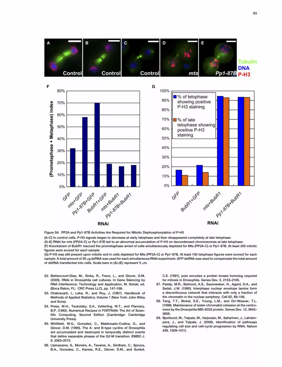
Figure S4. PP2A and Pp1-87B Activities Are Required for Mitotic Dephosphorylation of P-H3
(A–C) In control cells, P-H3 signals began to decrease at early telophase and then disappeared completely at late telophase.
(D–E) RNAi for mts (PP2A-C) or Pp1-87B led to an abnormal accumulation of P-H3 on decondensed chromosomes at late telophase.
(F) Knockdown of BubR1 rescued the prometaphase arrest of cells simultaneously depleted for Mts (PP2A-C) or Pp1-87B. At least 200 mitotic
figures were scored for each sample.
(G) P-H3 was still present upon mitotic exit in cells depleted for Mts (PP2A-C) or Pp1-87B. At least 100 telophase figures were scored for each
sample. A total amount of 30 mg dsRNA was used for each simultaneous RNAi experiment. GFP dsRNA was used to compensate the total amount
of dsRNA transfected into cells. Scale bars in (A)–(E) represent 5 mm.
S3
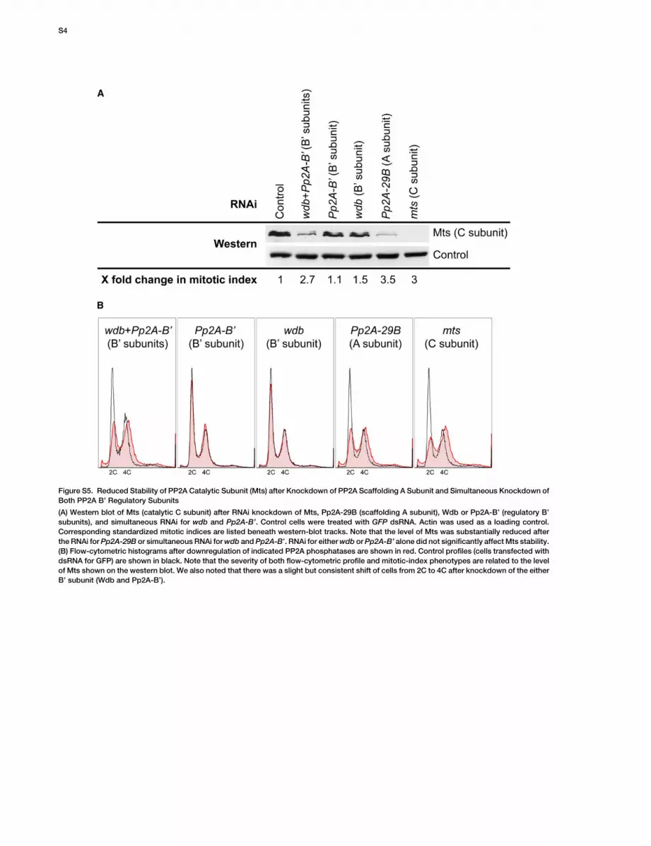
Figure S5. Reduced Stability of PP2A Catalytic Subunit (Mts) after Knockdown of PP2A Scaffolding A Subunit and Simultaneous Knockdown of
Both PP2A B’ Regulatory Subunits
(A) Western blot of Mts (catalytic C subunit) after RNAi knockdown of Mts, Pp2A-29B (scaffolding A subunit), Wdb or Pp2A-B’ (regulatory B’
subunits), and simultaneous RNAi for wdb and Pp2A-B’. Control cells were treated with GFP dsRNA. Actin was used as a loading control.
Corresponding standardized mitotic indices are listed beneath western-blot tracks. Note that the level of Mts was substantially reduced after
the RNAi for Pp2A-29B or simultaneous RNAi for wdb and Pp2A-B’. RNAi for either wdb or Pp2A-B’ alone did not significantly affect Mts stability.
(B) Flow-cytometric histograms after downregulation of indicated PP2A phosphatases are shown in red. Control profiles (cells transfected with
dsRNA for GFP) are shown in black. Note that the severity of both flow-cytometric profile and mitotic-index phenotypes are related to the level
of Mts shown on the western blot. We also noted that there was a slight but consistent shift of cells from 2C to 4C after knockdown of the either
B’ subunit (Wdb and Pp2A-B’).
S4

Figure S6. Depletion of MEI-S332 Did Not Affect the Normal Localization of Wdb
(A) Western blot showing that MEI-S332 protein was efficiently depleted after 4 days of RNAi. Note that an equal amount of total proteins
was loaded as suggested by the similar protein level of a nonspecific band recognized by the anti-MEI-S332.
(B) The normal localization of Wdb was not changed after depletion of MEI-S332. Note the absence of MEI-S332 staining on the metaphase
chromosomes after its RNAi knockdown. Scale bars represent 5 mm.
Figure S7. Illustration of the Method Used to Determine the Relative
Fluorescence Intensity of MEI-S332
To determine the signal intensity of MEI-S332 (S), a box of 8 3 8
pixels (indicated by arrows) was drawn and placed between the
centromeres as revealed by staining for the centromeric marker
CID, and the average fluorescence intensity was measured. The
same box was then placed directly adjacent to the metaphase plate
in a chromosome-free region to measure nonspecific ‘‘background’’
fluorescence ([B], see arrowheads). See Supplemental Experimental
Procedures for details. Scale bars represent 5 mm.
S5
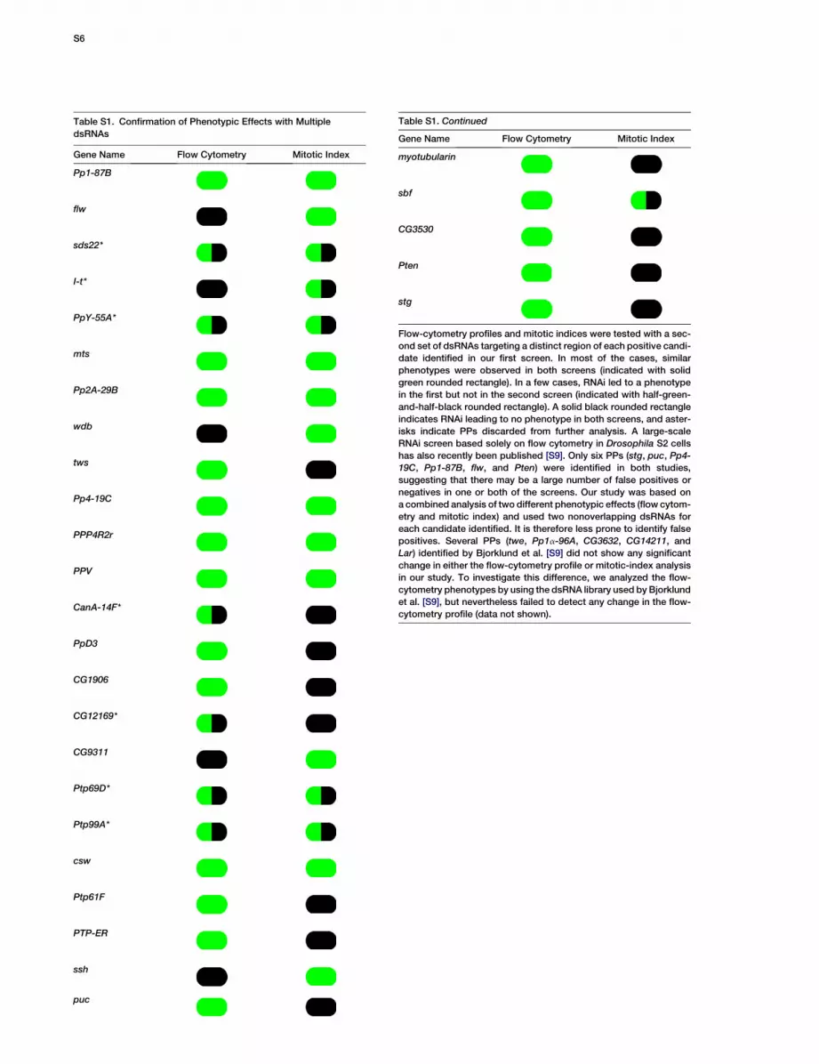
Table S1. Confirmation of Phenotypic Effects with Multiple
dsRNAs
Gene Name Flow Cytometry Mitotic Index
Pp1-87B
flw
sds22*
I-t*
PpY-55A*
mts
Pp2A-29B
wdb
tws
Pp4-19C
PPP4R2r
PPV
CanA-14F*
PpD3
CG1906
CG12169*
CG9311
Ptp69D*
Ptp99A*
csw
Ptp61F
PTP-ER
ssh
puc
Table S1. Continued
Gene Name Flow Cytometry Mitotic Index
myotubularin
sbf
CG3530
Pten
stg
Flow-cytometry profiles and mitotic indices were tested with a sec-
ond set of dsRNAs targeting a distinct region of each positive candi-
date identified in our first screen. In most of the cases, similar
phenotypes were observed in both screens (indicated with solid
green rounded rectangle). In a few cases, RNAi led to a phenotype
in the first but not in the second screen (indicated with half-green-
and-half-black rounded rectangle). A solid black rounded rectangle
indicates RNAi leading to no phenotype in both screens, and aster-
isks indicate PPs discarded from further analysis. A large-scale
RNAi screen based solely on flow cytometry in Drosophila S2 cells
has also recently been published [S9]. Only six PPs (stg, puc, Pp4-
19C, Pp1-87B, flw, and Pten) were identified in both studies,
suggesting that there may be a large number of false positives or
negatives in one or both of the screens. Our study was based on
a combined analysis of two different phenotypic effects (flow cytom-
etry and mitotic index) and used two nonoverlapping dsRNAs for
each candidate identified. It is therefore less prone to identify false
positives. Several PPs (twe, Pp1a-96A, CG3632, CG14211, and
Lar) identified by Bjorklund et al. [S9] did not show any significant
change in either the flow-cytometry profile or mitotic-index analysis
in our study. To investigate this difference, we analyzed the flow-
cytometry phenotypes by using the dsRNA library used by Bjorklund
et al. [S9], but nevertheless failed to detect any change in the flow-
cytometry profile (data not shown).
S6
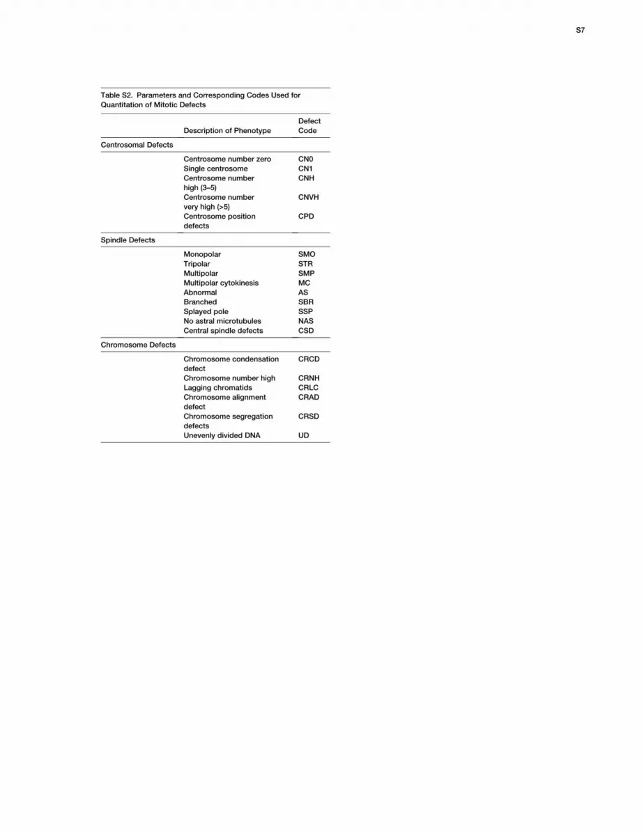
Table S2. Parameters and Corresponding Codes Used for
Quantitation of Mitotic Defects
Description of Phenotype
Defect
Code
Centrosomal Defects
Centrosome number zero CN0
Single centrosome CN1
Centrosome number
high (3–5)
CNH
Centrosome number
very high (>5)
CNVH
Centrosome position
defects
CPD
Spindle Defects
Monopolar SMO
Tripolar STR
Multipolar SMP
Multipolar cytokinesis MC
Abnormal AS
Branched SBR
Splayed pole SSP
No astral microtubules NAS
Central spindle defects CSD
Chromosome Defects
Chromosome condensation
defect
CRCD
Chromosome number high CRNH
Lagging chromatids CRLC
Chromosome alignment
defect
CRAD
Chromosome segregation
defects
CRSD
Unevenly divided DNA UD
S7


