Embed Size (px)
Citation preview
HAL Id: dumas-02868586https://dumas.ccsd.cnrs.fr/dumas-02868586
Submitted on 12 Oct 2020
HAL is a multi-disciplinary open accessarchive for the deposit and dissemination of sci-entific research documents, whether they are pub-lished or not. The documents may come fromteaching and research institutions in France orabroad, or from public or private research centers.
L’archive ouverte pluridisciplinaire HAL, estdestinée au dépôt et à la diffusion de documentsscientifiques de niveau recherche, publiés ou non,émanant des établissements d’enseignement et derecherche français ou étrangers, des laboratoirespublics ou privés.
Distributed under a Creative Commons Attribution - NonCommercial - ShareAlike| 4.0International License
Prostatic artery embolization in 420 patients with lowerurinary tract symptoms or chronic urinary retention:
early outcomes and risk factors in a French multicentricretrospective study
Asmaa Belaouni
To cite this version:Asmaa Belaouni. Prostatic artery embolization in 420 patients with lower urinary tract symptomsor chronic urinary retention: early outcomes and risk factors in a French multicentric retrospectivestudy. Human health and pathology. 2019. �dumas-02868586�
UNIVERSITE DE MONTPELLIER FACULTE DE MEDECINE MONTPELLIER-NIMES
THESE
Pour obtenir le titre de DOCTEUR EN MEDECINE
Présentée et soutenue publiquement
Par Asmaa BELAOUNI
Le 4 Octobre 2019
Prostatic artery embolization in 420 patients with lower urinary tract
symptoms or chronic urinary retention: early outcomes and risk factors in a
French multicentric retrospective study.
Directeur de thèse : Dr Julien FRANDON
JURY Président : Pr Jean Paul BEREGI Assesseurs : Pr Hélène VERNHET-KOVACSIK Pr Stéphane DROUPY Dr Julien FRANDON

ANNEE UNIVERSITAIRE 2018 - 2019
PERSONNEL ENSEIGNANT
Professeurs Honoraires ALLIEU Yves ALRIC Robert ARNAUD Bernard ASTRUC Jacques AUSSILLOUX Charles AVEROUS Michel AYRAL Guy BAILLAT Xavier BALDET Pierre BALDY-MOULINIER Michel BALMES Jean-Louis BALMES Pierre BANSARD Nicole BAYLET René BILLIARD Michel BLARD Jean-Marie BLAYAC Jean Pierre BLOTMAN Francis BONNEL François BOUDET Charles BOURGEOIS Jean-Marie BRUEL Jean Michel BUREAU Jean-Paul BRUNEL Michel CALLIS Albert CANAUD Bernard CASTELNAU Didier CHAPTAL Paul-André CIURANA Albert-Jean CLOT Jacques D’ATHIS Françoise DEMAILLE Jacques DESCOMPS Bernard DIMEGLIO Alain
DUBOIS Jean Bernard DUMAS Robert DUMAZER Romain ECHENNE Bernard FABRE Serge FREREBEAU Philippe GALIFER René Benoît GODLEWSKI Guilhem GRASSET Daniel GROLLEAU-RAOUX Robert GUILHOU Jean-Jacques HERTAULT Jean HUMEAU Claude JAFFIOL Claude JANBON Charles JANBON François JARRY Daniel JOYEUX Henri LAFFARGUE François LALLEMANT Jean Gabriel LAMARQUE Jean-Louis LAPEYRIE Henri LESBROS Daniel LOPEZ François Michel LORIOT Jean LOUBATIERES Marie Madeleine MAGNAN DE BORNIER Bernard MARY Henri MATHIEU-DAUDE Pierre MEYNADIER Jean MICHEL François-Bernard MICHEL Henri
MION Charles MION Henri MIRO Luis NAVARRO Maurice NAVRATIL Henri OTHONIEL Jacques PAGES Michel PEGURET Claude PELISSIER Jacques POUGET Régis PUECH Paul PUJOL Henri PUJOL Rémy RABISCHONG Pierre RAMUZ Michel RIEU Daniel RIOUX Jean-Antoine ROCHEFORT Henri ROSSI Michel ROUANET DE VIGNE LAVIT Jean Pierre SAINT AUBERT Bernard SANCHO-GARNIER Hélène SANY Jacques SEGNARBIEUX François SENAC Jean-Paul SERRE Arlette SIMON Lucien SOLASSOL Claude THEVENET André VIDAL Jacques VISIER Jean Pierre
Professeurs Emérites
ARTUS Jean-Claude BLANC François BOULENGER Jean-Philippe BOURREL Gérard BRINGER Jacques CLAUSTRES Mireille DAURES Jean-Pierre DAUZAT Michel DEDET Jean-Pierre ELEDJAM Jean-Jacques GUERRIER Bernard JOURDAN Jacques
MARES Pierre MAURY Michèle MILLAT Bertrand MAUDELONDE Thierry MONNIER Louis PREFAUT Christian PUJOL Rémy SULTAN Charles TOUCHON Jacques VOISIN Michel ZANCA Michel
3
Professeurs des Universités - Praticiens Hospitaliers
PU-PH de classe exceptionnelle ALBAT Bernard - Chirurgie thoracique et cardiovasculaire ALRIC Pierre - Chirurgie vasculaire ; médecine vasculaire (option chirurgie vasculaire) BACCINO Eric - Médecine légale et droit de la santé BASTIEN Patrick - Parasitologie et mycologie BONAFE Alain - Radiologie et imagerie médicale CAPDEVILA Xavier - Anesthésiologie-réanimation COLSON Pascal – Anesthésie-réanimation COMBE Bernard - Rhumatologie COSTA Pierre - Urologie COTTALORDA Jérôme - Chirurgie infantile COUBES Philippe – Neurochirurgie COURTET Philippe – Psychiatrie d’adultes, adictologie CRAMPETTE Louis - Oto-rhino-laryngologie CRISTOL Jean Paul - Biochimie et biologie moléculaire DAVY Jean Marc - Cardiologie DE LA COUSSAYE Jean Emmanuel - Anesthésiologie-réanimation DELAPORTE Eric - Maladies infectieuses ; maladies tropicales DEMOLY Pascal – Pneumologie, addictologie DE WAZIERES Benoît - Médecine interne ; gériatrie et biologie du vieillissement, médecine générale, addictologie DOMERGUE Jacques - Chirurgie générale DUFFAU Hugues - Neurochirurgie DUJOLS Pierre - Biostatistiques, informatique médicale et technologies de la communication ELIAOU Jean François - Immunologie FABRE Jean Michel - Chirurgie générale FRAPIER Jean-Marc – Chirurgie thoracique et cardiovasculaire GUILLOT Bernard - Dermato-vénéréologie HAMAMAH Samir-Biologie et Médecine du développement et de la reproduction ; gynécologie médicale HEDON Bernard-Gynécologie-obstétrique ; gynécologie médicale HERISSON Christian-Médecine physique et de réadaptation JABER Samir-Anesthésiologie-réanimation JEANDEL Claude-Médecine interne ; gériatrie et biologie du vieillissement, médecine générale, addictologie JONQUET Olivier-Réanimation ; médecine d’urgence JORGENSEN Christian-Thérapeutique ; médecine d’urgence ; addictologie KOTZKI Pierre Olivier-Biophysique et médecine nucléaire LANDAIS Paul-Epidémiologie, Economie de la santé et Prévention LARREY Dominique-Gastroentérologie ; hépatologie ; addictologie LEFRANT Jean-Yves-Anesthésiologie-réanimation LE QUELLEC Alain-Médecine interne ; gériatrie et biologie du vieillissement, médecine générale, addictologie MARTY-ANE Charles - Chirurgie thoracique et cardiovasculaire MERCIER Jacques - Physiologie MESSNER Patrick – Cardiologie MONDAIN Michel – Oto-rhino-laryngologie PELISSIER Jacques-Médecine physique et de réadaptation RENARD Eric-Endocrinologie, diabète et maladies métaboliques ; gynécologie médicale REYNES Jacques-Maladies infectieuses, maladies tropicales RIBSTEIN Jean-Médecine interne ; gériatrie et biologie du vieillissement, médecine générale, addictologie RIPART Jacques-Anesthésiologie-réanimation
4
ROUANET Philippe-Cancérologie ; radiothérapie SCHVED Jean François-Hématologie; Transfusion TAOUREL Patrice-Radiologie et imagerie médicale UZIEL Alain -Oto-rhino-laryngologie VANDE PERRE Philippe-Bactériologie-virologie ; hygiène hospitalière YCHOU Marc-Cancérologie ; radiothérapie PU-PH de 1re classe AGUILAR MARTINEZ Patricia-Hématologie ; transfusion AVIGNON Antoine-Nutrition AZRIA David -Cancérologie ; radiothérapie BAGHDADLI Amaria-Pédopsychiatrie ; addictologie BEREGI Jean-Paul-Radiologie et imagerie médicale BLAIN Hubert-Médecine interne ; gériatrie et biologie du vieillissement, médecine générale, addictologie BLANC Pierre-Gastroentérologie ; hépatologie ; addictologie BORIE Frédéric-Chirurgie digestive BOULOT Pierre-Gynécologie-obstétrique ; gynécologie médicale CAMBONIE Gilles -Pédiatrie CAMU William-Neurologie CANOVAS François-Anatomie CARTRON Guillaume-Hématologie ; transfusion CHAMMAS Michel-Chirurgie orthopédique et traumatologique CHANQUES Gérald – Anesthésie-réanimation CORBEAU Pierre-Immunologie COSTES Valérie-Anatomie et cytologie pathologiques CYTEVAL Catherine-Radiologie et imagerie médicale DADURE Christophe-Anesthésiologie-réanimation DAUVILLIERS Yves-Physiologie DE TAYRAC Renaud-Gynécologie-obstétrique, gynécologie médicale DEMARIA Roland-Chirurgie thoracique et cardio-vasculaire DEREURE Olivier-Dermatologie – vénéréologie DE VOS John – Cytologie et histologie DROUPY Stéphane -Urologie DUCROS Anne-Neurologie GARREL Renaud – Oto-rhino-laryngologie HAYOT Maurice - Physiologie KLOUCHE Kada-Réanimation ; médecine d’urgence KOENIG Michel-Génétique moléculaire LABAUGE Pierre- Neurologie LAFFONT Isabelle-Médecine physique et de réadaptation LAVABRE-BERTRAND Thierry-Cytologie et histologie LAVIGNE Jean-Philippe – Bactériologie – virologie, hygiène hospitalière LECLERCQ Florence-Cardiologie LEHMANN Sylvain-Biochimie et biologie moléculaire LE MOING Vincent – Maladies infectieuses, maladies tropicales LUMBROSO Serge-Biochimie et Biologie moléculaire MARIANO-GOULART Denis-Biophysique et médecine nucléaire MATECKI Stéfan -Physiologie MEUNIER Laurent-Dermato-vénéréologie MOREL Jacques - Rhumatologie MORIN Denis-Pédiatrie NAVARRO Francis-Chirurgie générale PETIT Pierre-Pharmacologie fondamentale ; pharmacologie clinique ; addictologie PERNEY Pascal-Médecine interne ; gériatrie et biologie du vieillissement, médecine générale, addictologie PRUDHOMME Michel - Anatomie PUJOL Jean Louis-Pneumologie ; addictologie PUJOL Pascal-Biologie cellulaire PURPER-OUAKIL Diane-Pédopsychiatrie ; addictologie
5
QUERE Isabelle-Chirurgie vasculaire ; médecine vasculaire (option médecine vasculaire) SOTTO Albert-Maladies infectieuses ; maladies tropicales TOUITOU Isabelle-Génétique TRAN Tu-Anh-Pédiatrie VERNHET Hélène-Radiologie et imagerie médicale PU-PH de 2ème classe ASSENAT Éric-Gastroentérologie ; hépatologie ; addictologie BERTHET Jean-Philippe-Chirurgie thoracique et cardiovasculaire BOURDIN Arnaud-Pneumologie ; addictologie CANAUD Ludovic-Chirurgie vasculaire ; Médecine Vasculaire CAPDEVIELLE Delphine-Psychiatrie d'Adultes ; addictologie CAPTIER Guillaume-Anatomie CAYLA Guillaume-Cardiologie COLOMBO Pierre-Emmanuel-Cancérologie ; radiothérapie COSTALAT Vincent-Radiologie et imagerie médicale COULET Bertrand-Chirurgie orthopédique et traumatologique CUVILLON Philippe-Anesthésiologie-réanimation DAIEN Vincent-Ophtalmologie DORANDEU Anne-Médecine légale - DUPEYRON Arnaud-Médecine physique et de réadaptation FAILLIE Jean-Luc – Pharmacologie fondamentale, pharmacologie clinique, addictologie FESLER Pierre-Médecine interne ; gériatrie et biologie du vieillissement, médecine générale, addictologie GAUJOUX Viala Cécile-Rhumatologie GENEVIEVE David-Génétique GODREUIL Sylvain-Bactériologie-virologie ; hygiène hospitalière GUILLAUME Sébastien-Urgences et Post urgences psychiatriques - GUILPAIN Philippe-Médecine Interne, gériatrie et biologie du vieillissement; addictologie GUIU Boris-Radiologie et imagerie médicale HERLIN Christian – Chirurgie plastique, reconstructrice et esthétique, brulologie HOUEDE Nadine-Cancérologie ; radiothérapie JACOT William-Cancérologie ; Radiothérapie JUNG Boris-Réanimation ; médecine d'urgence KALFA Nicolas-Chirurgie infantile KOUYOUMDJIAN Pascal-Chirurgie orthopédique et traumatologique LACHAUD Laurence-Parasitologie et mycologie LALLEMANT Benjamin-Oto-rhino-laryngologie LE QUINTREC Moglie - Néphrologie LETOUZEY Vincent-Gynécologie-obstétrique ; gynécologie médicale LONJON Nicolas - Neurologie LOPEZ CASTROMAN Jorge-Psychiatrie d'Adultes ; addictologie LUKAS Cédric-Rhumatologie MAURY Philippe-Chirurgie orthopédique et traumatologique MILLET Ingrid-Radiologie et imagerie médicale MORANNE Olvier-Néphrologie NAGOT Nicolas-Biostatistiques, informatique médicale et technologies de la communication NOCCA David-Chirurgie digestive PANARO Fabrizio-Chirurgie générale PARIS Françoise-Biologie et médecine du développement et de la reproduction ; gynécologie médicale PASQUIE Jean-Luc-Cardiologie PEREZ MARTIN Antonia-Physiologie POUDEROUX Philippe-Gastroentérologie ; hépatologie ; addictologie RIGAU Valérie-Anatomie et cytologie pathologiques RIVIER François-Pédiatrie ROGER Pascal-Anatomie et cytologie pathologiques ROSSI Jean François-Hématologie ; transfusion ROUBILLE François-Cardiologie SEBBANE Mustapha-Anesthésiologie-réanimation
6
SIRVENT Nicolas-Pédiatrie SOLASSOL Jérôme-Biologie cellulaire STOEBNER Pierre – Dermato-vénéréologie SULTAN Ariane-Nutrition THOUVENOT Éric-Neurologie THURET Rodolphe-Urologie VENAIL Frédéric-Oto-rhino-laryngologie VILLAIN Max-Ophtalmologie VINCENT Denis -Médecine interne ; gériatrie et biologie du vieillissement, médecine générale, addictologie VINCENT Thierry-Immunologie WOJTUSCISZYN Anne-Endocrinologie-diabétologie-nutrition
PROFESSEURS DES UNIVERSITES
1re classe : COLINGE Jacques - Cancérologie, Signalisation cellulaire et systèmes complexes 2ème classe : LAOUDJ CHENIVESSE Dalila - Biochimie et biologie moléculaire VISIER Laurent - Sociologie, démographie
PROFESSEURS DES UNIVERSITES - Médecine générale 1re classe : LAMBERT Philippe 2ème classe : AMOUYAL Michel
PROFESSEURS ASSOCIES - Médecine Générale
CLARY Bernard DAVID Michel
PROFESSEUR ASSOCIE - Médecine BESSIS Didier - Dermato-vénéréologie MEUNIER Isabelle – Ophtalmologie MULLER Laurent – Anesthésiologie-réanimation PERRIGAULT Pierre-François - Anesthésiologie-réanimation ; médecine d'urgence ROUBERTIE Agathe – Pédiatrie
Maîtres de Conférences des Universités - Praticiens Hospitaliers
MCU-PH Hors classe BOULLE Nathalie – Biologie cellulaire CACHEUX-RATABOUL Valère-Génétique CARRIERE Christian-Bactériologie-virologie ; hygiène hospitalière CHARACHON Sylvie-Bactériologie-virologie ; hygiène hospitalière FABBRO-PERAY Pascale-Epidémiologie, économie de la santé et prévention HILLAIRE-BUYS Dominique-Pharmacologie fondamentale ; pharmacologie clinique ; addictologie GIANSILY-BLAIZOT Muriel – Hématologie, transfusion PELLESTOR Franck-Cytologie et histologie PUJOL Joseph-Anatomie RICHARD Bruno-Thérapeutique ; addictologie RISPAIL Philippe-Parasitologie et mycologie SEGONDY Michel-Bactériologie-virologie ; hygiène hospitalière MCU-PH de 1re classe BADIOU Stéphanie-Biochimie et biologie moléculaire
7
BOUDOUSQ Vincent-Biophysique et médecine nucléaire BOURGIER Céline-Cancérologie ; Radiothérapie BRET Caroline -Hématologie biologique COSSEE Mireille-Génétique Moléculaire GABELLE DELOUSTAL Audrey-Neurologie GIRARDET-BESSIS Anne-Biochimie et biologie moléculaire LAVIGNE Géraldine-Hématologie ; transfusion LESAGE François-Xavier – Médecine et santé au travail MATHIEU Olivier-Pharmacologie fondamentale ; pharmacologie clinique ; addictologie MENJOT de CHAMPFLEUR Nicolas-Neuroradiologie MOUZAT Kévin-Biochimie et biologie moléculaire PANABIERES Catherine-Biologie cellulaire PHILIBERT Pascal-Biologie et médecine du développement et de la reproduction RAVEL Christophe - Parasitologie et mycologie SCHUSTER-BECK Iris-Physiologie STERKERS Yvon-Parasitologie et mycologie TUAILLON Edouard-Bactériologie-virologie ; hygiène hospitalière YACHOUH Jacques-Chirurgie maxillo-faciale et stomatologie MCU-PH de 2éme classe BERTRAND Martin-Anatomie DE JONG Audrey – Anesthésie-réanimation DU THANH Aurélie-Dermato-vénéréologie GALANAUD Jean Philippe-Médecine Vasculaire GOUZI Farès-Physiologie HERRERO Astrid – Chirurgie générale JEZIORSKI Éric-Pédiatrie KUSTER Nils-Biochimie et biologie moléculaire MAKINSON Alain-Maladies infectieuses, Maladies tropicales MURA Thibault-Biostatistiques, informatique médicale et technologies de la communication OLIE Emilie-Psychiatrie d'adultes ; addictologie PANTEL Alix – Bactériologie-virologie, hygiène hospitalière PERS Yves-Marie – Thérapeutique, addictologie SABLEWSKI Vanessa – Anatomie et cytologie pathologiques THEVENIN-RENE Céline-Immunologie
MAITRES DE CONFERENCES DES UNIVERSITES - Médecine Générale Maîtres de conférence de 1ère classe COSTA David Maîtres de conférence de 2ème classe FOLCO-LOGNOS Béatrice OUDE-ENGBERINK Agnès
8
MAITRES DE CONFERENCES ASSOCIES - Médecine Générale GARCIA Marc MILLION Elodie PAVAGEAU Sylvain REBOUL Marie-Catherine SERAYET Philippe
MAITRES DE CONFERENCES DES UNIVERSITES
Maîtres de Conférences hors classe BADIA Eric - Sciences biologiques fondamentales et cliniques Maîtres de Conférences de classe normale BECAMEL Carine - Neurosciences BERNEX Florence - Physiologie CHAUMONT-DUBEL Séverine - Sciences du médicament et des autres produits de santé CHAZAL Nathalie - Biologie cellulaire DELABY Constance - Biochimie et biologie moléculaire GUGLIELMI Laurence - Sciences biologiques fondamentales et cliniques HENRY Laurent - Sciences biologiques fondamentales et cliniques LADRET Véronique - Mathématiques appliquées et applications des mathématiques LAINE Sébastien - Sciences du Médicament et autres produits de santé LE GALLIC Lionel - Sciences du médicament et autres produits de santé LOZZA Catherine - Sciences physico-chimiques et technologies pharmaceutiques MAIMOUN Laurent - Sciences physico-chimiques et ingénierie appliquée à la santé MOREAUX Jérôme - Science biologiques, fondamentales et cliniques MORITZ-GASSER Sylvie - Neurosciences MOUTOT Gilles - Philosophie PASSERIEUX Emilie - Physiologie RAMIREZ Jean-Marie - Histologie TAULAN Magali - Biologie Cellulaire
PRATICIENS HOSPITALIERS UNIVERSITAIRES
CLAIRE DAIEN-Rhumatologie BASTIDE Sophie-Epidémiologie, économie de la santé et prévention GATINOIS Vincent-Histologie, embryologie et cytogénétique PINETON DE CHAMBRUN Guillaume-Gastroentérologie ; hépatologie ; addictologie SOUCHE François-Régis – Chirurgie générale TORRE Antoine-Gynécologie-obstétrique ; gynécologie médicale
9
REMERCIEMENTS Aux membres du Jury de thèse : A Monsieur le Professeur Jean-Paul BEREGI : Vous me faites le très grand honneur de présider le jury de cette thèse. Je vous
remercie pour vos conseils avisés tout au long de mes semestres passés à Nîmes.
J’admire votre parcours, votre volonté, votre courage immense pour défendre vos
idées et en faire profiter le monde entier. Je sais la chance que j’ai de pouvoir
bénéficier de votre enseignement et de vos immenses connaissances. Je vous prie de
recevoir l’expression de mon profond respect et de ma sincère reconnaissance.
A Monsieur le Professeur Hélène VERNHET-KOVACSIK :
Vous me faites le très grand honneur de siéger parmi ce jury de thèse. Je suis une
grande admiratrice de votre parcours et de votre dévouement pour ce métier. J’ai été
honorée de recevoir votre enseignement et plus particulièrement dans le domaine de
l’imagerie cardiaque que j’espère avoir l’opportunité de poursuivre dans les prochaines
années. Je vous prie de recevoir l’expression de mon profond respect et de ma sincère
reconnaissance.
A Monsieur le Professeur Stéphane DROUPY :
Je vous remercie de me faire l’honneur de siéger à ce jury de thèse. Merci également
d’avoir accepté d’évaluer mon travail sur ce sujet qui j’espère vous a été utile. Je vous
prie de recevoir l’expression de mon respect et de ma sincère reconnaissance.
A Monsieur le Docteur Julien FRANDON :
Merci infiniment pour ta bonne humeur, tes taquineries, tes blagues (parfois
inadaptées), ta motivation sans limite qui m’ont donné envie de travailler avec toi. Tu
as le don d’obtenir ce que tu veux des autres et je dis ça en pensant au TIPS qu’on a
fait ensemble ce fameux samedi matin. Grâce à toi, j’ai fait le tour de la France à
l’affut des données sans trop me poser de questions. En travaillant avec toi, j’ai su

10
que je voulais faire partie de ton équipe. J’ai encore plus d’attrait pour la radiologie
interventionnel. Tu as toujours su te montrer rassurant quoi qu’il arrivât en salle et
quelques soit les erreurs que je pouvais faire. Tu sais aussi te montrer moqueur dans
les situations les plus inconfortables, mais c’est ce qui fait le charme de faire partie
de ton équipe. Ton nom et ton ambition sont une marque, j’ai beaucoup de respect
pour ça et j’espère être à la hauteur de tes attentes. J’ai encore beaucoup à
apprendre et j’en suis impatiente. J’ai une très grande estime pour toi. Je te remercie
de m’avoir impliquée dans ce travail qui ne fait à vrai dire que commencer.
11
A ma famille,
A mon papou, J’aurai tellement aimé que tu sois là, t’avoir auprès de moi aurait été
mon voeux le plus cher, toi qui a toujours su me soutenir même quand je n’avais pas
été à la hauteur. Tu es parti beaucoup trop tôt. Malgré cela, tu m’as transmis une
force mentale qui m’a permis de tenir jusqu’au bout et de garder la tête froide à
chaque moment clé de mes études. J’ai toujours eu un amour et une admiration
inconditionnels pour toi. Parfois je sens encore ta présence comme pour me donner
plus de force quand j’en ai besoin. Je t’aime Papa.
A maman, ma tendre et douce maman, notre relation si fusionnelle a forgé une
partie de la personne que je suis aujourd’hui. Je t’ai toujours prise pour exemple,
honnête, studieuse, forte, sérieuse, ambitieuse, courageuse, exigeante envers toi-
même, voilà ce que j’ai toujours espéré prendre de toi. Tu as été mon pilier toutes
ces années, ton soutien en toute épreuve m’a aidé à tout surmonter. Tendre mais
caractérielle à la fois, je crois qu’on ne doit pas être si différentes toi et moi. Je te
serai toujours reconnaissante de m’avoir fait confiance, de m’avoir encouragée à
m’engager dans cette voie-là, sans jamais émettre le moindre doute sur mes
capacités, de m’avoir épaulée durant tous ces moments de stress, qui ont pourtant
été longs. Cela a été presque vital pour moi, tu m’as rendue plus forte que ce que
j’aurai pu être. J’ai toujours voulu te rendre fière car cela a été la source d’un
bonheur essentiel dans ma vie. Avec du recul, je pense que j’étais un petit toi en plus
timide et réservée mais ayant toujours su que je voulais aller le plus loin possible
dans mes ambitions, et sans aucun doute, c’est toi qui me l’a transmis. Enfin, merci
pour ton amour inconditionnel, ton affection sans limite et ta bienveillance. Un jour,
j’aurai peut-être des enfants et j’espère leur donner autant d’amour que j’ai reçu. Je
t’aime Maman.
A mon grand frère Khalid, mon héros, mon meilleur exemple dans la vie, tu es
beau intelligent et grand bosseur, ceux qui me comparent à toi me font le plus beau
des compliments. J’admire autant ta force mentale que physique, ton énergie
inépuisable te permettant de gérer tout aussi bien ton travail que ta petite famille.
Malgré notre différence d’âge, nous n’avons jamais été aussi proche
qu’actuellement, tu es une source de lumière dans ma vie et c’est grâce à toi que j’en
12
suis là. Toute petite déjà, je te vouais une grande admiration, un immense respect, et
j’aspirai à être ta version féminine, cela n’a jamais changé. Je te suis reconnaissante
de m’avoir encouragé à faire ce métier, c’est principalement grâce à toi que je me
suis engagée dans cette voie, tes conseils m’ont toujours été très précieux (même si
tu aurais préféré que je sois dentiste :p). Je te remercie également pour tes
encouragements et ta présence inconditionnelle, tant dans les moments de joies que
les moments difficiles, ton amour et ta bienveillance. Enfin, merci d’avoir été là quand
papa est parti, j’aurai surement abandonné mon concours sans ton soutien.
Je t’aime tellement fort grand frère chéri.
A Nadia, tu es comme une grande sœur pour moi, tu m’as toujours accueillie avec
beaucoup de bienveillance. Tu as toujours été là quand j’avais besoin. Je te suis
reconnaissante pour tes nombreux conseils en tout domaine. J’admire ton
intelligence, ta capacité incroyable à résoudre le moindre problème et à savoir gérer
des milliers de choses à la fois. Tu as ce côté bavard qui chez toi constitue une si
grande qualité que j’apprécie spécialement. Mon frère et toi êtes complémentaires
sur bien des choses et vous continuerez à l’être. Je t’aime fort.
A Lilya et Adam, mes petits neveux d’amour, je suis heureuse de vous voir grandir,
chaque moment passé avec vous est un bonheur pour moi. Lilou, je suis fière de ton
parcours et ce que tu as déjà accompli du haut de tes 12 ans. Tu es ma petite
danseuse étoile préférée. Gracieuse mais aussi intelligente, l’avenir ne te réservera
que de belles aventures, mais n’oublie pas de rester toi-même. Adam, entre
l’équitation et le foot, ton cœur balance, mais à 6 ans tu es déjà un petit garçon bien
déterminé, fort de caractère mais d’une tendresse intense. Je vous aime mes petits
loups.
A ma sœur jumelle, Hasna, à notre complicité incontestable toutes ces années, à
être inséparables malgré nos chamailleries d’enfants, à nos différences aussi qui ont
forgé nos personnalités peu comparables. J’espère que la distance n’aura pas raison
de notre relation si unique. Je te souhaite de tout mon cœur de réussir dans tes
projets, même si tu t’es éloignée un peu plus. Je t’aime très fort.
A Mima, ma 2ème maman, je te suis profondément reconnaissante pour l’éducation
que tu m’as donnée, si pure, bienveillante, généreuse et tendre, tu m’as appris à
toujours vivre dans le respect envers soi et les autres. Tu fais partie de ceux qui ont
13
rendu mon enfance si heureuse et insouciante. Dès l’âge de 5 ans tu me
surnommais déjà « le petit docteur », aujourd’hui je le suis vraiment et j’espère que
tu aurais été fière de moi. Je te dédie ce travail. Je t’aime Mima. A mes tatas et tontons du Maroc, Fadela, Wassila, Youssef, Rachid (2), vous
m’avez vu grandir, m’avez chéri et aimé comme votre fille. Vous m’avez apporté
beaucoup d’amour et de tendresse, m’avez gâtée d’attention et de cadeaux. Malgré
la distance qui nous sépare, votre place dans mon cœur a toujours été grande et le
restera. Je vous aime tous très fort.
A mes cousins Salma, Soussou et Meriem, mon enfance n’aurait pas été la même
sans vous, tous ces bons moments passés avec vous, à jouer, à rire, à se chamailler
me manquent tellement. Nous avons été si proches étions si différents et
complémentaires à la fois et malgré la distance qui nous sépare aujourd’hui, les
retrouvailles font parties de ces meilleurs moments de l’année. A Salma, je te
souhaite une très belle nouvelle vie à Toulouse avec ta petite famille. A Souhail,
Singapour et compagnie ça suffit, j’ai bien hâte que tu reviennes après ces 6 années.
A Mimi, tu as un bel avenir qui t’attend. Je vous aime fort.
A Rizou, Patrick, merci infiniment d’avoir été là pour moi tout au long des années de
médecine et tout particulièrement la 1ère. Vous m’avez si bien accueilli dans votre
environnement à Rennes. J’ai tellement aimé passer tous mes moments libres avec
vous, même s’ils se faisaient rares. Votre aide et soutien ont été précieux pour moi et
je vous dois la réussite de ce fameux concours de 1ère année. Je me suis éloignée
mais vous me manquez et comptez tellement pour moi. A Adam, mon amour de petit
cousin, qui à lui seul a su remplir votre vie de bonheur, et la mienne également. Je
vous aime très fort.
A Lamia, Mickael, Camille et Clément, mon petit noyau familial montpelliérain, je
vous suis reconnaissante pour votre présence et ces bons moments passés avec
vous, ces fêtes de Noel et nouvel an, ces week-end en Espagne et ces soirées
tapas.

14
A mon cousin Tarik, ta douceur et attitude protectrice envers moi m’ont toujours fait
me sentir comme ta petite sœur, sans oublier ton petit côté taquin. Je t’admire
beaucoup.
A ma famille hollandaise, à tous ces moments partagés avec vous lors de mon
enfance, à tous ces instants de jeux, ces journées passées à la plage, ces soirées à
se promener.
15
A mes ami(e)s,
A Alice, je suis ta plus grande admiratrice et je m’estime heureuse de faire partie de
ta vie. D’une énergie inépuisable, une gentillesse et bienveillance sans égales, un
dévouement pour tes plus proches mais aussi pour ton métier que je ne saurai
qualifier, une intelligence exceptionnelle, à toi je te serai l’une des plus fidèles amies.
Merci de m’avoir initiée à la planche à voile, au surf et autres activités plus ou moins
dangereuses. A tous nos moments passés ensemble. Je t’aime très fort et te
souhaite une vie remplie d’aventures, de bonheur et d’amour avec ta tendre Floriane.
A Cécile, ma popinou d’amour, ma co équipière de sports de glisse en tout genre.
On s’est adorées dès le 1er regard, je me souviendrai toujours de la première où je
t’ai rencontré, depuis mon affection pour toi n’a cessé de grandir. J’adore ta bonne
humeur, ton coté aventurière, toujours pétillante et motivée. Nous avons encore
tellement de bons moments devant à nous à partager. Je vous souhaite à Pierre et
toi tout le bonheur du monde. Je t’aime fort.
A Ghita, mon amie d’enfance, ma seconde moitié, on a toujours su être
complémentaire de part nos caractères et nos différences, on a grandi ensemble,
inséparables au collège puis au lycée. Depuis ce temps, on a su entretenir cette
belle amitié et continuer à partager de délicieux moments de complicité. Je te
souhaite d’être heureuse dans tes différents projets. Je t’aime fort.
A Julien, on s’est connu en 1ère année de médecine sans plus jamais se quitter. Tu
es l’un de mes plus chers amis et je te dois tellement. Merci a toi et ta famille de
m’avoir toujours accueilli à bras ouverts avec tellement de douceur de bienveillance
et de générosité. A Cécile, ta maman, Nicolas, ton papa, Marie et Claire, tes deux
petites sœurs, ma famille adoptive que j’aime tout autant. Merci de m’avoir supporté
ces quelques années d’externat passées ensemble à Rennes. Merci pour m’avoir
soutenue psychologiquement face à l’une des pires situations que j’ai pu vivre
jusqu’ici. Je vous souhaite plein de bonheur avec Marion. Je t’aime très fort.
16
A Bahia, ma petite Baba au Rhum d’amour, tu m’as prise sous ton aile en 1ère année
de médecine et tu as largement participé à ma réussite, grâce à ton soutien
psychologique, à ta bienveillance, à ta douceur, j’avais trouvé une grande sœur à ce
moment-là. Je n’oublierai pas nos années de collocations alternant révisions, soirées
médecines, moments de rire et d’autres à pleurer. Aujourd’hui te voila grande,
mariée et prête à t’installer dans le fin fond de la Bretagne, mais malgré cette
distance j’espère que l’on restera aussi proches et je te souhaite le meilleur pour la
suite. Je t’aime fort.
A Marlène, ma petite Marly, une fille exceptionnelle à tout point de vue, en plus de
ton look sorti tout droit d’un magazine, toujours motivée, pétillante, pleine de bonnes
idées et d’une intelligence peu commune. Tu sais autant t’amuser qu’être sérieuse et
toujours dans la rigolade. A nos fameuse fraises Tagada paillettes. A Charles-Eric, Charlichou, à tes blagues, ton flegme, toutes tes bêtises
incessantes qui me manquent parfois.
A mes copains du Maroc, Tarik, Tarik, Tarek, Hmidou, à toutes ces soirées,
sorties, WE à Marrakech qu’on a pu faire ensemble. Vous avez toujours été
bienveillants envers moi, faciles à vivre et surtout vous avez pu supporter mon petit
caractère toutes ces années. J’ai pu compter sur chacun de vous dans n’importe
quelle situation.
A Hicham, à nos quelques années passées ensemble qui font parties de moi, ma vie
et m’ont aidé à grandir et me construire. Tu auras toujours une place importante pour
moi.
A mes amis de Montpellier, A Jenny, ou Jenay pour ma part, j’admire ton intelligence assez exceptionnelle qui
se cache derrière tant de modestie. Travailler avec toi a été un pur bonheur, toujours
motivée, sérieuse et rigolote à la fois, le mélange parfait. J’ai également eu la chance
de te découvrir de façon plus intime, à savoir dans des conditions « extrêmes » de
trekking en Jordanie où l’on a parfaitement su co-habiter dans la même tente sans
17
douche pendant 4 jours, et je n’en garde que des bons souvenirs. Je te souhaite de
tout réussir, tant sur le plan professionnel que personnel. Je t’aime fort ma Jenini.
A Philou, si différents et pourtant tu es ma plus longue relation de cohabitation après
mes parents… Merci de nous avoir supporté mon caractère et moi ces 4 années
passées en colocation. Avant de te connaitre je ne savais pas qu’on pouvait être
autant passionné de poissons et d’aquarium. Tu es aussi original que tes hobbies et
cela fait tout ton charme. « Asmou ! je vais nous faire un nouvel aquarium avec des ..
crevettes, …, je vais peindre une baleine bleue sur le mur du salon, je vais faire
pousser un ananas du Japon, … ». Ton imagination débordante et sans limite sera
toujours une intrigue pour moi.
A Sandra, à ton naturel, ta beauté et ton dynamisme qui l’ont fait craquer. A votre
bonheur et nouvelle vie à tous les 3 qui sera remplie de bonheur, d’amour et
d’attention.
A Christophe, ou Chrissou, je suis parmi tes plus grandes admiratrices et tu es mon
meilleur coup de cœur de l’internat. Tout est dit.
A la promo « Bisounours » : A Benjou, tu as été mon 1er co-interne, on a réussi à se soutenir dans les moments
les plus difficiles. Chaque stage après ça avec toi a été un régal du point de vue
professionnel et surtout personnel. Ta bonne humeur m’a toujours épatée et rendu
mes journées tellement plus agréables. Merci pour ta confiance, ta patience, ta
présence tout court.
A Antonia, on a plus partagé de soirées, randonnées et autres sorties que de stages
ensemble. Simplicité, générosité et fidélité sont tes plus belles qualités. Toujours
motivée pour toute sorte de loisir, j’ai adoré passer ces nombreux moments avec toi.
A Charlotte, à ta constante bonne humeur, à ta douceur, ta générosité et
bienveillance, ta motivation et ton courage dans tout ce que tu entreprends.
A Anne Hélène, à ton rire, à ta douceur, tu es ma petite lilloise préférée. Tout plein
de bonnes choses t’attendent avec ton petit Roméo.
18
A Taki, merci pour tes bons conseils, de toujours mettre une bonne ambiance, tu as
ce côté serein et apaisant qui rend le travail si agréable avec toi.
A mes co-internes,
A Lylia, ta douceur, ta gentillesse, ta motivation et ton sérieux qui font de toi cette
jeune femme si unique et que j’apprécie tant. Je regrette ton départ et te souhaite
une belle réussite dans ta carrière et tes projets personnels.
A Benoit, à tes blagues bizarres et parfois incompréhensibles, à ton rire inimitable,
toujours prêt à rendre service, je n’oublierai jamais ce jour où tu m’as sauvée d’une
garde alors que je pensais mourir d’une grippe. J’aurai apprécié travailler d’avantage
avec toi les prochaines années. En tout cas je te souhaite le meilleur.
A Alexandre S., ce 1er stage de radio à GdC n’aurait pas été le même sans toi. Tu
sais instaurer un climat de sérénité dans la bonne humeur et le sérieux. J’admire ton
assurance et ta fausse nonchalance sans oublier ton franc coté blagueur. J’ai bien
hâte de (re)travailler avec toi.
A Elaura, merci pour ta douceur et bonne humeur, sans oublier ton joli sourire qui
me mettait tout le temps de bonne humeur. Je sens qu’on va continuer à bien
s’entendre et surtout à partager quelques activités ensemble (oooooom).
A Mehdi, avec ton joli sourire toujours prêt à faire une pause-café quoiqu’il arrive. Je
pensais être tête en l’air, mais après 6 mois de stage avec toi j’ai compris qu’il y avait
bien pire. J’ai été ravie de te découvrir autant en stage qu’en dehors. Merci pour ta
bienveillance et d’avoir pris soin de ce « petit poussin » qui te suivait partout dans les
rues parisiennes.
A Helene, à ton incroyable bienveillance, ta douceur, ton coté fêtarde que tu as su
allier à ta nouvelle vie de maman. Tu es un exemple pour bon nombre d’entre nous.
A ta nouvelle vie en Bretagne.
19
A Nadir, admirative de ta bienveillance, ton sérieux et ton dévouement pour les
autres, tu as su te démarquer par ta motivation. J’ai été ravie de travailler avec toi et
bien heureuse de ton retour.
A Antoine L., ton énergie, ta motivation inépuisables et ton incroyable intelligence
que j’admire tant, restent un mystère pour moi. Notre irremplaçable organisateur
officiel de soirées de la radiologie : à toutes ces soirées qui restent à venir.
A Kim, à ton humour et second degré, à ta future carrière de radiologue-paysagiste.
A Margaux H., j’admire ton sérieux, ton intelligence, surtout ton coté perfectionniste
où rien ne peut t’échapper, tout cela avec des petites pointes d’humour qui rendent le
travail agréable avec toi.
A tous mes autres co-internes, aux plus vieux Aymeric, Lauranne, Pierre L., Adel, William, Nadia, Satcha, Maelis, Aureline et aux plus jeunes Quentin D, Phil L,
merci de m’avoir supportée tous ces stages durant.
A tous les autres internes ou chefs avec qui j’ai eu l’occasion de partager des
gardes, des pots de thèse ou boire des coups, Carole, Céline, Caroline, Brice.
A mes chefs,
A Florian, mon tout premier chef de clinique, tu m’as connue « bébé » interne et m’a
vu grandir et évoluer. C’est toi qui m’a appris à interpréter mes premières imageries,
qui m’a aidé me surpasser pendant ces moments de stress des premières gardes, tu
as su te montrer ferme quand il le fallait tout autant qu’amusant avec ta bonne
humeur et tes « non mais laissez-moi, non mais laissez-moi manger ma banane.. ».
En plus d’avoir été un super chef tu es un artiste exceptionnel.
Aux nîmois A Alban, je ne saurai quantifier ou qualifier la grande admiration et le grand respect
que j’ai pour toi, j’aurai aimé profiter de tes connaissances plus longtemps, mais ton
bonheur avant tout. Merci pour ton enseignement, ton dévouement pour tes internes,
pour tous ces cafés et sans oublier tes discours dignes d’un grand poète.
20
A Iskander, co-interne puis chef, merci pour ta solidarité lors de certains moments
passés à GDC, ton enseignement, avec toi tout est dans la précision, le savoir et la
maitrise des choses, sans oublier bien évidemment ces nombreuses imitations en
allant de NL, VC, PM à JF, JG qui m’ont tellement faites rires toutes ces vacations
passées avec toi, tu as un talent exceptionnel.
A Jean, à ta patience inépuisable, enfin non pas du tout, à ta sagesse et ta capacité
à canaliser Julien. J’apprécie travailler avec toi et ce sera un grand plaisir de
continuer à apprendre ce métier à tes cotés.
A Mélinée, à ton dynamisme, constante bonne humeur et bienveillance. Merci pour
ta disponibilité en toute circonstance, tous ces avis en pédiatrie et pour toutes les
pauses café/potins. Je suis ravie de continuer à travailler avec toi.
A Romain, à ta constante bonne humeur, à ton rire, merci pour ta patience avec moi,
tu m’as appris pas mal de tips en geste interventionnel. J’ai beaucoup aimé être ton
interne, et je suis ravie de travailler avec toi ces prochaines années.
A Elise, à ton dynamisme, ta bonne humeur, ton investissement auprès de tes
internes.
A Ahmed, le Don de la radiologie, mon exemple dans la rigueur, à toutes ces
pauses café qu’on faisait parmi les 25 IRM de la vac, mêlées à une gestion parfaite
du timing,
A Anca, Anna, Conny, Cyrine merci pour votre disponibilité, votre enseignement et
votre bienveillance.
A Pierre, Pierre et Fehmi, les osseux nîmois, merci pour votre enseignement en
imagerie ostéo-articulaire, et surtout pour votre patience.
A l’équipe de radio-pédiatrie, a Cathy, merci pour ton enseignement, ta patience,
et ton extrême bienveillance, à Ikram, merci pour ta douceur, tes précieux conseils et
ces petits schémas lors des vacations d’écho, à Julie, à ta bonne humeur et ton
21
dynamisme, à Olivier et tes petites blagues, Stéphanie, Nancy, merci pour votre
enseignement. A l’équipe de Lapeyronie, au Pr Taourel, à Ingrid, Cécile, Marion, Claire, Emma, Fernanda, merci pour tout ce que vous m’avez appris, pour votre soutien lors des
gardes et votre bienveillance.
A Yann, à toutes ces blagues que je n’ai pas souvent comprises, à ta patience et tes
connaissances infinies, A Benjamin, merci pour ces petits bonbons lors des gardes,
et surtout pour avoir veillé sur nous en 1er semestre.
A l’équipe D’Arnaud de Villeneuve, à Sébastien, merci pour ton enseignement, ta
confiance et pour m’avoir initiée aux ponctions thoraciques.., à Hamid, à Valérie, à Juliette et à Cyril, merci pour votre enseignement en imagerie cardio-vasculaire.
A l’équipe de st Eloi, à Marie-Ange, Elisabeth, Valentina, Laure, Stéphanie merci
de m’avoir tout appris de l’imagerie digestive.
A l’équipe de Gui de Chauliac, au Pr Bonafe, à Vincent, Nicolas M., Nicolas L., Gregory, Carlos, à ce 1er stage de radiologie en tant que « bébé » interne. Merci de
m’avoir initié à l’imagerie en général et à la neuro-radiologie en particulier.
A l’équipe de Perpignan, à Geoffroy (mon jojo), merci de m’avoir initié et fait
aimer la radiologie interventionnelle, à ta patience et ta qualité d’enseignement que
je n’oublierai pas, Aymeric, merci pour ta bonne humeur et tout ce que tu m’as
appris, Maxime, Sabine, Anna Maria, Sandra, Philippe, Jean-Louis, vous m’avez
formé et fait aimer la radiologie sous toutes ses formes.
A toutes les équipes de manips radio et de secrétaires, je ne peux vous citer un à un mais merci pour tout.

22
A Charlie, à ta douceur, cachée derrière ta virilité, tes petites attentions, ton humour,
ta générosité et surtout ta capacité à lire dans mes pensées (une exceptionnelle
qualité) qui m’ont fait craquée et tenir ces derniers mois, en toute maitrise de moi-
même et de la situation. J’admire ta détermination, ton optimisme, ton sens de
l’organisation, sans oublier ton audace. Que de belles choses à venir, de belles
aventures et de projets.
23
SUMMARY
I. Abstract……………………………………………………………………………...24
II. Introduction………………………………………………………………………….26
III. Materials and methods…………………………………………………………….38
IV. Results……………………………………………………………………………….50
V. Discussion…………………………………………………………………………..58
VI. Conclusion…………………………………………………………………………..71
VII. Bibliography…………………………………………………………………………72
VIII. Annexes……………………………………………………………………………..78
IX. List of abreviations………………………………………………………………….85
24
ABSTRACT
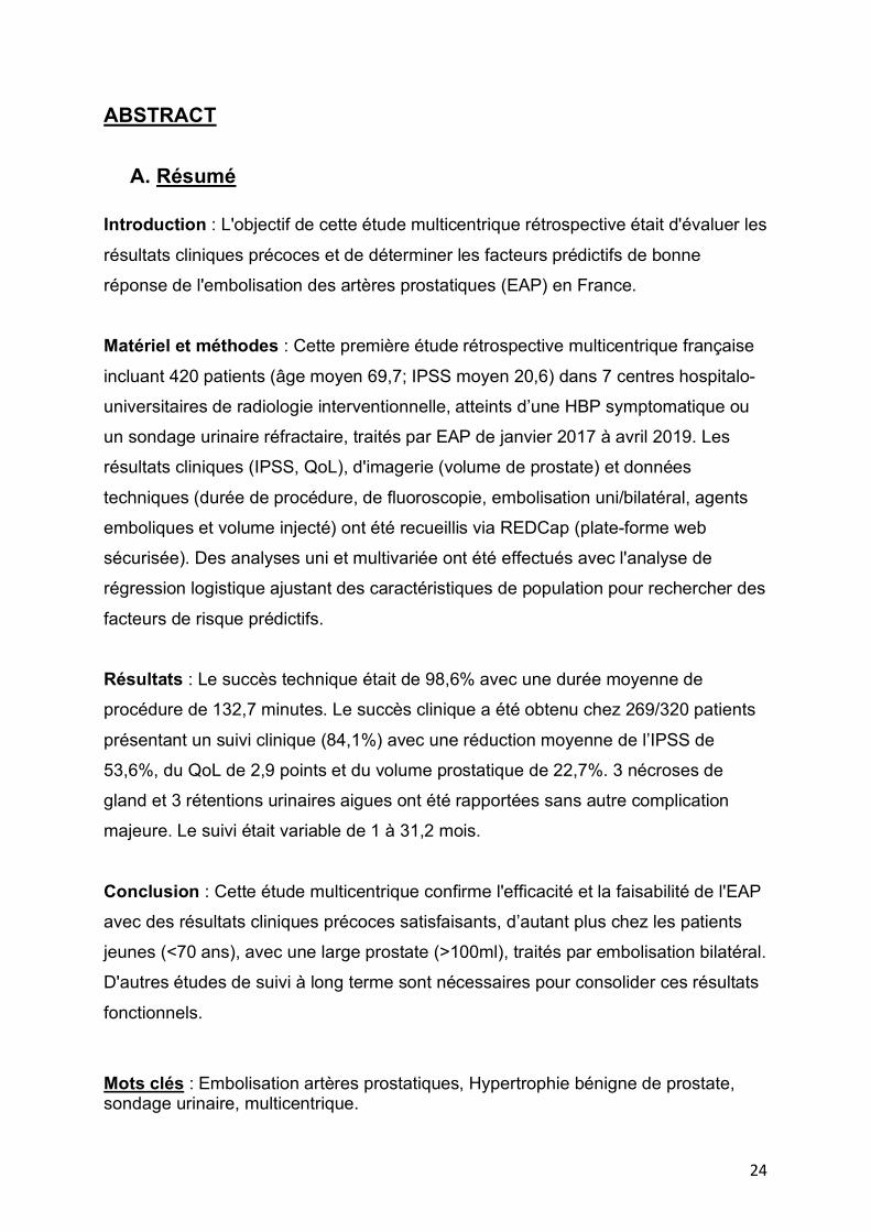
A. Résumé
Introduction : L'objectif de cette étude multicentrique rétrospective était d'évaluer les
résultats cliniques précoces et de déterminer les facteurs prédictifs de bonne
réponse de l'embolisation des artères prostatiques (EAP) en France.
Matériel et méthodes : Cette première étude rétrospective multicentrique française
incluant 420 patients (âge moyen 69,7; IPSS moyen 20,6) dans 7 centres hospitalo-
universitaires de radiologie interventionnelle, atteints d’une HBP symptomatique ou
un sondage urinaire réfractaire, traités par EAP de janvier 2017 à avril 2019. Les
résultats cliniques (IPSS, QoL), d'imagerie (volume de prostate) et données
techniques (durée de procédure, de fluoroscopie, embolisation uni/bilatéral, agents
emboliques et volume injecté) ont été recueillis via REDCap (plate-forme web
sécurisée). Des analyses uni et multivariée ont été effectués avec l'analyse de
régression logistique ajustant des caractéristiques de population pour rechercher des
facteurs de risque prédictifs.
Résultats : Le succès technique était de 98,6% avec une durée moyenne de
procédure de 132,7 minutes. Le succès clinique a été obtenu chez 269/320 patients
présentant un suivi clinique (84,1%) avec une réduction moyenne de l’IPSS de
53,6%, du QoL de 2,9 points et du volume prostatique de 22,7%. 3 nécroses de
gland et 3 rétentions urinaires aigues ont été rapportées sans autre complication
majeure. Le suivi était variable de 1 à 31,2 mois.
Conclusion : Cette étude multicentrique confirme l'efficacité et la faisabilité de l'EAP
avec des résultats cliniques précoces satisfaisants, d’autant plus chez les patients
jeunes (<70 ans), avec une large prostate (>100ml), traités par embolisation bilatéral.
D'autres études de suivi à long terme sont nécessaires pour consolider ces résultats
fonctionnels.
Mots clés : Embolisation artères prostatiques, Hypertrophie bénigne de prostate, sondage urinaire, multicentrique.
25
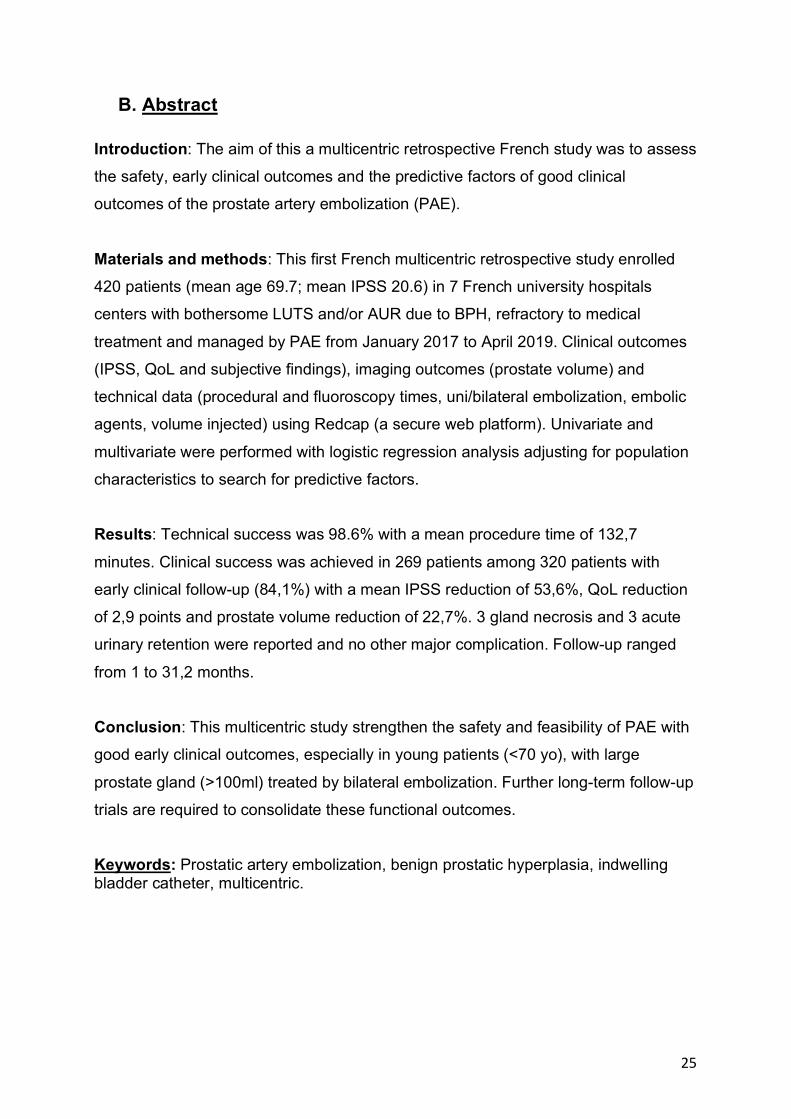
B. Abstract
Introduction: The aim of this a multicentric retrospective French study was to assess
the safety, early clinical outcomes and the predictive factors of good clinical
outcomes of the prostate artery embolization (PAE).
Materials and methods: This first French multicentric retrospective study enrolled
420 patients (mean age 69.7; mean IPSS 20.6) in 7 French university hospitals
centers with bothersome LUTS and/or AUR due to BPH, refractory to medical
treatment and managed by PAE from January 2017 to April 2019. Clinical outcomes
(IPSS, QoL and subjective findings), imaging outcomes (prostate volume) and
technical data (procedural and fluoroscopy times, uni/bilateral embolization, embolic
agents, volume injected) using Redcap (a secure web platform). Univariate and
multivariate were performed with logistic regression analysis adjusting for population
characteristics to search for predictive factors.
Results: Technical success was 98.6% with a mean procedure time of 132,7
minutes. Clinical success was achieved in 269 patients among 320 patients with
early clinical follow-up (84,1%) with a mean IPSS reduction of 53,6%, QoL reduction
of 2,9 points and prostate volume reduction of 22,7%. 3 gland necrosis and 3 acute
urinary retention were reported and no other major complication. Follow-up ranged
from 1 to 31,2 months.
Conclusion: This multicentric study strengthen the safety and feasibility of PAE with
good early clinical outcomes, especially in young patients (<70 yo), with large
prostate gland (>100ml) treated by bilateral embolization. Further long-term follow-up
trials are required to consolidate these functional outcomes.
Keywords: Prostatic artery embolization, benign prostatic hyperplasia, indwelling bladder catheter, multicentric.
26
INTRODUCTION:
Background:
Benign prostatic hyperplasia (BPH), as the most common male disease in urological
pathology, represents a serious public health problem in our society. It affects about
50% of men aged 50-60 years and 90% of men aged 85 years or older1.
In France, there are near two million of men with lower urinary tract symptoms and 100
000 new patients each year (urofrance.org).
Benign prostatic hyperplasia is a histological term and represents the most frequent
chronic benign neoplasm in aging men. It can lead to an enlargement of prostatic
transitional zone (TZ) due to the proliferation of glandular and fibromuscular elements
resulting in the formation of nodules androgen-dependant. There is an imbalance
between growth and death programs of stroma cells, leading to increased final stromal
volume2.
Although it is benign, in one half of cases, benign prostatic enlargement causes lower
urinary tract symptoms (LUTS) related to benign prostatic obstruction (BPO). LUTS
are defined by storing (irritative) symptoms, voiding (obstructive) symptoms and post
micturition symptoms3. These several symptoms have been shown to cause a negative
impact on the health-related health of life4. Symptoms such as urinary incontinence,
urgentury, frequency and nocturia are such a serious therapeutic problem due to its
great impact on the quality of life5. The clinical diagnosis of LUTS due to BPH is a
multistep diagnosis.
Voiding difficulties due to BPH can be quantified with the International Prostatic
Symptom Score (IPSS) consisting of seven symptom categories rating an increasingly
27
severe symptom scores from 0 through 35. Severity categories admitted are: mildly
symptomatic (1-7), moderately symptomatic (8-19), and severely symptomatic (20-35).
The IPSS is the most commonly used score but is considered as a subjective
evaluation quite as well as the Quality of life score (QoL) related to LUTS, ranging from
0 (delighted) to 6 (terrible) based on eight questions.
Urodynamic testing is more accurate to identify the functional mechanisms that result
in LUTS. It provides functional information about bladder filling, urine storage,
emptying, distinguish benign prostatic obstruction from other causes of LUTS, and
reproduce patient’s symptoms to obtain an accurate diagnosis of primary causes of
LUTS. The American Urological Association recommends using urodynamic studies
to identify LUTS due to BPH if invasive and potentially irreversible treatments are
considered. Predicting outcomes after invasive treatments relies on pressure flow
study based on maximum flow rate (Qmax) and post void residual (PVR) urine volume.
One of the Bladder outlet obstruction complications due to BPH is indwelling bladder
catheterization.
Management of BPH and existing therapies:
The primary treatment goal for LUTS due to BPH is to reduce bothersome symptoms,
improving quality of life, urinary flow and prevent long term complications such as acute
urinary retention. A wide variety of surgical and medical options are currently available
for the management of BPO.
The standard management of symptomatic BPH usually depends on the severity of
the symptoms and the patient’s state of health.
Watchful waiting is indicated in men with mild symptoms, coupled with lifestyle
modification, and do not usually require an active intervention but might be monitored
28
annually by the physician. Knowing that BPH is generally a progressive disease with
symptom deterioration, there is no established evidence concerning the timeliest
moment to introduce an appropriate treatment6.
For Patients with moderate symptoms, the first line therapy consists on medical
treatment with two main categories: alpha-blockers and 5 alpha-reductase inhibitors or
a combination therapy. There are six Pharmacological classes available alone or in
combination: alpha-blockers, 5 alpha-reductase inhibitors, Phytotherapeutics, Anti-
muscarinics (AMs), Beta-3 agonists and Phosphodiesterase type 5 inhibitors.
Alpha-blockers are effective in men with small or large prostate (>40mL) and have a
rapid onset of action interacting with the contraction of prostatic smooth muscle.
However, 5 alpha-reductase inhibitors are more effective on large prostates and
contribute to reduce its size and increase the peak urinary flow rate. European
Association of Urology (EAU) recommend the combination therapy in patients with
moderate to severe symptoms and in cases of large gland and/or a high PSA7.
Approximately half of the patients get a medical treatment while surgical approaches
are the treatment of choice for patients’ refractory or intolerant to medical therapy.
Pharmacological options have many secondary effects such as erectile dysfunction,
retrograde ejaculation, decreased libido, orthostatic hypotension and dizziness leading
to stop them.
When medical treatments fail to satisfactorily treat bothersome LUTS, either because
of medication non-compliance, medication failure or disease progression, procedural
treatments are indicated. 25-30% of patients have no response and 7% progress
despite therapy8.
The American Urological Association (AUA) recommends surgery in cases of BPH-
related complications including recurrent urinary tract infections, renal insufficiency,
29
refractory urinary retention, recurrent bladder stones or gross hematuria, due to BPH
and/or with LUTS refractory to other therapies9.
Surgical treatment of LUTS due to BPH comprises a variety of treatment modalities.
Two groups of surgical techniques can be distinguished:
-Invasive surgery considered as the best long-term solution such as Transurethral
Resection of the Prostate (TURP), open prostatectomy, laser surgery, laparoscopic
and robotic prostatectomy.
-Minimally invasive surgery including water vapor therapy, transurethral microwave
therapy;
During the last sixty years, monopolar-TURP has been the reference standard for
surgical treatment of patients with LUTS due to BPH consisting on removing tissue
from the transitional zone of the gland, with a well-documented long-term treatment
efficacy10 with IPSS reduced on average by 70% . Open prostatectomy is performed
in cases with large adenomas with volumes greater than 80-100mL. Other various
alternative transurethral procedures have been introduced to overcome M-TURPS’
flaws. The main new techniques of tissue ablation consist on resection, enucleation
and vaporization. Trans-urethral vaporization of the prostate (TUVP) results in
equivalent short-term improvement in symptoms and flow rate compared to TURP but
with higher rate of post-operative irritative voiding symptoms, dysuria, acute urinary
retention and recatheterization1. Thus, the major contemporary surgical treatments for
BPH comprise enucleative and resective techniques, with no difference in efficacy
profile at 24 months post operatively but with better efficacy and safety in enucleation11.
According to the French urological society, there are three validated laser techniques
which are the Holmium laser enucleation of the prostate (HoLEP) technique, the
resective laser Thulium technique and the photoselective vaporization (PVP)
30
technique. PVP and HoLEP are preferred in patient with bleeding risk12,13. For PV
greater than 60 ml, HoLEP seems to provide better outcomes than PVP and is now
recommended as a size-independent technique (based on AUA and European
guidelines).
Minimal invasive techniques include prostatic urethral lift, radiofrequency (TUNA), and
thermotherapy (TUMT).
Re-operative rates with M-TURP reported in literature range from 5.6% to 15.5% with
an impressive follow-up up to 22 years TURP14–17. Madersbacher et al. reported a
global incidence of a secondary procedure after TURP of 5.8%, 12.3% and 14.7% at
one, five and years of follow-up16 of 20 671 patients.
Short-term complications reported with surgical treatments include transurethral
resection syndrome (dilutional hyponatremia due to absorbed irrigant solution into
bloodstream), hematuria, blood loss, blood transfusion, clot retention,
recatheterization, urinary retention, dysuria and urinary tract infection. While long-term
complications comprise erectile dysfunction (10%), ejaculatory disorder (65%),
urethral stricture (7%), urinary incontinence (2%), bladder neck contracture and
reoperation18.
In TURP, the morbidity rate reported is around 11-20 %19,20; ejaculatory disorder
remains the primary side effect in 50-70% of patients21.
Despite of being the “gold standard”, prostatic surgery is not available for everyone
and the population suitable to it is limited by selection criteria and contraindication,
therefore, the number of TURPs performed has declined in the last three decades22.
31
Prostatic Arterial Embolization:
Prostatic artery embolization (PAE) is an interventional radiological technique which
has emerged as a new evolving minimally invasive treatment option for patients with
symptomatic benign prostatic hyperplasia. It is also one of the treatment techniques
for hematuria caused by prostatic pathology such as HBP, traumatic bladder
catheterization, Prostatitis, anticoagulant treatment, surgery etc.
This technique has been introduced since the 1970s to control refractory hematuria of prostatic origin or bleeding after prostatectomy or prostate biopsy23,24
In 2000, Demerrit et al. published the first PAE case about a patient with recurrent
severe gross hematuria failing to surgical treatments and who also developed an acute
urinary retention. In addition to the cessation of bleeding, they noticed for the first time,
improved voiding symptoms and a prostate volume reduction with no minor or major
complication except transient temperature25.
After that in 2010, Carnevale et al. published the first preliminary case of PAE as an
intentional treatment to BPH, using microspheres as a primary treatment in two
patients with acute urinary retention (AUR) due to BPH26, confirming the efficacy of the
procedure.
Since then, it is being performed by a growing number of interventional radiologists in
the world and is becoming a recognized option for the management of benign prostatic
obstruction.
This radiological method has been then more recently considered as a minimally
invasive alternative therapy to surgery for benign prostatic obstruction (BPO) and the
relief of lower urinary symptoms, especially for patients with comorbidities and
contraindications to surgery27.
32
There is up to six years follow up of patients in the literature with an established safety
and efficacy. Pisco et al. has proven in a retrospective study, a technical success of
98% among 630 patients and a bilateral PAE in 92%. Clinical success was obtained in
85.1% at short-term, 81.9% at medium-term, and 76.1% at long-term (up to 6.5
years).27
The rationale of PAE is that the prostate gland receives its blood supplies from prostatic
arteries arising from each pelvic side. Embolization leads to necrosis of the gland and
shrinkage as a consequence.
Indication:
Patients eligible for PAE are those with symptomatic BPH, with failure of medical
treatment, presenting a contra indication or refusing to surgery. Authors have proposed
PAE for different inclusion criteria. In the UK-ROPE study patients enrolled had IPSS
³14, QoL >3 and PV greater than 40 mL28. Amouyal et al. included patients with IPSS
≥8 and Qol ≥329.
However, PAE is not indicated in cases of urinary obstruction due to causes other than
BPH including urethral stricture, sphincter abnormalities bladder dysfunction such as
bladder atonia, neurogenic bladder disorders,
A complex arterial anatomy:
One of the major challenge is identification and the navigation through pelvic and
prostatic vascular anatomy. As the prostate is a central pelvic organ, it is usually
vascularized by arteries from each side. But Vascular anatomy related to PAE is
variable among individuals and also between each side of one patient.
33
This complex arterial anatomy with multiple variants of the pelvic vascularization, and
many possibilities of arterial anastomoses represent a high risk of non-target
embolization. Knowledge of the branching patterns of internal iliac arteries is essential
to perform prostatic arterial embolization. Various nomenclatures have been used for
the anatomy of artery supplies to the prostatic gland.
According to /Using Adachi’s classification (1928), Yamaki et al. proposed, in 1998, a
simplified classification of the pelvic vascular anatomy based on a dissection of 645
pelvic halves of Japanese cadavers30. The branching patterns of the internal iliac artery
(IIA) were separated in 4 types (A, B, C and D; Annexes A) and 19 groups; type A
being the most frequent. Bilhim et al. studied imaging findings (angio-CT, angio-MRI
and Digital Angiography) of the main branching patterns of the male internal iliac
arteries in 42 pelvic sides31, confirming the simple application of the Yamaki’s
classification, being be the most reproductible for this complex vascular system and
showing a good correlation between cadaveric and imaging findings. As well as in the
literature, Group A is the main branching pattern found, corresponding to the superior
gluteal artery arising independently and a common gluteal-pudendal trunk (49.1-
70.4%).
Generally, IIA is subdivided into two large trunks: anterior (giving rises to the superior
and inferior vesical arteries, obturator, middle rectal, inferior gluteal and internal
pudendal arteries) and posterior (giving rises to superior gluteal, ilio-lombar and lateral
sacral arteries). De Assis et al. proposed an anatomic classification of the arterial
prostatic vascularization reviewing angiographic findings in 286 pelvic sides from PAE
procedures32. The origin of the Inferior Vesical Artery (IVA) was classified into 5
subtypes (annexe A):
34
- Type I: from the anterior division of the IIA, from a common trunk with the
superior vesical artery (SVA) (28.7%).
- Type II: from the anterior division of the IIA, inferior to the SVA (14.7).
- Type III: from the obturator artery (18.9).
- Type IV: from the internal pudendal artery (31.1%).
- Type V: other less common origins.
Carnevale33 illustrated the pelvic vascular anatomy relevant to PAE using Digital
angiography, based on the PROVISO acronym to recognize the anterior branches
related to prostate arterial supply during digital subtract arteriogram21 (internal
Pudendal, middle Rectal, Obturator, Vesical Inferior and Superior under Oblique view).
The main artery that supplies the major part of the prostatic gland is the superior
prostatic pedicle also named the prostatic-vesical artery or the inferior vesical artery
(IVA). Generally, one main prostatic artery is found each side, arising as the second
or third branch of the anterior trunk of the IIA, but the main artery or additional
accessory prostatic branch can arise from the superior vesical, internal pudendal,
obturator and middle rectal arteries.
In fact, the prostatic artery can arise from several origins and usually bifurcates into
two branches: antero-medial for the central gland and postero-lateral for the peripheral
zone and the apex.
Technique:
PAE is performed in an interventional operating room and it does not require
administration of general anesthetic, making it suitable for managing patients with
significant comorbidities and may carry a high risk for surgery. The arterial approach
is most commonly performed via the right femoral artery, or less frequently left femoral,
35
and more recently left radial artery approach. Pelvic navigation is performed with
catheter in large and medium vessels and microcatheter in selective small vessels.
Angiography is used with pump injection to evaluate iliac vessels and identify vascular
supplies to the prostatic gland. Global Cone-Beam-Computed Tomography is usually
used to improve pelvic navigation, to identify prostatic arteries and avoid non-target
embolization.
In practice, after crossing the aortic bifurcation, selective angiogram of the internal iliac
artery is performed to identify the anterior division. Digital Subtraction Angiography
(DSA) of the anterior division is performed with ipsilateral anterior oblique view (30-
40°). Once the prostatic artery traced, selective catheterization using microcatether is
achieved. Embolic agents widely employed are calibrated PVA Particles. These are
mixed up with iodinated contrast and saline and are slowly injected until complete
stasis. Slow injection under fluoroscopy guidance is recommended to avoid early
proximal occlusion and enable diffuse gland parenchymal ischemia21.
There have been some variations in procedure techniques with the “Proximal
Embolization First Then Embolize Distal” (PErFecTED) technique consisting in starting
by a proximal super-selective embolization in the prostatic artery, then pushing the
micro-catheter more distally and injecting more microparticles. This technique has
been described by Carnevale and has shown a significantly lower proportion of
symptoms recurrence34.
Post-operative hospitalization is not clearly defined, it depends on the departments’
routine. From ambulatory to few days of hospitalization (1-2 days) are admitted.
Imaging data:
36
Pre and post procedural prostatic imaging for PAE is not clearly consensual and the
three imaging modalities can be involved in patients’ management: Ultrasonography
(US), Computed Tomography (CT) and MRI. The first aim of the imaging the prostate
gland is to precise its volume, usually employing US or MRI, both to precise the
indication and evaluate the morphological data along the follow-up period.
Pre-PAE CT angiogram have been shown to improve prostatic arteries identification35.
It is usually performed with administration of sublingual Glyceryl Trinitrate in order to
increase the detectability of PA origins, tortuosity, atherosclerotic disease and
troublesome anastomoses and facilitate procedural planning. Maclean et al. showed
that CT angiography successfully identified prostatic arteries in 214/220 pelvic sides
(97.3%) but with a low sensitivity in detecting anastomoses (59.0%).
There is also a lack of sensitivity in identifying small PAs36 and defining prostatic artery
origin is paramount to allow selective catheterization.
More recently, per-procedural Cone Beam CT (CBCT) angiography has been
introduced and increasingly used for pelvic arterial cartography during PAE. This
modality provides high resolution three-dimensional and cross-sectional images that
can be reconstructed into angiographic images like and might be performed either
global or selective. Adding CBCT to DSA has been established to upgrade
identification of PAs and adjacent anastomoses.36–40
MRI is admitted being the most adequate and powerful imaging technique to analyze
prostatic gland41, providing accurate volumetric assessment and characterization. It is
also likely to bring quantitative and qualitative information after PAE such as the
changes in T2 signal intensity, the enhancement of the transitional zone, the signal of
the peri-prostatic fat, etc. One of the features observed in early follow up MRI is
prostatic ischemia which has been shown as a predictive factor of clinical success 42,43.
37
Prostatic artery embolization is considered as one of the most technically challenging
interventional radiology procedure. Challenges such as anatomical variations,
navigating arteries with atherosclerosis, avoiding the non-target embolization to the
pelvic structures are common during embolization and a technical skill is required to
catheterize such small arteries. PAE remains a relatively new technique and no
scientist society has admitted it yet as a therapy for BPH, considering PAE as a
technique still being experimental.
The purpose of this retrospective study is to evaluate the clinical outcomes of PAE, for
patients with LUTS/BPH or with indwelling bladder catheter and to establish the
predictive factors of clinical success from a multicentric French registry.
38
MATERIELS AND METHODS:
Study population:
This French multi-centric retrospective study was initiated in our local institution and
ethical approval was taken from the Institutional Review Board (IRB). Every single
center gave its authorization to process the existing data. Study data were collected
and managed using REDCap electronic data capture tools hosted at the university
hospital of Nimes. REDCap (Research Electronic Data Capture) is a secure, web-
based software platform designed to support data capture for research studies,
providing 1) an intuitive interface for validated data capture; 2) audit trail for tracking
data manipulation and export procedures; 3) automated export procedures for
seamless data downloads to common statistical packages; and 4) procedures for data
integration and interoperability for with external sources44,45. The variables were
validated by an administrator making sure of medical confidentiality. Data was
collected by a single radiologist who had to move around all the centers using the local
clinical and imaging softwires. From January 2017 to March 2019, patients who
underwent PAE for the relief of LUTS due to BPH and/or with indwelling bladder
catheterization, failing medial management, were included (420/477; 87,6%). Of 420
patients, 82 (19,6%) had been recused to surgery, 151/420 (36,1%) were eligible but
refused surgery or wanted to try a less invasive technique, 122 were supposed to be
directed by their urologist as a first treatment after medical therapy failure, and we
didn’t find the information for 63/420 (15,1%) patients. 342 patients were treated for
moderate to severe LUTS/BPH and 78 presented acute urinary retention or chronic
indwelling catheter bladder.
39
Patients excluded were those with potentially confounding bladder disease (detrusor
hyperactivity, hypocontractility, neurogenic/inflammatory bladder disease, bladder
stones) or urethral disease (urethral stricture), active bladder cancer or known prostate
cancer, previous prostate surgery or embolization and all other urinary obstruction due
to other causes than BPH.
After we applied inclusion and exclusion criteria, 57 patients were excluded for various
reasons. Thus, 420 patients were included in our study.
All procedures were performed by interventional radiologists of each center, with a
various level of experience, knowing that most of them were trained during the HEGP
(Hopitâl Européen Georges Pompidou) interventional radiology department’s
workshops.
Patient evaluation:
All measurements and variable were recorded retrospectively. There consisted of
International Prostate Symptom Score (IPSS), Quality-of-Life-related symptoms (QoL),
uroflowmetry variables including Qmax and post void residual volume (PVR) and
serum prostate-specific antigen (PSA). Baseline values are shown in Table 1.
Comorbidities were also noticed and reported as cardio-vascular, anticoagulant
treatment, chronic respiratory disease, chronic renal insufficiency, neurologic disease
and evolutive neoplasm.
Prostate volume and Imaging characteristics
Prostate size evaluation was calculated by the same radiologist when a prostatic MRI
imaging was available with axial and sagittal T2 sequences. The three incidences
formula used for volume calculation was transversal diameter x cranio-caudal diameter
40
x antero-posterior diameter x 0.52. When MRI was not accessible or not performed,
volume was calculated using pre-procedural CT. Every time a pre-procedural prostate
US was performed, volume was recorded from the radiologist report.
Main MRI sequences were axial and sagittal T2-weighted turbo/fast spin echo
sequence and dynamic pre and post-contrast T1-weighted volumetric interpolated
sequences. In addition of prostate volume, pre-procedural MRI characteristics included
central volume (CV), peripheral volume (PV.
The post procedural MRI characteristics reported were the volume features (global
volume, CV and PV) on the basis of which we calculated the percentage of reduction
for each numerical data.
Pre-procedural pelvic angio-CT imaging characteristics were analyzed to identify
arterial atherosclerotic lesions assessed as none, middle, moderate or severe, existing
arterial occlusions, iliac vessels tortuosity qualified following the same patterns and the
viewability of the main artery supplying the prostate gland.
All imaging techniques were analyzed by one radiologist.
Embolization procedure
A variety of technical data relating to the procedure was collected. As the procedures
were performed by a heterogenous group of radiologists and departments we noted
for each patient number of embolization, whether it was under local or general
anesthesia, the arterial puncture site (right femoral, left femoral, humeral or radial
approach), the diameter of the introducer (4, 5 or 6-French introducer), the catheter
type (Cobra, Side, Vertebral, Multipurpose or other), the microcatheter size (1.7, 2.0,
2.4, 2.7, 2.8 or other), the embolic agent (Microparticles, Glue or coil), the size of
microparticles (<100, 100-300, 300-500), the volume of diluted microparticles injected
41
in each side, the microparticles’ brand (Meritt Medical, Terumo, Boston scientific, BTG,
other). The PErFecTED technique was recorded if noted in the procedure report with
specifying right, left, both sides or none. Protective embolization of extra prostatic
anastomoses to avoid non-target pelvic structures was notified outlining the side (right
or left) and the pelvic artery concerned (middle rectal, vesical, internal pudendale,
accessory internal pudendal or penile artery).
All these variables were reported using the radiologist report and the procedure
imaging (DSA, CBCT). We specified whenever bilateral embolization was achieved. If
unilateral embolization only was performed, the side (right or left) was indicated. The
cause(s) of the other side’s failure was taking into consideration anatomical factor
(occlusion, inaccessibility of prostatic artery, spasm or dissection and non-target
dangerous anastomoses), technical factor such as material failure, patient condition
and long-lasting procedure not allowing further treatment.
Procedural data were also collected including overall procedural time (minutes),
fluoroscopy time (minutes), skin dose (mGy), dose area product (mGy.cm2), Kair
(mGy) and number of images when available.
Immediate and short-term post procedural complications were searched in the clinical
reports and summarized in Table 5. Minor complications were considered if the patient
did not undergo any extended hospitalization or severe life/health consequence
including most kind of pains (pelvic, perineal, urethral), hematuria, dysuria, fever,
hematochezia, hematospermia, puncture site complications. Major complications
included death, severe infection, severe hematuria, renal insufficiency and loco-
regional necrosis (penile, rectal or vesical).
Post PAE syndrome was defined by mild symptoms occurring 2-5 days after
embolization including dysuria, urethral burning, pelvic and perineal pain and fever.
42
Adverse events were evaluated for severity using Clavien Dindo classification which is
based on the therapy used to correct a specific complication in order to rank in an
objective manner. It consists on 7 grades (Annexes G).
PAE Follow up
Patients’ follow-up after PAE was divided in clinical and imaging data. Concerning
clinical data, we yielded if patients were lost to follow-up and if not, reported the dates
of monitoring and evaluation. Concerning patients treated for AUR or chronic use of
indwelling catheter bladder, clinical success was assessed as catheter removal,
precising the delay with PAE, and successful permanent spontaneous voiding.
LUTS severity post embolization was assessed using urologists’ and radiologists’
reports. Clinical numerical scores (IPSS, QoL, IIEF) and subjective evaluation of the
physician were reported when indicated in order to estimate patient’s symptoms
improvement. It was noticed as: no remaining symptom, middle, moderate or severe
remaining symptoms. Percentage reduction of the clinical scores were calculated.
Uroflowmetry (Qmax, PVR) and PSA variables were collected when available.
We pointed out every post procedural MRI precising its delay (<1 month, between 1-3
months, 3-6 months, 6-12 months and > 12 months after embolization). Post
procedural MRI characteristics were compared to pre-procedural when available.
Intercurrent events including death, prostatic surgery, acute urinary retention, etc.,
were collected.
Technical and clinical success
Technical success was considered when at least one side of selective prostatic arterial
catheterization and embolization of prostatic arteries was achieved.
We defined clinical success by the following criteria:
43
- Concerning patients treated for AUR or chronic use of indwelling catheter
bladder, clinical success was assessed as catheter removal, precising the delay
with PAE, and successful permanent spontaneous voiding.
- LUTS improvements were assessed according to IPSS, QoL and when
unavailable, by subjective evaluation according to physician report. Clinical
success was defined by IPSS £ 8 or a decrease rate ³ 25%, QoL £ 3 or decrease
by ³ 1 point29,46,47.
Recurrence of symptoms was defined if there were a need of reintervention including
surgery or second PAE.
44
Fig. 1: flow chart. AUR= acute urinary retention; PAE = prostate artery embolization, BPH = benign prostatic hyperplasia
Patient undergoing PAE
n = 477
Excluded n = 57- Prostate or vesical neoplasm
- Previous prostate surgery-Bladder disease
-PAE for hematuria
Included in statistical analysis
n = 420 / 477
AUR n = 78/420
Symptomatic BPHn = 342 / 420
Bordeauxn= 54/420 (12,9%)
Grenoblen= 25/420 (5,9%)
HEGPn= 219/420 (52,1%)
Lyonn= 26/420 (6,2%)
Marseillen= 28/420 (6,7%)
Montpelliern= 36/420 (8,6%)
Nimesn= 32/420 (7,6%)
45
Table 1: Baseline data in the study population according to individual center
IPSS= International Prostate Symptom Score; Qol= quality of life; PV= Prostate volume; PVR= post-void residual; PSA= Prostate specific Antigen; Qmax= maximal flow rate.
Mean +/-SD Centers
Age IPSS Qol PV (cm3)
PVR (ml)
PSA (ng/ml)
Qmax (ml/s)
Bordeaux n= 54
69,9 +/- 11,1
17,8 +/- 6,6
4,6 +/- 1,3
95,7 +/- 41,0
164,5 +/- 162,2
5,93 +/- 5,35
10,06 +/- 4,65
Grenoble n=25
74,3 +/- 10,6
19,1 +/- 3,9
4,2 +/- 0,8
97,1 +/- 44,1
192,8 +/- 204,7
9,85 +/- 7,71
7,60 +/- 1,67
Lyon n=26
66,0 +/- 11,8
18,1 +/- 9,1
4,1 +/- 1,8
87,2 +/- 55,4
124,6 +/-162,6
5,70 +/- 3,12
7,69 +/- 4,56
Marseille n=28
72,9 +/- 10,6
20,8 +/- 6,4
4,7 +/- 0,7
119,0 +/- 76,5
102 +/- 107,1
4,15 +/- 5,40
_
Montpellier n=36
71,3 +/- 5,9
19,9 +/- 8,5
4,2 +/- 1,2
111,3 +/- 57,6
92,4 +/- 80,1
5,85 +/- 5,72
9,10 +/- 3,53
Nimes n=32
76,5 +/- 9,8
18,7 +/- 6,8
4,5 +/- 1,1
99,1 +/- 44,9
130 +/- 112,2
8,54 +/- 8,23
4,00 _
Paris n=219
67,2 +/- 9,1
21,0 +/- 7,0
5,8 +/- 1,2
92,8 +/- 41,6
129,0 +/- 131,9
6,05 +/- 4,50
8,12 +/- 3,80
Total / Mean n=420
71,2 19,3 5,2 100 ,3 133,6 6,58 7,76
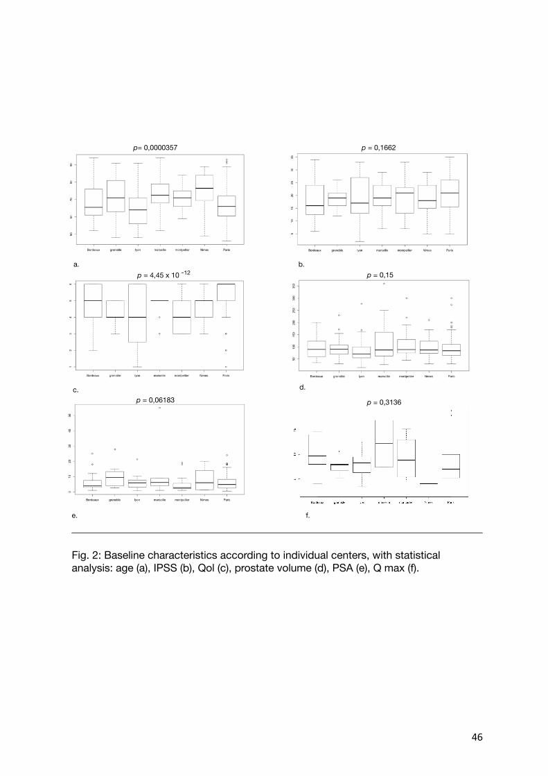
46
b. a.
c. d.
e. f.
Fig. 2: Baseline characteristics according to individual centers, with statistical analysis: age (a), IPSS (b), Qol (c), prostate volume (d), PSA (e), Q max (f).
p= 0,0000357 p = 0,1662
p = 4,45 x 10 p = 0,15
p = 0,06183
-12
p = 0,3136
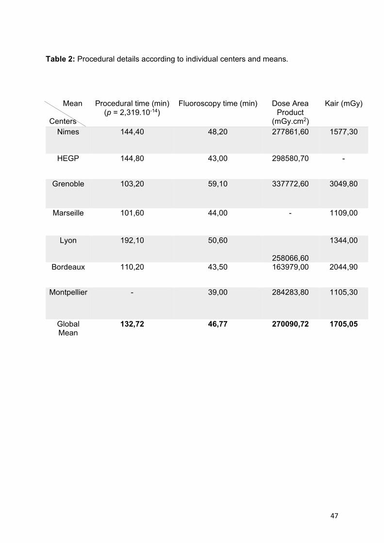
47
Table 2: Procedural details according to individual centers and means.
Mean Centers
Procedural time (min) (p = 2,319.10-14)
Fluoroscopy time (min)
Dose Area Product
(mGy.cm2)
Kair (mGy)
Nimes 144,40 48,20 277861,60 1577,30
HEGP 144,80 43,00 298580,70 -
Grenoble 103,20 59,10 337772,60 3049,80
Marseille 101,60 44,00 - 1109,00
Lyon 192,10 50,60
258066,60
1344,00
Bordeaux 110,20 43,50 163979,00 2044,90
Montpellier - 39,00 284283,80 1105,30
Global Mean
132,72 46,77 270090,72 1705,05
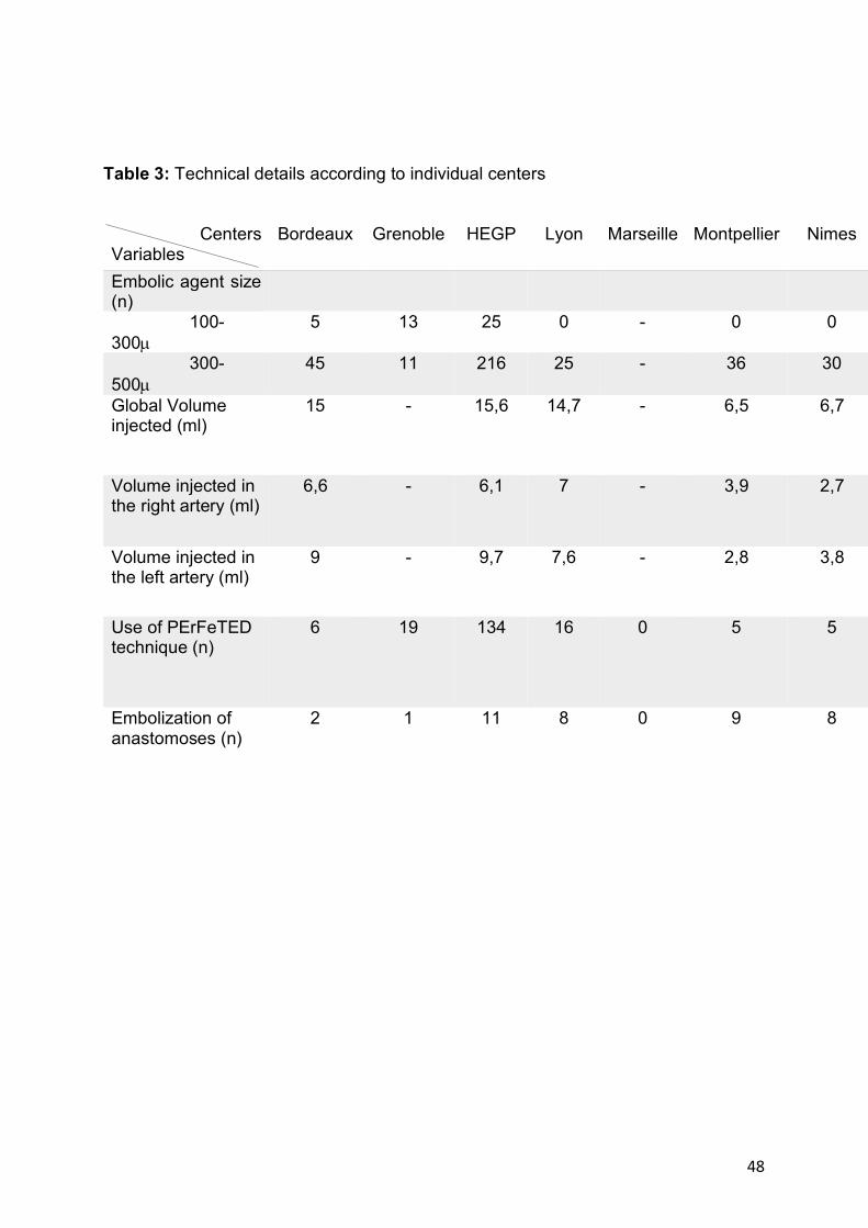
48
Table 3: Technical details according to individual centers
Centers Variables
Bordeaux Grenoble HEGP Lyon Marseille Montpellier Nimes
Embolic agent size (n)
100-300µ
5 13 25 0 - 0 0
300-500µ
45 11 216 25 - 36 30
Global Volume injected (ml)
15 - 15,6 14,7 - 6,5 6,7
Volume injected in the right artery (ml)
6,6 - 6,1 7 - 3,9 2,7
Volume injected in the left artery (ml)
9 - 9,7 7,6 - 2,8 3,8
Use of PErFeTED technique (n)
6 19 134 16 0 5 5
Embolization of anastomoses (n)
2 1 11 8 0 9 8
49
STATISTICAL ANALYSES Patients were separated into two groups: patients with bothersome LUTS due to BPH
and patients treated for acute/chronic urinary retention. Descriptive statistics, including
means and standard deviation, and ranges were used to summarize the continuous
variables in this study. Continuous variables included for analysis were demographical
and baseline data (age, prostate volume, PSA, IPSS, Qol, urine flow rate), imaging
data (global prostate volume, central prostate volume, percentage of prostate volume
decrease for global and central after embolization) and embolization data (technical
and procedural details), were analyzed using an analysis of variance (ANOVA).
Categoric variables were expressed as percentage.
Demographic data were compared using t test for the continuous variables and Chi-
square test for the categorical variable. Univariate and multivariate analyses were
performed with logistic regression analysis adjusting for population characteristics to
search for risk factors to predict early outcomes. The significative threshold was set as
p<0.02.
50
RESULTS
We collected data from seven different radiology departments set within university
hospitals and reported characteristics on Table 1.
Technical data:
PAE was technically successful in patients (98.5%). Bilateral embolization was
performed in 368 (89,3%) and unilateral in 38 (9,2%). PAE was carried out in one side
essentially because of anatomical reasons with PA occluded by atherosclerosis or in
out of reach because of extreme tortuoristy, atheromatous stenosis, spasme or
dissection. In some reports, unilateral failure was explained by the existence of
dangerous anastomoses, not reachable for protective embolization. Technical failure
was mainly due to atherosclerosis (5 patients).
PAE procedure time ranged from 40 to 300 minutes (mean 132,7 +/- 39 minutes) with
a mean Fluoroscopy time of 46,8 +/- 6,1 min. No skin injuries or other complication
related to radiation exposure have been reported during follow up. Procedures were
achieved under local anesthesia (Scarpa’s triangle) and/or sedation in the majority of
centers, aside Nimes’ radiological department were a systematic anesthetist
consultation recommending in some cases general anesthesia (GA) for patients’
comfort (19 patients among 23 cases of GA). The most common approach was the
single right femoral with 5F introducer (84,5%). Bilateral femoral approach was used
in 7 patients due to severe iliac artery tortuosity. Various types of hydrophilic intra
vascular catheters shape (Cobra, Sidewire, etc) and microcatheters were used, the
most frequently was the 2.0 ProgreatÒ (Terumo, Tokyo, Japan), to facilitate distal
catheterization and embolization. Cone-Beam computed Tomography (CBCT) was
noted in 348 patients, and not used in cases of inadequate morphology, in some
51
situations of inoperative machine or if not needed. We also considered CBCT as not
performed in cases where procedural imaging was not available and with no mention
about CBCT in the PAE report. The most frequent embolic agent used above the
different department was a trisacryl microparticles (EmbosphereÒ 300-500µ, Merit
Medical, Salt Lake City, USA) with a mixture of contrast and saline. The mean global
volume of embolic agent injected was 14,8 mL +/- 7,2 mL with a mean of 5,6 mL in the
right arteries and 7,6 mL in the left arteries. The PErFecTED technique as described
by Carnevale, was performed in 183 patients. Preventive embolization of dangerous
anastomoses was performed in 38 patients using micro-coils (1 or 2 mm).
Clinical data:
Among this series of 420 patients, 84 were referred to PAE after being recused to
surgery, 255 patients with no contra-indication to surgery, choose PAE as an
alternative treatment or were directly addressed by their urologist, 41 refused surgery
and for 40 patients we did not find the reason why PAE was indicated. Global
comorbidities were reported for 193 patients and cardiovascular comorbidities for 111
patients. Urological mean follow-up before PAE was estimated at 48,5 months. We
reported 350 patients undergoing medical treatment before PAE, among them 302
were treated by alpha-blockers, 73 by 5 alpha-reductase inhibitors and of which 66 by
combination therapy. Most of the patients with no medical treatment before PAE had
to stop them because of the secondary effects affecting the quality of life such as
retrograde ejaculation. Mean follow up after PAE was 82,2 days with a maximum follow
up of 937 days.
Out of 105 patients who had indwelling bladder catheterization due to acute urinary
retention 59 were removed among 85 patients with follow up (69,4%).
52
At the latest follow-up available, out of patients who were treated for severe LUTS,
IPSS mean improvement was 53,6 % (+/- 6,4%). Mean QoL after embolization was
1,73 with a mean improvement of 2,89 points. Global Clinical success during follow up,
including patients treated for LUTS/BPH and patients with indwelling catheter, was
achieved in 269 patients among 320 (84,1%). Global mean Qmax post embolization
was 12,3 mL/s with an average increase of 3,4 points.
PSA decreased below baseline at follow up by an average of 2,1 +/- 0,8 points with a
mean of 3,55 ng/mL after PAE which is considered as normal (< 4 ng/ml). An average
decrease in PVR post embolization of 51,8 +/- 26,8 ml (mean +/- SD) was noted.
91 patients were lost to follow up.
Complications observed were essentially minor with no need of hospitalization
including pelvic pain (18), perineal (4), rectal (3), or penile pain (3), dysuria (10), minor
hematuria (5), hematospermia (3), rectal bleeding (2) and isolated fever (15). More
serious complications were noted in few patients such as transitional penile ulceration
in 3 patients, resolutive after local treatments and acute urinary retention (3). We
reported one case of prostatitis with abscess of the prostatic gland. No case of
retrograde ejaculation or erectile dysfunction were reported outside of BPH medical
treatment’s side effects existing before PAE. No major complication such as death,
blood loss, severe urosepsis or prolongated hospitalization for other reasons due to
PAE were observed.
Imaging data:
We reported a total of 502 MRI (261 before PAE and 241 after PAE) and could analyze
188 MRI in this study, among them 57 patients had pre and post procedural MRI. Not
all patients had an MRI follow up and an unknown number of patients have had their
53
MRI pre/post PAE outside the different radiology departments where data were
collected, and we couldn’t have access to the imaging data. Imaging follow-up was
essentially through MRI from 1 to 12 months after PAE. We found 50 MRI performed
at 1 month or before, 128 between 1 and 3 months, 41 between 3 and 6 months and
22 after 6 months. We noted some variations concerning the delay between the
departments in this study, with early imaging follow-up in 2 centers (< 1 month), to
assess the infarct area expansion and more delayed in the others (> 1 month) in order
to evaluate the prostate volume decrease and the regression of adenomatous zones.
MRI sequences usually included a two plan T2-weighted and fat-suppressed T1-
Weighted pre and post-contrast. This was globally similar except in one center in which
early MRI protocol consisted on at-suppressed T1-Weighted pre and post-contrast to
for infarct volumes. Mean prostate global volume (PV) reduction was 22,7 %, and
central volume (CV) 16,7%.
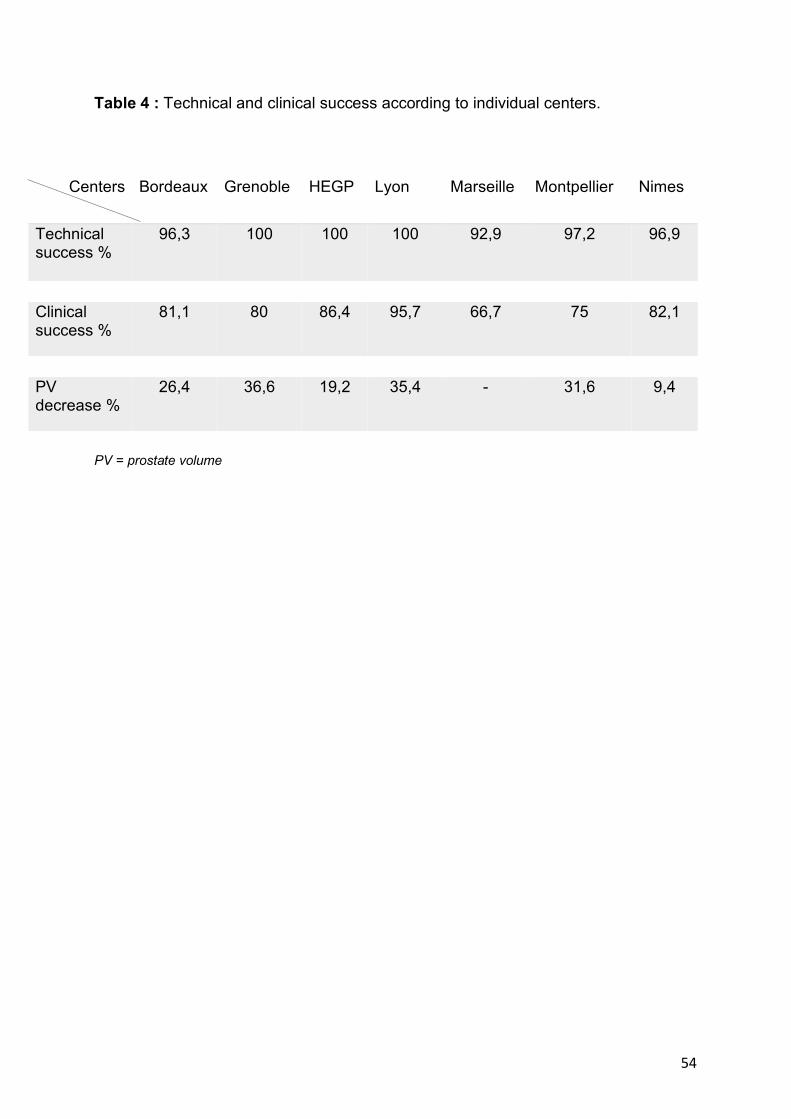
54
Table 4 : Technical and clinical success according to individual centers.
PV = prostate volume
Centers Bordeaux Grenoble HEGP Lyon Marseille Montpellier Nimes
Technical success %
96,3 100 100 100 92,9 97,2 96,9
Clinical success %
81,1 80 86,4 95,7 66,7 75 82,1
PV decrease %
26,4 36,6 19,2 35,4 - 31,6 9,4
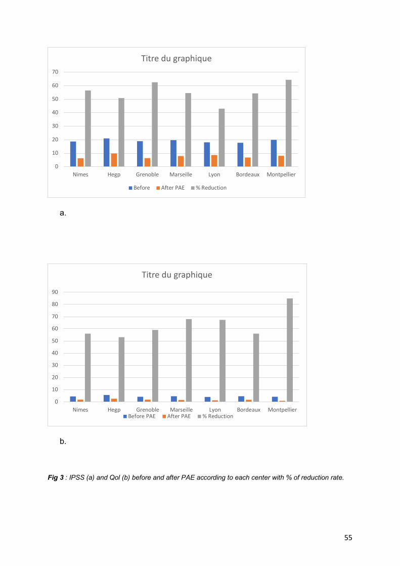
55
a.
b.
Fig 3 : IPSS (a) and Qol (b) before and after PAE according to each center with % of reduction rate.
0
10
20
30
40
50
60
70
Nimes Hegp Grenoble Marseille Lyon Bordeaux Montpellier
Titre du graphique
Before After PAE % Reduction
0
10
20
30
40
50
60
70
80
90
Nimes Hegp Grenoble Marseille Lyon Bordeaux Montpellier
Titre du graphique
Before PAE After PAE % Reduction
56
Table 5: PAE-related minor and major complications Adverse events N° patients %
Major 5 1,2 -Penile ulceration 3 0,7 -AUR 3 0,7 -Prostatis + abcess 1 0,2 Minor 60 14,5 -Dysuria 10 2,4 -Hematuria 5 1,2 -Hematospermia `3 0,7 -Pelvic pain 18 4,3 -Perineal pain 4 1 -Penile pain 3 0,7 -Rectal pain 3 0,7 -Rectal bleeding 2 0,5 -Fever 15 3,6
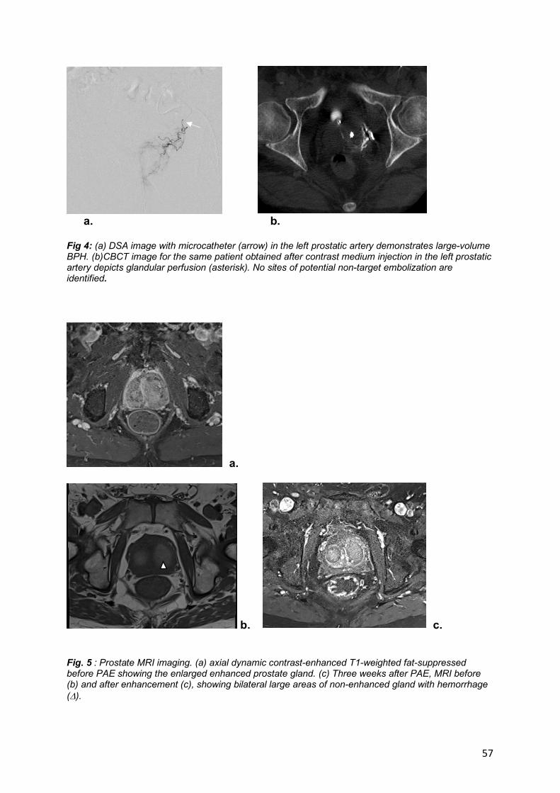
57
a. b.
Fig 4: (a) DSA image with microcatheter (arrow) in the left prostatic artery demonstrates large-volume BPH. (b)CBCT image for the same patient obtained after contrast medium injection in the left prostatic artery depicts glandular perfusion (asterisk). No sites of potential non-target embolization are identified.
a.
b. c. Fig. 5 : Prostate MRI imaging. (a) axial dynamic contrast-enhanced T1-weighted fat-suppressed before PAE showing the enlarged enhanced prostate gland. (c) Three weeks after PAE, MRI before (b) and after enhancement (c), showing bilateral large areas of non-enhanced gland with hemorrhage (D).
58
DISCUSSION
PAE clinical outcomes:
Minimally invasive procedures for the treatment of LUTS/BPH has become a
substantial research area.
This study supports findings of previous studies which suggest that the procedure of
PAE is safe and with low rates of morbidity and good outcomes. None of these several
reports was based on a French multicentric study. This paper tends to describe the
results of a large multicenter observational study of French patients across 7 centers.
A large area of indications for PAE and patients’ characteristics with no real limits
concerning age, prostate volume (ranging from 15 to 360 ml), IPSS (³ 5), Qol (³ 3),
Qmax (from 2,5 to 19 ml/s), PVR (from 0 to 650 ml) was reported and compared
between the different centers. Significative difference in populations’ mean age was
found between the studied centers (p<0,02), with the younger patients in Paris,
Bordeaux and Lyon (respectively 66, 65,5 and 64 yo). Patients were usually sent by
their urologists whose convictions in PAE efficacy might differ, some prefers to address
older patients with comorbidities hopping to avoid surgery, and others were maybe
more permissive to other mini-invasive treatment for their younger patients. Also,
numeral patients have chosen PAE when urologists expose the RTUP secondary
effects and other alternatives such as embolization, however we cannot affirm the
incidence as we didn’t classify all patients to whom PAE was proposed. No statistically
significant difference was observed in pre-procedural prostate volume and IPSS score,
these variables being certainly correlated to PAE indications46,48,49.
Differences in PSA, Qol and Qmax were to take with caution since significant data
were missing while collecting.
59
Through this work, we did demonstrate that PAE has several potential advantages
over traditional surgery therapies. It is usually performed under conscious sedation,
minimally invasive (via single arterial puncture), with a feasible outpatient procedure.
Global clinical success obtained was 84,0 % +/- 9,0 (mean +/- SD) with no statistically
significance difference between centers and in accordance with previous studies (80,7
– 97,1%). We present a median post PAE IPSS score of 7,7 with a mean reduction of
11,4 points and 53,1%. In terms of QoL, our PAE patients made a mean improvement
of 2,9 +/- 0,3 points with a mean Qol after PAE of 1,7 +/- 0,5.
We also found that PAE is more effective in case of symptomatic BPH than in
indwelling bladder catheter. This can result from the fact that patients with indwelling
catheters are older with more pathological vessels, impacting the embolization success
(89,3% of success rate for patients with symptomatic LUTS and versus 69,4% in
patients with indwelling catheter). We can also assume that this group of patients is
more refractory because of an advanced pathological condition.
The rate of technical and clinical success between the different departments is not
significantly different. However, there are significant difference in baseline patients’
characteristics concerning age, Qol score, PSA and Qmax. These differences in
results can be explained by the fact that each clinical team has its own clinical
judgement to make patients suitable for PAE and that the inclusion et exclusion criteria
are more flexible than randomized studies where morphological, clinical and
urodynamics parameters are more severe. Most clinical failures occurred at short-term
follow up (1 to 3 months).
Previous study showed a clinical success rate of 76.3% at long-term follow-up with low
morbidity, indicating PAE as a leading treatment option for patients with symptomatic
BPH27.
60
A statistically significant difference was observed concerning the procedural time with
a mean of 132,7 +/- 35,1 min. PAE requires a well-trained interventional radiologist
with a high level of expertise because of the complex prostatic vascular anatomy. We
know that there is a learning curve of 10-20 patients until the radiologist develops
appropriate skills. At the beginning of our experience, patients addressed for PAE
were essentially elderly men with high comorbidities recused to surgery and with no
other alternative therapy. These patients had atherosclerosis with stenosis and
tortuosity making the procedure more difficult and challenging, increasing the
procedure time, fluoroscopy time and radiation dose. More recently, PAE has been
offered to patients being refractory to medical treatment, and eligible to surgery, or
some of them refusing invasive therapies, being afraid from the secondary effects such
as erectile dysfunction et retrograde ejaculation and with less (vascular) comorbidities.
This recent enrolled population have contributed to improve technical and clinical
results. According to these facts and differences of BPH management and patients’
eligibility to PAE, we may explain this observation.
Factors affecting clinical outcomes
Baseline characteristics:
No difference in clinical success was revealed between the different centers, HEGP
being the reference trainer center. We can explain this similitude by the national
workshops organized by the HEGP. This suggest that despite of its technical
difficulties, PAE is widely feasible by interventional radiologists.
Age: a higher clinical success rate was observed for patients < 70 (88%) versus >70
(80%).
61
Medical treatment for LUTS due to BPH before PAE didn’t significantly influence the
clinical success of PAE. However, this variable must be taken carefully because of the
lack of uniformity even though the reference treatment consists on the alpha-blockers,
patients reports annoying side effects influencing their compliance.
Technical factors:
Embolic agent: Clinical success obtained was correlated to the volume of
microparticles injected with a higher rate when the global volume was > 15mL (92%)
versus > 15mL (85%), but with no significant difference. We also found a non-
significant difference of volume injected between right and left arteries, to the
advantage of the left artery which is almost always the first one to be embolized. This
can be explained by the existing intra prostatic anastomoses which are not necessarily
seen during the procedure.
Questions have been raised about the embolic agent sizes. In the literature, clinical
success has been achieved with different types of embolic agent and sizes ranging
from 100 to 500µm. In the present series, the most commonly used embolic agent
were the 300-500µm microsphere PVA particles. We can explain this similitude by the
national workshops organized by the HEGP. It has been presumed in some studies
that smaller particles would induce greater ischemia and PV reduction based on the
theory that it would permit a better penetration into the prostatic microvascular bed and
occlude the small intraprostatic anastomoses (<100µm) without additional
complications50. Hwang et al. has shown a significant difference in PV decrease after
PAE with microspheres compared to non-spherical PVA assuming that microspheres
have more chance to perform distal embolization. Torres et al. compared in a
prospective randomized trial, three groups of patients undergoing PAE with 100-300
62
versus 300-500 versus 100-300 + 300-500µm, resulting in no significant difference in
outcomes at short and medium-term follow-up (18 months)51.
Procedure time and fluoroscopy: significant variability in procedure time have been
reported in this study as well as in the literature (De assis et al.52: 158 et 54min; Pisco
et al.48 2013: 76 et 18 min; respectively procedure time and fluoroscopy time). Iliac
vessel tortuosity, pelvic vasculature variability and atherosclerosis may contribute to
procedural difficulty and challenge53. Du Pisani et al. found that subjective iliac artery
tortuosity grade and presence of intern iliac atherosclerosis are associated with
increased dose area product, fluoroscopy time and contrast volume54. Besides, this
study concerns only university hospitals counting a numerous of radiologists operating
with a various level of experience. Variables such as learning curve, small caliber
branches and elderly are thus involved in PAE increased difficulty. Knowledge of pelvic
arterial anatomy and peri procedural planning are essential to reduce procedure time
and non-target-embolization.
Unilatéral vs bilateral PAE: we reported 9,2% of unilateral PAE, considered as a partial
technical success with some operators planning a systematic re-embolization to further
enhance PAE improvement, when possible. Unilateral embolization reported rates
range approximatively from 0 to 15 %55,56. Some studies have shown clinical relief
following unilateral PAE. Herein, we reported a better clinical success with bilateral
embolization (86%) compared to unilateral (68%) with a statistically significant
difference (p<0,02). Bilhim et al. reported better clinical outcomes with bilateral PAE
compared to unilateral (75 % vs 50 %)55. This was strengthened by Carnevale et al.
finding more LUTS recurrence after unilateral PAE34. Our analyses support prior
63
observations, that is to say unilateral PAE was performed due to cases of
atherosclerosis stenosis/occlusion, vessels being too tortuous, which are correlated to
patient’s age.
Use of CBCT vs pre PAE CT
Correctly identifying PAs can be difficult and the use of ipsilateral oblique angiography
is not always sufficient especially with the overlapping of pelvic arteries. Pre-procedural
conventional angio-CT, with or without sublingual Glyceryl Trinitrate, have been shown
to improve PAs identification but is less used in our series. Pre-PAE CT has a
significant advantage in planning the procedure taking into account atherosclerotic
disease, evaluating stenosis and tortuosity, leading to an alternative primary access
site, and even aborting the procedure if technical success is compromised. Angio-
CBCT has become a common technique during PAE, allowing a three-dimensional
view of the detailed pelvic vascular anatomy and visualization of the parenchymal
enhancement of the prostatic gland. The rare cases where CBCT wasn’t performed
concerned overweighed patients. CBCT is a per procedural technique, which can be
performed either global, selective or both, and despite the increase in noise and
artifact, the intra-aortic/artery injection of contrast result in improvement of contrast to
noise ratio allowing a high-quality imaging recognizing PAs and dangerous
anastomoses. There is no RCT comparing prospectively the two techniques which can
be interesting to initiate in order to unify the planification of PAE procedure.
Other:
Arterial access: PAE is typically performed via transfemoral access (TFA) which is
widely used in our series (383/420; 91,2 % of procedures). However trans-radial
64
access is an increasing common method for arterial embolization in radiological
interventional procedures57–59. A more recent study concluded that PAE via trans-
radial or ulnar artery was not only safe but also associated with shorter procedure and
fluoroscopy time and lower contrast volume than TFA60. In our case, trans-radial
access was performed in a few proportion (35/420; 8,3%) and in more experienced
centers, because of the potential challenge related to the small artery diameter,
accessibility to appropriate material and the increased distance from the wrist to pelvic
vasculature not being in radiologists’ practice habits. Another advantage to be noticed
is the shorter time to ambulation resulting of less access site complications.
Clinical and imaging factors:
Prostate size: we found a correlation between prostate volume and clinical
improvement with PAE being more effective in large prostate gland higher than 100g.
This result represents the literature reports, demonstrating PAE effectiveness on large
prostate volume. Three published study of prospective single centers have shown PAE
efficacy with prostate volume greater than 80 and 90 ml52,61,62, including less than 90
patients. Herein, we strengthened these findings by evaluating this variable in 281
patients. Its reliability as a predictor of good clinical outcome has been contentious in
the literature, some studies supporting prostate size as an influence factor 63,64 and
others disproving that48. Some patients presented a significant decrease of PV with no
clinical improvement, this can be explained by the existing of other causes of LUTS
not sufficiently explored before PAE such as bladder dysfonction. To optimize PAE
results it can be interesting to recommend a complete urodynamic assessment to help
insure that LUTS are due to BPH.
65
Thus, the potential for PAE as an alternative treatment for prostates > 80-90g is
promising compared to surgical therapies, especially with OP remaining the reference
of care in men with very enlarged prostatic gland compared with TURP not being
considered for this population. In this perspective, we should also take into account the
volume decreases after PAE making patients eligible to TURP if additional treatment
is needed.
Post PAE MRI is widely used to evaluate the prostate volume because of its
reproducibility but also to assess structural and perfusion changes, estimating the
infarct pattern with the presence or absence of areas of devascularization65. The
percentage of necrosis can be correlated to the percentage of volume decrease over
time because the expected shrinking of the infarcts zones. It can be interesting to
analyze these variables in a long-term follow up.
Good clinical outcomes in both obstructive and irritative symptoms after PAE suggest
that prostate tissue changes, along with volume reduction, take part in improving
symptoms. However, the reasons to explain why some develops infarcts areas and
others not, remain unclear. Some authors suggest potential factors such as
embolization technique, type and volume of embolic agents, existing of collaterals and
tissue component43. T2 signal intensity in prostatic gland is known to be associated
with the histological substance of hyperplasia e.g. glandular and/or stromal. We can
hypothesize that these different components can be related to the vascularization of
the gland and that the correlation between T2 signal intensity and perfusion in pre-
procedural MRI and decrease of prostatic gland after PAE can be potentially
associated. Herein, neither these variables nor exact volume of infarction areas were
66
evaluated and that might present an interesting information in the comparative
statistics within the predictive factors.
Adverse events/Complications:
PAE has a low complication rate and no major complication was observed in the
present series. Three patients presented a transitional penile ulceration, three others
an acute urinary retention post embolization and one case of prostatitis with abscess
of the prostatic gland was reported. The other minor complications were locoregional
pain with the post-embolization syndrome and few cases of dysuria.
No patient needed a prolongated hospitalization except for elderly patients with
comorbidities. The role of Cone-beam 3D angiography paired with digital subtraction
angiography is known to be useful to analyze carefully anastomoses and identify non-
target vessels to avoid non-prostatic embolization. However, we surely underestimated
the rate of minor complications, especially the post embolization syndrome, because
patients were either examined by an outside physician with no tracing of the
information or did not think necessary to consult as they were well informed about the
minor risks and were provided appropriate preventive treatments.
Additionally, not only PAE can be an alternative to patients recused to surgery because
of comorbidities, but the demonstrated advantages of PAE regarding to outcome such
as no sexual dysfunction or ejaculatory disorder, low morbidity, no need of
hospitalization with a quick recovery and the possibility of ending the medical therapy
make it a good alternative for relatively young patients without comorbidities, refusing
surgery or taking an opportunity to postpone it. Furthermore, we strengthened that this
mini-invasive therapy has also shown its efficacy for large prostate gland21,52,62,66.
67
A variety of other minimally invasive therapies have been developed in recent years to
limit surgical morbidity. This includes the water vapor thermal therapy (RezumÔ
system, Boston Scientific), transforming sterile water into thermal energy causing
prostate cell necrosis via a transurethral approach. It has shown its safety and
effectiveness with significant mean improvement in IPSS and Qol, and no major
reports of sexual dysfunction67. However, it concerns PV<80 ml and no evidence of
the long-term efficacy has been demonstrated yet. Aquablation consists on dissection
of prostate tissue via a high-pressure saline, with a procedure time under 10 minutes,
but is not suitable for large prostates (>100mL) and with no long-term study. The
transurethral microwave therapy (TUMT) is not developed in France and is less
efficient than RTUP especially in cases of AUR. Mechanical strategies include prostatic
urethral lift (UroliftÔ), intra prostatic stents and temporary implantable nitinol device
which are quick procedures, under local anesthesia, yet limited techniques in cases of
obstructive median lobe, previous prostate surgery and large prostate volume. These
techniques provide a less significant symptom improvement compared to TURP9.
Intra-Prostatic stents are also not well tolerated (increase of LUTS and perineal pain)
with a failure rate of 16% at one year and 27% at 5 years and should be proposed in
exceptional cases of surgical contra indication9.
All these techniques, which many are under study, challenge the reference technique
(RTUP), allowing personalized approaches for a select group of patients and with less
urinary and sexual side effects. Nevertheless, prostate size represents their major limit
(less than 100-120mL)68.
PAE has some disadvantages, it can be technically limited by major atherosclerosis
condition with stenosis, occlusion and vessels tortuosity. Minor side effects suggest
68
that nontarget embolization may occur even if not detected during the procedure
because of very small anastomoses hardly detected despite the available techniques.
Radiation exposure should be carefully monitored because of the prolongated
fluoroscopy time that occasionally happens. PAE clinical failure rate can be explained
by multiple reasons. First, the very large inclusion criteria reported in this study
compared to the literature, especially the prostate volume size and IPSS score
variables might contribute to the non-response rate and the fact that we obtained a
clinical success rate in the low average of previous study reports. Besides, we
compared PAE with a heterogenous population of radiologists performing this
challenging procedure with its complex pelvic artery anatomy.
This study has some limitations. The most important one was the retrospective
multicentric characteristic generating some bias and several patients were lost to
follow-up. In this large multicentric study, heterogeneous groups of patients’
management, various techniques and practices of PAE with diverse follow-up terms
and imaging modalities were collected. In addition, in this non-randomized study, there
was no control group of patients undergoing other BPH therapies.
Furthermore, there is a significant gap in the number of patients recruited for PAE
between centers, some of them performing few procedures per a month compared to
a bigger department of interventional radiology such as “HEGP”, operating 3 to 4 PAE
per a week, probably influencing the clinical and technical success. In addition,
procedures can be executed by a various population of radiologists which we didn’t
aim to analyze in this study but can be an impacting factor on some variables.
As a consequence, the heterogeneity of radiologists’ practice and experience level can
confound associations found in this study. Clinical outcomes may therefore not reflect
69
the real potential of PAE for the treatment of symptomatic BPH. Besides, operating
time and dose outcomes can also be related to radiologist’s experience, which is
necessary to optimize techniques.
Different modalities of volume measurements were used including MRI, US and CT
imaging with US involving different operator with a considerable variability generating
an important measurement bias.
The patients recruited and treated by PAE may have different epidemiological
characteristics above the different centers, partly explained by urologists’ patterns of
working and convictions, PAE being considered as an experience procedure due to
long-term follow up lacking compared to TURP. Available equipment may also differ
between centers and lead to confounding results in addition to radiologist experience
while performing PAE.
A High non-response rate of the patients to the questionnaires (IPSS, QoL) was
observed. The IPSS overall response rate after PAE was 106 over 310 patients with
symptomatic BPH.
Adding to this, a monitoring bias and the lack of long-term follow up data are to take
into account, especially in comparison with conventional surgical treatments. Knowing
that revascularization and regrowth of the prostate gland may occur at long-term follow
up causing symptoms recurrence, rigorous longer monitoring up to 6 years and more
is to be intended.
We didn’t report in the present study the cases where medical treatment has been
definitely/never stopped or took back in some period. This variable is important
because some patients were eligible to PAE because of the secondary effects of the
medical treatments and in some cases, they had to pursue this therapy because of
insufficient effect. Furthermore, some physicians maintain voluntarily the medical
70
therapy after PAE especially in cases of severe LUTS and that represents another
confounding variable in our study. In fact, it influences results after PAE skewing the
clinical scores and particularly the sexual function.
Data collection and image interpretation was an obstacle in this retrospective study,
and all of the variables were gathered by a fifth-year radiological student in a short time
period with possible mistakes. Some variables had a low patient number compared to
others which can explain their limit of significance in statistical analysis.
However, this registry study design has the benefit to point out a more real-world
regarding to PAE in France. The greatest advantage of this study was to provide a
multicentric data collection with a consequent number of patients (420), which is
lacking in the literature. Although it has been recommended to select patients eligible
for PAE with strict inclusion criteria, we obtained good clinical outcomes among the
different centers with their similarities and discrepancies. Along with predictive models,
these results can help patient selection, planning procedural technique and initiate
trainee to this challenging technique.
71
Conclusion
The main aim of this study was to provide a snapshot of PAE technique in France.
We also confirm the safety and feasibility of PAE in the different centers with no
mortality and a very low rate of morbidity.
The findings can inform future clinical practice and policy guidance on the treatment of
LUTS / BPH. Although it is not a randomized controlled trial (RCT), this multicentric
French study based on PAE can be a complementary perspective to international
RCTs, with reflecting a more real-life practice by recruiting a varied patient group.
72
Bibliography 1. McVary KT, Roehrborn CG, Avins AL, et al. Update on AUA Guideline on the
Management of Benign Prostatic Hyperplasia. J Urol. 2011;185(5):1793-1803. doi:10.1016/j.juro.2011.01.074
2. Fibbi B, Penna G, Morelli A, Adorini L, Maggi M. Chronic inflammation in the pathogenesis of benign prostatic hyperplasia: BPH and inflammation. Int J Androl. 2009;33(3):475-488. doi:10.1111/j.1365-2605.2009.00972.x
3. Calogero AE, Burgio G, Condorelli RA, Cannarella R, La Vignera S. Lower urinary tract symptoms/benign prostatic hyperplasia and erectile dysfunction: from physiology to clinical aspects. Aging Male. 2018;21(4):261-271. doi:10.1080/13685538.2018.1430758
4. Kupelian V. Prevalence of Lower Urinary Tract Symptoms and Effect on Quality of Life in a Racially and Ethnically Diverse Random Sample: The Boston Area Community Health (BACH) Survey. Arch Intern Med. 2006;166(21):2381. doi:10.1001/archinte.166.21.2381
5. Eckhardt MD, Van Venrooij GEPM, Van Melick HHE, Boon TA. PREVALENCE AND BOTHERSOMENESS OF LOWER URINARY TRACT SYMPTOMS IN BENIGN PROSTATIC HYPERPLASIA AND THEIR IMPACT ON WELL-BEING. J Urol. 2001;166(2):563-568. doi:10.1016/S0022-5347(05)65985-X
6. Presicce F, De Nunzio C, Tubaro A. Is Early Benign Prostatic Hyperplasia (BPH) Treatment Worthwhile? Urol J. 2017;84(3):142-147. doi:10.5301/uj.5000251
7. Gratzke C, Bachmann A, Descazeaud A, et al. EAU Guidelines on the Assessment of Non-neurogenic Male Lower Urinary Tract Symptoms including Benign Prostatic Obstruction. Eur Urol. 2015;67(6):1099-1109. doi:10.1016/j.eururo.2014.12.038
8. Gul ZG, Kaplan SA. BPH: Why Do Patients Fail Medical Therapy? Curr Urol Rep. 2019;20(7):40. doi:10.1007/s11934-019-0899-z
9. Foster HE, Barry MJ, Dahm P, et al. Surgical Management of Lower Urinary Tract Symptoms Attributed to Benign Prostatic Hyperplasia: AUA Guideline. J Urol. 2018;200(3):612-619. doi:10.1016/j.juro.2018.05.048
10. Reich O, Gratzke C, Stief CG. Techniques and Long-Term Results of Surgical Procedures for BPH. Eur Urol. 2006;49(6):970-978. doi:10.1016/j.eururo.2005.12.072
11. Zhang Y, Yuan P, Ma D, et al. Efficacy and safety of enucleation vs. resection of prostate for treatment of benign prostatic hyperplasia: a meta-analysis of randomized controlled trials. Prostate Cancer Prostatic Dis. February 2019. doi:10.1038/s41391-019-0135-4
12. Reimann M, Fishman N, Lichy I, et al. Outcome of Photoselective Vaporization of the Prostate with the GreenLight-XPS 180 Watt System Compared to
73
Transurethral Resection of the Prostate. J Clin Med. 2019;8(7):1004. doi:10.3390/jcm8071004
13. Boeri L, Capogrosso P, Ventimiglia E, et al. Clinical Comparison of Holmium Laser Enucleation of the Prostate and Bipolar Transurethral Enucleation of the Prostate in Patients Under Either Anticoagulation or Antiplatelet Therapy. Eur Urol Focus. March 2019:S240545691930077X. doi:10.1016/j.euf.2019.03.002
14. Koshiba K, Egawa S, Ohori M, Uchida T, Yokoyama E, Shoji K. Does Transurethral Resection of the Prostate Pose a Risk to Life? 22-Year Outcome. J Urol. 1995;153(5):1506-1509. doi:10.1016/S0022-5347(01)67447-0
15. Sidney S, Quesenberry CP, Sadler MC, Cattolica EV, Lydick EG, Guess HA. Reoperation and Mortality After Surgical Treatment of Benign Prostatic Hypertrophy in a Large Prepaid Medical Care Program: Med Care. 1992;30(2):117-125. doi:10.1097/00005650-199202000-00003
16. Madersbacher S, Lackner J, Brössner C, et al. Reoperation, Myocardial Infarction and Mortality after Transurethral and Open Prostatectomy: A Nation-Wide, Long-Term Analysis of 23,123 Cases. Eur Urol. 2005;47(4):499-504. doi:10.1016/j.eururo.2004.12.010
17. Eredics K, Wachabauer D, Röthlin F, Madersbacher S, Schauer I. Reoperation Rates and Mortality After Transurethral and Open Prostatectomy in a Long-term Nationwide Analysis: Have We Improved Over a Decade? Urology. 2018;118:152-157. doi:10.1016/j.urology.2018.04.032
18. Magistro G, Chapple CR, Elhilali M, et al. Emerging Minimally Invasive Treatment Options for Male Lower Urinary Tract Symptoms. Eur Urol. 2017;72(6):986-997. doi:10.1016/j.eururo.2017.07.005
19. Roehrborn CG. Morbidity, Mortality and Early Outcome of Transurethral Resection of the Prostate: A Prospective Multicenter Evaluation of 10,654 Patients. Yearb Urol. 2008;2008:93-95. doi:10.1016/S0084-4071(08)79167-X
20. McWilliams JP, Kuo MD, Rose SC, et al. Society of Interventional Radiology Position Statement: Prostate Artery Embolization for Treatment of Benign Disease of the Prostate. J Vasc Interv Radiol. 2014;25(9):1349-1351. doi:10.1016/j.jvir.2014.05.005
21. Carnevale FC, Antunes AA. Prostatic Artery Embolization for Enlarged Prostates Due to Benign Prostatic Hyperplasia. How I Do It. Cardiovasc Intervent Radiol. 2013;36(6):1452-1463. doi:10.1007/s00270-013-0680-5
22. Young M, Elmussareh M, Morrison T, Wilson J. The changing practice of transurethral resection of the prostate. Ann R Coll Surg Engl. 2018;100(4):326-329. doi:10.1308/rcsann.2018.0054
23. Mitchell ME, Waltman AC, Athanasoulis CA, Kerr WS, Dretler SP. Control of Massive Prostatic Bleeding with Angiographic Techniques. J Urol. 1976;115(6):692-695. doi:10.1016/S0022-5347(17)59339-8
74
24. Michel F, Dubruille T, Cercueil J-P, Paparel P, Cognet F, Krause D. Arterial Embolization for Massive Hematuria Following Transurethral Prostatectomy. J Urol. 2002;168(6):2550-2551. doi:10.1016/S0022-5347(05)64200-0
25. DeMeritt JS, Elmasri FF, Esposito MP, Rosenberg GS. Relief of Benign Prostatic Hyperplasia-related Bladder Outlet Obstruction after Transarterial Polyvinyl Alcohol Prostate Embolization. J Vasc Interv Radiol. 2000;11(6):767-770. doi:10.1016/S1051-0443(07)61638-8
26. Carnevale FC, Antunes AA, da Motta Leal Filho JM, et al. Prostatic artery embolization as a primary treatment for benign prostatic hyperplasia: preliminary results in two patients. Cardiovasc Intervent Radiol. 2010;33(2):355-361. doi:10.1007/s00270-009-9727-z
27. Pisco JM, Bilhim T, Pinheiro LC, et al. Medium- and Long-Term Outcome of Prostate Artery Embolization for Patients with Benign Prostatic Hyperplasia: Results in 630 Patients. J Vasc Interv Radiol. 2016;27(8):1115-1122. doi:10.1016/j.jvir.2016.04.001
28. Hacking N, Vigneswaran G, Maclean D, et al. Technical and Imaging Outcomes from the UK Registry of Prostate Artery Embolization (UK-ROPE) Study: Focusing on Predictors of Clinical Success. Cardiovasc Intervent Radiol. 2019;42(5):666-676. doi:10.1007/s00270-018-02156-8
29. Amouyal G, Thiounn N, Pellerin O, et al. Clinical Results After Prostatic Artery Embolization Using the PErFecTED Technique: A Single-Center Study. Cardiovasc Intervent Radiol. 2016;39(3):367-375. doi:10.1007/s00270-015-1267-0
30. Yamaki K-I, Saga T, Doi Y, Aida K, Yoshizuka M. A Statistical Study of the Branching of the Human Internal Iliac Artery. Kurume Med J. 1998;45(4):333-340. doi:10.2739/kurumemedj.45.333
31. Bilhim T, Casal D, Furtado A, Pais D, O’Neill JEG, Pisco JM. Branching patterns of the male internal iliac artery: imaging findings. Surg Radiol Anat. 2011;33(2):151-159. doi:10.1007/s00276-010-0716-3
32. de Assis AM, Moreira AM, de Paula Rodrigues VC, et al. Pelvic Arterial Anatomy Relevant to Prostatic Artery Embolisation and Proposal for Angiographic Classification. Cardiovasc Intervent Radiol. 2015;38(4):855-861. doi:10.1007/s00270-015-1114-3
33. Carnevale FC, Soares GR, de Assis AM, Moreira AM, Harward SH, Cerri GG. Anatomical Variants in Prostate Artery Embolization: A Pictorial Essay. Cardiovasc Intervent Radiol. 2017;40(9):1321-1337. doi:10.1007/s00270-017-1687-0
34. Carnevale FC, Moreira AM, Harward SH, et al. Recurrence of Lower Urinary Tract Symptoms Following Prostate Artery Embolization for Benign Hyperplasia: Single Center Experience Comparing Two Techniques. Cardiovasc Intervent Radiol. 2017;40(3):366-374. doi:10.1007/s00270-017-1569-5
75
35. Bilhim T, Pisco JM, Rio Tinto H, et al. Prostatic Arterial Supply: Anatomic and Imaging Findings Relevant for Selective Arterial Embolization. J Vasc Interv Radiol. 2012;23(11):1403-1415. doi:10.1016/j.jvir.2012.07.028
36. Bagla S, Rholl KS, Sterling KM, et al. Utility of Cone-Beam CT Imaging in Prostatic Artery Embolization. J Vasc Interv Radiol. 2013;24(11):1603-1607. doi:10.1016/j.jvir.2013.06.024
37. Chiaradia M, Radaelli A, Campeggi A, Bouanane M, De La Taille A, Kobeiter H. Automatic Three-Dimensional Detection of Prostatic Arteries Using Cone-Beam CT during Prostatic Arterial Embolization. J Vasc Interv Radiol. 2015;26(3):413-417. doi:10.1016/j.jvir.2014.11.009
38. Wang MQ, Duan F, Yuan K, Zhang GD, Yan J, Wang Y. Benign Prostatic Hyperplasia: Cone-Beam CT in Conjunction with DSA for Identifying Prostatic Arterial Anatomy. Radiology. 2017;282(1):271-280. doi:10.1148/radiol.2016152415
39. Desai H, Yu H, Ohana E, Gunnell ET, Kim J, Isaacson A. Comparative Analysis of Cone-Beam CT Angiogram and Conventional CT Angiogram for Prostatic Artery Identification Prior to Embolization. J Vasc Interv Radiol. 2018;29(2):229-232. doi:10.1016/j.jvir.2017.09.020
40. Chen C-B, Chou C-T, Chen Y-L. Cone-beam CT findings during prostate artery embolization for benign prostatic hyperplasia-induced lower urinary tract symptoms: a case report. BMC Urol. 2017;17(1):120. doi:10.1186/s12894-017-0311-6
41. Schnall MD, Pollack HM. Magnetic resonance imaging of the prostate gland. Urol Radiol. 1990;12(1):109-114. doi:10.1007/BF02923982
42. Frenk NE, Baroni RH, Carnevale FC, et al. MRI Findings After Prostatic Artery Embolization for Treatment of Benign Hyperplasia. Am J Roentgenol. 2014;203(4):813-821. doi:10.2214/AJR.13.11692
43. Kisilevzky N, Faintuch S. MRI assessment of prostatic ischaemia: best predictor of clinical success after prostatic artery embolisation for benign prostatic hyperplasia. Clin Radiol. 2016;71(9):876-882. doi:10.1016/j.crad.2016.05.003
44. Harris PA, Taylor R, Thielke R, Payne J, Gonzalez N, Conde JG. Research electronic data capture (REDCap)—A metadata-driven methodology and workflow process for providing translational research informatics support. J Biomed Inform. 2009;42(2):377-381. doi:10.1016/j.jbi.2008.08.010
45. Harris PA, Taylor R, Minor BL, et al. The REDCap consortium: Building an international community of software platform partners. J Biomed Inform. 2019;95:103208. doi:10.1016/j.jbi.2019.103208
46. Gao Y, Huang Y, Zhang R, et al. Benign Prostatic Hyperplasia: Prostatic Arterial Embolization versus Transurethral Resection of the Prostate—A Prospective, Randomized, and Controlled Clinical Trial. Radiology. 2014;270(3):920-928. doi:10.1148/radiol.13122803
76
47. Grosso M, Balderi A, Arnò M, et al. Prostatic artery embolization in benign prostatic hyperplasia: preliminary results in 13 patients. Radiol Med (Torino). 2015;120(4):361-368. doi:10.1007/s11547-014-0447-3
48. Pisco JM, Rio Tinto H, Campos Pinheiro L, et al. Embolisation of prostatic arteries as treatment of moderate to severe lower urinary symptoms (LUTS) secondary to benign hyperplasia: results of short- and mid-term follow-up. Eur Radiol. 2013;23(9):2561-2572. doi:10.1007/s00330-012-2714-9
49. Lebdai S, Delongchamps NB, Sapoval M, et al. Early results and complications of prostatic arterial embolization for benign prostatic hyperplasia. World J Urol. 2016;34(5):625-632. doi:10.1007/s00345-015-1665-6
50. Wang MQ, Zhang JL, Xin HN, et al. Comparison of Clinical Outcomes of Prostatic Artery Embolization with 50-μm Plus 100-μm Polyvinyl Alcohol (PVA) Particles versus 100-μm PVA Particles Alone: A Prospective Randomized Trial. J Vasc Interv Radiol. 2018;29(12):1694-1702. doi:10.1016/j.jvir.2018.06.019
51. Torres D, Costa NV, Pisco J, Pinheiro LC, Oliveira AG, Bilhim T. Prostatic Artery Embolization for Benign Prostatic Hyperplasia: Prospective Randomized Trial of 100–300 μm versus 300–500 μm versus 100- to 300-μm + 300- to 500-μm Embospheres. J Vasc Interv Radiol. 2019;30(5):638-644. doi:10.1016/j.jvir.2019.02.014
52. de Assis AM, Moreira AM, de Paula Rodrigues VC, et al. Prostatic Artery Embolization for Treatment of Benign Prostatic Hyperplasia in Patients with Prostates > 90 g: A Prospective Single-Center Study. J Vasc Interv Radiol. 2015;26(1):87-93. doi:10.1016/j.jvir.2014.10.012
53. Wolf YG, Tillich M, Lee WA, Rubin GD, Fogarty TJ, Zarins CK. Impact of aortoiliac tortuosity on endovascular repair of abdominal aortic aneurysms: Evaluation of 3D computer-based assessment. J Vasc Surg. 2001;34(4):594-599. doi:10.1067/mva.2001.118586
54. du Pisanie J, Abumoussa A, Donovan K, Stewart J, Bagla S, Isaacson A. Predictors of Prostatic Artery Embolization Technical Outcomes: Patient and Procedural Factors. J Vasc Interv Radiol. 2019;30(2):233-240. doi:10.1016/j.jvir.2018.09.014
55. Bilhim T, Pisco J, Rio Tinto H, et al. Unilateral Versus Bilateral Prostatic Arterial Embolization for Lower Urinary Tract Symptoms in Patients with Prostate Enlargement. Cardiovasc Intervent Radiol. 2013;36(2):403-411. doi:10.1007/s00270-012-0528-4
56. Gonçalves OM, Carnevale FC, Moreira AM, Antunes AA, Rodrigues VC, Srougi M. Comparative Study Using 100–300 Versus 300–500 μm Microspheres for Symptomatic Patients Due to Enlarged-BPH Prostates. Cardiovasc Intervent Radiol. 2016;39(10):1372-1378. doi:10.1007/s00270-016-1443-x
57. Fischman AM, Horn CJ, Resnick NJ, et al. Uterine artery embolization using a transradial approach: initial experience and technique. J Vasc Interv Radiol. 2014;25(3):S67-S68. doi:10.1016/j.jvir.2013.12.169
77
58. Posham R, Biederman DM, Patel RS, et al. Transradial Approach for Noncoronary Interventions: A Single-Center Review of Safety and Feasibility in the First 1,500 Cases. J Vasc Interv Radiol. 2016;27(2):159-166. doi:10.1016/j.jvir.2015.10.026
59. Thakor AS, Alshammari MT, Liu DM, et al. Transradial Access for Interventional Radiology: Single-Centre Procedural and Clinical Outcome Analysis. Can Assoc Radiol J. 2017;68(3):318-327. doi:10.1016/j.carj.2016.09.003
60. Bhatia S, Harward SH, Sinha VK, Narayanan G. Prostate Artery Embolization via Transradial or Transulnar versus Transfemoral Arterial Access: Technical Results. J Vasc Interv Radiol. 2017;28(6):898-905. doi:10.1016/j.jvir.2017.02.029
61. Kurbatov D, Russo GI, Lepetukhin A, et al. Prostatic Artery Embolization for Prostate Volume Greater Than 80 cm3: Results From a Single-center Prospective Study. Urology. 2014;84(2):400-404. doi:10.1016/j.urology.2014.04.028
62. Bagla S, Smirniotopoulos JB, Orlando JC, van Breda A, Vadlamudi V. Comparative Analysis of Prostate Volume as a Predictor of Outcome in Prostate Artery Embolization. J Vasc Interv Radiol. 2015;26(12):1832-1838. doi:10.1016/j.jvir.2015.08.018
63. Wang M, Guo L, Duan F, et al. Prostatic arterial embolization for the treatment of lower urinary tract symptoms caused by benign prostatic hyperplasia: a comparative study of medium- and large-volume prostates. BJU Int. 2016;117(1):155-164. doi:10.1111/bju.13147
64. Maclean D, Harris M, Drake T, et al. Factors Predicting a Good Symptomatic Outcome After Prostate Artery Embolisation (PAE). Cardiovasc Intervent Radiol. 2018;41(8):1152-1159. doi:10.1007/s00270-018-1912-5
65. Brook OR, Faintuch S, Brook A, Goldberg SN, Rofsky NM, Lenkinski RE. Embolization therapy for benign prostatic hyperplasia: Influence of embolization particle size on gland perfusion: Embolization for BPH. J Magn Reson Imaging. 2013;38(2):380-387. doi:10.1002/jmri.23981
66. Bhatia S, Sinha VK, Harward S, Gomez C, Kava BR, Parekh DJ. Prostate Artery Embolization in Patients with Prostate Volumes of 80 mL or More: A Single-Institution Retrospective Experience of 93 Patients. J Vasc Interv Radiol. 2018;29(10):1392-1398. doi:10.1016/j.jvir.2018.05.012
67. Kaplan SA. Re: Three-Year Outcomes of the Prospective, Randomized Controlled Rezūm System Study: Convective Radiofrequency Thermal Therapy for Treatment of Lower Urinary Tract Symptoms Due to Benign Prostatic Hyperplasia. J Urol. 2018;199(5):1085-1088. doi:10.1016/j.juro.2018.02.046
68. Srinivasan A, Wang R. An Update on Minimally Invasive Surgery for Benign Prostatic Hyperplasia: Techniques, Risks, and Efficacy. World J Mens Health. 2019;37. doi:10.5534/wjmh.190076
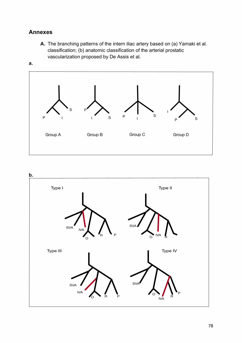
78
Annexes
A. The branching patterns of the intern iliac artery based on (a) Yamaki et al. classification; (b) anatomic classification of the arterial prostatic vascularization proposed by De Assis et al.
a.
b.
S
IP
P
I S P IS
I
P S
Group A Group B Group C Group D
SVA IVA
OPR
OO
O
SVA
SVASVA
IVA
R R
R
PPIVA
IVA
Type I Type II
Type III Type IV
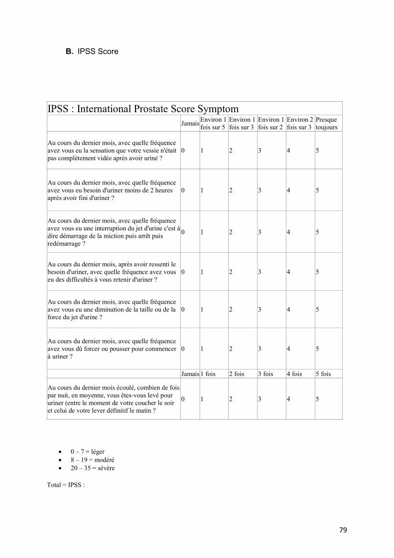
79
B. IPSS Score
IPSS : International Prostate Score Symptom Jamais Environ 1
fois sur 5 Environ 1 fois sur 3
Environ 1 fois sur 2
Environ 2 fois sur 3
Presque toujours
Au cours du dernier mois, avec quelle fréquence avez vous eu la sensation que votre vessie n'était pas complètement vidée après avoir uriné ?
0 1 2 3 4 5
Au cours du dernier mois, avec quelle fréquence avez vous eu besoin d'uriner moins de 2 heures après avoir fini d'uriner ?
0 1 2 3 4 5
Au cours du dernier mois, avec quelle fréquence avez vous eu une interruption du jet d'urine c'est à dire démarrage de la miction puis arrêt puis redémarrage ?
0 1 2 3 4 5
Au cours du dernier mois, après avoir ressenti le besoin d'uriner, avec quelle fréquence avez vous eu des difficultés à vous retenir d'uriner ?
0 1 2 3 4 5
Au cours du dernier mois, avec quelle fréquence avez vous eu une diminution de la taille ou de la force du jet d'urine ?
0 1 2 3 4 5
Au cours du dernier mois, avec quelle fréquence avez vous dû forcer ou pousser pour commencer à uriner ?
0 1 2 3 4 5
Jamais 1 fois 2 fois 3 fois 4 fois 5 fois
Au cours du dernier mois écoulé, combien de fois par nuit, en moyenne, vous êtes-vous levé pour uriner (entre le moment de votre coucher le soir et celui de votre lever définitif le matin ?
0 1 2 3 4 5
• 0 – 7 = léger • 8 – 19 = modéré • 20 – 35 = sévère
Total = IPSS :

80
C. Qol score Évaluation de la qualité de vie liée aux symptômes urinaires Très
satisfait Satisfait Plutôt satisfait
Partagé (ni satisfait, ni ennuyé)
Plutôt ennuyé Ennuyé Très
ennuyé
Si vous deviez vivre le restant de votre vie avec cette manière d'uriner, diriez-vous que vous en seriez :
0 1 2 3 4 5 6
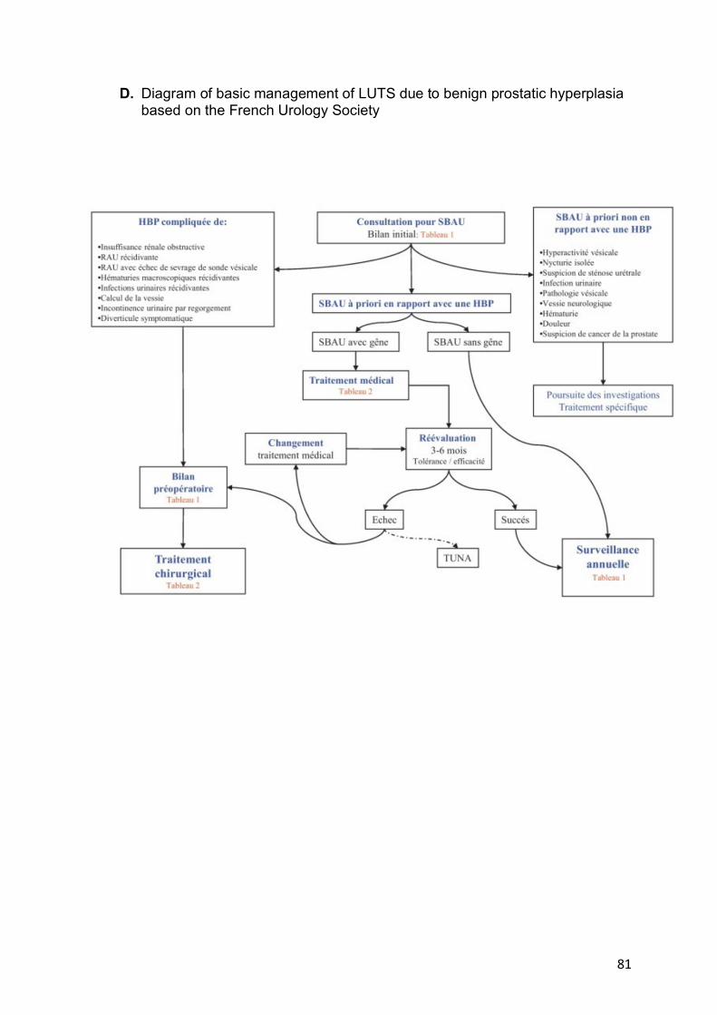
81
D. Diagram of basic management of LUTS due to benign prostatic hyperplasia based on the French Urology Society
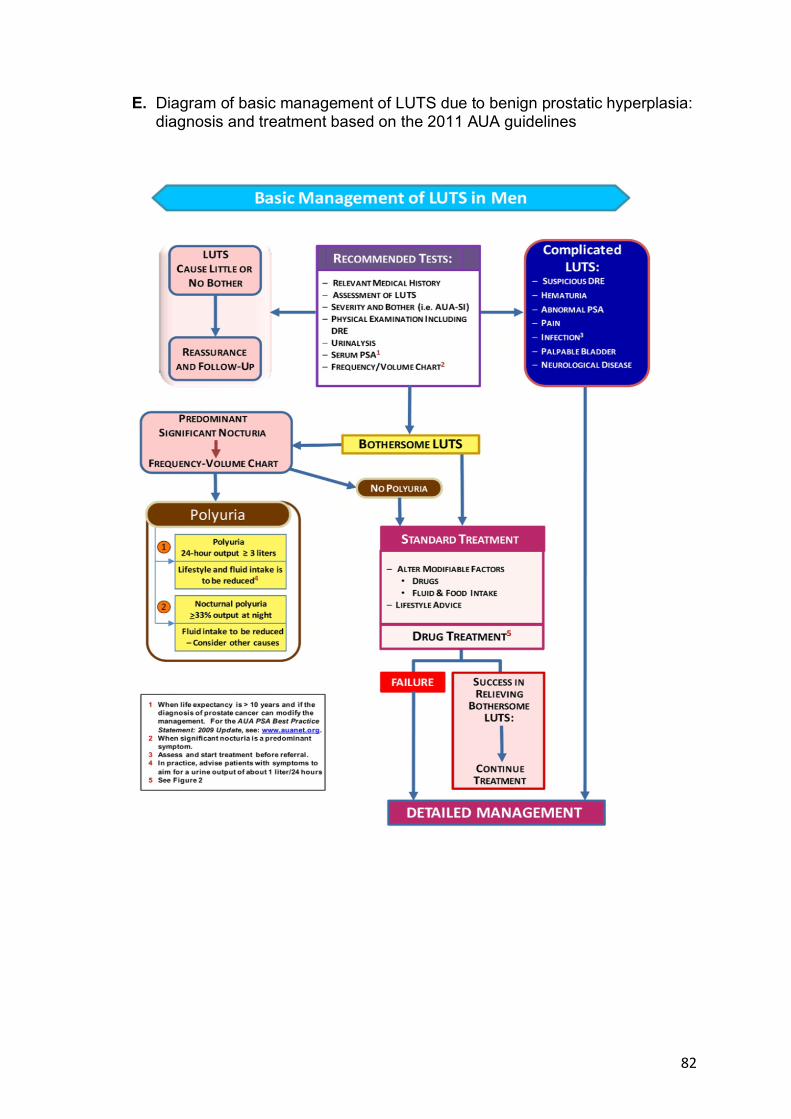
82
E. Diagram of basic management of LUTS due to benign prostatic hyperplasia: diagnosis and treatment based on the 2011 AUA guidelines
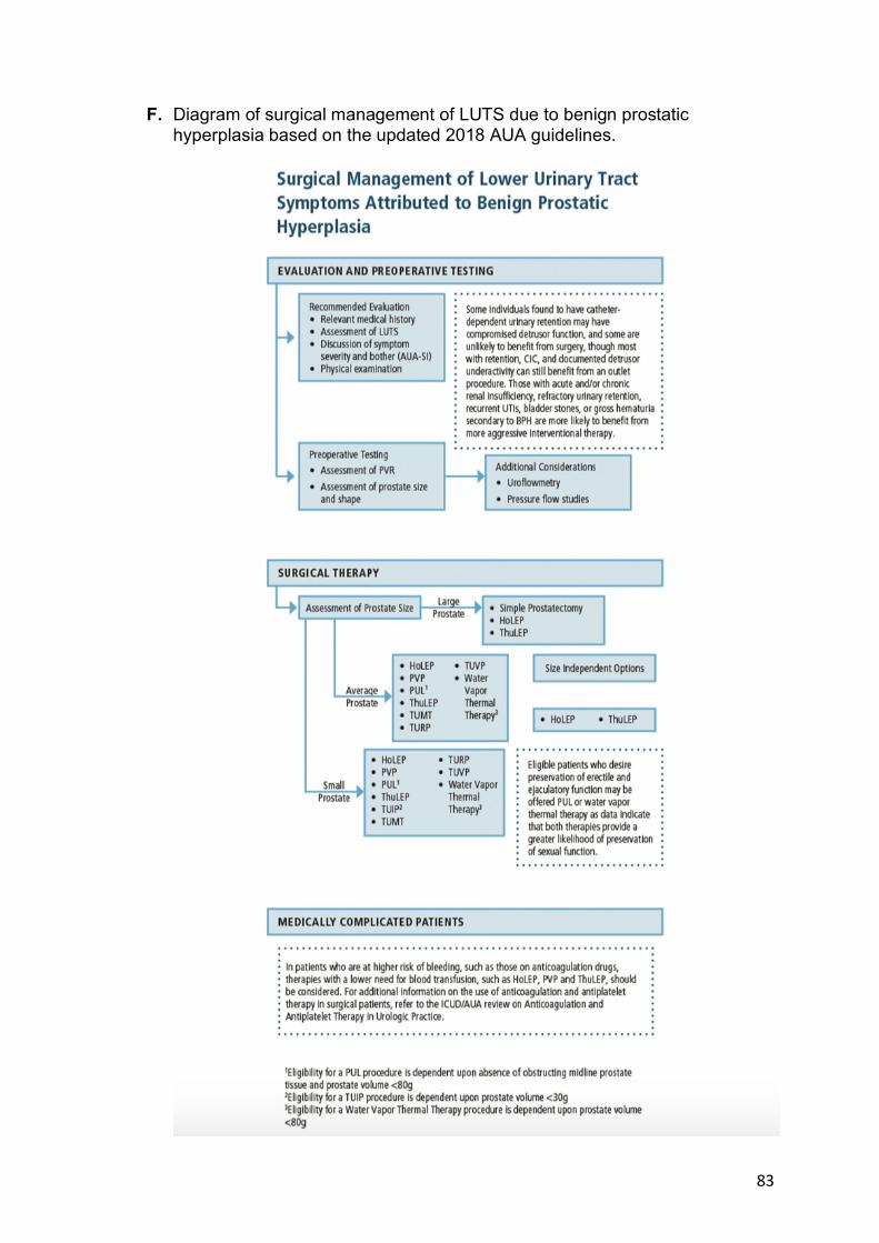
83
F. Diagram of surgical management of LUTS due to benign prostatic hyperplasia based on the updated 2018 AUA guidelines.
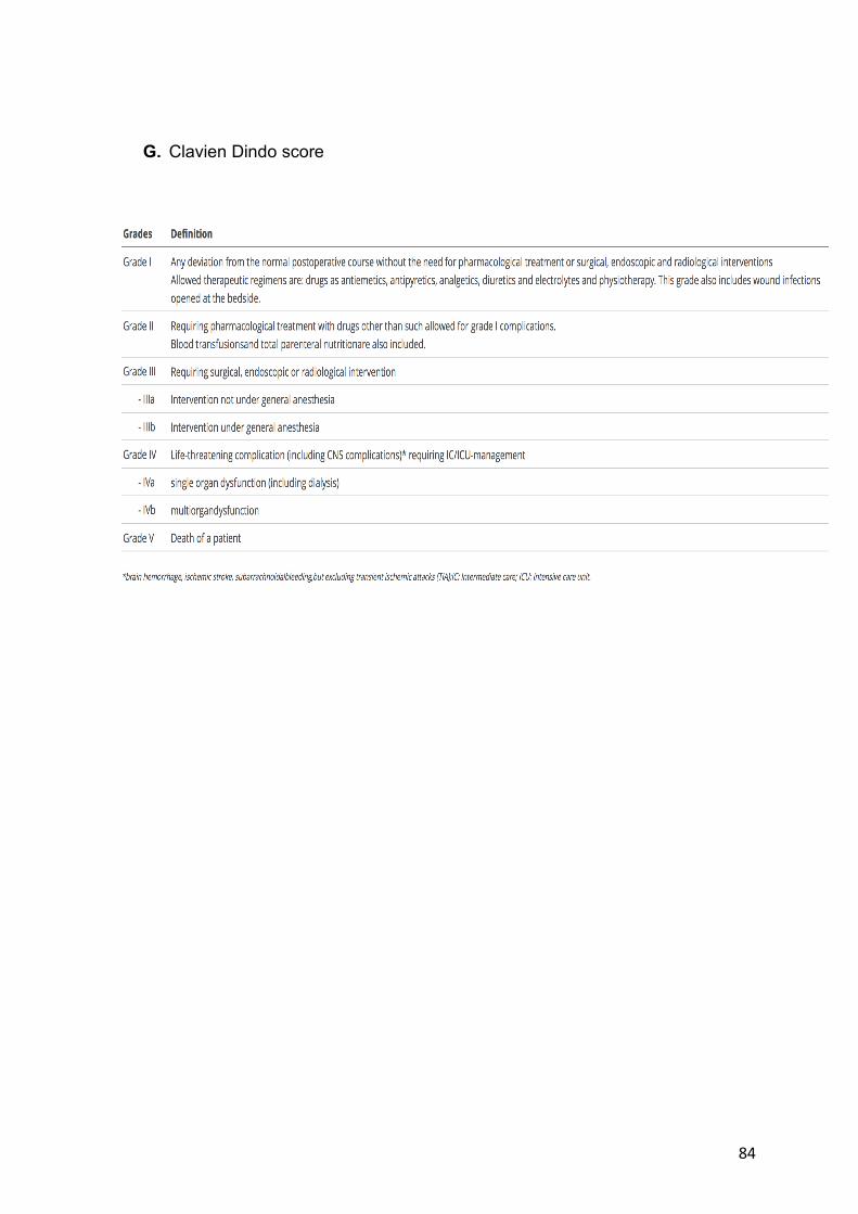
84
G. Clavien Dindo score
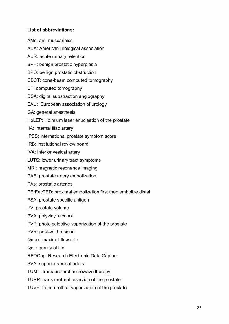
85
List of abbreviations:
AMs: anti-muscarinics
AUA: American urological association
AUR: acute urinary retention
BPH: benign prostatic hyperplasia
BPO: benign prostatic obstruction
CBCT: cone-beam computed tomography
CT: computed tomography
DSA: digital substraction angiography
EAU: European association of urology
GA: general anesthesia
HoLEP: Holmium laser enucleation of the prostate
IIA: internal iliac artery
IPSS: international prostate symptom score
IRB: institutional review board
IVA: inferior vesical artery
LUTS: lower urinary tract symptoms
MRI: magnetic resonance imaging
PAE: prostate artery embolization
PAs: prostatic arteries
PErFecTED: proximal embolization first then embolize distal
PSA: prostate specific antigen
PV: prostate volume
PVA: polyvinyl alcohol
PVP: photo selective vaporization of the prostate
PVR: post-void residual
Qmax: maximal flow rate
QoL: quality of life
REDCap: Research Electronic Data Capture
SVA: superior vesical artery
TUMT: trans-urethral microwave therapy
TURP: trans-urethral resection of the prostate
TUVP: trans-urethral vaporization of the prostate

86
TZ : transitionnel zone
UK-Rope : UK registry of protate artery embolization
87
SERMENT
Ø En présence des Maîtres de cette école, de mes chers condisciples et
devant l’effigie d’Hippocrate, je promets et je jure, au nom de l’Etre suprême, d’être fidèle aux lois de l’honneur et de la probité dans l’exercice de la médecine.
Ø Je donnerai mes soins gratuits à l’indigent et n’exigerai jamais un
salaire au-dessus de mon travail. Ø Admis (e) dans l’intérieur des maisons, mes yeux ne verront pas ce qui
s’y passe, ma langue taira les secrets qui me seront confiés, et mon état ne servira pas à corrompre les mœurs, ni à favoriser le crime.
Ø Respectueux (se) et reconnaissant (e) envers mes Maîtres, je rendrai à
leurs enfants l’instruction que j’ai reçue de leurs pères. Ø Que les hommes m’accordent leur estime si je suis fidèle à mes
promesses. Que je sois couvert (e) d’opprobre et méprisé (e) de mes confrères si j’y manque.

89
Résumé Introduction : L'objectif de cette étude multicentrique rétrospective était d'évaluer les
résultats cliniques précoces et de déterminer les facteurs prédictifs de bonne
réponse de l'embolisation des artères prostatiques (EAP) en France.
Matériel et méthodes : Cette première étude rétrospective multicentrique française
incluant 420 patients (âge moyen 69,7; IPSS moyen 20,6) dans 7 centres hospitalo-
universitaires de radiologie interventionnelle, atteints d’une HBP symptomatique ou
un sondage urinaire réfractaire, traités par EAP de janvier 2017 à avril 2019. Les
résultats cliniques (IPSS, QoL), d'imagerie (volume de prostate) et données
techniques (durée de procédure, de fluoroscopie, embolisation uni/bilatéral, agents
emboliques et volume injecté) ont été recueillis via REDCap (plate-forme web
sécurisée). Des analyses uni et multivariée ont été effectués avec l'analyse de
régression logistique ajustant des caractéristiques de population pour rechercher des
facteurs de risque prédictifs.
Résultats : Le succès technique était de 98,6% avec une durée moyenne de
procédure de 132,7 minutes. Le succès clinique a été obtenu chez 269/320 patients
présentant un suivi clinique (84,1%) avec une réduction moyenne de l’IPSS de
53,6%, du QoL de 2,9 points et du volume prostatique de 22,7%. 3 nécroses de
gland et 3 rétentions urinaires aigues ont été rapportées sans autre complication
majeure. Le suivi était variable de 1 à 31,2 mois.
Conclusion : Cette étude multicentrique confirme l'efficacité et la faisabilité de l'EAP
avec des résultats cliniques précoces satisfaisants, d’autant plus chez les patients
jeunes (<70 ans), avec une large prostate (>100ml), traités par embolisation bilatéral.
D'autres études de suivi à long terme sont nécessaires pour consolider ces résultats
fonctionnels.
Mots clés : Embolisation artères prostatiques, Hypertrophie bénigne de prostate, sondage urinaire, multicentrique.