Embed Size (px)
Citation preview
Nb-containing hematites Fe2�xNbxO3: The role of Nb5+ on the reactivityin presence of the H2O2 or ultraviolet light
Adilson C. Silva a, Diana Q.L. Oliveira a, Luiz C.A. Oliveira a,*, Alexandre S. Anastacio a,Teodorico C. Ramalho a, Joao H. Lopes b, Hudson W.P. Carvalho c, Claudia E. Rodriguez Torres d
a Department of Chemistry, Federal University of Lavras, Lavras-MG 37200-000, Brazilb Department of Chemistry, UNICAMP, Campinas-SP, Brazilc Department of Physical-Chemistry, State University Julio de Mesquita Filho, Araraquara-SP 14801-970, Brazild Department of Physical, National University of La Plata, La Plata - Bs.As 1900, Argentina
Applied Catalysis A: General 357 (2009) 79–84
A R T I C L E I N F O
Article history:
Received 6 October 2008
Received in revised form 7 January 2009
Accepted 9 January 2009
Available online 16 January 2009
Keywords:
Hematite
Niobium
Organic oxidation
ESI-MS
A B S T R A C T
A series of Nb-containing hematites, Fe2�xNbxO3 (%Nb = 0.00, 1.49, 5.00 and 9.24) was prepared using the
conventional co-precipitation method. Mossbauer and temperature-programmed reduction (TPR)
measurements suggested the formation of the crystalline phase with partial substitution of Fe3+ by Nb5+
in the structure. N2 adsorption/desorption revealed that the presence of Nb has a remarkable effect on
the textural properties of the material with an increase in the BET surface area. The reactivity of
Fe2�xNbxO3 was investigated using the oxidation of the methylene blue dye used as a model pollutant.
The obtained results showed that the presence of Nb seems not to act directly promoting the H2O2
decomposition, but improving the dye oxidation. The analysis using the ESI-MS technique showed
partial oxidation observed through different intermediates before the mineralization. This suggests the
use of Nb-doped hematite as an efficient catalyst in degradation reactions in the presence of H2O2 or
ultraviolet light.
� 2009 Elsevier B.V. All rights reserved.
Contents lists available at ScienceDirect
Applied Catalysis A: General
journa l homepage: www.e lsev ier .com/ locate /apcata
1. Introduction
Dye wastewater is characterized by large amounts ofdischarge, high concentration, and complex composition. Con-ventional dye wastewater treatment methods are graduallybecoming inadequate to obtain higher environmental qualityrequirements. In recent years, various advanced oxidationprocesses such as, ozonation, wet air oxidation, supercriticalwater oxidation and photocatalytic oxidation have been proposedas substitutes for the conventional treatment techniques [1–4]. Anovel and promising catalytic application for destruction oforganic contaminants in wastewaters is the utilization of ironoxides and H2O2 in a heterogeneous Fenton system [5–10]. In thisheterogeneous Fenton system the iron oxide activates H2O2 togenerate radicals, especially HO�, which can completely oxidizeorganics present in the aqueous medium [11]. It has beenobserved that magnetite, Fe3O4, is especially active for oxidationof organics in the presence of H2O2. This activity was assigned tothe presence of Fe2+ species in the magnetite structure, which can
* Corresponding author. Tel.: +55 35 3829 1626; fax: +55 35 3829 1271.
E-mail address: [email protected] (Luiz C.A. Oliveira).
URL: http://www.gqa.dqi.ufla.br
0926-860X/$ – see front matter � 2009 Elsevier B.V. All rights reserved.
doi:10.1016/j.apcata.2009.01.014
activate H2O2, by a Haber Weiss mechanism [12]. On the otherhand, the activity of other iron oxide phases such as hematite,Fe2O3, can be improved by the presence of different metals in thestructure. For example, the introduction of lanthanum orneodymium in the hematite structure remarkably increased thereactivity of the dehydrogenation reaction [13]. In heterogeneouscatalysis, numerous applications involve Nb compounds aspromoters and support for other metals mainly due to theincrease of catalytic activity and stability of the catalyst. Recently,the photocatalytic properties of niobia compounds have beenexplored by many authors [14].
In this work, niobium was introduced in the hematitestructure to produce for the first time an active heterogeneoussystem with H2O2 and also to improve the photocatalyticactivity of the material. The reactions in the presence of H2O2 orultraviolet light were carried out using the organic dyemethylene blue as probe molecule. Niobium shows interestingfeatures for this system such as: ionic ray compatible with theFe structural and the high reactivity towards H2O2 activation[15,16]. Many studies in the literature with iron and niobiumoxides can be found, however, no systematic investigation onNb-doped hematite and no catalytic studies with this oxide inthe presence of hydrogen peroxide or ultraviolet light have beencarried out.
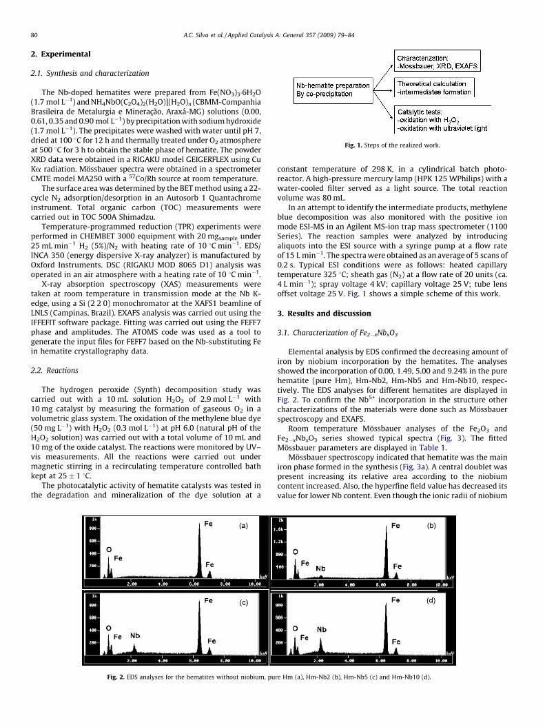
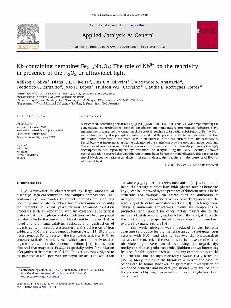
Fig. 1. Steps of the realized work.
A.C. Silva et al. / Applied Catalysis A: General 357 (2009) 79–8480
2. Experimental
2.1. Synthesis and characterization
The Nb-doped hematites were prepared from Fe(NO3)3�6H2O(1.7 mol L�1) and NH4NbO(C2O4)2(H2O)](H2O)n (CBMM-CompanhiaBrasileira de Metalurgia e Mineracao, Araxa-MG) solutions (0.00,0.61, 0.35 and 0.90 mol L�1) by precipitation with sodium hydroxide(1.7 mol L�1). The precipitates were washed with water until pH 7,dried at 100 8C for 12 h and thermally treated under O2 atmosphereat 500 8C for 3 h to obtain the stable phase of hematite. The powderXRD data were obtained in a RIGAKU model GEIGERFLEX using CuKa radiation. Mossbauer spectra were obtained in a spectrometerCMTE model MA250 with a 57Co/Rh source at room temperature.
The surface area was determined by the BET method using a 22-cycle N2 adsorption/desorption in an Autosorb 1 Quantachromeinstrument. Total organic carbon (TOC) measurements werecarried out in TOC 500A Shimadzu.
Temperature-programmed reduction (TPR) experiments wereperformed in CHEMBET 3000 equipment with 20 mgsample under25 mL min�1 H2 (5%)/N2 with heating rate of 10 8C min�1. EDS/INCA 350 (energy dispersive X-ray analyzer) is manufactured byOxford Instruments. DSC (RIGAKU MOD 8065 D1) analysis wasoperated in an air atmosphere with a heating rate of 10 8C min�1.
X-ray absorption spectroscopy (XAS) measurements weretaken at room temperature in transmission mode at the Nb K-edge, using a Si (2 2 0) monochromator at the XAFS1 beamline ofLNLS (Campinas, Brazil). EXAFS analysis was carried out using theIFFEFIT software package. Fitting was carried out using the FEFF7phase and amplitudes. The ATOMS code was used as a tool togenerate the input files for FEFF7 based on the Nb-substituting Fein hematite crystallography data.
2.2. Reactions
The hydrogen peroxide (Synth) decomposition study wascarried out with a 10 mL solution H2O2 of 2.9 mol L�1 with10 mg catalyst by measuring the formation of gaseous O2 in avolumetric glass system. The oxidation of the methylene blue dye(50 mg L�1) with H2O2 (0.3 mol L�1) at pH 6.0 (natural pH of theH2O2 solution) was carried out with a total volume of 10 mL and10 mg of the oxide catalyst. The reactions were monitored by UV–vis measurements. All the reactions were carried out undermagnetic stirring in a recirculating temperature controlled bathkept at 25 � 1 8C.
The photocatalytic activity of hematite catalysts was tested inthe degradation and mineralization of the dye solution at a
Fig. 2. EDS analyses for the hematites without niobium, pu
constant temperature of 298 K, in a cylindrical batch photo-reactor. A high-pressure mercury lamp (HPK 125 WPhilips) with awater-cooled filter served as a light source. The total reactionvolume was 80 mL.
In an attempt to identify the intermediate products, methyleneblue decomposition was also monitored with the positive ionmode ESI-MS in an Agilent MS-ion trap mass spectrometer (1100Series). The reaction samples were analyzed by introducingaliquots into the ESI source with a syringe pump at a flow rateof 15 L min�1. The spectra were obtained as an average of 5 scans of0.2 s. Typical ESI conditions were as follows: heated capillarytemperature 325 8C; sheath gas (N2) at a flow rate of 20 units (ca.4 L min�1); spray voltage 4 kV; capillary voltage 25 V; tube lensoffset voltage 25 V. Fig. 1 shows a simple scheme of this work.
3. Results and discussion
3.1. Characterization of Fe2�xNbxO3
Elemental analysis by EDS confirmed the decreasing amount ofiron by niobium incorporation by the hematites. The analysesshowed the incorporation of 0.00, 1.49, 5.00 and 9.24% in the purehematite (pure Hm), Hm-Nb2, Hm-Nb5 and Hm-Nb10, respec-tively. The EDS analyses for different hematites are displayed inFig. 2. To confirm the Nb5+ incorporation in the structure othercharacterizations of the materials were done such as Mossbauerspectroscopy and EXAFS.
Room temperature Mossbauer analyses of the Fe2O3 andFe2�xNbxO3 series showed typical spectra (Fig. 3). The fittedMossbauer parameters are displayed in Table 1.
Mossbauer spectroscopy indicated that hematite was the mainiron phase formed in the synthesis (Fig. 3a). A central doublet waspresent increasing its relative area according to the niobiumcontent increased. Also, the hyperfine field value has decreased itsvalue for lower Nb content. Even though the ionic radii of niobium
re Hm (a), Hm-Nb2 (b), Hm-Nb5 (c) and Hm-Nb10 (d).
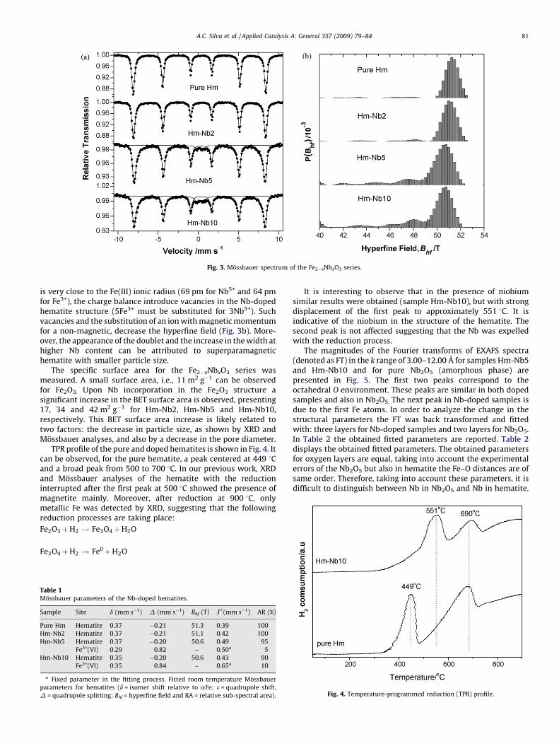
Fig. 3. Mossbauer spectrum of the Fe2�xNbxO3 series.
A.C. Silva et al. / Applied Catalysis A: General 357 (2009) 79–84 81
is very close to the Fe(III) ionic radius (69 pm for Nb5+ and 64 pmfor Fe3+), the charge balance introduce vacancies in the Nb-dopedhematite structure (5Fe3+ must be substituted for 3Nb5+). Suchvacancies and the substitution of an ion with magnetic momentumfor a non-magnetic, decrease the hyperfine field (Fig. 3b). More-over, the appearance of the doublet and the increase in the width athigher Nb content can be attributed to superparamagnetichematite with smaller particle size.
The specific surface area for the Fe2�xNbxO3 series wasmeasured. A small surface area, i.e., 11 m2 g�1 can be observedfor Fe2O3. Upon Nb incorporation in the Fe2O3 structure asignificant increase in the BET surface area is observed, presenting17, 34 and 42 m2 g�1 for Hm-Nb2, Hm-Nb5 and Hm-Nb10,respectively. This BET surface area increase is likely related totwo factors: the decrease in particle size, as shown by XRD andMossbauer analyses, and also by a decrease in the pore diameter.
TPR profile of the pure and doped hematites is shown in Fig. 4. Itcan be observed, for the pure hematite, a peak centered at 449 8Cand a broad peak from 500 to 700 8C. In our previous work, XRDand Mossbauer analyses of the hematite with the reductioninterrupted after the first peak at 500 8C showed the presence ofmagnetite mainly. Moreover, after reduction at 900 8C, onlymetallic Fe was detected by XRD, suggesting that the followingreduction processes are taking place:
Fe2O3þH2 ! Fe3O4þH2O
Fe3O4þH2 ! Fe0þH2O
Table 1Mossbauer parameters of the Nb-doped hematites.
Sample Site d (mm s�1) D (mm s�1) Bhf (T) G (mm s�1) AR (%)
Pure Hm Hematite 0.37 �0.21 51.3 0.39 100
Hm-Nb2 Hematite 0.37 �0.21 51.1 0.42 100
Hm-Nb5 Hematite 0.37 �0.20 50.6 0.49 95
Fe3+(VI) 0.29 0.82 – 0.50a 5
Hm-Nb10 Hematite 0.35 �0.20 50.6 0.43 90
Fe3+(VI) 0.35 0.84 – 0.65a 10
a Fixed parameter in the fitting process. Fitted room temperature Mossbauer
parameters for hematites (d = isomer shift relative to aFe; e = quadrupole shift,
D = quadrupole splitting; Bhf = hyperfine field and RA = relative sub-spectral area).
It is interesting to observe that in the presence of niobiumsimilar results were obtained (sample Hm-Nb10), but with strongdisplacement of the first peak to approximately 551 8C. It isindicative of the niobium in the structure of the hematite. Thesecond peak is not affected suggesting that the Nb was expelledwith the reduction process.
The magnitudes of the Fourier transforms of EXAFS spectra(denoted as FT) in the k range of 3.00–12.00 A for samples Hm-Nb5and Hm-Nb10 and for pure Nb2O5 (amorphous phase) arepresented in Fig. 5. The first two peaks correspond to theoctahedral O environment. These peaks are similar in both dopedsamples and also in Nb2O5. The next peak in Nb-doped samples isdue to the first Fe atoms. In order to analyze the change in thestructural parameters the FT was back transformed and fittedwith: three layers for Nb-doped samples and two layers for Nb2O5.In Table 2 the obtained fitted parameters are reported. Table 2displays the obtained fitted parameters. The obtained parametersfor oxygen layers are equal, taking into account the experimentalerrors of the Nb2O5 but also in hematite the Fe–O distances are ofsame order. Therefore, taking into account these parameters, it isdifficult to distinguish between Nb in Nb2O5 and Nb in hematite.
Fig. 4. Temperature-programmed reduction (TPR) profile.
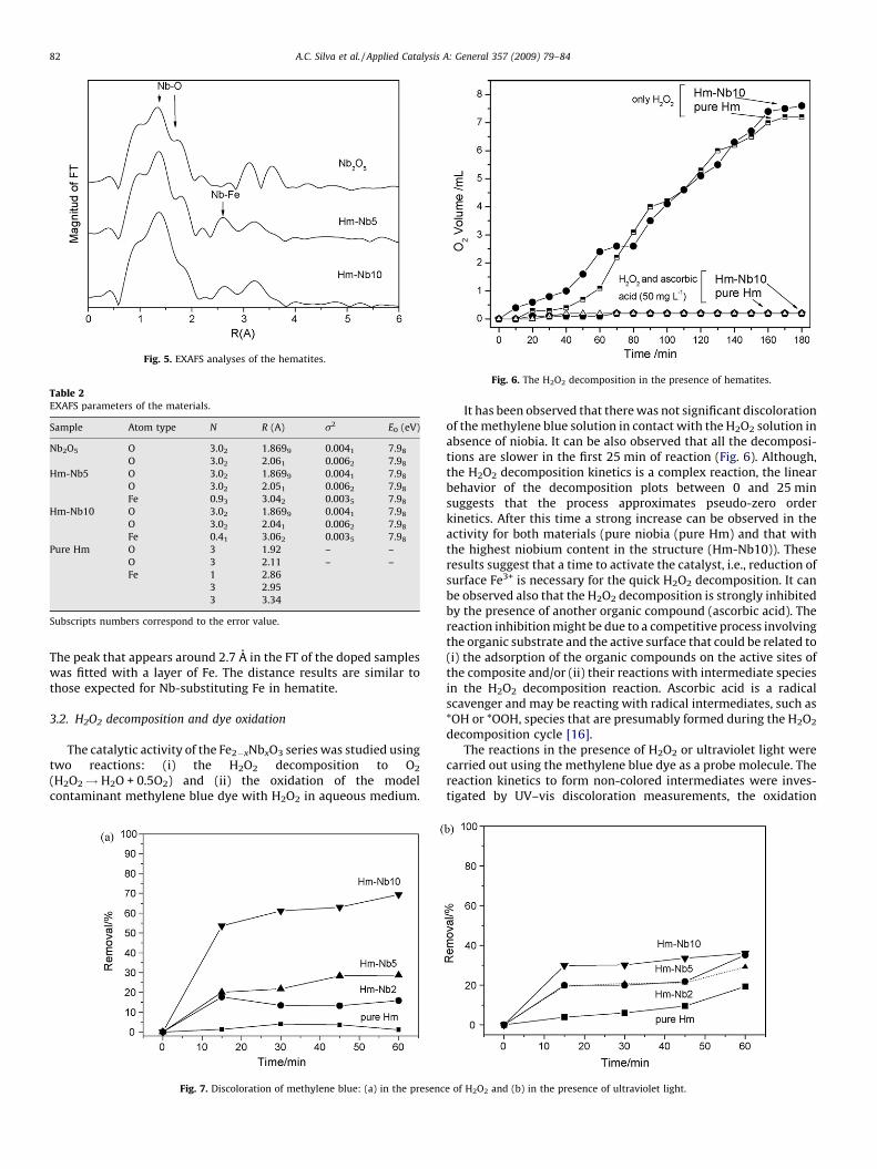
Table 2EXAFS parameters of the materials.
Sample Atom type N R (A) s2 E0 (eV)
Nb2O5 O 3.02 1.8699 0.0041 7.98
O 3.02 2.061 0.0062 7.98
Hm-Nb5 O 3.02 1.8699 0.0041 7.98
O 3.02 2.051 0.0062 7.98
Fe 0.93 3.042 0.0035 7.98
Hm-Nb10 O 3.02 1.8699 0.0041 7.98
O 3.02 2.041 0.0062 7.98
Fe 0.41 3.062 0.0035 7.98
Pure Hm O 3 1.92 – –
O 3 2.11 – –
Fe 1 2.86
3 2.95
3 3.34
Subscripts numbers correspond to the error value.
Fig. 6. The H2O2 decomposition in the presence of hematites.
Fig. 5. EXAFS analyses of the hematites.
A.C. Silva et al. / Applied Catalysis A: General 357 (2009) 79–8482
The peak that appears around 2.7 A in the FT of the doped sampleswas fitted with a layer of Fe. The distance results are similar tothose expected for Nb-substituting Fe in hematite.
3.2. H2O2 decomposition and dye oxidation
The catalytic activity of the Fe2�xNbxO3 series was studied usingtwo reactions: (i) the H2O2 decomposition to O2
(H2O2! H2O + 0.5O2) and (ii) the oxidation of the modelcontaminant methylene blue dye with H2O2 in aqueous medium.
Fig. 7. Discoloration of methylene blue: (a) in the presenc
It has been observed that there was not significant discolorationof the methylene blue solution in contact with the H2O2 solution inabsence of niobia. It can be also observed that all the decomposi-tions are slower in the first 25 min of reaction (Fig. 6). Although,the H2O2 decomposition kinetics is a complex reaction, the linearbehavior of the decomposition plots between 0 and 25 minsuggests that the process approximates pseudo-zero orderkinetics. After this time a strong increase can be observed in theactivity for both materials (pure niobia (pure Hm) and that withthe highest niobium content in the structure (Hm-Nb10)). Theseresults suggest that a time to activate the catalyst, i.e., reduction ofsurface Fe3+ is necessary for the quick H2O2 decomposition. It canbe observed also that the H2O2 decomposition is strongly inhibitedby the presence of another organic compound (ascorbic acid). Thereaction inhibition might be due to a competitive process involvingthe organic substrate and the active surface that could be related to(i) the adsorption of the organic compounds on the active sites ofthe composite and/or (ii) their reactions with intermediate speciesin the H2O2 decomposition reaction. Ascorbic acid is a radicalscavenger and may be reacting with radical intermediates, such as*OH or *OOH, species that are presumably formed during the H2O2
decomposition cycle [16].The reactions in the presence of H2O2 or ultraviolet light were
carried out using the methylene blue dye as a probe molecule. Thereaction kinetics to form non-colored intermediates were inves-tigated by UV–vis discoloration measurements, the oxidation
e of H2O2 and (b) in the presence of ultraviolet light.
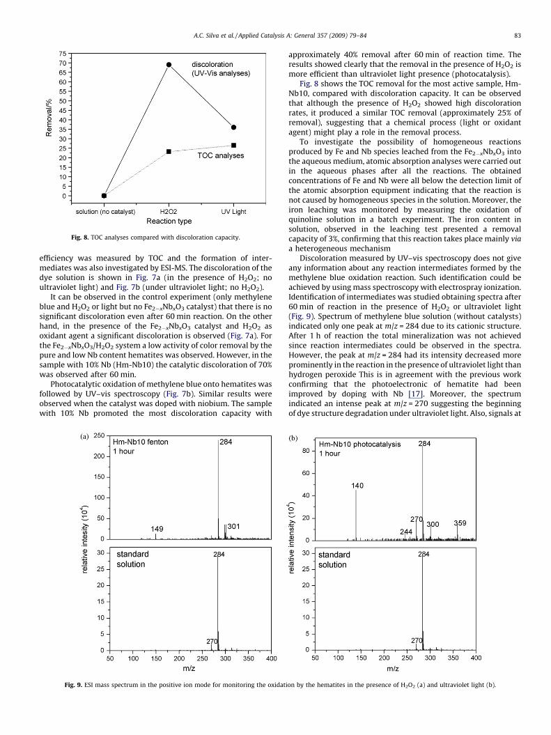
Fig. 8. TOC analyses compared with discoloration capacity.
A.C. Silva et al. / Applied Catalysis A: General 357 (2009) 79–84 83
efficiency was measured by TOC and the formation of inter-mediates was also investigated by ESI-MS. The discoloration of thedye solution is shown in Fig. 7a (in the presence of H2O2; noultraviolet light) and Fig. 7b (under ultraviolet light; no H2O2).
It can be observed in the control experiment (only methyleneblue and H2O2 or light but no Fe2�xNbxO3 catalyst) that there is nosignificant discoloration even after 60 min reaction. On the otherhand, in the presence of the Fe2�xNbxO3 catalyst and H2O2 asoxidant agent a significant discoloration is observed (Fig. 7a). Forthe Fe2�xNbxO3/H2O2 system a low activity of color removal by thepure and low Nb content hematites was observed. However, in thesample with 10% Nb (Hm-Nb10) the catalytic discoloration of 70%was observed after 60 min.
Photocatalytic oxidation of methylene blue onto hematites wasfollowed by UV–vis spectroscopy (Fig. 7b). Similar results wereobserved when the catalyst was doped with niobium. The samplewith 10% Nb promoted the most discoloration capacity with
Fig. 9. ESI mass spectrum in the positive ion mode for monitoring the oxidati
approximately 40% removal after 60 min of reaction time. Theresults showed clearly that the removal in the presence of H2O2 ismore efficient than ultraviolet light presence (photocatalysis).
Fig. 8 shows the TOC removal for the most active sample, Hm-Nb10, compared with discoloration capacity. It can be observedthat although the presence of H2O2 showed high discolorationrates, it produced a similar TOC removal (approximately 25% ofremoval), suggesting that a chemical process (light or oxidantagent) might play a role in the removal process.
To investigate the possibility of homogeneous reactionsproduced by Fe and Nb species leached from the Fe2�xNbxO3 intothe aqueous medium, atomic absorption analyses were carried outin the aqueous phases after all the reactions. The obtainedconcentrations of Fe and Nb were all below the detection limit ofthe atomic absorption equipment indicating that the reaction isnot caused by homogeneous species in the solution. Moreover, theiron leaching was monitored by measuring the oxidation ofquinoline solution in a batch experiment. The iron content insolution, observed in the leaching test presented a removalcapacity of 3%, confirming that this reaction takes place mainly via
a heterogeneous mechanismDiscoloration measured by UV–vis spectroscopy does not give
any information about any reaction intermediates formed by themethylene blue oxidation reaction. Such identification could beachieved by using mass spectroscopy with electrospray ionization.Identification of intermediates was studied obtaining spectra after60 min of reaction in the presence of H2O2 or ultraviolet light(Fig. 9). Spectrum of methylene blue solution (without catalysts)indicated only one peak at m/z = 284 due to its cationic structure.After 1 h of reaction the total mineralization was not achievedsince reaction intermediates could be observed in the spectra.However, the peak at m/z = 284 had its intensity decreased moreprominently in the reaction in the presence of ultraviolet light thanhydrogen peroxide This is in agreement with the previous workconfirming that the photoelectronic of hematite had beenimproved by doping with Nb [17]. Moreover, the spectrumindicated an intense peak at m/z = 270 suggesting the beginningof dye structure degradation under ultraviolet light. Also, signals at
on by the hematites in the presence of H2O2 (a) and ultraviolet light (b).
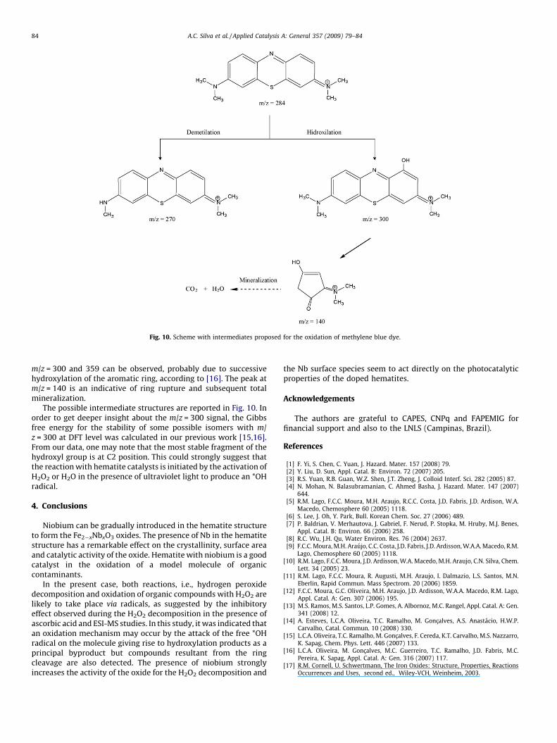
Fig. 10. Scheme with intermediates proposed for the oxidation of methylene blue dye.
A.C. Silva et al. / Applied Catalysis A: General 357 (2009) 79–8484
m/z = 300 and 359 can be observed, probably due to successivehydroxylation of the aromatic ring, according to [16]. The peak atm/z = 140 is an indicative of ring rupture and subsequent totalmineralization.
The possible intermediate structures are reported in Fig. 10. Inorder to get deeper insight about the m/z = 300 signal, the Gibbsfree energy for the stability of some possible isomers with m/z = 300 at DFT level was calculated in our previous work [15,16].From our data, one may note that the most stable fragment of thehydroxyl group is at C2 position. This could strongly suggest thatthe reaction with hematite catalysts is initiated by the activation ofH2O2 or H2O in the presence of ultraviolet light to produce an *OHradical.
4. Conclusions
Niobium can be gradually introduced in the hematite structureto form the Fe2�xNbxO3 oxides. The presence of Nb in the hematitestructure has a remarkable effect on the crystallinity, surface areaand catalytic activity of the oxide. Hematite with niobium is a goodcatalyst in the oxidation of a model molecule of organiccontaminants.
In the present case, both reactions, i.e., hydrogen peroxidedecomposition and oxidation of organic compounds with H2O2 arelikely to take place via radicals, as suggested by the inhibitoryeffect observed during the H2O2 decomposition in the presence ofascorbic acid and ESI-MS studies. In this study, it was indicated thatan oxidation mechanism may occur by the attack of the free *OHradical on the molecule giving rise to hydroxylation products as aprincipal byproduct but compounds resultant from the ringcleavage are also detected. The presence of niobium stronglyincreases the activity of the oxide for the H2O2 decomposition and
the Nb surface species seem to act directly on the photocatalyticproperties of the doped hematites.
Acknowledgements
The authors are grateful to CAPES, CNPq and FAPEMIG forfinancial support and also to the LNLS (Campinas, Brazil).
References
[1] F. Yi, S. Chen, C. Yuan, J. Hazard. Mater. 157 (2008) 79.[2] Y. Liu, D. Sun, Appl. Catal. B: Environ. 72 (2007) 205.[3] R.S. Yuan, R.B. Guan, W.Z. Shen, J.T. Zheng, J. Colloid Interf. Sci. 282 (2005) 87.[4] N. Mohan, N. Balasubramanian, C. Ahmed Basha, J. Hazard. Mater. 147 (2007)
644.[5] R.M. Lago, F.C.C. Moura, M.H. Araujo, R.C.C. Costa, J.D. Fabris, J.D. Ardison, W.A.
Macedo, Chemosphere 60 (2005) 1118.[6] S. Lee, J. Oh, Y. Park, Bull. Korean Chem. Soc. 27 (2006) 489.[7] P. Baldrian, V. Merhautova, J. Gabriel, F. Nerud, P. Stopka, M. Hruby, M.J. Benes,
Appl. Catal. B: Environ. 66 (2006) 258.[8] R.C. Wu, J.H. Qu, Water Environ. Res. 76 (2004) 2637.[9] F.C.C. Moura, M.H. Araujo, C.C. Costa, J.D. Fabris, J.D. Ardisson, W.A.A. Macedo, R.M.
Lago, Chemosphere 60 (2005) 1118.[10] R.M. Lago, F.C.C. Moura, J.D. Ardisson, W.A. Macedo, M.H. Araujo, C.N. Silva, Chem.
Lett. 34 (2005) 23.[11] R.M. Lago, F.C.C. Moura, R. Augusti, M.H. Araujo, I. Dalmazio, L.S. Santos, M.N.
Eberlin, Rapid Commun. Mass Spectrom. 20 (2006) 1859.[12] F.C.C. Moura, G.C. Oliveira, M.H. Araujo, J.D. Ardisson, W.A.A. Macedo, R.M. Lago,
Appl. Catal. A: Gen. 307 (2006) 195.[13] M.S. Ramos, M.S. Santos, L.P. Gomes, A. Albornoz, M.C. Rangel, Appl. Catal. A: Gen.
341 (2008) 12.[14] A. Esteves, L.C.A. Oliveira, T.C. Ramalho, M. Goncalves, A.S. Anastacio, H.W.P.
Carvalho, Catal. Commun. 10 (2008) 330.[15] L.C.A. Oliveira, T.C. Ramalho, M. Goncalves, F. Cereda, K.T. Carvalho, M.S. Nazzarro,
K. Sapag, Chem. Phys. Lett. 446 (2007) 133.[16] L.C.A. Oliveira, M. Goncalves, M.C. Guerreiro, T.C. Ramalho, J.D. Fabris, M.C.
Pereira, K. Sapag, Appl. Catal. A: Gen. 316 (2007) 117.[17] R.M. Cornell, U. Schwertmann, The Iron Oxides: Structure, Properties, Reactions
Occurrences and Uses, second ed., Wiley-VCH, Weinheim, 2003.


