Embed Size (px)
Citation preview

Molecules 2014, 19, 10115-10128; doi:10.3390/molecules190710115
molecules ISSN 1420-3049
www.mdpi.com/journal/molecules
Review
MicroRNAs and Bone Metastasis: A New Challenge
Martine Croset 1, Daniele Santini 2,*, Michele Iuliani 2, Marco Fioramonti 2, Alice Zoccoli 2,
Bruno Vincenzi 2, Giuseppe Tonini 2 and Francesco Pantano 2
1 Institut national de la santé et de la recherche médicale (INSERM), UMR 1033, Lyon F-69008,
France; E-Mail: [email protected] 2 Medical Oncology, Campus Bio-Medico University of Rome, Rome 00128, Italy;
E-Mails: [email protected] (M.I.); [email protected] (M.F.);
[email protected] (A.Z.); [email protected] (B.V.); [email protected] (G.T.);
[email protected] (F.P.)
* Author to whom correspondence should be addressed; E-Mail: [email protected];
Tel.: +39-06-2254-19117; Fax: +39-06-2254-11933.
Received: 4 April 2014; in revised form: 16 June 2014 / Accepted: 19 June 2014 /
Published: 11 July 2014
Abstract: The development of bone metastases requires multistep and multicellular
machinery consisting not only of processes shared with any type of metastases (formation
of a pre-metastatic niche, chemotaxis of tumor cells into the host tissue, tumor cells
escape from the microvasculature), but also biological interactions that are strictly related
to the particular bone microenvironment (bone marrow colonization by cancer cells,
osteomimicry, deregulation of bone homeostasis). MiRNAs are highly conserved, small
RNAs molecules that regulate gene expression. The functional consequence of miRNA
deregulation lies in the mRNA targets whose expression is altered. MiRNA networks
acting as upstream regulators of these genes interfere with the initial steps of tumor local
invasion and cancer cell intravasation, mainly by regulating the epithelial-mesenchymal
transition, the motility, invasiveness and survival abilities of these cells. The miRNA-mediated
regulation on the steps of bone tropism, anchorage, homing and finally bone colonization is
more tissue specific, being dependent on the expression pattern of target miRNAs in bone
marrow sinusoids, bone cells and microenvironment. In that, miRNA specific expression
signatures that can distinguish between primary tumors from their corresponding bone
metastases might be determinants of clinical aggressiveness. In this review, we focus on
the current advances on functions and molecular mechanisms by which miRNAs exert
their biological roles in regulating bone metastases development.
OPEN ACCESS
Molecules 2014, 19 10116
Keywords: bone metastases; microRNAs; epithelial-mesenchymal transition; osteomimicry
1. Introduction
Bone metastases are a common place complication of several types of cancers, including breast,
prostate and lung cancer [1]. The occurrence of these bone metastases deeply impairs the prognosis
and quality of life of patients and is responsible for significant morbidity (bone pain, pathological
fractures, nerve compression, hypercalcemia). Bone metastases are often osteolytic (due to significant
bone destruction), sometimes sclerotic (due to an excess of bone formation) or mixed [2]. Indeed,
tumor cells secrete factors that may disrupt physiological bone remodeling processes through the
deregulation of the normal osteoclast and osteoblast functions. For example, osteolysis is the result of
stimulation by tumor cells of osteoclast activity and inhibition of osteoblast, while an opposite
phenomenon occurs during osteosclerosis. Moreover, the mineralized bone matrix plays an important
role in metastasis formation acting as a reservoir of growth factors and calcium that, once released
from the matrix during degradation, can have a mitogenic effect on the tumor cells, thus leading to a
vicious cycle where the resorption/bone formation and tumor proliferation feed off each other.
The complex sequence of events that lead to the onset of bone metastases not only involve processes
common to any other metastasis (invasion of blood vessels from the primary tumor, establishing of a
pre-metastatic niche in the host tissue, chemotaxis of tumor cells into the host tissue, extravasation of
tumor cells from blood vessels) but also processes that are more specific to the bone tissue
(bone marrow colonization, osteomimicry, deregulation of osteoblast/osteoclast activity) [3]. All steps
of this sequence are regulated by multiple factors and molecular pathways, mainly through the tight
control of genes expressed by interacting cells [4–6]. The small non-coding microRNAs (miRNAs)
which have the capacity to regulate multiple genes are master regulators of gene expression and thus
redirect or reprogram biological pathways [7]. They also are capable of controlling the aberrant
biological activities characteristic of tumor cells [8]; consequently, in cancer, they can act either as
promoters or suppressors of tumor development and metastatic progression [7,9–11].
2. Bone Marrow Colonization
It has been described that about 30% of breast cancer patients may harbor bone marrow
micrometastases at the time of primary cancer diagnosis [12], but half of them will never develop
clinically established metastases. Furthermore, patients with breast and prostate cancer may remain
metastasis free even for decades after surgical removal of the primary cancer. Indeed, according to
Paget’s “seed and soil theory”, metastatic cells can enter into bone marrow and remain alive and start
to proliferate or remain in a quiescent status for many years. Preclinical evidences suggest that
metastasis arises from a small fraction of cells with the capacity to self-renew and to differentiate to
specialized cell types with limited proliferative potential, often termed cancer stem cells (CSCs) [13,14].
CSCs are proposed to acquire a great part of their biological features through undergoing the so called
epithelial-mesenchymal transition (EMT). EMT is a biologic process that allows a polarized epithelial
cell, which normally interacts with basement membrane via its basal surface to undergo multiple
Molecules 2014, 19 10117
biochemical changes that enable it to assume a mesenchymal cell phenotype. During EMT,
transformation of normal epithelial cells disrupts cell-cell and cell extracellular matrix contacts and
migrate to other locations in the body [15] providing these early cancer cells with the capacity to
infiltrate the surrounding tissue and ultimately metastasize to distant sites [16]. Once these cells reach
the bone marrow, they interact with anatomical entities in contact with the bone called “niches”. Two
different niches exist: “endosteal niche” where stem cells are closely associated with spindle-shaped
N-cadherin-positive osteoblasts (SNO) involved in the maintenance of stem cells quiescence and
“vascular niche” within central marrow, where more differentiated hematopoietic cells generally are
located [17]. Bone metastatic disseminated tumor cells (DTCs) target the niches where hematopoietic
stem cells (HSCs) reside and compete with them for occupancy. Indeed, DTCs and HSCs are believed
to use similar mechanisms to gain access to the HSC niche. After entering the niche, DTCs are likely
to evict HSCs into the peripheral blood or drive them into progenitor pools. In the competition for the
HSC niche, PCa cells may directly and indirectly drive HSC maturity, therefore vacating the niche.
As a result of the competition with DTCs, more hematopoietic progenitor cells (HPCs) are found
circulating in the peripheral blood of patients with metastatic PCa than either patients with localized
PCa or healthy age-matched controls. Under normal physiologic conditions, HSCs are believed to
reside mainly within the bone marrow HSC niche, while some HSCs are known to leave the marrow,
differentiate into HPCs, and circulate throughout the body. However, the question as to whether
HPCs mobilized by DTCs are functionally the same as normal HPCs remains unanswered [18].
Several receptors and their ligands have been implicated in these intercellular interactions; for example
CXCR-4 (receptor on HSC)/SDF-1 (ligand secreted by osteoblastic lineage cells) axis represents one
of the most extensively investigated while hyaluronan and osteopontin (ligands expressed in the
bone)/CD44 (receptor and putative marker for CSC) binding suggest a possible adhesive interaction
for circulating tumor cell arrest. Moreover these steps involved in cancer metastasis development have
been shown to be orchestrated by pleiotropically acting molecules—transcription factors and miRNAs.
Several transcription factors, including Snail [19], Slug [20], Twist [21], ZEB1, and ZEB2 [22], have
been identified as inducers of EMT and tumor metastasis. More recently, miR-200s and miR-205
family [23,24], miR-143 and miR-145 [25,26], let-7 [27], miR-203 [28], miR-34a [29] have emerged
as new epithelial markers and repressors of EMT and stem cell properties (Table 1).
2.1. miR-200s and miR-205 Family
Oncogenic EMT is programmed by transcription factors such as Snail, Slug and Twist, which are
potent E-cadherin suppressors, and is stimulated by TGFb. The miR-200 family and miR-205 are
crucial regulators of EMT/MET by targeting ZEB1 and ZEB2 mRNAs [23,24]. MiRNAs that target
CSCs affect the self-renewal and differentiation capacities of these cells and impair metastatic
progression. Let-7 miRNAs which expression is consistently downregulated in metastatic cancer
cells [30], inhibit cell stemness and breast cancer metastasis through silencing of MYC, Ras and
HMGA2 [27], while miR-200c inhibits cell stemness and metastasis by targeting ZEBs, TGFb and
BMI1 [31]. Furthermore, miRNAs regulate the expression of genes which have been reported as pro-
or anti-metastatic in one or several steps of metastatic dissemination to the bone: the gain in enhanced
migratory capacity and invasiveness, the production of ECM components, angiogenesis and elevated
Molecules 2014, 19 10118
resistance to apoptosis and the bone homing and colonization [32]. The miR-200 family has also been
shown to promote metastatic colonization at distal sites by altering the tumor secretome through direct
targeting of Sec23 homolog A (SEC23A) [8]. It should also be noticed that experimental evidence
suggest that interference with the microenvironmental support impairs the de novo formation of bone
metastases in vivo [33]. miRNAs produced in the bone microenvironment might interfere with bone
metastasis. Indeed, miRNAs can be transported via gap-junctions or exosomes from bone marrow
stroma to breast cancer cells and modulate the dormancy of DTCs [34]. Although still confusing,
increasing evidences demonstrated how tumor-associated microenvironment can produce exosomes
and both cancer cell-derived or stromal cell-derived exosomes are thus able to alter the tumor
environment and may participate in forming a distant metastatic niche to promote metastasis [35,36].
Regarding gap-junctions, the exchange of miRNA through those between the bone marrow stroma and
breast cancer cells may be an important factor that contributes to the dormancy of the cancer cells to
impart a quiescent phenotype in bone marrow. The gap-junctions identified between bone marrow
stroma and breast cancer cells in the endosteal region is consistent with the failure of high-dose
chemotherapy in autologous hematopoietic stem cell transplantation [37].
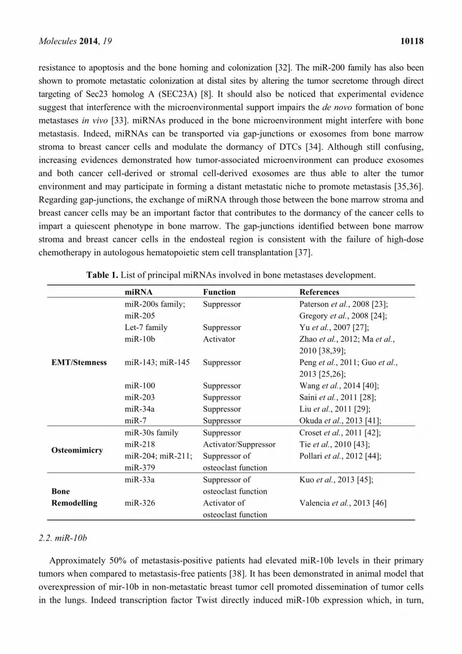
Table 1. List of principal miRNAs involved in bone metastases development.
miRNA Function References
EMT/Stemness
miR-200s family; miR-205
Suppressor Paterson et al., 2008 [23]; Gregory et al., 2008 [24];
Let-7 family Suppressor Yu et al., 2007 [27]; miR-10b Activator Zhao et al., 2012; Ma et al.,
2010 [38,39]; miR-143; miR-145 Suppressor Peng et al., 2011; Guo et al.,
2013 [25,26]; miR-100 Suppressor Wang et al., 2014 [40]; miR-203 Suppressor Saini et al., 2011 [28]; miR-34a Suppressor Liu et al., 2011 [29]; miR-7 Suppressor Okuda et al., 2013 [41];
Osteomimicry
miR-30s family Suppressor Croset et al., 2011 [42]; miR-218 Activator/Suppressor Tie et al., 2010 [43]; miR-204; miR-211; miR-379
Suppressor of osteoclast function
Pollari et al., 2012 [44];
Bone Remodelling
miR-33a Suppressor of osteoclast function
Kuo et al., 2013 [45];
miR-326 Activator of osteoclast function
Valencia et al., 2013 [46]
2.2. miR-10b
Approximately 50% of metastasis-positive patients had elevated miR-10b levels in their primary
tumors when compared to metastasis-free patients [38]. It has been demonstrated in animal model that
overexpression of mir-10b in non-metastatic breast tumor cell promoted dissemination of tumor cells
in the lungs. Indeed transcription factor Twist directly induced miR-10b expression which, in turn,

Molecules 2014, 19 10119
inhibited the expression of the transcription factor homeobox D10 (HOXD10). Interestingly, Twist1,
which has been reported as a link between EMT and stemness in breast carcinomas [38], is expressed
in disseminated breast cancer cells that persist in the bone marrow after chemotherapy. Moreover
serum miR-10b concentrations were found to be significantly higher in breast cancer patients with
bone metastases than in patients without bone metastases, suggesting that the Twist-induced miR-10b
expression could be also involved in bone metastasis formation [39]. The potential of miR-10b as
target for anti-metastatic therapy has been therefore studied in animal model of breast cancer
metastasis [39]. In this study, miR-10b antagonist was efficiently delivered to rapidly growing tumor
cells and it prevented metastasis dissemination to secondary organs without marks of cytotoxicity.
2.3. miR-143, miR-145
Comparison of array-based miRNA profiles of human primary prostate tumors and matched bone
metastasis showed that expression levels of miR-145, -143, -33a, -100 and -508-5p were the most
highly down-regulated in bone metastasis specimens [25]. Ectopic expression of miR-143 and miR-145
in the aggressive and androgen-insensitive PC-3 prostate cancer cell line reduced the migratory and
invasive capacities of these tumor cells in vitro and their propensity to metastasize to bone in vivo. The
expression of the mesenchymal markers, fibronectin and vimentin, was also decreased suggesting a
direct involvement in modulating EMT [25]. In a retrospective investigation of 12 patients without
bone metastasis and 10 patients with bone metastasis the expression levels of these two miRNAs were
significantly lower in bone metastatic patients. Moreover miRs-143 and -145 expression levels were
inversely correlated to free PSA and the Gleason score [25]. These findings suggest that miR-143 and
miR-145 could be used as biomarkers in discriminating different stages of prostate carcinomas and
predicting bone metastasis.
Recently it has been demonstrated that AGO2 was a direct target of miR-100 which is involved in
regulation of bone metastatic process in prostate cancer. In particular downregulation of AGO2 by
miR-100 repressed migration, invasion, EMT and stemness of prostate cancer cells. Furthermore,
miR-100 expression was negatively correlated with bone metastasis of prostate cancer patients [40].
2.4. miR-203
An antimetastatic role for miR-203 in prostate cancer progression and bone metastasis has been
reported [28]. The expression level of miR-203 in human prostate adenocarcinoma is lower than in
normal adjacent tissue. Low miR-203 expression correlates with advanced clinicopathological stage
and high Gleason score in patients with prostate cancer. In addition, miR-203 expression is down-regulated
in prostate cancer cell lines derived from bone metastasis (PC3, MDA-PCa-2b and VCaP cells).
Ectopic expression of miR-203 significantly decreases the metastatic capacity of PC-3 cells in a mouse
model of bone metastasis. The phenotype of miR-203-transfected cells (marked morphological
changes, from fibroblasts to epithelial-like phenotype, and reduced migration/invasion) is reminiscent
of a reverse transition from an EMT to a mesenchymal-to-epithelial state (MET). Importantly, miR-203
can pleiotropically regulate a cohort of metastatic effectors that includes apart from ZEB2, Bmi1 and
survivin, involved in the development of hormone resistance in PCa and associated with unfavorable

Molecules 2014, 19 10120
outcome. Finally miR-203 regulates bone-specific effectors including Runx2, a master regulator of
bone metastasis [28].
2.5. miR-34a/miR-7
miR-34a has been identified as a potent inhibitor of prostate CSCs and metastasis by directly
repressing CD44 [29]. The authors purified CSC population from prostate cancer xenograft models.
These CD44-positive CSCs harbor enhanced clonogenicity, tumor-initiating and metastatic properties
and are also depleted in miR-34a expression, compared to CD44-negative CSCs. Restoring miR-34a
expression inhibits clonogenic expansion, tumor development and metastasis by targeting CD44
expression. Similarly, Okuda et al. [41] reported that CD44-expressing CSCs isolated from MDA-MB-231
breast cancer cell subpopulations which were highly metastatic to brain and bone expressed low levels
of miR-7 and that this miRNA modulated KLF4, one of the essential genes for induced pluripotent
stem cell. Forced expression of miR-7 suppressed the ability of CSCs to metastasize to brain but not to
bone. Thus, the miRNA-mediated regulation on gene expression seems to be not only cell specific
(CSCc vs. non CSCs) but also tissue specific (bone vs. brain).
3. Osteomimicry
It has been postulated that tumor cells which spread in the bone marrow must then adapt to the bone
environment for the subsequent development of overt metastasis. It is probably for this reason that the
DTCs that are in the bone marrow remain for several years in a state of quiescence before suddenly be
reactivated and start to grow. This adaptation requires that tumor cells begin to express genes
that are normally expressed by bone cells. This process is called osteomimicry. For example,
immunohistochemical analysis of samples of primary tumor and matched metastases (liver, lung, bone) of
breast cancer patients showed that only metastatic cells express proteins of bone marrow origin
(cathepsin K, Integrin Beta 3 osteonectin, cadherin-11, connexin-43 and Runx2) [47,48].
3.1. miR-30s Family
It has been demonstrated that the miRNA-30s are expressed at higher levels in hormone-dependent
and well differentiated breast tumors, compared the levels observed in hormone-refractory tumors [49].
miRNA-30s have been reported acting as a tumor suppressor in lung [50], breast [51] and anaplasic
thyroid [52] cancers, mostly by regulating EMT. Their lower expression levels in lymph node
metastases vs. primary tumors suggests a role for miR-30s during metastatic dissemination of breast
carcinomas [53]. Moreover miRNA-30s regulate osteoblast differentiation [53] leading to the hypothesis
of their involvement in regulating cancer cell osteomimetic phenotype. Indeed differential miRNAs
profiling expression in breast cancer cell lines MDA-MB-231 and B02 (subpopulation of the parental
MDA-MB-231 cell line that has been selected for its high propensity to metastasize to bone in murine
models [54]) that the expression level of each of the members of the miRNA-30 family (miRNA-
30a,b,c,d,e) is downregulated within MDA-BO2 cells compared to parental cell line [42]. Restoring
the expression of the miRNA-30s in MDA-BO2 cells decreased significantly bone metastasis
progression in mice mainly targeting osteomimetic genes like connective tissue growth factor (CTGF),

Molecules 2014, 19 10121
connexin 43, integrinβ3 (ITGb3), cadherin-11, and Runx2 were associated to osteomimicry of
MDA-B02 cells.
3.2. miR-218
Regulation of osteomimicry-related tumor activity has also been reported for miR-218 [43]. Mir-218
appears as an “osteo-miR” by controlling bone formation through the upregulaton of Runx2 and the
downregulation of Wnt signaling inhibitors (sclerostin, dickkopf-2 and secreted frizzled-related protein2)
in osteoblasts. It also appears as a pro-metastatic miRNA through stimulation of the expression of
Wnt-related proteins (bone sialoprotein and osteopontin) in metastatic breast cancer cells. In addition
miR-218 may facilitate MDA-MB-231 breast cancer bone metastasis by up-regulating CXCR4, a
chemokine receptor supporting breast cancer cell migration to bone and mediating tumor growth in
bone. Although miR-218 regulates positively the molecular mechanisms underlying bone metastasis
formation it appears as tumor suppressor in other types of cancers [43], pointing out that its biological
activity depends on the cellular microenvironment.
4. Deregulation of Osteoblast/Osteoclast Activity
Bone lesions result in the interaction between cancer cells, bone microenvironment and bone cells
themselves. The tumor cells use bone-derived growth factors involved in the coupling between
osteoclasts/osteoblasts to promote their own development. In turn, they secrete factors (PTHrP, IL-1,
IL-6, IL-8, IL-11, M-CSF) that act in a paracrine fashion to activate osteoclast-mediated bone
resorption. In addition, cancer cells can release other growth factors that can promote (ET1, IGFs) or
inhibit (DKK1, Noggin, sclerostin) osteoblast activity. These processes are accompanied by the release
of growth factors and cytokines (TGFb, IGFs) embedded in the matrix during bone formation.
These molecules released from the resorbed bone matrix act in turn on cancer cells to promote their
proliferation [55–57].
4.1. miR-204, miR-211, miR-379
TGFb is involved in the vicious cycle between bone and tumor cells. It is released from bone in the
microenvironment during bone resorption and it stimulates cancer cells to produce pro-osteolytic
factors such as IL-11. To identify miRNAs that potentially regulate this process, Pollari et al. [44]
profiled the miRNA expression in the MDA-MB-231 cell line and a highly bone metastatic variant and
found that miRNAs were differently expressed between the two cell lines. Among these miRNAs, the
authors identify 3 miRNAs, miR-204, miR-211 and miR-379, as potent inhibitors of TGFb-induced
IL-11 secretion. In addition, gene expression analysis showed that miR-204 and miR-379
downregulated a set of genes involved in TGFb signalling.
4.2. miR-33a and miR-336
It has been reported that PTHrP, is a direct target of miR-33a in lung cancer. miR-33a levels are
inversely correlated with PTHrP expression between human normal bronchial cell line and lung cancer
cell lines. Restoring miR-33a expression reduces the stimulatory effect of lung cancer cells on the

Molecules 2014, 19 10122
production of osteoclastogenesis activator RANKL, (receptor activator of nuclear factor kappa-B
ligand) and M-CSF (macrophage colony-stimulating factor) on osteoblasts, while the expression of
PTHrP and IL-8 are decreased [45]. Thus a low miR-33a expression contributes to cancer-mediated
bone destruction and may even predict a poor prognosis for lung cancer patients. Indeed, miRNAs are
promising candidates as prognosis markers in cancer in regard of their high stability and their accessibility
in serum patients. In this respect, Valencia et al. [46] assessed the validity of measuring serum miRNAs
in a murine model of human lung cancer bone metastasis and compared miRNA serum levels with
those of standard biochemical markers of bone turnover such as PINP (procollagen I amino-terminal
propeptide), BGP (osteocalcin) and CTX (carboxyterminal telopeptide). Using this model of lung
cancer, PINP (procollagen I amino-terminal propeptide) exhibited a strong correlation with osteolytic
lesions and tumor burden at early and late stages of bone colonization. In contrast, BGP and CTX
demonstrated a strong correlation only at late stages. Interestingly, in mice model serum levels of
miR-326 strongly associated with tumor burden and PINP in vehicle-treated animals, whereas no
association was found in animals treated with an inhibitor of bone resorption (zoledronate). These
results suggest that miR-326 could serve as a biochemical marker for monitoring bone metastatic
progression in advanced lung cancer.
5. Clinical Usefulness of miRNAs in Bone Metastatic Cancer
MiRNAs expression represents a specific pattern for individual tissues and both miRNA family and
levels differ between cancer and normal tissues, constituting the so-called “miRNA signature” [58].
MiRNAs are stable in different biological fluids such as plasma, urine, saliva, seminal, amniotic and
pleural effusions and for this reason they are considered as good diagnostic and prognostic markers
and predictors of chemotherapeutic response. In particular, was found that miRNAs are very useful to
perform an accurate diagnosis and therapy for breast and prostate cancer that highly metastatize to
the bone [59].
In breast carcinomas, aberrant concentrations of miRNAs in serum correlate disease progression
and metastatic spread. Patients with lymph node-positive breast cancer and those with node-negative
are characterized by different plasma levels of miR-10b and miR-373 [60]. Increased concentrations of
miR-373 were also associated with HER2 negative status of the primary tumor. Also hormone receptor
status could be detected by miRNA signature, because miR-17 is deregulated in hormone receptor-positive
patients, whereas serum levels of miR-34a, miR-21, miR-126, miR-155, miR-199a and miR-335 are
deregulated in hormone receptor-negative patient. In addition, patients with breast cancer with
advanced-stage disease had significantly more miR-34a in their blood stream than patients at early
stages of the disease, and changes in serum levels of miR-34a together with miR-10b and miR-155
correlated with the presence of metastases [59].
Urine levels miR-107 and miR-574-3p were significantly higher in men with prostate cancer than in
healthy men. Metastatic patients exhibit plasma levels of miR-21, miR-221 and particularly miR-141
significantly higher than patient with locally advanced-stage disease [59].
MiR-141, but also miR-375 and miR-378, were significantly overexpressed in patient with
castration-resistant prostate cancer (CRPC), whereas expression of miR-409-3p was reduced.
Also hormone- dependence status could be monitored through miRNA signature, with serum levels of

Molecules 2014, 19 10123
miR-21 in patient with hormone-refractory prostate cancer that were higher than in patients with
androgen-dependent and localized prostate cancer. Finally plasma levels of miR-20a differentiate
in patients with stage III tumours from stage I and II [59].
The future potential of miRNA-based therapy is in its infancy: while the effectiveness of specific
miRNAs in metastatic bone disease is still under investigation. Pre-clinical mouse studies have
demonstrated promising results in targeting the metastatic tumor cells and a Phase I clinical trial of
miR-34 replacement therapy is currently underway [61].
6. Conclusions
Recent advances have shown the important role played by adaptation of metastatic cells in the bone
environment and the subsequent crosstalk between tumour and host tissue, underlining their
involvement in skeletal metastasis growth. It is still not clear whether cancer cells already possess an
osteomimetic phenotype when they detach from the primary tumour or whether these characteristics
are instead acquired when they colonize the bone niche. Anyway it has been demonstrated that CSCs
are able to localize in new sites where the “soil” can exert an important role on their fate and
that cancer cells need a biological signature to invade bone. Analysis of this bone-specific
metastasis-signatures in primary carcinomas has provided biological insight into the mechanisms
driving metastasis and identified bone-specific metastasis genes. The miRNA-mediated regulation on
these gene expression leads to a cascade of events that alter gene interaction networks at different
steps of bone metastasis progression. In this, miRNAs are reported as general regulators of tumor
progression, metastatic dissemination and immune invasion whereas their activity in interfering with
bone colonization lies more specifically in the mRNA targets whose expression is altered in bone
metastasis. Uncovering the genes under the control of these miRNAs and how they integrate into the
bone metastatic gene interaction network will further help our understanding of the disease and lead to
new therapy. To accomplish this, identifying the genome-wide targets of miRNAs and transferring
their bone metastasis-suppressive activity from bench into clinical settings for predictive biomarkers
and/or treatment of bone lesions seem essential. Although the miRNA-based therapeutic approach to
interfere with bone metastasis is an attractive one, it remains puzzling in regard of the large numbers of
transcripts, sometimes with opposing functional consequences, that could be targeted by a single
miRNA. The efficiency and safe delivery of agents such as miRNA mimics or antagonist to a growing
metastatic tumour or to already-established metastasis need to further studied.
Acknowledgments
The authors thank Philippe Clèzardin for his scientific contribution.
Author Contributions
Each author has participated sufficiently in the work in order to take public responsibility for the
article content. F.P. and D.S. made substantial contributions to conception and design this review;
M.C., M.I., M.F. and A.Z. participated in drafting the article and prepared it in the model of the

Molecules 2014, 19 10124
journal; B.V. and G.T. prepared the revised version; All author gave final contributions and approved
the final version to be submitted.
Conflicts of Interest
The authors declare no conflict of interest.
References
1. Yin, J.J.; Pollock, C.B.; Kelly, K. Mechanisms of cancer metastasis to the bone. Cell Res. 2005,
15, 57–62.
2. Suva, L.J.; Washam, C.; Nicholas, R.W.; Griffin, R.J. Bone metastasis: Mechanisms and
therapeutic opportunities. Nat. Rev. Endocrinol. 2011, 7, 208–218.
3. Weilbaecher, K.N.; Guise, T.A.; McCauley, L.K. Cancer to bone: A fatal attraction. Nat. Rev. Cancer
2011, 11, 411–425.
4. Nguyen, D.X.; Massagué, J. Genetic determinants of cancer metastasis. Nat. Rev. Genet. 2007, 8,
341–352.
5. Smid, M.; Wang, Y.; Klijn, J.G.; Sieuwerts, A.M.; Zhang, Y.; Atkins, D.; Martens, J.W.;
Foekens, J.A. Genes associated with breast cancer metastatic to bone. J. Clin. Oncol. 2006, 24,
2261–2267.
6. Van’t Veer, L.J.; Dai, H.; van de Vijver, M.J.; He, Y.D.; Hart, A.A.; Mao, M.; Peterse, H.L.;
van der Kooy, K.; Marton, M.J.; Witteveen, A.T.; et al. Gene expression profiling predicts
clinical outcome of breast cancer. Nature 2002, 415, 530–536.
7. Bartel, D.P. MicroRNAs: Genomics, biogenesis, mechanism, and function. Cell 2004, 116,
281–297.
8. Browne, G.; Taipaleenmäki, H.; Stein, G.S.; Stein, J.L.; Lian, J.B. MicroRNAs in the control of
metastatic bone disease. Trends Endocrinol. MeTable 2014, 25, 320–327.
9. Iorio, M.V.; Croce, C.M. MicroRNA involvement in human cancer. Carcinogenesis 2012, 33,
1126–1133.
10. Profumo, V.; Gandellini, P. MicroRNAs: Cobblestones on the road to cancer metastasis.
Crit. Rev. Oncog. 2013, 18, 341–355.
11. Wang, L.; Wang, J. MicroRNA-mediated breast cancer metastasis: From primary site to distant
organs. Oncogene 2012, 31, 2499–2511.
12. Pantel, K.; Alix-Panabières, C.; Riethdorf, S. Cancer micrometastases. Nat. Rev. Clin. Oncol.
2009, 6, 339–351.
13. Al-Hajj, M.; Wicha, M.S.; Benito-Hernandez, A.; Morrison, S.J.; Clarke, M.F. Prospective
Identification of tumorigenic breast cancer cells. Proc. Natl. Acad. Sci. USA 2003, 100, 3983–3988.
14. Ricci-Vitiani, L.; Lombardi, D.G.; Pilozzi, E.; Biffoni, M.; Todaro, M.; Peschle, C.; de Maria, R.
Identification and expansion of human colon-cancer initiating cells. Nature 2007, 445, 111–115.
15. Turley, E.A.; Veiseh, M.; Radisky, D.C.; Bissell, M.J. Mechanisms of disease:
Epithelial-mesenchymal transition-does cellular plasticity fuel neoplastic progression? Nat. Clin.
Pract. Oncol. 2008, 5, 280–290.

Molecules 2014, 19 10125
16. Radisky, D.C.; LaBarge, M.A. Epithelial-mesenchymal transition and the stem cell phenotype.
Cell Stem Cell 2008, 2, 511–512.
17. Yin, T.; Li, L. The stem cell niches in bone. J. Clin. Investig. 2006, 116, 1195–1201.
18. Shiozawa, Y.; Pedersen, E.A.; Havens, A.M.; Jung, Y.; Mishra, A.; Joseph, J.; Kim, J.K.;
Patel, L.R.; Ying, C.; Ziegler, A.M.; et al. Human prostate cancer metastases target the
hematopoietic stem cell niche to establish footholds in mouse bone marrow. J. Clin. Investig.
2011, 121, 1298–1312.
19. Cano, A.; Pérez-Moreno, M.A.; Rodrigo, I.; Locascio, A.; Blanco, M.J.; del Barrio, M.G.; Portillo, F.;
Nieto, M.A. The transcription factor snail controls epithelial–mesenchymal transitions by
repressing E-cadherin expression. Nat. Cell Biol. 2000, 2, 76–83.
20. Hajra, K.M.; Chen, D.Y.; Fearon, E.R. The SLUG zinc-finger protein represses E-cadherin in
breast cancer. Cancer Res. 2002, 62, 1613–1618.
21. Yang, J.; Mani, S.A.; Donaher, J.L.; Ramaswamy, S.; Itzykson, R.A.; Come, C.; Savagner, P.;
Gitelman, I.; Richardson, A.; Weinberg, R.A. Twist, a master regulator of morphogenesis, plays
an essential role in tumor metastasis. Cell 2004, 117, 927–939.
22. Eger, A.; Aigner, K.; Sonderegger, S.; Dampier, B.; Oehler, S.; Schreiber, M.; Berx, G.; Cano, A.;
Beug, H.; Foisner, R. DeltaEF1 is a transcriptional repressor of E-cadherin and regulates
epithelial plasticity in breast cancer cells. Oncogene 2005, 24, 2375–2385.
23. Paterson, E.L.; Kolesnikoff, N.; Gregory, P.A.; Bert, A.G.; Khew-Goodall, Y.; Goodall, G.J.
The microRNA-200 family regulates epithelial to mesenchymal transition. Sci. World J. 2008, 8,
901–904.
24. Gregory, P.A.; Bert, A.G.; Paterson, E.L.; Barry, S.C.; Tsykin, A.; Farshid, G.; Vadas, M.A.;
Khew-Goodall, Y.; Goodall, G.J. The miR-200 family and miR-205 regulate epithelial to
mesenchymal transition by targeting ZEB1 and SIP1. Nat. Cell Biol. 2008, 10, 593–601.
25. Peng, X.; Guo, W.; Liu, T.; Wang, X.; Tu, X.; Xiong, D.; Chen, S.; Lai, Y.; Du, H.; Chen, G.; et al.
Identification of miRs-143 and -145 that is associated with bone metastasis of prostate cancer and
involved in the regulation of EMT. PLoS One 2011, 6, e20341.
26. Guo, W.; Ren, D.; Chen, X.; Tu, X.; Huang, S.; Wang, M.; Song, L.; Zou, X.; Peng, X.
HEF1 promotes epithelial mesenchymal transition and bone invasion in prostate cancer under the
regulation of microRNA-145. J. Cell. Biochem. 2013, 114, 1606–1615.
27. Yu, F.; Yao, H.; Zhu, P.; Zhang, X.; Pan, Q.; Gong, C.; Huang, Y.; Hu, X.; Su, F.; Lieberman, J.;
et al. let-7 regulates self renewal and tumorigenicity of breast cancer cells. Cell 2007, 131,
1109–1123.
28. Saini, S.; Majid, S.; Yamamura, S.; Tabatabai, L.; Suh, S.O.; Shahryari, V.; Chen, Y.; Deng, G.;
Tanaka, Y.; Dahiya, R. Regulatory Role of mir-203 in Prostate Cancer Progression and
Metastasis. Clin. Cancer Res. 2011, 17, 5287–5298.
29. Liu, C.; Kelnar, K.; Liu, B.; Chen, X.; Calhoun-Davis, T.; Li, H.; Patrawala, L.; Yan, H.;
Jeter, C.; Honorio, S.; et al. The microRNA miR-34a inhibits prostate cancer stem cells and
metastasis by directly repressing CD44. Nat. Med. 2011, 17, 211–215.
30. Vimalraj, S.; Miranda, P.J.; Ramyakrishna, B.; Selvamurugan, N. Regulation of breast cancer and
bone metastasis by microRNAs. Dis. Markers 2013, 35, 369–387.
Molecules 2014, 19 10126
31. Brabletz, S.; Brabletz, T. The ZEB/miR-200 feedback loop—A motor of cellular plasticity in
development and cancer? EMBO Rep. 2010, 11, 670–677.
32. Abraham, B.K.; Fritz, P.; McClellan, M.; Hauptvogel, P.; Athelogou, M.; Brauch, H. Prevalence
of CD44+/CD24-/low cells in breast cancer may not be associated with clinical outcome but may
favor distant metastasis. Clin. Cancer Res. 2005, 11, 1154–1159.
33. Van der Pluijm, G.; Que, I.; Sijmons, B.; Buijs, J.T.; Löwik, C.W.; Wetterwald, A.; Thalmann, G.N.;
Papapoulos, S.E.; Cecchini, M.G. Interference with the microenvironmental support impairs the
de novo formation of bone metastases in vivo. Cancer Res. 2005, 65, 7682–7690.
34. Lim, P.K.; Bliss, S.A.; Patel, S.A.; Taborga, M.; Dave, M.A.; Gregory, L.A.; Greco, S.J.;
Bryan, M.; Patel, P.S.; Rameshwar, P. Gap junction-mediated import of microRNA from bone
marrow stromal cells can elicit cell cycle quiescence in breast cancer cells. Cancer Res. 2011, 71,
1550–1560.
35. Hoffman, R.M. Stromal-cell and cancer-cell exosomes leading the metastatic exodus for the
promised niche. Breast Cancer Res. 2013, 15, doi:10.1186/bcr3426.
36. Luga, V.; Zhang, L.; Viloria-Petit, A.M.; Ogunjimi, A.A.; Inanlou, M.R.; Chiu, E.; Buchanan, M.;
Hosein, A.N.; Basik, M.; Wrana, J.L. Exosomes mediate stromal mobilization of autocrine
Wnt-PCP signaling in breast cancer cell migration. Cell 2012, 151, 1542–1556.
37. Gregory, L.A.; Ricart, R.A.; Patel, S.A.; Lim, P.K.; Rameshwar, P. microRNAs, Gap Junctional
intercellular communication and mesenchymal stem cells in breast cancer metastasis.
Curr. Cancer Ther. Rev. 2011, 7, 176–183.
38. Zhao, F.L.; Hu, G.D.; Wang, X.F.; Zhang, X.H.; Zhang, Y.K.; Yu, Z.S. Serum overexpression of
microRNA-10b in patients with bone metastatic primary breast cancer. J. Int. Med. Res. 2012, 40,
859–866.
39. Ma, L.; Reinhardt, F.; Pan, E.; Soutschek, J.; Bhat, B.; Marcusson, E.G.; Teruya-Feldstein, J.;
Bell, G.W.; Weinberg, R.A. Therapeutic silencing of miR-10b inhibits metastasis in a mouse
mammary tumor model. Nat. Biotechnol. 2010, 28, 341–347.
40. Wang, M.; Ren, D.; Guo, W.; Wang, Z.; Huang, S.; Du, H.; Song, L.; Peng, X. Loss of miR-100
enhances migration, invasion, epithelial-mesenchymal transition and stemness properties in
prostate cancer cells through targeting Argonaute 2. Int. J. Oncol. 2014, 45, 362–372.
41. Okuda, H.; Xing, F.; Pandey, P.R.; Sharma, S.; Watabe, M.; Pai, S.K.; Mo, Y.Y.; Iiizumi-Gairani, M.;
Hirota, S.; Liu, Y.; et al. miR-7 suppresses brain metastasis of breast cancer stem-like cells by
modulating KLF4. Cancer Res. 2013, 73, 1434–1444.
42. Croset, M.; Bachelier, R.; Allioli, N.; Hong, S.-S.; Agami, R.; Clézardin, P. Targeting breast
cancer osteomimetic genes by miRNA-30s decreases the formation of metastatic osteolytic
lesions in mice. Bull. Cancer 2011, 98, S29–S30.
43. Tie, J.; Pan, Y.; Zhao, L.; Wu, K.; Liu, J.; Sun, S.; Guo, X.; Wang, B.; Gang, Y.; Zhang, Y.; et al.
MiR-218 inhibits invasion and metastasis of gastric cancer by targeting the Robo1 receptor.
PLoS Genet. 2010, 6, e1000879.
44. Pollari, S.; Leivonen, S.K.; Perälä, M.; Fey, V.; Käkönen, S.M.; Kallioniemi, O. Identification of
microRNAs inhibiting TGF-β-induced IL-11 production in bone metastatic breast cancer cells.
PLoS One 2012, 7, e37361.

Molecules 2014, 19 10127
45. Kuo, P.L.; Liao, S.H.; Hung, J.Y.; Huang, M.S.; Hsu, Y.L. MicroRNA-33a functions as a bone
metastasis suppressor in lung cancer by targeting parathyroid hormone related protein. Biochim.
Biophys. Acta 2013, 1830, 3756–3766.
46. Valencia, K.; Martín-Fernández, M.; Zandueta, C.; Ormazábal, C.; Martínez-Canarias, S.;
Bandrés, E.; de la Piedra, C.; Lecanda, F. miR-326 associates with biochemical markers of bone
turnover in lung cancer bone metastasis. Bone 2013, 52, 532–539.
47. Le Gall, C.; Bellahcène, A.; Bonnelye, E.; Gasser, J.A.; Castronovo, V.; Green, J.; Zimmermann, J.;
Clézardin, P. A cathepsin K inhibitor reduces breast cancer induced osteolysis and skeletal tumor
burden. Cancer Res. 2007, 67, 9894–9902.
48. Bellahcène, A.; Bachelier, R.; Detry, C.; Lidereau, R.; Clézardin, P.; Castronovo, V.
Transcriptome analysis reveals an osteoblast-like phenotype for human osteotropic breast cancer
cells. Breast Cancer Res. Treat. 2007, 101, 135–148.
49. Iorio, M.V.; Ferracin, M.; Liu, C.G.; Veronese, A.; Spizzo, R.; Sabbioni, S.; Magri, E.; Pedriali, M.;
Fabbri, M.; Campiglio, M.; et al. MicroRNA gene expression deregulation in human breast cancer.
Cancer Res. 2005, 65, 7065–7070.
50. Kumarswamy, R.; Mudduluru, G.; Ceppi, P.; Muppala, S.; Kozlowski, M.; Niklinski, J.; Papotti, M.;
Allgayer, H. MicroRNA-30a inhibits epithelial-to-mesenchymal transition by targeting Snai1 and
is downregulated in non-small cell lung cancer. Int. J. Cancer 2012, 130, 2044–2053.
51. Zhang, N.; Wang, X.; Huo, Q.; Sun, M.; Cai, C.; Liu, Z.; Hu, G.; Yang, Q. MicroRNA-30a
suppresses breast tumor growth and metastasis by targeting metadherin. Oncogene 2013,
doi:10.1038/onc.2013.286.
52. Visone, R.; Pallante, P.; Vecchione, A.; Cirombella, R.; Ferracin, M.; Ferraro, A.; Volinia, S.;
Coluzzi, S.; Leone, V.; Borbone, E.; et al. Specific microRNAs are downregulated in human
thyroid anaplastic carcinomas. Oncogene 2007, 26, 7590–7595.
53. Wu, T.; Zhou, H.; Hong, Y.; Li, J.; Jiang, X.; Huang, H. miR-30 family members negatively
regulate osteoblast differentiation. J. Biol. Chem. 2012, 287, 7503–7511.
54. Peyruchaud, O.; Winding, B.; Pécheur, I.; Serre, C.M.; Delmas, P.; Clézardin, P. Early detection
of bone metastases in a murine model using fluorescent human breast cancer cells: Application to
the use of the bisphosphonate zoledronic acid in the treatment of osteolytic lesions. J. Bone
Miner. Res. 2001, 16, 2027–2034.
55. Santini, D.; Pantano, F.; Vincenzi, B.; Tonini, G.; Bertoldo, F. The role of bone
microenvironment, vitamin D and calcium. Recent Results Cancer Res. 2012, 192, 33–64.
56. Santini, D.; Galluzzo, S.; Vincenzi, B.; Schiavon, G.; Fratto, E.; Pantano, F.; Tonini, G.
New developments of aminobisphosphonates: The double face of Janus. Ann. Oncol. 2007, 18,
164–167.
57. Zoccoli, A.; Iuliani, M.; Pantano, F.; Imperatori, M.; Intagliata, S.; Vincenzi, B.; Marchetti, P.;
Papapietro, N.; Denaro, V.; Tonini, G.; et al. Premetastatic niche: Ready for new therapeutic
interventions? Expert Opin. Ther. Targets 2012, 16, 119–129.
58. Volinia, S.; Calin, G.A.; Liu, C.G.; Ambs, S.; Cimmino, A.; Petrocca, F.; Visone, R.; Iorio, M.;
Roldo, C.; Ferracin, M.; et al. A microRNA expression signature of human solid tumors defines
cancer gene targets. Proc. Natl. Acad. Sci. USA 2006, 103, 2257–2261.

Molecules 2014, 19 10128
59. Schwarzenbach, H.; Nishida, N.; Calin, G.A.; Pantel, K. Clinical relevance of circulating cell-free
microRNAs in cancer. Nat. Rev. Clin. Oncol. 2014, 11, 145–156.
60. Roth, C.; Rack, B.; Müller, V.; Janni, W.; Pantel, K.; Schwarzenbach, H. Circulating microRNAs
as blood-based markers for patients with primary and metastatic breast cancer. Breast Cancer Res.
2010, 12, doi:10.1186/bcr2766.
61. Bouchie, A. First microRNA mimic enters clinic. Nat. Biotechnol. 2013, 31,
doi:10.1038/nbt0713-577.
© 2014 by the authors; licensee MDPI, Basel, Switzerland. This article is an open access article
distributed under the terms and conditions of the Creative Commons Attribution license
(http://creativecommons.org/licenses/by/3.0/).


