Embed Size (px)
Citation preview

Please indicate author’s corrections in blue, setting errors in red
149436 NEON ART.NO 947-97 (737) ORD.NO 234737.Z
Journal of Neuro-Oncology 37: 285–293, 1998. 1998 Kluwer Academic Publishers. Printed in the Netherlands.
Clinical Study
Bone marrow metastasis in astrocytic gliomata
E. Hsu,1 D. Keene,1 E. Ventureyra,1 M.A. Matzinger,2 C. Jimenez,3 H.S. Wang,4 and L. Grimard5
1Pediatric Neuro-oncology Service, 2Department of Radiology, 3Department of Laboratory Medicine, 4De-partmentof Genetics, Children’s Hospital of Eastern Ontario, 5Department of Radiation Oncology, OttawaRegional Cancer Centre and the University of Ottawa, Ottawa, Canada
Key words: astrocytoma, extraneural metastasis, bone marrow
Summary
With the increasing survival time of many pediatric patients with malignancies, unexpected symptoms or signsrequire diligent search for rare complications or second cancers related to the disease or treatment. We re-cently encountered a patient with extensive glioblastoma multiforme who developed pancytopenia sixmonths after completion of treatment with craniospinal radiation and chemotherapy with etoposide and cy-clophosphamide. Bone marrow aspirate and biopsy confirmed bone marrow metastasis from the brain tumor.He showed good partial remission with chemotherapy with carmustine and cis-platinum as demonstrated byserial bone marrow aspirate for cytology and cytogenetics and enjoyed good quality of life for eight months. 14other patients with astrocytic glioma, two of whom are children, are reported in the literature to have diffusebone marrow metastasis. Therefore, in patients with malignant astrocytic tumor, bone marrow metastasis,though not common, should be considered when bone pain or cytopenias occur, especially when prolonged.
Introduction
Brain tumors are the most common solid tumors inchildhood. In four large series of retrospective anal-ysis of childhood brain tumors, extraneural metas-tasis was noted in 35 of 1368 patients (2.5%), pri-marily in patients with medulloblastoma. The inci-dence of extraneural metastasis in glioma was 0.2%[1–4]. Extensive diffuse bone marrow involvement,commonly seen with many adult and childhoodcancers, is distinctly unusual in central nervous sys-tem (CNS) malignancies [5]. Diagnosis of bonemarrow involvement may be difficult, because pa-tients frequently manifest evidence of bone marrowdepression when treated aggressively with neuraxisradiation and/or chemotherapy. Bone marrow aspi-ration with or without biopsy is necessary to delin-eate the etiology and to facilitate further manage-ment when cytopenias are prolonged or unex-plained [6]. We report the course of disease and
gratifying response to treatment for the bone mar-row metastases of a 13 year old patient who present-ed initially with multifocal high grade glioma andleptomeningeal spread. We also reviewed the re-cords of our patients with astrocytic gliomata for ex-traneural metastasis and surveyed the literature forbone marrow metastasis in patients with astrocytictumors.
Case history
A 13 years old right handed male presented in Octo-ber 1994 with steadily increasing frontal-parietal in-tense squeezing head pain for 3 months, occasional-ly associated with photophobia and vomiting. Aswell, he had coccygeal pain radiating into his legsand increasing difficulty with his gait. On severaloccasions, he had lost bladder control. On examin-ation, he was afebrile, alert, oriented, and in moder-

Please indicate author’s corrections in blue, setting errors in red
149436 NEON ART.NO 947-97 (737) ORD.NO 234737.Z
286
A
B
Figure 1a & 1b. Bifrontal, partly cystic, partly solid space occupy-ing lesions with hydrocephalus and enhancing dural invasion.
C
Figure 1c. MR of cervical spine showing abnormal leptomenin-geal enhancement in spinal canal.
ate distress from pain. Head circumference was53.5 cm. His general examination was unremark-able. The abnormal neurological findings includedbilateral papilledema and unusual gait, walking insmall steps hunched over with minimal ability toflex the spine. Straight leg raising bilaterally waslimited to 10°–20°.
Computed tomography (CT) (Figure 1a) and Ga-dolinium (Gd)-enhanced T1 weighted magneticresonance imaging (MRI) (Figure 1b) of the headrevealed large bifrontal, partly cystic and partly sol-id space occupying lesions associated with moder-ate hydrocephalus, edema, effacement of the leftfrontal horn and dural invasion. Non-CNS malig-nancy was ruled out by normal CT studies of chest,abdomen and normal bone scan, as well as radio-graphs of the head and spine. His hematologicalparameters were normal. At surgery, a tumor masswas noted in both frontal regions with the one in theright being mainly cystic. There was a 1.0 × 0.8 ×

Please indicate author’s corrections in blue, setting errors in red
149436 NEON ART.NO 947-97 (737) ORD.NO 234737.Z
287
Figure 2a. Meningeal nodule showing variably sized epithelioid cells with focal single file assays, pseudoacinar, pseudopapillary andpseudoglandular profiles. Hematoxylins and eosin × 180. 2b. Decalcified bone marrow biopsy showing clusters of contracted leucocytecommon antigen non-reactive neoplastic cells within the fibrotic marrow. The focal areas with faint fuzzy appearance of the cytoplasmrepresent GFAP reactivity. Glial fibrillary acid protein reaction × 340.
0.3 cm meningeal nodule overlying but demarcatedfrom the left frontal lobe cortex, extending into thefloor of the anterior fossa and adherent to the falx.The tumor mass was debulked and the meningealnodule excised.
Histologically (Figure 2a), the meningeal nodulewas composed of moderately large, pleomorphic,mitotically active epithelioid tumor cells formingsingle file ribbons with perivascular, papillary andpseudoacinar profiles as well as multifocal geo-graphic necrosis, but was devoid of pseudopallisad-ed tumor cells. There were a few multinucleated gi-ant tumor cells without bizarre nuclei. Vascularchannels were difficult to visualize in the intenselydesmoplastic meningeal stroma. The neoplasticcells were moderately reactive with glial fibrillaryacidic protein (GFAP) and nonreactive with cyto-
keratin, synaptophysin, PLAP, AFP, and epithelialmembrane antibodies, confirming the diagnosis ofglioblastoma multiforme. No meningeal, neuro-blastic, sarcomatous or epithelial differentiationwas demonstrated ultrastructurally. Continuity ofthe meningeal tumor with malignant astrocytic cellsin the underlying cortex was seen in one section.
Subsequently, MRI of the spine showed intrame-dullary Gd enhancement in the upper cervical spi-nal cord (Figure 1c), leptomeningeal spread at T5, 6,7, 10 and evidence of syrinx of the lower spinal cord.Postoperatively, his neurological symptoms abated.He tolerated three monthly courses of chemother-apy consisting of etoposide at 200 mg/m2 daily forthree days and cyclophosphamide at 150 mg/m2 q6hfor 12 doses. His back pain improved during thetreatment. From January to February 1995, he re-

Please indicate author’s corrections in blue, setting errors in red
149436 NEON ART.NO 947-97 (737) ORD.NO 234737.Z
288
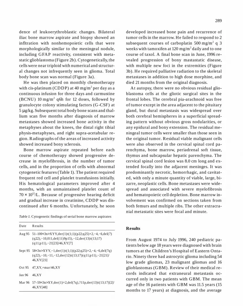
Figure 3a. Normal bone scan. Figure 3b. Multiple bone metastases in lower extremities.
ceived radiation to the craniospinal axis of 50.4 Gyfractions of 1.2 Gy per fraction twice daily with aboost of 16.8 Gy in 14 fractions to gross residual ar-eas of brain tumor to a total of 67.2 Gy, and 7.2 Gyin 6 fractions to the spinal metastasis with a totaldose of 57.6 Gy in 48 fractions.
He was clinically well except for occasional legpain and headache relieved with acetaminophen.He attended school and travelled extensively to Eu-rope within 6 months of diagnosis. At 8 months af-
ter diagnosis, follow up MRI of the spine and thehead showed minimal increase in size of the cervi-comedullary enhancing tumor nodule; and investi-gation showed: hemoglobin (Hb) 105 Gm/.L, whiteblood cell count (WBC) 2.67 × 10⁹/L, neutrophil1.39 × 109/L, and platelet (plt) count 146 × 1012/L.One month later in August, he became more fa-tigued and had bruising, but no other new findings.His Hb dropped to 63 Gm/L, WBC to 2.11 × 10⁹/L,and platelet count to 85 × 1012/L. There was no evi-
Please indicate author’s corrections in blue, setting errors in red
149436 NEON ART.NO 947-97 (737) ORD.NO 234737.Z
289
Table 1. Cytogenetic findings of serial bone marrow aspirates
Date Results
Aug 95 51–100<3n>XYY,der(1)t(1;1)(p22;q25)×2,−4,−6,del(7)(q22),−10,011,del(11)9p15),−12,der(13)t(13;17)(q11;p11),−21[23]/46,XY[7]
Sept 95 58<3n>XYY,−1,der(1)t(1;1)(p22;q25)×2,−4,−6,del(7q)(q22),−10,−11,−12,der(12)t(13;17)(q11;p11),−21[21]/46,XY[23]
Oct 95 47,XY,+mar/46,XY
Jan 96 46,XY
Mar 96 57–59<3n>XY,der(1)×2,del(7q),?11p,der(13)t(13;17)[2]/46,XY[48]
dence of leukoerythroblastic changes. Bilateraliliac bone marrow aspirate and biopsy showed aninfltration with nonhemopoietic cells that weremorphologically similar to the meningeal nodule,including GFAP reactivity, consistent with meta-static glioblastoma (Figure 2b). Cytogenetically, thecells were near triploid with numerical and structur-al changes not infrequently seen in glioma. Totalbody bone scan was normal (Figure 3a).
He was then placed on monthly chemotherapywith cis-platinum (CDDP) at 40 mg/m2 per day as acontinuous infusion for three days and carmustine(BCNU) 10 mg/m2 q6h for 12 doses, followed bygranulocyte colony stimulating factors (G-CSF) at5 µg/kg. Subsequent total body bone scans and thal-lium scan five months after diagnosis of marrowmetastases showed increased bone activity in themetaphyses about the knees, the distal right tibialphysis-metaphyses, and right supra-acetabular re-gion. Radiographs of the areas of increased activityshowed increased bony sclerosis.
Bone marrow aspirate repeated before eachcourse of chemotherapy showed progressive de-crease in myelofibrosis, in the number of tumorcells, and in the proportion of cells with abnormalcytogenetic features (Table 1). The patient requiredfrequent red cell and platelet transfusions initially.His hematological parameters improved after 4months, with an unmaintained platelet count of70 × 1012/L. Because of progressive hearing deficitand gradual increase in creatinine, CDDP was dis-continued after 6 months. Unfortunately, he soon
developed increased bone pain and recurrence oftumor cells in the marrow. He failed to respond to 2subsequent courses of carboplatin 500 mg/m2 q 3weeks with tamoxifen at 120 mg/m2 daily and to onecourse of taxol. A final bone scan in June, 1996 re-vealed progression of bony mastastatic disease,with multiple new foci in the extremities (Figure3b). He required palliative radiation to the skeletalmetastases in addition to high dose morphine, anddied 21 months from the original diagnosis.
At autopsy, there were no obvious residual glio-blastoma cells at the gliotic surgical sites in thefrontal lobes. The cerebral pia-arachnoid was freeof tumor except in the area adjacent to the pituitarygland, but dural metastasis was widespread overboth cerebral hemispheres in a superficial spread-ing pattern without obvious gross nodularities, orany epidural and bony extension. The residual me-ningeal tumor cells were smaller than those seen inthe original tumor. Residual viable malignant cellswere also observed in the cervical spinal cord pa-renchyma, bone marrow, periadrenal soft tissue,thymus and subcapsular hepatic parenchyma. Thecervical spinal cord lesion was 8.0 cm long and ex-tended focally into the adjacent meninges. It waspredominantly necrotic, hemorrhagic, and cavitat-ed, with only a minute quantity of viable, large, bi-zarre, neoplastic cells. Bone metastases were wide-spread and associated with severe myelofibrosisand hematopoietic cell depletion. Bone marrow in-volvement was confirmed on sections taken fromboth femurs and multiple ribs. The other extracra-nial metastatic sites were focal and minute.
Results
From August 1974 to July 1996, 240 pediatric pa-tients below age 18 years were diagnosed with braintumors at the Children’s Hospital of Eastern Onta-rio. Ninety three had astrocytic glioma including 54low grade gliomas, 23 malignant gliomas and 16glioblastomas (GBM). Review of their medical re-cords indicated that extraneural metastasis oc-curred only in two patients with GBM. The meanage of the 16 patients with GBM was 11.5 years (15months to 17 years) at diagnosis, and the average

Please indicate author’s corrections in blue, setting errors in red
149436 NEON ART.NO 947-97 (737) ORD.NO 234737.Z
290
Table 2. Marrow metastasis in astrocytic gliomata in the literature
Age Sex Locationof tumor
Initial Rx Shunt BM metastasis
Time Symptoms& findings
Otherextraneuralsites ofmetastasis
Rx Survivalfrom BMmetastasis
Ref
Low grade glioma19 M hypothal biopsy XRT ? 14M anemia, back
painbones LM CPPD
BCNU12M+ 15
Malignant astrocytoma36 F thalamus biopsy 54Gy no 7M low back pain LM skeleton 6 weeks 1637 M intracerebral XRT no 15M low back pain
anemiamyelofibrosis
retroperi nodes 5M 17
40 F intra-medullary
biopsy 55Gy no fewmonths
painmyelofibrosis
LM diffuseskeleton
? 18
51 M frontal 50Gy wholebrain 60Gy totumor
no 24M low back painlow plateletsmyelofibrosis
LM 6M 19
52 F parietal subtotalresection 60GyBCNU
no 24M anemia lowplateletsmyelofibrosis
diffuse bones 3M 20
83 F temporal 70Gy no 6 PM lung, liver ? 21
Glioblastoma multiforme11 F post fossa 50Gy ‘8 in 1’ yes 4M back pain LM lung bones,
pelvisthiotepa 2M 22
12 M parietal occip resection 40Gy no 6M hip pain anemia LM, pelvis liver 4M 2313 M bifrontal LM
intramedresection 72GyVP16 cyclo
no 10M low back pain,pancytopenia
LM, skeletonliver
BCNUCDDP taxol
10M **
24 F frontal subtotalresection 60Gy
no 6M low back pain,low platelets
none 2M 24
31 M occipital partial resectionXRT, AZQBCNU
no 8M low back pain multiple bone 3 weeks 25
41 M temporal surgery XRTBCNU
no 6 Dx anemia, lowplatelets
LM 7M 26
55 F tempo-parietal
resection 60Gy no 4M petechiaepancytopenia
axial skeleton,liver
5M 27
57 M tempo-occipital
subtotalresection 60GyBCNU
no 12M hip pain lowplatelets
LM, axialskeleton pleura
shortly afterdiagnosis
24
LM-leptomeningeal. ** present case.
survival was 15 months (4–46 months). All but theyoungest child received radiation threrapy to thetumor bed. Eleven received a variety of postoper-ative chemotherapy combinations consisting of twoor more of vincristine, cyclophosphamide, carmus-tine (BCNU), lomustine (CCNU), procarbazine,CDDP, etoposide, carboplatin, ifosfamide, taxoland tamoxifen. For the 4 of 16 patients with GBM in
whom consent for autopsy was obtained, one hadextensive brain tumor only; a second with a ventri-culo-peritoneal (VP) shunt had an isolated extra-neural metastasis to the appendix. The third pa-tient, an 11 year old boy with Li-Fraumeni syn-drome and previous soft tissue and osteogenic sar-coma, was confirmed 10 months after surgicalresection and chemotherapy for occipital glioblas-

Please indicate author’s corrections in blue, setting errors in red
149436 NEON ART.NO 947-97 (737) ORD.NO 234737.Z
291
toma, to have acute myelomonocytic leukemia twodays before he succumbed to fulminant disseminat-ed intravascular coagulation. The case of the fourthpatient was the case presented above.
Discussion
Brain tumors account for 20–25% of all pediatricmalignancies, about half of which are of astrocyticorigin [7]. Majority of the astrocytic tumors are lowgrade, occur mainly in the posterior fossa, and havea favorable prognosis. High grade gliomas affectpredominantly the older children and teenagers,occurring mainly in the supratentorial region, com-prising 7–11% of pediatric brain tumors and are ma-lignant [8]. In contrast to adult glioma, pediatricgliomas are rarely associated with abnormalities inthe p53 gene, epidermal growth factor, or deletionsof chromosome 10, suggesting that they are formeddifferently [9]. Finlay reported addition of chemo-therapy to radiation and surgery in high grade glio-ma to have a favorable advantage, especially in pa-tients younger than 3 years of age [10].
Local relapse and neuraxis spread are still themost likely sites of disease recurrence. Extraneuralmetastasis from brain tumors, though rare, has beenwell reported since 1928, occurring late in thecourse of the disease, after a median of two years[11]. In glioblastomas, the organs involved includepleura/lung (60%), lymph nodes (51%), bones(31%) and liver (22%) [12]. VP-shunt-associatedmetastases generally involve abdomen and lung [1].In 10% of the patients, extracranial metastases de-velop without a shunt, or are present at diagnosis.Despite the absence of an identifiable lymphaticsystem in the CNS [13], there have been reports ofregional lymph node involvement, commonly attri-buted to lymphatic spread. Direct access of tumorvia the dural vessels to the extrameningeal tissue isthe most frequent single factor in the developmentof extraneural metastases [14].
There are 15 cases in the literature of diffuse bonemarrow metastases associated with astrocytic glio-ma (Table 2) [15–27], of which 1 was low grade, 6malignant glioma and 8 GBM. The age group spansfrom 11 to 83 years, with evidence of metastasis oc-
curring from diagnosis to 24 months, the averagebeing 12 months. There was a trend to developmentof metastases later in the more benign tumor. Twoof the cases had complete resection of the brain tu-mors. All received radiation treatment to the pri-mary tumor, with boosts given to leptomeningealdisease, when present. Shunts were placed in 2 pa-tients only. Presenting symptoms of marrow metas-tases include low back pain in 11, anemia in 6, andthrombocytopenia in 7 of the 15 patients. A secondcancer was considered to be more likely for patientswith hematologic abnormalities until histologicalproof of glioma was made.
Myelosuppression commonly accompanies che-motherapy, with recovery usually occurring withinweeks after completion of treatment. Early marrowmetastasis from brain tumors may not be recog-nized because of a normal hemogram. Leukoeryth-roblastic changes are seen in only about 30% of pa-tients [12]. Bone marrow aspirate may only showmyelofibrosis, necessitating diagnosis of extraneu-ral metastasis with open bone or bone marrow biop-sy [19, 24]. Cohen and Duffner suggest that all pa-tients presenting with anemia or thrombocytopeniashould be suspected of extraneural metastases [6].
Back pain occurs in 70% of patients with marrowmetastases. Many patients also have evidence ofleptomeningeal disease and bony metastases, mak-ing it difficult to decide the reason for the pain. Onlyone patient reported in the literature had isolatedbone marrow metastasis [24]. Bony metastases, themost common site being the vertebral spine, aremanifested as lytic or sclerotic lesions on radio-graphs [28]. Radiological changes may occur longbefore symptoms [29]. Increased uptake of techne-tium-99 m-MDP, a bone matrix imaging agent, isnot always evident with bone marrow metastasis[24]. Diffuse bony involvement is unusual and isonly seen with high grade astrocytomas [22]. MRI isthe imaging modality of choice for investigation ofdisease of the bone marrow because of its sensitiv-ity. However, in patients who received radiationpreviously, the signal intensity may be abnormal formany years, therefore making it difficult to investi-gate suspected marrow metastasis. Concomitantuse of G-CSF may further confuse the issue by caus-ing strong marrow signal [30]. Although the MRI

Please indicate author’s corrections in blue, setting errors in red
149436 NEON ART.NO 947-97 (737) ORD.NO 234737.Z
292
appearance of marrow metastases is non-specificand not effective in separating malignant from be-nign disease, it is still a useful method to identify thepresence and extent of metastases and to plan radi-ation treatment portals [31]. In our patient the ini-tial negative bone scan and spinal radiographs sug-gested the pain to be secondary to cervical myelo-pathy. The bone scan only became positive after hewas started on treatment for the marrow metasta-ses.
Cytogenetic and molecular characterization areuseful in initial diagnosis, identification of residualtumor, as well as determination of etiology. The ab-sence of the 11q23 abnormality typically seen in leu-kemias due to etoposide, as well as the presence ofan almost triploid karyotype helped to suggest thatabnormal marrow cells arose from our patient’sglioma. Shapiro et al. postulated that hyperdiploidcells with extensive ploidy changes and chromo-some rearrangement are more sensitive to BCNUbecause the mechanism for the repair of drug in-duced damage are not functional [32]. Good re-sponse to BCNU lasted for more than 6 months inour patient. The tumor cytogenetics at subsequentrelapse were similar to that seen at the time of initialmarrow involvement, suggesting that there wereother mechanisms for his ultimate tumor resistance.Unfortunately, tissue culture of the original braintumor did not yield cells in metaphase to allow forcytogenetic analysis and comparison to the bonemarrow metastasis.
Sequential use of CPPD and BCNU was found tobe effective in adjuvant therapy for high grade glio-ma [33]. Low grade gliomas with metastases alsoshowed good response to vincristine and cyclophos-phamide [34] or BCNU and CPPD [15]. In our pa-tient, progressive hearing and renal impairment ne-cessitated the discontinuation of CDDP after 6months. In all the cases of bone marrow metastasisfor which treatment was initiated, there was evi-dence of clearing of marrow disease. Although theresponse time is usually short, extra treatment ap-pears to offer survival advantage and better qualityof life.
In summary, in patients with brain tumor andbone pain or hematological abnormalities, bonescintigraphy and plain radiographs are useful to
suggest the presence of underlying bone marowmetastatic disease. This can be pursued with MRI,bone marrow aspirate and biopsy. Chemotherapyhas been shown to be effective in alleviating symp-toms of pain and reduce significantly marrow infil-tration. With the availability of peripheral stem celltransplant after high dose chemotherapy as a re-search method, improved survival can still be hopedfor, as previously seen in those patients with medul-loblastoma. There needs to be better understandingabout extraneural metastasis of gliomas so thatstrategies may be used to prevent this unusual oc-currence.
Acknowledgements
The authors wish to thank Dr. Marc Rosenblum ofMemorial Sloan-Kettering Cancer Center for thereview of the histological material, Dr. David J.Stewart of the Ontario Cancer Treatment and Re-search Centre for the critical review of the manu-script and Margaret Humphreys for secretarial as-sistance.
References
1. Berger MS, Baumeister B, Geyer JR, Milstein J, Kanev PM,LeRoux PD: The risks of metastases from shunting in chil-dren with primary central nervous system tumors. J Neuro-surg 74: 872–877, 1991
2. Campbell AN, Chan HSL, Becker LE, Daneman A, ParkTS, Hoffman HJ: Extracranial metastases in childhood pri-mary intracranial tumors. Cancer 53: 974–981, 1984
3. Duffner PF, Cphen ME: Extraneural metastasis in child-hood brain tumor. Ann Neurol 10: 261–265, 1981
4. Marchese MF, Chang CH: Malignant astrocytic gliomas inchildren. Cancer 65: 2771–2778, 1990
5. Papac RJ: Bone marrow metastases. Cancer 74: 2403–2413,1994
6. Cohen ME, Duffner PK: Extraneural metastasis in child-hood brain tumors. In: Brain tumor in children: Principles ofDiagnosis and Treatment, 2nd edition. Raven Press, Ltd.,New York, 1994
7. Pollack I: Childhood gliomas: an overview. J Neuro-Oncol28: 117–120, 1996
8. Heideman RL, Packer RJ, Albright LA, Freeman CR,Rorke LB: Tumors of the central nervous system. In: PizzoPA, Poplack DG (eds) Principle and Practice of PediatricOncology. 3rd edition Lippincott-Raven. 1997, pp 664

Please indicate author’s corrections in blue, setting errors in red
149436 NEON ART.NO 947-97 (737) ORD.NO 234737.Z
293
9. Raffel C: Molecular biology of pediatric gliomas. J Neuro-oncol 28: 121–128, 1996
10. Finlay JL, Boyett JM, Yates AJ, Wisoff JH, Milestein HM,Geyer JR, Bertolone SJ, McGuire P, Cherlow JM, Tefft M,Turski PA, Wara WW, Edwards M, Sutton LN, Berger MS,Epstein F, Ayers G, Allen JC, Pachu RJ, for the ChildrensCancer Group: Randomized Phase III trial in childhoodhigh-grade astrocytoma comparing vincristine, lomustine,and prednisone with the eight-drugs-in-1-day regimen. JClin Oncol 13: 112–123, 1995
11. Hoffman HJ, Duffner PK: Extraneural metastases of cen-tral nervous system tumors. Cancer 56: 1778–1782, 1985
12. Pasquier B, Pasquier D, N’Golet A, Panh MH, Couderc P:Extraneural metastases of astrocytomas and gliomas. Clin-ico-pathologocal study of two cases and review of literature.Cancer 45: 112–125, 1980
13. Bernstein JJ, Woodard CA: Glioblastoma cells do not in-travasate into blood vessels. Neurosurgery 36: 124–132,1995
14. Huang P, Allam A, Taghian A, Freeman J, Duffy M, SuitHL: Growth and metastatic behavior of five human glioblas-toma compared with nine other histological types of humantumor xenografts in SCID mice. J Neurosurg 83: 308–315,1995
15. Johnston S: Complete response by systemic chemotherapyfor the treatment of simultaneous bone and bone marrowmetastasis with meningeal gliomatosis as a progression fromhypothalamic low grade astrocytoma. International Sympo-sium of Pediatric Neuro-Oncology. Abstract Washington,Abstract # 56, 1996
16. Dewar JM, Dady PG, Balakrishnan V: Metastatic astrocyto-ma. Australian & New Zealand J Med 15: 745–747, 1985
17. Rubinstein LJ: Development of extracranial metastasesfrom a malignant astrocytoma in the absence of previouscraniotomy. J Neurosurg 26: 542–547, 1967
18. Newman RP, Schaefer EJ, Thomas CB, Oldfield EH: A betalipropoteinemia and metastatic spinal cord glioblastoma.Arch Neurol 41: 554–556, 1984
19. Liuwicz BH, Rubinstein LJ: The pathways of extraneuralspread in metastasizing gliomas. Hum Pathol 10: 453–467,1979
20. Friedman JH, Liu HM, Spremulli E, Calabresi P: Distantmetastases from a malignant glioma with unusual complica-tions associated with treatment of a glioblastoma: Distantmetastases and focal white matter degeneration [Letter]. JNeurol Neurosurg Psychiat 50: 237–238, 1987
21. Kawasaki H, Shimada H, Nakura H, Tomonaga M: Peculiarpattern of glial fibrillary acidic protein production in intra-
cranial metastatic tumor cells of malignant astrocytoma.Acta Neuropathol 74: 89–91, 1987
22. Gamis AS, Egelhoff J, Roloson G, Young J, Woods GM,Newman T, Freeman AJ: Diffuse bony metastases at pre-sentation in a child with glioblastoma multiforme. Cancer66: 180–184, 1990
23. Terheggen HG, Muller W: Extracerebrospinal metastases inglioblastoma. Eur J Pediat 124: 155–164, 1977
24. Yung KA, Tepper JS, Young DF: Diffuse bone marrow me-tastasis by glioblastoma: Premortem diagnosis by perox-idase-antiperoxidase staining for glial fibrillary acidic pro-tein. Ann Neurol 14: 581–585, 1983
25. Haddon M, Slavin JD, Spencer RP: Multiple bone metasta-sis in a patient with glioblastoma multiforme. Clin Nucl Med14: 13–14, 1989
26. LoRusso PM, Tapazoglou E, Zarko RJ, Cullis PA, Austin D,Al-Sarraf M: Intracranial astrocytoma with diffuse bonemarrow metastasis: a case report and review of the litera-ture. J Neuro-oncol 6: 53–59, 1988
27. Mousavi M: Bone marrow metastasis from glioblastomamultiforme. J Med Soc New Jersey 77: 904–905, 1980
28. Mihara F, Ikeda M, Rothman MI, Numaguchi Y, Kristt D:Vertebral body metastasis of glioblastoma multiforme withepidural mass formation: contrast-enhanced MRI study.Clin Imag 18: 286–289, 1994
29. Schatzki SC, McIlmoyle G, Lowis S: Diffuse osteoblasticmetastasis from an intracranial glioma. Am J Roentgenol128: 321–323, 1977
30. Cavenaugh EC, Weinberger E, Shaw DWW, White KS,Geyer JRL: Hematopoietic marrow regeneration in pediat-ric patients undergoing spinal irradiation: MR depiction.Am J Neuroradiol 16: 461–467, 1995
31. Steiner TM, Mitchell DG, Rao VM, Schweitzer ME: Mag-netic resonance imaging of diffuse bone marrow disease.Radiol Clin North America 31: 383–408, 1993
32. Shapiro JR, Yu PY, Mohamed AN, Galicich JH, EbrahimSAD, Shapiro WR: Chromosome number and carmustinesensitivity in human gliomas. Cancer 71: 4007–4021, 1993
33. Yung WKA, Janus TJ, Maor M, Feun LG: Adjuvant chemo-therapy with carmustine and cisplatin for patients with ma-lignant gliomas. J Neurooncol 12: 131–135, 1992
34. Longee DC, Friedman HS, Phillips PC, Burger PC, Oakes J,Heffez D, Wharam M, Strauss L, Fuller GN, Schold SC:Osteoblastic metastases from astrocytomas: A report of twocases. Med Pediat Oncol 19: 318–324, 1991
Address for offprints: E. Hsu, Children’s Hospital of EasternOntario, 401 Smyth, K1H 8L1 Ottawa, Ontario, Canada


