Embed Size (px)
Citation preview

SAGE-Hindawi Access to ResearchAutoimmune DiseasesVolume 2011, Article ID 807426, 27 pagesdoi:10.4061/2011/807426
Review Article
MicroRNAs and Multiple Sclerosis
Kemal Ugur Tufekci, Meryem Gulfem Oner, Sermin Genc, and Kursad Genc
Department of Neuroscience, Health Science Institute, Dokuz Eylul University, Inciralti, 35340 Izmir, Turkey
Correspondence should be addressed to Kursad Genc, [email protected]
Received 1 September 2010; Accepted 16 October 2010
Academic Editor: Sreeram Ramagopalan
Copyright © 2011 Kemal Ugur Tufekci et al. This is an open access article distributed under the Creative Commons AttributionLicense, which permits unrestricted use, distribution, and reproduction in any medium, provided the original work is properlycited.
MicroRNAs (miRNAs) have recently emerged as a new class of modulators of gene expression. miRNAs control protein synthesisby targeting mRNAs for translational repression or degradation at the posttranscriptional level. These noncoding RNAs areendogenous, single-stranded molecules approximately 22 nucleotides in length and have roles in multiple facets of immunity,from regulation of development of key cellular players to activation and function in immune responses. Recent studies have shownthat dysregulation of miRNAs involved in immune responses leads to autoimmunity. Multiple sclerosis (MS) serves as an exampleof a chronic and organ-specific autoimmune disease in which miRNAs modulate immune responses in the peripheral immunecompartment and the neuroinflammatory process in the brain. For MS, miRNAs have the potential to serve as modifying drugs.In this review, we summarize current knowledge of miRNA biogenesis and mode of action and the diverse roles of miRNAs inmodulating the immune and inflammatory responses. We also review the role of miRNAs in autoimmunity, focusing on emergingdata regarding miRNA expression patterns in MS. Finally, we discuss the potential of miRNAs as a disease marker and a noveltherapeutic target in MS. Better understanding of the role of miRNAs in MS will improve our knowledge of the pathogenesis ofthis disease.
1. Introduction
MicroRNAs (miRNAs) represent a class of noncoding RNAmolecules that play pivotal roles in cellular and devel-opmental processes by regulating gene expression at theposttranscriptional level. miRNAs are endogenous, evolu-tionarily conserved, single-stranded RNAs approximately22 nucleotides in length that suppress the expression ofprotein-coding genes by directing translational repressionthrough base-pairing with complementary messenger RNA(mRNA)and/or by promoting degradation of target mRNAdegradation [1, 2]. Since the identification of the miRNA lin-4 as a regulator of developmental timing in the nematodeCaenorhabditis elegans (C. elegans) in 1993 [3, 4], morethan 17000 miRNAs have been recognized in 142 species.Currently, 1048 human miRNAs are registered in themiRNA registry (miRBase) which is the most commonlyused database for miRNA (September 2010, release 16,http://www.mirbase.org/) [5]. miRBase reports 672 miRNAsin mouse and 408 miRNAs in rat, with new miRNAs
constantly being identified, though the biologic function ofonly a fraction of miRNAs has been elucidated. miRNAs arepredicted to regulate up to one-third of all human protein-coding genes. Unraveling the miRNA translational silencingnetwork remains a challenge in part because individualmiRNAs typically target several transcripts rather just onespecific gene and a single mRNA can be regulated byseveral distinct miRNAs that act cooperatively [2]. Ribo-some profiling experiments showed that miRNAs mediatedestabilization of target mRNAs resulting in reduced proteinlevels [6]. miRNAs play an important role in diverse biologicprocesses such as development, cell proliferation and differ-entiation, apoptosis, oncogenesis, metabolism, angiogenesis,and inflammation. The expression of miRNAs is initiallycontrolled at the level of transcriptionby transcription factorsthat regulate the production of miRNA-containing primarytranscripts in specific cell types during development or inresponse to different environmental signals. Dysregulation ofmiRNA expression and function is associated with a variety

2 Autoimmune Diseases
of human diseases including cancer, neurodegeneration andautoimmunity [7, 8].
The regulation of mammalian immune responses bymiRNAs is a concept currently evidenced by rapidly accumu-lating data [9, 10]. miRNAs have unique expression profilesin cells of the innate and adaptive immune systems andhave pivotal roles in the regulation of both cell developmentand function. Recent studies focused on the networkwiderole of miRNA or the functions of individual miRNAs haverevealed that these small noncoding RNAs are involved in Tand B cell differentiation in the thymus and bone marrow,respectively. During the effector phases of adaptive immu-nity, miRNAs contribute to the differentiation of T cellsinto functional lineages, class switching and germinal centreformation in B cells and activation of antigen-presentingcells (APCs) through pattern recognition pathways [11].miRNAs are also directly involved in innate immunity andtransduction signalling by Toll-like receptors (TLRs) andthe ensuing cytokine response [12]. Up to one half ofinnate immune genes are predicted to be under the directregulation of miRNAs. With the capacity of miRNAs toregulate the survival and death of T and B cells, control overmiRNA expression is essential to prevent adaptive immunecells from unregulated proliferation leading to cancer orautoimmunity [13, 14]. miRNAs are differentially expressedin autoimmune diseases and miRNA regulation may have animpact on the development or prevention of autoimmunity.miRNA dysregulation is linked to autoimmune diseasessuch as rheumatoid arthritis, systemic lupus erythematosus,Sjogren’s syndrome, psoriasis, and MS [15–17]. MS is themost common autoimmune disease of the central nervoussystem (CNS). It is a chronic, neuroinflammatory, anddemyelinating disease in which myelin specific autoreactiveCD4+ T cells become activated in the peripheral immunecompartment, cross the blood-brain barrier (BBB), andpromote neurological disability [18, 19]. Both genetic (HLAtype) and environmental causes for MS have been suggested.Recently, genomewide association studies have identifiedadditional potential MS susceptibility loci [20, 21]. Recentstudies suggest that miRNA dysregulation may contributeto the pathogenesis of MS. Thus, better understanding ofmiRNA mechanisms might shed light, not only on thepathogenesis of MS but also on potential approaches formanaging or even suppressing the disease. In this review,we briefly overview the biogenesis and action mechanisms ofmiRNAs and summarize recent advances in our understand-ing of both the intended functions of miRNAs in managingimmune responses. We then review evolving knowledge onthe role of miRNA in autoimmunity and emerging dataregarding miRNA expression patterns in MS. Finally, wealso discuss the potential of miRNAs as a diagnostic andprognostic indicators of disease type and status and as a noveltherapeutic target in MS.
2. MicroRNAs
2.1. MicroRNA Biogenesis. All miRNAs are processed andmaturated through a complex biogenesis process involving
multiple protein catalysts, accessory proteins, and macro-molecular complexes following a coordinated series ofevents. The reader is referred to excellent recent reviews fordetailed discussions of miRNA biogenesis and its regulation[22–25]. MicroRNAs can be encoded by independent genesbut may also be processed from a variety of different RNAspecies, including introns, 3′-UTR of mRNAs, long non-coding RNAs, transposable elements, and genomic repeats[26–32]. miRNAs are expressed as 21–23 nucleotide RNAmolecules initially transcribed by RNA polymerase II aslong primary miRNAs (pri-microRNAs). Although most ofthe miRNA genes are transcribed by RNA Polymerase II,a cluster of human miRNAs have recently been shown toutilize RNA Polymerase III for their transcription [33]. Pri-miRNAs are typically 3 to 4 kilobases long single-strandedRNAs with 5′ cap, 3′ poly(A) tail and complicated secondarystructure [34–37]. Pri-miRNAs are processed in the nucleusinto one or more precursor-miRNAs (pre-miRNAs) with anapproximately 70-nt loop structure. Processing is performedby a protein complex named microprocessor complex con-sisting of the nuclease Drosha (nuclear RNase III) and thestranded RNA-binding protein, human DiGeorge syndromecritical region gene 8 (DGCR8) (also known as Pasha inflies) [35, 37–42]. Drosha functions as the catalytic subunitwhile DGCR8 recognizes the RNA substrate. Pre-miRNAsare exported from the nucleus to the cytoplasm by exportin-5which specifically recognizes the characteristic end structureof pre-miRNAs [43–46]. In the cytoplasm, another RNase III,known as Dicer, further processes the pre-miR into maturemiRNA, which is double stranded (miRNA duplex) [47, 48].After Dicer processing, the miRNA duplex is unwound anda strand (known as miRNA strand or guide strand) bindsto an Argonaute 2 (Ago 2) protein (eIF2C2 in human) ina process that is referred to as miRNA loading or assembly,while the complementary strand (known as miRNA∗ strand,star strand or passenger strand) is degraded. The effectorcomplex that mediates catalytic mRNA cleavage is knownas RNA-induced silencing complex (RISC), and the effectorcomplex that mediates translational repression directed bymiRNAs is known as micro-ribonucleoprotein complex(miRNP) [49–51]. The single stranded mature microRNAmust associate with the RISC. Mature microRNAs areincorporated into a miRNP (Figure 1). In this complex,which includes the Dicer-transactivation-responsive RNA-binding protein (TRBP)-PACT-Ago 2, microRNAs can directdownregulation by two mechanisms: translational inhibitionand target mRNA cleavage [52–55]. Perfect match with thetarget results in mRNA degradation whereas partial matchleads to translational inhibition.
Inflammation has been reported to regulate miRNAbiogenesis; TLR ligands, antigens, or cytokines can modulatemiRNA expression level through regulation of specifictranscription factors [2, 9, 56]. Cytokines have been shownto regulate Dicer expression resulting in alteration of pre-miRNA processing. Interferon-beta (IFN-β) has been shownto inhibit Dicer expression, which results in decrease ofpre-miRNA processing, whereas IFN-γ induces pre-miRNAprocessing [57].
Autoimmune Diseases 3
2.2. Detection of MicroRNAs. Information about miRNA andtarget expression patterns can help to assess the likelihoodthat a predicted miRNA-target relationship is relevant invivo [58]. Expression of a miRNA can be measured bymolecular biology techniques, such as Northern blotting,RNase protection assay, polymerase chain reaction- (PCR-)based techniques, and high throughput assays [59–61].miRNA expression profiles were first generated by smallRNA cloning and Northern blotting [4, 62–67]. The smallsize of miRNAs initially hampered PCR-based methods [61].However, since the development of quantitative real-timePCR, PCR-based techniques have become very popular dueto their high sensitivity [62, 68, 69]. In situ hybridizationhas provided further insight into the tissue-specific expres-sion of pri- and mature miRNAs [62, 70–74]. Microarraytechniques are widely used to comprehensively assay theentire miRNome (the global miRNA expression profile) intissues or in cell lines [62, 68, 75–83]. In addition, serialanalyses of gene expression (SAGE) adapted for small RNAshave been used to obtain miRNomes [84]. Interest in theSAGE approach was stimulated by recent innovations innext generation (deep) sequencing methods that provide apowerful tool for various genomics studies [85–87]. Overall,these technical improvements are expected to greatly widenthe repertoire of known miRNAs in a variety of biologicalsystems [61]. Emerging techniques for miRNA detection andquantification, including luminescence-based, fluorescence-based, electrochemical, colorimetric, and enzyme-based, andnanotechnology-based methods have recently been reviewed[88]. Whereas expression analyses are required to identifymiRNAs with altered expression patterns in diseased tissues,functional analyses of the ability of these miRNAs to regulateexpression of target mRNAs are essential to understand theirimpact on pathogenic pathways and processes.
3. MicroRNAs and Immunity
Clearly, both innate and adaptive immune responses areextremely highly regulated. Recent work from a number oflaboratories has revealed that miRNAs play an importantrole in this intricate system (Table 1). miRNAs have uniqueexpression patterns in immune cells and play a pivotal role intheir development, maturation, and function.
3.1. Role of MicroRNAs in Immune Cell Development. miR-NAs have an important role in regulating stem cell self-renewal and differentiation by repressing the translationof selected mRNAs in stem cells and differentiating intodaughter cells. Such a role has been shown in embryonicstem cells, germline stem cells and various somatic tissuestem cells [89]. The first studies implicating miRNAs inimmunological processes were originated from expressionprofiling of haematopoietic cells during their development.Haematopoietic stem cells reside mainly in the bone marrowand give rise to all blood cell lineages, including cells thatconstitute the immune system [9]. These cells must maintaina precise balance between self-renewal and differentiationinto multipotent progenitors, which subsequently give rise
to both the common lymphoid and common myeloidprogenitors of the haematopoietic system [9, 90]. Systematicinvestigation of miRNA levels in hematopoietic cell lineageshas identified miRNAs that are now considered as markers ofthese lineages [91–93]. Peculiar miRNA profiles in differenthaematopoietic organs and cell types suggest that miRNAsare dynamically regulated during early haematopoiesis, lin-eage commitment, and the development of immune cells andare involved in the regulation of these processes.
One of the first miRNAs described to have a role inimmune cell development was miR-181a which is highlyexpressed in thymus cells and expressed at lower levels inthe heart, lymph nodes, and bone marrow [91, 94]. Inbone marrow-derived B cells, miR-181a expression has beenshown to decrease during B cell development from thepro-B to the pre-B cell stage [91]. miR-181a inhibits thetransition of pro-B to the pre-B cell stage. Moreover, miR-181a was identified as a positive regulator of B lymphocytedifferentiation based on evidence that expression of miR-181a in hematopoietic stem and progenitor cells resulted inan increase in CD19+ B cells and a decrease in CD8+ T cells[91]. Interestingly, miR-181a is also involved in thymic T celldifferentiation, by defining the activation threshold of T cellreceptor (TCR) [94]. This miRNA modulates TCR signaling,thus affecting the sensitivity of T cells to antigens [94].Other examples of miRNA-mediated regulation of immunecell development include miR-223 which was identified asan essential modulator of granulocytic differentiation [95]and miR-150 which has been shown to be critical forB cell differentiation [96, 97]. Collectively, these studiesdemonstrate that miRNAs play critical roles at distinct stagesof immune cell development.
3.2. MicroRNAs in Adaptive Immune Responses. The adaptiveor acquired immune system involves the selective recognitionand removal of nonself by the TCRs on T cells and antibodiesproduced by B cells. The maturation, proliferation, differen-tiation, and activation of T and B cells are complex processestightly controlled at different levels including miRNA-mediated posttranscriptional gene regulation [11]. Adaptiveimmunity refers to immune responses to antigens thatundergo learning processes and provide specific memory.Once APCs capture a pathogen, they display foreign anti-gens complexed with major histocompatibility complexes(MHCs) on their surface to enable recognition of the antigenby naıve T cells to induce the adaptive immune response[19]. The combination of this interaction further drives theupregulation of both CD80 and CD86 on the surface ofAPCs. CD80 and CD86 identify two additional receptors,CD28 and Cytotoxic T-lymphocyte antigen 4 (CTLA4), onthe surface of the T cells to provide a second signal toAPCs [19]. CD28 is associated with activation of the T cellwhereas CTLA4 is more regulatory. After this second signal,the T cells become activated, and APCs begin to secreteimportant cytokines, including IL-12 and IL-23, which bindto specific receptors on T cells and drive them to secretedifferent cytokines, such as IFN-γ or IL-17, depending onthe cytokine milieu. T cells also begin to secrete IL-2, which

4 Autoimmune Diseases
Pri-miRNA
Pre-miRNA
DGCR8
Drosha
Processing
RAN-GTP
Exp-5
TRBP
Pre-miRNA
Dicer
miRNA duplex
RISC complex
mRNA degradation
CytoplasmNucleus
Translational repression
Transcription
Figure 1: miRNA genes are transcribed in the form of Pri-miRNA. The DGCR8-Drosha complex processes in the form Pre-miRNA followedby transport into cytoplasm by Exportin-5. In cytoplasm, Pre-miRNA is processed by Dicer into miRNA duplex. Of miRNA duplex, onestrand is loaded into RISC complex, which functions for either mRNA degradation or translational repression.
then activates its own IL-2 receptor [19]. Upon activationof their TCR in the presence of costimulatory molecules,naıve T cells differentiate into various subsets of effector Tcells with distinct effector functions (e.g., Th1, Th2, Th17,Th9). This differentiation is directed by a specific cytokinemilieu leading to the expression of transcription factorsspecific for the respective lineages. The expression levelsof all molecules involved in adaptive immune responses(transcription factors, cell surface receptors, cytokines, andtheir receptors) may be regulated by miRNAs as discussedbelow.
3.2.1. T Cells. The development of T cells in the thymus andtheir activation in the periphery are controlled by complexprotein signalling networks that are subject to regulation bymiRNAs [9, 98]. miRNA expression profiles vary betweenT cell subsets and different developmental stages [92, 99].Specific deletion of Dicer in the T cell lineage resulted inimpaired T cell development and aberrant T helper celldifferentiation and cytokine production [100]. A severeblock in peripheral CD8+ T cell development was observed
upon Dicer deletion in the thymus. However, Dicer-deficientCD4+ T cells, although reduced in numbers, were viableand could be analyzed further. These cells were defectivein microRNA processing, and upon stimulation, they pro-liferated poorly and underwent increased apoptosis [100].Deletion of Dicer at an early stage of T cell developmentcompromised the survival of alpha-beta lineage cells whereasthe numbers of gamma-delta-expressing thymocytes werenot affected in developing thymocytes [101]. Mice withhigher expression of miR-17–92 in lymphocytes developedlymphoproliferative disease and autoimmunity and diedprematurely. Lymphocytes from these mice showed moreproliferation and less activation-induced cell death. ThemiR-17–92 miRNA suppressed expression of the tumorsuppressor PTEN and the proapoptotic protein Bim [102].T cell sensitivity to antigen is intrinsically regulated duringmaturation to ensure proper development of immunityand tolerance. Increasing miR-181a expression in matureT cells augments the sensitivity to peptide antigens whileinhibiting miR-181a expression in the immature T cellsreduces sensitivity and impairs both positive selection andnegative selection [94]. These effects are in part achieved by

Autoimmune Diseases 5
the downregulation of multiple phosphatases, which leads toelevated steady-state levels of phosphorylated intermediatesand a reduction of the TCR signaling threshold. T cellactivation requires signaling through the TCR and costim-ulatory molecules, such as CD28. Costimulation-dependentupregulation of miR-214 promotes T cell activation bytargeting the negative regulator Pten. Thus, the requirementfor T cell costimulation is, in part, related to its ability toregulate expression of miRNAs that control T cell activation[103].
Recent data have also indicated a role for miRNAs in thedifferentiation of T cells into distinct effector T helper cellsubsets. miR-155 has an important role in the mammalianimmune system, specifically in regulating T helper celldifferentiation and the germinal center reaction to producean optimal T cell-dependent antibody response [104]. miR-155 exerts this control, at least in part, by regulating cytokineproduction. Many types of specialized Th cells, includingTh1, Th2, Th17, Th9, follicular helper T, and Treg, havebeen identified. Different Th cells are committed to theirpaths but recent emerging evidence suggests that undercertain conditions, seemingly committed Th cells possessplasticity and may convert into other types of effector cells[105]. There is growing evidence that clinically similar formsof autoimmune demyelinating disease can be driven bymyelin-specific T cells of distinct lineages with differentdegrees of dependence on IL-17 production to achieve theirpathological effects [106]. miRNAs play an important role inthe development of Th17 cells [107]. Bcl-6, a transcriptionalrepressor, binds to the promoters of the Th1 and Th17cell transcriptional regulators T-bet and RORgammat andrepresses IFN-γ and IL-17 production. Bcl-6 also repressesexpression of many miRNAs predicted to control the T fol-licular cell signature, including miR-17-92, which repressesCXCR5 expression. Thus, Bcl-6 positively directs T follicularcell differentiation, through combined repression of miRNAsand transcription factors [108]. miRNAs are also essential inthe development, differentiation, and function of Treg cellswhich are potent immune regulators [109]. Recent studiesshowed a crucial role for miRNAs in Treg cell biology andthe prevention of spontaneous autoimmunity [110–112].
miR-155 deficiency in Treg cells results in increasedsuppressor of cytokine signaling 1 (SOCS1) expressionaccompanied by impaired activation of signal transducer andactivator of transcription 5 (STAT5) transcription factor inresponse to limiting amounts of IL-2. Forkhead box P3-(Foxp3-) dependent regulation of miR155 maintains com-petitive fitness of Treg cell subsets by targeting SOCS1 [113].miR-155-deficient mice have reduced numbers of Tregs, bothin the thymus and periphery, due to impaired development.However, no evidence for defective suppressor activity ofmiR-155-deficient Tregs was found, either in vitro or in vivo,suggesting that miR-155 contributes to Treg development,but that additional miRNAs control Treg function [114].The expression of miR-142-3p was recently shown to berepressed by Foxp3, leading to increased production of cyclicAMP and suppressor function of Treg cells [115]. DepletingmiRNAs by eliminating Dicer reduces Treg cell numbersand results in immune pathology [116]. Dicer facilitates, in
a cell-autonomous fashion, the development of Treg cells inthe thymus and the efficient induction of Foxp3 by trans-forming growth factor-beta (TGF-β). These results suggestthat Treg cell development involves Dicer-generated RNAsawaiting functional assessment. miR-31 negatively regulatesFoxp3 expression by binding directly to its potential targetsite in the 3′-UTR of Foxp3 mRNA whereas miR-21 acts asa positive, though indirect, regulator of Foxp3 expression[117]. Finally, miR-155 inhibition sensitizes CD4+ Th cellsfor Treg-mediated suppression [118].
3.2.2. B Cells. The generation of B cells that express highaffinity antigen receptors involves two main stages: antigen-independent development in the bone marrow and antigen-dependent selection in the secondary lymphoid organs, bothof which are associated with dynamic regulation by miRNAs[9, 119]. Antigen receptors on the surface of B cells triggeradaptive immune responses after encountering their cognateantigens but also control a series of antigen-independentcheckpoints during B cell development. These physiologicalprocesses are regulated by the expression and function ofcell surface receptors, intracellular signaling molecules, tran-scription factors, and miRNAs [119]. Temporal regulation ofseveral different miRNAs was observed and putative new celltype-specific miRNAs were identified in the development ofB cells, suggesting the involvement of many, but undefined,regulatory pathways in B cell development and maturation[9]. The role of miRNAs in controlling the early developmentof B cells is now thought to involve the modulation ofkey protein factors that control these aspects of B celldevelopment [97]. miR-181 is preferentially expressed in theB-lymphoid cells of mouse bone marrow and its ectopicexpression in hematopoietic stem/progenitor cells leads toan increased fraction of B-lineage cells, without increase ofT cells or myeloid cells in both tissue-culture differentiationassays and adult mice [91].
In contrast, mice with a conditional deletion of Dicerin B cells had a complete block in B cell development[120]. This block is related to dysregulated expression ofthe proapoptotic protein Bim, probably during the selec-tion of effective antigen receptors. These results suggest adefect in the regulation of B cell selection. Regulation ofapoptosis and cell cycle progression plays an essential rolein the maintenance of B-cell homeostasis, because a finebalance of survival and expansion is critical for preventinglymphocytic disorders. Interestingly, the changes observed bygene expression profiling of Dicer-deficient B cell precursorsare generally similar to those observed in B cells lacking themiR-17–92 family. Absence of miR-17–92 leads to increasedlevels of the proapoptotic protein Bim and inhibits B celldevelopment at the pro-B to pre-B transition [121]. Inaddition to effects on antigen receptor selection, miRNAsalso regulate the transcription factors involved in early Bcell development [9]. Constitutive expression of miR-150,which is highly upregulated at the immature B cell stage,leads to a block at a proximal stage of B cell development,the pro-B to pre-B cell transition, indicating that miR-150most likely downregulates mRNAs that are important for

6 Autoimmune Diseases
pre- and pro-B cell formation or function [96]. miR-150controls B cell differentiation by targeting the transcriptionfactor c-Myb [97]. miR-125b also promotes B cell diver-sification in the germinal center by inhibiting prematureutilization of essential transcription factors for plasma celldifferentiation [122].
The contribution of miRNAs in the antigen-drivenstages of the humoral response in secondary lymphoidorgans has also been described [9]. miR-155 is requiredin B cell responses to thymus-dependent and -independentantigens [123]. B cells lacking miR-155 generated reducedextrafollicular and germinal center responses and failed toproduce high-affinity IgG1 antibodies. When transcriptionfactor Pu.1 is overexpressed in wild-type B cells, fewer IgG1
cells are produced, suggesting that loss of Pu.1 regulationis a contributing factor to the miR-155-deficient phenotype[123]. The miR-23a cluster is a downstream target of PU.1involved in antagonizing lymphoid cell fate determina-tion [124]. miR-155 represses activation-induced cytidinedeaminase, which is required for immunoglobulin genediversification in B lymphocytes [125, 126]. A recent studyshowed that numerous miRNAs were expressed in a stage- ortransformation-specific fashion in B cells, suggesting specificfunctional or pathological roles [127].
3.3. MicroRNAs in Innate Immune Responses. The innateimmune response provides the initial defense against infec-tion by external pathogens and is predominantly mediatedvia myeloid cells such as macrophages, DCs, monocytes,neutrophils, as well as natural killer (NK) cells. The presenceof pathogens is commonly detected by tissue APCs such asmacrophages and DCs via families of pattern recognitionreceptors that bind nonself-antigens such as microbialproducts. Many families of pattern recognition receptorshave been identified, although the best characterised are theTLR which are composed of 11 members and the interleukinIL-1 receptors which have 10 members. On ligation, the APCis activated by the Nuclear factor kappa B (NF-κB) pathwaythat leads to the production of type 1 IFNs, including IFN-β. These processes are stereotypical and do not generateimmunological memory. The distinction between the body’scells and unwanted foreign invaders becomes obscured inautoimmune diseases. Thus, the innate immune systemplays an important role in autoimmunity. Emerging datahave identified an important contribution of miRNAs tothe development and function of innate immune cells.Furthermore, studies investigating myeloid cell developmentand function have identified a common theme of a dynamicinterplay between lineage-specific transcription factors andmiRNAs. miRNAs involved in the regulation of granulocytes,monocytes, macrophages, DCs, NK, and natural killer T cellshave been identified [9, 98].
Several studies have shown that transcription factorsinvolved in monocytopoiesis are regulated by, and/or regu-late, specific miRNAs, which indicates a connection betweenthese molecular species during development [9, 98]. Studiesin human umbilical cord blood CD34+ haematopoieticprogenitor cells induced to differentiate into monocytes
upon exposure to macrophage-colony stimulating factor(M-CSF) showed that monocytopoiesis is controlled by acircuitry involving sequentially three miRNAs (i.e., miR-17-5p, miR-20a, and miR-106a, members of the miR-17–92 and related miR-106a–92 families) and the transcriptionfactor acute myeloid leukaemia-1 (AML1) [128]. Duringmonocytic differentiation, the expression of these miRNAsis downregulated, whereas the transcription factor AML1is upregulated at the protein but not mRNA level. Accord-ingly, this process promotes M-CSF receptor (M-CSFR)transcription, which therefore enhances the differentiationand maturation of monocytes. While these miRNAs targetAML1, this transcription factor binds and transcription-ally inhibits expression of miR-17-5p, miR-20a, and miR-106a in a mutual negative feedback loop [128]. PU.1 isanother transcription factor that is crucial for monocyteand macrophage differentiation [129]. PU.1 activates thetranscription of miR-424, and this upregulation is involvedin stimulating monocyte differentiation through miR-424-dependent translational repression of nuclear factor I/A(NFI-A). In turn, the decrease in NFI-A levels is importantfor the activation of differentiation-specific genes such as M-CSFR [129]. Translational repression of NFI-A by miR-233 isalso involved in myeloid cell differentiation [95].
Neutrophils arise from granulocyte-monocyte progeni-tors under the influence of the transcription factor growthfactor independent 1 (GfI1) [9]. GfI1 was recently shownto bind to the promoter regions of pri-miR-21 and pri-miR-196b and repress their expression [130]. The sustainedexpression of miR-155 can increase immature granulocytenumbers in vivo, and several of its targets, including SH2-domain-containing inositol-5-phosphatase 1 (SHIP1), areprobably involved in this process [131, 132]. In addition toregulating neutrophil development, miRNAs also regulategranulocyte function. Genetic deletion of miR-223 canpositively influence myeloid cell development and functionin vivo [133]. miR-223 is induced by the myeloid transcrip-tion factors PU.1 and CCAAT/enhancer-binding protein-β(C/EbPβ), and it negatively regulates both the proliferationand activation of neutrophils. Myeloid Elf1-like factor 2C(MEF2C) has been shown to be a direct target of miR-223. TLR4-activated NF-κB rapidly increases the expressionof miR-9 that operates a feedback control of the NF-κB-dependent responses by fine tuning the expression of a keymember of the NF-κB family [134]. Brief exercise alters themiRNA profile in circulating neutrophils in humans [135].
miRNAs regulate distinct aspects of DC biology andso are involved in the crucial connection between innateand adaptive immune responses. miR-34 and miR-21 havebeen shown to be important for human myeloid-derived DCdifferentiation by targeting the mRNAs encoding Jagged1and WNT1 [136]. Myeloid-derived DCs from Bic−/− (miR-155-deficient) mice showed defects in antigen presentationto T cells [137]. In addition, miR-155 downregulated expres-sion of DC-specific ICAM3-grabbing nonintegrin (DC-SIGN; also known as CD209) by human monocyte-derivedDCs through suppression of PU.1 expression [138]. DC-SIGN is a cell surface C-type lectin that binds pathogens,implicating miRNAs in the regulation of pathogen uptake by
Autoimmune Diseases 7
DCs. In human myeloid-derived DCs, knockdown of miR-155 expression significantly increased protein expression ofthe proinflammatory cytokine interleukin-1β (IL-1β) [139].miR-146a acts as a regulator of monocyte and DC activationbut not myeloid/DC subset differentiation [140].
miRNAs have been implicated in the development andfunction of NK cells which are important componentsof immune surveillance against cancer and viral infection[9]. NK cells express the receptor natural killer group 2,member D (NKG2D), which recognizes ligands, MHCclass I polypeptide-related sequence A (MICA), and MICB,expressed by cells undergoing stress triggered by events suchas viral infection or cell transformation [9, 141]. Engagementof NKG2D on NK cells leads to direct killing of the target cell.A recent study showed that a set of miRNAs, many of whichare overexpressed by various cancer cells, binds to MICA andMICB 3′-UTR sequences and maintains expression of MICAand MICB protein under a certain threshold and facilitatesacute upregulation of MICA and MICB during cellularstres [142]. Certain herpesvirus family members, namely,cytomegalovirus, Epstein-Barr virus and Kaposi’s sarcoma-associated herpesvirus, produce miRNAs that target MICBmRNA, suggesting a miRNA-based immunoevasion mecha-nism that appears to be exploited by human viruses [143].Lipopolysaccharide (LPS) stimulation decreased expressionof miRNAs, miR-17-5, miR-20a, and miR-93, which targetMICA, implicating a novel role for miRNAs in NKG2Dligand expression. These results suggest that TLR stimulationallows expression of NKG2D ligands through multiplepathways, including downmodulation of specific miRNAs[144]. Invariant NKT (iNKT) cells are a class of innate-like T cells that express an invariant TCR that recognizeslipids presented by the MHC class I-like CD1d moleculeand regulate diverse immune responses [9, 145]. Two recentstudies showed that differentiation and homeostasis of iNKTcells require Dicer in a cell-autonomous fashion [146, 147].Dicer deletion results in a substantial reduction of iNKTcells in thymus and their disappearance from the periphery.Without Dicer, iNKT cells do not complete their innateeffector differentiation and display a defective homeostasisdue to increased cell death.
Numerous studies clearly demonstrate that miRNAs playan essential role in the regulation of various aspects ofinnate immunity, including the regulation of direct microbialkilling, the production of cytokines, and antigen presentationby MHC molecules. All of these mechanisms are importantfor host defense and are instrumental in initiating antigen-specific responses by cells of the adaptive immune system [9].
Both the induction and repression of miRNA expressionin response to inflammatory stimuli can influence severalbiological processes and exert pro- or antinflammatoryeffects [16]. Microbial products are important proinflam-matory stimuli and activation by TLR ligands has beenshown to modulate several miRNAs including miR-9, miR-125b, miR-146a, and miR-155 [98]. Of these miRNAs, onlymiR-146a and miR-155 appear to be induced in multiplecell types. In macrophages and DCs, stimulation by TLRsligands results in miR-155 induction via NFκB pathwayand signalling through the c-jun-N-terminal kinase (JNK)
pathway [148, 149]. The induction of miR-155 by LPS hasalso been demonstrated in vivo and is accompanied by adecrease in miR-125b expression [149].
The downregulation of miR-125b appears to be necessaryin macrophages to prevent suppression of Tumor necrosisfactor-alpha (TNF-α) during the inflammatory response.miR-155 is upregulated in murine macrophages by thesynthetic triacylated lipopeptide Pam3CSK4, the syntheticdouble stranded RNA analog poly(I:C), LPS, and CpGoligonucleotides, suggesting that several TLR ligands caninduce miR-155 expression and that miR-155 is involved inthe regulation of both bacterial and viral innate immuneresponses [148]. Fas-associated death domain protein, IkBkinase ε and receptor interacting serine-threonine kinase1 were experimentally validated as targets of miR-155[139, 148]. Involvement of miR-155 in the TLR-inducedantigen presentation pathway was confirmed by a studyshowing that miR-155-deficient DCs are unable to induceefficient T-cell activation, with impaired antigen presen-tation and costimulation [137]. miR-9 is upregulated inboth polymorphonuclear neutrophils and monocytes afterTLR4 activation. This miRNA is also induced by TLR2and TLR7/8 agonists and by the proinflammatory cytokinesTNF-α and IL-1β [134]. MiR-146, miR-147, and miR-21are also upregulated after the activation of TLR4 uponstimulation via LPS [150–152]. However, in contrast tomiR-155, these miRNAs are negative regulators of pattern-recognition response. miR-146a reduces the translation oftumour necrosis factor receptor-associated factor-6 (TRAF6)and IL-1 receptor-activated kinase-1 (IRAK1), which are twokey components of the TLR signalling pathway [150]. Thesestudies indicate an essential role of miRNAs as importantregulators of inflammation.
4. MicroRNAs and Autoimmunity
The roles of miRNAs are only beginning to be exploredin the context of autoimmunity, in which they may beinvolved in regulating immune responses against self-tissues[9]. Immune responses are normally targeted against micro-bial pathogens and not self-antigens by mechanisms thatare only partially understood. Over the past few decades,multiple mechanisms have emerged that operate to prunethe lymphocyte repertoire of self-reactive specificities andmaintain immunological tolerance. miRNAs target immunetranscripts to fine-tune gene expression and turn on negativefeedback loops. Both of these actions are crucial to limitcostimulation, set precise cellular activation thresholds, cur-tail inflammation, control lymphocyte growth, and maintainregulatory T cell homeostasis and suppressive function [153].miRNA expression is tightly regulated during hematopoiesisand lymphoid cell differentiation and disruption of theentire miRNA network or selected miRNAs may lead todysregulated immune responses. Dysregulation of singleor a few miRNAs or miRNA clusters can result fromgenetic variation, hormonal influences, or environmentaltriggers including infections. In the light of this vast andpromiscuous miRNA-mediated regulation of autoimmune

8 Autoimmune Diseases
genes, it is anticipated that changes in miRNA levels or theirtarget sequences may help explain susceptibility to complexautoimmune diseases. Abnormalities in miRNA expressionrelated to inflammatory cytokines, Th17 and Treg cells, aswell as B cells have been described in several autoimmunediseases [9, 13, 14, 154].
In 2007, the involvement of miRNA in a new pathwayregulating autoimmunity was discovered in T lymphocytesin the sanroque mouse [155]. The sanroque mouse wasoriginally selected from screening mutant mice derived fromthe chemical mutagen N-ethyl-N-nitrosourea and has beenshown to result from a mutation in the gene Roquin thatencodes a RING-type ubiquitin ligase [14]. In normal Tcells, Roquin normally limits the expression of inducibleT-cell costimulator (ICOS) by promoting the degradationof ICOS mRNA. In sanroque mice, however, the absenceof this regulation leads to an accumulation of lymphocytesthat is associated with a lupus-like autoimmune syndrome.Yu et al. reported that miR-101 is required for the Roquin-mediated degradation of ICOS mRNA [155]. Introducingmutations into the miR-101 binding sites in the 3′-UTR ofICOS mRNA disrupted the repressive activity of Roquin.These results revealed a critical miRNA-mediated regula-tory pathway that prevents lymphocyte accumulation andautoimmunity. More recently, deletion studies showed thattargeted deletion of miRNAs in hematopoietic stem cellsor in thymus disrupts T cell homeostasis and results inautoimmunity and abnormal cytokine production. Recentstudies revealed the importance of miRNA regulation insafeguarding Treg function in the prevention of autoimmu-nity. miRNA biogenesis is indispensable for the functionof Treg cells. Specific deletion of either Drosha or Dicerphenocopies mice lacking a functional Treg cell-specifictranscription factor Foxp3 gene or Foxp3(+) cells whereasdeletion throughout the T cell compartment also results inspontaneous inflammatory disease, but later in life [112].Treg cell-mediated immune tolerance is critically dependenton the Dicer-controlled miRNA pathway. Mice with condi-tional Dicer knockout within the Treg cell lineage rapidlydeveloped fatal systemic autoimmune disease resembling theFoxp3 knockout phenotype [110, 111]. Although thymicTreg cells developed normally in Dicer-deficient mice, thecells exhibited altered differentiation and dysfunction in theperiphery. Interestingly, Dicer-deficient Treg cells retainedsome suppressive activity, albeit reduced compared to wild-type mice [111]. However, under inflammatory conditionsDicer-deficient Treg cells were completely devoid of anysuppressor activity and instead showed a robust in vitroproliferative response leading to autoimmunity suggestingthat miRNAs preserve the Treg cell functional program underinflammatory conditions. These findings support a centralrole for miRNAs in maintaining the stability of differentiatedTreg cell function in vivo and homeostasis of the adaptiveimmune system.
Further support for a causal relationship between specificmiRNAs and the onset of autoimmunity has come fromstudies involving miR-17–92 overexpression in mice [102].Mice with higher expression of miR-17–92 in lymphocytesdeveloped lymphoproliferative disease and autoimmunity
and died prematurely. miR-17–92 overexpression promotedmarked lymphoproliferation, the presence of serum autoan-tibodies, and tissue changes such as lymphoid infiltratesand antibody deposition. T cells seem to develop normallyin these mice, but the number of mature CD4+ T cellswas markedly increased and they had a highly activatedprofile, suggesting a failure of peripheral tolerance. Lym-phocytes from these mice showed more proliferation andless activation-induced cell death. The miR-17–92 miRNAsuppressed expression of the tumor suppressor Phosphataseand tensin homolog (Pten) and the proapoptotic proteinBim [102]. This mechanism probably contributed to thelymphoproliferative disease and autoimmunity of miR-17–92-transgenic mice. Dysregulation of miRNAs involved inimmune cell development may cause autoimmunity. Arecent study has shown that inhibition of miR-181a in T cellsduring thymic development converts endogenous positivelyselecting peptides into autoantigens [156].
Emerging evidence has demonstrated that miRNAs aredifferentially expressed in autoimmune diseases and miRNAregulation may impact in the development or prevention ofautoimmunity. miRNA dysregulation is linked to autoim-mune diseases that include rheumatoid arthritis, systemiclupus erythematosus, primary biliary cirrhosis, ulcerativecolitis, psoriasis, Idiopathic thrombocytopenic purpura, pri-mary Sjogren’s syndrome, and MS.
These molecules have also been shown to be useful asdiagnostic and prognostic indicators of disease type andseverity [15–17]. Many autoimmunity and disease suscep-tibility genes are targeted by several miRNAs [153, 157].The precise mechanisms miRNAs use to promote or hinderautoimmunity have yet to be elucidated. However, severalpotential mechanisms deserve consideration, including lossor downregulation of miRNA expression due to mutation,epigenetic activation, aberrant processing, or transcriptionaldownregulation; overexpression of particular miRNA conse-quent to gene amplification or mutation, especially miRNApromoter regions, or due to transcriptional upregulationthat may result in the suppressed production of its targetproteins; and mutation at the 3′-UTR of the target mRNAor its gene [14]. In most cases the role of specific miRNAsin autoimmune diseases has been established in vitro byassociation, and that causal roles in vivo remain a matter ofinvestigation [14].
Clinical characteristics along with pathological hetero-geneity make MS appealing to study many aspects of miRNAsin an organ-specific autoimmune disease, such as theirpotential as diagnostic or prognostic biomarkers and theirrole in pathogenesis of autoimmunity, neuroinflammation,and organ dysfunction. Thus, we will focus on the involve-ment of specific miRNAs in MS pathogenesis following thegeneral overview of the immunopathobiology of the disease.
5. MicroRNAs and Multiple Sclerosis
5.1. Genetic and Epigenetic Factors in Multiple Sclerosis.MS is a chronic inflammatory demyelinating disease ofthe CNS that primarily affects young adults. Prevalence
Autoimmune Diseases 9
rates for MS vary between 2 and 160 per 100,000 indifferent countries, and more than 2 million individuals areaffected by this disease worldwide [158]. Autoreactive T cell-mediated autoimmune response to myelin antigens resultsin both inflammation and axonal degeneration accountingfor the disability of patients with MS [19]. The exactfactors that initiate inflammation are unknown, but it isgenerally believed that MS is caused by environmentalfactors in a genetically susceptible host that trigger a T-cellautoimmune response against the CNS [18]. In the literature,several genetic factors have been described to influence thedevelopment and severity of MS and are responsible fordisease susceptibility [20]. The major genetic factor in MS isthe major histocompatibility complex [159], however recentgenomewide association studies revealed new susceptibilityalleles for MS that are all related with immune functions(e.g., CASP8, CD58, STAT3, interleukin 7 receptor (IL7RA;CD127), interleukin 2 receptor A (IL2RA)) [20, 160, 161].Nevertheless, no locus has been detected of constant formin all the studies, suggesting the existence of genetic hetero-geneity. MS is likely to be the result of interactions betweenenvironmental stimuli (e.g., infection), susceptibility genes(which predispose individuals to the development of neu-roinflammation), and modifier genes (which affect diseasephenotype in susceptible subjects). Although viruses maytrigger MS relapses, there is no definitive evidence that thereis an MS virus or an ongoing chronic infection of the nervoussystem. It is possible, however, that a self-limited CNSinfection in childhood could trigger MS, and epidemiologicalevidence suggests that Epstein-Barr virus (EBV) may playa key role in MS [18]. Other nongenetic but neverthelessgene-regulation factors including epigenetic mechanismssuch as DNA methylation and histone modification andmiRNA-mediated posttranscriptional gene regulation mightindividually influence both susceptibility and severity of thedisease [162].
Experimental allergic encephalomyelitis (EAE) serves asthe primary and most widely used animal model for MSand can be induced in susceptible rodent strains by activeimmunization of myelin antigens [163, 164]. Different typesof the model have been developed that mimic virtually allthe clinical features of MS including relapsing, relapsingremitting, progressive, and opticospinal forms. The majorityof treatments for MS have stemmed from studies in the EAEmodel, further supporting the concept that autoimmuneprocesses in the EAE model are relevant to MS [18].However, there are also examples of mechanisms that haveworked in EAE but have failed in the clinic, such as the TNF-α antagonists and anti-p40 (a subunit of IL-12 and IL-23)[163, 164].
5.2. Immunopathobiology of Multiple Sclerosis. The clinicalcourse of MS varies, with 80% of patients presenting withepisodes of disability followed by a period of recoveryclassified as relapsing-remitting while 10%–15% exhibit amore progressive disease without remission, namely, primaryprogressive [165, 166]. The patient has a yearly risk ofabout 3% for a transition from the relapsing-remitting phase
to the chronic, progressive form of MS. Over a period of10 years, roughly half of relapsing-remitting patients entera secondary progressive stage of disease characterized byaccumulating disability while recovery between episodesdiminishes.
There is consensus that a dysregulated immune systemplays a critical role in the pathogenesis of MS. Relapsesare driven by the adaptive immune system and involvewaves of Th1, Th17, and CD8+ cells that infiltrate the CNSand provoke an attack. These cells are modulated by Tregand B cells. MS is initiated and maintained by continuousmigration of inflammatory immune cells from the peripheryinto the target organ. The three ways that lymphocytes canenter the CNS include entry from the bloodstream acrossthe choroid plexus into the Cerebrospinal fluid (CSF), fromthe blood in the subanachroid space into the CSF, or directlyinto the parenchyma under permissible conditions, such asinflammation, controlled by cell adhesion molecules andcytokines [167]. Subpopulations of T cells may employdifferent trafficking mechanisms [163]. Infiltration of T cellsinto the CNS initiates a complex immunological cascadeconsisting of epitope spreading, which triggers new attacks,and activation of the innate immune system composed ofmicroglia, dendritic cells, and astrocytes [18]. The secondaryprogressive phase is due to neurodegeneration triggered byneuroinflammation and is driven by the innate immunesystem. The loss of axons and their neurons in the courseof chronic neuroinflammation is a major factor determininglong-term disability in patients and neurodegeneration asthe major cause of irreversible neurological disability in MSpatients. Thus, in the relapsing stage, a proinflammatorymilieu that combines both the innate and adaptive immunesystem is present whereas in the progressive stage abnormal-ities of the innate immune system predominate [18].
5.2.1. Adaptive Immune Responses in Multiple Sclerosis
Pathogenic T Cells. Among cells isolated from the inflam-matory infiltrate in actively demyelinating MS lesions,approximately 10% are T cells [168, 169]. There, multipleT-cell subsets have been implicated: CD4+ Th1 and Th17,γ/δ T cells, CD8+, and Treg cells [163]. CD4+ T cells are themost prominent cells in active MS lesions but are not presentin chronic MS lesions [170]. It is generally believed that theacute MS lesion is initiated by a myelin-reactive CD4+ T cellthat is stimulated in the periphery and enters the brain andspinal cord [18]. Recent research has focused on the differentroles of subsets of CD4+ T cells in MS and other autoimmunediseases. Th1 cells classically express IFN-γ, TNF-β, IL-2, andnitric oxide [171] and activate macrophages to stimulate cell-mediated immunity [168]. Th2 cells release IL-4, IL-5, IL-6, IL-10, IL-13, and TGF-β [168]. These cytokines may beassociated with disease recovery in MS. Th17 cells are CD4+T cells subtype that are associated with autoimmune diseases.Th17 cells are dependent on IL-23, TGF-β, IL-6, and IL-1 [163]. Th17 cells produce IL-17A and IL-17F, which areupregulated in chronic lesions [172, 173], and IL-22 whichis also involved in MS pathogenesis. It is now recognized that
10 Autoimmune Diseases
Th17 cells play a crucial role in autoimmunity in the EAEmodel [174]. However, recent work by Haak et al. [175] hasdemonstrated that overexpression of IL17 in T cells did notexacerbate EAE. If Th17 cells are given with Th1 cells, thenfull disease induction occurs [176]. These results suggestedthat pure Th17 cells are not pathogenic. Both types of cells(Th1 and Th17) may play a role in MS and could account forthe immunological and clinical heterogeneity of the disease[18, 177].
Most TCRs are composed of two linked polypeptides,α and β, which participate in the recognition of foreignantigen plus self-MHC [168, 178]. However, a small subtypeof circulating lymphocytes expresses γ/δ TCR polypeptideswhich function in both innate and adaptive immunity [168,179]. Clonal expansion of activated lymphocytes bearingthe γ/δ TCR has been demonstrated in samples isolatedfrom the CSF of patients with recent-onset MS but notfrom patients with chronic MS [180]. Recently, investigatorsdemonstrated that γ/δ T cell-deficient mice were unable torecover from EAE [168, 179, 181]. Histopathologically, therewas a prolonged presence of monocytes and lymphocytesin the CNS [179, 181]. CD8+ T cells are also implicated inMS pathology. Within MS plaques, clonal and oligoclonalexpansion of CD8+ T cells reactive to myelin antigens hasbeen observed [182]. A new effector T cell subset, Th9 cells,has been identified. Jager et al. showed that Th9 effectorcells participate in induction of EAE [183]. These resultssuggested that Th9 cells may participate in MS pathogenesis.
Regulatory T Cells. Defects in Treg-cell function have beendescribed in MS, and a major goal of MS immunotherapyis to induce regulatory cells in a physiological fashion [184–186]. Clinical studies in MS patients showed that Treg celldysfunction occurred in the initial stages of the disease [168].In addition, experimental data suggest that regulatory cellsmay not be effective if there is ongoing CNS inflammation[187].
B Cells and Antibodies. MS is generally thought to bea T cell-mediated immune disease although there is animportant role of humoral immunity in pathogenesis of MS.Intrathecal antibody synthesis is a hallmark of the diseaseprocess and, in most of cases, consists of oligoclonal IgGproduction [18, 188]. A direct correlation has been reportedbetween levels of immunoglobulin production and MSdisease severity [189, 190]. Antibodies to self-antigen suchas Myelin Basic Protein (MBP) and myelin oligodendrocyteglycoprotein (MOG) have been identified in the serum ofpatients with MS and clinically isolated syndromes (CISs)[191, 192]. B cells and plasma cells have been detected inbrains and CSF of patients with MS [189]. Characterizationof the B-cell compartment within the CSF of MS patientsshows that short-lived plasmablasts, not plasma cells, are thepredominant antibody-secreting cell in MS CSF [189, 193]and the B cell to monocyte ratio correlates with the rateof disease progression. B cells are also potent APCs andmay play a prominent role in T-cell antigenic stimulation.
Thus, B cells may well be active participants in initiating andmaintaining disease [168].
5.2.2. Innate Immune Responses in Multiple Sclerosis. Theinnate immune system consists of monocytes, dendriticcells, and microglia. The innate immune system plays animportant role in the immunopathogenesis of MS. Thesecondary progressive phase of MS has been believed tobe related to neurodegenerative changes in the CNS [18].Furthermore, chronic microglial activation occurs in MS[194]. The peripheral innate immune system changes causethe transition from the relapsing-remitting to the progressivestage. This raises important questions regarding the patho-genesis and treatment of different stages of MS. A majorquestion is whether aggressive and early anti-inflammatorytreatment will prevent the secondary progressive form of thedisease. There are no specific therapies designed to affect theinnate immune system in MS. Furthermore, like the adaptiveimmune system, there are different classes of innate immuneresponses, for example, protective and tolerogenic versuspathogenic and proinflammatory [18]. This fact should bekept in mind for new drug development studies that targetinnate immunity.
Antigen Presenting Cells. Macrophages are the major MHCClass II positive cell in the CSF. Macrophages in EAEhave an integral role in initiating disease, and deple-tion of macrophages significantly inhibits disease [195].Macrophages are not the only class II positive cells that canpresent myelin antigens. Monocytes, DCs, microglia, andastrocytes have all been implicated in presenting antigen andinvolved in MS pathogenesis [163]. Greter et al. demon-strated that mice with MHC class II expression limited toDC can still develop disease [196]. DCs can be furthersubdivided into myeloid (mDCs) and plasmacytoid DCs(pDCs) depending on their lineage, and they also differ infunction [163]. pDCs are the major CNS-infiltrating DCpopulation during EAE and pDCs have both stimulatory andregulatory effects on T cells [197]. pDCs negatively regulateCD4+ inflammatory responses in the CNS [197]. Depletionof pDCs during either the acute or relapse phase of EAEresulted in exacerbation of disease severity [163]. In MSpatients, pDC from peripheral blood showed an immaturephenotype. The pDC had a lower capacity to secrete IFN-α upon TLR-9 stimulation. This may indicate why commoninfectious agents trigger MS attacks [198]. mDCs within theCNS activate myelin specific T cells that are recruited to theinflamed tissue and facilitate differentiation into Th1 andTh17 cells [199, 200]. However, Deshpande et al. reportedthat mDC isolated from the peak of disease are less efficientAPCs than those isolated at disease onset, suggesting thatchanges in DC phenotype may contribute to remissions[200].
Microglial cells seem to be crucial for maintainingautoimmune responses in the CNS. It has been demonstratedthat both a microglial cell-specific deficiency of CD40expression and a transient inactivation of microglial cellsreduce disease severity [201].

Autoimmune Diseases 11
Astrocytes also express MHC Class II after IFNγ expo-sure and it has been reported that astrocytes can presentantigen [202]. Astrocytes from mice deficient in Class IItransactivator (CIITA) failed to activate MOG-specific CD4+T cells due to a lack of MHC Class II expression [163,203]. However, CIITA-deficient mice still were susceptibleto EAE [203]. However, human astrocytes do not effectivelyactivate encephalitogenic T cells in vitro [204]. They mayalso influence the disease by secretion of cytokines andchemokines.
5.3. Neurodegeneration in Multiple Sclerosis. The identifica-tion of MS susceptibility loci, of which at least 15 have aprimary function in immunological systems, favors earlyimmune dysregulation followed by secondary neurodegen-erative processes [163]. Indeed, MS is not exclusively a whitematter disease. Specific cognitive deficits such as memoryimpairment, attention deficit, and reduced mental reasoningare increasingly being explained by damage to neurons inthe gray matter, which affects 45%–65% of MS patients[205]. Although the precise trigger for MS remains elusive,it is understood that autoimmune mechanisms underlie thepathology, and furthermore that activated T cells migratethrough the BBB where they accumulate and proliferatebecause of antigen restimulation. These cells release a host ofproinflammatory molecules, which, in turn, further activatemicroglia or infiltrated macrophages and B cells. Axonaland neuronal injury occurs as an early event in the diseaseand is strongly correlated with the degree of inflammationin the brain [206–208]. In MS, neurons in the cortexand spinal cord are also affected, albeit to varying extents[209, 210]. The latest events in the chain of neuronaldamage processes following focal axonal lesions includeaxon degeneration and atrophy of neuronal cell bodies anddendrites [165]. The loss of neurons and their processes isthe leading cause of atrophy and is the primary determinantof long-term disability in MS patients. This chain of eventsproduces a marked inflammatory response, which causesaxonal injury through various antigen specific and bystandermechanisms.
In MS, both soluble factors and surface moleculescould participate in neurodegeneration. Besides injuriousproinflammatory molecules, proapoptotic factors producedby T cells, including FasL, granzyme B, soluble TNF-related apoptosis-inducing ligand (TRAIL), glutamate, nitricoxide, and free radicals, are possible mediators of injury[208, 211–214]. Accumulating evidence suggests that theincreased energy demand of impulse conduction alongexcitable demyelinated axons and reduced axonal ATPproduction induce a chronic state of virtual hypoxia inchronically demyelinated axons, ultimately leading to exces-sive stimulation of Ca2+-dependent degradative pathways[215]. Glutamate and nitric oxide can lead to enhancedexpression of chemokine (C-C-motif) ligand 2 (CCL2) onastrocytes, which, in turn, leads to infiltration of CD11bcells and additional tissue damage [216]. Antiexcitotoxiccompounds have an ameliorating effect in EAE model [18].Another important component of neurodegeneration relates
to changes in Na+ channels, and these are targets of therapy[217].
Axonal injury can be directly caused by immune cells.CD4+ and CD8+ T-cell subsets, once activated, are highlyneurotoxic. These effects are mediated through a varietyof contact-dependent mechanisms involving cell surfacemolecules such as FasL, LFA-1, and CD40. Th1 and Th17proinflammatory classes of CD4+ T cells are neurotoxicwhereas the anti-inflammatory Th2 subset is not [218].Although activated T cells can clearly harm neurons, theconverse has also been observed. Activated T cells underwentapoptosis that was mediated through neurons via a FasL-dependent mechanism [219]. In another context, neuronsmay induce encephalitogenic T cells to convert to T-regulatory cells that inhibit encephalitogenic T-cell actionand suppress EAE [220]. It is likely that the adaptiveimmune system orchestrates the attack against CNS cells anddrives microglia and macrophages to attack oligodendrocytesand neurons. Activated microglia and peripherally derivedmacrophages are shifted towards a strongly proinflammatoryphenotype and produce apoptosis-inducing molecules suchas the TRAIL and the proinflammatory cytokines TNF-α andIL-1β as well as potentially neurotoxic substances includingnitric oxide, oxygen radicals and proteolytic enzymes [221,222].
5.3.1. Neurodegeneration and MicroRNAs. Many recent stud-ies provide a link between miRNA function and neurode-generation [223–225]. Complete loss of miRNA expressionin the brain leads to neurodegeneration in several animalmodels. Evidence from patient material is emerging thatmiRNA dysregulation could, indeed, contribute to neurode-generative disorders. The translation of proteins previouslyimplicated in familial forms of disease seems to be undercontrol of miRNAs, and changes in miRNAs might explainhow these proteins become affected in sporadic neurodegen-eration. Thus, miRNAs are rapidly moving to center stageas key regulators of neuronal development and function aswell as important contributors to neurodegeneration. Thelink between miRNAs and axonal neurodegeneration in thecontext of MS has not been focused on to date.
Endogenous tissue repair mechanisms such as myelinrepair, gliogenesis, and neurogenesis in MS may also bemodulated by specific miRNAs. Enhancing such repairmechanisms is an important, and increasingly realistic, ther-apeutic goal in MS [226]. Neurogenesis is defined as a processthat includes the proliferation of neural stem/progenitorcells (NPCs) and the differentiation of these cells into newneurons that integrate into the existing neuronal circuitry.Recent studies point to the importance of miRNAs inregulating lineage-specific gene expression and determiningneuronal identity during neurogenesis [227, 228]. Thesenew observations suggest that miRNAs could function atmany levels to regulate self-renewal of neural stem cellsand neuronal fate specification, implicating miRNAs in thecomplexity of neurogenesis. miRNAs are also involved inadult neurogenesis which may imply the possible role ofsome miRNAs in endogenous repair mechanisms in MS

12 Autoimmune Diseases
[229, 230]. In addition, cross talk between miRNA andepigenetic regulation contributes to the modulation of adultneurogenesis [231]. The modulation of miRNAs involved inadult neurogenesis may stimulate the differentiation of NPCsinto mature neurons that can replace neurons lost throughthe disease process in MS. Patient studies also suggest thepresence of neuronal precursor cells in MS lesions [232].
Within the CNS, myelin is produced by oligodendro-cytes. Developmentally, the oligodendrocyte lineage arisesfrom subventricular zone progenitors that give rise tooligodendrocyte progenitor cells (OPCs), which divide andmigrate throughout the CNS before terminally differenti-ating to generate mature oligodendrocytes which myelinatereceptive axons [233]. Each step of progression along thelineage is under tight transcriptional control; elucidationof this control is vital for understanding developmentalmyelination and for developing strategies to promote repairin demyelinating diseases.
Remyelination following CNS demyelination restoresrapid saltatory conduction of action potentials and con-tributes to the maintenance of axonal integrity [234].Chronic demyelination predisposes axons to atrophy, anirreversible event that is a major pathological correlate ofprogressive functional decline. Remyelination in MS is inmost cases insufficient, leading to irreversible disability.Different and nonexclusive factors account for this repairdeficit [235]. Local inhibitors of the differentiation of OPCsmight play a role as well as axonal factors impairing thewrapping process. Alternatively, a defect in the recruitmentof OPCs toward the demyelinated area may be involvedin lesions with oligodendroglial depopulation. Decipheringthe mechanisms underlying myelin repair success or failureshould open new avenues for designing strategies aimed atfavoring endogenous remyelination [235]. The few treat-ments that are available for combating myelin damage inMS, which largely comprise anti-inflammatory drugs, onlyshow limited efficacy in subsets of patients. More effectivetreatment of myelin disorders will probably be accomplishedby early intervention with combinatorial therapies that targetinflammation and other processes—for example, signalingpathways that promote remyelination [236]. However, theintegration of these pathways with transcriptional and post-transcriptional regulatory networks is not fully understood.The interplay of transcription factors and epigenetic modi-fiers including histone modifications, DNA methylation, andmiRNAs during development is essential for the acquisitionof specific cell fates [237]. Recent studies have identified anumber of new transcriptional regulators and miRNAs ashaving key roles in oligodendrocyte (OL) differentiation andCNS myelination, providing new targets for myelin repair[233].
Selective deletion of miRNA-processing enzyme, Dicer,in oligodendrocyte lineage cells results in severe myelinatingdeficits despite an expansion of the oligodendrocyte pro-genitor pool [238, 239]. Dugas et al. identified the miRNApathways responsible for myelination using Dicer1-deletedtransgenic mouse model [238]. In this study, they found theinhibition of OPC-OL miRNA processing resulting in defectsin mature miRNA processing. They also identified three
miRNAs: miR-219, miR-138, and miR-338. Of these miR-NAs, miR-219 is important for OL differentiation, directlyrepressing PDGFRalpha, Sox6, FoxJ3, and ZFP238 whichpromote OPC differentiation [238]. Postnatal Dicer ablationin mature OLs results in inflammatory neuronal degenera-tion through increased demyelination, lipid accumulation,and peroxisomal and oxidative damage and therefore indi-cates that miRNAs play an essential role in the maintenanceof lipids and redox homeostasis in mature OLs [240]. Asmall subset of miRNAs (e.g., miR-9, miR-23, miR-206,miR-219, miR-338, and miR-17-92 cluster), is importantto orchestrate the switch from OPCs to myelin-formingoligodendrocytes [238–244]. Transcription factors, myelinproteins, signaling molecules, and cytoskletal proteins wereidentified as validated targets of these miRNAs. Interestingly,the highest differentially expressed miRNAs demonstrateda similar pattern of expression throughout all stages ofdifferentiation, suggesting that they potentially regulate acommon target or set of targets in this process [245].
Dysfunction of the BBB is a major hallmark of MSand may impair tissue homoeostasis, which may haveeffects on disease progression, repair mechanisms, and drugdelivery [246–248]. Thus, restoration of BBB permeabilitymay help endogenous tissue repair. Although the pivotalrole of miRNAs in angiogenesis is well established [249–251], these molecules have not been focused on in thecontext of MS, BBB integrity, and cerebral angiogenesis.Only one study showed that a proapoptotic miRNA, miR-15a, was downregulated by peroxisome proliferator-activatedreceptor delta in brain endothelial cells [252]. Peroxisomeproliferator-activated receptor delta is a nuclear receptorwhose agonists have been shown to inhibit EAE [253–255]. However, the contribution of vascular protection byperoxisome proliferator-activated receptor delta throughmiRNA regulation in the recovery process is not known.
5.4. MicroRNA Studies in Multiple Sclerosis. Little is knownabout what drives the differential control of the immunesystem in MS patients compared to unaffected individu-als. Thus, it is important to reveal the aberrant miRNAexpression profiling in MS patients. To our knowledge therehave been only seven publications investigating the role ofmiRNAs in MS, six of which focus on the immune systemin MS and the other on active and inactive MS lesions(Table 2). Differences in miRNA expression patterns havebeen documented in MS compared to healthy controls andin relapse versus remission of the disease.
Studies in peripheral blood mononuclear cells (PBMCs)of patients with MS revealed different expression patternscompared to control individuals. Using qPCR, a pilot studyof the expression of 346 miRNAs in PBMCs obtained from asmall number of MS patients during relapse and remission,versus healthy controls, demonstrated differences in geneexpression patterns not only between the MS patients andhealthy controls but also between patients with and withoutactive disease [256]. Two miRNAs (miR-18b and miR-599)have been shown to be associated with relapse whereasanother miRNA (miR-96) was found to be involved in

Autoimmune Diseases 13
the remission of the disease. The genes targeted by miR-96are involved in immunological pathways such as interleukinsignaling and other pathways as wnt signaling [256]. Inanother recent study, Keller et al. [257] investigated theexpression profiles of 866 human miRNAs; in whole bloodcells of MS patients 165 miRNAs were identified that weresignificantly up- or downregulated in patients with RRMSas compared to healthy controls. The best single miRNAmarker, miR-145, allowed discriminating MS patients fromcontrols with a specificity of 89.5%, a sensitivity of 90.0%,and an accuracy of 89.7%. The authors concluded that singlemiRNAs, and even more so miRNA expression profiles, mayhave the potential to serve as diagnostic biomarkers forRRMS. However, MS patients in that study were treated witheither glatiramer acetate or interferon-beta while one patientwas not treated with anything. One of the difficulties ofstudying MS is the acquisition of samples unaffected by theinfluence of immunomodulatory treatment. These studiesdo not provide information about miRNA expression invarious cell subpopulations and their importance during thedifferentiation and activation of lymphocytes in MS.
The recent study by Du et al. [258] identified a Th17cell-associated miRNA, miR-326, as a major determinant ofMS in a Chinese population but not of neuromyelitis optica.Its expression was highly correlated with disease severity inpatients with MS and mice with EAE. In vivo silencing ofmiR-326 resulted in fewer Th17 cells and mild EAE, andits overexpression led to more Th17 cells and severe EAE.Du et al. also found that miR-326 promoted Th17 differ-entiation by targeting Ets-1, a negative regulator of Th17differentiation [258, 259]. These results suggest a criticalrole for miR-326 in the regulation of Th17 differentiationand the pathogenesis of MS. Although a more recent studydid not identify any statistically significant change in wholeblood miR-326 expression between MS patients and controls[260], one of the three most upregulated miRNA detectedin active MS lesions is miR-326 lending further support tothe relevance of this miRNA for MS pathogenesis [261]. Thediscrepancies between the results of clinical studies may becaused by differences observed in MS patients from Asian orCaucasian origin [260]. In a group of MS patients in relapse,glucocorticoid treatment downregulates miR-326 expressionindicating that this miRNA is under control of disease-modifying drugs and thus may be used in the monitoring oftherapy responses [258]. Further exploration of the functionof miR-326 in other cell types may be of great importance forunderstanding the immunopathogenesis of MS.
Although it is known that specific miRNAs are involvedin each step of the maturation of pluripotent hematopoieticstem cells into the various blood cell lineages including Band T cells [262], little is known about miRNA involvementin the differentiation during T-cell activation under diseaseconditions such as MS. A recent study has analyzed theexpression of 365 miRNA and revealed different miRNAexpression profiles in CD4+, CD8+, and B cells of peripheralblood from eight RRMS patients compared with ten healthyvolunteers and they have also validated miRNA in CD4+cells with qPCR [263]. Importantly, all the patients hadno immunomodulatory or other MS specific treatments in
the six months before or during the study. Ten miRNAsin CD4+, four miRNAs in CD8+, and six miRNAs in Bcells were differentially expressed in MS patients. Lindberget al. found distinct and cell-specific expression patternsof miRNA in all cell subpopulations, which is well in linewith reports about diverse miRNA expression in immunecells. Furthermore, the expression of potential target genesof these miRNA was altered. miR-17-5p, which is knownto be involved in the development of autoimmunity and innumerous lymphoproliferative diseases [264], was detectedin CD4+ lymphocytes of MS patients [263]. Functionalexperiments with a synthetic inhibitor of miR-17 alsosupported the link between miRNA expression and thealtered target gene expression. Moreover, authors have foundthat miRNAs were also differentially expressed in the twostudy groups following in vitro stimulation of CD4+ Tcells with anti-CD3/CD28. miR-17-5p and miR-193a werestrongly upregulated, in contrast to the downregulation ofmiR-497, miR-1, and miR-126. This was correlated withalterations in the expression of potential target genes ofmiR-17-5p, that is, phosphatase and tensin homology andphosphatidyl-inositol-3-kinase regulatory subunit 1, whichwere downregulated upon stimulation of CD4+ cells in vitro.Other deregulated miRNAs did not respond to the stimu-lation probably due to other, non-T-cell activation related,mechanisms in their mode of action. These results supportthe role of miRNA-dependent regulatory mechanisms in theimmunopathogenesis of MS. However, in a larger and morerecent study, Cox et al. showed that miR-17 is underexpressedin MS whole blood [260]. This discrepancy between thestudies may be due to methodological differences. Anothercause of the discrepancy may be the material analyzed inthose two studies, such as cell types. Patient number anddisease activity status may also change the outcome of theanalyses. In the study by Cox et al., the transcriptome ofcurrently known miRNAs was investigated using miRNAmicroarray analysis in peripheral blood samples of 59 MSpatients that were free of disease modifying therapy for atleast 3 months before the study and 37 healthy age-matchedcontrols. Of the patients, 18 had a primary progressive,17 a secondary progressive, and 24 a relapsing remittingdisease course. In all MS subtypes miR-17 and miR-20a weresignificantly underexpressed in MS, confirmed by qPCR.It was demonstrated that these miRNAs modulate T cellactivation genes in a knock-in and knock-down T cell model.The same T cell activation genes are also upregulated in MSwhole blood mRNA, suggesting that miR-17 and miR-20a areimplicated in the development of MS [260].
It is known that Tregs play a key role in the autoimmunebalance and their improper function may facilitate theexpansion of autoreactive T cell clones. CD4+CD25+Foxp3+
Treg cells play a pivotal role in the maintenance of self-tolerance and controlling autoimmunity [109].
Recent evidence has been provided for a potentialfunctional defect of CD4+CD25+Foxp3+ Treg cells in patientswith RRMS [265]. The fact that ablation of miRNAs in Tregcells completely phenocopies the loss of Foxp3 cells clearlyindicated that multiple immunosuppressive mechanismsused by Treg cells are ultimately controlled by miRNAs

14 Autoimmune Diseases
[109]. The miRNA expression profile in Treg cells fromtreatment naive RRMS patients has recently been analyzedby De Santis et al. [266]. The suppressive capacity of isolatedCD4+CD25+ has been verified by in vitro suppression assays.When the expression levels of 723 human miRNAs werecompared in CD4+CD25+ T cells obtained from 12 MSpatients and 14 healthy donors using microarray assay, 23human miRNAs were differentially expressed between studygroups. Among the deregulated miRNAs, members of miR-106b-25 were found to be downregulated in the Treg cellsof MS patients when compared to healthy donors as con-firmed by qPCR. Unexpectedly, in a preliminary experimentperformed in a very small number of subjects, the ratiobetween Treg cells (CD4+CD25+CD127DIM)/T effector cells(CD4+CD25+CD127high) showed an enrichment of thesemiRNA in Treg cells derived from patients as comparedto healthy controls [266]. miR-106b and miR-25 modulatethe TGF-beta signaling pathway through their action oncell cycle inhibitor CDKN1A/p21 and the proapoptotic geneBCL2L11/Bim. TGF-β is involved in Treg cell differentiationand maturation [267]. Therefore, the deregulation of thismiRNA cluster may alter Treg cell activity during the courseof MS, by altering TGF-β biological functions.
A recent in situ and in vitro study extends the currentconcepts of MS lesion activity to the level of miRNA-regulated gene expression and may have broad implicationsfor the regulation of macrophage activation in autoimmuneand inflammatory diseases in general [261]. In the studywhich used laser capture microdissection from active andinactive MS lesions to pool single cells and in vitro cultures,differentially expressed miRNAs were assigned to specificcell types by qPCR. Tissue specimens were obtained fromthe brains of 20 MS pateints and nine control subjectswithout any known neurological disease. In active MSlesions 20 miRNAs, including those that are associatedwith immune responses such as miR-155 and miR-146a,were at least twofold more abundant and eight miRNAswere at least twofold less abundant than in normal whitematter. Eight of the active MS brain specimens are derivedfrom MS cases with a very fulminant disease course calledMarburg’s variant. Some miRNAs were more prominentlyregulated in Marburg’s variant than those in the otheractive MS lesions, probably reflecting the more intense tissuedestruction in Marburg’s variant. In inactive MS lesions,22 miRNAs were at least twofold more abundant and 13miRNAs at least twofold less abundant than in normalwhite matter. Among the significantly altered miRNAs, someshowed differential regulation in active versus inactive lesionswhereas others were modified in the same direction. Junkeret al. found that three of the most upregulated miRNAsin active MS lesions, namely, miR-155, miR-326, and miR-34a, target CD47, which was one of the downregulatedtranscripts in the active lesions in comparison to normalbrain white matter. CD47 is a membrane glycoproteinand mediator of macrophage inhibition via its receptorsignal-regulatory protein alpha on myeloid cells. Using laserdissection microscopy combined with qPCR, CD47 geneexpression was found to be downregulated in the centerof chronic active and inactive MS lesions [268]. CD47
has been considered as a “don’t eat me signal” and itsreduction in brain cells of MS could promote phagocytosisof myelin by macrophage activation. Active MS lesions aredefined by the presence of myelin degradation productsin macrophages, and phagocytosis of myelin by activatedmacrophages/microglia is a crucial step in tissue destructionin MS. The results of the study by Junker et al. suggestthat miRNAs dysregulated in MS lesions reduce CD47 inbrain resident cells, releasing macrophages from inhibitorycontrol, thereby promoting phagocytosis of myelin [261].This mechanism may have broad implications for miRNA-regulated macrophage activation in inflammatory diseases.
Altered miRNA profiles detected in MS active lesionsmay reflect the presence of infiltrating immune cells, changesin brain resident cells such as glial cells, or both. MiRNAprofiling in isolated cells by laser capture microdissectionfrom active and inactive MS lesions showed that the mostprominently upregulated miRNAs in active MS lesions, miR-155, miR-650, miR-34a, and miR-326 were detected inboth microdissected astrocytes and infiltrating immune cells[261]. Novel techniques that allow detection of miRNAsand their targets at the same tissue sections may be usedto confirm these results [269, 270]. Under in vitro cultureconditions, human astrocytes contained all 10 miRNAthat were most strongly upregulated in active MS lesions,including miR-155, which is known to modulate immuneresponses in different ways but so far had not been assignedto CNS resident cells [104, 137, 271, 272]. When culturedastrocytes were stimulated with various cytokines (i.e., IL-1β,TNF-α, IFNγ, and TGF-β), miR-23a, miR-146a, and miR-155 were strongly induced in vitro [261]. These results werealso confirmed with cultured astroglial cells from miRNA-155−/−lacZ mice expressing lacZ reporter instead of miR-155[104, 261].
Although Junker et al. have focused on only threeupregulated miRNAs in active MS lesions (i.e., miR-155,miR-326, and miR-34a) and their common target CD47,other upregulated miRNAs, especially miR-146a and miR-34a deserve further mention. These miRNAs are known tomodulate immune responses in different ways. They are alsoimplicated in other CNS disorders accompanied by chronicneuroinflammatory conditions such as epilepsy, Alzheimer’sdisease (AD), and schizophrenia. miR-34a upregulation wasdetermined in peripheral blood cells of sporadic AD patients,cerebral cortex of APPswe/PSDeltaE9 mice, and prefrontalcortex of schizophrenic patients [273–275]. Cortical expres-sion of miR-34a was inversely correlated with the proteinlevel of Bcl2 in a double transgenic mouse model of AD.In vitro experiments in SH-SY5Y neuronal cells verifiedanti-apoptotic gene bcl2 as a target of miR-34a [274]. Themeaning of these results in the context of MS is currentlyunknown. Interestingly, mood stabilizators such as lithiumand valproate modulate the expression level of miR-34a bothin vitro and in vivo [262, 276, 277]. Metabotropic glutamatereceptor 7, which is an important regulator of glutamatergicfunction and of fear, aversion, and cognition, was identifiedas a target of miR-34a [277, 278]. Although changes in miR-34a expression levels are in opposite directions (downreg-ulation in rat hippocampus and upregulation in cultured
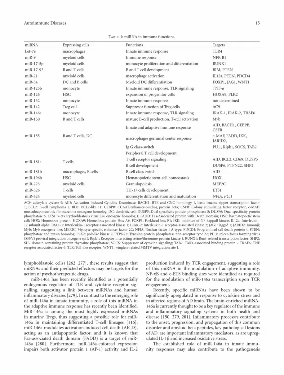
Autoimmune Diseases 15
Table 1: miRNA in immune functions.
miRNA Expressing cells Functions Targets
Let-7e macrophages Innate immune response TLR4
miR-9 myeloid cells Immune response NFK B1
miR-17-5p myeloid cells monocyte proliferation and differentiation RUNX1
miR-17-92 B and T cells B and T cell development BIM, PTEN
miR-21 myeloid cells macrophage activation IL12a, PTEN, PDCD4
miR-34 DC and B cells Myeloid DC differentiation FOXP1, JAG1, WNT1
miR-125b monocyte Innate immune response, TLR signaling TNF-α
miR-126 HSC expansion of progenitor cells HOXA9, PLK2
miR-132 monocyte Innate immune response not determined
miR-142 Treg cell Suppresor function of Treg cells AC9
miR-146a monocyte Innate immune response, TLR signaling IRAK-1, IRAK-2, TRAF6
miR-150 B and T cells mature B-cell production, T-cell activation Myb
miR-155 B and T cells, DC
Innate and adaptive immune responseAID, BACH1, CEBPB,CSFR
macrophages germinal center responsec-MAF, FADD, IKK,JARID2,
Ig G class-switch PU.1, Ripk1, SOCS, TAB2
Peripheral T cell development
miR-181a T cellsT cell receptor signaling AID, BCL2, CD69, DUSP5
B cell development DUSP6, PTPN22, SHP2
miR-181b macrophages, B cells B cell class switch AID
miR-196b HSC Hematopoietic stem-cell homeostasis HOX
miR-223 myeloid cells Granulopoiesis MEF2C
miR-326 T cells TH-17 cells development ETS1
miR-424 myeloid cells monocyte differentiation and maturation NFIA, PU.1
AC9: adenylate cyclase 9; AID: Activation-Induced Cytidine Deaminase; BACH1: BTB and CNC homology 1, basic leucine zipper transcription factor1; BCL2: B-cell lymphoma 2; BIM: BCL2-like 11; CEBPB: CCAAT/enhancer-binding protein beta; CSFR: Colony stimulating factor receptor; c-MAF:musculoaponeurotic fibrosarcoma oncogene homolog; DC: dendritic cell; DUSP5: Dual specificity protein phosphatase 5; DUSP6: Dual specificity proteinphosphatase 6; ETS1: v-ets erythroblastosis virus E26 oncogene homolog 1; FADD: Fas-Associated protein with Death Domain; HSC: haematopoetic stemcell; HOX: Homeobox protein; HOXA9: Homeobox protein Hox-A9; FOXP1: Forkhead box P1; IKK: inhibitor of NF-kappaB kinase; IL12a: Interleukin-12 subunit alpha; IRAK-1: Interleukin-1 receptor-associated kinase 1; IRAK-2: Interleukin-1 receptor-associated kinase 2; JAG1: jagged 1; JARID2: Jumonji;Myb: Myb oncogene-like; MEF2C: Myocyte-specific enhancer factor 2C; NFIA: Nuclear factor 1 A-type; PDCD4: Programmed cell death protein 4; PTEN:phosphatase and tensin homolog; PLK2: pololike kinase 2; PTPN22: Tyrosine-protein phosphatase non-receptor type 22; PU.1: spleen focus forming virus(SFFV) proviral integration oncogene spi1; Ripk1: Receptor-interacting serine/threonine-protein kinase 1; RUNX1: Runt-related transcription factor; SHP2:SH2 domain containing protein thyrosine phosphatase; SOCS: Suppressor of cytokine signaling; TAB2: TAK1-associated binding protein 2 TRAF6: TNFreceptor associated factor-6; TLR: Toll-like receptor; WNT1: wingless-related MMTV integration site 1.
lymphoblastoid cells) [262, 277], these results suggest thatmiRNAs and their predicted effectors may be targets for theaction of psychotherapeutic drugs.
miR-146a has been recently identified as a potentiallyendogenous regulator of TLR and cytokine receptor sig-nalling, suggesting a link between miRNAs and humaninflammatory diseases [279]. In contrast to the emerging roleof miR-146a in innate immunity, a role of this miRNA inthe adaptive immune response has recently been identified.MiR-146a is among the most highly expressed miRNAsin murine Tregs, thus suggesting a possible role for miR-146a in maintaining differentiated T-cell lineages [116].miR-146a modulates activation-induced cell death (AICD),acting as an antiapoptotic factor, and it is known thatFas-associated death domain (FADD) is a target of miR-146a [280]. Furthermore, miR-146a-enforced expressionimpairs both activator protein 1 (AP-1) activity and IL-2
production induced by TCR engagement, suggesting a roleof this miRNA in the modulation of adaptive immunity.NF-κB and c-ETS binding sites were identified as requiredfor the modulation of miR-146a transcription upon TCRengagement.
Recently, specific miRNAs have been shown to besignificantly upregulated in response to cytokine stress andin affected regions of AD brain. The brain-enriched miRNA-146a is currently thought to be a key regulator of the immuneand inflammatory signaling systems in both health anddisease [150, 279, 281]. Inflammatory processes contributeto the onset, progression, and propagation of this commondisorder and amyloid beta peptides, key pathological lesionsof AD, are important inflammatory mediators, as are upreg-ulated IL-1β and increased oxidative stress.
The established role of miR-146a in innate immu-nity responses may also contribute to the pathogenesis

16 Autoimmune Diseases
of neuroinflammatory CNS diseases such as MS and AD.miRNA-146a controls TLR and cytokine signaling through anegative feedback regulation loop involving downregulationof TRAF6 and IRAK1 levels [150]. In neuroinflammation,TLRs provide a critical link between immune stimulants andthe initiation of host defense, and TLR activation modulatesthe release of inflammatory cytokines [282, 283]. TLRs areexpressed on cells of the CNS and can influence local CNSimmune responses. There is a marked increase in expressionof TLRs in MS brain lesions and CSF mononuclear cells aswell as in EAE brain lesions [284, 285]. The secondary pro-gressive phase is characterized by progressive accumulationof disability in the absence of clinical attacks and is driven bythe innate immune system [18]. However, the exact role ofspecific miRNAs in these processes is unknown.
The miRNA profiling of microglial cells in both unstim-ulated and stimulated conditions has not been reported. Ourpreliminary study using microarray and qPCR revealed thatthe expression levels of a set of miRNA were deregulatedupon LPS stimulation in N9 murine microglial cell line(Table 3). Predicted target genes of upregulated miRNAsdetected in this preliminary study were also found to bedownregulated in a microarray study with N9 cells [286].Validation experiments for the predicted target genes areongoing. Interestingly, deregulated microglial miRNAs aresomewhat different from those detected in murine primarymacrophages or mouse Raw 264.7 macrophages upon LPSstimulation [149, 151, 287]. Further studies for the profilingof microglial miRNA expression in MS and EAE are stillwarranted.
6. Diagnostics and Therapeutic Perspectives
In patients with MS, intensive efforts are directed at identify-ing biomarkers in body fluids related to underlying diseasemechanisms, disease activity and progression, and thera-peutic response [288]. Without biomarkers, the clinical andpathological heterogeneity of MS makes treatment difficult.Thus, identification of biomarkers appears desirable for animproved diagnosis of MS as well as for monitoring of diseaseactivity and treatment response. Biomarkers are defined asparameters that are objectively measurable biological char-acteristics, which can be used as indicators of physiologic orpathologic processes. A valuable biomarker requires robustand reproducible assays that work in clinically availablesamples as well as archived material. As miRNAs are morestable than mRNAs, they are good candidates for use asdisease biomarkers and their use as biomarkers has gainedgrowing interest in the last few years [289]. Blood serumand plasma are important sample types for investigatingmiRNAs as biomarkers. Blood biomarkers are attractivebecause blood samples are easy to collect, cheaper, andnoninvasive. However, miRNA profiles in all body fluidssuch as CSF and their content including hematopoieticcells, exosomes, microvesicles, and microparticles can beused as diagnostic, prognostic, or monitoring marker [290,291]. miRNA profiling has been established only in an ADstudy [292] and has not been evaluated in the context of
MS. Besides magnetic resonance imaging parameters [166],CSF biomarkers provide important and specific informationsince changes in the CSF composition may reflect diseasemechanisms inherent to MS [288, 293]. miRNA profiling inCSF may provide valuable information about key patholog-ical processes of MS such as inflammation, demyelination,neuroaxonal injury, gliosis, and regeneration. In recent years,the field of biomarker discovery has gradually shifted fromthe aim of finding a single perfect surrogate marker to theconstruction of composite markers with higher performance.miRNA profiles may be coupled with diagnostic evaluationof miRNA targets, mRNAs, and protein output; thereforecomparison of miRNA analysis with transcriptomic andproteomic studies represents one of the major challenges forclinical application. Another major challenge is representedby technological aspects of miRNA detection aimed at highthroughput, sensitivity, and accurate analysis. The level ofmiRNAs in body fluid samples is very low and efficient andreproducible recovery of miRNA may be problematic. Dueto their short length and high sequence similarity withinmiRNA families, reliable and accurate quantification is stilla challenge. In addition, RNA-purified plasma can alsocontain inhibitors that affect qPCR efficiency [294]. Analysesof miRNA-associated SNPs (e.g., SNP in miRNA genes, inmiRNA binding sites in the target mRNA, or in miRNAbiogenesis pathway genes) are also potential biomarkers ofthe diseases associated with miRNA dysregulation [295–298]. While still not fully validated, profiling of blood cells,exosomes, or body fluid miRNAs would represent a tremen-dous and promising advance in noninvasive diagnosticsof CNS disorders. Identification of suitable miRNA-basedbiomarker sets for MS based on parameters in peripheralblood is only in its infancy.
While we have only just begun to gain insights intomiRNA biology, their apparent association with the onsetand progression of human diseases such as MS has pro-duced great interest in assessing the feasibility of thera-peutic regulation of miRNAs [299]. miRNA-based therapiescould involve administration of a specific miRNA mimicto downregulate target genes or antisense oligonucleotidefor the blocking of certain miRNA to increase expressionof target genes. Importantly, anti-miRNA strategies maybe preferred over antisense mRNA strategies in complexhuman diseases because of the potential of miRNA to affectthe regulation of multiple disease-related genes. However,because manipulation of one miRNA may have impact onmultiple mRNAs and because one mRNA may be regulatedby multiple miRNAs, it is important to guard against off-target effects. Also, miRNAs, instead of causing translationalrepression or mRNA degradation, may relieve translationalrepression and promote transcription [300, 301]. Anotherchallenge is the risk of triggering a cellular immune responsewith RNA therapy. A very promising approach may bethe use of LNAs (locked nucleic acids). These moleculescomprise a class of bicyclic conformational analogues ofRNA, which exhibit high binding affinity to complementaryRNA molecules and high stability in blood and tissuesin vivo [302]. Recent reports on LNA-mediated miRNAsilencing in primates support the potential of LNA-modified
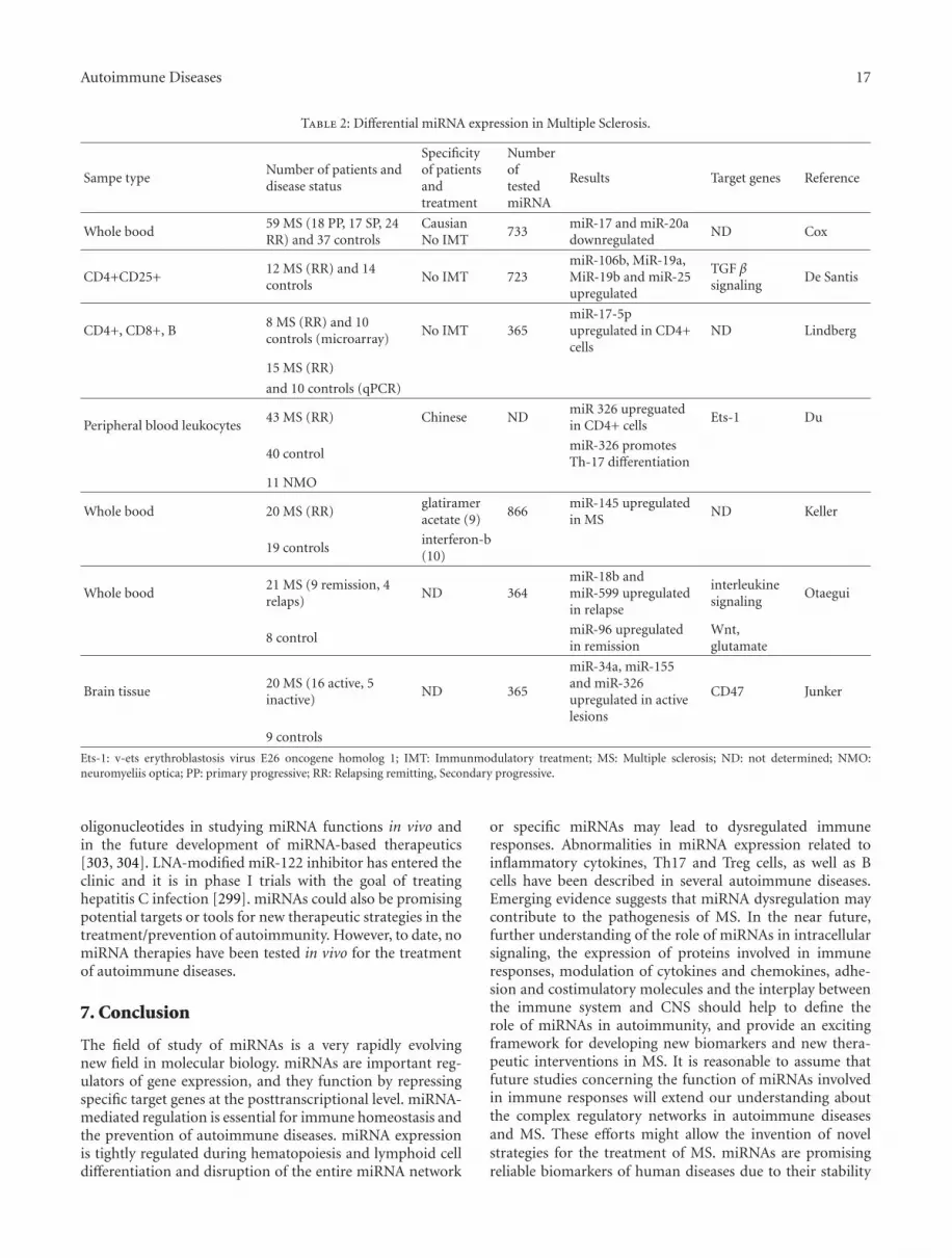
Autoimmune Diseases 17
Table 2: Differential miRNA expression in Multiple Sclerosis.
Sampe typeNumber of patients anddisease status
Specificityof patientsandtreatment
NumberoftestedmiRNA
Results Target genes Reference
Whole bood59 MS (18 PP, 17 SP, 24RR) and 37 controls
CausianNo IMT
733miR-17 and miR-20adownregulated
ND Cox
CD4+CD25+12 MS (RR) and 14controls
No IMT 723miR-106b, MiR-19a,MiR-19b and miR-25upregulated
TGF βsignaling
De Santis
CD4+, CD8+, B8 MS (RR) and 10controls (microarray)
No IMT 365miR-17-5pupregulated in CD4+cells
ND Lindberg
15 MS (RR)
and 10 controls (qPCR)
Peripheral blood leukocytes43 MS (RR) Chinese ND
miR 326 upreguatedin CD4+ cells
Ets-1 Du
40 controlmiR-326 promotesTh-17 differentiation
11 NMO
Whole bood 20 MS (RR)glatirameracetate (9)
866miR-145 upregulatedin MS
ND Keller
19 controlsinterferon-b(10)
Whole bood21 MS (9 remission, 4relaps)
ND 364miR-18b andmiR-599 upregulatedin relapse
interleukinesignaling
Otaegui
8 controlmiR-96 upregulatedin remission
Wnt,glutamate
Brain tissue20 MS (16 active, 5inactive)
ND 365
miR-34a, miR-155and miR-326upregulated in activelesions
CD47 Junker
9 controls
Ets-1: v-ets erythroblastosis virus E26 oncogene homolog 1; IMT: Immunmodulatory treatment; MS: Multiple sclerosis; ND: not determined; NMO:neuromyeliis optica; PP: primary progressive; RR: Relapsing remitting, Secondary progressive.
oligonucleotides in studying miRNA functions in vivo andin the future development of miRNA-based therapeutics[303, 304]. LNA-modified miR-122 inhibitor has entered theclinic and it is in phase I trials with the goal of treatinghepatitis C infection [299]. miRNAs could also be promisingpotential targets or tools for new therapeutic strategies in thetreatment/prevention of autoimmunity. However, to date, nomiRNA therapies have been tested in vivo for the treatmentof autoimmune diseases.
7. Conclusion
The field of study of miRNAs is a very rapidly evolvingnew field in molecular biology. miRNAs are important reg-ulators of gene expression, and they function by repressingspecific target genes at the posttranscriptional level. miRNA-mediated regulation is essential for immune homeostasis andthe prevention of autoimmune diseases. miRNA expressionis tightly regulated during hematopoiesis and lymphoid celldifferentiation and disruption of the entire miRNA network
or specific miRNAs may lead to dysregulated immuneresponses. Abnormalities in miRNA expression related toinflammatory cytokines, Th17 and Treg cells, as well as Bcells have been described in several autoimmune diseases.Emerging evidence suggests that miRNA dysregulation maycontribute to the pathogenesis of MS. In the near future,further understanding of the role of miRNAs in intracellularsignaling, the expression of proteins involved in immuneresponses, modulation of cytokines and chemokines, adhe-sion and costimulatory molecules and the interplay betweenthe immune system and CNS should help to define therole of miRNAs in autoimmunity, and provide an excitingframework for developing new biomarkers and new thera-peutic interventions in MS. It is reasonable to assume thatfuture studies concerning the function of miRNAs involvedin immune responses will extend our understanding aboutthe complex regulatory networks in autoimmune diseasesand MS. These efforts might allow the invention of novelstrategies for the treatment of MS. miRNAs are promisingreliable biomarkers of human diseases due to their stability

18 Autoimmune Diseases
Table 3: Significantly altered miRNAs upon stimulation LPS in N9 microglial cells.
MicroRNAMicroarray qPCR
TargetsFold change P Fold change P
mmu-miR-105 0.35 .02 ns not determined
mmu-miR-125b-3p 2.81 ns 0.16 .047 IL-1β, IL-13, TNF-α
mmu-miR-191 3.12 ns 0.21 .032 CCL9, CRP, IL-6, TLR-3
mmu-miR-193∗ 0.26 .03 0.28 .047 CCL6, IL-10, IL-12Rγ
mmu-miR-208a 3.01 ns 0.12 .015 CD8, IL-18BP, IL-24
mmu-miR-224 3.73 ns 0.09 .033 CD53, CXCL-14, IL-11
mmu-miR-297c∗ 0.31 ns 0.12 .033 not determined
mmu-miR-324-3p 0.33 ns 0.18 .049 not determined
mmu-miR-376c 2.99 .01 ns not determined
mmu-miR-421 0.35 ns 0.03 .033 not determined
mmu-miR-431∗ 4.62 ns 0.22 .034 CD5, CD81, DICER, IRAK1, TRAP 1
mmu-miR-669g 3.48 ns 0.15 .015 not determined
mmu-miR-1190 0.28 .01 0.12 .016 not determined
mmu-miR-1894-5p 0.34 ns 0.08 .017 not determined
CCL9: chemokine (C-C) motif ligand 9; CXCL-14: chemokine (C-X-C) motif ligand 14; CRP: c reactive protein; IL-1β: interleukin 1-β; IL-6: interleukin-6; IL-10: interleukine-10; IL-11: interleukin-11; IL-12Rγ: interleukin 12 receptor γ; IL-13: interleukin-13; IL-18BP: interleukin-18 binding protein; IL-24:interleukin-24; IRAK1: interleukin-1 receptor associated kinase-1; TLR-3: Toll-like receptor-3; TRAP 1: TNF receptor associated protein 1; TNF-α: tumornecrosis factor-α.
being less susceptible to chemical modification and RNasedegradation. Although there is much to be learned in thefield, the role of miRNAs in regulating a great variety oftargets and, as a consequence, multiple pathways makestheir use in diagnostics a powerful tool to be exploitedfor early detection of MS, assessment for risk disease,and monitoring both disease progression and therapeuticresponses to disease-modifying drugs.
8. Take-Home Messages
(i) miRNAs have recently emerged as a new class of mod-ulators of gene expression at the posttranscriptionallevel and are thought to play a critical role in manybiological processes.
(ii) miRNAs are involved in the development, matu-ration, and the functions of immune cells, whichsuggest that they are implicated in the developmentof autoimmune diseases.
(iii) Changes of expression of some miRNAs have beenreported in autoimmune pathologies such as rheu-matoid arthritis, systemic lupus erythematosus, andMS.
(iv) MS serves an example of a chronic and organ-specificautoimmune disease in which miRNAs modulateimmune responses in the peripheral immune com-partment and the neuroinflammatory process in thebrain.
(v) The differential expression of miRNAs and their rolein MS have been investigated by several studies.
(vi) miRNAs have the potential to serve as biomarkersfor the assessment of disease activity and therapeuticresponse to disease-modifying drugs in MS.
Conflict of Interest Disclosure
The authors declare no competing financial interests.
Acknowledgment
The authors thank Professor Anne Frary for critical readingof the manuscript for English.
References
[1] D. P. Bartel, “microRNAs: genomics, biogenesis, mechanism,and function,” Cell, vol. 116, no. 2, pp. 281–297, 2004.
[2] D. P. Bartel, “microRNAs: target recognition and regulatoryfunctions,” Cell, vol. 136, no. 2, pp. 215–233, 2009.
[3] R. C. Lee, R. L. Feinbaum, and V. Ambros, “The C. elegansheterochronic gene lin-4 encodes small RNAs with antisensecomplementarity to lin-14,” Cell, vol. 75, no. 5, pp. 843–854,1993.
[4] R. C. Lee and V. Ambros, “An extensive class of small RNAsin Caenorhabditis elegans,” Science, vol. 294, no. 5543, pp.862–864, 2001.
[5] S. Griffiths-Jones, “miRBase: microRNA sequences and an-notation,” in Current Protocols Bioinformatics, Chapter 12,Unit 12.9.1-10, 2010.
[6] H. Guo, N. T. Ingolia, J. S. Weissman, and D. P. Bartel,“Mammalian microRNAs predominantly act to decreasetarget mRNA levels,” Nature, vol. 466, no. 7308, pp. 835–840,2010.
[7] J. R. Kanwar, G. Mahidhara, and R. K. Kanwar, “MicroRNAin human cancer and chronic inflammatory diseases,” Fron-tiers in Bioscience, vol. 2, pp. 1113–1126, 2010.
[8] K.-C. Sonntag, “microRNAs and deregulated gene expressionnetworks in neurodegeneration,” Brain Research, vol. 1338,pp. 48–57, 2010.

Autoimmune Diseases 19
[9] R. M. O’Connell, D. S. Rao, A. A. Chaudhuri, and D. Balti-more, “Physiological and pathological roles for microRNAsin the immune system,” Nature Reviews Immunology, vol. 10,no. 2, pp. 111–122, 2010.
[10] C. Xiao and K. Rajewsky, “MicroRNA control in the immunesystem: basic principles,” Cell, vol. 136, no. 1, pp. 26–36, 2009.
[11] A. Liston, M. Linterman, and L.-F. Lu, “MicroRNA in theadaptive immune system, in sickness and in health,” Journalof Clinical Immunology, vol. 30, no. 3, pp. 339–346, 2010.
[12] M. P. Gantier, “New perspectives in MicroRNA regulationof innate immunity,” Journal of Interferon and CytokineResearch, vol. 30, no. 5, pp. 283–289, 2010.
[13] L. T. Jeker and J. A. Bluestone, “Small RNA regulators of Tcell-mediated autoimmunity,” Journal of Clinical Immunol-ogy, vol. 30, no. 3, pp. 347–357, 2010.
[14] K. M. Pauley, S. Cha, and E. K. L. Chan, “MicroRNAin autoimmunity and autoimmune diseases,” Journal ofAutoimmunity, vol. 32, no. 3-4, pp. 189–194, 2009.
[15] V. Furer, J. D. Greenberg, M. Attur, S. B. Abramson, and M.H. Pillinger, “The role of microRNA in rheumatoid arthritisand other autoimmune diseases,” Clinical Immunology, vol.136, no. 1, pp. 1–15, 2010.
[16] M. Iborra, F. Bernuzzi, P. Invernizzi, and S. Danese, “microR-NAs in autoimmunity and inflammatory bowel disease: cru-cial regulators in immune response,” Autoimmunity Reviews.In press.
[17] I. Alevizos and G. G. Illei, “microRNAs in Sjogren’s syndromeas a prototypic autoimmune disease,” Autoimmunity Reviews,vol. 9, no. 9, pp. 618–621, 2010.
[18] H. L. Weiner, “The challenge of multiple sclerosis: how do wecure a chronic heterogeneous disease?” Annals of Neurology,vol. 65, no. 3, pp. 239–248, 2009.
[19] L. H. Kasper and J. Shoemaker, “Multiple sclerosis immunol-ogy: the healthy immune system vs the MS immune system,”Neurology, vol. 74, supplement 1, pp. S2–S8, 2010.
[20] S. Hoffjan and D. A. Akkad, “The genetics of multiplesclerosis: an update 2010,” Molecular and Cellular Probes, vol.24, no. 5, pp. 237–243, 2010.
[21] J. R. Oksenberg and S. E. Baranzini, “Multiple sclerosisgenetics—is the glass half full, or half empty?” Nature ReviewsNeurology, vol. 6, no. 8, pp. 429–439, 2010.
[22] B. N. Davis and A. Hata, “Regulation of MicroRNA Bio-genesis: a miRiad of mechanisms,” Cell Communication andSignalling, vol. 7, p. 18, 2009.
[23] V. N. Kim, J. Han, and M. C. Siomi, “Biogenesis of smallRNAs in animals,” Nature Reviews Molecular Cell Biology, vol.10, no. 2, pp. 126–139, 2009.
[24] P.-W. Lau and I. J. MacRae, “The molecular machines thatmediate microRNA maturation,” Journal of Cellular andMolecular Medicine, vol. 13, no. 1, pp. 54–60, 2009.
[25] J. Winter, S. Jung, S. Keller, R. I. Gregory, and S. Diederichs,“Many roads to maturity: MicroRNA biogenesis pathwaysand their regulation,” Nature Cell Biology, vol. 11, no. 3, pp.228–234, 2009.
[26] X. Cai and B. R. Cullen, “The imprinted H19 noncodingRNA is a primary microRNA precursor,” RNA, vol. 13, no.3, pp. 313–316, 2007.
[27] S. He, H. Su, C. Liu et al., “MicroRNA-encoding long non-coding RNAs,” BMC Genomics, vol. 9, p. 236, 2008.
[28] J. Piriyapongsa, L. Marino-Ramırez, and I. K. Jordan, “Originand evolution of human microRNAs from transposableelements,” Genetics, vol. 176, no. 2, pp. 1323–1337, 2007.
[29] J. Piriyapongsa and I. K. Jordan, “A family of humanmicroRNA genes from miniature inverted-repeat transpos-able elements,” PloS ONE, vol. 2, no. 2, article e203, 2007.
[30] C. Sevignani, G. A. Calin, L. D. Siracusa, and C. M. Croce,“Mammalian microRNAs: a small world for fine-tuning geneexpression,” Mammalian Genome, vol. 17, no. 3, pp. 189–202,2006.
[31] N. R. Smalheiser and V. I. Torvik, “Mammalian microRNAsderived from genomic repeats,” Trends in Genetics, vol. 21,no. 6, pp. 322–326, 2005.
[32] T. Yoshimizu, A. Miroglio, M.-A. Ripoche et al., “The H19locus acts in vivo as a tumor suppressor,” Proceedings of theNational Academy of Sciences of the United States of America,vol. 105, no. 34, pp. 12417–12422, 2008.
[33] G. M. Borchert, W. Lanier, and B. L. Davidson, “RNA poly-merase III transcribes human microRNAs,” Nature Structuraland Molecular Biology, vol. 13, no. 12, pp. 1097–1101, 2006.
[34] X. Cai, C. H. Hagedorn, and B. R. Cullen, “Human microR-NAs are processed from capped, polyadenylated transcriptsthat can also function as mRNAs,” RNA, vol. 10, no. 12, pp.1957–1966, 2004.
[35] Y. Lee, C. Ahn, J. Han et al., “The nuclear RNase III Droshainitiates microRNA processing,” Nature, vol. 425, no. 6956,pp. 415–419, 2003.
[36] Y. Lee, M. Kim, J. Han et al., “MicroRNA genes aretranscribed by RNA polymerase II,” The EMBO Journal, vol.23, no. 20, pp. 4051–4060, 2004.
[37] H. K. Saini, S. Griffiths-Jones, and A. J. Enright, “Genomicanalysis of human microRNA transcripts,” Proceedings of theNational Academy of Sciences of the United States of America,vol. 104, no. 45, pp. 17719–17724, 2007.
[38] A. M. Denli, B. B. J. Tops, R. H. A. Plasterk, R. F. Ketting,and G. J. Hannon, “Processing of primary microRNAs bythe Microprocessor complex,” Nature, vol. 432, no. 7014, pp.231–235, 2004.
[39] R. I. Gregory, K.-P. Yan, G. Amuthan et al., “The Micropro-cessor complex mediates the genesis of microRNAs,” Nature,vol. 432, no. 7014, pp. 235–240, 2004.
[40] J. Han, Y. Lee, K.-H. Yeom, Y.-K. Kim, H. Jin, and V. N.Kim, “The Drosha-DGCR8 complex in primary microRNAprocessing,” Genes and Development, vol. 18, no. 24, pp.3016–3027, 2004.
[41] M. Landthaler, A. Yalcin, and T. Tuschl, “The human DiGe-orge syndrome critical region gene 8 and its D. melanogasterhomolog are required for miRNA biogenesis,” Current Biol-ogy, vol. 14, no. 23, pp. 2162–2167, 2004.
[42] Y. Zeng, R. Yi, and B. R. Cullen, “Recognition and cleavageof primary microRNA precursors by the nuclear processingenzyme Drosha,” The EMBO Journal, vol. 24, no. 1, pp. 138–148, 2005.
[43] M. T. Bohnsack, K. Czaplinski, and D. Gorlich, “Exportin 5 isa RanGTP-dependent dsRNA-binding protein that mediatesnuclear export of pre-miRNAs,” RNA, vol. 10, no. 2, pp. 185–191, 2004.
[44] A. M. Brownawell and I. G. Macara, “Exportin-5, a novelkaryopherin, mediates nuclear export of double-strandedRNA binding proteins,” Journal of Cell Biology, vol. 156, no.1, pp. 53–64, 2002.
[45] E. Lund, S. Guttinger, A. Calado, J. E. Dahlberg, and U. Kutay,“Nuclear export of MicroRNA precursors,” Science, vol. 303,no. 5654, pp. 95–98, 2004.
20 Autoimmune Diseases
[46] R. Yi, Y. Qin, I. G. Macara, and B. R. Cullen, “Exportin-5 mediates the nuclear export of pre-microRNAs and shorthairpin RNAs,” Genes and Development, vol. 17, no. 24, pp.3011–3016, 2003.
[47] G. Hutvagner, J. McLachlan, A. E. Pasquinelli, E. Balint, T.Tuschl, and P. D. Zamore, “A cellular function for the RNA-interference enzyme dicer in the maturation of the let-7 smalltemporal RNA,” Science, vol. 293, no. 5531, pp. 834–838,2001.
[48] R. F. Ketting, S. E. J. Fischer, E. Bernstein, T. Sijen, G. J.Hannon, and R. H. A. Plasterk, “Dicer functions in RNAinterference and in synthesis of small RNA involved indevelopmental timing in C. elegans,” Genes and Development,vol. 15, no. 20, pp. 2654–2659, 2001.
[49] T. A. Farazi, S. A. Juranek, and T. Tuschl, “The growingcatalog of small RNAs and their association with distinctArgonaute/Piwi family members,” Development, vol. 135, no.7, pp. 1201–1214, 2008.
[50] E. Maniataki and Z. Mourelatos, “A human, ATP-independ-ent, RISC assembly machine fueled by pre-miRNA,” Genesand Development, vol. 19, no. 24, pp. 2979–2990, 2005.
[51] Z. Mourelatos, J. Dostie, S. Paushkin et al., “miRNPs: a novelclass of ribonucleoproteins containing numerous microR-NAs,” Genes and Development, vol. 16, no. 6, pp. 720–728,2002.
[52] V. Ambros, “MicroRNA pathways in flies and worms: growth,death, fat, stress, and timing,” Cell, vol. 114, no. 2, p. 269,2003.
[53] E. Wienholds and R. H. A. Plasterk, “MicroRNA functionin animal development,” FEBS Letters, vol. 579, no. 26, pp.5911–5922, 2005.
[54] B. D. Harfe, “microRNAs in vertebrate development,” Cur-rent Opinion in Genetics and Development, vol. 15, no. 4, pp.410–415, 2005.
[55] R. I. Gregory, T. P. Chendrimada, N. Cooch, and R. Shiekhat-tar, “Human RISC couples microRNA biogenesis and post-transcriptional gene silencing,” Cell, vol. 123, no. 4, pp. 631–640, 2005.
[56] D. Anglicheau, T. Muthukumar, and M. Suthanthiran,“microRNAs: small RNAs with big effects,” Transplantation,vol. 90, no. 2, pp. 105–112, 2010.
[57] J. L. Wiesen and T. B. Tomasi, “Dicer is regulated by cellularstresses and interferons,” Molecular Immunology, vol. 46, no.6, pp. 1222–1228, 2009.
[58] N. Bushati and S. M. Cohen, “MicroRNA functions,” AnnualReview of Cell and Developmental Biology, vol. 23, pp. 175–205, 2007.
[59] E. Berezikov, E. Cuppen, and R. H. A. Plasterk, “Approachesto microRNA discovery,” Nature Genetics, vol. 38, no. 1, pp.S2–S7, 2006.
[60] K. A. Cissell and S. K. Deo, “Trends in microRNA detection,”Analytical and Bioanalytical Chemistry, vol. 394, no. 4, pp.1109–1116, 2009.
[61] S. L. Yong and A. Dutta, “microRNAs in cancer,” AnnualReview of Pathology, vol. 4, pp. 199–227, 2009.
[62] A. A. Aravin, M. Lagos-Quintana, A. Yalcin et al., “The smallRNA profile during Drosophila melanogaster development,”Developmental Cell, vol. 5, no. 2, pp. 337–350, 2003.
[63] E. Berezikov, G. Van Tetering, M. Verheul et al., “Many novelmammalian microRNA candidates identified by extensivecloning and RAKE analysis,” Genome Research, vol. 16, no.10, pp. 1289–1298, 2006.
[64] P. Y. Chen, H. Manninga, K. Slanchev et al., “The develop-mental miRNA profiles of zebrafish as determined by small
RNA cloning,” Genes and Development, vol. 19, no. 11, pp.1288–1293, 2005.
[65] M. Lagos-Quintana, R. Rauhut, W. Lendeckel, and T. Tuschl,“Identification of novel genes coding for small expressedRNAs,” Science, vol. 294, no. 5543, pp. 853–858, 2001.
[66] N. C. Lau, L. P. Lim, E. G. Weinstein, and D. P. Bartel, “Anabundant class of tiny RNAs with probable regulatory rolesin Caenorhabditis elegans,” Science, vol. 294, no. 5543, pp.858–862, 2001.
[67] J. G. Ruby, C. Jan, C. Player et al., “Large-scale sequencingreveals 21U-RNAs and additional microRNAs and endoge-nous siRNAs in C. elegans,” Cell, vol. 127, no. 6, pp. 1193–1207, 2006.
[68] A. M. Krichevsky, “MicroRNA profiling: from dark matterto white matter, or identifying new players in neurobiology,”The Scientific World Journal, vol. 7, no. 2, pp. 155–166, 2007.
[69] T. D. Schmittgen, E. J. Lee, J. Jiang et al., “Real-timePCR quantification of precursor and mature microRNA,”Methods, vol. 44, no. 1, pp. 31–38, 2008.
[70] W. P. Kloosterman, E. Wienholds, E. de Bruijn, S. Kauppinen,and R. H. A. Plasterk, “In situ detection of miRNAs in ani-mal embryos using LNA-modified oligonucleotide probes,”Nature Methods, vol. 3, no. 1, pp. 27–29, 2006.
[71] G. J. Nuovo, “In situ detection of precursor and maturemicroRNAs in paraffin embedded, formalin fixed tissues andcell preparations,” Methods, vol. 44, no. 1, pp. 39–46, 2008.
[72] E. Varallyay, J. Burgyan, and Z. Havelda, “Detection ofmicroRNAs by Northern blot analyses using LNA probes,”Methods, vol. 43, no. 2, pp. 140–145, 2007.
[73] E. Varallyay, J. Burgyan, and Z. Havelda, “MicroRNA detec-tion by northern blotting using locked nucleic acid probes,”Nature Protocols, vol. 3, no. 2, pp. 190–196, 2008.
[74] E. Wienholds, W. P. Kloosterman, E. Miska et al., “MicroRNAexpression in zebrafish embryonic development,” Science,vol. 309, no. 5732, pp. 310–311, 2005.
[75] S. Baskerville and D. P. Bartel, “Microarray profiling ofmicroRNAs reveals frequent coexpression with neighboringmiRNAs and host genes,” RNA, vol. 11, no. 3, pp. 241–247,2005.
[76] W. Li and K. Ruan, “MicroRNA detection by microarray,”Analytical and Bioanalytical Chemistry, vol. 394, no. 4, pp.1117–1124, 2009.
[77] C.-G. Liu, G. A. Calin, S. Volinia, and C. M. Croce,“MicroRNA expression profiling using microarrays,” NatureProtocols, vol. 3, no. 4, pp. 563–578, 2008.
[78] C.-G. Liu, R. Spizzo, G. A. Calin, and C. M. Croce,“Expression profiling of microRNA using oligo DNA arrays,”Methods, vol. 44, no. 1, pp. 22–30, 2008.
[79] E. A. Miska, E. Alvarez-Saavedra, M. Townsend et al.,“Microarray analysis of microRNA expression in the devel-oping mammalian brain,” Genome biology, vol. 5, no. 9, p.R68, 2004.
[80] P. T. Nelson, D. A. Baldwin, L. M. Scearce, J. C. Oberholtzer,J. W. Tobias, and Z. Mourelatos, “Microarray-based, high-throughput gene expression profiling of microRNAs,” NatMethods, vol. 1, no. 2, pp. 155–161, 2004.
[81] R. Saba and S. A. Booth, “Target labelling for the detectionand profiling of microRNAs expressed in CNS tissue usingmicroarrays,” BMC Biotechnology, vol. 6, article 47, 2006.
[82] J. M. Thomson, J. Parker, C. M. Perou, and S. M. Hammond,“A custom microarray platform for analysis of microRNAgene expression,” Nat Methods, vol. 1, no. 1, pp. 47–53, 2004.
Autoimmune Diseases 21
[83] J. Q. Yin, R. C. Zhao, and K. V. Morris, “Profiling microRNAexpression with microarrays,” Trends in Biotechnology, vol.26, no. 2, pp. 70–76, 2008.
[84] J. M. Cummins, Y. He, R. J. Leary et al., “The colorectalmicroRNAome,” Proceedings of the National Academy ofSciences of the United States of America, vol. 103, no. 10, pp.3687–3692, 2006.
[85] S. V. Anisimov, “Serial Analysis of Gene Expression (SAGE):13 years of application in research,” Current PharmaceuticalBiotechnology, vol. 9, no. 5, pp. 338–350, 2008.
[86] M. R. Friedlander, W. Chen, C. Adamidi et al., “DiscoveringmicroRNAs from deep sequencing data using miRDeep,”Nature Biotechnology, vol. 26, no. 4, pp. 407–415, 2008.
[87] E. R. Mardis, “Next-generation DNA sequencing methods,”Annual Review of Genomics and Human Genetics, vol. 9, pp.387–402, 2008.
[88] W. Kong, J.-J. Zhao, H. E. Lili, and J. Q. Cheng, “Strategiesfor profiling MicroRNA expression,” Journal of CellularPhysiology, vol. 218, no. 1, pp. 22–25, 2009.
[89] V. K. Gangaraju and H. Lin, “microRNAs: key regulators ofstem cells,” Nature Reviews Molecular Cell Biology, vol. 10, no.2, pp. 116–125, 2009.
[90] F. Navarro and J. Lieberman, “Small RNAs guide hematopoi-etic cell differentiation and function,” Journal of Immunology,vol. 184, no. 11, pp. 5939–5947, 2010.
[91] C.-Z. Chen, L. Li, H. F. Lodish, and D. P. Bartel, “microRNAsmodulate hematopoietic lineage differentiation,” Science, vol.303, no. 5654, pp. 83–86, 2004.
[92] S. Monticelli, K. M. Ansel, C. Xiao et al., “MicroRNAprofiling of the murine hematopoietic system,” Genomebiology, vol. 6, no. 8, article R71, 2005.
[93] J. R. Neilson, G. X. Y. Zheng, C. B. Burge, and P. A. Sharp,“Dynamic regulation of miRNA expression in ordered stagesof cellular development,” Genes and Development, vol. 21, no.5, pp. 578–589, 2007.
[94] Q.-J. Li, J. Chau, P. J. R. Ebert et al., “miR-181a is an intrinsicmodulator of T cell sensitivity and selection,” Cell, vol. 129,no. 1, pp. 147–161, 2007.
[95] F. Fazi, A. Rosa, A. Fatica et al., “A minicircuitry comprised ofmicroRNA-223 and transcription factors NFI-A and C/EBPαregulates human granulopoiesis,” Cell, vol. 123, no. 5, pp.819–831, 2005.
[96] B. Zhou, S. Wang, C. Mayr, D. P. Bartel, and H. F. Lodish,“miR-150, a microRNA expressed in mature B and T cells,blocks early B cell development when expressed prema-turely,” Proceedings of the National Academy of Sciences of theUnited States of America, vol. 104, no. 17, pp. 7080–7085,2007.
[97] C. Xiao, D. P. Calado, G. Galler et al., “miR-150 controls B celldifferentiation by targeting the transcription factor c-Myb,”Cell, vol. 131, no. 1, pp. 146–159, 2007.
[98] E. Tsitsiou and M. A. Lindsay, “microRNAs and the immuneresponse,” Current Opinion in Pharmacology, vol. 9, no. 4, pp.514–520, 2009.
[99] H. Wu, J. R. Neilson, P. Kumar et al., “miRNA profiling ofnaıve, effector and memory CD8 T cells,” PloS ONE, vol. 2,no. 10, Article ID e1020, 2007.
[100] S. A. Muljo, K. Mark Ansel, C. Kanellopoulou, D. M.Livingston, A. Rao, and K. Rajewsky, “Aberrant T cell dif-ferentiation in the absence of Dicer,” Journal of ExperimentalMedicine, vol. 202, no. 2, pp. 261–269, 2005.
[101] B. S. Cobb, T. B. Nesterova, E. Thompson et al., “T cell lineagechoice and differentiation in the absence of the RNase III
enzyme Dicer,” Journal of Experimental Medicine, vol. 201,no. 9, pp. 1367–1373, 2005.
[102] C. Xiao, L. Srinivasan, D. P. Calado et al., “Lymphoprolifera-tive disease and autoimmunity in mice with increased miR-17-92 expression in lymphocytes,” Nature Immunology, vol.9, no. 4, pp. 405–414, 2008.
[103] P. T. Jindra, J. Bagley, J. G. Godwin, and J. Iacomini,“Costimulation-dependent expression of microRNA-214increases the ability of T cells to proliferate by targeting Pten,”Journal of Immunology, vol. 185, no. 2, pp. 990–997, 2010.
[104] T.-H. Thai, D. P. Calado, S. Casola et al., “Regulation of thegerminal center response by MicroRNA-155,” Science, vol.316, no. 5824, pp. 604–608, 2007.
[105] Y. Y. Wan, “Multi-tasking of helper T cells,” Immunology, vol.130, no. 2, pp. 166–171, 2010.
[106] B. M. Segal, “Th17 cells in autoimmune demyelinatingdisease,” Seminars in Immunopathology, vol. 32, no. 1, pp. 71–77, 2010.
[107] B. Wei and G. Pei, “microRNAs: critical regulators in Th17cells and players in diseases,” Cellular & Molecular Immunol-ogy, vol. 7, no. 3, pp. 175–181, 2010.
[108] D. Yu, S. Rao, L. M. Tsai et al., “The transcriptional repressorBcl-6 directs T follicular helper cell lineage commitment,”Immunity, vol. 31, no. 3, pp. 457–468, 2009.
[109] L. Zhou, K. H. Seo, H. K. Wong, and Q.-S. Mi, “microRNAsand immune regulatory T cells,” International Immunophar-macology, vol. 9, no. 5, pp. 524–527, 2009.
[110] X. Zhou, L. T. Jeker, B. T. Fife et al., “Selective miRNA dis-ruption in T reg cells leads to uncontrolled autoimmunity,”Journal of Experimental Medicine, vol. 205, no. 9, pp. 1983–1991, 2008.
[111] A. Liston, L.-F. Lu, D. O’Carroll, A. Tarakhovsky, and A. Y.Rudensky, “Dicer-dependent microRNA pathway safeguardsregulatory T cell function,” Journal of Experimental Medicine,vol. 205, no. 9, pp. 1993–2004, 2008.
[112] M. M. W. Chong, J. P. Rasmussen, A. Y. Rudensky, and D.R. Littman, “The RNAseIII enzyme Drosha is critical in Tcells for preventing lethal inflammatory disease,” Journal ofExperimental Medicine, vol. 205, no. 9, pp. 2005–2017, 2008.
[113] L.-F. Lu, T.-H. Thai, D. P. Calado et al., “Foxp3-dependentmicroRNA155 confers competitive fitness to regulatory Tcells by targeting SOCS1 protein,” Immunity, vol. 30, no. 1,pp. 80–91, 2009.
[114] S. Kohlhaas, O. A. Garden, C. Scudamore, M. Turner, K.Okkenhaug, and E. Vigorito, “Cutting edge: the Foxp3 targetmiR-155 contributes to the development of regulatory Tcells,” Journal of Immunology, vol. 182, no. 5, pp. 2578–2582,2009.
[115] B. Huang, J. Zhao, Z. Lei et al., “miR-142-3p restricts cAMPproduction in CD4+CD25- T cells and CD4+CD25+ TREGcells by targeting AC9 mRNA,” The EMBO Reports, vol. 10,no. 2, pp. 180–185, 2009.
[116] B. S. Cobb, A. Hertweck, J. Smith et al., “A role for Dicer inimmune regulation,” Journal of Experimental Medicine, vol.203, no. 11, pp. 2519–2527, 2006.
[117] R. Rouas, H. Fayyad-Kazan, N. El Zien et al., “Humannatural Treg microRNA signature: role of microRNA-31 andmicroRNA-21 in FOXP3 expression,” European Journal ofImmunology, vol. 39, no. 6, pp. 1608–1618, 2009.
[118] H. F. Stahl, T. Fauti, N. Ullrich et al., “miR-155 inhibitionsensitizes CD4+ Th cells for TREG mediated suppression,”PloS ONE, vol. 4, no. 9, article e7158, 2009.

22 Autoimmune Diseases
[119] T. Kurosaki, H. Shinohara, and Y. Baba, “B cell signaling andfate decision,” Annual Review of Immunology, vol. 28, pp. 21–55, 2010.
[120] S. B. Koralov, S. A. Muljo, G. R. Galler et al., “Dicer ablationaffects antibody diversity and cell survival in the B lympho-cyte lineage,” Cell, vol. 132, no. 5, pp. 860–874, 2008.
[121] A. Ventura, A. G. Young, M. M. Winslow et al., “Targeteddeletion reveals essential and overlapping functions of themiR-17∼92 family of miRNA clusters,” Cell, vol. 132, no. 5,pp. 875–886, 2008.
[122] M. Gururajan, C. L. Haga, S. Das et al., “MicroRNA 125binhibition of B cell differentiation in germinal centers,”International Immunology, vol. 22, no. 7, pp. 583–592, 2010.
[123] E. Vigorito, K. L. Perks, C. Abreu-Goodger et al.,“microRNA-155 regulates the generation of immunoglob-ulin class-switched plasma cells,” Immunity, vol. 27, no. 6,pp. 847–859, 2007.
[124] K. Y. Kong, K. S. Owens, J. H. Rogers et al., “MIR-23AmicroRNA cluster inhibits B-cell development,” Experimen-tal Hematology, vol. 38, no. 8, pp. 629–640, 2010.
[125] Y. Dorsett, K. M. McBride, M. Jankovic et al., “microRNA-155 suppresses activation-induced cytidine deaminase-mediated Myc-Igh translocation,” Immunity, vol. 28, no. 5,pp. 630–638, 2008.
[126] G. Teng, P. Hakimpour, P. Landgraf et al., “microRNA-155 is a negative regulator of activation induced cytidinedeaminase,” Immunity, vol. 28, no. 5, pp. 621–629, 2008.
[127] K. Basso, P. Sumazin, P. Morozov et al., “Identification of thehuman mature B cell miRNome,” Immunity, vol. 30, no. 5,pp. 744–752, 2009.
[128] L. Fontana, E. Pelosi, P. Greco et al., “microRNAs 17-5p-20a-106a control monocytopoiesis through AML1 targeting andM-CSF receptor upregulation,” Nature Cell Biology, vol. 9, no.7, pp. 775–787, 2007.
[129] A. Rosa, M. Ballarino, A. Sorrentino et al., “The interplaybetween the master transcription factor PU.1 and miR-424 regulates human monocyte/macrophage differentiation,”Proceedings of the National Academy of Sciences of the UnitedStates of America, vol. 104, no. 50, pp. 19849–19854, 2007.
[130] C. S. Velu, A. M. Baktula, and H. L. Grimes, “Gfi1 regulatesmiR-21 and miR-196b to control myelopoiesis,” Blood, vol.113, no. 19, pp. 4720–4728, 2009.
[131] R. M. O’Connell, D. S. Rao, A. A. Chaudhuri et al., “Sustainedexpression of microRNA-155 in hematopoietic stem cellscauses a myeloproliferative disorder,” Journal of ExperimentalMedicine, vol. 205, no. 3, pp. 585–594, 2008.
[132] R. M. O’Connell, A. A. Chaudhuri, D. S. Rao, and D.Baltimore, “Inositol phosphatase SHIP1 is a primary targetof miR-155,” Proceedings of the National Academy of Sciencesof the United States of America, vol. 106, no. 17, pp. 7113–7118, 2009.
[133] J. B. Johnnidis, M. H. Harris, R. T. Wheeler et al., “Regulationof progenitor cell proliferation and granulocyte function bymicroRNA-223,” Nature, vol. 451, no. 7182, pp. 1125–1129,2008.
[134] F. Bazzoni, M. Rossato, M. Fabbri et al., “Induction andregulatory function of miR-9 in human monocytes and neu-trophils exposed to proinflammatory signals,” Proceedingsof the National Academy of Sciences of the United States ofAmerica, vol. 106, no. 13, pp. 5282–5287, 2009.
[135] S. Radom-Aizik, F. Zaldivar Jr., S. Oliver, P. Galassetti,and D. M. Cooper, “Evidence for microRNA involvement
in exercise-associated neutrophil gene expression changes,”Journal of Applied Physiology, vol. 109, no. 1, pp. 252–261,2010.
[136] S. T. Hashimi, J. A. Fulcher, M. H. Chang, L. Gov, S.Wang, and B. Lee, “microRNA profiling identifies miR-34aand miR-21 and their target genes JAG1 and WNT1 in thecoordinate regulation of dendritic cell differentiation,” Blood,vol. 114, no. 2, pp. 404–414, 2009.
[137] A. Rodriguez, E. Vigorito, S. Clare et al., “Requirement ofbic/microRNA-155 for normal immune function,” Science,vol. 316, no. 5824, pp. 608–611, 2007.
[138] R. T. Martinez-Nunez, F. Louafi, P. S. Friedmann, and T.Sanchez-Eisner, “microRNA-155 modulates the pathogenbinding ability of dendritic cells (DCs) by down-regulation ofDC-specific intercellular adhesion molecule-3 grabbing non-integrin (DC-SIGN),” Journal of Biological Chemistry, vol.284, no. 24, pp. 16334–16342, 2009.
[139] M. Ceppi, A. M. Pereira, I. Dunand-Sauthier et al.,“microRNA-155 modulates the interleukin-1 signaling path-way in activated human monocyte-derived dendritic cells,”Proceedings of the National Academy of Sciences of the UnitedStates of America, vol. 106, no. 8, pp. 2735–2740, 2009.
[140] J. Jurkin, Y. M. Schichl, R. Koeffel et al., “miR-146a isdifferentially expressed by myeloid dendritic cell subsets anddesensitizes cells to TLR2-dependent activation,” Journal ofImmunology, vol. 184, no. 9, pp. 4955–4965, 2010.
[141] N. Stern-Ginossar and O. Mandelboim, “An integrated viewof the regulation of NKG2D ligands,” Immunology, vol. 128,no. 1, pp. 1–6, 2009.
[142] N. Stern-Ginossar, C. Gur, M. Biton et al., “Human microR-NAs regulate stress-induced immune responses mediated bythe receptor NKG2D,” Nature Immunology, vol. 9, no. 9, pp.1065–1073, 2008.
[143] N. Stern-Ginossar, N. Elefant, A. Zimmermann et al., “Hostimmune system gene targeting by a viral miRNA,” Science,vol. 317, no. 5836, pp. 376–381, 2007.
[144] P. Eissmann, J. H. Evans, M. Mehrabi, E. L. Rose, S.Nedvetzki, and D. M. Davis, “Multiple mechanisms down-stream of TLR-4 stimulation allow expression of NKG2Dligands to facilitate macrophage/NK cell crosstalk,” Journal ofImmunology, vol. 184, no. 12, pp. 6901–6909, 2010.
[145] K. Sakuishi, S. Miyake, and T. Yamamura, “Role of NK cellsand invariant NKT cells in multiple sclerosis,” Results andProblems in Cell Differentiation, vol. 51, pp. 127–147, 2010.
[146] M. Fedeli, A. Napolitano, M. P. Wong et al., “Dicer-dependent microRNA pathway controls invariant NKT celldevelopment,” Journal of Immunology, vol. 183, no. 4, pp.2506–2512, 2009.
[147] L. Zhou, K.-H. Seo, H.-Z. He et al., “Tie2cre-inducedinactivation of the miRNA-processing enzyme Dicer disruptsinvariant NKT cell development,” Proceedings of the NationalAcademy of Sciences of the United States of America, vol. 106,no. 25, pp. 10266–10271, 2009.
[148] R. M. O’Connell, K. D. Taganov, M. P. Boldin, G. Cheng,and D. Baltimore, “microRNA-155 is induced during themacrophage inflammatory response,” Proceedings of theNational Academy of Sciences of the United States of America,vol. 104, no. 5, pp. 1604–1609, 2007.
[149] E. Tili, J.-J. Michaille, A. Cimino et al., “Modulation of miR-155 and miR-125b levels following lipopolysaccharide/TNF-α stimulation and their possible roles in regulating theresponse to endotoxin shock,” Journal of Immunology, vol.179, no. 8, pp. 5082–5089, 2007.
Autoimmune Diseases 23
[150] K. D. Taganov, M. P. Boldin, K.-J. Chang, and D. Baltimore,“NF-κB-dependent induction of microRNA miR-146, aninhibitor targeted to signaling proteins of innate immuneresponses,” Proceedings of the National Academy of Sciencesof the United States of America, vol. 103, no. 33, pp. 12481–12486, 2006.
[151] G. Liu, A. Friggeri, Y. Yang, Y.-J. Park, Y. Tsuruta, and E. Abra-ham, “miR-147, a microRNA that is induced upon toll-likereceptor stimulation, regulates murine macrophage inflam-matory responses,” Proceedings of the National Academy ofSciences of the United States of America, vol. 106, no. 37, pp.15819–15824, 2009.
[152] F. J. Sheedy, E. Palsson-McDermott, E. J. Hennessy et al.,“Negative regulation of TLR4 via targeting of the proinflam-matory tumor suppressor PDCD4 by the microRNA miR-21,” Nature Immunology, vol. 11, no. 2, pp. 141–147, 2010.
[153] C. G. Vinuesa, R. J. Rigby, and D. Yu, “Logic and extent ofmiRNA-mediated control of autoimmune gene expression,”International Reviews of Immunology, vol. 28, no. 3-4, pp.112–138, 2009.
[154] C. Carissimi, V. Fulci, and G. Macino, “microRNAs: novelregulators of immunity,” Autoimmunity Reviews, vol. 8, no.6, pp. 520–524, 2009.
[155] D. I. Yu, A. H.-M. Tan, X. Hu et al., “Roquin repressesautoimmunity by limiting inducible T-cell co-stimulatormessenger RNA,” Nature, vol. 450, no. 7167, pp. 299–303,2007.
[156] P. J. Ebert, S. Jiang, J. Xie, Q. J. Li, and M. M. Davis, “Anendogenous positively selecting peptide enhances mature Tcell responses and becomes an autoantigen in the absence ofmicroRNA miR-181a,” Nature immunology, vol. 10, no. 11,pp. 1162–1169, 2009.
[157] T. B. Tomasi, W. J. Magner, J. L. Wiesen et al., “MHCclass II regulation by epigenetic agents and microRNAs,”Immunologic Research, vol. 46, no. 1–3, pp. 45–58, 2010.
[158] M. Pugliatti, S. Sotgiu, and G. Rosati, “The worldwideprevalence of multiple sclerosis,” Clinical Neurology andNeurosurgery, vol. 104, no. 3, pp. 182–191, 2002.
[159] S. V. Ramagopalan, J. C. Knight, and G. C. Ebers, “Multiplesclerosis and the major histocompatibility complex,” CurrentOpinion in Neurology, vol. 22, no. 3, pp. 219–225, 2009.
[160] M. Camina-Tato, M. Fernandez, C. Morcillo-Suarez et al.,“Genetic association of CASP8 polymorphisms with primaryprogressive multiple sclerosis,” Journal of Neuroimmunology,vol. 222, no. 1-2, pp. 70–75, 2010.
[161] E. Jakkula, V. Leppa, A.-M. Sulonen et al., “Genome-wideassociation study in a high-risk isolate for multiple sclerosisreveals associated variants in STAT3 gene,” American Journalof Human Genetics, vol. 86, no. 2, pp. 285–291, 2010.
[162] R. G. Urdinguio, J. V. Sanchez-Mut, and M. Esteller,“Epigenetic mechanisms in neurological diseases: genes,syndromes, and therapies,” Lancet Neurology, vol. 8, no. 11,pp. 1056–1072, 2009.
[163] A. Slavin, L. Kelly-Modis, M. Labadia et al., “Pathogenicmechanisms and experimental models of multiple sclerosis,”Autoimmunity. In press.
[164] E. Mix, H. Meyer-Rienecker, H.-P. Hartung, and U. K.Zettl, “Animal models of multiple sclerosis-Potentials andlimitations,” Progress in Neurobiology, vol. 92, no. 3, pp. 386–404.
[165] V. Siffrin, J. Vogt, H. Radbruch, R. Nitsch, and F. Zipp,“Multiple sclerosis—candidate mechanisms underlying CNSatrophy,” Trends in Neurosciences, vol. 33, no. 4, pp. 202–210,2010.
[166] R. A. Rudick and C. H. Polman, “Current approaches to theidentification and management of breakthrough disease inpatients with multiple sclerosis,” The Lancet Neurology, vol.8, no. 6, pp. 545–559, 2009.
[167] I. Hamann, F. Zipp, and C. Infante-Duarte, “Therapeutictargeting of chemokine signaling in Multiple Sclerosis,”Journal of the Neurological Sciences, vol. 274, no. 1-2, pp. 31–38, 2008.
[168] J. L. Bennett and O. Stuve, “Update on inflammation, neu-rodegeneration, and immunoregulation in multiple sclerosis:therapeutic implications,” Clinical Neuropharmacology, vol.32, no. 3, pp. 121–132, 2009.
[169] I. F. Charo and R. M. Ransohoff, “Mechanisms of disease:the many roles of chemokines and chemokine receptors ininflammation,” New England Journal of Medicine, vol. 354,no. 6, pp. 610–621, 2006.
[170] T. Chitnis, “The role of CD4 T cells in the pathogenesis ofmultiple sclerosis,” International Review of Neurobiology, vol.79, pp. 43–72, 2007.
[171] T. R. Mosmann, H. Cherwinski, and M. W. Bond, “Twotypes of murine helper T cell clone. I. Definition accordingto profiles of lymphokine activities and secreted proteins,”Journal of Immunology, vol. 136, no. 7, pp. 2348–2357, 1986.
[172] H. Kebir, K. Kreymborg, I. Ifergan et al., “Human TH17lymphocytes promote blood-brain barrier disruption andcentral nervous system inflammation,” Nature Medicine, vol.13, no. 10, pp. 1173–1175, 2007.
[173] C. Lock, G. Hermans, R. Pedotti et al., “Gene-microarrayanalysis of multiple sclerosis lesions yields new targets vali-dated in autoimmune encephalomyelitis,” Nature Medicine,vol. 8, no. 5, pp. 500–508, 2002.
[174] E. Bettelli, T. Korn, M. Oukka, and V. K. Kuchroo, “Inductionand effector functions of TH17 cells,” Nature, vol. 453, no.7198, pp. 1051–1057, 2008.
[175] S. Haak, A. L. Croxford, K. Kreymborg et al., “IL-17A andIL-17F do not contribute vitally to autoimmune neuro-inflammation in mice,” Journal of Clinical Investigation, vol.119, no. 1, pp. 61–69, 2009.
[176] R. A. O’Connor, C. T. Prendergast, C. A. Sabatos et al.,“Cutting edge: Th1 cells facilitate the entry of Th17 cells tothe central nervous system during experimental autoimmuneencephalomyelitis,” Journal of Immunology, vol. 181, no. 6,pp. 3750–3754, 2008.
[177] I. M. Stromnes, L. M. Cerretti, D. Liggitt, R. A. Harris, andJ. M. Goverman, “Differential regulation of central nervoussystem autoimmunity by T H1 and TH17 cells,” NatureMedicine, vol. 14, no. 3, pp. 337–342, 2008.
[178] P. Marrack and J. Kappler, “The T cell receptor,” Science, vol.238, no. 4830, pp. 1073–1079, 1987.
[179] E. D. Ponomarev and B. N. Dittel, “γδ T cells regulate theextent and duration of inflammation in the central nervoussystem by a fas ligand-dependent mechanism,” Journal ofImmunology, vol. 174, no. 8, pp. 4678–4687, 2005.
[180] R. Shimonkevitz, C. Colburn, J. A. Burnham, R. S. Murray,and B. L. Kotzin, “Clonal expansions of activated γ/δ T cellsin recent-onset multiple sclerosis,” Proceedings of the NationalAcademy of Sciences of the United States of America, vol. 90,no. 3, pp. 923–927, 1993.
[181] E. D. Ponomarev, M. Novikova, M. Yassais, M. Szczepanik,J. Gorski, and B. N. Dittel, “γδ T cell regulation of IFN-γproduction by central nervous system-infiltrating encephali-togenic T cells: correlation with recovery from experimentalautoimmune encephalomyelitis,” Journal of Immunology, vol.173, no. 3, pp. 1587–1595, 2004.

24 Autoimmune Diseases
[182] H. Babbe, A. Roers, A. Waisman et al., “Clonal expansions ofCD8+ T cells dominate the T cell infiltrate in active multiplesclerosis lesions as shown by micromanipulation and singlecell polymerase chain reaction,” Journal of ExperimentalMedicine, vol. 192, no. 3, pp. 393–404, 2000.
[183] A. Jager, V. Dardalhon, R. A. Sobel, E. Bettelli, and V.K. Kuchroo, “Th1, Th17, and Th9 effector cells induceexperimental autoimmune encephalomyelitis with differentpathological phenotypes,” Journal of Immunology, vol. 183,no. 11, pp. 7169–7177, 2009.
[184] V. Viglietta, C. Baecher-Allan, H. L. Weiner, and D. A.Hafler, “Loss of functional suppression by CD4+CD25+regulatory T cells in patients with multiple sclerosis,” Journalof Experimental Medicine, vol. 199, no. 7, pp. 971–979, 2004.
[185] I. Martinez-Forero, R. Garcia-Munoz, S. Martinez-Pasamaret al., “IL-10 suppressor activity and ex vivo Tr1 cell functionare impaired in multiple sclerosis,” European Journal ofImmunology, vol. 38, no. 2, pp. 576–586, 2008.
[186] A. L. Astier, G. Meiffren, S. Freeman, and D. A. Hafler, “Alter-ations in CD46-mediated Tr1 regulatory T cells in patientswith multiple sclerosis,” Journal of Clinical Investigation, vol.116, no. 12, pp. 3252–3257, 2006.
[187] T. Korn, J. Reddy, W. Gao et al., “Myelin-specific regulatory Tcells accumulate in the CNS but fail to control autoimmuneinflammation,” Nature Medicine, vol. 13, no. 4, pp. 423–431,2007.
[188] T. Ganes, N. J. Brautaset, R. Nyberg-Hansen, and B. Vandvik,“Multimodal evoked responses and cerebrospinal fluid oligo-clonal immunoglobulins in patients with multiple sclerosis,”Acta Neurologica Scandinavica, vol. 73, no. 5, pp. 472–476,1986.
[189] S. Cepok, B. Rosche, V. Grummel et al., “Short-lived plasmablasts are the main B cell effector subset during the courseof multiple sclerosis,” Brain, vol. 128, no. 7, pp. 1667–1676,2005.
[190] L. M. Villar, J. Masjuan, P. Gonzalez-Porque et al., “Intrathe-cal IgM synthesis in neurologic diseases: relationship withdisability in MS,” Neurology, vol. 58, no. 5, pp. 824–826, 2002.
[191] T. Berger, P. Rubner, F. Schautzer et al., “Antimyelin anti-bodies as a predictor of clinically definite multiple sclerosisafter a first demyelinating event,” The New England Journal ofMedicine, vol. 349, no. 2, pp. 139–145, 2003.
[192] S. Gaertner, K. L. de Graaf, B. Greve, and R. Weissert,“Antibodies against glycosylated native MOG are elevated inpatients with multiple sclerosis,” Neurology, vol. 63, no. 12,pp. 2381–2383, 2004.
[193] K. M. Winges, D. H. Gilden, J. L. Bennett, X. Yu, A. M.Ritchie, and G. P. Owens, “Analysis of multiple sclerosiscerebrospinal fluid reveals a continuum of clonally relatedantibody-secreting cells that are predominantly plasmablasts,” Journal of Neuroimmunology, vol. 192, no. 1-2, pp.226–234, 2007.
[194] A. Kutzelnigg, C. F. Lucchinetti, C. Stadelmann et al., “Corti-cal demyelination and diffuse white matter injury in multiplesclerosis,” Brain, vol. 128, no. 11, pp. 2705–2712, 2005.
[195] F. L. Heppner, M. Greter, D. Marino et al., “Experimen-tal autoimmune encephalomyelitis repressed by microglialparalysis,” Nature Medicine, vol. 11, no. 2, pp. 146–152,2005.
[196] M. Greter, F. L. Heppner, M. P. Lemos et al., “Dendritic cellspermit immune invasion of the CNS in an animal model ofmultiple sclerosis,” Nature Medicine, vol. 11, no. 3, pp. 328–334, 2005.
[197] S. L. Bailey-Bucktrout, S. C. Caulkins, G. Goings, J. A.A. Fischer, A. Dzionek, and S. D. Miller, “Cutting edge:central nervous system plasmacytoid dendritic cells reg-ulate the severity of relapsing experimental autoimmuneEncephalomyelitis,” Journal of Immunology, vol. 180, no. 10,pp. 6457–6461, 2008.
[198] A. Bayas, M. Stasiolek, N. Kruse, K. V. Toyka, K. Selmaj, andR. Gold, “Altered innate immune response of plasmacytoiddendritic cells in multiple sclerosis,” Clinical and Experimen-tal Immunology, vol. 157, no. 3, pp. 332–342, 2009.
[199] S. L. Bailey, B. Schreiner, E. J. McMahon, and S. D. Miller,“CNS myeloid DCs presenting endogenous myelin peptides’preferentially’ polarize CD4+ TH-17 cells in relapsing EAE,”Nature Immunology, vol. 8, no. 2, pp. 172–180, 2007.
[200] P. Deshpande, I. L. King, and B. M. Segal, “Cutting edge:CNS CD11c+ cells, from mice with encephalomyelitis polar-ize Th17 cells, and support CD25+CD4+ T cell-mediatedimmunosuppression, suggesting dual roles in the diseaseprocess,” Journal of Immunology, vol. 178, no. 11, pp. 6695–6699, 2007.
[201] B. Becher, B. G. Durell, A. V. Miga, W. F. Hickey, and R.J. Noelle, “The clinical course of experimental autoimmuneencephalomyelitis and inflammation is controlled by theexpression of CD40 within the central nervous system,”Journal of Experimental Medicine, vol. 193, no. 8, pp. 967–974, 2001.
[202] F. Weber, E. Meinl, F. Aloisi et al., “Human astrocytes areonly partially competent antigen presenting cells: possibleimplications for lesion development in multiple sclerosis,”Brain, vol. 117, no. 1, pp. 59–69, 1994.
[203] O. Stuve, S. Youssef, A. J. Slavin et al., “The role of theMHC class II transactivator in class II expression andantigen presentation by astrocytes and in susceptibility tocentral nervous system autoimmune disease,” Journal ofImmunology, vol. 169, no. 12, pp. 6720–6732, 2002.
[204] B.-G. Xiao, A. Diab, J. Zhu, P. Van Der Meide, and H. Link,“Astrocytes induce hyporesponses of myelin basic protein-reactive T and B cell function,” Journal of Neuroimmunology,vol. 89, no. 1-2, pp. 113–121, 1998.
[205] J. J. Geurts and F. Barkhof, “Grey matter pathology inmultiple sclerosis,” The Lancet Neurology, vol. 7, no. 9, pp.841–851, 2008.
[206] T. Kuhlmann, G. Lingfeld, A. Bitsch, J. Schuchardt, and W.Bruck, “Acute axonal damage in multiple sclerosis is mostextensive in early disease stages and decreases over time,”Brain, vol. 125, no. 10, pp. 2202–2212, 2002.
[207] J. M. Frischer, S. Bramow, A. Dal-Bianco et al., “The relationbetween inflammation and neurodegeneration in multiplesclerosis brains,” Brain, vol. 132, no. 5, pp. 1175–1189, 2009.
[208] V. W. Yong and S. Marks, “The interplay between the immuneand central nervous systems in neuronal injury,” Neurology,vol. 74, supplement 1, pp. S9–S16, 2010.
[209] C. P. Gilmore, G. C. Deluca, L. Bo et al., “Spinal cordneuronal pathology in multiple sclerosis,” Brain Pathology,vol. 19, no. 4, pp. 642–649, 2009.
[210] D. Papadopoulos, S. Dukes, R. Patel, R. Nicholas, A. Vora,and R. Reynolds, “Substantial archaeocortical atrophy andneuronal loss in multiple sclerosis,” Brain Pathology, vol. 19,no. 2, pp. 238–253, 2009.
[211] I. Medana, Z. Li, A. Flugel, J. Tschopp, H. Wekerle, and H.Neumann, “Fas ligand (CD95L) protects neurons againstperforin-mediated T lymphocyte cytotoxicity,” Journal ofImmunology, vol. 167, no. 2, pp. 674–681, 2001.
Autoimmune Diseases 25
[212] O. Aktas, A. Smorodchenko, S. Brocke et al., “Neuronal dam-age in autoimmune neuroinflammation mediated by thedeath ligand TRAIL,” Neuron, vol. 46, no. 3, pp. 421–432,2005.
[213] F. Giuliani, C. G. Goodyer, J. P. Antel, and V. Wee Yong, “Vul-nerability of human neurons to T cell-mediated cytotoxicity,”Journal of Immunology, vol. 171, no. 1, pp. 368–379, 2003.
[214] M. Vercellino, A. Merola, C. Piacentino et al., “Altered glu-tamate reuptake in relapsing-remitting and secondary pro-gressive multiple sclerosis cortex: correlation with microgliainfiltration, demyelination, and neuronal and synaptic dam-age,” Journal of Neuropathology and Experimental Neurology,vol. 66, no. 8, pp. 732–739, 2007.
[215] B. D. Trapp and P. K. Stys, “Virtual hypoxia and chronicnecrosis of demyelinated axons in multiple sclerosis,” TheLancet Neurology, vol. 8, no. 3, pp. 280–291, 2009.
[216] A. S. Basso, D. Frenkel, F. J. Quintana et al., “Reversal ofaxonal loss and disability in a mouse model of progressivemultiple sclerosis,” Journal of Clinical Investigation, vol. 118,no. 4, pp. 1532–1543, 2008.
[217] S. G. Waxman, “Mechanisms of disease: sodium channelsand neuroprotection in multiple sclerosis—current status,”Nature Clinical Practice Neurology, vol. 4, no. 3, pp. 159–169,2008.
[218] S. Hendrix and R. Nitsch, “The role of T helper cells in neuro-protection and regeneration,” Journal of Neuroimmunology,vol. 184, no. 1-2, pp. 100–112, 2007.
[219] A. Flugel, F. W. Schwaiger, H. Neumann et al., “NeuronalFasL induces cell death of encephalitogenic T lymphocytes,”Brain Pathology, vol. 10, no. 3, pp. 353–364, 2000.
[220] Y. Liu, I. Teige, B. Birnir, and S. Issazadeh-Navikas, “Neuron-mediated generation of regulatory T cells from encephalito-genic T cells suppresses EAE,” Nature Medicine, vol. 12, no. 5,pp. 518–525, 2006.
[221] R. M. Ransohoff and V. H. Perry, “Microglial physiology:unique stimuli, specialized responses,” Annual Review ofImmunology, vol. 27, pp. 119–145, 2009.
[222] S. Genc, S. Kizildag, K. Genc, H. Ates, and N. Atabey, “Erra-tum: Interferon gamma and lipopolysaccharide upregulateTNF-related apoptosis-inducing ligand expression in murinemicroglia,” Immunology Letters, vol. 85, no. 3, pp. 271–274,2003.
[223] S. M. Eacker, T. M. Dawson, and V. L. Dawson, “Under-standing microRNAs in neurodegeneration,” Nature ReviewsNeuroscience, vol. 10, no. 12, pp. 837–841, 2009.
[224] S. V. Yelamanchili and H. S. Fox, “Defining larger roles for“tiny” RNA molecules: role of miRNAs in neurodegenerationresearch,” Journal of Neuroimmune Pharmacology, vol. 5, no.1, pp. 63–69, 2009.
[225] N. Bushati and S. M. Cohen, “microRNAs in neurodegener-ation,” Current Opinion in Neurobiology, vol. 18, no. 3, pp.292–296, 2008.
[226] V. W. Yong, “Prospects of repair in multiple sclerosis,” Journalof the Neurological Sciences, vol. 277, supplement 1, pp. S16–S18, 2009.
[227] C. Liu and X. Zhao, “microRNAs in Adult and EmbryonicNeurogenesis,” NeuroMolecular Medicine, vol. 11, no. 3, pp.141–152, 2010.
[228] X. Li and P. Jin, “Roles of small regulatory RNAs indetermining neuronal identity,” Nature Reviews Neuroscience,vol. 11, no. 5, pp. 329–338, 2010.
[229] L.-C. Cheng, E. Pastrana, M. Tavazoie, and F. Doetsch, “MiR-124 regulates adult neurogenesis in the subventricular zone
stem cell niche,” Nature Neuroscience, vol. 12, no. 4, pp. 399–408, 2009.
[230] Q. Shen and S. Temple, “Fine control: microRNA regulationof adult neurogenesis,” Nature Neuroscience, vol. 12, no. 4, pp.369–370, 2009.
[231] K. E. Szulwach, X. Li, R. D. Smrt et al., “Cross talk betweenmicroRNA and epigenetic regulation in adult neurogenesis,”Journal of Cell Biology, vol. 189, no. 1, pp. 127–141, 2010.
[232] A. Chang, M. C. Smith, X. Yin, R. J. Fox, S. M. Staugaitis, andB. D. Trapp, “Neurogenesis in the chronic lesions of multiplesclerosis,” Brain, vol. 131, no. 9, pp. 2366–2375, 2008.
[233] B. Emery, “Transcriptional and post-transcriptional controlof CNS myelination,” Current Opinion in Neurobiology, vol.20, no. 5, pp. 601–607, 2010.
[234] C. C. Bruce, C. Zhao, and R. J. M. Franklin, “Remyelina-tion—an effective means of neuroprotection,” Hormones andBehavior, vol. 57, no. 1, pp. 56–62, 2010.
[235] G. Piaton, A. Williams, D. Seilhean, and C. Lubetzki,“Remyelination in multiple sclerosis,” Progress in BrainResearch, vol. 175, pp. 453–464, 2009.
[236] C. Taveggia, M. L. Feltri, and L. Wrabetz, “Signals to promotemyelin formation and repair,” Nature Reviews Neurology, vol.6, no. 5, pp. 276–287, 2010.
[237] J. Liu and P. Casaccia, “Epigenetic regulation of oligodendro-cyte identity,” Trends in Neurosciences, vol. 33, no. 4, pp. 193–201, 2010.
[238] J. C. Dugas, T. L. Cuellar, A. Scholze et al., “Dicer1 and miR-219 Are required for normal oligodendrocyte differentiationand myelination,” Neuron, vol. 65, no. 5, pp. 597–611, 2010.
[239] X. Zhao, X. He, X. Han et al., “microRNA-mediated controlof oligodendrocyte differentiation,” Neuron, vol. 65, no. 5, pp.612–626, 2010.
[240] D. Shin, J.-Y. Shin, M. T. McManus, L. J. Ptacek, and Y.-H.Fu, “Dicer ablation in oligodendrocytes provokes neuronalimpairment in mice,” Annals of Neurology, vol. 66, no. 6, pp.843–857, 2009.
[241] A. Lehotzky, P. Lau, N. Tokesi, N. Muja, L. D. Hudson,and J. Ovadi, “Tubulin polymerization-promoting protein(TPPP/p25) is critical for oligodendrocyte differentiation,”GLIA, vol. 58, no. 2, pp. 157–168, 2010.
[242] P. Lau, J. D. Verrier, J. A. Nielsen, K. R. Johnson, L.Notterpek, and L. D. Hudson, “Identification of dynamicallyregulated microRNA and mRNA networks in developingoligodendrocytes,” Journal of Neuroscience, vol. 28, no. 45, pp.11720–11730, 2008.
[243] H. Budde, S. Schmitt, D. Fitzner, L. Opitz, G. Salinas-Riester,and M. Simons, “Control of oligodendroglial cell number bythe miR-17-92 cluster,” Development, vol. 137, no. 13, pp.2127–2132, 2010.
[244] S.-T. Lin and Y.-H. Fu, “miR-23 regulation of lamin B1 iscrucial for oligodendrocyte development and myelination,”DMM Disease Models and Mechanisms, vol. 2, no. 3-4, pp.178–188, 2009.
[245] B. S. Letzen, C. Liu, N. V. Thakor, J. D. Gearhart, A.H. All, and C. L. Kerr, “microRNA expression profilingof oligodendrocyte differentiation from human embryonicstem cells,” PloS ONE, vol. 5, no. 5, Article ID e10480, 2010.
[246] S. McQuaid, P. Cunnea, J. McMahon, and U. Fitzgerald, “Theeffects of blood-brain barrier disruption on glial cell functionin multiple sclerosis,” Biochemical Society Transactions, vol.37, no. 1, pp. 329–331, 2009.
[247] N. Weiss, F. Miller, S. Cazaubon, and P.-O. Couraud, “Theblood-brain barrier in brain homeostasis and neurological

26 Autoimmune Diseases
diseases,” Biochimica et Biophysica Acta, vol. 1788, no. 4, pp.842–857, 2009.
[248] J. Watzlawik, A. E. Warrington, and M. Rodriguez, “Impor-tance of oligodendrocyte protection, BBB breakdown andinflammation for remyelination,” Expert Review of Neu-rotherapeutics, vol. 10, no. 3, pp. 441–457, 2010.
[249] F. Wu, Z. Yang, and G. Li, “Role of specific microRNAsfor endothelial function and angiogenesis,” Biochemical andBiophysical Research Communications, vol. 386, no. 4, pp.549–553, 2009.
[250] S. Wang and E. N. Olson, “AngiomiRs-Key regulators ofangiogenesis,” Current Opinion in Genetics and Development,vol. 19, no. 3, pp. 205–211, 2009.
[251] Y. Suarez and W. C. Sessa, “microRNAs as novel regulators ofangiogenesis,” Circulation Research, vol. 104, no. 4, pp. 442–454, 2009.
[252] K.-J. Yin, Z. Deng, M. Hamblin et al., “Peroxisome prolifera-tor-activated receptor δ regulation of miR-15a in ischemia-induced cerebral vascular endothelial injury,” Journal ofNeuroscience, vol. 30, no. 18, pp. 6398–6408, 2010.
[253] S. E. Dunn, R. Bhat, D. S. Straus et al., “Peroxisomeproliferator-activated receptor δ limits the expansion ofpathogenic Th cells during central nervous system autoim-munity,” Journal of Experimental Medicine, vol. 207, no. 8, pp.1599–1608, 2010.
[254] S. Kanakasabai, W. Chearwae, C. C. Walline, W. Iams, S. M.Adams, and J. J. Bright, “Peroxisome proliferator-activatedreceptor δ agonists inhibit T helper type 1 (Th1) andTh17 responses in experimental allergic encephalomyelitis,”Immunology, vol. 130, no. 4, pp. 572–588, 2010.
[255] P. E. Polak, S. Kalinin, C. Dello Russo et al., “Protective effectsof a peroxisome proliferator-activated receptor-β/δ agonistin experimental autoimmune encephalomyelitis,” Journal ofNeuroimmunology, vol. 168, no. 1-2, pp. 65–75, 2005.
[256] D. Otaegui, S. E. Baranzini, R. Armananzas et al., “Differen-tial micro RNA expression in PBMC from multiple sclerosispatients,” PloS ONE, vol. 4, no. 7, Article ID e6309, 2009.
[257] A. Keller, P. Leidinger, J. Lange et al., “Multiple sclero-sis: microRNA expression profiles accurately differentiatepatients with relapsing-remitting disease from healthy con-trols,” PloS ONE, vol. 4, no. 10, Article ID e7440, 2009.
[258] C. Du, C. Liu, J. Kang et al., “microRNA miR-326 regulatesTH-17 differentiation and is associated with the pathogenesisof multiple sclerosis,” Nature Immunology, vol. 10, no. 12, pp.1252–1259, 2009.
[259] J. Moisan, R. Grenningloh, E. Bettelli, M. Oukka, and I.-C.Ho, “Ets-1 is a negative regulator of Th17 differentiation,”Journal of Experimental Medicine, vol. 204, no. 12, pp. 2825–2835, 2007.
[260] M. B. Cox, M. J. Cairns, K. S. Gandhi et al., “microRNAs miR-17 and miR-20a inhibit T cell activation genes and are under-expressed in MS whole blood,” PloS ONE, vol. 5, no. 8, ArticleID e12132, 2010.
[261] A. Junker, M. Krumbholz, S. Eisele et al., “microRNAprofiling of multiple sclerosis lesions identifies modulatorsof the regulatory protein CD47,” Brain, vol. 132, no. 12, pp.3342–3352, 2009.
[262] H. Chen, N. Wang, M. Burmeister, and M. G. McInnis,“microRNA expression changes in lymphoblastoid cell linesin response to lithium treatment,” International Journal ofNeuropsychopharmacology, vol. 12, no. 7, pp. 975–981, 2009.
[263] R. L.P. Lindberg, F. Hoffmann, M. Mehling, J. Kuhle, andL. Kappos, “Altered expression of miR-17-5p in CD4+ lym-phocytes of relapsing-remitting multiple sclerosis patients,”
European Journal of Immunology, vol. 40, no. 3, pp. 888–898,2010.
[264] A. Bonauer and S. Dimmeler, “The microRNA-17∼92 clus-ter: still a miRacle?” Cell Cycle, vol. 8, no. 23, pp. 3866–3873,2009.
[265] K. Venken, N. Hellings, R. Liblau, and P. Stinissen, “Dis-turbed regulatory T cell homeostasis in multiple sclerosis,”Trends in Molecular Medicine, vol. 16, no. 2, pp. 58–68, 2010.
[266] G. De Santis, M. Ferracin, A. Biondani et al., “AlteredmiRNA expression in T regulatory cells in course of multiplesclerosis,” Journal of Neuroimmunology, vol. 226, no. 1-2, pp.165–171, 2010.
[267] S. Yamagiwa, J. D. Gray, S. Hashimoto, and D. A. Horwitz,“A role for TGF-β in the generation and expansion ofCD4+CD25+ regulatory t cells from human peripheralblood,” Journal of Immunology, vol. 166, no. 12, pp. 7282–7289, 2001.
[268] N. Koning, L. Bo, R. M. Hoek, and I. Huitinga, “Downregula-tion of macrophage inhibitory molecules in multiple sclerosislesions,” Annals of Neurology, vol. 62, no. 5, pp. 504–514,2007.
[269] G. J. Nuovo, T. S. Elton, P. Nana-Sinkam, S. Volinia, C.M. Croce, and T. D. Schmittgen, “A methodology for thecombined in situ analyses of the precursor and mature formsof microRNAs and correlation with their putative targets,”Nature Protocols, vol. 4, no. 1, pp. 107–115, 2009.
[270] G. J. Nuovo, “In situ detection of microRNAs in paraffinembedded, formalin fixed tissues and the co-localization oftheir putative targets,” Methods. In press.
[271] I. Faraoni, F. R. Antonetti, J. Cardone, and E. Bonmas-sar, “miR-155 gene: a typical multifunctional microRNA,”Biochimica et Biophysica Acta, vol. 1792, no. 6, pp. 497–505,2009.
[272] E. Tili, C. M. Croce, and J. J. Michaille, “miR-155: on thecrosstalk between inflammation and cancer,” Internationalreviews of immunology, vol. 28, no. 5, pp. 264–284, 2009.
[273] H. M. Schipper, O. C. Maes, H. M. Chertkow, and E. Wang,“microRNA expression in Alzheimer blood mononuclearcells,” Gene Regulation and Systems Biology, vol. 1, pp. 263–274, 2007.
[274] X. Wang, P. Liu, H. Zhu et al., “miR-34a, a microRNA up-regulated in a double transgenic mouse model of Alzheimer’sdisease, inhibits bcl2 translation,” Brain Research Bulletin,vol. 80, no. 4-5, pp. 268–273, 2009.
[275] A. H. Kim, M. Reimers, B. Maher et al., “microRNA expres-sion profiling in the prefrontal cortex of individuals affectedwith schizophrenia and bipolar disorders,” SchizophreniaResearch. In press.
[276] T. G. Dinan, “microRNAs as a target for novel antipsychotics:a systematic review of an emerging field,” InternationalJournal of Neuropsychopharmacology, vol. 13, no. 3, pp. 395–404, 2010.
[277] R. Zhou, P. Yuan, Y. Wang et al., “Evidence for selectivemicroRNAs and their effectors as common long-term targetsfor the actions of mood stabilizers,” Neuropsychopharmacol-ogy, vol. 34, no. 6, pp. 1395–1405, 2009.
[278] R. M. O’Connor, B. C. Finger, P. J. Flor, and J. F. Cryan,“Metabotropic glutamate receptor 7: at the interface ofcognition and emotion,” European Journal of Pharmacology,vol. 639, no. 1–3, pp. 123–131, 2010.
[279] L. Li, X.-P. Chen, and Y.-J. Li, “microRNA-146a and humandisease,” Scandinavian Journal of Immunology, vol. 71, no. 4,pp. 227–231, 2010.

Autoimmune Diseases 27
[280] G. Curtale, F. Citarella, C. Carissimi et al., “An emergingplayer in the adaptive immune response: microRNA-146a isa modulator of IL-2 expression and activation-induced celldeath in T lymphocytes,” Blood, vol. 115, no. 2, pp. 265–273,2010.
[281] W. J. Lukiw, Y. Zhao, and G. C. Jian, “An NF-κB-sensitive micro RNA-146a-mediated inflammatory circuit inalzheimer disease and in stressed human brain cells,” Journalof Biological Chemistry, vol. 283, no. 46, pp. 31315–31322,2008.
[282] M. M. Buchanan, M. Hutchinson, L. R. Watkins, and H.Yin, “Toll-like receptor 4 in CNS pathologies,” Journal ofNeurochemistry, vol. 114, no. 1, pp. 13–27, 2010.
[283] M. Marta, U. C. Meier, and A. Lobell, “Regulation of autoim-mune encephalomyelitis by toll-like receptors,” Autoimmu-nity Reviews, vol. 8, no. 6, pp. 506–509, 2009.
[284] M. Fernandez, X. Montalban, and M. Comabella, “Orches-trating innate immune responses in multiple sclerosis:molecular players,” Journal of Neuroimmunology, vol. 225, no.1-2, pp. 5–12, 2010.
[285] R. Gandhi, A. Laroni, and H. L. Weiner, “Role of the innateimmune system in the pathogenesis of multiple sclerosis,”Journal of Neuroimmunology, vol. 221, no. 1-2, pp. 7–14,2010.
[286] C. S. McKimmie, D. Roy, T. Forster, and J. K. Fazakerley,“Innate immune response gene expression profiles of N9microglia are pathogen-type specific,” Journal of Neuroim-munology, vol. 175, no. 1-2, pp. 128–141, 2006.
[287] J. Worm, J. Stenvang, A. Petri et al., “Silencing of microRNA-155 in mice during acute inflammatory response leads toderepression of c/ebp Beta and down-regulation of G-CSF,”Nucleic Acids Research, vol. 37, no. 17, pp. 5784–5792, 2009.
[288] H. Tumani, H.-P. Hartung, B. Hemmer et al., “Cerebrospinalfluid biomarkers in multiple sclerosis,” Neurobiology ofDisease, vol. 35, no. 2, pp. 117–127, 2009.
[289] E. de Smaele, E. Ferretti, and A. Gulino, “microRNAs asbiomarkers for CNS cancer and other disorders,” BrainResearch, vol. 1338, pp. 100–111, 2010.
[290] M. G. Harrington, A. N. Fonteh, E. Oborina et al., “Themorphology and biochemistry of nanostructures provideevidence for synthesis and signaling functions in humancerebrospinal fluid,” Cerebrospinal Fluid Research, vol. 6,article 10, 2009.
[291] M. P. Hunter, N. Ismail, X. Zhang et al., “Detection ofmicroRNA expression in human peripheral blood microvesi-cles,” PloS ONE, vol. 3, no. 11, Article ID e3694, 2008.
[292] J. P. Cogswell, J. Ward, I. A. Taylor et al., “Identification ofmiRNA changes in Alzheimer’s disease brain and CSF yieldsputative biomarkers and insights into disease pathways,”Journal of Alzheimer’s Disease, vol. 14, no. 1, pp. 27–41, 2008.
[293] A. Awad, B. Hemmer, H.-P. Hartung, B. Kieseier, J. L.Bennett, and O. Stuve, “Analyses of cerebrospinal fluid in thediagnosis and monitoring of multiple sclerosis,” Journal ofNeuroimmunology, vol. 219, no. 1-2, pp. 1–7, 2010.
[294] D. Andreasen, J. U. Fog, W. Biggs, J. Salomon, I. K. Dahslveen,and A. Baker, “Improved microRNA quantification in totalRNA from clinical samples,” Methods, vol. 50, no. 4, pp. S6–S9, 2010.
[295] G. V. Glinsky, “SNP-guided microRNA maps (MirMaps) of16 common human disorders identify a clinically accessibletherapy reversing transcriptional aberrations of nuclearimport and inflammasome pathways,” Cell Cycle, vol. 7, no.22, pp. 3564–3576, 2008.
[296] G. V. Glinsky, “An SNP-guided microRNA map of fifteencommon human disorders identifies a consensus diseasephenocode aiming at principal components of the nuclearimport pathway,” Cell Cycle, vol. 7, no. 16, pp. 2570–2583,2008.
[297] M. S. Nicoloso, H. Sun, R. Spizzo et al., “Single-nucleotidepolymorphisms inside microRNA target sites influencetumor susceptibility,” Cancer Research, vol. 70, no. 7, pp.2789–2798, 2010.
[298] M. A. Saunders, H. Liang, and W.-H. Li, “Human polymor-phism at microRNAs and microRNA target sites,” Proceedingsof the National Academy of Sciences of the United States ofAmerica, vol. 104, no. 9, pp. 3300–3305, 2007.
[299] A. G. Seto, “The road toward microRNA therapeutics,”International Journal of Biochemistry and Cell Biology, vol. 42,no. 8, pp. 1298–1305, 2010.
[300] M. Beitzinger and G. Meister, “microRNAs: from decay todecoy,” Cell, vol. 140, no. 5, pp. 612–614, 2010.
[301] M. R. Fabian, N. Sonenberg, and W. Filipowicz, “Regulationof mRNA translation and stability by microRNAs,” Annualreview of biochemistry, vol. 79, pp. 351–379, 2010.
[302] J. Stenvang, M. Lindow, and S. Kauppinen, “Targeting ofmicroRNAs for therapeutics,” Biochemical Society Transac-tions, vol. 36, no. 6, pp. 1197–1200, 2008.
[303] J. Elmen, M. Lindow, S. Schutz et al., “LNA-mediatedmicroRNA silencing in non-human primates,” Nature, vol.452, no. 7189, pp. 896–899, 2008.
[304] R. E. Lanford, E. S. Hildebrandt-Eriksen, A. Petri et al.,“Therapeutic silencing of microRNA-122 in primates withchronic hepatitis C virus infection,” Science, vol. 327, no.5962, pp. 198–201, 2010.


