Embed Size (px)
Citation preview
Annals of Hepatology 7(2) 2008: 104-113104
www.medigraphic.com
Annals of Hepatology 2008; 7(2): April-June: 104-113
Annalsof
Hepatology
Concise Review
The role of microRNAs in primary liver cancer
Heike Varnholt1
«A small rock holds back a great wave»[Homer, The Odyssey, 800 B.C.–700 B.C.]
1 University of North Carolina, Chapel Hill, Department ofPathology.
Address for correspondence:Heike Varnholt, M.D.University of North CarolinaDepartment of PathologyCB #7525 Brinkhous-Bullit-Bldg.Chapel Hill, NC 27599-7525 USAPhone: +1 (919) 966-4676E-mail: [email protected]
Manuscript received and accepted: 27 March 2008
Abstract
Small RNA molecules such as microRNAs, for manyyears considered to be superfluous genomic material,are now known to play important regulatory roles inapoptosis, cell proliferation and differentiation, angio-genesis and thus in carcinogenesis. Primary liver carci-nomas such as hepatocellular carcinomas, cholangio-carcinomas and mixed variants show a rising incidencewith high mortality among affected patients but lack ef-fective targeted therapies except the new multiple kinaseinhibitor Sorafenib. This review elucidates the recentcontributions of miRNA gene expression analyses to abetter understanding of the complex molecular interac-tions in liver carcinogenesis and highlights their futurepromise to provide novel tools for improved diagnos-tics, more accurate prognostic assessment and tailoredmolecular therapies in liver cancer.
Key words: MicroRNA, liver carcinoma, gene expres-sion profile.
Introduction
MicroRNAs
MicroRNAs (miRNAs) have taken center stage in cancerresearch during the short span of the last 6 years since theyhave been shown to be functional in humans.1-7 They repre-sent one of the most abundant classes of regulatory genes inmammals and likely influence over one third of all protein-encoding human genes.8-12 MiRNAs themselves do not en-code proteins but rather function by targeting specific mes-senger RNAs for degradation or translational inhibition and
thus decrease the expression of the resulting protein.13,14 Al-though only about 550 miRNAs have been identified thusfar, estimates about their total number exceed 1,000, andthey may comprise approximately 1-3% of the currentlyknown genes in the human genome.12,15,16 Many miRNAsare expressed exclusively or preferentially in certain tissuetypes, e.g. miR-122 in liver parenchyma.16-21
miRNA biosynthesis and regulation
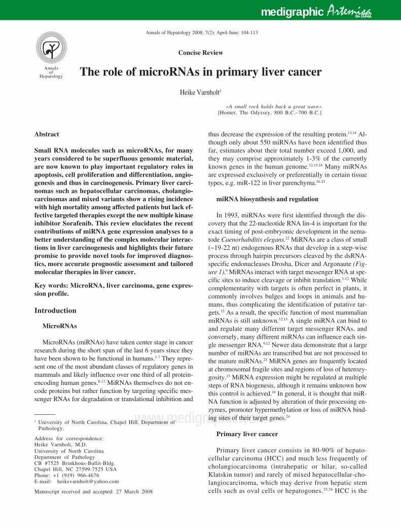
In 1993, miRNAs were first identified through the dis-covery that the 22-nucleotide RNA lin-4 is important for theexact timing of post-embryonic development in the nema-tode Caenorhabditis elegans.22 MiRNAs are a class of small(~19-22 nt) endogenous RNAs that develop in a step-wiseprocess through hairpin precursors cleaved by the dsRNA-specific endonucleases Drosha, Dicer and Argonaute (Fig-ure 1).9 MiRNAs interact with target messenger RNA at spe-cific sites to induce cleavage or inhibit translation.1-12 Whilecomplementarity with targets is often perfect in plants, itcommonly involves bulges and loops in animals and hu-mans, thus complicating the identification of putative tar-gets.12 As a result, the specific function of most mammalianmiRNAs is still unknown.12,13 A single miRNA can bind toand regulate many different target messenger RNAs, andconversely, many different miRNAs can influence each sin-gle messenger RNA.9,12 Newer data demonstrate that a largenumber of miRNAs are transcribed but are not processed tothe mature miRNAs.23 MiRNA genes are frequently locatedat chromosomal fragile sites and regions of loss of heterozy-gosity.13 MiRNA expression might be regulated at multiplesteps of RNA biogenesis, although it remains unknown howthis control is achieved.10 In general, it is thought that miR-NA function is adjusted by alteration of their processing en-zymes, promoter hypermethylation or loss of miRNA bind-ing sites of their target genes.24
Primary liver cancer
Primary liver cancer consists in 80-90% of hepato-cellular carcinoma (HCC) and much less frequently ofcholangiocarcinoma (intrahepatic or hilar, so-calledKlatskin tumor) and rarely of mixed hepatocellular-cho-langiocarcinoma, which may derive from hepatic stemcells such as oval cells or hepatogones.25,26 HCC is the
Artemisamedigraphic en línea
H Varnholt. MicroRNAs in primary liver cancer 105
www.medigraphic.com
fifth most common cancer worldwide, accounts for ap-proximately 500,000 deaths annually and continues toincrease in incidence despite vaccination against Hepa-titis B virus (HBV).26-29 HCC often develops in the set-ting of liver cirrhosis caused by its main risk factors,HBV or Hepatitis C Virus (HCV) infection, excessive al-cohol consumption or hemochromatosis.26,30,31 It isthought that most HCCs develop through a progressivepathway from premalignant nodular lesions such asdysplastic nodules.32 Remarkably, even after studyingthousands of HCCs, only limited knowledge has beengathered regarding genomic alterations during the de-velopment and progression of HCCs in humans.26,28
Overall, the molecular mechanisms of hepatocarcino-genesis are still poorly understood.28 Pathways and mol-ecules that have been identified to play crucial roles inhepatic cancers are cell cycle regulatory proteins suchas p53, c-Myc, Cyclin D1, the Wnt/β-catenin signallingpathway, and multiple tyrosine kinase growth factorligands and receptors, including epidermal growth fac-tor, hepatocyte growth factor, fibroblast growth factorand vascular endothelial growth factor.33,34 In contrast,cholangiocarcinomas often occur in the absence of livercirrhosis.35-38 They have a poor prognosis with a mediansurvival of only 13 months.39 Since they are highlychemoresistant, the need for improved treatment op-tions is particularly urgent for cholangiocarcinomas.37
The mechanisms regulating cholangiocarcinoma growthand resistance to chemotherapy are poorly understood.37
Identification of new target molecules that are criticallyinvolved in the development of primary liver carcino-
mas and dysregulated specifically in tumors will be es-sential to understand the mechanisms and improveprognostication and therapeutic intervention of hepaticcancers.
microRNAs in human cancer
We are still in the early stages of understanding theintricacies of the miRNA puzzle that contributes to dis-ease development in humans.40-42 Some miRNAs, similarto messenger RNAs, are expressed in a tissue-specificmanner, and human adult tissues have unique miRNAprofiles.43,44 Cancer is a complex genetic disease involv-ing structural and expression abnormalities of both cod-ing and noncoding genes.43,45 MiRNAs interact with clas-sic oncogene and tumor suppressor networks and therebycontribute to the initiation and progression of many ifnot all human malignancies.41,43-46 MiRNAs that aredownregulated in cancer and target oncogenes act as tu-mor suppressors, while miRNAs that are upregulated incancer and target tumor suppressor genes act as onco-genes.41,43-47 Several individual miRNAs stand out andhave been implicated in the development of human ma-lignancies, for example miR-145 in carcinoma originat-ing in the colon, breast, lung or prostate,4,48-52 miR-15aand miR-16-1 in chronic lymphocytic leukemia, mir-221in papillary thyroid carcinoma,53 the miR-17-92 polycis-tronic cluster in lung carcinoma11 and miR-21 in glio-blastoma.54 Several of the abnormally expressed miRNAsin human cancers target transcripts of protein-encodinggenes well-known to be involved in carcinogenesis, such
Figure 1. MicroRNA biosyn-thesis in the hepatocyte. Precur-sor microRNA is initially crop-ped into the characteristic hair-pin structure in the nucleus bythe enzymes RNA PolymeraseII and Drosha, then transferredto the cytoplasm by Exportin-5and further processed by Dicerand Argonaute into the matureshort single-stranded microRNA, which exerts its regulatingfunction on the appropriatemessenger RNA after incorpo-ration into the RISC (RNA-in-duced silencing complex).
microRNAgene
microRNAgene
RNA Pol IIRNA Pol II
AAAAAA
DroshaDrosha
AAA
Dicer
Argonaute
mRNAcleavage
translationalrepression
Exportin 5
Pri-miRNA(500-3000 nt)Pri-miRNA
(500-3000 nt)
Pre-miRNA(~70 nt)
AAAPre-miRNA(~70 nt)
AAA
Pre-miRNA(~70 nt)
miRNA-miRNAduplex
(transient,~22 nt)
Maturesingle-
strandedmiRNAin
RISC
Annals of Hepatology 7(2) 2008: 104-113106
www.medigraphic.com
as the BCL-2 anti-apoptotic gene by the miR-15a/miR-16-1 cluster, the Ras oncogenes by let-7 family members,the E2F1 transcription factor by the miR-17-92 cluster orthe BCL-6 anti-apoptotic gene by miR-127.2,11,55 Al-though the analysis of single miRNAs provides a fo-cussed understanding of their influence on known mo-lecular pathways, the real power of miRNA research liesin large-scale miRNA expression fingerprints of manyhundreds of miRNAs in tumors of varying etiologies. Re-cently, studies have emerged directly implicating miR-NAs in cancer and thus giving rise to a new moleculartaxonomy of human cancers based on miRNA profil-ing.5,44 A comprehensive analysis of the miRNA expres-sion in diverse neoplasms showed an even higher accura-cy for tumor diagnosis using the miRNA genetic finger-print than using a profile of more than 16,000 messengerRNAs.56 Thus, miRNA profiles are excellently suited forthe classification and diagnosis of human malignan-cies.4,43,47 In addition, the prognosis of patients with cer-tain carcinomas can be determined using miRNA profil-ing.52,57 Further, miRNA signatures have been shown tocorrelate with the degree of histological tumor differenti-ation in HCCs58 or with specific pathologic features suchas estrogen and progesterone receptor expression inbreast carcinomas.50 In the future, miRNA profiles couldpotentially aid in determining the primary site of a tumoror metastasis of unknown origin, providing a unique op-portunity for targeted therapy and eliminating the use ofempiric chemotherapy.59 Since miRNA research has pro-gressed rapidly over the past several years, a promisingfuture for miRNAs in the realm of cancer diagnosticsseems likely.4
microRNAs in hepatocellular carcinoma
miRNAs in the non-neoplastic liver and in premalig-nant liver lesions
Hepatocellular tumor development is thought todevelop in a multi-step process requiring the accumu-lation of several structural and genomic alterationsand affecting many different pathways.28,59 It has beensuggested that many of the miRNA changes that occurduring hepatocarcinogenesis do so early, so that manychanges that predispose to HCC have already takenplace in liver cirrhosis and other premalignant le-sions.33 Subsequent changes in the miRNA expressionin the transition from cirrhosis to HCC seem to bemuch less marked.33 A progressive downregulation ofmiR-145 and miR-198 from cirrhotic tissue to dysplas-tic nodules and further to HCCs of increasing histo-logical grades has been observed.60 The fact that ab-normal miRNA expression patterns are already presentin premalignant lesions has also been shown for otherorgan sites such as miR-143 and miR-145, which aredownregulated in colonic adenomas as well as adeno-
carcinomas51 and miR-221, which is upregulated inpapillary thyroid carcinomas and also in peritumoralthyroid parenchyma.53 Changes of miRNA patternshave been demonstrated to occur before tumor forma-tion in a HCC-model of rats exposed to tamoxifen.61
Therefore, it remains a tantalizing possibility thatmiRNAs could serve as early warning markers for can-cer initiation or progression.4,43,44
miRNAs in hepatocellular carcinoma
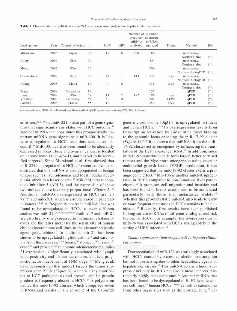
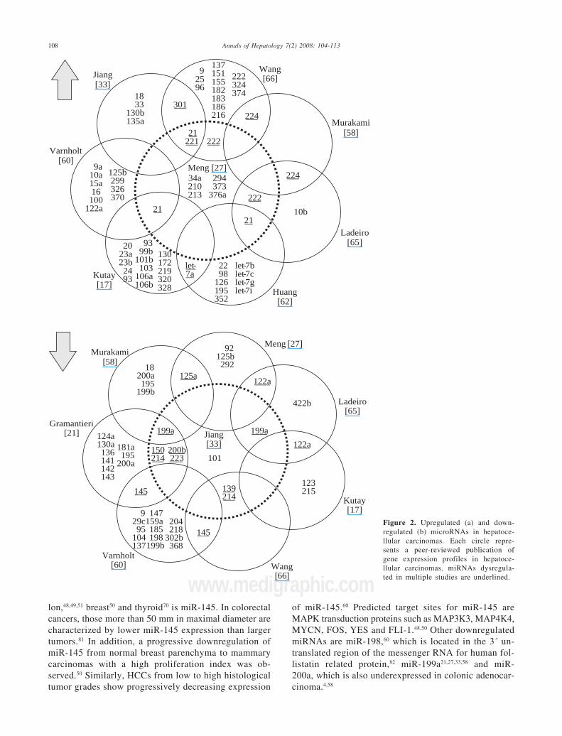
Since the first publication of a miRNA gene expres-sion profile by Murakami et al. in 2005, a total of 9comprehensive miRNA analyses in HCCs have been re-ported thus far (Table I). Geographical origins of pa-tients included those from the USA, Italy, France, Ger-many, Japan, Singapore and China. While the predis-posing risk factors and etiologies of HCCs in thesestudies were quite inhomogenous, one study selectivelyincluded a large number of only HCV-infected pa-tients60 and other authors limited their samples to thosethat were not infected with hepatotropic viruses.62 Overthe past few years, a trend of methodologies from mi-croarray platforms coupled with Northern blot confir-mation toward widespread application of real-timequantitative PCR is evident (Table I). Although it hasbeen shown that formalin-fixed paraffin-embedded(FFPE) tissue can be reliably be used for miRNA expres-sion analyses,18,63 most studies collected data fromsnap-frozen liver tumor material with the exception ofone study that utilized FFPE samples.60 Many hundredprecursor and mature miRNAs have been examined, butonly limited overlap exists between the results of upreg-ulated (Figure 2a) and downregulated (Figure 2b) miR-NAs in HCCs. Reasons for the diverse miRNA gene sig-natures could be variations in methodologies or in thesample origin spanning different geographical regionsand ethnic groups. The overexpressed miRNAs let-7a,miR-21, miR-221, miR-222, miR-224, miR-301 and theunderexpressed miR-122a, miR-125a, miR-139, miR-145, miR-150, miR-199a, miR-200b, miR-214, miR-223have been found to be dysregulated by more than onegroup of authors and are thus more likely to be of sig-nificance in hepatocellular carcinogenesis. These miR-NAs are further characterized in Table II.
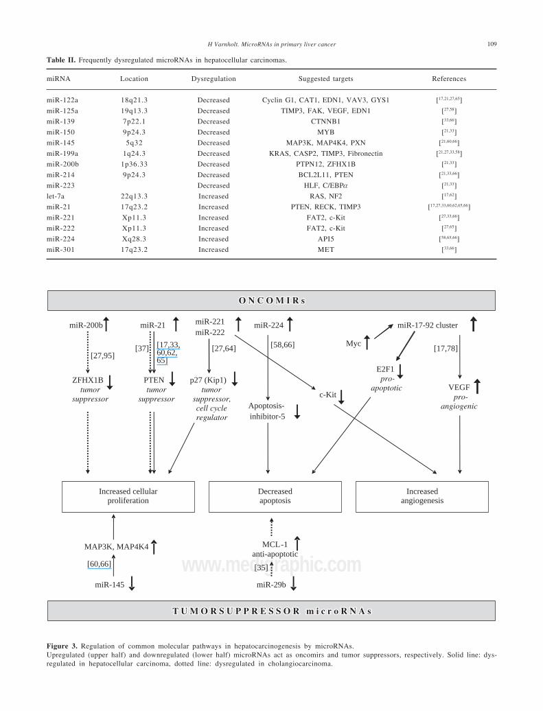
OncomiRs (upregulated) in hepatocellular carcinoma
Mir-221 and miR-222 are encoded in tandem on theX-chromosome (Table II) and, since their overexpres-sion directly results in upregulation of the tumor sup-pressor and cell cycle regulator p27(Kip1) (Figure 3),they can be viewed as a new family of oncogenes tar-geting p27(Kip1).64 Not only are both miR-221 andmiR-222 significantly overexpressed in HCCs whencompared to benign liver tumors and non-tumorous liv-
H Varnholt. MicroRNAs in primary liver cancer 107
www.medigraphic.com
er tissues,21,27,65 but miR-221 is also part of a gene signa-ture that significantly correlates with HCC outcome.33
Another miRNA that constitutes this prognostically im-portant miRNA gene signature is miR-100. It is like-wise upregulated in HCCs and thus acts as an on-comiR.60 MiR-100 has also been found to be aberrantlyexpressed in breast, lung and ovarian cancer, is locatedon chromosome 11q23-q24-D, and has yet to be identi-fied targets.13 Since Murakami et al. first showed thatmiR-224 is upregulated in HCCs,58 recent studies dem-onstrated that this miRNA is also upregulated in benigntumors such as liver adenomas and focal nodular hyper-plasia, albeit to a lesser degree.65 MiR-224 targets apop-tosis inhibitor-5 (API-5), and the expression of thesetwo molecules are inversely proportional (Figure 3).66
Additional miRNAs overexpressed in HCCs are let-7a17,62 and miR-301, which is also increased in pancreat-ic cancer.33,67 A frequently aberrant miRNA that wasfound to be upregulated in HCCs in seven differentstudies was miR-21.17,27,33,60,62,65,66 Both let-7 and miR-21are also highly overexpressed in malignant cholangio-cytes and the latter increases the sensitivity of humancholangiocarcinoma cell lines to the chemotherapeuticagent gemcitabine.37 In addition, mir-21 has beenshown to be upregulated in glioblastoma54 and carcino-ma from the pancreas,68,69 breast,50 stomach,68 thyroid,70
colon71 and prostate.68 In colonic adenocarcinoma, miR-21 expression is significantly associated with lymphnode positivity and distant metastases, and is a prog-nostic factor independent of TNM stage.72,73 Meng et al.have demonstrated that miR-21 targets the tumor sup-pressor gene PTEN (Figure 3), which is a key contribu-tor to HCC pathogenesis and growth, and its proteinproduct is frequently absent in HCCs.27 A polycistronnamed the miR-17-92 cluster, which comprises sevenmiRNAs and resides in the intron 2 of the C13orf25
gene at chromosome 13q31.3, is upregulated in rodentand human HCCs.11,17,33 Its overexpression results fromtranscription activation by c-Myc after direct bindingto the genomic locus encoding the miR-17-92 cluster(Figure 3).74-76 It is known that miRNAs from the miR-17-92 cluster act as oncogenes by influencing the trans-lation of the E2F1 messenger RNA.77 In addition, sincemiR-17-92-transduced cells form larger, better perfusedtumors and the Myc proto-oncogene sustains vascularendothelial growth factor (VEGF) production, it hasbeen suggested that the miR-17-92 cluster exerts a pro-angiogenic effect.78 Mir-10b is another miRNA upregu-lated in HCCs compared to non-tumorous liver paren-chyma.65 It promotes cell migration and invasion andhas been found in breast carcinomas to be associatedparticularly with those that metastasize widely.79
Whether this pro-metastatic miRNA also leads to earlyor more frequent metastases in HCCs remains to be elu-cidated.65 Recently, first results have been publishedlinking certain miRNAs to different etiologies and riskfactors in HCCs. For example, the overexpression ofmiR-96 was associated with HCCs arising solely in thesetting of HBV infection.65
Tumor suppressor (downregulated) in hepatocellularcarcinoma
Downregulation of miR-126 was strikingly associatedwith HCCs caused by excessive alcohol consumptionbut not those arising due to other hepatotoxic agents orhepatotropic viruses.65 This miRNA acts as a tumor sup-pressor not only in HCCs but also in breast cancers, par-ticularly highly metastatic ones.80 Another miRNA thathas been found to be dysregulated in HuH7 hepatic can-cer cell lines,48 human HCCs21,60,66 as well as carcinomasfrom other organ sites such as the prostate, lung,52 co-
Table I. Characteristics of published microRNA gene expression analyses in hepatocellular carcinoma.
Number of Numberprecursor of maturemiRNAs miRNAs
Lead author Year Country of origin n HCV HBV analyzed analyzed Tissue Method Ref
Murakami 2005 Japan 25 17 6 260 180 microarray/Northern blot [58]
Kutay 2006 USA 2 0 8 5 microarray/Northern blot [17]
Meng 2007 USA 2 0 206 microarray/Northern blot/qPCR [27]
Gramantieri 2007 Italy 6 0 4 4 1 1 143 238 cryo microarray/Northern blot/qPCR [21]
Huang 2008 China 10 0 0 331 cryo microarray/Northern blot [62]
Wang 2008 Singapore 1 9 157 qPCR [66]Jiang 2008 USA 5 4 1 1 7 182 196 cryo qPCR [33]Varnholt 2008 Germany 5 2 5 2 0 8 0 FFPE qPCR [60]Ladeiro 2008 France 55 13 17 250 cryo qPCR [65]
cryo=snap-frozen; FFPE: formalin-fixed paraffin-embedded; qPCR: quantitative real-time PCR; Ref: Reference
Annals of Hepatology 7(2) 2008: 104-113108
www.medigraphic.comlon,48,49,51 breast50 and thyroid70 is miR-145. In colorectalcancers, those more than 50 mm in maximal diameter arecharacterized by lower miR-145 expression than largertumors.81 In addition, a progressive downregulation ofmiR-145 from normal breast parenchyma to mammarycarcinomas with a high proliferation index was ob-served.50 Similarly, HCCs from low to high histologicaltumor grades show progressively decreasing expression
of miR-145.60 Predicted target sites for miR-145 areMAPK transduction proteins such as MAP3K3, MAP4K4,MYCN, FOS, YES and FLI-1.48,50 Other downregulatedmiRNAs are miR-198,60 which is located in the 3´ un-translated region of the messenger RNA for human fol-listatin related protein,82 miR-199a21,27,33,58 and miR-200a, which is also underexpressed in colonic adenocar-cinoma.4,58
Figure 2. Upregulated (a) and down-regulated (b) microRNAs in hepatoce-llular carcinomas. Each circle repre-sents a peer-reviewed publication ofgene expression profiles in hepatoce-llular carcinomas. miRNAs dysregula-ted in multiple studies are underlined.
Jiang[33]
Murakami[58]
Meng [27]
Ladeiro[65]
Huang[62]
Kutay[17]
Varnholt[60]
9a10a15a16100
122a
125b299326370
1833
130b135a
34a210213
294373
376a
224
222
10b21
let-7a
2298
126195352
let-7blet-7clet-7glet-7i
2023a23b2493
21221
21
Wang[66]
130172219320328
9399b
101b103
106a106b
224
92596
222
137151155182183186216
222324374
301
Jiang[33]
Murakami[58]
Meng [27]
Ladeiro[65]
Kutay[17]
Varnholt[60] Wang
[66]
Gramantieri[21]
145
145
199a 199a
139214
150214 101
122a
122a125a
200b223
123215
929c95
104137
147159a185198
199b
204218
302b368
124a130a136141142143
181a195
200a
18200a195
199b
92125b292
422b
H Varnholt. MicroRNAs in primary liver cancer 109
www.medigraphic.com
Table II. Frequently dysregulated microRNAs in hepatocellular carcinomas.
miRNA Location Dysregulation Suggested targets References
miR-122a 18q21.3 Decreased Cyclin G1, CAT1, EDN1, VAV3, GYS1 [17,21,27,65]
miR-125a 19q13.3 Decreased TIMP3, FAK, VEGF, EDN1 [27,58]
miR-139 7p22.1 Decreased CTNNB1 [33,66]
miR-150 9p24.3 Decreased MYB [21,33]
miR-145 5q32 Decreased MAP3K, MAP4K4, PXN [21,60,66]
miR-199a 1q24.3 Decreased KRAS, CASP2, TIMP3, Fibronectin [21,27,33,58]
miR-200b 1p36.33 Decreased PTPN12, ZFHX1B [21,33]
miR-214 9p24.3 Decreased BCL2L11, PTEN [21,33,66]
miR-223 Decreased HLF, C/EBPα [21,33]
let-7a 22q13.3 Increased RAS, NF2 [17,62]
miR-21 17q23.2 Increased PTEN, RECK, TIMP3 [17,27,33,60,62,65,66]
miR-221 Xp11.3 Increased FAT2, c-Kit [27,33,66]
miR-222 Xp11.3 Increased FAT2, c-Kit [27,65]
miR-224 Xq28.3 Increased API5 [58,65,66]
miR-301 17q23.2 Increased MET [33,66]
Figure 3. Regulation of common molecular pathways in hepatocarcinogenesis by microRNAs.Upregulated (upper half) and downregulated (lower half) microRNAs act as oncomirs and tumor suppressors, respectively. Solid line: dys-regulated in hepatocellular carcinoma, dotted line: dysregulated in cholangiocarcinoma.
Increased cellularproliferation
Decreasedapoptosis
Increasedangiogenesis
miR-200b
ZFHX1B
tumor
suppressor
[27,95]
miR-21
PTEN
tumor
suppressor
[37]
miR-221
miR-222
p27 (Kip1)
tumor
suppressor,
cell cycle
regulator
miR-224
Apoptosis-
inhibitor-5
miR-17-92 cluster
Myc
VEGF
pro-
angiogenic
[17,78]
E2F1
pro-
apoptoticc-Kit
[58,66][27,64]
miR-29b
MCL-1
anti-apoptotic
T U M O R S U P P R E S S O R m i c r o R N A sT U M O R S U P P R E S S O R m i c r o R N A s
O N C O M I R sO N C O M I R s
[35]
[17,33,60,62,65]
miR-145
MAP3K, MAP4K4
[60,66]
Annals of Hepatology 7(2) 2008: 104-113110
www.medigraphic.com
ESTE DOCUMENTO ES ELABORADO POR MEDI-GRAPHIC
miR-122
MiR-122 plays a crucial role in the understanding ofliver disease, because it is expressed exclusively in the liv-er where it constitutes 70% of the total miRNA con-tent.17,20,83 While miR-122 is the most abundant miRNA inadult livers, other miRNAs such as miR-92a and miR-483are highly expressed in fetal livers.84 The depletion ofmiR-122 compromises liver function and reduces choles-terol levels by targeting the expression of genes involvedin cholesterol biosynthesis.85,86 In HCV infection, miR-122facilitates viral replication by an unknown mechanism andseems to be required for efficient viral RNA expression.20
This is evident because HCV RNA can replicate in HuH7liver carcinoma cells, which express miR-122, but not inHepG2 cells, which do not express miR-122.20 The firstsuggestion that miR-122 may play an important role in thedevelopment of HCCs was made by Etiemble et al. in 1989in woodchucks.87 It took another 13 years until the humanequivalent of miR-122 was discovered,88 and, since then,most studies have found miR-122 to be significantlydownregulated in HCCs.17,21,27,65 However, one study re-ported miR-122 to be upregulated in a patient populationbeing entirely HCV-infected.60 These differences may berelated to the close interactions between miR-122 and thehepatitis C virus and remain to be elucidated in detail. Re-cently, Cyclin G1 has been identified as one of the targetsof miR-122.21 Anti-miR-122 oligonucleotides have beenshown to lead to specific, dose-dependent silencing ofmiR-122 without signficiant hepatotoxicity in mice andtherefore promise to represent the first of a novel class ofsmall molecules that could be used in molecular targetedtherapy in liver diseases.85,89-91
miRNAs in prognosis and metastasis of hepatocellu-lar carcinoma
A miRNA gene signature consisting of 20 miRNAsthat is significantly associated with venous metastases inHCCs has recently been reported.92 Determining the ex-pression levels of these miRNAs may thus be a usefultool to classify patients with HCCs at an early stage andimprove their clinical outcome.92 Dysregulation of select-ed miRNAs is associated with an altered response of tu-mors to commonly used chemotherapeutic agents. Forexample, miR-214 induces cell survival and cisplatin re-sistance through targeting the PTEN/Akt pathway93
while inhibition of miR-21 and miR-200b increases thesensitivity of cholangiocarcinoma cells to gemcitabine.37
MiRNA profiles may in the future provide information toguide oncologists in choosing a tailored therapy for indi-vidual patients.40,43,59 In combination with recently devel-oped technologies that allow the systemic delivery ofsmall RNA mimics or inhibitors to humans, these discov-eries hold great promise for the development of innova-tive therapeutic strategies.85
microRNA in cholangiocarcinoma
Gene expression profiles of miRNAs are much less de-tailed in cholangiocarcinomas than in hepatocellular car-cinomas. Cholangiocarcinomas are highly chemoresis-tant biliary malignancies with poorly understood mecha-nisms of growth regulation.35-39 Most studies thus farhave utilized cell cultures or rodent models.35-38 In cho-langiocarcinomas, upregulated oncomiRs are miR-141,miR-21, miR-23a, miR-27a, let-7a and miR-200b, whiledownregulated tumor suppressor miRNAs are miR-29band miR-370.35-38,94 MiR-141 is highly overexpressed inmalignant cholangiocytes and may target the CLOCKgene, which regulates circadian rhythms and can act as atumor suppressor.37 Inhibition of miR-141 decreases cellgrowth of cholangiocarcinoma cells.37 Let-7a and miR-21have been found to be overexpressed in bothHCCs17,27,33,60,62,65,66 and cholangiocarcinomas.37,38 The un-derlying mechanism, at least in cholangiocytes, may bemediated by interleukin-6 and contribute to a constitu-tive phosphorylation of Stat-3 by NF2.38 The oncogenicmiR-21 has already been characterized above in the sec-tion on HCCs and is likewise upregulated in cholangio-carcinoma.37,94 In summary, miR-21 targets PTEN and isan anti-apoptotic and pro-survival factor.54,94 Human cho-langiocarcinoma cells are increasingly sensitive to theanti-tumor agent gemcitabine with inhibition of miR-21and miR-200b. Interestingly, miR-200b shows inverselyproportional expression levels in HCCs and cholangio-carcinomas since it is downregulated in the former21,33
and upregulated the latter.37,94 A suggested target genefor miR-200b is the PTPN12, which, if dysregulated, maycontribute to tumor cell survival and carcinogenesis.37
MiR-200b has also been proposed to repress the expres-sion of ZFHX1B, a transcription factor involved in theTGFβ signalling pathway and in processes of epithelialto mesenchymal transition via regulation of E-Cad-herin.95
MiR-29b expression is reduced in cholangiocarcino-ma cell lines and thus acts as a tumor suppressor (Figure3).35 It has also been found to be downregulated in chron-ic lymphocytic leukemia, colon cancer and breast can-cer.4,8,50,52 Of note, a reduced expression of mir-29b wasassociated in particular with breast carcinomas thatlacked estrogen and progesterone receptors and dis-played an aggressive behavior.50 MiR-29b is present at alocus on chromosome 7q32, which coincides with thecommon fragile site FRA7H, thus possibly explainingthe frequent miR-29b downregulation in a number ofcancers.13 The suppression of miR-29b expression incholangiocarcinoma cells leads to an overexpression ofMcl-1 and renders the cells resistant to cell death.35 Theexpression of miR-370 is reduced in malignant cholang-iocytes compared to non-malignant cholangiocytes.36
Since miR-370 is also decreased in the early phase ofhepatotoxicity by acetaminophen or carbon tetrachlo-
H Varnholt. MicroRNAs in primary liver cancer 111
www.medigraphic.com
ride, it is speculated to regulate an oxidative-stress-relat-ed gene.96 MiR-370 is embedded in a CpG island and tar-gets MAP3K8, which is consequently upregulated incholangiocarcinoma cells lines as well as in tumor cellxenografts in vivo.36,94 Tight epigenetic regulation ofmiR-370 occurs by hypermethylation and through inter-leukin-6.36,94 Enhanced miR-370 expression suppressesgrowth of malignant human cholangiocytes and maytherefore be suited as a novel target for molecular thera-peutic strategies using small RNAs. Clearly, additionaldetailed investigations about the miRNA expression us-ing human cholangiocarcinoma samples are needed inthe future to elucidate the mechanisms involved in theoncogenesis of these enigmatic tumors.
Future outlook
We are now catching a first glimpse of a hopefullybrighter future for patients with primary liver carcinomasthan currently available. Small molecules such as mi-croRNAs could potentially lead the way not only to earlyand accurate diagnoses but also to novel targeted thera-peutic strategies for hepatic cancers.
References
1. Bartel DP. MicroRNAs: Genomics, biogenesis, mechanism andfunction. Cell 2004; 116: 281-97
2. Calin GA, Croce CM. MicroRNA-Cancer Connection: The Be-ginning of a New Tale. Cancer Res 2006; 66: 7390-4.
3. Dalmay T, Edwards DR. MicroRNAs and the hallmarks of can-cer. Oncogene 2006; 25: 6170-5.
4. Cummins JM, Velculescu VE. Implications of micro-RNA pro-filing for cancer diagnosis. Oncogene 2006; 25: 6220-7.
5. Kent OA, Mendell JT. A small piece in the cancer puzzle:microRNAs as tumor suppressors and oncogenes. Oncogene 2006;25: 6188-96.
6. Pfeffer S, Voinnet O. Viruses, microRNAs and cancer. Oncogene2006; 25: 6211-9.
7. Weber MJ. New human and mouse microRNA genes found byhomology search. FEBS J 2005; 272: 59-73.
8. Calin GA, Ferracin M, Cimmino A, Di Leva G, Shimizu M,Wojcik SE, Iorio MV, et al. A MicroRNA Signature Associatedwith Prognosis and Progression in Chronic Lymphocytic Leuke-mia. N Engl J Med 2005; 353: 1793-801.
9. Kim VN. MicroRNA biogenesis: coordinated cropping and dic-ing. Nat Rev 2005; 6: 376-85.
10. Engels BM, Hutvagner G. Principles and effects of microRNA-mediated post-transcriptional gene regulation. Oncogene 2006;25: 6163-9.
11. Hayashita Y, Osada H Tatematsu Y, Yamada H, Yanagisawa K,Tomida S, Yatabe Y, et al. A polycistronic microRNA cluster,miR-17-92, is overexpressed in human lung cancers and en-hances cell proliferation. Cancer Res 2005; 65: 9628-32.
12. John B, Enright AJ, Aravin A, Tuschl T, Sander C, Marks DS.Human microRNA targets. Plos Biol 2004; 2: 1862-79.
13. Calin GA, Sevignani C, Dumitru CD, Hyslop T, Noch E,Yendamuri S, Shimizu M, et al. Human microRNA genes arefrequently located at fragile sites and genomic regions involvedin cancers. Proc Natl Acad Sci USA 2004; 101: 2999-3004.
14. Lee YS, Kim HK, Chung S, Kim KS, Dutta A. Depletion of humanmicro-RNA miR-125b reveals that it is critical for the proliferation
of differentiated cells but not for the down-regulation of putativetargets during differentiation. J Biol Chem 2005; 17: 16635-41.
15. Bentwich I, Avniel A, Karov Y, Aharonov R, Gilad S, Barad O,Barzilai A, et al. Identification of hundreds of conserved andnonconserved human microRNAs. Nat Genet 2005; 37: 766-70.
16. Shingara J, Keiger K, Shelton J, Losinchai-Wolf W, Powers P,Conrad R, Brown D, et al. An optimized isolation and labellingplatform for accurate microRNA expression profiling. RNA 2005;11: 1461-70.
17. Kutay H, Bai S, Datta J, Motiwala T, Pogribny I, Frankel W, JacobST, et al. Downregulation of miR-122 in the rodent and humanhepatocellular carcinomas. J Cell Biochem 2006; 99: 671-8.
18. Liang Y, Ridzon D, Wong L, Chen C. Characterization ofmicroRNA expression profiles in normal human tissues. BMCGenomics 2007; 8: 166.
19. Jopling CL, Norman KL, Sarnow P. Positive and negative modu-lation of viral and cellular mRNAs by liver-specific microRNAmiR-122. Cold Spring Harb Symp Quant Biol 2006; 71: 369-76.
20. Jopling CL, Yi M, Lancaster AM, Lemon SM, Sarnow P. Modu-lation of Hepatitis C virus RNA abundance by a liver-specificmicroRNA. Science 2005; 309: 1577-81.
21. Gramantieri L, Ferracin M, Fornari F, Veronese A, Sabbioni S,Liu CG, Calin GA, et al. Cyclin G1 is a target of miR-122a, amicroRNA frequently down-regulated in human hepatocellularcarcinoma. Cancer Res 2007; 67: 6092-9.
22. Lee RC, Feinbaum RL, Ambros V. The C. elegans heterochronicgene lin-4 encodes small RNAs with antisense complementarityto lin-14. Cell 1993; 75: 843-54.
23. Lee EJ, Baek M, Gusev Y, Brackett DJ, Nuovo GJ, SchmittgenTD. Systematic evaluation of microRNA processing patterns intissues, cell lines, and tumors. RNA 2008; 14: 1-8.
24. Sassen S, Miska EA, Caldas C. MicroRNA – implications forcancer. Virchows Arch 2008; 452: 1-10.
25. Hunt JP, Varnholt H. Mixed hepatocellular-cholangiocarcinomamay derive from «hepatogones». Hepatobiliary Pancreat Dis Int2008; 358: 852.
26. Llovet JM, Burroughs A, Bruix J. Hepatocellular carcinoma.Lancet 2003; 362: 1907-17.
27. Meng F, Henson R, Wehbe-Janek H, Ghoshal K, Jacob ST, PatelT. MicroRNA-21 regulates expression of the PTEN tumor sup-pressor gene in human hepatocellular carcinoma. Gastroenterol-ogy 2007; 133: 647-58.
28. Thorgeirsson SS, Grisham JW. Molecular pathogenesis of hu-man hepatocellular carcinoma. Nat Gen 2002; 31: 339-46.
29. Motola-Kuba D, Zamora-Valdes D, Uribe M, Mendez-SanchezN. Hepatocellular carcinoma. An overview. Ann Hepatol 2006;5: 16-24.
30. Levrero M. Viral hepatitis and liver cancer: the case of hepatitisC. Oncogene 2006; 25: 3834-47.
31. Lee JS, Chu IS, Heo J, Calvisi DF, Sun Z, Roskams T, Durnez A,et al. Classification and prediction of survival in hepatocellularcarcinoma by gene expression profiling. Hepatology 2004; 40:667-76.
32. Kojiro M, Roskams T. Early hepatocellular carcinoma and dys-plastic nodules. Sem Liver Dis 2005; 25: 133-42.
33. Jiang J, Gusev Y, Aderca I, Mettler TA, Nagorney DM, BrackettDJ, Roberts LR, et al. Association of microRNA expression inhepatocellular carcinomas with hepatitis infection, cirrhosis, andpatient survival. Clin Cancer Res 2008; 14: 419-27.
34. Roberts LR, Gores GJ. Hepatocellular carcinoma: molecularpathways and new therapeutic targets. Semin Liver Dis 2005;25: 212-25.
35. Mott JL, Kobayashi S, Bronk SF, Gores GJ. miR-29 regulatesMcl-1 protein expression and apoptosis. Oncogene 2007; 26:6133-40.
36. Meng F, Wehbe-Janek H, Henson R, Smith H, Patel T. Epigeneticregulation of microRNA-370 by interleukin-6 in malignant hu-man cholangiocytes. Oncogene 2008; 27: 378-86.
37. Meng F, Henson R, Lang M, Wehbe H, Maheshwari S, MendellJT, Jiang J, et al. Involvement of human micro-RNA in growth
Annals of Hepatology 7(2) 2008: 104-113112
www.medigraphic.com
and response to chemotherapy in human cholangiocarcinomacell lines. Gastroenterology 2006; 130: 2113-29.
38. Meng F, Henson R, Wehbe-Janek H, Smith H, Ueno Y, Patel T.The microRNA let-7a modulates interleukin-6-dependent STAT-3 survival signalling in malignant human cholangiocytes. J BiolChem 2007; 282: 8256-64.
39. Weimann A, Varnholt H, Schlitt HJ, Lang H, Flemming P, HustedtC, Tusch G, et al. Retrospective analysis of prognostic factorsafter liver resection and transplantation for cholangiocellular car-cinoma. Br J Surg 2000; 87: 1182-7.
40. Ross JS, Carlson JA, Brock G. miRNA: The new gene silencer.Am J Clin Pathol 2007; 128: 830-6.
41. He X, He L, Hannon GJ. The guardian’s little helper: MicroRNAsin the p53 tumor suppressor network. Cancer Res 2007; 67:11099-101.
42. Yang J, Zhou F, Xu T, Deng H, Ge YY, Zhang C, Li J, et al.Analysis of sequence variations in 59 microRNAs in hepatocellu-lar carcinomas. Mutat Res 2008; 638: 205-9.
43. Calin GA, Croce CM. MicroRNA signatures in human cancers.Nat Rev Cancer 2006; 6: 857-66.
44. Caldas C, Brenton JD. Sizing up miRNAs as cancer genes. NatMed 2005; 11: 712-4.
45. Hutvagner G. MicroRNAs and cancer: issue summary. Oncogene2006; 25: 6154-5.
46. Hwang HW, Mendell JT. MicroRNAs in cell proliferation, celldeath, and tumorigenesis. Br J Cancer 2006; 94: 776-80.
47. Garzon R, Fabbri M, Cimmino A, Calin GA, Croce CM.MicroRNA expression and function in cancer. Trends Mol Med2006; 12: 580-7.
48. Akao Y, Nakagawa Y, Naoe T. MicroRNAs 143 and 145 arepossible common onco-microRNAs in human cancers. OncolRep 2006; 16: 845-50.
49. Bandres E, Cubedo E, Agirre X, Malumbres R, Zarate R, RamirezN, Abajo A, et al. Identification by Real-time PCR of 13 maturemicroRNAs differentially expressed in colorectal cancer and non-tumoral tissues. Mol Cancer 2006; 5: 29.
50. Iorio MV, Ferracin M, Liu CG, Veronese A, Spizzo R, SabbioniS, Magri E, et al. MicroRNA gene expression deregulation inhuman breast cancer. Cancer Res 2005; 65: 7065-70.
51. Michael MZ, O´Connor SM, Van Holst Pellekaan NG, YoungGP, James RJ. Reduced accumulation of specific microRNAs incolorectal neoplasia. Mol Cancer Res 2003; 1: 882-91.
52. Yanaihara N, Caplen N, Bowman E, Seike M, Kumamoto K, Yi M,Stephens RM, et al. Unique microRNA molecular profiles in lungcancer diagnosis and prognosis. Cancer Cell 2006; 9: 189-98.
53. He H, Jazdzewski K, Li W, Liyanarachchi S, Nagi R, Volinia S,Calin GA, et al. The role of microRNA genes in papillary thyroidcarcinoma. Proc Natl Acad Sci USA 2005; 102: 19075-80.
54. Chan JA, Krichevsky AM, Kosik KS. MicroRNA-21 is anantiapoptotic factor in human glioblastoma cells. Cancer Res2005; 65: 6029-33.
55. Cimmino A, Calin GA, Fabbri M, Iorio MV, Ferracin M, ShimizuM, Wojcik SE, et al. miR-15 and miR-16 induce apoptosis bytargeting BCL2. Proc Natl Acad Sci USA 2005; 102: 13944-9.
56. Lu J, Getz G, Miska EA, Alvarez-Saavedra E, Lamb J, Peck D,Sweet-Cordero A, et al. MicroRNA expression profiles classifyhuman cancers. Nature 2005; 435: 834-8.
57. Takamizawa J, Konishi H, Yanagisawa K, Tomida S, Osada H,Endoh H, Harano T, et al. Reduced expression of the let-7microRNAs in human lung cancers in association with a short-ened postoperative survival. Cancer Res 2004; 64: 3753-6.
58. Murakami Y, Yasuda T, Saigo K, Urashima T, Toyoda H,Okanoue T, Shimotohno K. Comprehensive analysis of microRNAexpression patterns in hepatocellular carcinoma and non-tumor-ous tissues. Oncogene 2006; 25: 2537-45.
59. Roessler S, Budhu A, Wang XW. Future of molecular profilingof human hepatocellular carcinoma. Future Oncol 2007; 3:429-39.
60. Varnholt H, Drebber U, Schulze F, Wedemeyer I, Schirmacher P,Dienes HP, Odenthal M. MicroRNA gene expression profile of
hepatitis C virus-associated hepatocellular carcinoma. Hepatology2008; 47: 1223-32.
61. Pogribny IP, Tryndyak VP, Boyko A, Rodriguez-Juarez R, BelandFA, Kovalchuk O. Induction of microRNAome deregulation inrat liver by long-term tamoxifen exposure. Mutat Res 2007; 619:30-7.
62. Huang YS, Dai Y, Yu XF, Bao SY, Yin YB, Tang M, Hu CX.Microarray analysis of microRNA expression in hepatocellularcarcinoma and non-tumorous tissues without viral hepatitis. JGastroenterol Hepatol 2008; 23: 87-94.
63. Xi Y, Nakajima G, Gavin E, Morris CG, Kudo K, Hayashi K, JuJ. Systematic analysis of microRNA expression of RNA extractedfrom fresh frozen and formalin-fixed paraffin-embedded samples.RNA 2007; 13: 1668-74.
64. Galardi S, Mercatelli N, Giorda E, Massalini S, Frajese GV, CiafreSA, Farace MG. miR-221 and miR-222 expression affects theproliferation potential of human prostate carcinoma cell lines bytargeting p27Kip1. J Biol Chem 2007; 282: 23716-24.
65. Ladeiro Y, Couchy G, Balabaud C, Bioulac-Sage P, Pelletier L,Rebouissou S, Zucman-Rossi J. MicroRNA profiling in hepato-cellular tumors is associated to clinical features an oncogene/tumor suppressor gene mutations. Hepatology 2008; (Epub aheadof print).
66. Wang Y, Lee AT, Ma JZ, Wang J, Ren J, Yang Y, Tantoso E, etal. Profiling microRNA expression in hepatocellular carcinomareveals microRNA-224 up-regulation and apoptosis inhibitor-5as a microRNA-224-specific target. J Biol Chem 2008; March 4(Epub ahead of print).
67. Lee EJ, Gusev Y, Jiang J, Nuovo GJ, Lerner MR, Frankel WL,Morgan DL, et al. Expression profiling identifies microRNA sig-natures in pancreatic cancer. Int J Cancer 2007; 120: 1046-54.
68. Volinia S, Calin GA, Liu CG, Ambs S, Cimmino A, Petrocca F,Visone R, et al. A microRNA expression signature of humansolid tumors defines cancer gene targets. Proc Natl Acad Sci USA2006; 103: 2257-61.
69. Roldo C, Missiaglia E, Hagan JP, Falconi M, Capelli P, Bersani S,Calin GA, et al. MicroRNA expression abnormalities in pancre-atic endocrine and acinar tumors are associated with distinctivepathologic features and clinical behavior. J Clin Oncol 2006; 24:4677-84.
70. Tetzlaff MT, Liu A, Xu X, Master SR, Baldwin DA, Tobias JW,Livolsi VA, et al. Differential expression of miRNAs in papillarythyroid carcinoma compared to multinodular goiter using for-malin fixed paraffin embedded tissues. Endocr Pathol 2007; 18:163-73.
71. Asangani IA, Rasheed SA, Nikolova DA, Leupold JH, ColburnNH, Post S, Allgayer H. MicroRNA-21 (miR-21) post-transcrip-tionally downregulates tumor suppressor Pdcd4 and stimulatesinvasion, intravasation and metastasis in colorectal cancer.Oncogene 2007; Oct 29 (Epub ahead of print).
72. Schetter AJ, Leung SY, Sohn JJ, Zanetti KA, Bowman ED,Yanaihara N, Yuen ST, et al. MicroRNA expression profiles as-sociated with prognosis and therapeutic outcome in colon adeno-carcinoma. JAMA 2008; 299: 425-36.
73. Zhu S, Wu H, Nie D, Sheng S, Mo YY. MicroRNA-21 targetstumor suppressor genes in invasion and metastasis. Cell Res 2008;February 12 (Epub ahead of print).
74. Chang TC, Yu D, Lee YS, Wentzel EA, Arking DE, West KM,Dang CV, et al. Widespread microRNA repression by Myc con-tributes to tumorigenesis. Nat Genet 2008; 40: 43-50.
75. O´Donnell KA, Wentzel EA, Zeller KI, Dang CV, Mendell JT: c-Myc-regulated microRNAs modulate E2F1 expression. Nature2005; 435: 839-43.
76. Ota A, Tagawa H, Karnan S, Tsuzuki S, Karpas A, Kira S, YoshidaY, et al. Identification and characterization of a novel gene,C13orf25, as a target for 13q31-q32 amplification in malignantlymphoma. Cancer Res 2004; 64: 3087-95.
77. He L, Thomson JM, Hemann MT, Hernando-Monge E, Mu D,Goodson S, Powers S, et al. A microRNA polycistron as a poten-tial human oncogenes. Nature 2005; 435: 828-33.
H Varnholt. MicroRNAs in primary liver cancer 113
www.medigraphic.com
78. Dews M, Homayouni A, Yu D, Murphy D, Sevignani C,Wentzel E, Furth EE, et al. Augmentation of tumor angiogen-esis by a Myc-activated microRNA cluster. Nat Genet 2006;38: 1060-5.
79. Ma L, Teruya-Felstein, Weinberg RA. Tumour invasion andmetastasis initiated by microRNA-10b in breast cancer. Nature2007; 449: 682-9.
80. Tavazoie SF, Alarcon C, Oskarsson T, Padua D, Wang Q, BosPD, Gerald WL, et al. Endogenous human microRNAs that sup-press breast cancer metastasis. Nature 2008; 451: 147-52.
81. Slaby O, Svoboda M, Fabian P, Smerdova T, Knoflickova D,Bednarikova M, Nenutil R, et al. Altered expression of miR-21,miR-31, miR-143 and miR-145 is related to clinicopathologicfeatures of colorectal cancer. Oncology 2007; 72: 397-4021.
82. Kong Y, Han JH. MicroRNA: Biological and computational per-spective. Genomics Proteomics Bioinformatics 2005; 3: 62-72.
83. Chang J, Nicolas E, Marks D, Sander C, Lerro A, Buendia MA,Xu C, et al. miR-122, a mammalian liver-specific microRNA, isprocessed from hcr mRNA and may downregulate the highaffinity cationic amino acid transporter CAT-1. RNA Biol 2004;1: 106-13.
84. Girard M, Jacquemin E, Munnich A, Lyonnet S, Henrion-CaudeA. miR-122, a paradigm for the role of microRNAs in the liver.J Hepatol 2008; 48: 648-56.
85. Krützfeld J, Rajewsky N, Braich R, Rajeev KG, Tuschl T,Manoharan M, Stoffel M. Silencing of microRNAs in vivo with‘antagomirs’. Nature 2005; 438: 685-9.
86. Esau C, Davis S, Murray SF, Yu XX, Pandey SK, Pear M, WattsL, et al. miR-122 regulation of lipid metabolism revealed by invivo antisense targeting. Cell Met 2006; 3: 87-98.
87. Etiemble J, Moroy T, Jacquemin E, Tiollais P, Buendia MA.Fused transcripts of c-myc and a new cellular locus, hcr, in aprimary liver tumor. Oncogene 1989; 4: 51-7.
88. Lagos-Quintana M, Rauhut R, Yalcin A, Meyer J, Lendeckel W,Tusch T. Identification of tissue-specific microRNAs from mouse.Curr Biol 2002; 12: 735-9.
89. Elmen J, Lindow M, Silahtaroglu A, Bak M, Christensen M,Lind-Thomsen A, Hedtjärn M, et al. Antagonism of microRNA-122 in mice by systemically administered LNA-antimiR leads toup-regulation of a large set of predicted target mRNAs in theliver. Nucl Acids Res 2008; 36: 1153-62.
90. Fabiani MM, Gait MJ. miR-122 targeting with LNA/2´-O-methyloligonucleotide mixmers, peptide nucleic acids (PNA), and PNA-peptide conjugates. RNA 2008; 14: 1-11.
91. Shan Y, Zheng J, Lambrecht RW, Bonkovsky HL. Reciprocaleffects of micro-RNA-122 on expression of heme oxygenase-1and hepatitis C virus genes in human hepatocytes. Gastroenterol-ogy 2007; 133: 1166-74.
92. Budhu A, Jia HL, Forgues M, Liu CG, Goldstein D, Lam A,Zanetti KA, et al. Identification of metastasis-relatedmicroRNAs in hepatocellular carcinoma. Hepatology 2008;47: 897-907.
93. Yang H, Kong W, He L, Zhao JJ, O´Donnell JD, Wang J, WenhamRM, et al. MicroRNA expression profiling in human ovariancancer: miR-214 induces cell survival and cisplatin resistance bytargeting PTEN. Cancer Res 2008; 68: 425-33.
94. Stutes M, Tran S, DeMorrow S. Genetic and epigenetic changesassociated with cholangiocarcinoma: From DNA methylation tomicroRNAs. World J Gastroenterology 2007; 13: 6465-9.
95. Christoffersen NR, Silahtaroglu A, Orom UA, Kauppinen S, LundAH. miR-200b mediates post-transcriptional repression ofZFHX1B. RNA 2007; 13: 1172-8.
96. Fukushima T, Hamada Y, Yamada H, Horii I. Changes of micro-RNA expression in rat liver treated by acetaminophen or carbontetrachloride – regulating role of micro-RNA for RNA expres-sion -. J Tox Sci 2007; 32: 401-9.


