Embed Size (px)
Citation preview

Biological Chemistry ‘Just Accepted’ Papers
Biological Chemistry ‘Just Accepted’ Papers are papers published online, in advance of appearing in the print journal. They have been peer-reviewed, accepted and are online published in manuscript form, but have not been copy edited, typeset, or proofread. Copy editing may lead to small differences between the Just Accepted version and the final version. There may also be differences in the quality of the graphics. When papers do appear in print, they will be removed from this feature and grouped with other papers in an issue.
Biol Chem ‘Just Accepted’ Papers are citable; the online publication date is indicated on the Table of Contents page, and the article’s Digital Object Identifier (DOI), a unique identifier for intellectual property in the digital environment (e.g.,10.1515/hsz-2011-xxxx), is shown at the top margin of the title page. Once an article is published as Biol Chem ‘Just Accepted’ Paper (and before it is published in its final form), it should be cited in other articles by indicating author list, title and DOI.
After a paper is published in Biol Chem ‘Just Accepted’ Paper form, it proceeds through the normal production process, which includes copy editing, typesetting and proofreading. The edited paper is then published in its final form in a regular print and online issue of Biol Chem. At this time, the Biol Chem ‘Just Accepted’ Paper version is replaced on the journal Web site by the final version of the paper with the same DOI as the Biol Chem ‘Just Accepted’ Paper version.
Disclaimer
Biol Chem ‘Just Accepted’ Papers have undergone the complete peer-review process. However, none of the additional editorial preparation, which includes copy editing, typesetting and proofreading, has been performed. Therefore, there may be errors in articles published as Biol Chem ‘Just Accepted’ Papers that will be corrected in the final print and online version of the Journal. Any use of these articles is subject to the explicit understanding that the papers have not yet gone through the full quality control process prior to advanced publication.
Authenticated | [email protected] author's copyDownload Date | 11/15/12 11:52 PM
Biological Chemistry ’Just Accepted’ paper
ISSN (online) 1437-4315
DOI: 10.1515/hsz-2012-0228
1 / 21
Minireview
Melanoma resistance to photodynamic therapy: new insights
Ying-Ying Huang1,2,3, Daniela Vecchio1,2, Pinar Avci1, Rui Yin1,2,4, Maria Garcia-Diaz1,5,6,
and Michael R. Hamblin1,2,7,*
1Wellman Center for Photomedicine, Massachusetts General Hospital, Boston, MA 02114,
USA 2Department of Dermatology, Harvard Medical School, Boston, MA 02115, USA 3Department of Pathology, Guangxi Medical University, Nanning, Guangxi, 530027, China 4Department of Dermatology, Southwest Hospital, Third Military Medical University,
Chongqing, China, 400038 5Molecular Engineering Group, IQS School of Engineering, Universitat Ramon Llull,
Barcelona, Spain 6Departament de Bioquimica i Biologia Molecular, Universitat de Barcelona, Barcelona,
Spain 7Harvard-MIT Division of Health Sciences and Technology, Cambridge, MA 02139, USA
*Corresponding author
e-mail: [email protected]
Authenticated | [email protected] author's copyDownload Date | 11/15/12 11:52 PM

Melanoma Resistance to Photodynamic Therapy
2 / 21
Abstract
Melanoma is the most dangerous form of skin cancer, with a steeply rising incidence and a
poor prognosis in its advanced stages. Melanoma is highly resistant to traditional
chemotherapy and radiotherapy, although modern targeted therapies such as BRAF inhibitors
are showing some promise. Photodynamic therapy (PDT, the combination of photosensitizing
dyes and visible light) has been tested for melanoma with some promising results, but
melanoma is generally considered to also be resistant to PDT. Optical interference by the
highly-pigmented melanin, the anti-oxidant effect of melanin, the sequestration of
photosensitizers inside melanosomes, defects in apoptotic pathways, and the efflux of
photosensitizers by ATP-binding cassette (ABC) transporters have all been implicated in
melanoma resistance to PDT. Approaches to overcoming melanoma resistance to PDT
include: the discovery of highly active photosensitizers absorbing in the 700-800-nm near
infrared spectral region; interventions that can temporarily reduce the amount or the
pigmentation of the melanin; compounds that can reverse apoptotic defects or inhibit
drug-efflux of photosensitizers; and immunotherapy approaches that can take advantage of the
ability of PDT to activate the host immune system to the treated tumor.
Keywords: melanoma, photodynamic therapy, resistance mechanisms, melanosomes,
photosensitizers, drug efflux systems, depigmentation, anti-tumor immune
response
Authenticated | [email protected] author's copyDownload Date | 11/15/12 11:52 PM

Melanoma Resistance to Photodynamic Therapy
3 / 21
Introduction
Melanoma is the malignancy responsible for the highest incidence of deaths from skin cancer.
Genetic and environmental factors such as UV damage can cause the transformation of skin
melanocytes into a tumorigenic melanoma (Carlson, Ross et al. 2009). Melanocytes are the
main cells responsible for the production of melanin, the pigment that protects the skin from
sun damage by absorbing UV light (Slominski, Tobin et al. 2004). Although the chronic and
intermittent exposure to UV leads to tanning that protects the skin from DNA damage, intense
exposure leading to sunburn can lead to DNA damage and genetic alterations in melanocytes.
Malignant melanomas can be pigmented (melanotic), characterized by black lesions due to
melanin accumulation or can be unpigmented (amelanotic) if the melanocytes involved are
less differentiated and therefore produce less melanin. It has been claimed that in recent years
there has been an “epidemic” of melanoma because it is being diagnosed at more than double
the rate it was in 1986, increasing faster than any other major cancer (Burton, Coates et al.
1993). However, there is disagreement on this point as some dermatologists assert
(Glusac 2011) that the increasing numbers represent not an epidemic of melanoma, but an
epidemic of melanoma screening, and a study lends support to this view
(Aguilar, Schoendorff et al. 1991). Melanoma is resistant to most traditional forms of
chemotherapy and radiotherapy, and for this reason many alternative treatments have been
investigated (Jilaveanu, Aziz et al. 2009).
PDT and melanoma
Photodynamic therapy is an effective treatment for several different cancers (Agostinis, Berg
et al. 2011). Its efficacy has been shown in non-melanoma skin cancers and other skin cancers
such as lymphoma and in dermatologic disorders like vitiligo and psoriasis (Babilas, Schreml
et al. 2010). PDT involves systemic or local administration of a photosensitizer, which
localizes in the tumor. The photosensitizers are activated by irradiation at a specific
wavelength and in presence of oxygen generate short-lived reactive oxygen species (ROS)
(Dougherty, Gomer et al. 1998). The ROS generated by the photosensitizer are responsible
for the selective tumor destruction, tumor-associated vascular damage, and activation of
antitumor immune responses (Castano, Mroz et al. 2006). This treatment offers many
advantages such as a low systemic cumulative toxicity; the selectivity and noninvasiveness of
Authenticated | [email protected] author's copyDownload Date | 11/15/12 11:52 PM
Melanoma Resistance to Photodynamic Therapy
4 / 21
the method; the possibility of repeating the treatment many times without serious effects.
Figure 1 shows the generation of ROS from the excited PS (represented by a Jablonski
diagram) and the destruction of tumor cells by apoptosis and necrosis.
One of the first studies carried out in 1988 to verify the efficacy of PDT on malignant
melanoma compared the effect of hematoporphyrin derivate (photofrin II) on melanotic and
amelanotic malignant melanoma in athymic nude mice. This study demonstrated effective
effect of PDT on amelanotic cancer but not in melanotic melanoma (Nelson, McCullough
et al. 1988). The authors concluded that the resistance of malignant melanotic melanoma to
PDT was due to the presence of the melanin that competed with the photosensitizer for the
absorption of photons or in the energy transfer process from the excited triplet state of the
sensitizer to melanin instead of cellular oxygen. PDT is a photochemical reaction, thus the
energy of the photon is absorbed by PS, which can transfer its energy to the target molecule.
Usually, PDT induces tumor necrosis by transferring energy from the excited triplet state of
the PS to ground state molecular oxygen, producing excited state singlet oxygen, which
causes irreversible oxidation of some essential cellular components. The presence of melanin,
a stable protein-complex with a wide absorption spectrum, in the same tissue, competed with
PS for photons resulting in inefficient phototoxicity (Nelson, McCullough et al. 1988).
Thereafter, subsequent studies were directed to investigate and synthesize new
photosensitizers able to exert their action after irradiation at different (longer) wavelengths
from the melanin absorption spectrum. The employment of selected second-generation
photosensitizing agents, such as Si(IV)-naphthalocyanine, bacteriochlorin a and
Lu(III)-texaphyrin, characterized by an extended macrocycle and high molar absorptivity in
the 750–800 nm spectral interval improved the efficacy of PDT on experimentally implanted
melanotic melanoma (Schuitmaker, van Best et al. 1990; Biolo, Jori et al. 1996; Woodburn,
Fan et al. 1998). Ten years later since the first study, Busetti et al. showed that if the
melanosomes were preirradiated with high peak power pulsed laser radiation at 1064 nm,
PDT treatment was improved (Busetti, Soncin et al. 1999). In 1999 the same group
investigated the effect of PDT using a benzoporphyrin derivative monoacid ring A
(verteporfin, BPD-MA), against B16 pigmented melanoma, after preirradiation.
Several studies have been carried out to investigate and improve the efficacy of PDT against
melanoma. PDT techniques were initially developed in experiments on animals
(Kostenich, Zhuravkin et al. 1994). Some years later Sheleg et al.(Sheleg, Zhavrid et al.
Authenticated | [email protected] author's copyDownload Date | 11/15/12 11:52 PM
Melanoma Resistance to Photodynamic Therapy
5 / 21
2004) investigated PDT using chlorin (e6), in phase I clinical trials, on skin melanoma
metastases in humans. The patients in this study received five PDT courses during a year and
a half. No new skin metastases from melanoma were detected in the patient within 2 years
after the treatment. PDT with chlorin (e6) for skin metastases from pigmented melanoma was
well tolerated and effective, especially in cases of isolated melanoma skin metastases. The
study was limited with only 14 cases thus more clinical investigation is necessary.
The PS employed is highly important in PDT, and some PS frequently used in clinical
practice are not always effective in melanoma skin cancer. Cordoba et al. (Cordoba, Braathen
et al. 2005) analyzed effects of 5-aminolaevulinic acid (5-ALA), widely used in clinical
applications in dermatology, on melanoma cell lines and on experimental melanomas. To
mimic the clinical situation, a transgenic model of skin melanoma was developed. The results
show that, although even MT-ret melanoma cells were vulnerable to 5-ALA PDT in vitro,
malignant MT-ret melanomas in vivo were quite resistant to this type of therapy at doses
which are highly effective in vitro.
PDT is a good potential candidate for the treatment of melanoma and much knowledge has
been acquired during these years of intense research, but more research will be necessary to
overcome the resistance of melanoma to PDT. There remains the need for the development of
novel and effective approaches to treat melanoma and photodynamic therapy (PDT) could be
applied as an adjuvant therapy alone or in combination with current therapeutics to combat
melanoma (Davids and Kleemann 2011).
Mechanisms of melanoma resistance to standard therapy
There are currently only a few FDA-approved treatments for metastatic melanoma
(Tarhini and Agarwala 2006), including conventional chemotherapy (single agent and
combination chemotherapy), cytokine-based therapies (such as interferon and interleukin-2),
and recently developed targeted therapy with monoclonal antibodies and small molecule
kinase inhibitors. Wide range of anticancer treatments is ineffective at killing melanoma cells,
which implies that the resistance mechanisms in melanoma are complex. Even though
melanoma is thought to be uniquely susceptible novel target therapy and immunotherapies,
such treatments only succeed to benefit a small subset of patients (Atkins, Lotze et al. 1999).
However, multidrug resistance still remains a big problem.
Authenticated | [email protected] author's copyDownload Date | 11/15/12 11:52 PM
Melanoma Resistance to Photodynamic Therapy
6 / 21
Drug resistance mechanisms in human melanoma are not well understood, and they are likely
to depend on the chemotherapeutic agent and the tumor entity. In the first phase, drug
effect-specific mechanisms may intervene in drug–target interaction by drug transport
mechanisms and detoxification or target modulation. Other mechanisms reverse and
compensate drug effects before the cell death cascades are initiated. Another drug-resistance
mechanism may lies in dysregulation of apoptotic pathways leading to apoptosis deficiency
and prevention of cell death (Helmbach, Rossmann et al. 2001). The general resistance
mechanisms of melanoma will be described first, before analyzing the resistance to PDT.
Melanogenesis-mediated MDR
The general resistance mechanisms for solid tumors can be applied to explain the MDR of
melanoma. However, it does not explain why melanomas are particularly insensitive to
conventional chemotherapy and radiotherapy compared to many other non-melanoma
cancers. The major difference between melanoma and non-melanoma cancer cells lies in a
unique subcellular organelle termed the melanosome, a lysosome-related organelle modified
for melanin synthesis, which has been implicated in drug trapping and export (Chen, Valencia
et al. 2009). Chen developed a melanogenesis model theory to better explain how the
melanosomes affect the drug sensitivity and is involved in intrinsic multidrug resistence
(MDR) in melanoma. Melanogenesis include three major proceses: melanosome biogenesis,
melanin synthesis, and endogenous melanogenic cytotoxicity related homeostasis.
Melanogenesis is involved in the regulation of drug sensitivity. Melanogenesis has three
distinct phases depending on the 4 stage of melanosome biogenesis. In phase I melanogenesis,
the melanosome termed as “premelanosome”, is at an early stage I and II of melanosome
biogenesis without melanin synthesis. These premelanosome possesses the ability to trap and
export cytotoxic drugs such as cisplatin (CDDP). In phase II, melanosomes are predominately
in stage III of melanosome biogenesis with active melanin synthesis. They possess a maximal
capacity to trap cytotoxic drug in the nascent melanin, and thus they are likely to be involved
in drug resistance. In phase III, melanosomes could generate endogenous melanogenic
cytotoxic byproducts causing an autophagic program on damaged stage IV melanosomes.
This in turn causes them to be more susceptible to cytotoxic drugs. At the end of
melanogenesis process the mature melanosomes are transferred from the dendrites of the
melanocytes into the surrounding keratinocytes where they form the melanin granules
responsible for the sun-protection properties. Moreover, tyrosinase-related protein-2
Authenticated | [email protected] author's copyDownload Date | 11/15/12 11:52 PM
Melanoma Resistance to Photodynamic Therapy
7 / 21
(TYRP2), a melanogenic enzyme, was shown to confer resistance to cisplatin in melanoma
cells (Chen, Valencia et al. 2009). Figure 2 illustrates the process of melanogenesis and
depicts some of the resistance mechanisms specific to melanomas.
ABC transporter mediated MDR
The most common cause of multidrug resistance in human cancers is the expression and
function of one or more ATP-binding cassette (ABC) transporters that efflux anticancer drugs
from cells (Chen, Valencia et al. 2009). ABC transporters (48 identified in the human
genome) are located at the cell membrane and many subcellular organelles, conveying
structurally diverse molecules across biological membranes in an ATP-dependent manner. A
cluster of ABC transporters is expressed in melanomas; ABCB5 is the most frequently found
in melanoma. Moreover, transfected full length of ABCB5 conferred drug resistance to two
cell lines (Chen, Szakacs et al. 2005) therefore over-expression of ABCB5 protein is likely to
be a major mechanism of the MDR of melanoma cells. Furthermore ABCB5 was considered
to be marker of melanoma stem cells, isolated ABCB5+ subpopulation cells were shown to
have high tumorigenicity in a mouse model compared to ABCB5- control (Schatton, Murphy
et al. 2008).
Chen et al. (Chen, Valencia et al. 2009) also integrated ABC transporters in the melanogesis
model. Melanoma cells utilize additional and specific subcellular organelles (melanosomes)
for subcellular drug trapping or sequestration, beside other subcellular compartments
(vesicles, lysosomes and endosomes) in non-melanoma cancer cells. The distribution and
regional intensity of this transporters network in diverse subcellular organelles would
constitute a buffer system that prevents nuclear or mitochondrial import of cytotoxic drugs,
thereby protecting the cells from endogenous melanogenic cytotoxicity generated by
late-stage melanosomes.
DNA repair mechanisms mediated MDR
A mechanism to counteract the deleterious effects of DNA-damaging drugs could be a
hyperactivation of DNA repair mechanisms, either by upregulating mismatch repair genes or
by potentiating enzymes that remove DNA-alkylation damage (Soengas and Lowe 2003).
Drug-resistant melanoma cell lines were demonstrated in fotemustine- and cisplatin-resistant
human melanoma cells, exhibiting increased repair of DNA (Runger, Emmert et al. 2000).
Authenticated | [email protected] author's copyDownload Date | 11/15/12 11:52 PM

Melanoma Resistance to Photodynamic Therapy
8 / 21
DNA-mismatch repair (MMR) deficiency results in drug resistance by impairing the ability of
cells to repair DNA damage (Fink, Aebi et al. 1998).
Dysregulation of apoptosis pathway
The molecular mechanism for the drug resistance is poorly understood; however, it has been
proposed that defects in the apoptotic pathway may present a critical event in melanoma
progression. One of the genes related to tumorigenesis and chemoresistance in melanoma is
p53. Although p53 mutation are rare in melanoma, apoptosis protease-activating factor-1
(APAF-1), a critical downstream effector of p53-dependent mitochondrial apoptotic pathway,
is deleted and inactivated by methylation in metastatic melanomas. Disruption of Apaf-1in
cells dramatically reduces p53-dependent apoptosis and facilitates oncogenic transformation
(Campioni, Santini et al. 2005). Alternatively, activation of an anti-apoptotic factor such as
Bcl-2, impairment of a pro-apoptotic pathway or loss/inactivation a pro-apoptotic factor like
BAX, may lead to resistance of apoptotic induced agents (Soengas and Lowe 2003).
Mechanism of Resistance to MAPK pathway inhibitors
The mitogen-activated protein kinase (MAPK) pathway is a key regulator of cell progression
and is commonly activated in human tumors though mutation of three-tiered kinase cascade
consisting of RAF, MEK (MAPK kinase), and ERK (extracellular signaling regulated kinase).
In normal cells, the RAS (K-, N-, and HRAS) small GTPase proteins regulate activation of
the RAF kinase (ARAF, BRAF, and CRAF/RAF1). Activated RAF initiated a series of
phosphorylation events including the serial phosphorylation of the MEK and ERK kinases.
When phosphorylated, ERK enters the nucleus where it phosphorylates transcription factors,
and thus promotes cell cycle progression and proliferation (Nissan and Solit 2011). BRAF
mutation has been found in 50% of melanomas. By far, the best validated targeted therapies in
melanoma are the BRAF inhibitors (Bollag, Hirth et al. 2010). However, response to RAF
inhibitors are transient, resistance to the inhibitors develops, and tumors invariably recur.
Multiple mechanisms involved in the resistance of BRAF inhibitors. In most instances,
reactivation of MAPK pathway is required to circumvent chronic BRAF inhibition and
resume proliferation. Melanoma cells initially addicted to BRAF can switch to one of the
other RAF isoforms (most CRAF, or ARAF) to continue proliferating (Dummer and Flaherty
2012). Additionally, overexpression of CRAF or the COT can also lead to MAPK
reactivation. Alternatively, treatment with BRAF inhibitors could select for minor, preexist
Authenticated | [email protected] author's copyDownload Date | 11/15/12 11:52 PM

Melanoma Resistance to Photodynamic Therapy
9 / 21
NRAS mutants clones which do not response to BRAF inhibitors but paradoxically
hyperactivate the MAPK pathway. Moreover, activation of receptor tyrosine kinases (RTKs),
in particular insulin-like growth factor receptor I (IGF-IR) and platelet-derived growth factor
receptor beta (PDGFRβ) can “bypass” BRAF inhibition by activating parallel
PI3K/AKT/ mTOR signaling pathway and modulate survival.
Melanoma resistance to PDT
Due to melanoma’s intrinsic resistance to radiation and chemotherapeutic drugs, PDT has
been suggested as an alternative therapeutic modality, which involved light induced
destruction of cells or target tissues through sensitization to light by various photosensitizing
agents; hypericin (Hadjur, Richard et al. 1996), benzoporphyrin derivatives (Busetti, Soncin
et al. 1999) or protophorphyrin IX (PPIX) being just a few examples (Kiesslich, Krammer
et al. 2006). However, several resistance mechanisms such as; optical interference
(Hadjur, Richard et al. 1996; Busetti, Soncin et al. 1999), anti-oxidant defense mechanisms of
melanin (Hadjur, Richard et al. 1996; Davids, Kleemann et al. 2009), cytoprotective response
through induction of autophagy (Davids, Kleemann et al. 2009), melanosomes protecting
melanocytes and melanoma cells against harmful effects of toxic intermediates
(Davids, Kleemann et al. 2009) and lastly ABCG2 transporters acting as efflux pumps
making cells capable of eliminating toxic amounts of porphyrins (Bebes, Nagy et al. 2011)
cause reduction in efficacy of PDT on melanoma cells.
The resistance of pigmented melanomas over their unpigmented counterparts, to various
therapies including PDT suggests that, presence of melanin plays a role in rendering these
cells less susceptible to cell death (K Sharma, Dai et al. 2011). One explanation for this
phenomenon is that melanin may act as a filter by preventing any in-depth penetration of
light, shielding certain cellular targets from light as well as absorbing and scattering
therapeutic light (Hadjur, Richard et al. 1996). It has been showed in a study that in
500-600nm interval, melanin is the dominant absorber and the optical penetration depth
demonstrated that light transmittance of melanotic melanomas occurs only above 700-nm
(Kollias, Sayre et al. 1991). Competitive absorbance of melanin at certain wavelengths might
also reduce the efficiency of the photosensitization by certain photosensitizing agents such as
hypericin (Hadjur, Richard et al. 1996). Especially in heavily pigmented melanomas, it is
already known that PDT with Photofrin is ineffective owing to the strong absorbance of
Authenticated | [email protected] author's copyDownload Date | 11/15/12 11:52 PM
Melanoma Resistance to Photodynamic Therapy
10 / 21
melanin in the 630-nm range, which is the wavelength that is used to activate Photofrin in
clinical PDT. However, studies demonstrated that by using second-generation
photosensitizing agents characterized by high molar absorbance in the 750-800nm spectral
interval such as Si (IV)-naphthalocyanine, bacteriochlorin a and Lu (III)-texapyrin, where
melanin exhibits a small residual absorbance, thereby minimizing the optical filtering action,
melanotic melanoma can be made responsive to PDT (Busetti, Soncin et al. 1999).
Melanin has a significant role in cytoprotection by acting as an intracellular antioxidant,
decreasing high levels of reactive oxygen species (ROS) by acting as a ROS scavenger
(Sharma, Bowers et al. 2011). It has been demonstrated that melanocytes with high melanin
content were more resistant to ROS than ones containing low melanin (Hadjur, Richard et al.
1996). Suzukawa found that melanins were found to bind to the minor grooves of DNA,
guaranteeing close proximity to DNA and potentially causing the observed high levels of
strand breaks. Moreover, they also found that after melanins interact with 1O2, they exhibit a
lower ability to induce DNA breakage (Suzukawa, Vieira et al. 2012). On the other hand,
PDT shows its effect on melanoma cells by inducing photo-oxidative stress and cytotoxicity,
where general response of cells is to upregulate their endogenous antioxidant systems, which
in return neutralizes the efficacy of PDT treatment. The formation of melanin is via the
melanogenic pathway that comprises a series of reactions driven by a rate-limiting enzyme,
tyrosinase (TYR). Sharma et al. (Sharma, Bowers et al. 2011) recently demonstrated that the
combining the inhibition of melanogenesis with PDT could be explored as a valid therapeutic
target for the management of advanced melanoma. They found that when a reversible TYR
inhibitor phenylthiourea (PTU) is used in conjunction with hypericin-mediated PDT
(HYP-PDT), melanotic phenotype reduced PTU+HYP+PDT toxicity. Conversely, the
inhibition by PTU increased the susceptibility of these melanoma cells to HYP-PDT. In
addition to this, when PTU is removed and melanin formation is allowed, the pigmented
melanoma cells showed an increased resistance to PDT-induced cell death. Peroxidation of
membrane phospholipids is believed to be another principle target for PDT, and this event can
be measured by change in thiobarbituric acid-reacting substances (TBAR). In the presence of
melanin, it has also been found that TBAR concentration remained unchanged after
HYP-PDT, suggesting that melanin could play a protective role in the hypericin photodamage
of membrane lipids (Hadjur, Richard et al. 1996).
Melanosomes are membrane-bound organelles found both in melanocytes and melanoma
cells. In the process of melanin synthesis, they are responsible for protecting these cells from
Authenticated | [email protected] author's copyDownload Date | 11/15/12 11:52 PM

Melanoma Resistance to Photodynamic Therapy
11 / 21
harmful effects of toxic intermediate products, through compartmentalizing cytotoxic melanin
intermediates from spilling into the cytoplasm (Davids, Kleemann et al. 2009; K Sharma, Dai
et al. 2011). It has been assumed that presences of melanosomes serve as targets for PDT and
different modes of cell death in pigmented and unpigmented melanomas are thought to be
associated with melanosomes themselves. In a study conducted by Davids et al.
(Davids, Kleemann et al. 2008) when cells were treated with HYP-PDT, it was observed that
pigmented cells died by necrosis, whereas unpigmented cells died by apoptosis. However,
initially both melanoma cell types showed a similar cytoprotective response through the
induction of autophagy, which is a cell survival program of cells undergoing environmental
stress (Davids, Kleemann et al. 2009).
One of the most commonly used photosensitizers is protoporphyrin IX (PPIX), which is
synthesized by the target cells from the applied prodrugs, δ-aminolevulinic acid (ALA) or its
methyl ester (Bebes, Nagy et al. 2011). On the other hand, while PPIX and heme (iron-PPIX)
are crucial for cellular homeostasis, free uncommitted pools of these molecules are extremely
toxic. This leads to tightly control of intracellular concentrations of free porphyrins by
biosynthetic components and efflux systems. The ABC transporter superfamily member
ABCG2 is a member of this heme efflux system, which by extruding a variety of cytotoxic
substrates from cancer cells, has a significant role in the acquired multidrug resistance of
tumors, and its expression has been detected in the basal layer of keratinocytes of the murine
and human epidermis (Bebes, Nagy et al. 2011). As a double-edged sword, while high-level
expression of ABCG2 makes the cells capable of eliminating toxic amounts of porphyries, it
also causes increased resistance to PDT. It has been reported that, low-dose methotrexate
enhances ALA-based PDT in skin carcinoma cells (Anand, Honari et al. 2009) and this
enhancement might be attributed to methotrexate being a substrate for the ABCG2 transporter
and therefore interfering with the porphyrin transport of ABCG2. Bebes and colleagues also
investigated the specific inhibition of ABCG2 by Ko-134, a non-toxic fumitremorgin C
analog and demonstrated that pre-treatment in combination with Ko-134 significantly and
dose-dependently increased the sensitivity of HaCaT keratinocytes to a dose of 1.5J/cm2 red
light (Bebes, Nagy et al. 2011).
Authenticated | [email protected] author's copyDownload Date | 11/15/12 11:52 PM
Melanoma Resistance to Photodynamic Therapy
12 / 21
Overcoming melanoma resistance to PDT
Many efforts have been devoted to the development of new strategies addressing the
challenge of treating melanoma with PDT, motivated by the good results of this therapy in
non-melanoma skin cancers and the immune system activation that could minimize the
metastatic potential of this type of cancer (Gupta and Ryder 2003; Sidoroff and Thaler 2010;
Choudhary, Tang et al. 2011). Use of near infrared (NIR) absorbing PS, decreasing the
melanin levels, as well as the combination with other therapies have shown considerable
promise.
Near infrared absorbing PS
In contrast to other skin cancers, melanomas grow aggressively both in both radial and
vertical directions, invading through the basement membrane into the deeper layer of the skin.
Moreover, its high content of melanin absorbs light over practically entire visible spectral
range used for PDT (400-750 nm) (Ma, Nielsen et al. 2007). PDT treatment therefore requires
a deep penetrating light source through the tissue, and a PS that absorbs in the region
bypassing the melanin absorption. The limitations of the clinically approved
porphyrin-derived PSs that normally absorb light below 700 nm prompted the synthesis of
near infrared (NIR) absorbing PS (see Figure 3). Bacteriochlorins, due to their large
absorption peak in the NIR spectrum, are considered one of the most interesting families of
PS. The initial unstable naturally-derived bacteriochlorins have given way to stable synthetic
molecules with favorable photophysical properties and promising photodynamic results. The
water-soluble bacteriochlorin, 5,10,15,20-tetrakis(2-chloro-5-sulfophenyl) bacteriochlorin
(TCBSO3H) showed preferential accumulation in S91 mouse melanoma and long-time tumor
growth inhibition (Dabrowski, Urbanska et al. 2011). Mroz et al. (Mroz, Huang et al. 2010)
studied a set of three chemically stable bacteriochlorins (BC1, 2, 3) in a panel of pigmented
mouse melanoma cell lines, B16 with different levels of pigmentation. All bacteriochlorins
localized in melanosomes leading to melanosomal destruction after illumination. Figure 4
shows colocalization studies in pigmented B16F10 cells with probes for mitochondria,
lysosomes and melanosomes. The best in vitro performing bacteriochlorins (BC3) was tested
in vivo in a B16F10 mouse melanoma model that expressed green-fluorescent protein
resulting in a marked reduction in tumor size and significant survival advantage with 20% of
cures as shown in Figure 5. Besides bacteriochlorins, different NIR photosensitizers have
been proposed for melanoma treatments. PDT of the heavily pigmented metastatic B16F10
Authenticated | [email protected] author's copyDownload Date | 11/15/12 11:52 PM

Melanoma Resistance to Photodynamic Therapy
13 / 21
melanoma using a diamagnetic water-soluble lutetium texaphyrin (PCI-0123) resulted in
tumor apoptosis with a significant tumor regrowth decay and increase of survival
(Woodburn, Fan et al. 1998). Biolo et al. (Biolo, Jori et al. 1994) used a liposomal
formulation of Si(IV)-naphthalocyanine for induction of tumor necrosis and delay of tumor
regrowth in B16 pigmented melanoma, although no tumor selectivity was observed.
Depigmentation strategies
Some studies have pointed out differences in photodynamic susceptibility between pigmented
and unpigmented melanoma (Mroz, Huang et al. 2010; Sharma, Bowers et al. 2011). The
optical interference and ROS photoprotection related with the presence of polymeric melanin
could be minimized using one of the strategies proposed for depigmentation of melanoma
cells. Sharma et al. (Sharma, Bowers et al. 2011) suggested a new adjuvant using a known
tyrosinase inhibitor, phenylthiourea (PTU) for suppression of melanogenesis. Depigmented
melanoma cells showed an enhanced death susceptibility to PDT, which approached that of
unpigmented melanoma cells. This inhibition of melanin formation is reversible and thus,
upon removing PTU, the pigmented cells showed again resistance to PDT-induced cell death.
Another approach is the photobleaching of melanin. Ma et al. (Ma, Nielsen et al. 2007) used
violet light (420 nm) to bleach melanin in melanotic tumors and therefore increased their
sensitivity to PDT treatments. B16F10 melanomas were then treated with topical application
of methyl 5-aminolevulinate (MAL) and irradiated with red light, resulting in a larger growth
inhibition of tumors. Based on the same principle, Bussetti et al. (Busetti, Soncin et al. 1998)
used 1064 nm light from a Q-switched Nd: YAG laser to cause instantaneous bleaching of the
pigmented tissue. This pretreatment appeared to enhance the susceptibility to conventional
PDT, without alteration of pharmacokinetics of the PS.
Combination with hyperthermia
Therapeutic hyperthermia is a cancer treatment in which body tissue is exposed to high
temperatures (40-45ºC). Its combination with radiotherapy and/or chemotherapy has been
performed for many years, with remarkable success (Rao, Deng et al. 2010). Synergistic
effects of PDT and hyperthermia in melanoma treatment have also been demonstrated. The
photooxidative damage induced by PDT is amplified by free radicals generated by thermal
lipid peroxidation. Photodynamic hyperthermal therapy (PHT) exposes cells to increased
temperature (43ºC) at the same time as irradiation. This combination induced early apoptosis
Authenticated | [email protected] author's copyDownload Date | 11/15/12 11:52 PM

Melanoma Resistance to Photodynamic Therapy
14 / 21
of murine melanoma B16F10 with short duration treatment (Radzi, Osaki et al. 2011). A
synergic effect was also achieved by heating cells after AlPcS PDT treatment
(Glassberg, Lewandowski et al. 1991). A 150% increased selective toxicity and therapeutic
ratios were achieved compared to those obtained with PDT alone.
Immune stimulation strategies
In situ photoimmunotherapy (ISPI) is a promising modality for the treatment of metastatic
melanoma that combines photodynamic therapy with immunological stimulation
(Naylor, Chen et al. 2006). A continued local application of topical imiquimoid
(a toll-like receptor 7 agonist) in combination with indocyanine green (ICG) PDT treatment
increased the 12-month overall survival up to 70% in melanoma patients (Li, Naylor et al.
2010). ISPI not only exert a complete local response but also it was demonstrated the
effective immune response against metastatic nodules. The immune response can also be
stimulated by means of intratumoral injection of dendritic cells (DC), which combined with
local photodynamic therapy induces a striking antitumor effect with potent systemic
antitumor immunity (Saji, Song et al. 2006). PDT creates the perfect microenvironment for
tumor antigen acquisition and DCs activation, alleviating the need to do in vitro loading of
DCs with tumor antigens. The studies show that these combined treatments induce strong and
durable tumor-specific immunity that results in the destruction not only of targeted tumors but
also at distant sites. The potential of this synergy was corroborated using B16 tumor model, a
poorly immunogenic and highly aggressive melanoma, with strong and durable
tumor-specific results (Saji, Song et al. 2006).
Conclusion
Although it was thought for many years that melanoma was particularly resistant to PDT,
recent insights have suggested that in fact there may actually be feasible approaches to
overcome this resistance. The development of highly active PS absorbing in the NIR spectral
region (700-900-nm) will allow testing of PDT even in highly pigmented melanomas.
Interventions that can (even temporarily) reduce the amount or reduce the optical absorption
of the interfering melanin may allow better photoactivation of the PS inside the melanoma
tumor due to better light penetration. Highly active research efforts into the molecular
resistance mechanisms of melanomas to cytotoxic drugs and also to targeted therapies may
Authenticated | [email protected] author's copyDownload Date | 11/15/12 11:52 PM
Melanoma Resistance to Photodynamic Therapy
15 / 21
have future applications to PDT as well. For instance if efflux-pump inhibitors can be
developed they may be combined with PS that are substrates of the efflux pumps. Compounds
that overcome overactive kinases and deficiencies in apoptotic pathways may also be
combined with PDT. Finally the realization that the host immune response can be activated by
PDT combined with the known immunogenic potential of melanomas in general may lead to
immunotherapy combinations with the hope that even advanced metastatic melanomas could
by treated with PDT.
Acknowledgments
This work was supported by US NIH (R01AI050875). Daniela Vecchio was supported by
Fondazione MPS, Italy. Rui Yin was supported by National Natural Science Foundation of
China (Grant No: 81172495), Maria Garcia-Diaz was supported by a predoctoral fellowship
from the Comissionat per a Universitats i Recerca del Departament d’Innovació, Universitats i
Empresa de la Generalitat de Catalunya i del Fons Social Europeu.
Authenticated | [email protected] author's copyDownload Date | 11/15/12 11:52 PM

Melanoma Resistance to Photodynamic Therapy
16 / 21
References
Agostinis, P., K. Berg et al. (2011). Photodynamic therapy of cancer: an update. CA: Cancer J Clin. 61, 250-281.
Aguilar, A., C. Schoendorff et al. (1991). Epidermotropic metastases from internal carcinomas. Am J Dermatopathol. 13, 452-458.
Anand, S., G. Honari et al. (2009). Low-dose methotrexate enhances aminolevulinate-based photodynamic therapy in skin carcinoma cells in vitro and in vivo. Clinical cancer research : an official journal of the American Association for Cancer Research. 15, 3333-3343.
Atkins, M. B., M. T. Lotze et al. (1999). High-dose recombinant interleukin 2 therapy for patients with metastatic melanoma: analysis of 270 patients treated between 1985 and 1993. J Clin Oncol. 17, 2105-2116.
Babilas, P., S. Schreml et al. (2010). Photodynamic therapy in dermatology: state-of-the-art. Photodermatology, photoimmunology & photomedicine. 26, 118-132.
Bebes, A., T. Nagy et al. (2011). Specific inhibition of the ABCG2 transporter could improve the efficacy of photodynamic therapy. J Photochem Photobiol B. 105, 162-166.
Biolo, R., G. Jori et al. (1994). Photodynamic therapy of B16 pigmented melanoma with liposome-delivered Si(IV)-naphthalocyanine. Photochem Photobiol. 59, 362-365.
Biolo, R., G. Jori et al. (1996). Effect of photosensitizer delivery system and irradiation parameters on the efficiency of photodynamic therapy of B16 pigmented melanoma in mice. Photochem Photobiol. 63, 224-228.
Bollag, G., P. Hirth et al. (2010). Clinical efficacy of a RAF inhibitor needs broad target blockade in BRAF-mutant melanoma. Nature. 467, 596-599.
Burton, R. C., M. S. Coates et al. (1993). An analysis of a melanoma epidemic. Int J Cancer. 55, 765-770.
Busetti, A., M. Soncin et al. (1998). Treatment of malignant melanoma by high-peak-power 1064 nm irradiation followed by photodynamic therapy. Photochemistry and photobiology. 68, 377-381.
Busetti, A., M. Soncin et al. (1999). High efficiency of benzoporphyrin derivative in the photodynamic therapy of pigmented malignant melanoma. Br J Cancer. 79, 821-824.
Campioni, M., D. Santini et al. (2005). Role of Apaf-1, a key regulator of apoptosis, in melanoma progression and chemoresistance. Experimental dermatology. 14, 811-818.
Carlson, J. A., J. S. Ross et al. (2009). New techniques in dermatopathology that help to diagnose and prognosticate melanoma. Clin Dermatol. 27, 75-102.
Castano, A. P., P. Mroz et al. (2006). Photodynamic therapy and anti-tumour immunity. Nat Rev Cancer. 6, 535-545.
Chen, K. G., G. Szakacs et al. (2005). Principal expression of two mRNA isoforms (ABCB 5alpha and ABCB 5beta ) of the ATP-binding cassette transporter gene ABCB 5 in melanoma cells and melanocytes. Pigment Cell Res. 18, 102-112.
Chen, K. G., J. C. Valencia et al. (2009). Involvement of ABC transporters in melanogenesis and the development of multidrug resistance of melanoma. Pigment cell & melanoma research. 22, 740-749.
Authenticated | [email protected] author's copyDownload Date | 11/15/12 11:52 PM
Melanoma Resistance to Photodynamic Therapy
17 / 21
Choudhary, S., J. Tang et al. (2011). Lasers in the treatment of nonmelanoma skin cancer. Dermatol Surg. 37, 409-425.
Cordoba, F., L. R. Braathen et al. (2005). 5-aminolaevulinic acid photodynamic therapy in a transgenic mouse model of skin melanoma. Exp Dermatol. 14, 429-437.
Dabrowski, J. M., K. Urbanska et al. (2011). Biodistribution and photodynamic efficacy of a water-soluble, stable, halogenated bacteriochlorin against melanoma. ChemMedChem. 6, 465-475.
Davids, L. M. and B. Kleemann (2011). Combating melanoma: the use of photodynamic therapy as a novel, adjuvant therapeutic tool. Cancer Treat Rev. 37, 465-475.
Davids, L. M., B. Kleemann et al. (2009). Melanomas display increased cytoprotection to hypericin-mediated cytotoxicity through the induction of autophagy. Cell Biol Int. 33, 1065-1072.
Davids, L. M., B. Kleemann et al. (2008). Hypericin phototoxicity induces different modes of cell death in melanoma and human skin cells. J Photochem Photobiol B. 91, 67-76.
Dougherty, T. J., C. J. Gomer et al. (1998). Photodynamic therapy. Journal of the National Cancer Institute. 90, 889-905.
Dummer, R. and K. T. Flaherty (2012). Resistance patterns with tyrosine kinase inhibitors in melanoma: new insights. Current opinion in oncology. 24, 150-154.
Fink, D., S. Aebi et al. (1998). The role of DNA mismatch repair in drug resistance. Clin Cancer Res. 4, 1-6.
Glassberg, E., L. Lewandowski et al. (1991). Hyperthermia potentiates the effects of aluminum phthalocyanine tetrasulfonate-mediated photodynamic toxicity in human malignant and normal cell lines. Lasers Surg Med. 11, 432-439.
Glusac, E. J. (2011). The melanoma 'epidemic', a dermatopathologist's perspective. J Cutan Pathol. 38, 264-267.
Gupta, A. K. and J. E. Ryder (2003). Photodynamic therapy and topical aminolevulinic acid: an overview. Am J Clin Dermatol. 4, 699-708.
Hadjur, C., M. J. Richard et al. (1996). Photodynamic effects of hypericin on lipid peroxidation and antioxidant status in melanoma cells. Photochem Photobiol. 64, 375-381.
Helmbach, H., E. Rossmann et al. (2001). Drug-resistance in human melanoma. Int J Cancer. 93, 617-622.
Jilaveanu, L. B., S. A. Aziz et al. (2009). Chemotherapy and biologic therapies for melanoma: do they work? Clin Dermatol. 27, 614-625.
K Sharma, S., T. Dai et al. (2011). Drug discovery of antimicrobial photosensitizers using animal models. Current pharmaceutical design. 17, 1303-1319.
Kiesslich, T., B. Krammer et al. (2006). Cellular mechanisms and prospective applications of hypericin in photodynamic therapy. Curr Med Chem. 13, 2189-2204.
Kollias, N., R. M. Sayre et al. (1991). Photoprotection by melanin. J Photochem Photobiol B. 9, 135-160.
Kostenich, G. A., I. N. Zhuravkin et al. (1994). Experimental grounds for using chlorin e6 in the photodynamic therapy of malignant tumors. J Photochem Photobiol B. 22, 211-217.
Li, X., M. F. Naylor et al. (2010). Clinical effects of in situ photoimmunotherapy on late-stage melanoma patients: a preliminary study. Cancer Biol Ther. 10, 1081-1087.
Authenticated | [email protected] author's copyDownload Date | 11/15/12 11:52 PM
Melanoma Resistance to Photodynamic Therapy
18 / 21
Ma, L. W., K. P. Nielsen et al. (2007). A new method for photodynamic therapy of melanotic melanoma -- effects of depigmentation with violet light photodynamic therapy. J Environ Pathol Toxicol Oncol. 26, 165-172.
Mroz, P., Y. Y. Huang et al. (2010). Stable synthetic bacteriochlorins overcome the resistance of melanoma to photodynamic therapy. Faseb J. 24, 3160-3170.
Naylor, M. F., W. R. Chen et al. (2006). In situ photoimmunotherapy: a tumour-directed treatment for melanoma. Br J Dermatol. 155, 1287-1292.
Nelson, J. S., J. L. McCullough et al. (1988). Photodynamic therapy of human malignant melanoma xenografts in athymic nude mice. J Natl Cancer Inst. 80, 56-60.
Nissan, M. H. and D. B. Solit (2011). The "SWOT" of BRAF inhibition in melanoma: RAF inhibitors, MEK inhibitors or both? Curr Oncol Rep. 13, 479-487.
Radzi, R., T. Osaki et al. (2011). Photodynamic Hyperthermal Therapy with Indocyanine Green (ICG) induces Apoptosis and Cell Cycle Arrest in B16F10 Murine Melanoma Cells. J Vet Med Sci.
Rao, W., Z. S. Deng et al. (2010). A review of hyperthermia combined with radiotherapy/chemotherapy on malignant tumors. Crit Rev Biomed Eng. 38, 101-116.
Runger, T. M., S. Emmert et al. (2000). Alterations of DNA repair in melanoma cell lines resistant to cisplatin, fotemustine, or etoposide. J Invest Dermatol. 114, 34-39.
Saji, H., W. Song et al. (2006). Systemic antitumor effect of intratumoral injection of dendritic cells in combination with local photodynamic therapy. Clin Cancer Res. 12, 2568-2574.
Schatton, T., G. F. Murphy et al. (2008). Identification of cells initiating human melanomas. Nature. 451, 345-349.
Schuitmaker, J. J., J. A. van Best et al. (1990). Bacteriochlorin a, a new photosensitizer in photodynamic therapy. In vivo results. Invest Ophthalmol Vis Sci. 31, 1444-1450.
Sharma, K. V., N. Bowers et al. (2011). Photodynamic therapy-induced killing is enhanced in depigmented metastatic melanoma cells. Cell Biol Int. 35, 939-944.
Sheleg, S. V., E. A. Zhavrid et al. (2004). Photodynamic therapy with chlorin e(6) for skin metastases of melanoma. Photodermatol Photoimmunol Photomed. 20, 21-26.
Sidoroff, A. and P. Thaler (2010). Taking treatment decisions in non-melanoma skin cancer--the place for topical photodynamic therapy (PDT). Photodiagnosis Photodyn Ther. 7, 24-32.
Slominski, A., D. J. Tobin et al. (2004). Melanin pigmentation in mammalian skin and its hormonal regulation. Physiol Rev. 84, 1155-1228.
Soengas, M. S. and S. W. Lowe (2003). Apoptosis and melanoma chemoresistance. Oncogene. 22, 3138-3151.
Suzukawa, A. A., A. Vieira et al. (2012). Novel properties of melanins include promotion of DNA strand breaks, impairment of repair, and reduced ability to damage DNA after quenching of singlet oxygen. Free Radic Biol Med. 52, 1945-1953.
Tarhini, A. A. and S. S. Agarwala (2006). Cutaneous melanoma: available therapy for metastatic disease. Dermatol Ther. 19, 19-25.
Woodburn, K. W., Q. Fan et al. (1998). Photodynamic therapy of B16F10 murine melanoma with lutetium texaphyrin. J Invest Dermatol. 110, 746-751.
Authenticated | [email protected] author's copyDownload Date | 11/15/12 11:52 PM
Melanoma Resistance to Photodynamic Therapy
19 / 21
Figure
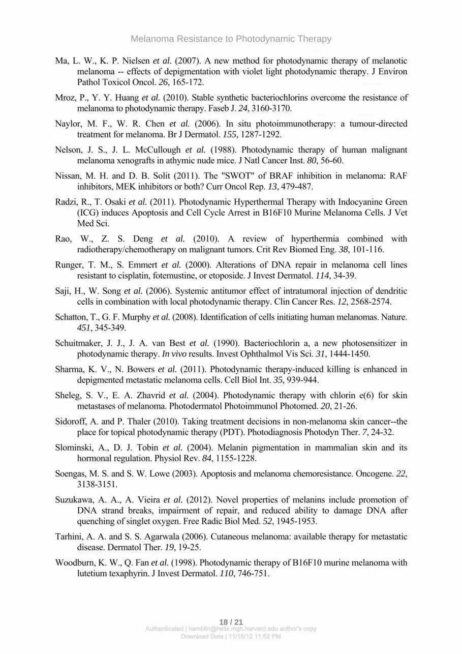
Figure 1 Mechanisms of PDT.
The ground state PS is initially excited to an excited singlet state that undergoes a transition to
a long-lived triplet state that can interact with oxygen in a Type I mechanism to produce
hydroxyl radicals or in a Type II mechanism to produce reactive singlet oxygen. These ROS
can cause death of tumor cells by apoptosis or necrosis and destroy the tumor.
Figure 2 Process of melanogenesis and resistance mechanisms of melanoma
Melanosomes mature through stages 1-4 and are finally transferred to keratinocyes where
they release melanin granules. Defects in apoptosis (BCL2), BRAF-mutation activated
MAPK/ERK pathway, and ABC-transporter drug efflux pumps contribute to melanoma
resistance.
Authenticated | [email protected] author's copyDownload Date | 11/15/12 11:52 PM
Melanoma Resistance to Photodynamic Therapy
20 / 21
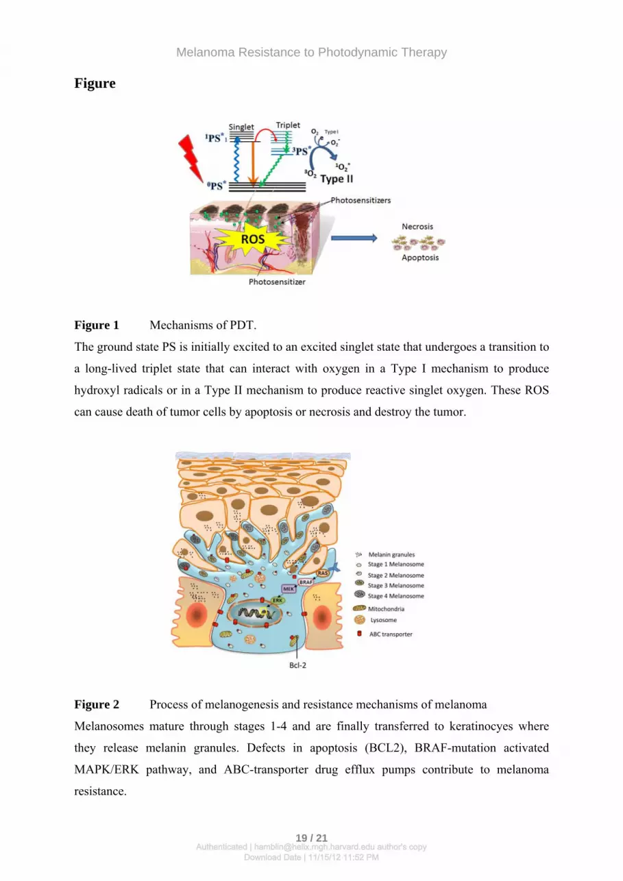
Figure 3 Chemical structures of NIR absorbing photosensitizers and the optical window
in tissue.
(A) Bacteriochlorin TCBSO3H from (Dabrowski, Urbanska et al. 2011). (B) Bacteriochlorin 3
from (Mroz, Huang et al. 2010). (C) Lutetium texaphyrin from (Woodburn, Fan et al. 1998).
(D) Si(IV)-naphthalocyanine (Isobosinc) from (Biolo, Jori et al. 1994).
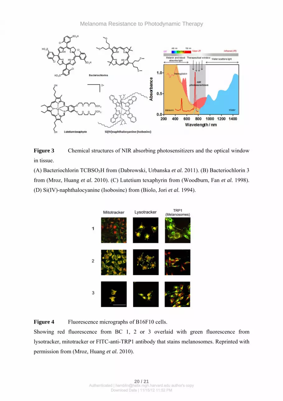
Figure 4 Fluorescence micrographs of B16F10 cells.
Showing red fluorescence from BC 1, 2 or 3 overlaid with green fluorescence from
lysotracker, mitotracker or FITC-anti-TRP1 antibody that stains melanosomes. Reprinted with
permission from (Mroz, Huang et al. 2010).
Authenticated | [email protected] author's copyDownload Date | 11/15/12 11:52 PM
Melanoma Resistance to Photodynamic Therapy
21 / 21
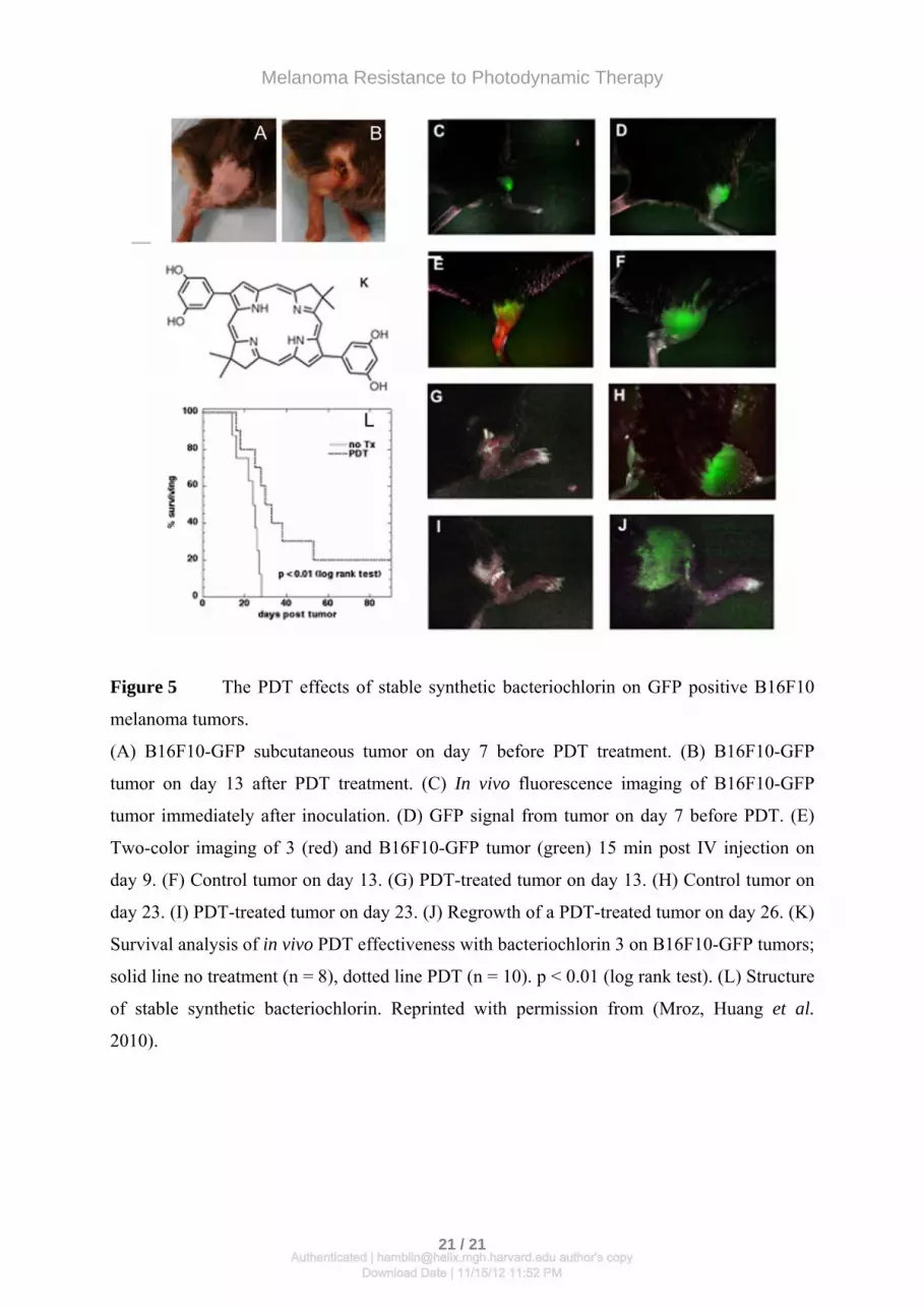
Figure 5 The PDT effects of stable synthetic bacteriochlorin on GFP positive B16F10
melanoma tumors.
(A) B16F10-GFP subcutaneous tumor on day 7 before PDT treatment. (B) B16F10-GFP
tumor on day 13 after PDT treatment. (C) In vivo fluorescence imaging of B16F10-GFP
tumor immediately after inoculation. (D) GFP signal from tumor on day 7 before PDT. (E)
Two-color imaging of 3 (red) and B16F10-GFP tumor (green) 15 min post IV injection on
day 9. (F) Control tumor on day 13. (G) PDT-treated tumor on day 13. (H) Control tumor on
day 23. (I) PDT-treated tumor on day 23. (J) Regrowth of a PDT-treated tumor on day 26. (K)
Survival analysis of in vivo PDT effectiveness with bacteriochlorin 3 on B16F10-GFP tumors;
solid line no treatment (n = 8), dotted line PDT (n = 10). p < 0.01 (log rank test). (L) Structure
of stable synthetic bacteriochlorin. Reprinted with permission from (Mroz, Huang et al.
2010).
Authenticated | [email protected] author's copyDownload Date | 11/15/12 11:52 PM


