Embed Size (px)
Citation preview
Endodontic photodynamic therapy ex vivo
Raymond Ng, DDS*, Fiza Singh, DDS*, Despoina A. Papamanou, DDS, Xiaoqing Song, MD,MS, Chitrang Patel, BS, Colleen Holewa, BS, Niraj Patel, BS, MS, Vanja Klepac-Ceraj, PhD,Carla R. Fontana, DDS, PhD, Ralph Kent, ScD, Tom C. Pagonis, DDS, MS, Philip P.Stashenko, DMD, PhD, and Nikolaos S. Soukos, DDS, PhDDrs. Ng and Singh are residents in Advanced Graduate Endodontics, Division of Endodontics,Harvard School of Dental Medicine, Boston, Massachusetts; Drs. Vanja Klepac-Ceraj and CarlaR. Fontana are post-doctoral research fellows, Dr. Papamanou, Dr. Song, and Mr. N. Patel areresearch fellows, and Mr. C. Patel and Ms. Holewa are research assistants, Applied MolecularPhotomedicine Laboratory, The Forsyth Institute, Boston, Massachusetts; Dr. Kent is the Head ofthe Department of Biostatistics, The Forsyth Institute; Dr. Pagonis is a Clinical Instructor inAdvanced Graduate Endodontics, Division of Endodontics, Harvard School of Dental Medicine;Dr. Stashenko is a Senior Scientist, Department of Cytokine Biology, The Forsyth Institute; Dr.Soukos is the Director of the Applied Molecular Photomedicine Laboratory, The Forsyth Institute.
AbstractObjective—To evaluate the anti-microbial effects of photodynamic therapy (PDT) on infectedhuman teeth ex vivo.
Materials and Methods—Fifty-two freshly extracted teeth with pulpal necrosis and associatedperiradicular radiolucencies were obtained from 34 subjects. Twenty-six teeth with 49 canalsreceived chemomechanical debridement (CMD) with 6% NaOCl and twenty-six teeth with 52canals received CMD plus PDT. For PDT, root canal systems were incubated with methylene blue(MB) at concentration of 50 µg/ml for 5 minutes followed by exposure to red light at 665 nm withan energy fluence of 30 J/cm2. The contents of root canals were sampled by flushing the canals atbaseline and following CMD alone or CMD+PDT and were serially diluted and cultured on bloodagar. Survival fractions were calculated by counting colony-forming units (CFU). Partialcharacterization of root canal species at baseline and following CMD alone or CMD+PDT wasperformed using DNA probes to a panel of 39 endodontic species in the checkerboard assay.
Results—The Mantel-Haenszel chi-square test for treatment effects demonstrated the betterperformance of CMD+PDT over CMD (P=0.026). CMD+PDT significantly reduced the frequencyof positive canals relative to CMD alone (P=0.0003). Following CMD+PDT, 45 of 52 canals(86.5%) had no CFU as compared to 24 of 49 canals (49%) treated with CMD (canal flushsamples). The CFU reductions were similar when teeth or canals were treated as independententities. Post-treatment detection levels for all species were markedly lower for canals treated byCMD+PDT than were for those treated by CMD alone. Bacterial species within dentinal tubules
© 2010 American Association of Endodontics. Published by Elsevier Inc. All rights reserved.Address requests for reprints to Dr. Nikolaos Soukos, Applied Molecular Photomedicine Laboratory, The Forsyth Institute, 245 FirstStreet, Cambridge, MA 02142. Tel: (617) 892-8467. Fax: (617) 892-8609. [email protected].*The contributions of the first two authors were equalPublisher's Disclaimer: This is a PDF file of an unedited manuscript that has been accepted for publication. As a service to ourcustomers we are providing this early version of the manuscript. The manuscript will undergo copyediting, typesetting, and review ofthe resulting proof before it is published in its final citable form. Please note that during the production process errors may bediscovered which could affect the content, and all legal disclaimers that apply to the journal pertain.The authors deny any conflicts of interest related to this study.
NIH Public AccessAuthor ManuscriptJ Endod. Author manuscript; available in PMC 2012 February 1.
Published in final edited form as:J Endod. 2011 February ; 37(2): 217–222. doi:10.1016/j.joen.2010.10.008.
NIH
-PA Author Manuscript
NIH
-PA Author Manuscript
NIH
-PA Author Manuscript
were detected in 17/22 (77.3%) and 15/29 (51.7%) of canals in the CMD and CMD+PDT group,respectively (P= 0.034).
Conclusion—Data indicate that PDT significantly reduces residual bacteria within the root canalsystem, and that PDT, if further enhanced by technical improvements, holds substantial promise asan adjunct to CMD.
KeywordsPhotodynamic therapy; methylene blue; endodontic disinfection; ex vivo
INTRODUCTIONEndodontic treatment is the clinical management of a microbiological problem (1) and themain target of treatment is the microorganisms residing within the root canal system (2).However, the complexity of the root canal system makes complete debridement and removalof bacteria with instrumentation, irrigation and intracanal medicaments virtually impossible(3). In addition, current endodontic procedures require very good technical skills, and usemedicaments whose effectiveness has never been definitively proven in human clinicaltrials. Three systematic reviews (4–6) on the outcome of primary non-surgical root canaltreatment summarized findings from longitudinal clinical studies published up to 2006, inwhich treatments were carried out by undergraduate students, graduate students, generaldental practitioners or specialists. The estimated success reported in these studies was 75%(6) and 78% (4,5). In a recent systematic review by Ng et al. (2010) that included fourteenstudies published between 1993 and 2007, the pooled proportion of teeth surviving over 2–10 years following root canal treatment was found to range between 86% and 93% (7).However, Wu et al. (2009) reported several factors that contribute to the overestimation ofsuccessful outcomes after primary root canal treatment: A high percentage of casesconfirmed healthy by periapical radiography reveal apical periodontitis on cone beamcomputed tomography and by histology; extractions and retreatments were rarely recordedas failures; and the recall rate was often < 50% in longitudinal clinical studies (8). Generaldentists perform about 75% of root canal procedures (9), and thus it might be anticipatedthat failure rates are even greater in general practice (6). When strict radiographic criteriawere used, the success rates were approximately 66%, 75%, 77% and 85% for treatmentscarried out by general dental practitioners, undergraduate students, graduate students andspecialists, respectively (6). Given that more than 20 million root canals are performedyearly in the U.S. (10), approximately 2 million endodontic failures could be avoided bybetter disinfection procedures. The development of adjunctive antibacterial therapeuticstrategies to CMD therefore becomes important in the evolution of methods to targetresidual microorganisms in the root canal system.
Photodynamic therapy (PDT) was developed as a therapy for cancer and is based on theconcept that a non-toxic photosensitizing agent, known as photosensitizer, can bepreferentially localized in premalignant and malignant tissues and subsequently activated bylight of the appropriate wavelength to generate singlet oxygen and free radicals that arecytotoxic to cells of the target tissue (11). In recent years, PDT has been employed to targetmicroorganisms in root canals in vitro (12–28) and in vivo (29–32) suggesting its usefulnessas an adjunct to current endodontic disinfection techniques. Methylene blue (MB) is a well-established photosensitizer that has been used in PDT for targeting various gram-positiveand gram-negative oral bacteria (33) and was previously employed to study the effect ofPDT on endodontic disinfection (14,19,20,22,25,26). MB has been used as aphotosensitizing agent for almost nine decades (34). It has been used for the detection ofmucosal premalignant lesions (35) and as a marker dye in surgery (36). The hydrophilicity
Ng et al. Page 2
J Endod. Author manuscript; available in PMC 2012 February 1.
NIH
-PA Author Manuscript
NIH
-PA Author Manuscript
NIH
-PA Author Manuscript
of MB (37), along with its low molecular weight and positive charge allows passage acrossthe porin-protein channels in the outer membrane of gram-negative bacteria (38). MB,whose intravenous administration is FDA approved for methemoglobinemia, predominantlyinteracts with the anionic macromolecule lipopolysaccharide and results in the generation ofMB dimers (38), which participate in the photosensitization process (38).
The objective of the present study was to evaluate the antimicrobial effects of MB-mediatedPDT in a stringent and clinically relevant evaluation using naturally human infected teeth exvivo treated immediately upon their extraction. Teeth with radiographic evidence ofperiradicular lesions were chosen because they were guaranteed to be grossly infected,which mimics the clinical situation that leads to higher failure rates (2). The use of naturally-infected teeth, which contain a much broader range of pathogens and deeper penetration intotubules than any in vitro model system provides an excellent test of the potential of PDT inachieving root canal disinfection.
MATERIALS AND METHODSCollection of teeth and groups
Fifty-two freshly extracted teeth with pulpal necrosis and radiographic evidence ofperiradicular lesions were obtained from 34 subjects in the Department of Oral andMaxillofacial Surgery, Massachusetts General Hospital, Boston. Permission to collectextracted teeth was authorized by Institutional Review Board-approved informed consent.Patients had no systemic disease and had not taken any antibiotics in the previous 3 months.Following extraction, teeth were placed into individual sterile vials and transferred within 30minutes to the Applied Molecular Photomedicine Laboratory at The Forsyth Institute forpreparation and experimentation. The external surface of each tooth was cleaned with 10%povidone-iodine. After 5 min, the disinfectant was removed from the surface with isopropylalcohol and the tooth was decoronated apical to the roof of the pulp chamber with a sterilerotating diamond saw (#911H, Brasseler USA, Savannah, GA) set at 20,000 rpm. Teethwere assigned to two groups. The first group comprised 26 teeth (5 incisors, 3 canines, 6premolars, 12 molars) with 49 canals that received only chemomechanical debridement(CMD group) (Fig. 1). The second group comprised 26 teeth (4 incisors, 2 canines, 6premolars, 14 molars) with 52 canals that received CMD followed by PDT (CMD+PDTgroup) (Fig. 1). In eight subjects, more than one tooth was obtained (22 teeth). In thesecases, teeth were randomly allocated to one of the above groups.
Baseline bacterial samplingsIn both groups, a baseline microbial sample of the root canal was taken. The canal wascompletely filled with pre-reduced anaerobically sterilized (PRAS) Ringer’s solution using asterile Monoject tuberculin syringe with 27-G X 1/2-in. detachable needle (SherwoodMedical, St. Louis, MO). A sample was collected by introducing an ISO size 10 K-type fileto a working length of 0.5 mm short of the apical foramen and then agitated in the canalsolution in the canal for 60 seconds. The file was then removed and the file handle was cutoff under aseptic conditions and put in a 1.5 ml microcentrifuge tube containing 1 ml PRASRinger’s solution. The canal contents were aspirated using the same syringe as above andadded in the tube containing the file.
Root canal treatmentChemomechanical debridement was performed in teeth of both groups using standard Kfiles and 0.04 mm/mm taper number 7 series 29 Ni-Ti rotary Profiles™ (Dentsply Maillefer,Tulsa, OK) to achieve a master apical file size of .465 (ISO equivalent) for distal canals ofmandibular molars, palatal canals of maxillary molars, and all single rooted teeth. Number 6
Ng et al. Page 3
J Endod. Author manuscript; available in PMC 2012 February 1.
NIH
-PA Author Manuscript
NIH
-PA Author Manuscript
NIH
-PA Author Manuscript
0.04 mm/mm taper Ni-Ti rotary Profiles™ were used to achieve a master apical file size of .360 (ISO equivalent) for buccal canals of maxillary molars and mesial canals of mandibularmolars. RC Prep® (Premium Products, Plymouth Meeting, PA) was used as a lubricantduring instrumentation and canals were irrigated with 10 cc of 6% sodium hypochlorite(NaOCl) throughout the instrumentation sequence. All irrigants used were dispensed using a30 gauge Max-I-Probe (Dentsply Maillefer). After canal preparation an aliquot of 1 ml of17% ethylenediaminetetraacetic acid (EDTA) solution was left in situ for 3 min for smearlayer removal, and was replaced by 1 ml of 6% NaOCl for 3 minutes.
Post-SET bacterial samplingImmediately after chemomechanical disinfection, each specimen in the CMD group wasaseptically mounted on a rubber dam attached to a rack. The contents of root canals weresampled by flushing the root canals with a coronal application of 1-ml of sterile phosphatebuffered saline (PBS) with a Pro Rinse® 30 gauge irrigation needle (Dentsply Maillefer)(Fig. 2). The bacterial suspension was collected in a 1.5 ml microcentrifuge tube positionedbelow the apical foramen and bacterial yielding was measured spectrophotometrically foreach sample. After vortexing for 20 seconds, serial dilutions were prepared and 100 µlaliquots were inoculated onto blood agar and incubated anaerobically for 7 days.
Photodynamic therapyFollowing CMD, specimens in the CMD+PDT group were treated by MB-mediated PDT.Methylene blue (Sigma, St Louis, MO) was dissolved in sterile PBS and filter-sterilizedimmediately prior to use. The final concentration used was 50 µg/ml (134 µM). Theultraviolet-visible absorption spectra of MB in PBS were recorded from 200 to 800 nmusing quartz cuvettes with 1 cm path length on a diode-array spectrophotometer and werecharacterized by a long-wavelength maximum at 665 nm as shown previously (14).
All individual specimens were aseptically mounted on a rubber dam, with the rubber damframe attached to a rack. Then the canals were filled to the level of the access cavity withMB solution using a Pro Rinse® 30 gauge irrigation needle (Dentsply Maillefer) for 5minutes. Following incubation, the canal was dried with a paper cone. Light was thenapplied in the root canal system of the specimens in appropriate groups for 2.5 minutesfollowed by a break of 2.5 minutes and a second light exposure for 2.5 minutes. Theirradiation source was a diode laser (BWTEK Inc., Newark, DE) with an output power of 1Watt and a central wavelength of 665 nm. The system was coupled to a 250-µm diameteroptical fiber (22) that was mechanically notched over a one-centimeter length atapproximately one-millimeter intervals (Schoelly Imaging Inc., Worcester, MA). The fiberwas able to uniformly distribute light at 36° within the root canal. The power density was100 mW/cm2 and the total energy fluence dose was 30 J/cm2. The fiber optic was wipedwith ethanol after the completion of each light exposure.
Post-PDT bacterial samplingThe contents of root canals were sampled by flushing the root canals as described above.Serial dilutions were prepared and 100 µl aliquots were inoculated onto blood agar andincubated anaerobically for 7 days.
Dentinal ShavingsFollowing flushing of tooth specimens, intracanal dentinal shavings were removed from theCMD group (9 teeth with 22 canals) and CMD+PDT group (12 teeth with 29 canals) andgathered in an microcentrifuge tube containing 1.5 ml of BHI. Briefly, a 21 mm lengthnickel-titanium rotary file (#25, Sequence, Brasseler, Savannah, Georgia) with a tip diameter
Ng et al. Page 4
J Endod. Author manuscript; available in PMC 2012 February 1.
NIH
-PA Author Manuscript
NIH
-PA Author Manuscript
NIH
-PA Author Manuscript
of 0.25 mm and a taper of 0.06 mm/mm was inserted to length in each 12 and 14 mm toothspecimen (i.e. at length, the 21 mm Sequence file protruded 9 mm and 7 mm beyond each12 mm and 14 mm tooth specimen root tip respectively). This generated a circumferentialdentinal tubule penetration of 205 to 455µ or 205 to 485µ for each 12 mm or 14 mm toothspecimen respectively measured from each root tip to coronal level.
Microbial analysisThe microbial composition of root canals before and after treatment was assayed using awhole genomic probe assay as described previously (22). Tris-EDTA buffer (1.5 ml) wasadded to the plates and the bacterial colonies were harvested using glass rods. The cellsuspensions were placed into individual Eppendorf tubes and sonicated for 10 sec to breakup clumps. The optical density (OD) of each suspension was adjusted to a final OD of 1.0,which corresponded to approximately 109 cells. Ten µl of the suspension (107 cells) wasremoved and placed in another Eppendorf tube with 140 µl of TE buffer and 150 µl of 0.5MNaOH. The samples were lysed and the DNA was placed in lanes on a positively chargednylon membrane using a Minislot device (Immunetics, Cambridge, MA, USA). Afterfixation of the DNA to the membrane, the membrane was placed in Miniblotter 45(Immunetics) with the lanes of DNA perpendicular to the lanes of the device. Digoxigenin-labeled whole genomic DNA probes against 39 species found in endodontic infections (39)were hybridized in individual lanes of the Miniblotter. After hybridization, the membraneswere washed at high stringency and the DNA probes were detected using antibody todigoxigenin conjugated with alkaline phosphatase for chemifluorescence detection. Signalswere detected using AttoPhos substrate (Amersham Life Science, Arlington Heights, IL,USA) and were scanned using a Storm Fluorimager (Molecular Dynamics, Sunnyvale, CA,USA). Computer-generated images were analyzed to determine the fluorescence intensityassociated with each sample and probe. Two lanes in each membrane contained DNAstandards with 1 ng (105 bacteria) and 10 ng (106 bacteria) of each species. The sensitivityof the assay was adjusted to permit detection of 104 cells of a given species by adjusting theconcentration of each DNA probe. The measured fluorescence intensities were converted toabsolute counts by comparison with the standards on the same membrane. Failure to detect asignal was recorded as zero.
Statistical AnalysisThe principal endpoint calculated for each canal was the residual level of colony-formingunits (CFUs) following treatment relative to the pretreatment CFU level (residual %CFUs).For multi-rooted teeth the value for each tooth was the average of the canal values.Treatment effects were evaluated in a logistic model using generalized estimating equations(GEE) to account for correlations between canals from the same tooth. An indicator (0/1)variable was included to estimate and adjust for single-rooted/multi-rooted teeth. Similaranalyses were done for teeth stratified as single/multi-rooted using Mantel-Haenszelanalysis.
RESULTSFigure 1 shows the distribution of 52 teeth in the CMD (26 teeth) and CMD+PDT (26 teeth)groups. The number of canals from each tooth that received either treatment is alsoprovided. These numbers are highlighted in bold when teeth were incompletely disinfectedfollowing treatment and the number of positive canals is given in parentheses. The resultsclearly demonstrated the better performance of CMD+PDT over CMD. The summaryMantel-Haenszel chi-square test for treatment effects was significant (P= 0.026). Overall, 13of 26 teeth (50%) were positive following CMD, whereas 6 of 20 teeth (30%) were positivefollowing CMD+PDT. Among single-rooted teeth, 5 of 14 teeth (35.7%) were positive
Ng et al. Page 5
J Endod. Author manuscript; available in PMC 2012 February 1.
NIH
-PA Author Manuscript
NIH
-PA Author Manuscript
NIH
-PA Author Manuscript
following CMD, whereas only 1 of 12 teeth (8.3%) was positive following CMD+PDT.Among multi-rooted teeth, 8 of 12 teeth (66.7%) were positive following CMD and 5 of 14teeth (35.7%) were positive following CMD +PDT.
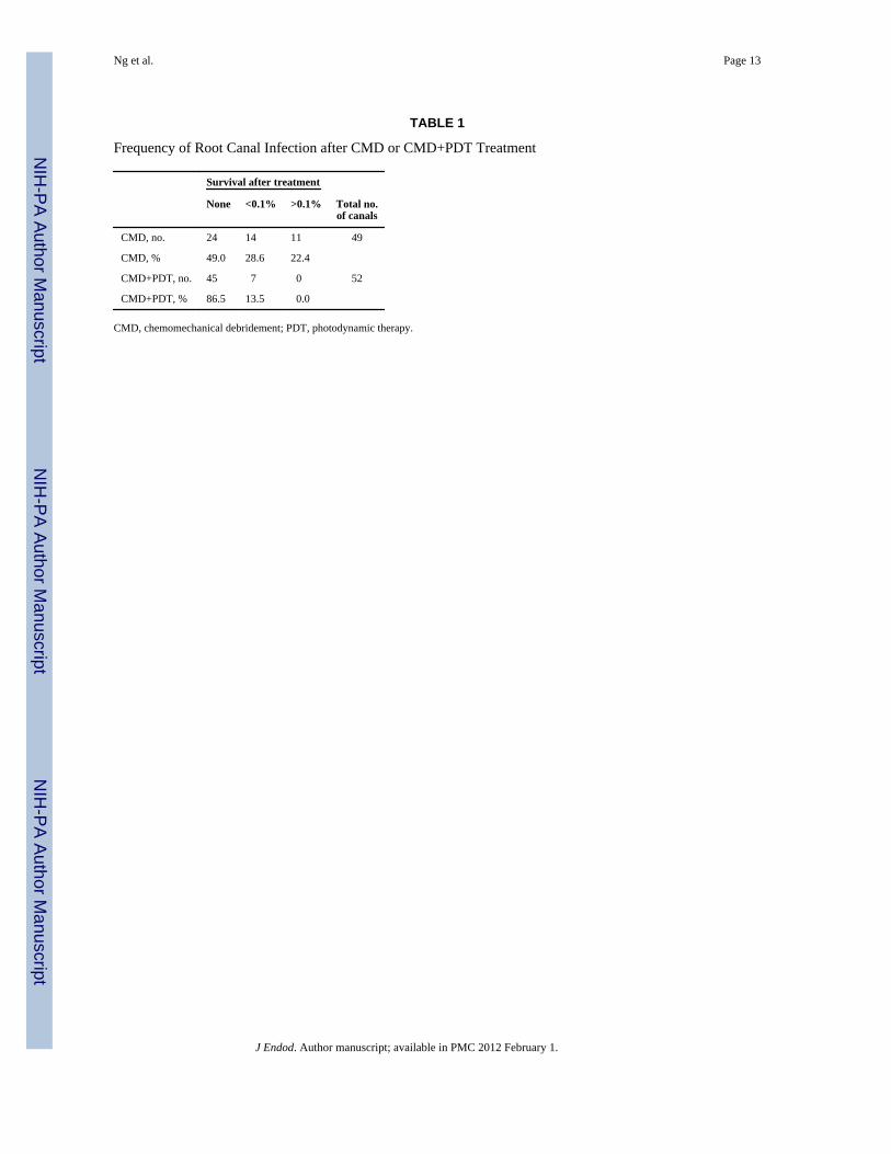
CMD+PDT significantly reduced the frequency of positive canals relative to CMD alone(P= 0.0003) (Table 1). Canals from single-rooted teeth were less likely to be positive post-treatment than canals from multi-rooted teeth (P= 0.10). Following CMD+PDT, 45 of 52canals (86.5%) had no CFU as compared to 24 of 49 canals (49%) treated with CMD (Table1) (canal flush samples). Post-treatment microbial levels were low as a percent ofpretreatment levels (%CFU). For CMD+PDT only 7 of 52 canals (13.5%) were positive andall had %CFU less than 0.1% of pretreatment CFU levels. However, following CMD 25 of49 canals were positive (51%) and 22.4% of canals had post-treatment values greater than0.1% of pretreatment levels (Table 1). The CFU reductions were similar when teeth orcanals were treated as independent entities. Analysis stratified by tooth type indicated thatpost treatment %CFU values were more often positive and also at higher levels of infectionin canals that received CMD relative to canals that have received CMD+PDT (P<0.0001).
The microbial composition of canal biofilms (canal flush samples) was studied bycheckerboard DNA-DNA hybridization. Pre- and post-treatment frequencies (+/−) wereobtained for 39 species found in endodontic infections with whole genomic probes for 45canals that received CMD+PDT and 44 that received CMD alone (Fig. 3). The number ofcanals positive for each species pretreatment was quite high and the pattern was similar forboth treatments. Post-treatment detection levels for all species were systematically andmarkedly lower for canals treated by CMD+PDT than for those treated by CMD alone. Keyendodontic pathogens resisting intracanal disinfection procedures (40) were dramaticallyreduced (Fig. 3, highlighted in grey).
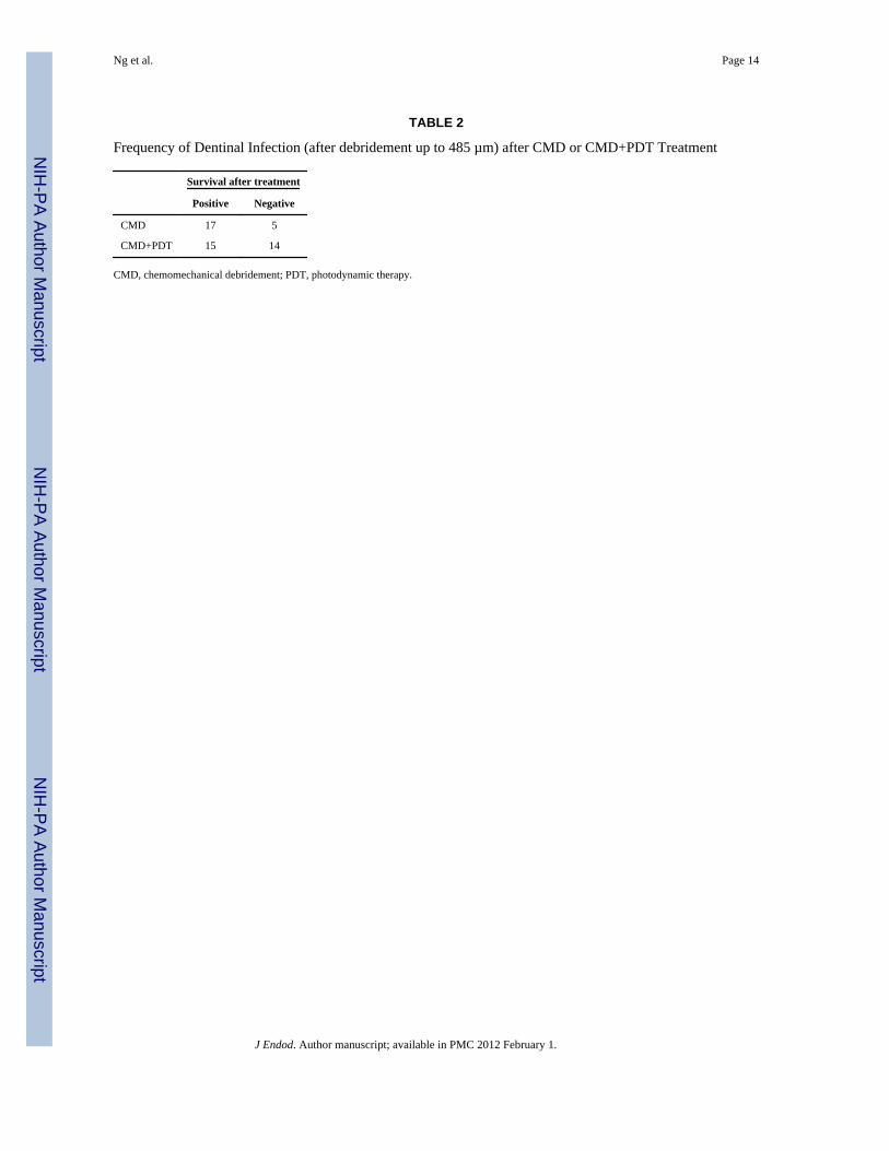
The frequency of dentinal infection (after debridement up to 485 µm) was also evaluated(Table 2). In the CMD group, tubules from 17/22 canals (77.3%) were positive aftertreatment, while in the CMD+PDT group tubules from 15/29 (51.7%) canals were positive(P= 0.034).
DISCUSSIONThe present study was built on the interdependent foundations of: 1) developing an in vitromodel for testing PDT (14,20,22); 2) the utilization of an FDA approved drug – methyleneblue (MB) – as the photosensitizer (14,20,22); 3) the development of a novel light deliverysystem that maximizes the distribution of light within the entire anatomy of the root canalsystem (22); 4) the ongoing refinement of light and drug dosimetry (14,20,22,41); and 5) theassessment of PDT safety (41). The hypothesis of this study was that near completeelimination of residual root canal bacteria could be achieved using PDT as an adjunctiveprocedure to SET in chronically-infected extracted human teeth ex vivo. Our findings showthat MB-mediated PDT significantly enhanced the effect of CMD. Four in vivo studies havealso suggested the potential of PDT as an adjunctive technique to eliminate residual rootcanal bacteria after CMD (29–32). Toluidine blue-mediated PDT offered a means ofdestroying microorganisms remaining after using sodium hypochlorite alone (29) or citricacid and sodium hypochlorite as co-irrigants (30). PDT significantly enhanced the effect ofCMD in teeth with necrotic pulps using a conjugate between polyethyleneimine chlorine e6conjugate (31) and toluidine blue (32).
The incomplete bacterial killing in dentinal tubules following PDT may be due to: a)Incomplete MB penetration in the tubules that may be related to binding interactions withdentin components; b) Failure of MB to penetrate canal biofilms; and c) Insufficient
Ng et al. Page 6
J Endod. Author manuscript; available in PMC 2012 February 1.
NIH
-PA Author Manuscript
NIH
-PA Author Manuscript
NIH
-PA Author Manuscript
oxygenation. We have proposed the encapsulation of MB within poly(D,L-lactide-co-glycolide) (PLGA) nanoparticles that may offer a novel nano-platform for enhanced drugdelivery and photodestruction of canal biofilms (28). These nanoparticles have ahydrophobic core part made up of PLGA (hydrophobic) and PEO-PPO (surfactant)molecules (polyethylene oxide-polypropylene oxide). Surfactant chains project outwardsfrom the surface of the core part because of their hydrophilic end groups (hydroxyl,quarternary ammonium). This creates a gradient from the hydrophilic end groups outside toincreased hydrophobicity towards the core of nanoparticles. Hydrophilic end groups providean anchoring effect for retention of nanoparticles to negatively-charged membranes. Due tothe hydrophobic-hydrophilic orientation of the surfactant molecules, they provide goodwettability to enhance interaction on/within bacterial membranes. Infiltration of dentinaltubules by MB-loaded nanoparticles has recently been demonstrated (28). George andKishen (19) dissolved MB in a mixture composed of glycerol, ethanol and water (30:20:50)and showed greater penetration of MB into dentinal tubules. Our future studies will explorethe use of ultrasonic waves for enhancement of the transdentinal movement and penetrationof MB in canal biofilms. It has been demonstrated that an irrigant in conjunction withultrasonic vibration, which generates acoustic streaming and continuous movement of theirrigant, increases the effectiveness of the cleaning of root canal (42). Regarding insufficientoxygenation, the application of perfluoro-decahydro-napthalene in the root canal system wasproposed as a carrier of oxygen for enhancement of the PDT effect (26). The basicproperties of perfluorocarbons and perfluorocarbon emulsions relevant to their use asoxygen delivery systems were briefly reviewed (43). A Phase III clinical trial incardiopulmonary bypass surgery, with a protocol that included both augmented-acutenormovolemic hemodilution and intraoperative autologous donation, was interruptedfollowing the observation of adverse events. At this time point, there is not enoughinformation concerning the toxicity of these compounds to utilize them.
The results obtained from this study are very promising. The use of naturally infected teeth,which contain a broader range of pathogens than in vitro model systems, provide anexcellent test of the potential of PDT in achieving root canal disinfection. However, sincesome living bacteria were still present in dentinal tubules following PDT, further refinementand enhancement of the PDT procedure may be necessary. The effect of biophysical meansand surface tension-reducing agents on the transdentinal penetration of MB as well as theeffect of supplemental hyper-oxygenation should be evaluated.
AcknowledgmentsThis work was supported by NIDCR grant RO1-DE-16922.
REFERENCES1. Figdor D. Apical Periodontitis: A very prevalent problem. Oral Surg Oral Med Oral Pathol Oral
Radiol Endod 2002;94:651–652. [PubMed: 12464886]2. Nair PN. Pathogenesis of apical periodontitis and the causes of endodontic failures. Crit Rev Oral
Biol Med 2004;15:348–381. [PubMed: 15574679]3. Siqueira JF Jr, Rôças IN, Paiva SS, Magalhães KM, Guimarães-Pinto T. Cultivable bacteria in
infected root canals as identified by 16S rRNA gene sequencing. Oral Microbiol Immunol2007;22:266–271. [PubMed: 17600539]
4. Lewsey JD, Gilthorpe MS, Gulabivala K. An Introduction to meta-analysis within the framework ofmultilevel modelling using the probability of success of root canal treatment as an illustration.Community Dent Health 2001;18:131–137. [PubMed: 11580087]
Ng et al. Page 7
J Endod. Author manuscript; available in PMC 2012 February 1.
NIH
-PA Author Manuscript
NIH
-PA Author Manuscript
NIH
-PA Author Manuscript
5. Basmadjian-Charles CL, Farge P, Bourgeois DM, Lebrun T. Factors influencing the long-termresults of endodontic treatment: a review of the literature. Int Dent J 2002;52:81–86. [PubMed:12013255]
6. Ng Y-L, Mann V, Rahbaran S, Lewsey J, Gulabivala K. Outcome of primary root canal treatment:systematic review of the literature - Part 1. Effects of study characteristics on probability of success.Int Endod J 2007;40:921–939. [PubMed: 17931389]
7. Ng Y-L, Mann V, Gulabivala K. Tooth survival following non-surgical root canal treatment: asystematic review in the literature. Int Endod J 2010;43:171–189. [PubMed: 20158529]
8. Wu M-K, Shemesh H, Wesselink PR. Limitations of previously published systematic reviewsevaluating the outcome of endodontic treatment. Int Endod J 2009;42:656–666. [PubMed:19548929]
9. Nash KD, Brown LJ, Hicks ML. Private practicing endodontists: Production of endodontic servicesand implications for workforce policy. J Endod 2002;28:699–705. [PubMed: 12398168]
10. Brown, LJ.; Nash, KD.; Johns, BA.; Warren, M. ADA Health policy Resources Center DentalHealth Policy Analysis Series. Chicago, IL: 2003. The Economics of Endodontics.
11. Dougherty TJ, Gomer CJ, Henderson BW, Jori G, Kessel D, Korbelik M, Moan J, Peng Q.Photodynamic therapy. J Natl Cancer Inst 1998;90:889–905. [PubMed: 9637138]
12. Silbert T, Bird PS, Milburn GJ, Walsh L. Disinfection of root canals by laser dyephotosensitization. J Dent Res 2000;79:569.
13. Seal GJ, Ng YL, Spratt D, Bhatti M, Gulabivala K. An in vitro comparison of the bactericidalefficacy of lethal photosensitization or sodium hyphochlorite irrigation on Streptococcusintermedius biofilms in root canals. Int Endod J 2002;35:268–274. [PubMed: 11985679]
14. Soukos NS, Chen PS, Morris JT, Ruggiero K, Abernethy AD, Som S, Foschi F, Doucette S,Bammann LL, Fontana CR, Doukas AG, Stashenko PP. Photodynamic therapy for endodonticdisinfection. J Endod 2006;32:979–984. [PubMed: 16982278]
15. Garcez AS, Núñez SC, Lage-Marques JL, Jorge AO, Ribeiro MS. Efficiency of NaOCl and laser-assisted photosensitization on the reduction of Enterococcus faecalis in vitro. Oral Surg Oral MedOral Pathol Oral Radiol Endod 2006;102:e93–e98. [PubMed: 16997103]
16. Williams JA, Pearson GJ, Colles MJ. Antibacterial action of photoactivated disinfection {PAD}used on endodontic bacteria in planktonic suspension and in artificial and human root canals. JDent 2006;34:363–371. [PubMed: 16239058]
17. Garcez AS, Ribeiro MS, Tegos GP, Núñez SC, Jorge AO, Hamblin MR. Antimicrobialphotodynamic therapy combined with conventional endodontic treatment to eliminate root canalbiofilm infection. Lasers Surg Med 2007;39:59–66. [PubMed: 17066481]
18. George S, Kishen A. Advanced noninvasive light-activated disinfection: assessment of cytotoxicityon fibroblast versus antimicrobial activity against Enterococcus faecalis. J Endod 2007;33:599–602. [PubMed: 17437881]
19. George S, Kishen A. Photophysical, photochemical, and photobiological characterization ofmethylene blue formulations for light-activated root canal disinfection. J Biomed Opt2007;12:034029. [PubMed: 17614737]
20. Foschi F, Fontana CR, Ruggiero K, Riahi R, Vera A, Doukas AG, Pagonis TC, Kent R, StashenkoPP, Soukos NS. Photodynamic inactivation of Enterococcus faecalis in dental root canals in vitro.Lasers Surg Med 2007;39:782–787. [PubMed: 18081066]
21. Bergmans L, Moisiadis P, Huybrechts B, Van Meerbeek B, Quirynen M, Lambrechts P. Effect ofphoto-activated disinfection on endodontic pathogens ex vivo. Int Endod J 2008;41:227–239.[PubMed: 18081808]
22. Fimple JL, Fontana CR, Foschi F, Ruggiero K, Song X, Pagonis TC, Tanner AC, Kent R, DoukasAG, Stashenko PP, Soukos NS. Photodynamic treatment of endodontic polymicrobial infection invitro. J Endod 2008;34:728–734. [PubMed: 18498901]
23. Fonseca MB, Júnior PO, Pallota RC, Filho HF, Denardin OV, Rapoport A, Dedivitis RA, VeroneziJF, Genovese WJ, Ricardo AL. Photodynamic therapy for root canals infected with Enterococcusfaecalis. Photomed Laser Surg 2008;26:209–213. [PubMed: 18484911]
Ng et al. Page 8
J Endod. Author manuscript; available in PMC 2012 February 1.
NIH
-PA Author Manuscript
NIH
-PA Author Manuscript
NIH
-PA Author Manuscript
24. George S, Kishen A. Augmenting the antibiofilm efficacy of advanced noninvasive light activateddisinfection with emulsified oxidizer and oxygen carrier. J Endod 2008;34:1119–1123. [PubMed:18718378]
25. George S, Kishen A. Influence of photosensitizer solvent on the mechanisms of photoactivatedkilling of Enterococcus faecalis. Photochem Photobiol 2008;84:734–740. [PubMed: 18435622]
26. Lim Z, Cheng JL, Lim TW, Teo EG, Wong J, George S, Kishen A. Light activated disinfection: analternative endodontic disinfection strategy. Aust Dent J 2009;54:108–114. [PubMed: 19473151]
27. Souza LC, Brito PRR, Machado de Oliveira JC, Alves FRF, Moreira EJL, Sampaio-Filbo HR,Rôças IN, Siqueira JF Jr. Photodynamic therapy with two different photosensitizers as asupplement to instrumentation/ irrigation procedures in promoting intracanal reduction ofEnterococcus Faecalis. J Endod 2010;36:292–296. [PubMed: 20113793]
28. Pagonis TC, Chen J, Fontana CR, Devalapally H, Ruggiero K, Song X, Foschi F, Dunham J, SkobeZ, Yamazaki H, Kent R, Tanner AC, Amiji MM, Soukos NS. Nanoparticle-based endodonticantimicrobial photodynamic therapy. J Endod 2010;36:322–328. [PubMed: 20113801]
29. Bonsor SJ, Nichol R, Reid TM, Pearson GJ. An alternative regimen for root canal disinfection. BritDent J 2006;22:101–105. [PubMed: 16865142]
30. Bonsor SJ, Nichol R, Reid TM, Pearson GJ. Microbiological evaluation of photo-activateddisinfection in endodontics (an in vivo study). Brit Dent J 2006;25:337–341. [PubMed: 16568063]
31. Garcez AS, Núñez SC, Hamblin MR, Ribeiro MS. Antimicrobial effects of photodynamic therapyon patients with necrotic pulps and periapical lesion. J Endod 2008;34:138–142. [PubMed:18215668]
32. Pinheiro SL, Schenka AA, Neto AA, de Souza CP, Rodriguez HM, Ribeiro MC. Photodynamictherapy in endodontic treatment of deciduous teeth. Lasers Med Sci 2009;24:521–526. [PubMed:18427873]
33. Harris F, Chatfield LK, Phoenix DA. Phenothiazinium based photosensitisers-photodynamicagents with a multiplicity of cellular targets and clinical applications. Curr Drug Targets2005;6:615–627. [PubMed: 16026282]
34. Wainwright M, Mohr H, Walker WH. Phenothiazinium derivatives for pathogen inactivation inblood products. J Photochem Photobiol B 2007;86:45–58. [PubMed: 16979899]
35. Ojetti V, Persiani R, Nista EC, Rausei S, Lecca G, Migneco A, Cananzi FC, Cammarota G, D’UgoD, Gasbarrini G, Gasbarrini A. A case-control study comparing methylene blue directed biopsiesand random biopsies for detecting pre-cancerous lesions in the follow-up of gastric cancer patients.Eur Rev Med Pharmacol Sci 2007;11:291–296. [PubMed: 18074937]
36. Creagh TA, Gleeson M, Travis D, Grainger R, McDermott TE, Butler MR. Is there a role for invivo methylene blue staining in the prediction of bladder tumour recurrence? Br J Urol1995;75:477–479. [PubMed: 7788259]
37. Wainwright M, Phoenix DA, Marland J, Wareing DR, Bolton FJ. A study of photobactericidalactivity in the phenothiazinium series. FEMS Immunol Med Microbiol 1997;19:75–80. [PubMed:9322071]
38. Usacheva MN, Teichert MC, Biel MA. The interaction of lipopolysaccharides with phenothiazinedyes. Lasers Surg Med 2003;33:311–319. [PubMed: 14677158]
39. Brito LC, Teles FR, Teles RP, França EC, Ribeiro-Sobrinho AP, Haffajee AD, Socransky SS. Useof multiple-displacement amplification and checkerboard DNA-DNA hybridization to examine themicrobiota of endodontic infections. J Clin Microb 2007;45:3039–3049.
40. Siqueira JF Jr, Rôças I. Clinical Implications and Microbiology of Bacterial Persistence afterTreatment Procedures. J Endod 2008;34:1291–1301. [PubMed: 18928835]
41. Xu Y, Young MJ, Battaglino RA, Morse LR, Fontana CR, Pagonis TC, Kent R, Soukos NS.Endodontic antimicrobial photodynamic therapy: safety assessment in mammalian cell cultures. JEndod 2009;35:1567–1572. [PubMed: 19840649]
42. Gutarts R, Nusstein J, Reader A, Beck M. In vivo debridement efficacy of ultrasonic irrigationfollowing hand-rotary instrumentation in human mandibular molars. J Endod 2005;31:166–170.[PubMed: 15735461]
43. Riess JG. Perfluorocarbon-based oxygen delivery. Artif Cells Blood Substit Immobil Biotechnol2006;34:567–580. [PubMed: 17090429]
Ng et al. Page 9
J Endod. Author manuscript; available in PMC 2012 February 1.
NIH
-PA Author Manuscript
NIH
-PA Author Manuscript
NIH
-PA Author Manuscript
Figure 1.Fifty-two teeth were assigned to two experimental groups (CMD and CMD+PDT). Thenumber of canals from each tooth that received treatment is provided. Numbers highlightedin bold indicate incompletely disinfected teeth. The number of positive canals followingtreatment is given in parentheses.
Ng et al. Page 10
J Endod. Author manuscript; available in PMC 2012 February 1.
NIH
-PA Author Manuscript
NIH
-PA Author Manuscript
NIH
-PA Author Manuscript
Figure 2.Post-flushing of tooth specimen under rubber dam.
Ng et al. Page 11
J Endod. Author manuscript; available in PMC 2012 February 1.
NIH
-PA Author Manuscript
NIH
-PA Author Manuscript
NIH
-PA Author Manuscript
Figure 3.Pre- and post-treatment detection frequencies for 39 species found in endodontic infectionsby checkerboard DNA-DNA hybridization with whole genomic probes for 45 canals treatedby CMD+PDT and 44 treated by CMD alone.◊ CMD, baseline ♦ post-CMD ○ PDT, baseline ● post-PDT
Ng et al. Page 12
J Endod. Author manuscript; available in PMC 2012 February 1.
NIH
-PA Author Manuscript
NIH
-PA Author Manuscript
NIH
-PA Author Manuscript
NIH
-PA Author Manuscript
NIH
-PA Author Manuscript
NIH
-PA Author Manuscript
Ng et al. Page 13
TABLE 1
Frequency of Root Canal Infection after CMD or CMD+PDT Treatment
Survival after treatment
None <0.1% >0.1% Total no.of canals
CMD, no. 24 14 11 49
CMD, % 49.0 28.6 22.4
CMD+PDT, no. 45 7 0 52
CMD+PDT, % 86.5 13.5 0.0
CMD, chemomechanical debridement; PDT, photodynamic therapy.
J Endod. Author manuscript; available in PMC 2012 February 1.
NIH
-PA Author Manuscript
NIH
-PA Author Manuscript
NIH
-PA Author Manuscript
Ng et al. Page 14
TABLE 2
Frequency of Dentinal Infection (after debridement up to 485 µm) after CMD or CMD+PDT Treatment
Survival after treatment
Positive Negative
CMD 17 5
CMD+PDT 15 14
CMD, chemomechanical debridement; PDT, photodynamic therapy.
J Endod. Author manuscript; available in PMC 2012 February 1.


