Embed Size (px)
Citation preview
ZEBRAFISH AS A MODEL FOR PATHOBIOLOGY (WOLFRAM GOESSLING, SECTION EDITOR)
Insights from Genetic Models of Melanoma in Fish
Viviana Anelli • Nicole Zettler • Marina Mione
Published online: 23 March 2014
� Springer Science+Business Media New York 2014
Abstract Fish melanoma models are increasingly used in
studies of both spontaneous and induced melanoma forma-
tion. The melanoma Xiphophorus model was the first
genetic model of melanoma available to researchers since
the 1920s (Klin Wochenschrift 7:1561–1562, 1928; Z Indukt
Abstammungs 44:253–257, 1927). Recently, transgenesis
has been used to develop zebrafish and medaka models for
melanoma research (Methods Mol Biol 461:521–539, 2008).
These models are now starting to produce a wealth of novel
information on the genetics of melanoma (including somatic
mutations), signaling pathways and molecular mechanisms,
immune responses, and determinants of metastasis. More-
over, the models are extremely well suited for drug/small-
molecule screens aimed at discovering new therapeutic
strategies in general treatment and personalized medicine
and for the study of cancer-immune cell interactions. This
review provides a summary of current and prospective
studies in fish models of melanoma and pinpoints the
translational potentials of melanoma models in fish.
Keywords Melanoma � Cancer � Genetic models �Zebrafish � Medaka � Chemical screen
Introduction
Melanoma is the most serious type of skin cancer, the most
aggressive and lethal, affecting more than 75,000 annually
in the US [4]. These malignant tumors originate from
melanocytes, the pigment-producing cells of vertebrates.
According to a classical model [5], melanoma begins with
UV light-induced mutations activating oncogenes, which
lead to melanocyte proliferation. The first response of
melanocytes to oncogenes, following proliferation, is
oncogene-induced senescence (OIS), which leads to the
formation of a benign nevus. Upon secondary mutations
inactivating tumor suppressors, OIS is overcome, and the
nascent melanoma proliferates first radially in the epider-
mis and then vertically until cells acquire the ability to
penetrate the basement membrane and further invade the
dermis. In the latter stage, melanoma cells acquire meta-
static characteristics and enter the blood stream or lym-
phatic vessels, colonizing distant tissues and organs [5].
However, not all cutaneous melanomas originate from
dysplastic nevi; about 50 % occur in normal skin, which
leads to the suggestion that alternative paths, including
origin from melanocytic progenitor or stem cells, different
transforming events or molecular pathways, are responsible
for half of the cutaneous melanoma developing in humans.
The Genetic Causes of Melanoma and Fish Models
Classical studies (candidate gene sequencing, i.e., COSMIC
[6]) have revealed that in over 90 % of cutaneous melanoma,
somatic mutations of a handful of genes taking place in
melanocytes as a result of environmental DNA damage (UV
light) are at the origin of melanoma development (reviewed
in [7]). These include activating mutations in oncogenes
V. Anelli
Departments of Surgery and Medicine, Weill Cornell Medical
College and New York Presbyterian Hospital, 1300 York
Avenue, New York, NY 10065, USA
e-mail: [email protected]
N. Zettler � M. Mione (&)
Institute of Toxicology and Genetics, Karlsruhe Institute of
Technology, Hermann-von-Helmholtz-Platz 1,
76344 Eggenstein-Leopoldshafen, Germany
e-mail: [email protected]
N. Zettler
e-mail: [email protected]
123
Curr Pathobiol Rep (2014) 2:85–92
DOI 10.1007/s40139-014-0043-1
(including B-RAF 45 %, N-RAS 13–25 % and H-RAS
1–5 %) and loss of function mutations in tumor suppressors
(CDKN2A, RB1,PTEN, P53). Already from these classical
studies it was clear that most oncogene-activating mutations
require additional factors to lead to melanoma development.
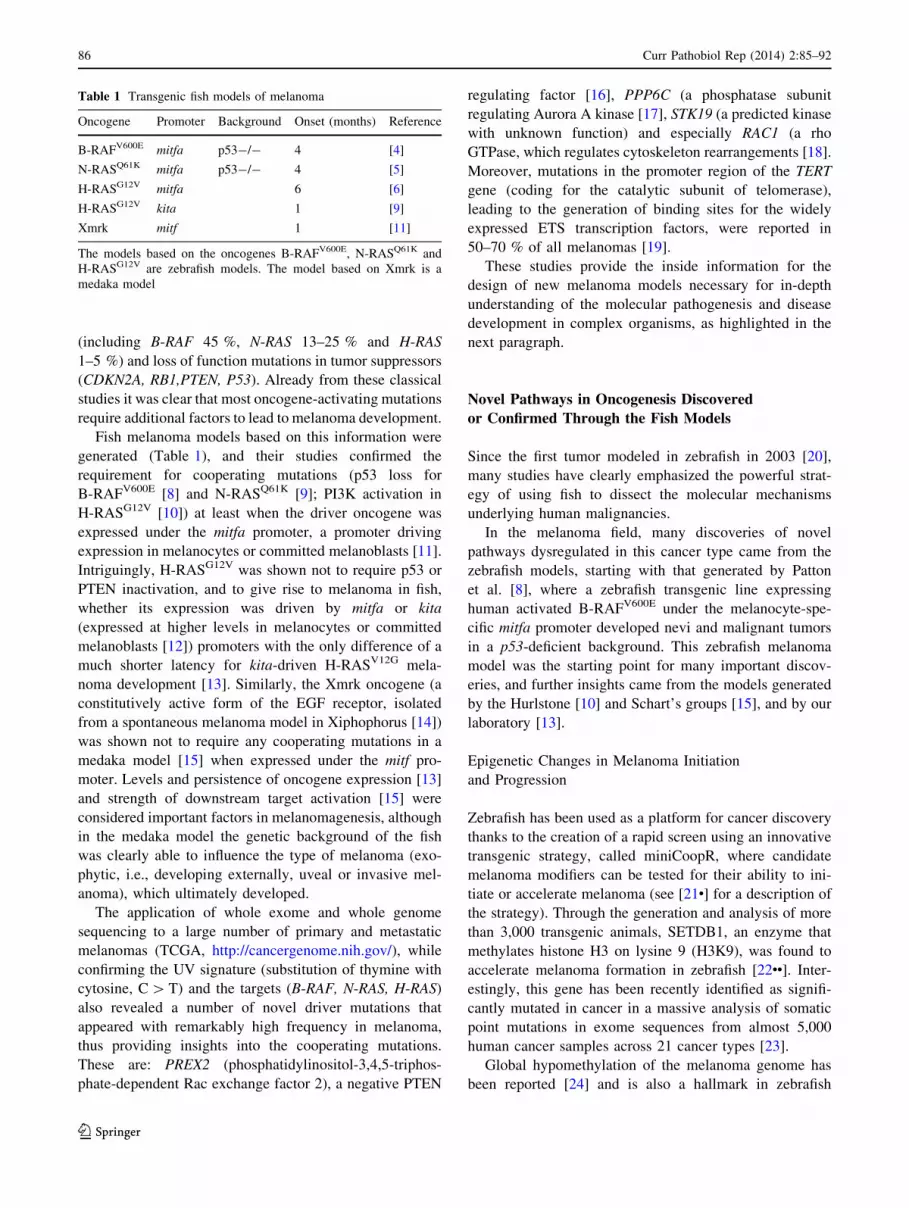
Fish melanoma models based on this information were
generated (Table 1), and their studies confirmed the
requirement for cooperating mutations (p53 loss for
B-RAFV600E [8] and N-RASQ61K [9]; PI3K activation in
H-RASG12V [10]) at least when the driver oncogene was
expressed under the mitfa promoter, a promoter driving
expression in melanocytes or committed melanoblasts [11].
Intriguingly, H-RASG12V was shown not to require p53 or
PTEN inactivation, and to give rise to melanoma in fish,
whether its expression was driven by mitfa or kita
(expressed at higher levels in melanocytes or committed
melanoblasts [12]) promoters with the only difference of a
much shorter latency for kita-driven H-RASV12G mela-
noma development [13]. Similarly, the Xmrk oncogene (a
constitutively active form of the EGF receptor, isolated
from a spontaneous melanoma model in Xiphophorus [14])
was shown not to require any cooperating mutations in a
medaka model [15] when expressed under the mitf pro-
moter. Levels and persistence of oncogene expression [13]
and strength of downstream target activation [15] were
considered important factors in melanomagenesis, although
in the medaka model the genetic background of the fish
was clearly able to influence the type of melanoma (exo-
phytic, i.e., developing externally, uveal or invasive mel-
anoma), which ultimately developed.
The application of whole exome and whole genome
sequencing to a large number of primary and metastatic
melanomas (TCGA, http://cancergenome.nih.gov/), while
confirming the UV signature (substitution of thymine with
cytosine, C [ T) and the targets (B-RAF, N-RAS, H-RAS)
also revealed a number of novel driver mutations that
appeared with remarkably high frequency in melanoma,
thus providing insights into the cooperating mutations.
These are: PREX2 (phosphatidylinositol-3,4,5-triphos-
phate-dependent Rac exchange factor 2), a negative PTEN
regulating factor [16], PPP6C (a phosphatase subunit
regulating Aurora A kinase [17], STK19 (a predicted kinase
with unknown function) and especially RAC1 (a rho
GTPase, which regulates cytoskeleton rearrangements [18].
Moreover, mutations in the promoter region of the TERT
gene (coding for the catalytic subunit of telomerase),
leading to the generation of binding sites for the widely
expressed ETS transcription factors, were reported in
50–70 % of all melanomas [19].
These studies provide the inside information for the
design of new melanoma models necessary for in-depth
understanding of the molecular pathogenesis and disease
development in complex organisms, as highlighted in the
next paragraph.
Novel Pathways in Oncogenesis Discovered
or Confirmed Through the Fish Models
Since the first tumor modeled in zebrafish in 2003 [20],
many studies have clearly emphasized the powerful strat-
egy of using fish to dissect the molecular mechanisms
underlying human malignancies.
In the melanoma field, many discoveries of novel
pathways dysregulated in this cancer type came from the
zebrafish models, starting with that generated by Patton
et al. [8], where a zebrafish transgenic line expressing
human activated B-RAFV600E under the melanocyte-spe-
cific mitfa promoter developed nevi and malignant tumors
in a p53-deficient background. This zebrafish melanoma
model was the starting point for many important discov-
eries, and further insights came from the models generated
by the Hurlstone [10] and Schart’s groups [15], and by our
laboratory [13].
Epigenetic Changes in Melanoma Initiation
and Progression
Zebrafish has been used as a platform for cancer discovery
thanks to the creation of a rapid screen using an innovative
transgenic strategy, called miniCoopR, where candidate
melanoma modifiers can be tested for their ability to ini-
tiate or accelerate melanoma (see [21•] for a description of
the strategy). Through the generation and analysis of more
than 3,000 transgenic animals, SETDB1, an enzyme that
methylates histone H3 on lysine 9 (H3K9), was found to
accelerate melanoma formation in zebrafish [22••]. Inter-
estingly, this gene has been recently identified as signifi-
cantly mutated in cancer in a massive analysis of somatic
point mutations in exome sequences from almost 5,000
human cancer samples across 21 cancer types [23].
Global hypomethylation of the melanoma genome has
been reported [24] and is also a hallmark in zebrafish
Table 1 Transgenic fish models of melanoma
Oncogene Promoter Background Onset (months) Reference
B-RAFV600E mitfa p53-/- 4 [4]
N-RASQ61K mitfa p53-/- 4 [5]
H-RASG12V mitfa 6 [6]
H-RASG12V kita 1 [9]
Xmrk mitf 1 [11]
The models based on the oncogenes B-RAFV600E, N-RASQ61K and
H-RASG12V are zebrafish models. The model based on Xmrk is a
medaka model
86 Curr Pathobiol Rep (2014) 2:85–92
123

melanoma [25]. More recently, loss of 5-hydroxymethyl-
cytosines (5-hmC) was identified as a discriminant between
nevi and malignant melanoma [26]. In the same study, the
miniCoopR vector was used to overexpress isocitrate
dehydrogenase 2 in a fish B-RAFV600E/p53-melanoma
model shown to promote an increase of 5-hmC levels and
to prolong tumor-free survival of transgenic fish.
The importance of epigenetic changes in the initial
transformation and progression of melanoma emerges from
results obtained in our laboratory [25]. We used the kita:
H-RASG12V melanoma model [13] to show that oncogenic
RAS is able to increase the levels of three microRNAs at
the onset of melanocyte transformation (miR-146a, miR-
146b and miR-193a). Interestingly, these three microRNAs
have as common target, Jmjd6, a jumonjiC domain protein
that functions as histone arginine demethylase [27] and
mRNA splicing regulator [28]. The upregulation of these
microRNAs decreased Jmjd6 levels at the onset of Ras
expression. However, at later stages of melanoma pro-
gression, Jmjd6 levels were found elevated, suggesting that
the microRNAs targeting Jmjd6 may be part of an anti-
cancer response activated by Ras. Similar levels of the
three microRNAs and JMD6 were also found in mouse
models, human cancer cell lines and melanoma tissue
obtained from several patients (Anelli et al., personal
communication), thus confirming the potential of zebrafish
melanoma as a model to identify novel molecular mecha-
nisms in human diseases.
Intriguingly, a recent paper [29] reported that JMJD6
and bromodomain-containing protein 4 (BRD4), acting as
functional partners, regulate Pol II promoter proximal
pausing in a large subset of genes based on their actions on
distal enhancers, termed anti-pause enhancer. In particular,
JMJD6 and BRD4 retain P-TEFb complex (a heterodimer
consisting of the cyclin-dependent kinase Cdk9 and a
cyclin component), leading to subsequent Pol II phos-
phorylation and efficient transcriptional elongation. This
finding is of particular interest as transcriptional elongation
has been found to be involved in neural crest specification
and melanocytes maturation [30], thus connecting results
obtained from different studies in zebrafish melanocytes
and melanoma.
In order to identify modifiers of RAS-induced mel-
anomagenesis, we developed a screening strategy based
on somatic expression of the oncogene and candidate
modifiers using the kita:gal4 line and a UAS vector to
express candidate modifier genes (Zettler et al., in prep-
aration), including epigenetic modifiers. The screen helped
to focus on JMJD6 as a microRNA target and RAS
cooperating factor. Figure 1 shows a comparison of the
two screening methods for melanoma modifiers described
in this section.
Rac1 in Melanoma
The Rho-like GTPase Rac1 stimulates cellular processes
such as proliferation, survival, motility and invasion [31],
which are deregulated in cancer. Its function may be
required for Ras-induced transformation, and recent NGS
data identified RAC1-activating mutations as drivers in
approximately 5 % of B-RAF and N-RAS negative cuta-
neous melanomas [18, 32].
Dalton and colleagues [33] contributed to understanding
the initial steps of Ras-induced transformation by dissect-
ing the role of Rac1 in melanoma initiation and progression
using a zebrafish melanoma model, where oncogenic
H-RAS is expressed in melanocytes under the mitfa pro-
moter. Results obtained in this study demonstrated that the
co-expression of a constitutively active form of RAC1
(V12RAC1) and oncogenic RAS accelerates nodular tumor
formation. Moreover, they found that the Rac activator
Tiam1 (T cell lymphoma invasion and metastasis 1) is
overexpressed in both zebrafish and human melanoma,
suggesting that Tiam1 may activate Rac1, specifically in
nodular tumors.
Mitf Levels in Melanoma
One of the important genes in melanocyte development and
melanoma is the conserved ‘‘master melanocyte transcrip-
tion regulator’’ microphthalmia-associated transcription
factor (MITF) [34•]. This pivotal transcription factor is
expressed in many melanomas, although its levels vary
between specimens.
A temperature-sensitive mitfa zebrafish mutant was used
to modulate endogenous MITF activity in melanoma
in vivo [35]. This mutant revealed two important aspects
regarding the role of Mitf in melanoma: first that low levels
of endogenous Mitf activity cooperate with BRAFV600E to
promote melanoma and, second, that the complete absence
of Mitf activity in BRAFV600E melanoma leads to dramatic
tumor regression, thus suggesting that Mitf acts as a
rheostat in melanocyte transformation. These observations
are in accordance with in vitro data in human cells showing
that MITF works as a rheostat, with low levels of func-
tional MITF present in G1-arrested melanocyte stem cells
and elevated levels present in G1-arrested differentiated
melanocytes or associated with proliferation of stem cells
[36]. However, studies of MITF level manipulation in vivo
were missing, and this clever zebrafish model provided
important insights into MITF biology in melanoma. The
study carried out in zebrafish could be relevant to find
therapeutic solutions for human melanoma, as they clearly
show that targeting MITF activity is a potent anti-tumor
mechanism. On the other side, they also reveal that partial
Curr Pathobiol Rep (2014) 2:85–92 87
123

or ineffective targeting of MITF is oncogenic, so drugs that
are not able to completely abrogate MITF activity could be
dangerous as they can promote the disease.
Telomerase
Increased telomerase activity has been detected in a large
number of cancers [37]. As recently shown by Huang et al.
[19], mutations of the TERT promoter carrying the clas-
sical UV-light signature (C [ T) were found to be present
in the majority (up to 70 %) of cutaneous melanomas.
These mutations generate de novo binding sites for ETS
transcription factors, leading to increased expression of the
catalytic subunit of telomerase and telomere elongation/
maintenance in proliferating melanoma cells. In zebrafish,
the BRAFV600E/p53-transgenic melanoma model in the
telomerase mutant background (hu3430 [38, 39]) has a
reduced melanoma burden (Ferreira MG et al., personal
communication), thus showing the importance of telomere
lengthening mechanisms in proliferative diseases in human
and zebrafish.
Mutational Analysis of Zebrafish Melanoma Models
The exome of 53 B-RAFV600E and N-RASQ61K zebrafish
melanomas has recently been sequenced at the Sanger
Fig. 1 Screening strategies to identify genetic modifiers of mela-
noma development in adult (A) or larval (B) zebrafish models.
A Candidate modifiers are placed under the mitfa promoter in the
MiniCoopR vector (a) and injected (b) for expression in tg(mit-
fa:BRAFV600E;p53-/-;mitfa-/-) 1 cell embryos. c Injected larvae
are screened for mosaic expression of mitfa and the candidate gene
(black spots). d Selected larvae raised to adults are scored for
melanoma development. e Melanoma-free survival curves are built on
several observations at different time points. Modified from [17, 18].
B Candidate modifiers are placed under the UAS regulatory sequences
for expression in the kita:gal4 transgenic line to identify modifiers of
ras-induced melanomagenesis. a Different fluorescent markers iden-
tify Ras and candidate genes. The strategy is the same for controls (b),
injected only with UAS:eGFP- HRASG12V (Ras), or for candidates
(UAS:gene X or UAS:gene Y), co-injected with UAS:eGFP-
HRASG12V (c, d). After injections, 1 dpf (day post-fertilization)
embryos are selected for the expression of Ras (green fluorescent
melanocytes) and candidate modifier gene (blue fluorescent nuclei,
plus green heart). At 5 dpf, selected larvae are evaluated for the
number of melanocyte spots. e Counts are plotted for the number of
larvae with 1–5 spots, 6–10 or[10. Injections with Ras alone give on
average 4–5 spots per larva (Zettler et al., in preparation)
88 Curr Pathobiol Rep (2014) 2:85–92
123
Center and the results reported by Yen et al. [40•]. Sur-
prisingly, the mutational landscape in these models, which
are not exposed to UV light, revealed a UV light signature
in an overall low mutation burden. Among the most
interesting variations, amplification of a region encoding
for a subunit of protein kinase A (prkacaa) was found in 13
genomes, providing evidence of an involvement of PKA
signaling in cooperation with MAPK and p53 loss in these
melanoma models.
Chemical Screens and Translational Impact
Due to its small size, large progeny clutch and embryonic
transparency, zebrafish serves as a superb in vivo animal
model for chemical compound screens and characterization
(reviewed in [41]). Ever since the first zebrafish-based
chemical screen was conducted in the year 2000 [42], many
functional chemicals have been discovered using this strat-
egy. Due to the fact that melanocytes in zebrafish are easy to
observe under a microscope, many screens have been per-
formed by incubating the embryos in the presence of drugs
at sphere stage (4–6 h post fertilization, hpf), well before the
neural crest develops, and observing the effects on mela-
nocyte number, size and migration compared to untreated
control at the end of the incubation (usually 48 hpf). By
screening 1,400 compounds from three different libraries at
10 lM final concentration, 50 small molecules were iden-
tified that affect a range of pigment cell processes including
specification, migration, proliferation and differentiation
[43]. Roscovitine, fiduxosin and cyclooxygenase (COX)
inhibitors were of particular interest from this screen, not
least because they are currently used in humans to treat a
range of conditions. In particular, roscovitine, a cyclin-
dependent kinase inhibitor, being developed as an anti-
cancer, anti-viral and anti-inflammatory drug, specifically
interfered with the numbers of melanocytes in the devel-
oping embryos. Fiduxosin, which is used in clinics as a
muscle relaxant, affected an iridophore differentiation tran-
scriptional program, and COX inhibitors were able to cause
spindly melanocytes at lower concentration or spot-like
melanocytes at higher concentration [43].
Another chemical screen of 6,000 compounds identified
two compounds inhibiting pigment formation in zebrafish,
12G9 and 36E9 [44]. TUNEL assay indicated that these
two compounds induced apoptosis of melanocytes, a
mechanism of action particularly attractive for treating
melanoma. Moreover, 12G9 was able to inhibit the via-
bility of a murine melanoma cell line but had no effect on a
human pancreatic carcinoma cell line, thus showing its
specificity for melanocytes.
Finally, with the aim to identify small-molecule sup-
pressors of neural crest progenitors, White and colleagues
[30] screened 2,000 small molecules using as a readout
crestin expression (a neural crest-specific marker [45]) by
in situ hybridization. The rationale for study pathways
involved in neural crest specification came from the notion
that melanocytes are originally derived from the embryonic
neural crest. Moreover, in adult zebrafish melanoma,
almost all tumor cells are positive for crestin [30], and the
gene expression signature of BRAFV600E/p53-adult mela-
noma overlaps with that of crestin transgenic cells in the
embryo. Neural crest progenitors were also found in a
human melanoma tissue array [30].
One class of compounds, inhibiting dihydroorotate
dehydrogenase and including leflunomide, was able to
completely abrogate neural crest development in zebrafish
embryos. Leflunomide inhibits the transcriptional elonga-
tion of genes that are required for neural crest development
and melanoma. Intriguingly, leflunomide, in combination
with a specific inhibitor of BRAFV600E, led to a marked
decrease in melanoma growth in both in vitro and mouse
xenograft models. This study could have important impli-
cations for understanding pathways involved in the initial
transformation events in melanoma and for therapy.
Other Developments of Fish Melanoma Models
The availability of several transgenic lines where specific
cell lineages such as endothelial cells or leukocytes are
marked with fluorescent proteins and the existence of
zebrafish compound mutant mostly transparent also as
adults (casper [46]) makes zebrafish a powerful vertebrate
model to visualize and manipulate in vivo inflammation,
angiogenesis and metastasis in the context of melanoma.
Melanoma and Immune Cells
It is known that the immune system is essential for various
steps of cancer initiation and progression, as it can both
identify and target transformed cells. However, it can also
be an active participant that can aid the expansion and
metastatic spread of a tumor. However, to detect the ear-
liest immune system-tumor interactions in murine models
is challenging, as transformed cells cannot be predictably
traced in the organism. In the zebrafish, cells of the innate
immune system emerge as early as 15 hpf and can respond
to infections and wounds from around 22 hpf [47, 48].
However, cells of the mature adaptive immune system
emerge later, at around 1 week of larval life [49, 50],
giving us the advantage of distinguishing between the
contribution of the innate immune cells and the adaptive
immune system in various processes involving the immune
system in larval zebrafish. Moreover, zebrafish transgenic
lines that have fluorescently labeled leukocyte subtypes in
Curr Pathobiol Rep (2014) 2:85–92 89
123

different colors are available [51–53], allowing easy
in vivo observations under a fluorescent microscope. Zeb-
rafish melanoma models were used by Feng [54] to
delineate possible early interactions between oncogene-
transformed melanoblasts and goblet cells and the immune
system in the skin of zebrafish larvae. Results of this study
indicate that transformed cells can activate and recruit host
leukocytes from very early developmental stages. In
addition, the initial recruitment to transformed cells is, at
least partially, triggered by local H2O2 synthesis. These
data reveal a previously unknown homology between the
transformed cell-induced host innate immune response and
a wound inflammatory response.
Two years later, the same author [55•], using both
genetic and pharmacological approaches, found that the
COX-2/PGE2 pathway is involved in the process of
expansion of transformed cells. In particular, at early stages
of cancer initiation, leukocytes generate PGE2, and this
mediator promotes the growth of transformed cells via the
specific receptor EP1, expressed in transformed cells.
Furthermore, using a zebrafish transgenic line in which
macrophages are labeled by low levels of a NFjB reporter,
they showed that reduced PGE2 production leads to altered
neutrophil migration and increased cell death of trans-
formed cells, followed by their rapid engulfment by mac-
rophages. These in vivo observations in zebrafish embryos
at the early stage of cancer initiation provide mechanistic
understanding for the reported effect of non-steroidal anti-
inflammatory drugs in reducing cancer incidence [55•, 56].
Zebrafish as a Tool to Study Melanoma Metastasis
Melanomas are highly metastatic skin tumors where the
presence of lymph nodes and visceral metastasis is directly
related to poor outcome, with a mean survival time of
7.5 months from diagnosis [57]. Metastases result from a
combination of several mechanisms including epithelial-
mesenchymal transition, loss of cell-to-cell adhesion, loss of
cell–matrix adhesion, matrix degradation, chemo-attraction/
repulsion and migration [58]. Although several signaling
pathways, including MAPK, PI3K and Wnt/b-catenin [59],
have been implicated in melanoma metastatic spread, many
questions remain still open, and studies uncovering molec-
ular pathways involved in melanoma metastasis in vivo are
needed. Zebrafish has proved to be a great model due to its
transparency at larval stages. However, as in murine and
human systems, the in vivo spatial resolution of the adult
zebrafish is limited because of the normal opacification of
the skin and subdermal structures. To overcome this prob-
lem and use the zebrafish to study metastasis in adult ani-
mals, a transparent zebrafish compound mutant, called
casper, lacking melanocytes and iridophores, was generated
[46]. Tumors carrying either the mutated human B-RAF or
N-RAS-GFP fusion derived from adult fish were disaggre-
gated into a single-cell suspension and transplanted either
into the ventral peritoneum or via intraventricular injection
into the irradiated casper recipient. Using this approach,
White and colleagues found that groups of tumor cells
expressing GFP are present in sites distant from the injec-
tions [46]. Importantly, in these fish, melanomas and their
derived metastasss can be imaged and quantified over long
periods using standard stereomicroscopy without the need to
sacrifice the animal. This model opens the doors to future
studies aiming at understanding the factors that determine
the capacity of melanoma cells to undergo early dissemi-
nation as well as which signaling pathways are involved in
specific organ colonizations, which will be facilitated by the
development of isogenic fish lines [60].
Many studies indicate chemokines as important signals in
tumor progression and metastasis. In human melanoma, high
levels of the chemokine receptors cxcr4 and cxcr7 have been
reported [61, 62]. Moreover, several in vitro and mouse
models of chemokine signaling indicated a small set of che-
mokine receptors guiding organ-specific melanoma metasta-
sis, including Sdf1/Cxcr4 [63]. The medaka melanoma model,
where the oncogene Xmrk, a homolog of the human epidermal
growth factor receptor, is expressed in melanocytes under the
mitf promoter, was used to investigate the influence of Sdf1
chemokine signaling in melanoma progression [64]. Results
obtained using this model showed that Sdf1 signaling via
Cxcr7 is able to constrain melanoma growth in vivo and that
these signals influence tumor outcome.
Conclusions
The complexity of melanoma is nicely reflected in the fish
models, which are now starting to provide novel insights into
the pathobiology of the disease. Newly discovered determi-
nants of melanoma development are scrutinized in fish genetic
models, where the cells of origin, the molecular drivers and the
biology of transformed cells in a whole organism are carefully
controlled and monitored through the evolution of the disease.
These models are continuously updated to reflect data
obtained from molecular and cellular snapshots of human
cases and in the future will also include the response to drug
treatments, which is one of the most urgent demands on the
experimental cancer research community.
Compliance with Ethics Guidelines
Conflict of Interest Viviana Anelli, Nicole Zettler and Marina
Mione declare that they have no conflict of interest.
Human and Animal Rights and Informed Consent This article
does not contain any studies with human or animal subjects
performed by any of the authors.
90 Curr Pathobiol Rep (2014) 2:85–92
123
References
Recently published papers of particular importance have
been highlighted as:• Of importance•• Of major importance
1. Haussler G (1928) Uber melanobildung bei bastarden von
Xiphophorus helleri and Platypoecilus maculatus var. rubra. Klin
Wochenschrift 7:1561–1562
2. Kosswig C (1927) Uber bastarde der teleostier Platypoecilus and
Xiphophorus. Z Indukt Abstammungs 44:253–257
3. Grabher C, Wittbrodt J (2008) Recent advances in meganuclease-
and transposon-mediated transgenesis of medaka and zebrafish.
Methods Mol Biol 461:521–539. doi:10.1007/978-1-60327-483-
8_36
4. Siegel R, Naishadham D, Jemal A (2013) Cancer statistic 2013.
CA Cancer J Clin 63(1):11–30. doi:10.3322/caac.21166
5. Thompson JF, Scolyer RA, Kefford RF (2005) Cutaneous mela-
noma. Lancet 365:687–701. doi:10.1016/S0140-6736(05)17951-3
6. Forbes SA, Bindal N, Bamford S et al (2011) COSMIC: mining
complete cancer genomes in the catalogue of somatic mutations
in cancer. Nucleic Acids Res 39:D945–D950. doi:10.1093/nar/
gkq929
7. Tsao H, Chin L, Garraway LA, Fisher DE (2012) Melanoma:
from mutations to medicine. Genes Dev 26:1131–1155. doi:10.
1101/gad.191999.112
8. Patton EE, Widlund HR, Kutok JL et al (2005) BRAF mutations
are sufficient to promote nevi formation and cooperate with p53
in the genesis of melanoma. Curr Biol 15:249–254. doi:10.1016/j.
cub.2005.01.031
9. Dovey M, White RM, Zon LI (2009) Oncogenic NRAS cooper-
ates with p53 loss to generate melanoma in zebrafish. Zebrafish
6:397–404. doi:10.1089/zeb.2009.0606
10. Michailidou C, Jones M, Walker P et al (2009) Dissecting the
roles of Raf- and PI3 K-signalling pathways in melanoma for-
mation and progression in a zebrafish model. Dis Model Mech
2:399–411. doi:10.1242/dmm.001149
11. Lister JA, Close J, Raible DW (2001) Duplicate mitf genes in
zebrafish: complementary expression and conservation of mela-
nogenic potential. Dev Biol 237:333–344. doi:10.1006/dbio.
2001.0379
12. Hultman KA, Bahary N, Zon LI, Johnson SL (2007) Gene
duplication of the zebrafish kit ligand and partitioning of mela-
nocyte development functions to kit ligand a. PLoS Genet 3:e17.
doi:10.1371/journal.pgen.0030017
13. Santoriello C, Gennaro E, Anelli V et al (2010) Kita driven
expression of oncogenic HRAS leads to early onset and highly
penetrant melanoma in zebrafish. PLoS One 5:e15170. doi:10.
1371/journal.pone.0015170
14. Wittbrodt J, Lammers R, Malitschek B et al (1992) The Xmrk
receptor tyrosine kinase is activated in Xiphophorus malignant
melanoma. EMBO J 11:4239–4246
15. Schartl M, Wilde B, Laisney JA et al (2010) A mutated EGFR is
sufficient to induce malignant melanoma with genetic back-
ground-dependent histopathologies. J Invest Dermatol
130:249–258. doi:10.1038/jid.2009.213
16. Berger MF, Hodis E, Heffernan TP et al (2012) Melanoma
genome sequencing reveals frequent PREX2 mutations. Nature
485:502–506. doi:10.1038/nature11071
17. Stefansson B, Ohama T, Daugherty AE, Brautigan DL (2008)
Protein phosphatase 6 regulatory subunits composed of ankyrin
repeat domains. Biochemistry 47:1442–1451. doi:10.1021/
bi7022877
18. Hodis E, Watson IR, Kryukov GV et al (2012) A landscape of
driver mutations in melanoma. Cell 150:251–263. doi:10.1016/j.
cell.2012.06.024
19. Huang FW, Hodis E, Xu MJ et al (2013) Highly recurrent TERT
promoter mutations in human melanoma. Science 339:957–959.
doi:10.1126/science.1229259
20. Langenau DM, Traver D, Ferrando AA et al (2003) Myc-induced
T cell leukemia in transgenic zebrafish. Science 299:887–890.
doi:10.1126/science.1080280
21. • Iyengar S, Houvras Y, Ceol CJ (2012) Screening for melanoma
modifiers using a zebrafish autochthonous tumor model. J Vis
Exp e50086. doi: 10.3791/50086. A detailed description of the
MiniCoopR screening strategy
22. •• Ceol CJ, Houvras Y, Jane-Valbuena J, et al. (2011) The histone
methyltransferase SETDB1 is recurrently amplified in melanoma
and accelerates its onset. Nature 471:513–517. doi: 10.1038/
nature09806. The B-RAFV600E/p53- model is used together with
the MiniCoopR strategy to identify SETB1 as a melanoma gene
23. Lawrence MS, Stojanov P, Polak P et al (2013) Mutational het-
erogeneity in cancer and the search for new cancer-associated
genes. Nature 499:214–218. doi:10.1038/nature12213
24. Hoon DS, Spugnardi M, Kuo C et al (2004) Profiling epigenetic
inactivation of tumor suppressor genes in tumors and plasma
from cutaneous melanoma patients. Oncogene 23:4014–4022.
doi:10.1038/sj.onc.1207505
25. Anelli V, Santoriello C, Distel M et al (2009) Global repression
of cancer gene expression in a zebrafish model of melanoma is
linked to epigenetic regulation. Zebrafish 6:417–424. doi:10.
1089/zeb.2009.0612
26. Lian CG, Xu Y, Ceol C et al (2012) Loss of 5-hydroxymethyl-
cytosine is an epigenetic hallmark of melanoma. Cell
150:1135–1146. doi:10.1016/j.cell.2012.07.033
27. Chang B, Chen Y, Zhao Y, Bruick RK (2007) JMJD6 is a histone
arginine demethylase. Science 318:444–447. doi:10.1126/
science.1145801
28. Webby CJ, Wolf A, Gromak N et al (2009) Jmjd6 catalyses lysyl-
hydroxylation of U2AF65, a protein associated with RNA
splicing. Science 325:90–93. doi:10.1126/science.1175865
29. Liu W, Ma Q, Wong K et al (2013) Brd4 and JMJD6-associated
anti-pause enhancers in regulation of transcriptional pause
release. Cell 155:1581–1595. doi:10.1016/j.cell.2013.10.056
30. White RM, Cech J, Ratanasirintrawoot S et al (2011) DHODH
modulates transcriptional elongation in the neural crest and
melanoma. Nature 471:518–522. doi:10.1038/nature09882
31. Mack NA, Whalley HJ, Castillo-Lluva S, Malliri A (2011) The
diverse roles of Rac signaling in tumorigenesis. Cell Cycle
10:1571–1581
32. Krauthammer M, Kong Y, Ha BH et al (2012) Exome sequencing
identifies recurrent somatic RAC1 mutations in melanoma. Nat
Genet 44:1006–1014. doi:10.1038/ng.2359
33. Dalton LE, Kamarashev J, Barinaga-Rementeria Ramirez I et al
(2013) Constitutive RAC activation is not sufficient to initiate
melanocyte neoplasia but accelerates malignant progression.
J Invest Dermatol 133:1572–1581. doi:10.1038/jid.2013.23
34. • Levy C, Khaled M, Fisher DE (2006) MITF: master regulator of
melanocyte development and melanoma oncogene. Trends Mol
Med 12:406–414. doi: 10.1016/j.molmed.2006.07.008. Effects of
mitf levels, modulated in vivo in a zebrafish model, on melanoma
development
35. Lister JA, Capper A, Zeng Z et al (2014) A conditional zebrafish
MITF mutation reveals MITF levels are critical for melanoma
promotion versus regression in vivo. J Invest Dermatol
134:133–140. doi:10.1038/jid.2013.293
36. Carreira S, Goodall J, Denat L et al (2006) Mitf regulation of
Dia1 controls melanoma proliferation and invasiveness. Genes
Dev 20:3426–3439. doi:10.1101/gad.406406
Curr Pathobiol Rep (2014) 2:85–92 91
123
37. Vinagre J, Almeida A, Populo H et al (2013) Frequency of TERT
promoter mutations in human cancers. Nat Commun 4:2185.
doi:10.1038/ncomms3185
38. Anchelin M, Alcaraz-Perez F, Martinez CM et al (2013) Pre-
mature aging in telomerase-deficient zebrafish. Dis Model Mech
6:1101–1112. doi:10.1242/dmm.011635
39. Henriques CM, Carneiro MC, Tenente IM et al (2013) Telome-
rase is required for zebrafish lifespan. PLoS Genet 9:e1003214.
doi:10.1371/journal.pgen.1003214
40. • Yen J, White RM, Wedge DC, et al. (2013) The genetic het-
erogeneity and mutational burden of engineered melanomas in
zebrafish models. Genome Biol 14:R113. doi: 10.1186/gb-2013-
14-10-r113. Comprehensive study of mutations arising in engi-
neered melanoma models in fish
41. Zon LI, Peterson RT (2005) In vivo drug discovery in the zeb-
rafish. Nat Rev Drug Discov 4:35–44. doi:10.1038/nrd1606
42. Peterson RT, Link BA, Dowling JE, Schreiber SL (2000) Small
molecule developmental screens reveal the logic and timing of
vertebrate development. Proc Natl Acad Sci USA
97:12965–12969. doi:10.1073/pnas.97.24.12965
43. Colanesi S, Taylor KL, Temperley ND et al (2012) Small mol-
ecule screening identifies targetable zebrafish pigmentation
pathways. Pigment Cell Melanoma Res 25:131–143. doi:10.1111/
j.1755-148X.2012.00977.x
44. Chen L, Ren X, Liang F et al (2012) Characterization of two
novel small molecules targeting melanocyte development in
zebrafish embryogenesis. Pigment Cell Melanoma Res
25:446–453. doi:10.1111/j.1755-148X.2012.01007.x
45. Luo R, An M, Arduini BL, Henion PD (2001) Specific pan-neural
crest expression of zebrafish Crestin throughout embryonic
development. Dev Dyn 220:169–174. doi: 10.1002/1097-
0177(2000)9999:9999\::AID-DVDY1097[3.0.CO;2-1
46. White RM, Sessa A, Burke C et al (2008) Transparent adult
zebrafish as a tool for in vivo transplantation analysis. Cell Stem
Cell 2:183–189. doi:10.1016/j.stem.2007.11.002
47. Redd MJ, Kelly G, Dunn G et al (2006) Imaging macrophage
chemotaxis in vivo: studies of microtubule function in zebrafish
wound inflammation. Cell Motil Cytoskelet 63:415–422. doi:10.
1002/cm.20133
48. Herbomel P, Thisse B, Thisse C (1999) Ontogeny and behaviour
of early macrophages in the zebrafish embryo. Development
126:3735–3745
49. Traver D, Herbomel P, Patton EE et al (2003) The zebrafish as a
model organism to study development of the immune system.
Adv Immunol 81:253–330
50. de Jong JL, Zon LI (2005) Use of the zebrafish system to study
primitive and definitive hematopoiesis. Annu Rev Genet
39:481–501. doi:10.1146/annurev.genet.39.073003.095931
51. Renshaw SA, Loynes CA, Trushell DM et al (2006) A transgenic
zebrafish model of neutrophilic inflammation. Blood
108:3976–3978. doi:10.1182/blood-2006-05-024075
52. Hall C, Flores MV, Storm T et al (2007) The zebrafish lysozyme
C promoter drives myeloid-specific expression in transgenic fish.
BMC Dev Biol 7:42. doi:10.1186/1471-213X-7-42
53. Ellett F, Pase L, Hayman JW et al (2011) mpeg1 promoter
transgenes direct macrophage-lineage expression in zebrafish.
Blood 117:e49–e56. doi:10.1182/blood-2010-10-314120
54. Feng Y, Santoriello C, Mione M et al (2010) Live imaging of
innate immune cell sensing of transformed cells in zebrafish
larvae: parallels between tumor initiation and wound inflamma-
tion. PLoS Biol 8:e1000562. doi:10.1371/journal.pbio.1000562
55. • Feng Y, Renshaw S, Martin P (2012) Live imaging of tumor
initiation in zebrafish larvae reveals a trophic role for leukocyte-
derived PGE(2). Curr Biol 22:1253–1259. doi: 10.1016/j.cub.
2012.05.010. Follow-up of a previous paper to identify the
molecular nature of the pro-melanoma effects of leukocytes
56. Sansom OJ, Stark LA, Dunlop MG, Clarke AR (2001) Suppres-
sion of intestinal and mammary neoplasia by lifetime adminis-
tration of aspirin in Apc(Min/?) and Apc(Min/?), Msh2(-/-)
mice. Cancer Res 61:7060–7064
57. Chudnovsky Y, Khavari PA, Adams AE (2005) Melanoma
genetics and the development of rational therapeutics. J Clin
Invest 115:813–824. doi:10.1172/JCI24808
58. Bonaventure J, Dominigues MJ, Larue L (2013) Cellular and
molecular mechanisms controlling the migration of melanocytes
and melanoma cells. Pigment Cell Melanoma Res 26(3):316–325.
doi:10.1111/pcrm.12080
59. Larue L, Beermann F (2007) Cutaneous melanoma in genetically
modified animals. Pigment Cell Res 20:485–497. doi:10.1111/j.
1600-0749.2007.00411.x
60. de Jong JL, Zon LI (2012) Histocompatibility and hematopoietic
transplantation in the zebrafish. Adv Hematol 2012:282318.
doi:10.1155/2012/282318
61. Robledo MM, Bartolome RA, Longo N et al (2001) Expression of
functional chemokine receptors CXCR3 and CXCR4 on human
melanoma cells. J Biol Chem 276:45098–45105. doi:10.1074/jbc.
M106912200
62. Widmer DS, Cheng PF, Eichhoff OM et al (2012) Systematic
classification of melanoma cells by phenotype-specific gene
expression mapping. Pigment Cell Melanoma Res 25:343–353.
doi:10.1111/j.1755-148X.2012.00986.x
63. Murakami T, Cardones AR, Hwang ST (2004) Chemokine
receptors and melanoma metastasis. J Dermatol Sci 36:71–78.
doi:10.1016/j.jdermsci.2004.03.002
64. Liedtke D, Erhard I, Abe K et al (2013) Xmrk-induced melanoma
progression is affected by Sdf1 signals through Cxcr7. Pigment
Cell Melanoma Res. doi:10.1111/pcmr.12188
92 Curr Pathobiol Rep (2014) 2:85–92
123


