Embed Size (px)
Citation preview
1 23
Journal of Fluorescence ISSN 1053-0509Volume 24Number 4 J Fluoresc (2014) 24:1297-1306DOI 10.1007/s10895-014-1414-z
Hydrophobic Interactions in Donor-Disulphide-Acceptor (DSSA) ProbesLooking Beyond Fluorescence ResonanceEnergy Transfer Theory
Shilpa Kammaradi Sanjeeva, SwathiKorrapati, Chandrasekhar B. Nair,P. V. Subba Rao, Phani Kumar Pullela,U. Vijayalakshmi, et al.
1 23
Your article is protected by copyright and all
rights are held exclusively by Springer Science
+Business Media New York. This e-offprint is
for personal use only and shall not be self-
archived in electronic repositories. If you wish
to self-archive your article, please use the
accepted manuscript version for posting on
your own website. You may further deposit
the accepted manuscript version in any
repository, provided it is only made publicly
available 12 months after official publication
or later and provided acknowledgement is
given to the original source of publication
and a link is inserted to the published article
on Springer's website. The link must be
accompanied by the following text: "The final
publication is available at link.springer.com”.
ORIGINAL PAPER
Hydrophobic Interactions in Donor-Disulphide-Acceptor (DSSA)Probes Looking Beyond Fluorescence Resonance EnergyTransfer Theory
Shilpa Kammaradi Sanjeeva & Swathi Korrapati & Chandrasekhar B. Nair &
P. V. Subba Rao & Phani Kumar Pullela & U. Vijayalakshmi & Ramamoorthy Siva
Received: 8 February 2014 /Accepted: 27 May 2014 /Published online: 10 June 2014# Springer Science+Business Media New York 2014
Abstract Donor –linker –acceptor (DSSA) is a concept influorescence chemistry with acceptor being a fluorescent com-pound (FRET) or quencher. The DSSA probes used to mea-sure thiol levels in vitro and in vivo. The reduction potential ofthese dyes are in the range of −0.60 V, much lower than thebest thiol reductant reported in literature, the DTT (−0.33 V).DSSA disulphide having an unusually low reduction potentialcompared to the typical thiol reductants is a puzzle. Secondly,DSSA probes have a cyclized rhodamine ring as acceptorwhich does not have any spectral overlap with fluorescein,but quenches its absorbance and fluorescence. To understandthe structural features of DSSA probes, we have synthesizedDSSANa and DSSAOr. The calculated reduction potential ofthese dyes suggest that DSSA probes have an alternate mech-anism from the FRET based quenching, namely hydrophobicinteraction or dye to dye quenching. The standard reductionpotential change with increasing complexity and steric hin-drance of the molecule is small, suggesting that ultra- low Eo’has no contribution from the disulphide linker and is based onstructural interactions between fluorescein and cyclized rho-damine. Our results help to understand the DSSA probequenching mechanism and provide ways to design fluorescentprobes.
Keywords DSSA . FRET . Probe . Fluorescence .
Dithiothreitol . Glutathione
AbbreviationDSSA Donor–Disulfide linker–acceptorDSSAAl –DSSA Probe with cystamine as a linker,
DSSAAr –DSSA probe probe withdiaminophenyl disulfide as a linker
DSSANa – DSSA Probe with 2,2’-dithiodi(1-naphthylamine) as a linker
DSSAOr – DSSA Probe with o-diamino diphenyldisulphide as a linker
FRET Forster (Fluorescence) resonanceenergy transfer, chemiluminescenceresonance energy transfer (CRET)
Introduction
Dithiols plays an important role in biology. They form thebasis of redox state in cells to give active confirmation forproteins. Cysteine is responsible for forming disulphides inproteins and glutathione (GSH)mediates the redox state insidecells by switching between reduced and oxidized glutathione(GSH/GSSG) [1, 2]. Most aquatic organisms like zebra fishhave thiol rich proteins in chorion to protect the embryo’sheavy metal contamination [3, 4]. The switch between re-duced and oxidised form help the capture and release of heavymetal toxins like Hg, Pb present in water. Considering thequantum of pollutants released in fresh water (aquatic bodies)every year the survival of the aquatic species is due to the inbuilt redox mechanism and switch of SH/S-S of chorionproteins. But the extent of the variability of the thiol redoxstate within different cellular micro environments is not wellunderstood. To address this we reported a set of fluorescent
S. K. Sanjeeva : C. B. Nair : P. V. S. Rao : P. K. Pullela :R. Siva (*)School of Bio Sciences and Technology, VIT University, Vellore,Tamil Nadu 632014, Indiae-mail: [email protected]
S. K. Sanjeeva : S. Korrapati : C. B. Nair : P. V. S. Rao : P. K. PullelaBigtec Labs, Bigtec Pvt. Ltd., 2nd Floor, 59th ‘C’ cross, Rajajinagar,Banglore 560 010, India
S. Korrapati : P. K. Pullela :U. VijayalakshmiSchool of Advanced Sciences, VIT University, Vellore, TamilNadu 632014, India
J Fluoresc (2014) 24:1297–1306DOI 10.1007/s10895-014-1414-z
Author's personal copy
probes called DSSA probes comprising of a donor and accep-tor separated by a disulphide [5]. The DSSA probe for in vivofluorescent imaging of cells and organisms has taken a rapidflourishing and there are more than forty different fluorescentprobes reported in literature for thiol determination [6–14].Some of these are now sold commercially for mitochondriaimaging systems based on cyclized rhodamine chemistry [15].FRET probes are the most commonly use in biological appli-cation, There are different types of FERT base probes arereported in literature [16, 17]. Qualitative measurement ofcellular thiols in cells using the DSSA probes has grown to aseparate fluorescence field. The thiol measurement tools of-fered by DSSA system are unique that it measures the redoxstate inside cell without affecting the cellular redox state asshown in Fig. 1. The best thiol reductant known in literature isDTT and it has standard reduction potential of −0.33 V [18].The DSSA probes are disulphides and surprisingly carry aredox potential of −0.6 Vand still have no significant effect onthe cellular redox state. Even the standard reductions potentialof other common reductants like BME (−0.26 V) and TCEP(−0.34 V) [5, 19, 20] are not even in the reach of the redoxpotential of DSSA probes. Understanding the unusually lowredox potential of DSSA probes offers designing in vivoimaging tools for biochemists and cell biologists.
Thiol Reactive Probes
Redox state inside cell is a result of dynamic equilibrium ofGSH/GSSG and protein thiols. Hence, static probes like thiolreactive compounds provide only information of redox state ata given time and not the dynamic equilibrium, hence redoxsensitive FRET probes were developed for measuring GSH/GSSG inside the cell [21] . The examples of static probes usedfor thiol quantitation in vitro are DTNB, maleimide substitut-ed compounds, acid chlorides, NBD-F, etc. [22]. Inside cellthiol associated enzymes like thioredoxin, glutathione s-transferase, glutathione oxidase etc. rebalance the GSH/GSSG equilibrium [23, 24]. The total thiol concentration ofa cell inclusive of GSH, GSSG, protein thiols constitutesabout 100 mM and a thiol reactive compound of 100 mMcannot be introduced inside a living cell. Hence, none of thethiol reactive probes are useful for in vivo imaging of live cells
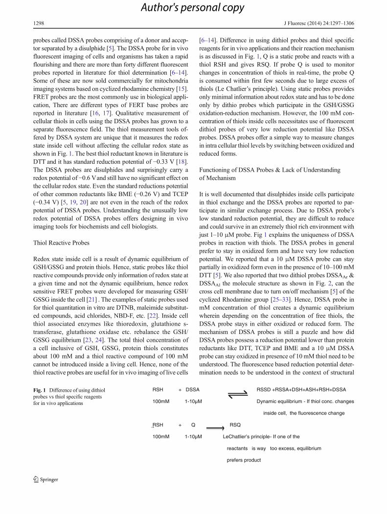
[6–14]. Difference in using dithiol probes and thiol specificreagents for in vivo applications and their reaction mechanismis as discussed in Fig. 1, Q is a static probe and reacts with athiol RSH and gives RSQ. If probe Q is used to monitorchanges in concentration of thiols in real-time, the probe Qis consumed within first few seconds due to large excess ofthiols (Le Chatlier’s principle). Using static probes providesonly minimal information about redox state and has to be doneonly by dithio probes which participate in the GSH/GSSGoxidation-reduction mechanism. However, the 100 mM con-centration of thiols inside cells necessitates use of fluorescentdithiol probes of very low reduction potential like DSSAprobes. DSSA probes offer a simple way to measure changesin intra cellular thiol levels by switching between oxidized andreduced forms.
Functioning of DSSA Probes & Lack of Understandingof Mechanism
It is well documented that disulphides inside cells participatein thiol exchange and the DSSA probes are reported to par-ticipate in similar exchange process. Due to DSSA probe’slow standard reduction potential, they are difficult to reduceand could survive in an extremely thiol rich environment withjust 1–10 μM probe. Fig 1 explains the uniqueness of DSSAprobes in reaction with thiols. The DSSA probes in generalprefer to stay in oxidized form and have very low reductionpotential. We reported that a 10 μM DSSA probe can staypartially in oxidized form even in the presence of 10–100 mMDTT [5]. We also reported that two dithiol probes DSSAAr &DSSAAl the molecule structure as shown in Fig. 2, can thecross cell membrane due to turn on/off mechanism [5] of thecyclized Rhodamine group [25–33]. Hence, DSSA probe inmM concentration of thiol creates a dynamic equilibriumwherein depending on the concentration of free thiols, theDSSA probe stays in either oxidized or reduced form. Themechanism of DSSA probes is still a puzzle and how didDSSA probes possess a reduction potential lower than proteinreductants like DTT, TCEP and BME and a 10 μM DSSAprobe can stay oxidized in presence of 10 mM thiol need to beunderstood. The fluorescence based reduction potential deter-mination needs to be understood in the context of structural
RSH + DSSA RSSD +RSSA+DSH+ASH+RSH+DSSA
100mM 1- Dynamic equilibrium - If thiol conc. changes
inside cell, the fluorescence change
RSH + Q RSQ
100mM 1-10µM LeChatlier’s principle- If one of the
reactants is way too excess, equilibrium
prefers product
Fig. 1 Difference of using dithiolprobes vs thiol specific reagentsfor in vivo applications
1298 J Fluoresc (2014) 24:1297–1306
Author's personal copy
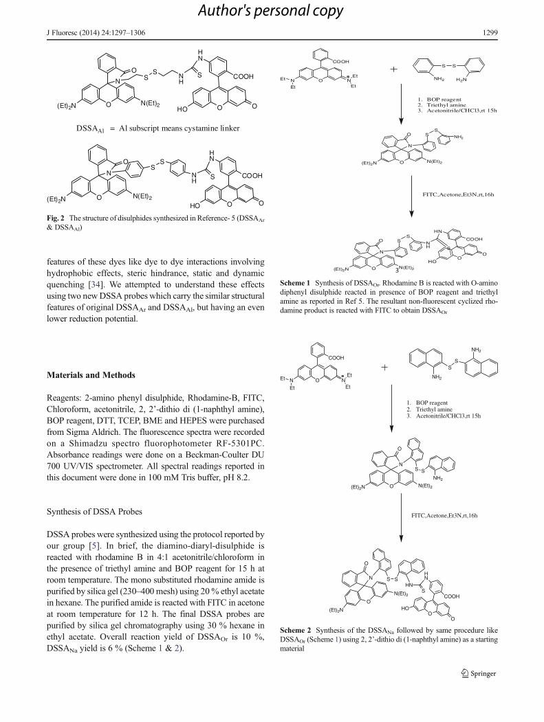
features of these dyes like dye to dye interactions involvinghydrophobic effects, steric hindrance, static and dynamicquenching [34]. We attempted to understand these effectsusing two newDSSA probes which carry the similar structuralfeatures of original DSSAAr and DSSAAl, but having an evenlower reduction potential.
Materials and Methods
Reagents: 2-amino phenyl disulphide, Rhodamine-B, FITC,Chloroform, acetonitrile, 2, 2’-dithio di (1-naphthyl amine),BOP reagent, DTT, TCEP, BME and HEPES were purchasedfrom Sigma Aldrich. The fluorescence spectra were recordedon a Shimadzu spectro fluorophotometer RF-5301PC.Absorbance readings were done on a Beckman-Coulter DU700 UV/VIS spectrometer. All spectral readings reported inthis document were done in 100 mM Tris buffer, pH 8.2.
Synthesis of DSSA Probes
DSSA probes were synthesized using the protocol reported byour group [5]. In brief, the diamino-diaryl-disulphide isreacted with rhodamine B in 4:1 acetonitrile/chloroform inthe presence of triethyl amine and BOP reagent for 15 h atroom temperature. The mono substituted rhodamine amide ispurified by silica gel (230–400 mesh) using 20% ethyl acetatein hexane. The purified amide is reacted with FITC in acetoneat room temperature for 12 h. The final DSSA probes arepurified by silica gel chromatography using 30 % hexane inethyl acetate. Overall reaction yield of DSSAOr is 10 %,DSSANa yield is 6 % (Scheme 1 & 2).
O
O
N SS
NH
S
HN
O
COOH
HO ON(Et)2(Et)2N
DSSAAl = Al subscript means cystamine linker
O
O
NS
S
NH
S
HN
O
COOH
HO ON(Et)2(Et)2N
Fig. 2 The structure of disulphides synthesized in Reference- 5 (DSSAAr
& DSSAAl)
3
O NN
Et
EtEt
Et
COOH
1. BOP reagent2. Triethyl amine3. Acetonitrile/CHCl3,rt 15h
FITC,Acetone,Et3N,rt,16h
O
O
N
SS
NH S
HN
O
COOH
HO
O
N(Et)2(Et)2N
SS
NH2 H2N
O
O
N
SS
NH2
N(Et)2(Et)2N
Scheme 1 Synthesis of DSSAOr. Rhodamine B is reacted with O-aminodiphenyl disulphide reacted in presence of BOP reagent and triethylamine as reported in Ref 5. The resultant non-fluorescent cyclized rho-damine product is reacted with FITC to obtain DSSAOr
O
O
N S SHN
S
HN
O
COOH
HO
O
N(Et)2
(Et)2N
O NN
Et
EtEt
Et
COOH
1. BOP reagent2. Triethyl amine3. Acetonitrile/CHCl3,rt 15h
FITC,Acetone,Et3N,rt,16h
O
O
NS S
NH2
N(Et)2(Et)2N
NH2
SS
NH2
Scheme 2 Synthesis of the DSSANa followed by same procedure likeDSSAOr (Scheme 1) using 2, 2’-dithio di (1-naphthyl amine) as a startingmaterial
J Fluoresc (2014) 24:1297–1306 1299
Author's personal copy
Purification of Probes for Characterization and FluorescenceStudies
DSSA dyes are purified by repeated preparative TLC to obtainnecessary purity for CHN analysis. The same product is usedfor absorbance and fluorescence studies.
DSSANa: Found (expected): C 71.37 (71.30), H 4.78(4.77), N 6.05 (6.02), Mass: 1162.3292 (1162.3993)
1H NMR (acetone-d6): 9.4 (s, 1H), 9.2 (s, 1H), 9.1 (s, 1H),8.2 (1H), 7.9 (2H), 7.6 (2H), 7.5 (6H), 7.4 (2H), 7.0 (2H), 6.8(2H), 6.6 (2H), 6.5 (8H), 6.4 (2H), 6.2 (2H), 3.3 (8H), 1.3(12H).
13C NMR (acetone-d6): 207, 186, 166, 165, 161, 156, 154(two peaks), 146, 140, 135, 134 (four peaks), 132, 130, 129,127 (six peaks), 125, 124 (four peaks), 116, 112, 110, 109,107, 105, 101, 98, 47, 13.
DSSAOr: Found (expected): C 68.99 (68.97), H 4.87(4.84), N 6.62 (6.59), Mass: 1062.2985 (1062.2819) 1HNMR (acetone-d6): 9.5 (s, 1H), 9.3 (s, 1H), 9.1 (s, 1H), 8.2(1H), 7.6 (2H), 7.7 (6H), 7.4 (2H), 7.1 (1H), 7.0 (3H), 6.5(8H), 6.4 (2H) 6.2 (2H), 3.4 (8H), 1.2 (12H).
13C NMR (acetone-d6): 203, 182, 167, 165, 158, 154, 153(two peaks), 147, 140, 137, 134, 133 (three peaks), 131, 130,128 (six peaks), 124 (three peaks), 117, 113, 110, 109, 106,104, 100, 98, 44, 12.
Results
Absorbance and Fluorescence of DSSA Probes
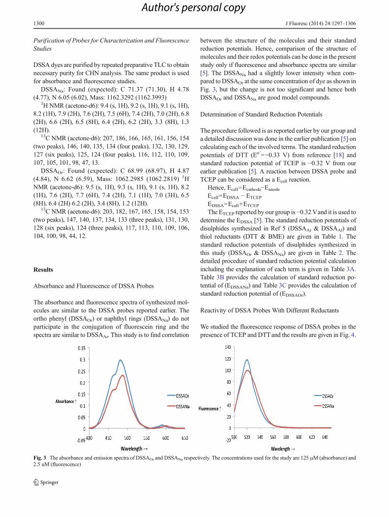
The absorbance and fluorescence spectra of synthesized mol-ecules are similar to the DSSA probes reported earlier. Theortho phenyl (DSSAOr) or naphthyl rings (DSSANa) do notparticipate in the conjugation of fluorescein ring and thespectra are similar to DSSAAr. This study is to find correlation
between the structure of the molecules and their standardreduction potentials. Hence, comparison of the structure ofmolecules and their redox potentials can be done in the presentstudy only if fluorescence and absorbance spectra are similar[5]. The DSSANa had a slightly lower intensity when com-pared to DSSAOr at the same concentration of dye as shown inFig. 3, but the change is not too significant and hence bothDSSAOr and DSSANa are good model compounds.
Determination of Standard Reduction Potentials
The procedure followed is as reported earlier by our group anda detailed discussion was done in the earlier publication [5] oncalculating each of the involved terms. The standard reductionpotentials of DTT (Eo’=−0.33 V) from reference [18] andstandard reduction potential of TCEP is −0.32 V from ourearlier publication [5]. A reaction between DSSA probe andTCEP can be considered as a Ecell reaction.
Hence, Ecell=Ecathode−Eanode
Ecell=EDSSA − ETCEPEDSSA=Ecell+ETCEPThe ETCEP reported by our group is −0.32Vand it is used to
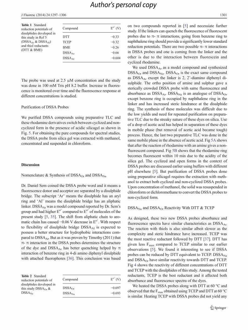
determine the EDSSA [5]. The standard reduction potentials ofdisulphides synthesized in Ref 5 (DSSAAr & DSSAAl) andthiol reductants (DTT & BME) are given in Table 1. Thestandard reduction potentials of disulphides synthesized inthis study (DSSAOr & DSSANa) are given in Table 2. Thedetailed procedure of standard reduction potential calculationincluding the explanation of each term is given in Table 3A.Table 3B provides the calculation of standard reduction po-tential of (EDSSANa) and Table 3C provides the calculation ofstandard reduction potential of (EDSSAOr).
Reactivity of DSSA Probes With Different Reductants
We studied the fluorescence response of DSSA probes in thepresence of TCEP and DTTand the results are given in Fig. 4.
Fig. 3 The absorbance and emission spectra of DSSAOr and DSSANa respectively. The concentrations used for the study are 125 μM (absorbance) and2.5 uM (fluorescence)
1300 J Fluoresc (2014) 24:1297–1306
Author's personal copy
The probe was used at 2.5 μM concentration and the studywas done in 100 mM Tris pH 8.2 buffer. Increase in fluores-cence is monitored over time and the fluorescence response atdifferent concentrations is studied.
Purification of DSSA Probes
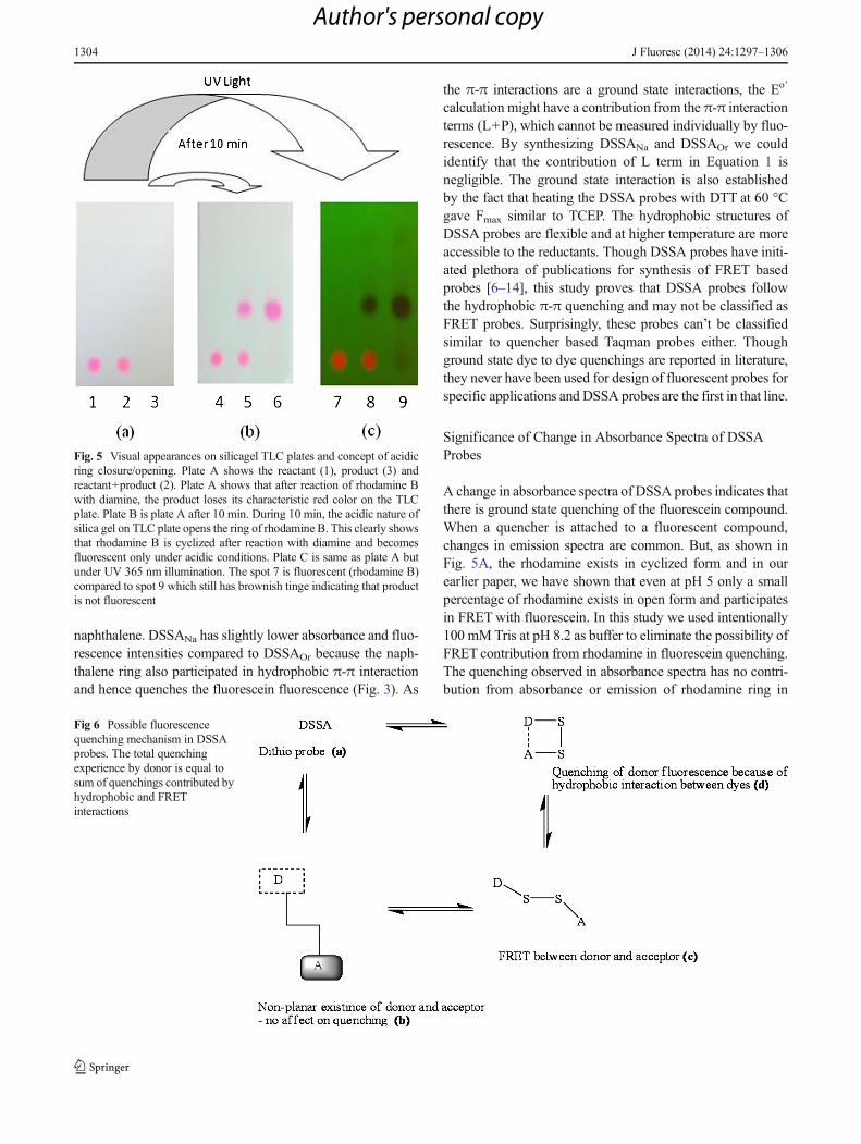
We purified DSSA compounds using preparative TLC andthese rhodamine derivatives switch between cyclized and non-cyclized form in the presence of acidic silicagel as shown inFig. 5. For obtaining the pure compounds for spectral studies,the DSSA probe from silica gel was extracted with methanol,concentrated and suspended in chloroform.
Discussion
Nomenclature & Synthesis of DSSAOr and DSSANa
Dr. Daniel Sem coined the DSSA probe word and it means afluorescence donor and acceptor are separated by a disulphidebridge. The subscript ‘Ar’ means the disulphide has an arylring and ‘Al’ means the disulphide bridge has an aliphaticlinker. DSSAAr was a model compound reported by Dr. Sem’sgroup and had higher Eo’ compared to Eo’ of molecules of thepresent study [5, 35]. The shift from aliphatic chain to aro-matic chain has caused −0.06 V decrease in Eo’. With respectto flexibility of disulphide bridge DSSAAl is expected topossess a better structure for hydrophobic interactions com-pared to DSSAAr. But as it was proven by Timothy (2011) thatπ- π interaction in the DSSA probes determines the structureof the dye and DSSAAr has better quenching helped by πinteraction of benzene ring in 4-di amino diphenyl disulphidewith attached fluorophores [36]. This conclusion was based
on two compounds reported in [5] and necessiate furtherstudy. If the linkers can quench the fluorescence of fluorescentprobes due to π- π interactions, going from benzene ring tonaphthalene ring should provide a significantly lower standardreduction potentials. There are two possible π- π interactionsin DSSA probes and one is coming from the linker and theother is due to the interaction between fluorescein andcyclized rhodamine.
We used DSSAAr as a model compound and synthesizedDSSAOr and DSSANa. DSSAOr is the exact same compoundas DSSAAr except the linker is 2, 2’-diamino diphenyl di-sulphide. The ortho position of amine and sulphur gave asterically crowded DSSA probe with same fluorescence andabsorbance as DSSAAr. DSSANa is an analogue of DSSAOr
except benzene ring is occupied by naphthalene ring in thelinker and has increased steric hindrance at the disulphidering. The synthesis of these molecules was difficult due tothe low yields and need for repeated purification on prepara-tive TLC due to the streaky nature of these dyes on silica. Useof a drop of acetic acid has helped in separation of these dyesin mobile phase (but removal of acetic acid became tough)process. Hence, the last two preparative TLC was done in thesamemobile phase in the absence of acetic acid. Fig 5A showsthat after the reaction of rhodamine with an amine gives a non-fluorescent compound. Fig 5B shows that the rhodamine ringbecomes fluorescent within 10 min due to the acidity of thesilica gel. The cyclized and open forms in the context ofDSSA probes are discussed earlier using buffers with differentpH elsewhere [5]. But purification of DSSA probes doneusing preparative silicagel requires the extraction with meth-anol to extract both cyclized and non-cyclized DSSA probes.Upon concentration of methanol, the solid was resuspended inchloroform or dichloromethane to convert the DSSA probes tonon-cyclized form.
DSSANa and DSSAOr Reactivity With DTT & TCEP
As designed, these two new DSSA probes absorbance andfluorescence spectra have similar characteristics as DSSAAr.
The reaction with thiols is also similar albeit slower as thecomplexity and steric hindrance have increased. TCEP wasthe most reactive reductant followed by DTT [37]. DTT hasgiven less Fmax compared to TCEP similar to our earlierobservations [5]. We found it interesting to see if DSSAprobes can be reduced by DTT equivalent to TCEP. DSSANa
and DSSAOr have similar reactivity towards DTT and TCEP.Fig 4 shows the reactivity of different concentrations of DTTand TCEPwith the disulphides of this study. Among the testedreductants, TCEP is the best reductant and it affected bothabsorbance and fluorescence spectra of the dyes.
We heated the DSSA probes along with DTT at 60 °C andobserved that the Fmax obtained using TCEP and DTTat 60 °Cis similar. Heating TCEP with DSSA probes did not yield any
Table 1 Standardreduction potentials ofdisulphides developed inthis study in Ref 5(DSSAAr & DSSAAl)and thiol reductants(DTT & BME)
Compound Eo’ (V)
DTT −0.33TCEP −0.32BME −0.26DSSAAr −0.66DSSAAl −0.604
Table 2 Standardreduction potentials ofdisulphides developed inthis study DSSAOr &DSSANa
Compound Eo’ (V)
DSSAOr −0.697DSSANa −0.693
J Fluoresc (2014) 24:1297–1306 1301
Author's personal copy
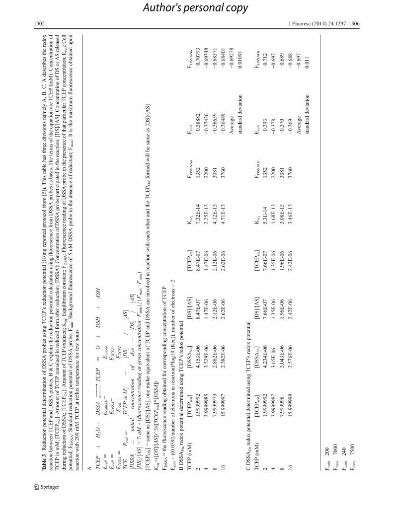
Table3
Reductio
npotentialdeterm
inationof
DSS
Aprobes
usingTCEP’sreductionpotential(U
sing
reported
protocol
from
[5]).T
histablehasthreedivisionsnamelyA,B
,C.A
describestheredox
reactio
nbetweenTCEPandDSSAprobes.B
&Cexplainthereductionpotentialcalculatio
nusingfluorescence
from
DSS
Aprobes
asbasis.The
term
sof
theequatio
nareTCEP(m
M):Concentratio
nof
TCEPinmM;[TCEPred]:Amountof
TCEPremainedinreducedform
afterreduction;[D
SSA]:Concentratio
nof
DSS
Aprobeparticipated
inthereactio
n;[D
S]/[AS]:C
oncentratio
nof
DSor
ASreleased
during
reductionof
DSSA
;[TCEP O
x]:Amountof
TCEPoxidized;K
eq:E
quilibriumconstant;F
DSSA:F
luorescencereadingof
DSS
Aprobeinthepresence
ofthatparticularTCEPconcentration;Ecell:C
ell
potential;EDSSA:Standardreductionpotentialof
DSS
Aprobe.F m
in:Backgroundfluorescence
of5uM
DSS
Aprobein
theabsenceof
reductant;F m
ax:Itisthemaxim
umfluorescence
obtained
upon
reactio
nwith
200mM
TCEPatreflux
temperature
forfewhours
A TCEP
þH
2Oþ
DSSA
���→
←���
TCEP
¼O
þDSH
þASH
Ecell¼
Ecathode−
Eanode
Ecell¼
EDSSA−
ETC
EP
EDSSA¼
Ecellþ
ETC
EP
TCE
Pred¼
TCEPin
M½
�−
DS
½�
=AS½�
DSSA
½�
¼initial
concentration
ofdye
–DS
½�
=AS½�
DS
½�=
AS½�¼
5uM
�flu
orescencereadingat
givenconcentration−
Fmin
ðÞ=
Fmax−Fmin
ðÞ
[TCEPOX]=sameas
[DS]/[AS],one
molar
equivalent
ofTCEPandDSS
Aareinvolved
inreactio
nwith
each
otherandtheTCEP O
Xform
edwill
besameas
[DS]/[A
S]
Keq=([DS]/[AS])^3/([TCEPred]*[D
SSA])
FDSSA=thefluorescence
readingobtained
forcorrespondingconcentrationof
TCEP
Ecell=((0.0592/num
berof
electronsin
reactio
n)*log10
(Keq)),num
berof
electrons=2
BDSSANaredoxpotentiald
etermined
usingTCEP’sredoxpotential
TCEP(m
M)
[TCEPred]
[DSSANa]
[DS]/[A
S][TCEPox]
Keq
FDSSANa
Ecell
EDSSANa
21.9999992
4.153E
-06
8.47E-07
8.47E-07
7.32E-14
1352
−0.38882
−0.70793
43.9999985
3.529E
-06
1.47E-06
1.47E-06
2.25E-13
2200
−0.37436
−0.69348
87.9999979
2.882E
-06
2.12E-06
2.12E-06
4.12E-13
3081
−0.36659
−0.68571
1615.999997
2.382E
-06
2.62E-06
2.62E-06
4.71E-13
3760
−0.36489
−0.68401
Average
−0.69278
standard
deviation
0.01091
CDSSAOrredoxpotentiald
etermined
usingTCEP’sredoxpotential
TCEP(m
M)
[TCEPred]
[DSSAOr]
[DS]/[A
S][TCEPox]
Keq
FDSSAOr
Ecell
EDSSAOr
21.9999992
4.234E
-06
7.66E-07
7.66E-07
5.3E
-14
1352
−0.393
−0.712
43.9999987
3.65E-06
1.35E-06
1.35E-06
1.68E-13
2200
−0.378
−0.697
87.999998
3.043E
-06
1.96E-06
1.96E-06
3.08E-13
3081
−0.370
−0.689
1615.999998
2.576E
-06
2.42E-06
2.42E-06
3.46E-13
3760
−0.369
−0.688
Average
−0.697
standard
deviation
0.011
Fmin
200
Fmax
7000
Fmin
240
Fmax
7500
1302 J Fluoresc (2014) 24:1297–1306
Author's personal copy
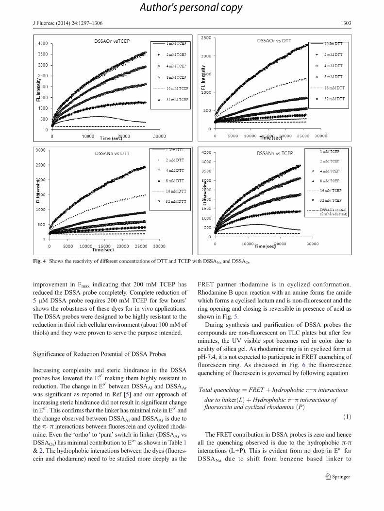
improvement in Fmax indicating that 200 mM TCEP hasreduced the DSSA probe completely. Complete reduction of5 μM DSSA probe requires 200 mM TCEP for few hours’shows the robustness of these dyes for in vivo applications.The DSSA probes were designed to be highly resistant to thereduction in thiol rich cellular environment (about 100 mM ofthiols) and they were proven to serve the purpose intended.
Significance of Reduction Potential of DSSA Probes
Increasing complexity and steric hindrance in the DSSAprobes has lowered the Eo’ making them highly resistant toreduction. The change in Eo’ between DSSAAl and DSSAAr
was significant as reported in Ref [5] and our approach ofincreasing steric hindrance did not result in significant changein Eo’. This confirms that the linker has minimal role in Eo’ andthe change observed between DSSAAl and DSSAAr is due tothe π- π interactions between fluorescein and cyclized rhoda-mine. Even the ‘ortho’ to ‘para’ switch in linker (DSSAAr vsDSSAOr) has minimal contribution to Eo’ as shown in Table 1& 2. The hydrophobic interactions between the dyes (fluores-cein and rhodamine) need to be studied more deeply as the
FRET partner rhodamine is in cyclized conformation.Rhodamine B upon reaction with an amine forms the amidewhich forms a cyclised lactum and is non-fluorescent and thering opening and closing is reversible in presence of acid asshown in Fig. 5.
During synthesis and purification of DSSA probes thecompounds are non-fluorescent on TLC plates but after fewminutes, the UV visible spot becomes red in color due toacidity of silica gel. As rhodamine ring is in cyclized form atpH-7.4, it is not expected to participate in FRET quenching offluorescein ring. As discussed in Fig. 6 the fluorescencequenching of fluorescein is governed by following equation
Total quenching ¼ FRET þ hydrophobic π−π interactions
due to linker Lð Þ þ Hydrophobic π−π interactions offluorescein and cyclized rhodamine Pð Þ
ð1Þ
The FRET contribution in DSSA probes is zero and henceall the quenching observed is due to the hydrophobic π-πinteractions (L+P). This is evident from no drop in Eo’ forDSSANa due to shift from benzene based linker to
Fig. 4 Shows the reactivity of different concentrations of DTT and TCEP with DSSANa and DSSAOr
J Fluoresc (2014) 24:1297–1306 1303
Author's personal copy
naphthalene. DSSANa has slightly lower absorbance and fluo-rescence intensities compared to DSSAOr because the naph-thalene ring also participated in hydrophobic π-π interactionand hence quenches the fluorescein fluorescence (Fig. 3). As
the π-π interactions are a ground state interactions, the Eo’
calculation might have a contribution from the π-π interactionterms (L+P), which cannot be measured individually by fluo-rescence. By synthesizing DSSANa and DSSAOr we couldidentify that the contribution of L term in Equation 1 isnegligible. The ground state interaction is also establishedby the fact that heating the DSSA probes with DTT at 60 °Cgave Fmax similar to TCEP. The hydrophobic structures ofDSSA probes are flexible and at higher temperature are moreaccessible to the reductants. Though DSSA probes have initi-ated plethora of publications for synthesis of FRET basedprobes [6–14], this study proves that DSSA probes followthe hydrophobic π-π quenching and may not be classified asFRET probes. Surprisingly, these probes can’t be classifiedsimilar to quencher based Taqman probes either. Thoughground state dye to dye quenchings are reported in literature,they never have been used for design of fluorescent probes forspecific applications and DSSA probes are the first in that line.
Significance of Change in Absorbance Spectra of DSSAProbes
A change in absorbance spectra of DSSA probes indicates thatthere is ground state quenching of the fluorescein compound.When a quencher is attached to a fluorescent compound,changes in emission spectra are common. But, as shown inFig. 5A, the rhodamine exists in cyclized form and in ourearlier paper, we have shown that even at pH 5 only a smallpercentage of rhodamine exists in open form and participatesin FRETwith fluorescein. In this study we used intentionally100 mM Tris at pH 8.2 as buffer to eliminate the possibility ofFRET contribution from rhodamine in fluorescein quenching.The quenching observed in absorbance spectra has no contri-bution from absorbance or emission of rhodamine ring in
Fig. 5 Visual appearances on silicagel TLC plates and concept of acidicring closure/opening. Plate A shows the reactant (1), product (3) andreactant+product (2). Plate A shows that after reaction of rhodamine Bwith diamine, the product loses its characteristic red color on the TLCplate. Plate B is plate A after 10 min. During 10 min, the acidic nature ofsilica gel on TLC plate opens the ring of rhodamine B. This clearly showsthat rhodamine B is cyclized after reaction with diamine and becomesfluorescent only under acidic conditions. Plate C is same as plate A butunder UV 365 nm illumination. The spot 7 is fluorescent (rhodamine B)compared to spot 9 which still has brownish tinge indicating that productis not fluorescent
Fig 6 Possible fluorescencequenching mechanism in DSSAprobes. The total quenchingexperience by donor is equal tosum of quenchings contributed byhydrophobic and FRETinteractions
1304 J Fluoresc (2014) 24:1297–1306
Author's personal copy

DSSA derivatives. Our current understanding of fluorescencechemistry suggests that the spectral overlap is a must forquenching and the quenching offered by hydrophobic inter-actions are very minimal. This perception probably requiresrelook due to the reported results from this paper and reference[5]. The hydrophobic interaction is a general term for non-spectral interactions between two molecules and it will bebased on chemical or structural interactions. The exact orien-tation of the fluorescein and cyclized rhodamine groups inDSSA probes can be understood only by crystallographicstudies and our efforts as of now in obtaining single crystalof DSSA probes were unsuccessful.
Impact of This Study on Future Design of FluorescentCompounds Based on π-π Interactions
Though FRETand static quenching are known for a long time,design of fluorescent probes specially utilizing staticquenching is rare. This is partly due to the requirement ofratiometric determination of two fluorophores to quantitate theanalyte in vivo. But the FRET probe efficiency is related toquite few parameters like overlap integral of donor fluores-cence and acceptor absorbance J (λ). In Fig. 7 we show twohypothetical fluorophores with spectral overlap integral J (λ)=0 (example Rhodamine B& an Infrared dye). By conventionalwisdom of FRET these two dyes can never be used as a FRETpair, but the results presented in this paper suggest a relook atpossibility of using fluorophores with no spectral overlap asFRET pairs. The DSSA probe has only one fluorophore and
the cyclized rhodamine is a non-fluorescent compound, sug-gesting that even a non-fluorescent compound can quenchfluorescence. Though Taqman probes use similar quencherbased systems, the quenchers share a spectral overlap withfluorescent compound (eg: FAM and BHQ-1) [38]. TheDSSA probes have a new mechanism for quenching withinteractions purely hydrophobic in nature. Foster radius be-tween donor and acceptor, quantum efficiency of donor, ori-entation of donor and acceptor etc. often make the choice offluorescent molecules difficult [39].
Most of the parameters discussed above are dependent onthe biomolecule used, or dependent too much on the inherentproperties of donors and acceptors and less to do with the skillof the researcher. But this study proves that static quenchingcan help design similar FRET probes with virtually no spectraloverlap [40]. It is because the quenching is caused by π-πinteraction and hence a near IR probe having absorbancemaximum of 1,000 nm can still quench fluorescence of acompound with λem of 600 nm by hydrophobic interactionhaving J (λ)=0. This removes the current limitation on theselection of fluorescent probes for FRET applications. As thehydrophobic interactions are known to work over an area of1,000 nm, designing fluorescent application becomes simpleand offers more versatility for biochemist.
Conclusion
This study has given four important informations for fluores-cence chemistry
a) The synthesis of DSSAOr and DSSANa and their respec-tive Eo’ has suggested that the ultra-low reduction poten-tials observed in DSSA probes is independent of sterichindrance.
b) The standard reduction potential of DSSA probes is min-imally influenced by the functional groups indicating theultra-low Eo’ is mostly coming from the interaction be-tween fluorescein and cyclized rhodamine.
c) The rhodamine ring in DSSA probes is in cyclized formand do not possess any excited state interactions withfluorescein. Quenching in DSSA probes is based on hy-drophobic interactions and not FRET based.
d) This study suggests that FRET probe design need not bebased on J (λ), and the biochemist could select anyfluorescent probe if the donor-acceptor pair is based onhydrophobic π-π interactions, making the typical param-eters like Forster radius in fluorescence chemistry irrele-vant. This conclusion is based on a single group ofcompounds (DSSA) and needs further study to validatethis hypothesis.
Fig 7 Schematic representation of the two different fluorophores spectraloverlap integral J (λ)=0 (example Rhodamine B & an Infrared dye). Byconvention wisdom of FRET these two dyes can never be used a FRETpair, but the results presented in this paper suggests a relook at possibilityof using fluorophores with no spectral overlap as FRET pairs. The DSSAprobe has only one fluorescence compound (fluorescein) and the secondmolecule in structure is a non-fluorescent compound (cyclized rhoda-mine) suggesting that even a non-fluorescent compound can quenchfluoresce. Though Taqman probes use similar quencher based systems,the quenchers share a spectral overlap with fluorescent compound (FAMand BHQ-1 are example for it). The DSSA probes have a third mecha-nism for quenching with is purely hydrophobic in nature
J Fluoresc (2014) 24:1297–1306 1305
Author's personal copy
References
1. Schafer FQ, Buettner GR (2001) Redox environment of the cell asviewed through the redox state of the glutathione disulphide/glutathione couple. Free Radical Bio Med 30:1191–1212
2. Reed MC, Thomas RL, Pavisic J, James SJ, Ulrich CM, Nijhout HFet al (2008) Amathematical model of glutathionemetabolism. Theor.Biol Med Model 5:3–16. doi:10.1186/1742-4682-5-8
3. Embry MR, Belanger SE, Braunbeck TA, Burgos MG, Halder M,Hinton DE, Léonard MA, Lillicrap A, King TN, Whalej G et al(2010) The fish embryo toxicity test as an animal alternative methodin hazard and risk assessment and scientific research. Aquat Toxicol97:79–87. doi:10.1016/j.aquatox.2009.12.008
4. Kirsten H (2011) Limits of the fish embryo toxicity test with Daniorerio as an alternative to the acute fish toxicity test. Dissertation.Ruperto – Carola University of Heidelberg.
5. Pullela PK, Chiku T, CarvanMJ, SemDS (2006) Fluorescence-baseddetection of thiols in vitro and in vivo using dithiol probes. AnalBiochem 352:265–273
6. Yin LL, Chen ZZ, Tong LL, Xu KH, Tang B (2009) Progress onFluorescent Probes for Thiols. Chinese J Analyt Chem 37:1073–1081. doi:10.1016/S1872-2040(08)60117-6
7. Bouffard J, Kim Y, Swager TM, Weissleder R, Hilderbrand SA(2008) A Highly Selective Fluorescent Probe for Thiol Bioimaging.Org Lett 10:37–40. doi:10.1021/ol702539v
8. MaedaH,Matsuno H, UshidaM, KatayamaK, Saeki K, Itoh N (2005)2,4-Dinitrobenzenesulfonyl Fluoresceins as Fluorescent Alternatives toEllman’s Reagent in Thiol-Quantification Enzyme Assays. AngewChem Int Ed 44:2922–2925. doi:10.1002/anie.200500114
9. Yi L, Li H, Sun L, Liu L, Zhang C, Xi Z (2009) A Highly SensitiveFluorescence Probe for Fast Thiol-Quantification Assay ofGlutathione Reductase. Angew Chem Int Ed 48:4034–4037. doi:10.1002/anie.200805693
10. Ahn YH, Lee JS, ChangYT (2007) Combinatorial Rosamine Libraryand Application to in Vivo Glutathione Probe. J Am Chem Soc 129:4510–4511. doi:10.1021/ja068230m
11. Lim CS, Masanta G, Kim HJ, Han JH, Kim HM, Cho BR (2011)Ratiometric Detection of Mitochondrial Thiols with a Two-PhotonFluorescent Probe. J Am Chem Soc 133:11132–11135. doi:10.1021/ja205081s
12. Lee MH, Han JH, Lee JH, Choi HG, Kang C, Kim JS (2012)Mitochondrial Thioredoxin-Responding Off−On FluorescentProbe. J Am Chem Soc 134:17314–17319. doi:10.1021/ja308446y
13. Zhu B, Zhang X, Li Y, Wang P, Zhang H, Zhuang X (2010) Acolorimetric and ratiometric fluorescent probe for thiols and itsbioimaging applications. Chem Commun 46:5710–5712. doi:10.1039/c0cc00477d
14. ChenX, ZhouY, PengX, Yoon J (2010) Fluorescent and colorimetricprobes for detection of thiols. Chem Soc Rev 39:2120–2135. doi:10.1039/b925092a
15. Chazotte B (2011) Labeling Mitochondria with TMRM or TMRE.CSH Protoc 7:895–897. doi:10.1101/pdb.prot5641
16. Zhang S, Yan Y, Bi S (2009) Design of Molecular Beacons asSignaling Probes for Adenosine Triphosphate Detection in CancerCells Based on Chemiluminescence Resonance Energy Transfer.Anal Chem 81:8695–8701
17. Habeeb Muhammed M A, Shaw AK, Pal SK, Pradeep T (2008).Quantum Clusters of Gold Exhibiting FRET, J. Phys. Chem. C,112(37), 14324–14330
18. ClelandWW (1964) Dithiothreitol, a New Protective Reagent for SHGroups. Biochemistry 3:480–482
19. Mickey B, Howard J (1995) Rigidity of Microtubules Is Increased byStabilizing Agents. J Cell Biol 130:909–917
20. Aitken CE, Marshall RA, Puglisi JD (2008) An Oxygen ScavengingSystem for Improvement of Dye Stability in Single-Molecule
Fluorescence Experiments. Biophys J 94:1826–1835. doi:10.1529/biophysj.107.117689
21. Kolossov VL, Spring BQ, Clegg RM, Henry JJ, Sokolowski A,Kenis PJA, Gaskins HR (2011) Development of a high-dynamicrange, GFP-based FRET probe sensitive to oxidative microenviron-ments. Exp Biol Med (Maywood) 236:681–691
22. Peng H, Chen W, Cheng Y, Hakuna L, Strongin R, Wang B (2012)Thiol Reactive Probes and Chemosensors. Sensors 12:15907–15946.doi:10.3390/s121115907
23. Akif M, Khare G, Tyagi AK, Mande SC, Sardesai AA et al (2008)Functional Studies of Multiple Thioredoxins from Mycobacteriumtuberculosis. J Bacteriol 190:7087–7095. doi:10.1128/JB.00159-08
24. Herzog CA (2011) Glutathione and non-glutathione-based oxidantcontrol in the endoplasmic reticulum. J Cell Sci 124:847–855. doi:10.1242/jcs.080895
25. Tang B, Xing Y, Li P, Zhang N, Yu F, Yang G (2007) A Rhodamine-Based Fluorescent Probe Containing a Se-N Bond for DetectingThiols and Its Application in Living Cells. J Am Chem Soc 129:11666–11667. doi:10.1021/ja072572q
26. Abe H, Ito Y, Shibata A, Ito M (2012) Thiol detection method US8187825 B2
27. Shibata A, Furukawa K, AbeH, Tsuneda S, Ito Y (2008) Rhodamine-based fluorogenic probe for imaging biological thiol. Bioorg MedChem Lett 18:2246–2249. doi:10.1016/j.bmcl.2008.03.014
28. Li H, Fan J, Wang J, Tian M, Du J, Sun S, Sun P, Peng X (2009) Afluorescent chemodosimeter specific for cysteine: effective discrim-ination of cysteine from homocysteine. Chem Commun 39:5904–5906. doi:10.1039/b907511a
29. Cai HH, Wang H,Wang J, Wei W, Yang PH, Cai J (2011) Naked eyedetection of glutathione in living cells using rhodamine B-functionalized gold nanoparticles coupled with FRET. DyesPigments 92:778–782. doi:10.1016/j.dyepig.2011.06.016
30. Jun ME, Roy B, Ahn KH (2011) Turn-on fluorescent sensing withreactive probes. Chem Commun 47:7583–7601. doi:10.1039/C1CC00014D
31. Long L, Lin W, Chen B, Gao W, Yuan L (2011) Construction of aFRET-based ratiometric fluorescent thiol probe. Chem Commun 47:893–895. doi:10.1039/c0cc03806g
32. Monostori P, Wittmann G, Karg E, Turi S (2009) Determination ofglutathione and glutathione disulfide in biological samples: An in-depth review. J Chromatogr B 877:3331–3346. doi:10.1016/j.jchromb.2009.06.016
33. Duan YL, Shi YG, Chen JH, Wu XH, Wang GK, Zhou Y, Zhang JF(2012) 1,8-Naphthyridine modified rhodamine B derivative andCu2+ complex: colorimetric sensing of thiols in aqueous media.Tetrahedron Lett 53:6544–6547
34. Lakowicz JR (2006) Handbook of principle of fluorescence spectros-copy book. University of Maryland School of. Medicine, USA
35. Sem DS (2009) Method for detecting kinase activity with thiolreactive fluorescent reagents-US 7585643, B2
36. Timothy J (2011) Fluorescence Probes for Cellular Thiol andDisulfide Detection: Sythesis and Biophysical Characterization.Marquette University, Dissertation
37. Getz EB, Xiao M, Chakrabarty T, Cooke R, Selvin PR et al (1999) AComparison between the Sulfhydryl Reductants Tris (2carboxyethyl) phosphine and Dithiothreitol for Use in ProteinBiochemistry. Anal Biochem 273:73–80
38. Johansson MK, Fidder H, Dick D, Cook RM (2002) Intramoleculardimers: a new strategy to fluorescence quenching in dual-labeledoligonucleotide probes. J Am Chem Soc 124:6950–6956. doi:10.1021/ja025678o
39. Marras SAE (2006) Handbook of Fluorescent Energy Transfer NucleicAcid Probes: Designs and Protocols. Methods Mol Biol 335:3–16
40. Johansson MK (2006) Handbook of Fluorescent Energy TransferNucleic Acid Probes: Designs and Protocols. Methods Mol Biol335:17–29
1306 J Fluoresc (2014) 24:1297–1306
Author's personal copy