Embed Size (px)
Citation preview

A U G U S T 2 0 0 8
Multidisciplinary Care
Provisional Restorations
�e Team ApproachJournalo f t h e c a l i f o r n i a d e n ta l a s s o c i at i o n
d e p a r t m e n t s The Associate Editor/Cost of Putting Patients First: $0; Professional Credibility: Priceless
Letters to the Editor
Impressions
Dr. Bob/Gold Fever
features TreaTmenT Planning: an arT or a Sci e nce ?
An introduction to the issue.
Sajid Jivraj, DDS, MSEd
The influence of PoSTerior occl u Si on W he n r e STor i ng a nTe r i or Te e Th
A prosthodontic perspective in management of patients requiring restoration of posterior support is given in this paper as well as how to transition patients from a tooth-supported to an implant-supported occlusion.
Mamaly Reshad, DDS, MSc, and Sajid Jivraj, DDS, MSEd
mulTidiSciPlinary care: Peri odonTa l aSPe cTS To Tr e aTme nT Pl a nni ng The a nTe r i or eSTheTic Zone
This article discusses the role of periodontal plastic and reconstructive surgery in treatment planning the anterior esthetic zone in interdisciplinary dental care.
Nicolas A. Ravon, DDS, MSD; Mark Handelsman, DDS; and David Levine, DDS
inTegraTing orThodonTicS f or The oPTi ma l Smi l e
The advantage of an interdisciplinary treatment approach and how knowledge of basic orthodontic techniques can help in producing a more comprehensive treatment plan are highlighted.
Frank K. Yorita, DDS
TranSiTioning PaTienTS from Te e Th To i m Pl a nTS u Ti l i Zi ng f i xe d r e STor aTi onS
Options other than removable prostheses in transitioning the patient from teeth to implant-supported restorations are described in this paper.
Sajid Jivraj, DDS, MSEd; Mamaly Reshad, DDS, MSc; and Winston W.L. Chee, DDS
The Team aPProach: SimPlify i ng comPl e x ca r e
The interaction between the oral and maxillofacial surgeon, the restorative dentist, and the rest of the dental and medical community is the foundation of daily patient care and management. The combination of the talents from each medical and dental discipline results in the highest quality of patient care. This article illustrates the power of the comprehensive team approach.
David Hochwald, DDS; Simona Arcan, DMD, MD; and Fariborz Farnad, DDS
537539541637
563
567
575
587
599
609
CDA JournalVolume 36, Number 8a u g u s t 2 0 0 8 Journal
c d a j o u r n a l , v o l 3 6 , n º 8
a u g u s t 2 0 0 8 537
Assoc. Editor
Cost of Putting Patients first: $0; Professional Credibility: Pricelesssteven a. gold, dds
ast week I was asked by two dif-ferent patients if I heard on the morning news that “… the FDA says that the mercury in silver fillings is bad for you.” Being
skeptical (without discrediting my pa-tients), I said I would “do a little research on it.”
Not by coincidence, I received an e-gram from the ADA exactly two days later explaining the nature of the settlement between the FDA and an antiamalgam group. Further details were provided on the ADA’s Web site. (For those unfamiliar with these developments, the information is currently located under the “Announce-ments” section of the home page.) Not only was I impressed by the timeliness of the ADA’s e-gram, but also by the breadth of informative statements available to the public on their Web site. These included statements about lead in dental prostheses and the safety of fluoride, two topics that continue to see exposure in the media.
What a valuable service this is that the ADA provides to its members; serving as a source of information to the public. Being a trusted source of information is one of the most important roles our local, state and, in this case, our national organization can fulfill. We as individual dentists could never amass the informa-tion the ADA can, nor could we make it available in a format so easy for the public to access and understand. Furthermore, legislators and other public policy mak-ers are bombarded by information from both individuals and organizations. They make decisions (hopefully more than occasionally) based on information they receive from trusted sources. Trustworthy organizations such as the ADA and CDA
are thus vital to the protection of both the public and the profession from poor public policy decisions.
The trust that is placed in a profes-sional organization, such as our dental tripartite, is both precious and delicate and it can be undermined by the very ac-tions of that profession. Most damaging are those messages that reach the public, which sound like either individual den-tists or the profession as a whole putting their well-being above that of the patient. The public rightfully holds us to this standard. They rightfully expect us to put them first. This should not be difficult to do. If we hearken back to the more idealis-tic days of our childhood, or young adult hood, or whatever point in our lives it was that we decided to be dentists, the notion of helping people was no doubt a big part of that decision for all of us.
Putting patients first does not cost anything, yet when we fail to do it, we lose some of the credibility we, as a profession have with them; a credibility which, as we see, is truly priceless. Therefore, we should critically evaluate those actions that can be perceived as being self-serving and ask ourselves how we can do better.
There is a myth that is often thrust upon us by some of the well-known dental “institutes,” consultants, members of the dental industry, and even our own
member dentists. This myth says that the quickest way to become financially suc-cessful in this profession is to help create a demand for elective, specifically cosmetic, services. Every time these messages reach the public, we lose some of that priceless credibility. Dentists are then implored to fill this demand, oftentimes by taking advantage of their patients’ trust and delivering gross overtreatment, including removing healthy tooth enamel to place porcelain veneers and other restorations when no other need for them exists.
Furthermore, this is often done on mul-tiple teeth with inappropriate and adverse changes to a patient’s occlusion without the patient being fully informed of the nature of such drastic treatment and its risks. Every time an improperly informed patient has an adverse treatment outcome after having his or her enamel stripped away and replaced with glass, we as a profession lose some of that priceless credibility.
Overly emphasized cosmetic treat-ment, and its demand, are often fueled by advertising that ignores long-standing principles reflective of a profession that places the care of patients first. Many of us have seen commercials and print ad-vertising more appropriate for selling big-screen TVs than for offering health care. Every day that the public is increasingly besieged by such advertisements hawking
l Being a trusted source of information is one of
the most important roles our local, state and, in
this case, our national organization can fulfill.
538 a u g u s t 2 0 0 8
c d a j o u r n a l , v o l 3 6 , n º 8
cosmetic dental care, we lose some of that priceless credibility. Dentists are also the targets of potentially credibility-damag-ing advertising.
There is a limitless stream of publica-tions containing articles and solicitations for services and “continuing education” courses aimed at making dentists as profitable as possible. The emphasis is so geared toward profitability that the mes-sage that genuine patient care is involved, if it is present at all, is lost. We would be foolish to think that these messages never reach the eyes and ears of the pub-lic, and when they do, we again lose some of that priceless credibility.
Messages that dentistry cares more about profit than patients can even be more subtle, but no less subversive to our credibility. Extreme caution must be used when dental organizations partner with
Address comments, letters, and questions to the editor at [email protected].
the for-profit industry side of the profes-sion; for such partnerships can become the source of these public perceptions. Pure altruism by a for-profit entity should not come with strings attached. Our organizations should not indulge them with commercial-like exposure in our scientific journals, on billboards, or other high visibility media. This can cause both questioning of the profession’s motives and dulling of the edge of science. Each time there is such fallout from unwise partnering with industry, we lose some of that priceless credibility.
These actions do not reflect a cul-ture of putting patients first. Whether intended or not, they carry the grave potential of simply damaging our credibil-ity with the public. The day an individual seeking dental information disregards the ADA as a credible source they will turn to
much less scientifically oriented sources. Furthermore, we will find ourselves at the mercy of politicians and government regulatory agencies who base oral health-related public policy decisions not on information provided by our profession, but on information provided by consumer watchdog groups, litigious attorneys, or other individuals and groups with far less scrupulous motives.
When we as a profession have lost our credibility, only then will we realize that we failed to make good on the immense responsibility of ensuring that the public image of dentistry is one of a noble, scientific-based profession comprised of caring individuals who always place the well-being of patients first.
Assoc. Editor
c d a j o u r n a l , v o l 3 6 , n º 8
a u g u s t 2 0 0 8 539
are extended Show, Venue Change Solutions to Crowded Spring Session?
never write letters to the editor. The Spring Scientific Session at Anaheim is becoming unbearable. The crowds!
Let me start by saying that I cannot even imagine the complexi-
ties of putting together a convention of this size. The financial commitment must be enormous too. The big problem is the crowds at this session were way too much for anybody to get anything productive done. It took me 45 minutes to drive the last three-quarters of a mile in front of the convention center. I finally said “It’s not worth it,” did a U-turn, and left. This was one hour before the convention hall even opened. I live in the next town over from Anaheim and am very familiar with the area and know how to get around, but it was just a mess.
As a dues-paying CDA member for the last 20 years, I think that this is a huge disservice to California dentists. Something must be done so that the dentists who support this organiza-tion can take advantage of this meeting. Perhaps the convention needs to run longer. Perhaps the convention needs to be offered twice in Southern Califor-nia. Perhaps a change of venue. Perhaps scheduling one day when only dentists or hygienists can get in. I love getting team members involved, but the dentists are the ones who are making the decisions in the practice; they are the ones who must be given access.
I never made it inside the hall, but I can imagine the scene inside. Look, we all want to see old friends and I understand these things are about making contacts etc., but I think most dentists simply
want to see what’s new, get some ques-tions answered about a couple of prod-ucts, and maybe take a class or two.
I’m not some old fuddy-duddy. I’m 45 years old. We all want to have some fun, but this convention only rolls around once a year and we need this chance to be able to have all these products and services under one roof, and be able to talk to the representatives.
I enjoy the dental convention. It’s a great time to get an update of what’s happening out there. Let’s start a dialogue about the best ways to handle crowd con-trol so this valuable meeting best serves its members.
er ic mey er , d d sFullerton, Calif.
you Can lead a horse to Water, but you Can’t make him Brush
I wanted to thank you for Dr. Gold’s editorial on parental responsibility for dental health in the May issue of the Journal of the California Dental Association (36:321-2).
As you say, we could question what parents value in their family lives — the $60K SUV, keeping up with the Joneses, etc., but the bottom line is that they will need to answer to the health of their chil-dren. Many times children are treated like a possession … my SUV needs mainte-nance, I take it to the mechanic. My child has cavities; I send them to the dentist to deal with it.
In my practice, I reinforce the concept that parents have a daily responsibility and accountability to their children to keep their teeth clean. I explain to them
that I only see their children twice a year; who will keep their teeth clean the other 363 days? When parents complain their children don’t like having their teeth brushed, we talk about the usual techniques mirroring the parent, having the parent brush after the child brushes, etc., but that’s all part of being a par-ent; brushing isn’t the only thing a child doesn’t like to do. How do you respond when your child acts that way? Merit re-ward? Flat out bribery? Whatever works?
I am not an uncaring soul; I have four children’s worth of experience. But at some point in our lives as parents, we must communicate a sense of responsibil-ity by the parent AND by the child.
I recently sat in on a focus market-ing group where there was a discussion about electric toothbrushes for kids. Most dentists’ opinions about children and toothbrushing seemed to be that WE as a profession need to find the magic wand that will solve all our children’s brush-ing problems; even the pedodontists in the group seemed intent on finding that instrument vs. creating accountability of the parent for the child’s dental health. A suggestion that the manufacturer create an interactive brushing program that involves the parent and the child got a lukewarm reception. If the parent cannot motivate the child at home, who can?
We as dentists can only do so much for our patients. I don’t think we need to feel bad if parents don’t recognize their accountability. Like the saying goes, you can only lead a horse to water …
j obie low, ddsSan Francisco
I
Letters

c d a j o u r n a l , v o l 3 6 , n º 8
a u g u s t 2 0 0 8 541
Impressions
continues on 545
Dan
Hub
ig
overfed but undernourishedby patty reyes
The United States has long been con-sidered the land of milk and honey. But judging by the expanding real estate of a typical citizen’s midriff of late, it’s also the home of milk chocolate candy the size of large bricks and honey-drenched cereals that only bees can appreciate.
Yes, that’s right, America is living large, but not in a good way.
In her presentation “Fattening of America — Where Does Dentistry Fit Into the Puzzle?” during the 2008 California Dental Association Spring Sci-entific Session, Lisa F. Harper Mallone, BSDH, MPH, RD/LD, associate profes-sor, Department of Dental Hygiene at Baylor College of Dentistry in Dallas, told the crowd about a 5-year-old boy who weighed 117 pounds and had a blood pressure reading of 148/86. (According to a chart by the NIH’s National Heart Lung and Blood Institute, a typical blood pres-sure reading for a 5-year-old, depending on height, is 95/53.) But with an estimat-ed 160 million U.S. citizens dining out daily, mostly at fast-food establishments, Mallone said, is it a surprise?
So, what’s eating America? Smoking
Journal Seeks new editorThe California Dental Association is looking for its next editor. The editor, who
must be a CDA member, has editorial control over the Journal of the California Dental
Association and serves as editorial adviser of the
Update, subject to policies of the association. The
editor also serves as an ex officio member of the
Executive Committee, House of Delegates, the
Board of Trustees and all other CDA councils
and committees, except the Nominating and
Volunteer Placement, without the right to vote.
applications for editor are due no later than
aug. 15, with an anticipated start date in
November. More details regarding the duties,
requirements, compensation and application
information are available online at cda.org/
about_cda/leadership/path_to_leadership.
Journal� � � � � � � � � � � � � � � � � � �� � � � � � � � �� � � �
� � � � � � � � � � � � � � � � � � �� � � � � � � � �� � � �Nerve Damage
History of Anesthesia
Surgical Templates
A P R I L 2 0 0 7
542 a u g u s t 2 0 0 8
c d a j o u r n a l , v o l 3 6 , n º 8
a u g . 0 8 i m p r e s s i o n s
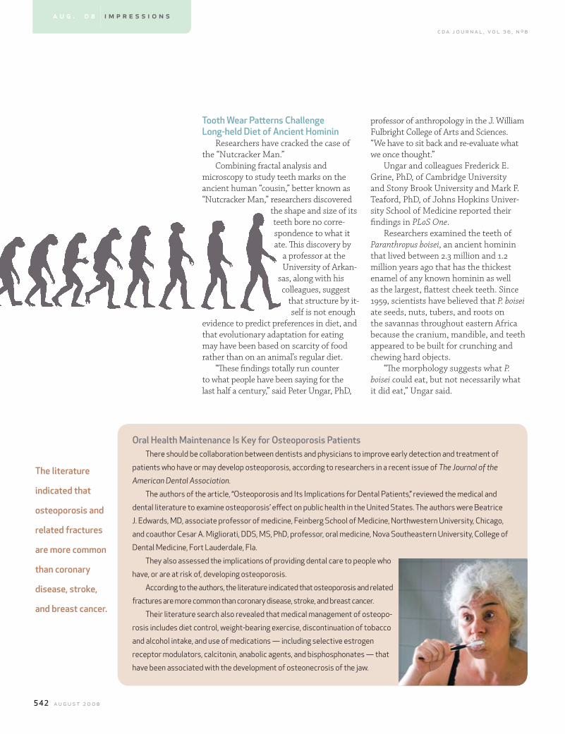
Tooth Wear Patterns Challenge long-held Diet of ancient hominin
Researchers have cracked the case of the “Nutcracker Man.”
Combining fractal analysis and microscopy to study teeth marks on the ancient human “cousin,” better known as “Nutcracker Man,” researchers discovered
the shape and size of its teeth bore no corre-spondence to what it ate. This discovery by
a professor at the University of Arkan-
sas, along with his colleagues, suggest
that structure by it-self is not enough
evidence to predict preferences in diet, and that evolutionary adaptation for eating may have been based on scarcity of food rather than on an animal’s regular diet.
“These findings totally run counter to what people have been saying for the last half a century,” said Peter Ungar, PhD,
professor of anthropology in the J. William Fulbright College of Arts and Sciences. “We have to sit back and re-evaluate what we once thought.”
Ungar and colleagues Frederick E. Grine, PhD, of Cambridge University and Stony Brook University and Mark F. Teaford, PhD, of Johns Hopkins Univer-sity School of Medicine reported their findings in PLoS One.
Researchers examined the teeth of Paranthropus boisei, an ancient hominin that lived between 2.3 million and 1.2 million years ago that has the thickest enamel of any known hominin as well as the largest, flattest cheek teeth. Since 1959, scientists have believed that P. boisei ate seeds, nuts, tubers, and roots on the savannas throughout eastern Africa because the cranium, mandible, and teeth appeared to be built for crunching and chewing hard objects.
“The morphology suggests what P. boisei could eat, but not necessarily what it did eat,” Ungar said.
oral health maintenance Is Key for osteoporosis Patients There should be collaboration between dentists and physicians to improve early detection and treatment of
patients who have or may develop osteoporosis, according to researchers in a recent issue of The Journal of the
American Dental Association.
The authors of the article, “Osteoporosis and Its Implications for Dental Patients,” reviewed the medical and
dental literature to examine osteoporosis’ effect on public health in the United States. The authors were Beatrice
J. Edwards, MD, associate professor of medicine, Feinberg School of Medicine, Northwestern University, Chicago,
and coauthor Cesar A. Migliorati, DDS, MS, PhD, professor, oral medicine, Nova Southeastern University, College of
Dental Medicine, Fort Lauderdale, Fla.
They also assessed the implications of providing dental care to people who
have, or are at risk of, developing osteoporosis.
According to the authors, the literature indicated that osteoporosis and related
fractures are more common than coronary disease, stroke, and breast cancer.
Their literature search also revealed that medical management of osteopo-
rosis includes diet control, weight-bearing exercise, discontinuation of tobacco
and alcohol intake, and use of medications — including selective estrogen
receptor modulators, calcitonin, anabolic agents, and bisphosphonates — that
have been associated with the development of osteonecrosis of the jaw.
The literature
indicated that
osteoporosis and
related fractures
are more common
than coronary
disease, stroke,
and breast cancer.
c d a j o u r n a l , v o l 3 6 , n º 8
a u g u s t 2 0 0 8 543
site of the American Journal of Public Health. “This is the first time we’ve seen a
connection between pregnancy and tooth loss affecting women at all socioeconomic levels in a large, heterogeneous sample of the U.S. population,” Russell commented.
Profound biological and behavioral changes related to pregnancy and child birth are likely to be a factor in tooth loss, Russell found. For example:
n Pregnancy can make women prone to gingivitis. Repeated pregnancies are likely to result in more frequent outbreaks of gingivitis that may lead to tooth loss in women with periodontitis.
n A woman may postpone seeking dental treatment because of financial concerns related to having children.
n Caring for more children may lead a mother to cut back on the time she devotes to her own oral health.
“Although further research is needed on the specific reasons for the link be-tween pregnancy and tooth loss, it is clear that women with multiple children need to be especially vigilant about their oral health,” said Russell.
nyu Dental researcher Validates old Wives’ Tale
Perhaps the source of crabbiness of the “Old Woman Who Lived in a Shoe” was due to her inability to enjoy peanut brittle or taffy.
Stefanie Russell, DDS, MPH, PhD, an assistant professor of Epidemiology and Health Promotion at New York Univer-sity, recently was able to substantiate the old wives’ tale that for every child birthed, the mother lost a tooth. With so many kids, “she didn’t know what to do,” it could be conjectured the shoe-dwelling, overwhelmed mom was more than likely fairly edentulous.
Women who have more children are more likely to have missing teeth, accord-ing to a nationwide study of 2,635 women, said Russell, who was able to base her conclusions on white and black non-Hispanic women between the ages of 18 to 64 who reported at least one pregnancy in the Third National Health and Nutri-tion Examination Survey, a representative study of the U.S. population.
Her findings were published on the Web
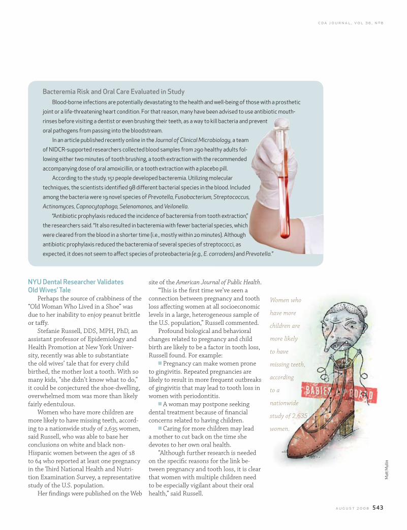
Bacteremia risk and oral Care evaluated in StudyBlood-borne infections are potentially devastating to the health and well-being of those with a prosthetic
joint or a life-threatening heart condition. For that reason, many have been advised to use antibiotic mouth-
rinses before visiting a dentist or even brushing their teeth, as a way to kill bacteria and prevent
oral pathogens from passing into the bloodstream.
In an article published recently online in the Journal of Clinical Microbiology, a team
of NIDCR-supported researchers collected blood samples from 290 healthy adults fol-
lowing either two minutes of tooth brushing, a tooth extraction with the recommended
accompanying dose of oral amoxicillin, or a tooth extraction with a placebo pill.
According to the study, 151 people developed bacteremia. Utilizing molecular
techniques, the scientists identified 98 different bacterial species in the blood. Included
among the bacteria were 19 novel species of Prevotella, Fusobacterium, Streptococcus,
Actinomyces, Capnocytophaga, Selenomonas, and Veilonella.
“Antibiotic prophylaxis reduced the incidence of bacteremia from tooth extraction,”
the researchers said. “It also resulted in bacteremia with fewer bacterial species, which
were cleared from the blood in a shorter time (i.e., mostly within 20 minutes). Although
antibiotic prophylaxis reduced the bacteremia of several species of streptococci, as
expected, it does not seem to affect species of proteobacteria (e.g., E. corrodens) and Prevotella.”
Women who
have more
children are
more likely
to have
missing teeth,
according
to a
nationwide
study of 2,635
women.
Matt
Mul
lin
544 a u g u s t 2 0 0 8
c d a j o u r n a l , v o l 3 6 , n º 8
new Procedure Can Speed up Process to Straighter Teeth
Teeth straightening and a killer smile in months instead of years? It can be done, say researchers at the University of Southern California School of Dentistry.
Hessam Nowzari DDS, PhD, director of the USC School of Dentistry and Advanced Education in Periodontology program, led a team of researchers who have published the first case study of the successful use of a patient’s own bone material for the grafting necessary in a surgical procedure developed by a Pennsylvania periodontist, Tom Wilcko, DMD. The report appeared in the May 2008 issue of the Compendium of Continuing Education in Dentistry.
“Given a choice for grafts, nothing is better than a patient’s own tissue,” Now-zari said. “It encourages new, healthy bone
formation in the grafted area. It’s very safe and eliminates the risk of any disease transmission.”
Wilcko offers courses in the proce-dure, trademarked as “Wilckodontics.” The dentists from USC used the proce-dure Periodontally Accelerated Osteo-genic Orthodontics, PAOO. With this technique, an oral surgeon or periodon-tist uses special instruments to score the bone holding the teeth in place and then applies bone graft material over the grooves. A local anesthetic is used in a dental office operatory.
The bone softens slightly as it starts to heal, allowing teeth to be moved into alignment with braces in a matter of months, instead of the years typically required by traditional orthodontics, ac-cording to a press release.
upcoming meetings
2 0 0 8
Sept. 6-9 94th annual meeting, american academy of Periodontology, Seattle, Wash., perio.org/meetings.
Sept. 12-14 CDa fall Scientific Session, San francisco, 800-CDa-SmIle (232-7645), cda.org.
Sept. 24-27 fDI annual World Dental Congress, Stockholm, [email protected].
oct. 16-19 american Dental association 149th annual Session, San antonio, Texas, ada.org.
oct. 25-29 american Public health association oral health Section’s annual meeting and exposition, San Diego, www.apha.org/meetings.
nov. 2-8 united States Dental Tennis association fall meeting, Palm Desert, dentaltennis.org.
2 0 0 9
may 14-17 CDa Spring Scientific Session, anaheim, 800-CDa-SmIle (232-7645), cda.org.
Sept. 11-13 CDa fall Scientific Session, San francisco, 800-CDa-SmIle (232-7645), cda.org.
oct. 1-4 american Dental association 150th annual Session, honolulu, hawaii, ada.org.
nov. 8-14 united States Dental Tennis association fall meeting, Scottsdale, ariz., dentaltennis.org.
To have an event included on this list of nonprofit association continuing education meetings, please send the information
to Upcoming Meetings, CDA Journal, 1201 K St., 16th Floor, Sacramento, CA 95814 or fax the information to 916-554-5962.
a u g . 0 8 i m p r e s s i o n s
“given a
choice for grafts,
nothing is better
than a patient’s
own tissue.”
HESSAM NOWzARI
DDS, PHD
c d a j o u r n a l , v o l 3 6 , n º 8
a u g u s t 2 0 0 8 545
cessation, medications, and not getting enough shut eye, are among the con-tributors to obesity. “You need a good six to eight good hours,” Mallone said of sleep. “If you don’t get enough, you’re so tired, you turn to food for energy.” Other factors Mallone mentioned may be attributed to:
n Geneticsn Illness, resulting in limited physical
activityn Food companies marketing to young
childrenn Fast food and its around-the-clock
availability as well as its expanded por-tions and inexpensive cost
Mallone, who referred to a knife and fork as potential weapons of mass destruction, said 1 in 5 children are over-weight. “Today’s kids are projected to live shorter lifespans than their parents; and 1 in 3 kids are at risk for type 2 diabetes.”
Rare is the case that someone can eat a plateful of 101 fried things and nosh on a slice of pepperoni pizza as “dessert” without deleterious effects to one’s health.
“We are overfed but undernourished, the foods we eat are calorie-dense, not nutrition-dense,” she said. “We’re eating more but doing less.”
Mallone held up her own family as an
example. She recounted an occasion when she and her husband wasted time looking for the remote control rather than getting off the sofa to switch on the TV and change the channels.
How does obesity affect the dental profession? For starters, the poor food choices and habits may increase a pa-tient’s caries risk as well as pose challeng-es to a treating dentist. “Obese patients may need to be placed upright due to pos-sible difficulty breathing when in a supine position for an extended period of time,” Mallone said.
Other ways, according to Mallone’s handout, dentists can be an integral part of their patient’s oral and overall health:
n Conduct a comprehensive review of the patient’s medical history to determine if the patient has other systemic diseases that coexist with increased weight that may prevent factors for treatment.
n Document any weight management medications, supplements or herbal prod-ucts in the dental chart.
n Look for signs of iron or vitamin deficiency.
n In the oral exam, be alert to changes
that might indicate deficiency such as glossitis, stomatitis, ulceration, and angular cheilitis. If present,
offer palliative oral health care tips.n Refer the patient to a registered
dietician and physician, if necessary, for additional evaluation and treatment of the etiology.
n Dental professionals can work with a registered dietician to offer support through nutrition counseling and weight management, as well as reinforce the ben-efits to a patient’s oral health from having a more healthful diet.
n Assess diet to determine intake of carbohydrates as part of caries risk man-agement.
n Give information about caries formation and its relationship to diet; provide education on caries control.
n Oral hygiene and daily fluoride may-be indicated, depending on the patient’s caries status.
n Emphasize the need for regular physical activity. And before doing so, remind them to visit their physician prior to starting an exercise program.
“Don’t discount the influence dentists can have on patients,” Mallone said.
overfed, co n tin u ed fr om 54 1
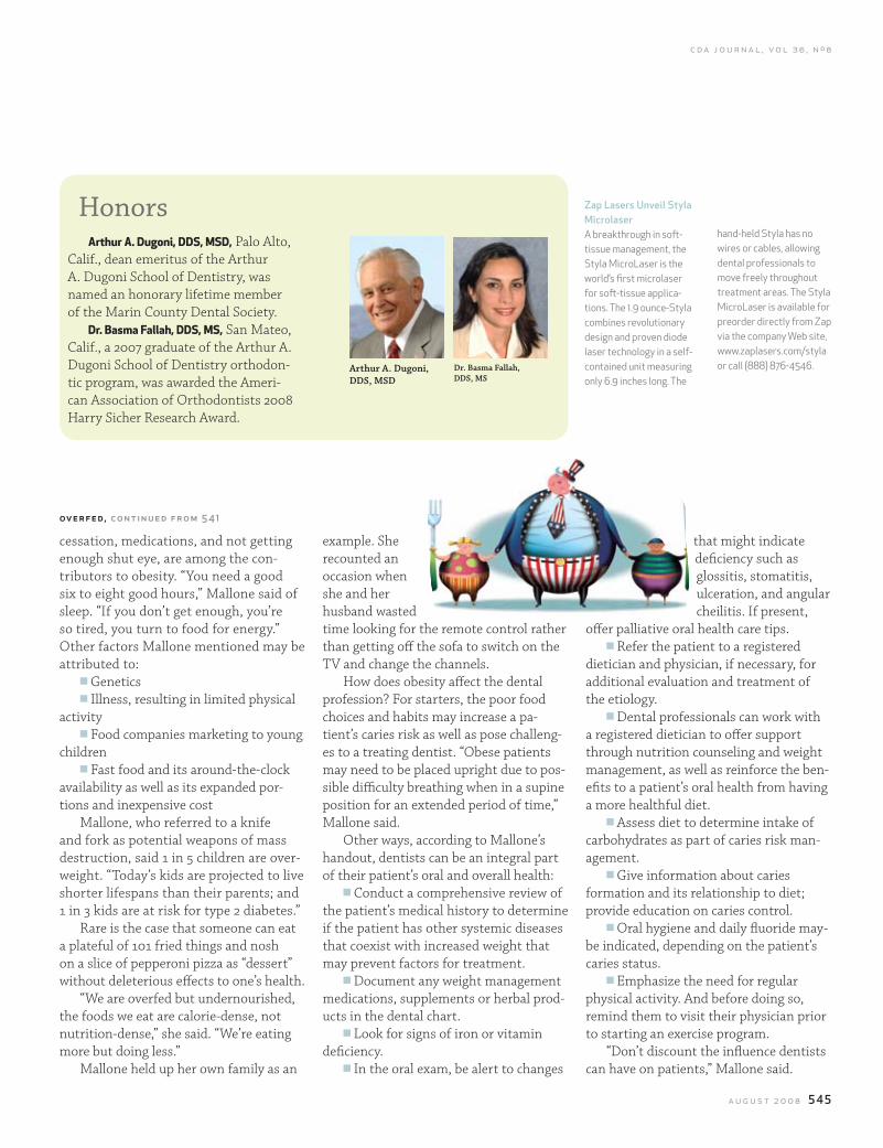
Honors Arthur A. Dugoni, DDS, MSD, Palo Alto,
Calif., dean emeritus of the Arthur A. Dugoni School of Dentistry, was named an honorary lifetime member of the Marin County Dental Society.
Dr. Basma Fallah, DDS, MS, San Mateo, Calif., a 2007 graduate of the Arthur A. Dugoni School of Dentistry orthodon-tic program, was awarded the Ameri-can Association of Orthodontists 2008 Harry Sicher Research Award.
Arthur A. Dugoni, DDS, MSD
Dr. Basma Fallah, DDS, MS
Zap lasers unveil Styla microlaserA breakthrough in soft-tissue management, the Styla MicroLaser is the world’s first microlaser for soft-tissue applica-tions. The 1.9 ounce-Styla combines revolutionary design and proven diode laser technology in a self-contained unit measuring only 6.9 inches long. The
hand-held Styla has no wires or cables, allowing dental professionals to move freely throughout treatment areas. The Styla MicroLaser is available for preorder directly from zap via the company Web site, www.zaplasers.com/styla or call (888) 876-4546.
546 a u g u s t 2 0 0 8
c d a j o u r n a l , v o l 3 6 , n º 8
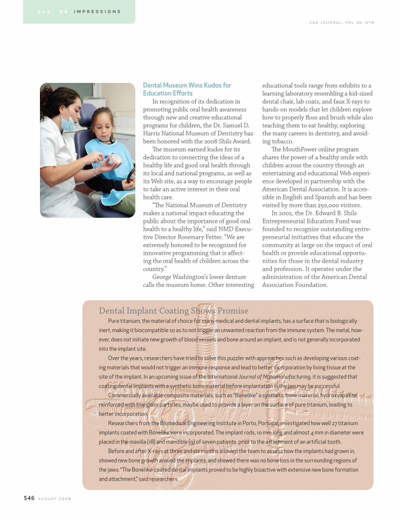
Dental museum Wins Kudos for education efforts
In recognition of its dedication in promoting public oral health awareness through new and creative educational programs for children, the Dr. Samuel D. Harris National Museum of Dentistry has been honored with the 2008 Shils Award.
The museum earned kudos for its dedication to connecting the ideas of a healthy life and good oral health through its local and national programs, as well as its Web site, as a way to encourage people to take an active interest in their oral health care.
“The National Museum of Dentistry makes a national impact educating the public about the importance of good oral health to a healthy life,” said NMD Execu-tive Director Rosemary Fetter. “We are extremely honored to be recognized for innovative programming that is affect-ing the oral health of children across the country.”
George Washington’s lower denture calls the museum home. Other interesting
educational tools range from exhibits to a learning laboratory resembling a kid-sized dental chair, lab coats, and faux X-rays to hands-on models that let children explore how to properly floss and brush while also teaching them to eat healthy, exploring the many careers in dentistry, and avoid-ing tobacco.
The MouthPower online program shares the power of a healthy smile with children across the country through an entertaining and educational Web experi-ence developed in partnership with the American Dental Association. It is acces-sible in English and Spanish and has been visited by more than 250,000 visitors.
In 2002, the Dr. Edward B. Shils Entrepreneurial Education Fund was founded to recognize outstanding entre-preneurial initiatives that educate the community at large on the impact of oral health or provide educational opportu-nities for those in the dental industry and profession. It operates under the administration of the American Dental Association Foundation.
a u g . 0 8 i m p r e s s i o n s
Dental Implant Coating Shows PromisePure titanium, the material of choice for many medical and dental implants, has a surface that is biologically
inert, making it biocompatible so as to not trigger an unwanted reaction from the immune system. The metal, how-
ever, does not initiate new growth of blood vessels and bone around an implant, and is not generally incorporated
into the implant site.
Over the years, researchers have tried to solve this puzzler with approaches such as developing various coat-
ing materials that would not trigger an immune response and lead to better incorporation by living tissue at the
site of the implant. In an upcoming issue of the International Journal of Nanomanufacturing, it is suggested that
coating dental implants with a synthetic bone material before implantation in the jaw may be successful.
Commercially available composite materials, such as “Bonelike” a synthetic bone material, hydroxyapatite
reinforced with tiny glass particles, maybe used to provide a layer on the surface of pure titanium, leading to
better incorporation.
Researchers from the Biomedical Engineering Institute in Porto, Portugal, investigated how well 27 titanium
implants coated with Bonelike were incorporated. The implant rods, 10 mm long and almost 4 mm in diameter were
placed in the maxilla (18) and mandible (9) of seven patients, prior to the attachment of an artificial tooth.
Before and after X-rays at three and six months allowed the team to assess how the implants had grown in,
showed new bone growth around the implants, and showed there was no bone loss in the surrounding regions of
the jaws. “The Bonelike-coated dental implants proved to be highly bioactive with extensive new bone formation
and attachment,” said researchers.
c d a j o u r n a l , v o l 3 6 , n º 8
a u g u s t 2 0 0 8 563
An Art or a Science?sajid jivraj, dds, msed
guest editor
Sajid Jivraj, dds, msed, is an associate clinical professor, former chair-man section of fixed prosthodontics and opera-tive dentistry University of Southern California, School of Dentistry, Los Angeles, and in private practice in Oxnard, Calif.
i n t r o d u c t i o n
Today, the practice of dentistry requires an interdisciplinary approach that
integrates the knowledge, skills, and experience of all the disciplines of
dentistry into a comprehensive treatment plan.
Prosthodontics can offer exceptional satisfaction to both the patient
and dentist. It can transform an unhealthy, unattractive dentition with
poor function into a comfortable, healthy occlusion capable of providing
years of further service, whilst at the same time greatly enhancing the
esthetic result.1
To obtain optimal results, meticulous attention must be paid to a
myriad of details. The process starts with the patient interview and me-
ticulous treatment planning, continues through to active treatment, and
culminates in regular planned follow-up care.
The objectives are to improve oral health, to establish proper occlusal
function, and to create the most ideal esthetic result possible. It is only
through an organized and systematic approach that appropriate diagno-
ses can be made, and based on these diagnoses, functional and esthetic
problems can be addressed predictably.
T r e aT m e n TP l a n n I n g :
564 a u g u s t 2 0 0 8
c d a j o u r n a l , v o l 3 6 , n º 8
Interdisciplinary therapy involves the combination of diagnostic, treatment planning, and therapeutic procedures. It is imperative the team leader appropriately selects a team of practitioners. The selec-tion process can either have a positive or a negative impact on the overall treatment. Each provider on the team must have an optimal level of skill in his or her area of ex-pertise to be a positive factor.2 The complex nature of dentofacial problems necessitates a highly organized method of communica-tion between the team members so that all aspects of treatment can be equally voiced.
It is through this communication that an interdisciplinary treatment plan can be
formulated prior to generation of a joint treatment letter. This treatment letter should include a discussion of aspects of treatment that will be provided by each team member, the time frame of the proposed treatment, the inherent risks involved, informed consent, and the financial responsibilities of the patient. It can be said that the quality of treat-ment is dependent upon the quality of the communication. It is critical the team leader maintains communication between the specialists both during treatment and once it has been completed. It is only through this approach that optimal care can be delivered and regular planned
follow-up care can be implemented.Treatment planning must begin
through visualization of the end result. By paying attention to details, systemati-cally analyzing each factor that affects the esthetic result, recognizing inadequacies in crown contour and gingival margin levels prior to restorative intervention, the restorative dentist can take advan-tage of the benefits of orthodontic and periodontal treatment to enhance the esthetic and functional outcomes.
The objective of each article is to outline how each specialty can enhance the final outcomes of treatment. Dr. Yorita will discuss the impact of orthodontics on treat-ment planning and how anchorage can be obtained when teeth are missing. Drs. Han-delsman, Ravon, and Levine will address management of the periodontium and how subtle procedures can enhance the esthetic outcome. Dr. Hochwald’s paper will describe how surgical procedures can re-establish op-timum occlusion and how communicating with the restorative dentist is key in obtain-ing optimal outcomes. Finally, Dr. Reshad and I will provide a prosthodontic perspec-tive in management of patients requiring restoration of posterior support and how to transition patients from a tooth-supported to an implant-supported occlusion.
My intention with this issue is to stimulate critical thinking and to offer the patient options for optimum care. Without an interdisciplinary approach, final outcomes can be compromised. With a team approach to the management of patients who require prosthodontic treatment, fewer compromises will occur and more ideal restorations can be developed.
r efer ences1. Rosenstiel SF, Land MF, Fujimoto J, Contemporary Fixed Prosthodontics, St. Louis, Mosby, pages 46-64, 1995.2. Roblee RD, Interdisciplinary Dentofacial Therapy. A Com-prehensive Approach to Optimal Patient Care, Quintessence Publishing Co., Inc., pages 17-43, 1994.
i n t r o d u c t i o n
c d a j o u r n a l , v o l 3 6 , n º 8
a u g u s t 2 0 0 8 567
here is a general consensus that tooth retention amongst the aging population pays credence to preventive dentistry and patient educa-
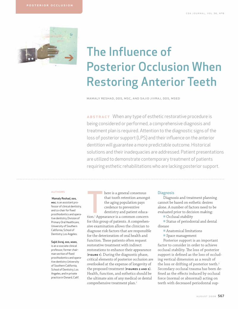
tion.1 Appearance is a common concern for this group of patients. A comprehen-sive examination allows the clinician to diagnose risk factors that are responsible for the deterioration of oral health and function. These patients often request restorative treatment with indirect restorations to enhance their appearance (figure 1). During the diagnostic phase, critical elements of posterior occlusion are overlooked at the expense of longevity of the proposed treatment (figures 2 and 3). Health, function, and esthetics should be the ultimate aim of any medical or dental comprehensive treatment plan.2
The Influence of Posterior occlusion When restoring anterior Teeth mamaly reshad, dds, msc, and sajid jivraj, dds, msed
abstract When any type of esthetic restorative procedure is being considered or performed, a comprehensive diagnosis and treatment plan is required. Attention to the diagnostic signs of the loss of posterior support (LPS) and their influence on the anterior dentition will guarantee a more predictable outcome. Historical solutions and their inadequacies are addressed. Patient presentations are utilized to demonstrate contemporary treatment of patients requiring esthetic rehabilitations who are lacking posterior support.
authors
mamaly reshad, dds, msc, is an assistant pro-fessor of clinical dentistry, and co-chair for fixed prosthodontics and opera-tive dentistry, Division of Primary Oral Healthcare, University of Southern California, School of Dentistry, Los Angeles.
Sajid Jivraj, dds, msed, is an a ssociate clinical professor, former chair-man section of fixed prosthodontics and opera-tive dentistry University of Southern California, School of Dentistry, Los Angeles, and in private practice in Oxnard, Calif.
p o s t e r i o r o c c l u s i o n
DiagnosisDiagnosis and treatment planning
cannot be based on esthetic desires alone. A number of factors need to be evaluated prior to decision making:
n Occlusal stabilityn Status of periodontal and dental
diseasen Anatomical limitations n Space management Posterior support is an important
factor to consider in order to achieve occlusal stability. The loss of posterior support is defined as the loss of occlud-ing vertical dimension as a result of the loss or drifting of posterior teeth.3 Secondary occlusal trauma has been de-fined as the effects induced by occlusal force (normal or abnormal) acting on teeth with decreased periodontal sup-
T
568 a u g u s t 2 0 0 8
c d a j o u r n a l , v o l 3 6 , n º 8
port.3 Hence, it is possible for a patient with an almost intact dentition, but with a reduced periodontium to pres-ent with the signs of LPS (figure 4).
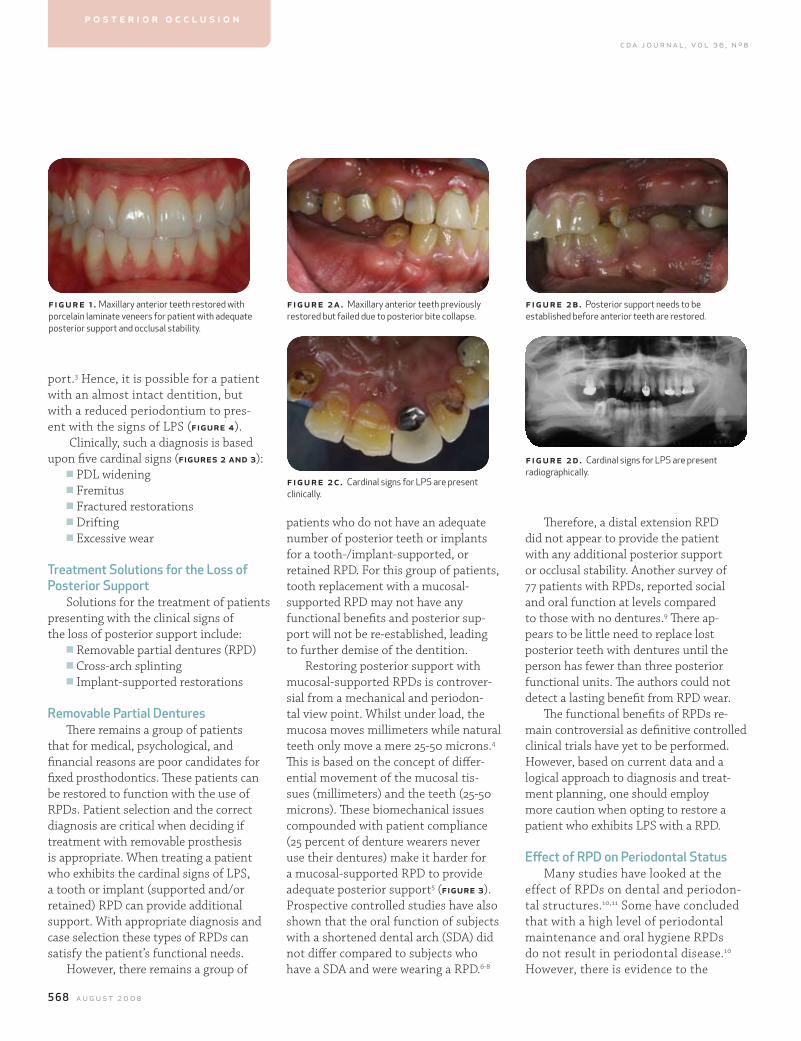
Clinically, such a diagnosis is based upon five cardinal signs (figures 2 and 3):
n PDL wideningn Fremitusn Fractured restorationsn Driftingn Excessive wear
Treatment Solutions for the loss of Posterior Support
Solutions for the treatment of patients presenting with the clinical signs of the loss of posterior support include:
n Removable partial dentures (RPD)n Cross-arch splintingn Implant-supported restorations
removable Partial Dentures There remains a group of patients
that for medical, psychological, and financial reasons are poor candidates for fixed prosthodontics. These patients can be restored to function with the use of RPDs. Patient selection and the correct diagnosis are critical when deciding if treatment with removable prosthesis is appropriate. When treating a patient who exhibits the cardinal signs of LPS, a tooth or implant (supported and/or retained) RPD can provide additional support. With appropriate diagnosis and case selection these types of RPDs can satisfy the patient’s functional needs.
However, there remains a group of
patients who do not have an adequate number of posterior teeth or implants for a tooth-/implant-supported, or retained RPD. For this group of patients, tooth replacement with a mucosal-supported RPD may not have any functional benefits and posterior sup-port will not be re-established, leading to further demise of the dentition.
Restoring posterior support with mucosal-supported RPDs is controver-sial from a mechanical and periodon-tal view point. Whilst under load, the mucosa moves millimeters while natural teeth only move a mere 25-50 microns.4 This is based on the concept of differ-ential movement of the mucosal tis-sues (millimeters) and the teeth (25-50 microns). These biomechanical issues compounded with patient compliance (25 percent of denture wearers never use their dentures) make it harder for a mucosal-supported RPD to provide adequate posterior support5 (figure 3). Prospective controlled studies have also shown that the oral function of subjects with a shortened dental arch (SDA) did not differ compared to subjects who have a SDA and were wearing a RPD.6-8
Therefore, a distal extension RPD did not appear to provide the patient with any additional posterior support or occlusal stability. Another survey of 77 patients with RPDs, reported social and oral function at levels compared to those with no dentures.9 There ap-pears to be little need to replace lost posterior teeth with dentures until the person has fewer than three posterior functional units. The authors could not detect a lasting benefit from RPD wear.
The functional benefits of RPDs re-main controversial as definitive controlled clinical trials have yet to be performed. However, based on current data and a logical approach to diagnosis and treat-ment planning, one should employ more caution when opting to restore a patient who exhibits LPS with a RPD.
effect of rPD on Periodontal StatusMany studies have looked at the
effect of RPDs on dental and periodon-tal structures.10,11 Some have concluded that with a high level of periodontal maintenance and oral hygiene RPDs do not result in periodontal disease.10 However, there is evidence to the
f igur e 1 . Maxillary anterior teeth restored with porcelain laminate veneers for patient with adequate posterior support and occlusal stability.
figure 2a. Maxillary anterior teeth previously restored but failed due to posterior bite collapse.
figure 2c. Cardinal signs for LPS are present clinically.
fig ur e 2b. Posterior support needs to be established before anterior teeth are restored.
fig ur e 2d . Cardinal signs for LPS are present radiographically.
p o s t e r i o r o c c l u s i o n
c d a j o u r n a l , v o l 3 6 , n º 8
a u g u s t 2 0 0 8 569
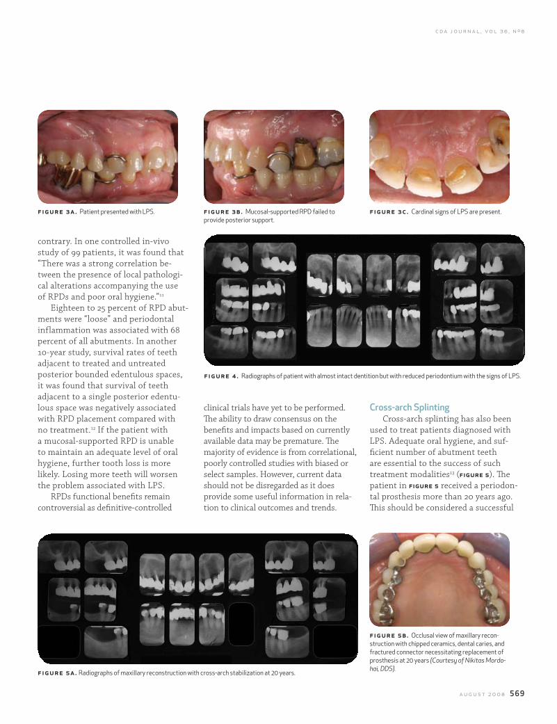
Cross-arch SplintingCross-arch splinting has also been
used to treat patients diagnosed with LPS. Adequate oral hygiene, and suf-ficient number of abutment teeth are essential to the success of such treatment modalities13 (figure 5). The patient in figure 5 received a periodon-tal prosthesis more than 20 years ago. This should be considered a successful
clinical trials have yet to be performed. The ability to draw consensus on the benefits and impacts based on currently available data may be premature. The majority of evidence is from correlational, poorly controlled studies with biased or select samples. However, current data should not be disregarded as it does provide some useful information in rela-tion to clinical outcomes and trends.
contrary. In one controlled in-vivo study of 99 patients, it was found that “There was a strong correlation be-tween the presence of local pathologi-cal alterations accompanying the use of RPDs and poor oral hygiene.”11
Eighteen to 25 percent of RPD abut-ments were “loose” and periodontal inflammation was associated with 68 percent of all abutments. In another 10-year study, survival rates of teeth adjacent to treated and untreated posterior bounded edentulous spaces, it was found that survival of teeth adjacent to a single posterior edentu-lous space was negatively associated with RPD placement compared with no treatment.12 If the patient with a mucosal-supported RPD is unable to maintain an adequate level of oral hygiene, further tooth loss is more likely. Losing more teeth will worsen the problem associated with LPS.
RPDs functional benefits remain controversial as definitive-controlled
f igure 3a. Patient presented with LPS.
f igure 5a. Radiographs of maxillary reconstruction with cross-arch stabilization at 20 years.
figure 3b. Mucosal-supported RPD failed to provide posterior support.
fig ur e 3 c. Cardinal signs of LPS are present.
fig ur e 5b. Occlusal view of maxillary recon-struction with chipped ceramics, dental caries, and fractured connector necessitating replacement of prosthesis at 20 years (Courtesy of Nikitas Mordo-hai, DDS).
figure 4 . Radiographs of patient with almost intact dentition but with reduced periodontium with the signs of LPS.
570 a u g u s t 2 0 0 8
c d a j o u r n a l , v o l 3 6 , n º 8
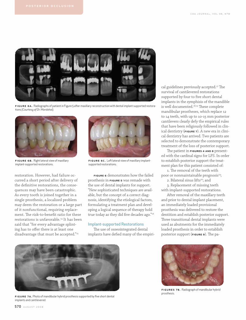
figure 6 demonstrates how the failed prosthesis in figure 5 was remade with the use of dental implants for support. “New sophisticated techniques are avail-able, but the concept of a correct diag-nosis, identifying the etiological factors, formulating a treatment plan and devel-oping a logical sequence of therapy hold true today as they did five decades ago.”16
Implant-supported restorationsThe use of osseointegrated dental
implants have defied many of the empiri-
restoration. However, had failure oc-curred a short period after delivery of the definitive restorations, the conse-quences may have been catastrophic. As every tooth is joined together in a single prosthesis, a localized problem may deem the restoration or a large part of it nonfunctional, requiring replace-ment. The risk-to-benefit ratio for these restorations is unfavorable.14 It has been said that “for every advantage splint-ing has to offer there is at least one disadvantage that must be accepted.”15
cal guidelines previously accepted.17 The survival of cantilevered restorations supported by four to five short dental implants in the symphisis of the mandible is well documented.18,19 These complete mandibular prostheses, which replace 12 to 14 teeth, with up to 10-15 mm posterior cantilevers clearly defy the empirical rules that have been religiously followed in clin-ical dentistry (figure 7). A new era in clini-cal dentistry has arrived. Two patients are selected to demonstrate the contemporary treatment of the loss of posterior support.
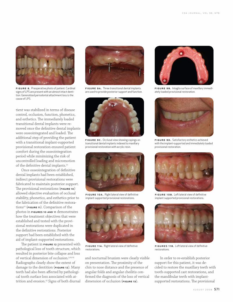
The patient in figures 4 and 8 present-ed with the cardinal signs for LPS. In order to establish posterior support the treat-ment plan for this patient consisted of:
1. The removal of the teeth with poor or nonmaintainable prognosis14;
2. Bilateral sinus lifts20; and3. Replacement of missing teeth
with implant-supported restorations.After removal of the maxillary teeth
and prior to dental implant placement, an immediately loaded provisional prosthesis was delivered to restore the dentition and establish posterior support. Three transitional dental implants were used as abutments for the immediately loaded prosthesis in order to establish posterior support (figure 9). The pa-
f igur e 6a. Radiographs of patient in Figure 5 after maxillary reconstruction with dental implant supported restora-tions (Courtesy of Dr. Mordohai).
f igur e 6b. Right lateral view of maxillary implant-supported restorations.
figure 6c. Left lateral view of maxillary implant-supported restorations.
f igur e 7a. Photo of mandibular hybrid prosthesis supported by five short dental implants and cantilevered.
fig ur es 7 b. Radiograph of mandibular hybrid prosthesis.
p o s t e r i o r o c c l u s i o n
c d a j o u r n a l , v o l 3 6 , n º 8
a u g u s t 2 0 0 8 571
tient was stabilized in terms of disease control, occlusion, function, phonetics, and esthetics. The immediately loaded transitional dental implants were re-moved once the definitive dental implants were osseointegrated and loaded. The additional step of providing the patient with a transitional implant-supported provisional restoration ensured patient comfort during the osseointegration period while minimizing the risk of uncontrolled loading and micromotion of the definitive dental implants.21
Once osseointegration of definitive dental implants had been established, indirect provisional restorations were fabricated to maintain posterior support. The provisional restorations (figure 10) allowed objective evaluation of occlusal stability, phonetics, and esthetics prior to the fabrication of the definitive restora-tions22 (figure 11). Comparison of the photos in figures 10 and 11 demonstrates how the treatment objectives that were established and tested with the provi-sional restorations were duplicated in the definitive restorations. Posterior support had been established with the aid of implant-supported restorations.
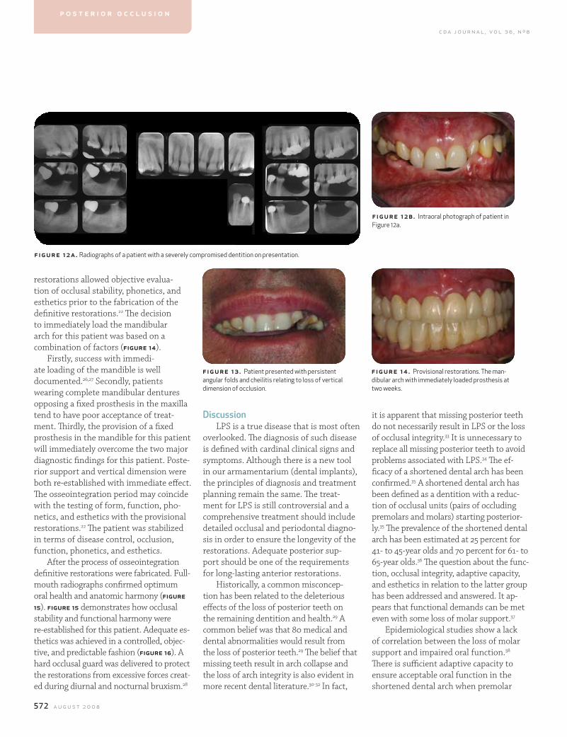
The patient in figure 12 presented with pathological loss of tooth structure, which resulted in posterior bite collapse and loss of vertical dimension of occlusion.3,23-25 Radiographs clearly show the extent of damage to the dentition (figure 12). Many teeth had also been affected by pathologi-cal tooth surface loss associated with at-trition and erosion.23 Signs of both diurnal
f igure 8 . Preoperative photo of patient. Cardinal signs of LPS are present with an almost intact denti-tion. Generalized periodontal attachment loss is the cause of LPS.
figure 9a . Three transitional dental implants are used to provide posterior support and function.
fig ur e 9 b. Intaglio surface of maxillary immedi-ately loaded provisional restoration.
figure 9c . Occlusal view showing copings on transitional dental implants indexed to maxillary provisional restoration with acrylic resin.
fig ur e 9 d . Satisfactory esthetics achieved with the implant-supported and immediately loaded provisional restoration.
figure 10a. Right lateral view of definitive implant-supported provisional restorations.
figure 11a. Right lateral view of definitive restorations.
fig ur e 10b. Left lateral view of definitive implant-supported provisional restorations.
fig ur es 11b . Left lateral view of definitive restorations.
and nocturnal bruxism were clearly visible on presentation. The proximity of the chin to nose distance and the presence of angular folds and angular cheilitis con-firmed the diagnosis of the loss of vertical dimension of occlusion (figure 13).
In order to re-establish posterior support for this patient, it was de-cided to restore the maxillary teeth with tooth-supported cast restorations, and the mandibular teeth with implant-supported restorations. The provisional
572 a u g u s t 2 0 0 8
c d a j o u r n a l , v o l 3 6 , n º 8
restorations allowed objective evalua-tion of occlusal stability, phonetics, and esthetics prior to the fabrication of the definitive restorations.22 The decision to immediately load the mandibular arch for this patient was based on a combination of factors (figure 14).
Firstly, success with immedi-ate loading of the mandible is well documented.26,27 Secondly, patients wearing complete mandibular dentures opposing a fixed prosthesis in the maxilla tend to have poor acceptance of treat-ment. Thirdly, the provision of a fixed prosthesis in the mandible for this patient will immediately overcome the two major diagnostic findings for this patient. Poste-rior support and vertical dimension were both re-established with immediate effect. The osseointegration period may coincide with the testing of form, function, pho-netics, and esthetics with the provisional restorations.22 The patient was stabilized in terms of disease control, occlusion, function, phonetics, and esthetics.
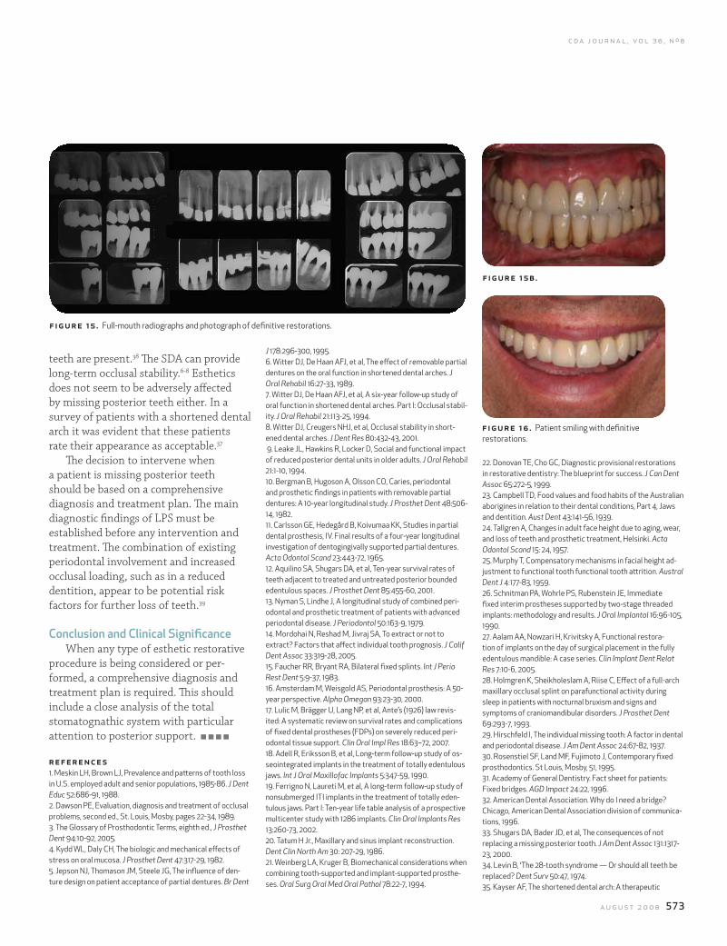
After the process of osseointegration definitive restorations were fabricated. Full-mouth radiographs confirmed optimum oral health and anatomic harmony (figure 15). figure 15 demonstrates how occlusal stability and functional harmony were re-established for this patient. Adequate es-thetics was achieved in a controlled, objec-tive, and predictable fashion (figure 16). A hard occlusal guard was delivered to protect the restorations from excessive forces creat-ed during diurnal and nocturnal bruxism.28
Discussion LPS is a true disease that is most often
overlooked. The diagnosis of such disease is defined with cardinal clinical signs and symptoms. Although there is a new tool in our armamentarium (dental implants), the principles of diagnosis and treatment planning remain the same. The treat-ment for LPS is still controversial and a comprehensive treatment should include detailed occlusal and periodontal diagno-sis in order to ensure the longevity of the restorations. Adequate posterior sup-port should be one of the requirements for long-lasting anterior restorations.
Historically, a common misconcep-tion has been related to the deleterious effects of the loss of posterior teeth on the remaining dentition and health.29 A common belief was that 80 medical and dental abnormalities would result from the loss of posterior teeth.29 The belief that missing teeth result in arch collapse and the loss of arch integrity is also evident in more recent dental literature.30-32 In fact,
it is apparent that missing posterior teeth do not necessarily result in LPS or the loss of occlusal integrity.33 It is unnecessary to replace all missing posterior teeth to avoid problems associated with LPS.34 The ef-ficacy of a shortened dental arch has been confirmed.35 A shortened dental arch has been defined as a dentition with a reduc-tion of occlusal units (pairs of occluding premolars and molars) starting posterior-ly.35 The prevalence of the shortened dental arch has been estimated at 25 percent for 41- to 45-year olds and 70 percent for 61- to 65-year olds.36 The question about the func-tion, occlusal integrity, adaptive capacity, and esthetics in relation to the latter group has been addressed and answered. It ap-pears that functional demands can be met even with some loss of molar support.37
Epidemiological studies show a lack of correlation between the loss of molar support and impaired oral function.38 There is sufficient adaptive capacity to ensure acceptable oral function in the shortened dental arch when premolar
f igur e 12a. Radiographs of a patient with a severely compromised dentition on presentation.
fig ur e 12b. Intraoral photograph of patient in Figure 12a.
figure 13 . Patient presented with persistent angular folds and cheilitis relating to loss of vertical dimension of occlusion.
fig ur e 14 . Provisional restorations. The man-dibular arch with immediately loaded prosthesis at two weeks.
p o s t e r i o r o c c l u s i o n
c d a j o u r n a l , v o l 3 6 , n º 8
a u g u s t 2 0 0 8 573
teeth are present.38 The SDA can provide long-term occlusal stability.6-8 Esthetics does not seem to be adversely affected by missing posterior teeth either. In a survey of patients with a shortened dental arch it was evident that these patients rate their appearance as acceptable.37
The decision to intervene when a patient is missing posterior teeth should be based on a comprehensive diagnosis and treatment plan. The main diagnostic findings of LPS must be established before any intervention and treatment. The combination of existing periodontal involvement and increased occlusal loading, such as in a reduced dentition, appear to be potential risk factors for further loss of teeth.39
Conclusion and Clinical SignificanceWhen any type of esthetic restorative
procedure is being considered or per-formed, a comprehensive diagnosis and treatment plan is required. This should include a close analysis of the total stomatognathic system with particular attention to posterior support.
r ef erences1. Meskin LH, Brown LJ, Prevalence and patterns of tooth loss in U.S. employed adult and senior populations, 1985-86. J Dent Educ 52:686-91, 1988.2. Dawson PE, Evaluation, diagnosis and treatment of occlusal problems, second ed., St. Louis, Mosby, pages 22-34, 1989.3. The Glossary of Prosthodontic Terms, eighth ed., J Prosthet Dent 94:10-92, 2005.4. Kydd WL, Daly CH, The biologic and mechanical effects of stress on oral mucosa. J Prosthet Dent 47:317-29, 1982.5. Jepson NJ, Thomason JM, Steele JG, The influence of den-ture design on patient acceptance of partial dentures. Br Dent
J 178:296-300, 1995.6. Witter DJ, De Haan AFJ, et al, The effect of removable partial dentures on the oral function in shortened dental arches. J Oral Rehabil 16:27-33, 1989.7. Witter DJ, De Haan AFJ, et al, A six-year follow-up study of oral function in shortened dental arches. Part I: Occlusal stabil-ity. J Oral Rehabil 21:113-25, 1994.8. Witter DJ, Creugers NHJ, et al, Occlusal stability in short-ened dental arches. J Dent Res 80:432-43, 2001. 9. Leake JL, Hawkins R, Locker D, Social and functional impact of reduced posterior dental units in older adults. J Oral Rehabil 21:1-10, 1994.10. Bergman B, Hugoson A, Olsson CO, Caries, periodontal and prosthetic findings in patients with removable partial dentures: A 10-year longitudinal study. J Prosthet Dent 48:506-14, 1982.11. Carlsson GE, Hedegård B, Koivumaa KK, Studies in partial dental prosthesis, IV. Final results of a four-year longitudinal investigation of dentogingivally supported partial dentures. Acta Odontol Scand 23:443-72, 1965.12. Aquilino SA, Shugars DA, et al, Ten-year survival rates of teeth adjacent to treated and untreated posterior bounded edentulous spaces. J Prosthet Dent 85:455-60, 2001.13. Nyman S, Lindhe J, A longitudinal study of combined peri-odontal and prosthetic treatment of patients with advanced periodontal disease. J Periodontol 50:163-9, 1979.14. Mordohai N, Reshad M, Jivraj SA, To extract or not to extract? Factors that affect individual tooth prognosis. J Calif Dent Assoc 33:319-28, 2005.15. Faucher RR, Bryant RA, Bilateral fixed splints. Int J Perio Rest Dent 5:9-37, 1983.16. Amsterdam M, Weisgold AS, Periodontal prosthesis: A 50-year perspective. Alpha Omegan 93:23-30, 2000.17. Lulic M, Brägger U, Lang NP, et al, Ante’s (1926) law revis-ited: A systematic review on survival rates and complications of fixed dental prostheses (FDPs) on severely reduced peri-odontal tissue support. Clin Oral Impl Res 18:63–72, 2007.18. Adell R, Eriksson B, et al, Long-term follow-up study of os-seointegrated implants in the treatment of totally edentulous jaws. Int J Oral Maxillofac Implants 5:347-59, 1990.19. Ferrigno N, Laureti M, et al, A long-term follow-up study of nonsubmerged ITI implants in the treatment of totally eden-tulous jaws. Part I: Ten-year life table analysis of a prospective multicenter study with 1286 implants. Clin Oral Implants Res 13:260-73, 2002.20. Tatum H Jr., Maxillary and sinus implant reconstruction. Dent Clin North Am 30: 207-29, 1986.21. Weinberg LA, Kruger B, Biomechanical considerations when combining tooth-supported and implant-supported prosthe-ses. Oral Surg Oral Med Oral Pathol 78:22-7, 1994.
22. Donovan TE, Cho GC, Diagnostic provisional restorations in restorative dentistry: The blueprint for success. J Can Dent Assoc 65:272-5, 1999.23. Campbell TD, Food values and food habits of the Australian aborigines in relation to their dental conditions, Part 4, Jaws and dentition. Aust Dent 43:141-56, 1939. 24. Tallgren A, Changes in adult face height due to aging, wear, and loss of teeth and prosthetic treatment, Helsinki. Acta Odontol Scand 15: 24, 1957.25. Murphy T, Compensatory mechanisms in facial height ad-justment to functional tooth functional tooth attrition. Austral Dent J 4:177-83, 1959.26. Schnitman PA, Wohrle PS, Rubenstein JE, Immediate fixed interim prostheses supported by two-stage threaded implants: methodology and results. J Oral Implantol 16:96-105, 1990.27. Aalam AA, Nowzari H, Krivitsky A, Functional restora-tion of implants on the day of surgical placement in the fully edentulous mandible: A case series. Clin Implant Dent Relat Res 7:10-6, 2005.28. Holmgren K, Sheikholeslam A, Riise C, Effect of a full-arch maxillary occlusal splint on parafunctional activity during sleep in patients with nocturnal bruxism and signs and symptoms of craniomandibular disorders. J Prosthet Dent 69:293-7, 1993.29. Hirschfeld I, The individual missing tooth: A factor in dental and periodontal disease. J Am Dent Assoc 24:67-82, 1937.30. Rosenstiel SF, Land MF, Fujimoto J, Contemporary fixed prosthodontics. St Louis, Mosby, 51, 1995.31. Academy of General Dentistry. Fact sheet for patients: Fixed bridges. AGD Impact 24:22, 1996.32. American Dental Association. Why do I need a bridge? Chicago, American Dental Association division of communica-tions, 1996.33. Shugars DA, Bader JD, et al, The consequences of not replacing a missing posterior tooth. J Am Dent Assoc 131:1317-23, 2000.34. Levin B, ‘The 28-tooth syndrome — Or should all teeth be replaced? Dent Surv 50:47, 1974.35. Kayser AF, The shortened dental arch: A therapeutic
f igure 15 . Full-mouth radiographs and photograph of definitive restorations.
figure 15b.
f ig ur e 16 . Patient smiling with definitive restorations.
574 a u g u s t 2 0 0 8
c d a j o u r n a l , v o l 3 6 , n º 8
concept in reduced dentitions and certain high-risk groups. Int J Perio Rest Dent 9:427-49, 1989.36. Bjorn AL, Owall B, Partial edentulism and its prosthetic treatment. A frequency study within a Swedish population. Swed Dent J 3:15-25, 1979.37. Kayser AF, Witter DJ, Oral functional needs and its conse-quences for dentulous older people. Community Dent Health 2:285-91, 1985.38. Kayser AF, Shortened dental arches and oral function. J Oral Rehabil 8:457-62, 1981.39. Witter DJ, De Haan AF, Kayser AF, Van Rossum GM, A six-year follow-up study of oral function in shortened dental arches. Part II: Craniomandibular dysfunction and oral comfort. J Oral Rehabil 21:353-66, 1994.
to request a printed copy of this article, please contact Mamaly Reshad DDS, MSc, University of Southern California, School of Dentistry, Department of Advanced Graduate Prosthodontics, 925 West 34th St., Los Angeles, Calif., 90089-0641.
c d a j o u r n a l , v o l 3 6 , n º 8
a u g u s t 2 0 0 8 575
multidisciplinary Care: Periodontal aspects to Treatment Planning the anterior esthetic Zonenicolas a. ravon, dds, msd; mark handelsman, dds; and david levine, dds
abstract The field of periodontology has changed dramatically during the past 30 years. The goal of periodontal therapy is not only to establish and maintain the dentition and the periodontium free of any oral infections, but also to provide an environment with optimal function and esthetics. Esthetics has become an integral portion of the overall treatment goal in periodontics. This article discusses the role of periodontal plastic and reconstructive surgery in treatment planning the anterior esthetic zone in interdisciplinary dental care.
authors
nicolas a. ravon, dds, msd, is a diplomate, Ameri-can Board Periodontology, and in private practice in Burbank, Calif.
mark handelsman, dds, is a diplomate, American Board Periodontology, and in private practice in Santa Monica, Calif.
David levine, dds, is a dip-lomate, American Board Periodontology, an associ-ate clinical professor at the University of Southern California, School of Dentistry, and in private practice in Burbank, Calif.
he signs of periodontal disease are often varied and complex. In order to deter-mine a meaningful treatment plan, a proper diagnosis is
essential. This can only be determined through a comprehensive examination and collection of the precise clinical characteristics of the patient’s overall periodontal condition. In performing the periodontal examination and develop-ing a treatment plan, care must be taken to consider all aspects of the patient, including their overall functional status.
Consultation with other special-ists (restorative dentist, oral surgeon, orthodontist, endodontist) regarding the restorative and endodontic health, and any occlusal, skeletal, and space problems, is required. All the treating specialists on the team need to collabo-
t r e a t m e n t p l a n n i n g
rate their findings. An evaluation and understanding of the etiology of the pathologic process is extremely impor-tant. A correct diagnosis with long-term prognostic information is mandatory to develop an interdisciplinary treatment plan. This article will review periodontal aspects of treatment planning with an emphasis on the anterior esthetic zone.
The American Academy of Periodon-tology has developed parameters on a comprehensive periodontal examination.1 These parameters are discussed below.
First in any examination should be a review of the patient’s medical history. Those systemic conditions or behav-ioral characteristics that may contribute or predispose to periodontal disease should be noted and discussed with the patient. Such conditions include but are not limited to diabetes, smok-
T
576 a u g u s t 2 0 0 8
c d a j o u r n a l , v o l 3 6 , n º 8
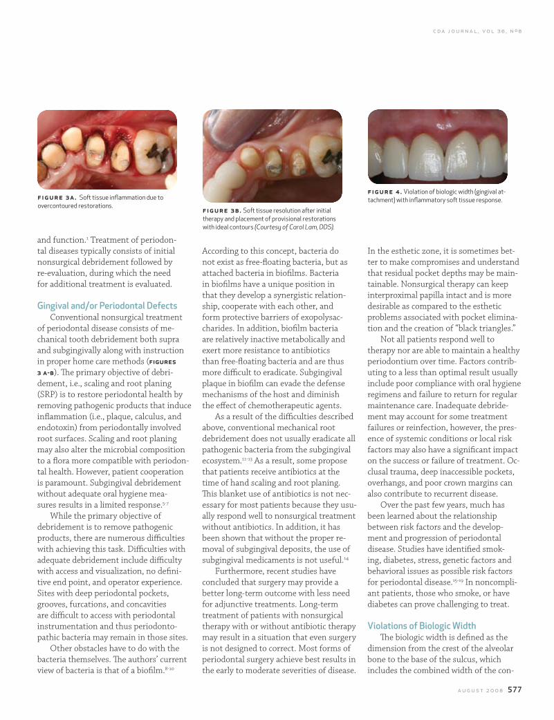
frontal view but also from the lateral view, both with the lip at rest and when smiling. Uneven gingival architecture, the position of teeth relative to the arch shape and opposing occlusion will all affect and dictate the decision-making process. The position of the incisal edge relative to facial proportions and lip dynamics is critical (figure 2 and table 1).
It is important to identify the prob-lems that will affect the desired esthetic outcome.
Esthetic periodontal defects include: n Residual gingival/periodontal defectsn Violations of biologic widthn Gingival asymmetriesn Inadequate amount of gingivan Gingival recessionsn Deficient pontic areasn Frena impinging on the gingival
marginn Excessive gingival displayn Deficient interproximal papillaeProper gingival esthetic involves
initially the restoration of periodontal health. The ultimate goal of periodontal therapy is to preserve the natural denti-tion, periodontium, and peri-implant tissues in health, comfort, esthetics,
ing, hypertension, and pregnancy.A review of the dental history is also
important. This information should include the patient’s main reason for seeking treatment as well as past dental treatment and previous radiographs. Has the patient been compliant and has the patient received adequate follow up care is also important information to know.
Extraoral and intraoral structures should be examined and evaluated. Any temporomandibular joint issues should be discussed. The oral mucosa, lips, floor of the mouth, muscles of mastica-tion, salivary glands, palate, and the oropharynx should all be evaluated.
An evaluation of the teeth should include observation of missing teeth, condition of restorations, caries, tooth mobility, tooth position, occlusal and interdental relationships, signs of parafunctional habits, and if applicable, pulpal status.2 Proximal contact rela-tionships are also important to note as some open contacts can impact food, which can contribute to the progres-sion of disease. Any furcation involve-ments should be evaluated and noted.
A comprehensive periodontal exami-nation includes the hard and soft sup-porting tissues of the dentition. Clinical findings and radiographic findings need to be evaluated. Radiographs should be eval-uated to help determine the status of the periodontium and dental implants. Radio-graphs should be diagnostic and based on the needs of the patient. Clinically, the pa-tient’s tissue biotype is classified accord-ing to how thick or thin the supporting bone and gingival soft tissues are defined.
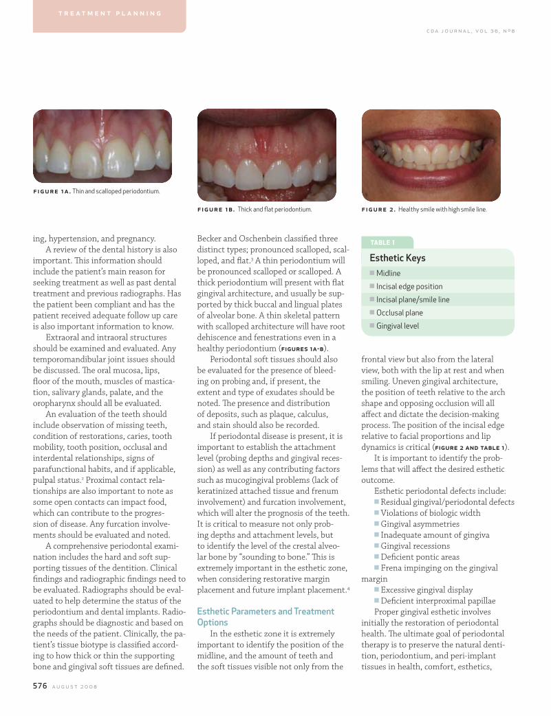
Becker and Oschenbein classified three distinct types; pronounced scalloped, scal-loped, and flat.3 A thin periodontium will be pronounced scalloped or scalloped. A thick periodontium will present with flat gingival architecture, and usually be sup-ported by thick buccal and lingual plates of alveolar bone. A thin skeletal pattern with scalloped architecture will have root dehiscence and fenestrations even in a healthy periodontium (figures 1a-b).
Periodontal soft tissues should also be evaluated for the presence of bleed-ing on probing and, if present, the extent and type of exudates should be noted. The presence and distribution of deposits, such as plaque, calculus, and stain should also be recorded.
If periodontal disease is present, it is important to establish the attachment level (probing depths and gingival reces-sion) as well as any contributing factors such as mucogingival problems (lack of keratinized attached tissue and frenum involvement) and furcation involvement, which will alter the prognosis of the teeth. It is critical to measure not only prob-ing depths and attachment levels, but to identify the level of the crestal alveo-lar bone by “sounding to bone.” This is extremely important in the esthetic zone, when considering restorative margin placement and future implant placement.4
esthetic Parameters and Treatment options
In the esthetic zone it is extremely important to identify the position of the midline, and the amount of teeth and the soft tissues visible not only from the
f igur e 1a. Thin and scalloped periodontium.
figure 1b . Thick and flat periodontium. fig ur e 2 . Healthy smile with high smile line.
t r e a t m e n t p l a n n i n g
tABle 1
esthetic Keysn Midlinen Incisal edge positionn Incisal plane/smile linen Occlusal planen Gingival level
c d a j o u r n a l , v o l 3 6 , n º 8
a u g u s t 2 0 0 8 577
In the esthetic zone, it is sometimes bet-ter to make compromises and understand that residual pocket depths may be main-tainable. Nonsurgical therapy can keep interproximal papilla intact and is more desirable as compared to the esthetic problems associated with pocket elimina-tion and the creation of “black triangles.”
Not all patients respond well to therapy nor are able to maintain a healthy periodontium over time. Factors contrib-uting to a less than optimal result usually include poor compliance with oral hygiene regimens and failure to return for regular maintenance care. Inadequate debride-ment may account for some treatment failures or reinfection, however, the pres-ence of systemic conditions or local risk factors may also have a significant impact on the success or failure of treatment. Oc-clusal trauma, deep inaccessible pockets, overhangs, and poor crown margins can also contribute to recurrent disease.
Over the past few years, much has been learned about the relationship between risk factors and the develop-ment and progression of periodontal disease. Studies have identified smok-ing, diabetes, stress, genetic factors and behavioral issues as possible risk factors for periodontal disease.15-19 In noncompli-ant patients, those who smoke, or have diabetes can prove challenging to treat.
Violations of Biologic WidthThe biologic width is defined as the
dimension from the crest of the alveolar bone to the base of the sulcus, which includes the combined width of the con-
and function.1 Treatment of periodon-tal diseases typically consists of initial nonsurgical debridement followed by re-evaluation, during which the need for additional treatment is evaluated.
gingival and/or Periodontal DefectsConventional nonsurgical treatment
of periodontal disease consists of me-chanical tooth debridement both supra and subgingivally along with instruction in proper home care methods (figures 3 a-b). The primary objective of debri-dement, i.e., scaling and root planing (SRP) is to restore periodontal health by removing pathogenic products that induce inflammation (i.e., plaque, calculus, and endotoxin) from periodontally involved root surfaces. Scaling and root planing may also alter the microbial composition to a flora more compatible with periodon-tal health. However, patient cooperation is paramount. Subgingival debridement without adequate oral hygiene mea-sures results in a limited response.5-7
While the primary objective of debridement is to remove pathogenic products, there are numerous difficulties with achieving this task. Difficulties with adequate debridement include difficulty with access and visualization, no defini-tive end point, and operator experience. Sites with deep periodontal pockets, grooves, furcations, and concavities are difficult to access with periodontal instrumentation and thus periodonto-pathic bacteria may remain in those sites.
Other obstacles have to do with the bacteria themselves. The authors’ current view of bacteria is that of a biofilm.8-10
According to this concept, bacteria do not exist as free-floating bacteria, but as attached bacteria in biofilms. Bacteria in biofilms have a unique position in that they develop a synergistic relation-ship, cooperate with each other, and form protective barriers of exopolysac-charides. In addition, biofilm bacteria are relatively inactive metabolically and exert more resistance to antibiotics than free-floating bacteria and are thus more difficult to eradicate. Subgingival plaque in biofilm can evade the defense mechanisms of the host and diminish the effect of chemotherapeutic agents.
As a result of the difficulties described above, conventional mechanical root debridement does not usually eradicate all pathogenic bacteria from the subgingival ecosystem.11-13 As a result, some propose that patients receive antibiotics at the time of hand scaling and root planing. This blanket use of antibiotics is not nec-essary for most patients because they usu-ally respond well to nonsurgical treatment without antibiotics. In addition, it has been shown that without the proper re-moval of subgingival deposits, the use of subgingival medicaments is not useful.14
Furthermore, recent studies have concluded that surgery may provide a better long-term outcome with less need for adjunctive treatments. Long-term treatment of patients with nonsurgical therapy with or without antibiotic therapy may result in a situation that even surgery is not designed to correct. Most forms of periodontal surgery achieve best results in the early to moderate severities of disease.
f igure 3a. Soft tissue inflammation due to overcontoured restorations. figure 3b. Soft tissue resolution after initial
therapy and placement of provisional restorations with ideal contours (Courtesy of Carol Lam, DDS).
fig ur e 4 . Violation of biologic width (gingival at-tachment) with inflammatory soft tissue response.
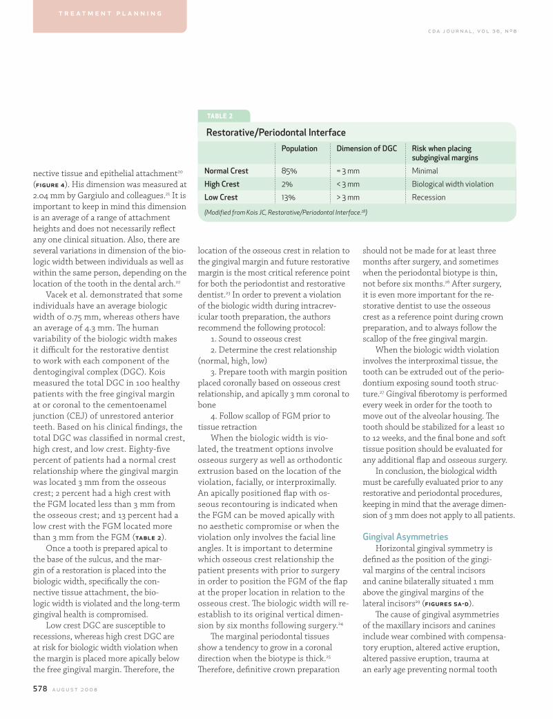
578 a u g u s t 2 0 0 8
c d a j o u r n a l , v o l 3 6 , n º 8
nective tissue and epithelial attachment20 (figure 4). His dimension was measured at 2.04 mm by Gargiulo and colleagues.21 It is important to keep in mind this dimension is an average of a range of attachment heights and does not necessarily reflect any one clinical situation. Also, there are several variations in dimension of the bio-logic width between individuals as well as within the same person, depending on the location of the tooth in the dental arch.22
Vacek et al. demonstrated that some individuals have an average biologic width of 0.75 mm, whereas others have an average of 4.3 mm. The human variability of the biologic width makes it difficult for the restorative dentist to work with each component of the dentogingival complex (DGC). Kois measured the total DGC in 100 healthy patients with the free gingival margin at or coronal to the cementoenamel junction (CEJ) of unrestored anterior teeth. Based on his clinical findings, the total DGC was classified in normal crest, high crest, and low crest. Eighty-five percent of patients had a normal crest relationship where the gingival margin was located 3 mm from the osseous crest; 2 percent had a high crest with the FGM located less than 3 mm from the osseous crest; and 13 percent had a low crest with the FGM located more than 3 mm from the FGM (table 2).
Once a tooth is prepared apical to the base of the sulcus, and the mar-gin of a restoration is placed into the biologic width, specifically the con-nective tissue attachment, the bio-logic width is violated and the long-term gingival health is compromised.
Low crest DGC are susceptible to recessions, whereas high crest DGC are at risk for biologic width violation when the margin is placed more apically below the free gingival margin. Therefore, the
location of the osseous crest in relation to the gingival margin and future restorative margin is the most critical reference point for both the periodontist and restorative dentist.23 In order to prevent a violation of the biologic width during intracrev-icular tooth preparation, the authors recommend the following protocol:
1. Sound to osseous crest2. Determine the crest relationship
(normal, high, low)3. Prepare tooth with margin position
placed coronally based on osseous crest relationship, and apically 3 mm coronal to bone
4. Follow scallop of FGM prior to tissue retraction
When the biologic width is vio-lated, the treatment options involve osseous surgery as well as orthodontic extrusion based on the location of the violation, facially, or interproximally. An apically positioned flap with os-seous recontouring is indicated when the FGM can be moved apically with no aesthetic compromise or when the violation only involves the facial line angles. It is important to determine which osseous crest relationship the patient presents with prior to surgery in order to position the FGM of the flap at the proper location in relation to the osseous crest. The biologic width will re-establish to its original vertical dimen-sion by six months following surgery.24
The marginal periodontal tissues show a tendency to grow in a coronal direction when the biotype is thick.25 Therefore, definitive crown preparation
should not be made for at least three months after surgery, and sometimes when the periodontal biotype is thin, not before six months.26 After surgery, it is even more important for the re-storative dentist to use the osseous crest as a reference point during crown preparation, and to always follow the scallop of the free gingival margin.
When the biologic width violation involves the interproximal tissue, the tooth can be extruded out of the perio-dontium exposing sound tooth struc-ture.27 Gingival fiberotomy is performed every week in order for the tooth to move out of the alveolar housing. The tooth should be stabilized for a least 10 to 12 weeks, and the final bone and soft tissue position should be evaluated for any additional flap and osseous surgery.
In conclusion, the biological width must be carefully evaluated prior to any restorative and periodontal procedures, keeping in mind that the average dimen-sion of 3 mm does not apply to all patients.
gingival asymmetriesHorizontal gingival symmetry is
defined as the position of the gingi-val margins of the central incisors and canine bilaterally situated 1 mm above the gingival margins of the lateral incisors29 (figures 5a-d).
The cause of gingival asymmetries of the maxillary incisors and canines include wear combined with compensa-tory eruption, altered active eruption, altered passive eruption, trauma at an early age preventing normal tooth
t r e a t m e n t p l a n n i n g
tABle 2
restorative/Periodontal InterfacePopulation Dimension of DgC risk when placing
subgingival margins
normal Crest 85% = 3 mm Minimal
high Crest 2% < 3 mm Biological width violation
low Crest 13% > 3 mm Recession
(Modified from Kois JC, Restorative/Periodontal Interface.28)
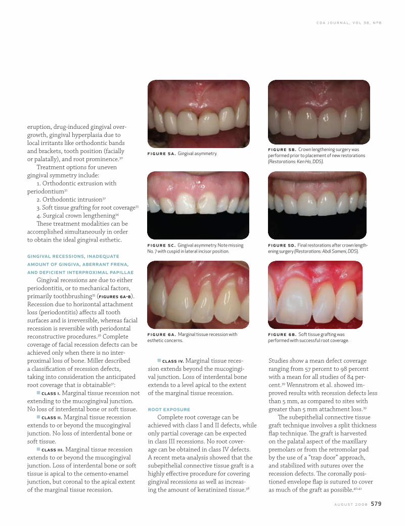
c d a j o u r n a l , v o l 3 6 , n º 8
a u g u s t 2 0 0 8 579
eruption, drug-induced gingival over-growth, gingival hyperplasia due to local irritants like orthodontic bands and brackets, tooth position (facially or palatally), and root prominence.30
Treatment options for uneven gingival symmetry include:
1. Orthodontic extrusion with periodontium31
2. Orthodontic intrusion32
3. Soft tissue grafting for root coverage33
4. Surgical crown lengthening34
These treatment modalities can be accomplished simultaneously in order to obtain the ideal gingival esthetic.
gingival recessions, inadequate amount of gingiva, aberrant frena, and deficient interproximal papillae
Gingival recessions are due to either periodontitis, or to mechanical factors, primarily toothbrushing35 (figures 6a-b). Recession due to horizontal attachment loss (periodontitis) affects all tooth surfaces and is irreversible, whereas facial recession is reversible with periodontal reconstructive procedures.36 Complete coverage of facial recession defects can be achieved only when there is no inter-proximal loss of bone. Miller described a classification of recession defects, taking into consideration the anticipated root coverage that is obtainable37:
n class i. Marginal tissue recession not extending to the mucogingival junction. No loss of interdental bone or soft tissue.
n class ii. Marginal tissue recession extends to or beyond the mucogingival junction. No loss of interdental bone or soft tissue.
n class iii. Marginal tissue recession extends to or beyond the mucogingival junction. Loss of interdental bone or soft tissue is apical to the cemento-enamel junction, but coronal to the apical extent of the marginal tissue recession.
n class iv. Marginal tissue reces-sion extends beyond the mucogingi-val junction. Loss of interdental bone extends to a level apical to the extent of the marginal tissue recession.
root exposureComplete root coverage can be
achieved with class I and II defects, while only partial coverage can be expected in class III recessions. No root cover-age can be obtained in class IV defects. A recent meta-analysis showed that the subepithelial connective tissue graft is a highly effective procedure for covering gingival recessions as well as increas-ing the amount of keratinized tissue.38
Studies show a mean defect coverage ranging from 57 percent to 98 percent with a mean for all studies of 84 per-cent.39 Wennstrom et al. showed im-proved results with recession defects less than 5 mm, as compared to sites with greater than 5 mm attachment loss.39
The subepithelial connective tissue graft technique involves a split thickness flap technique. The graft is harvested on the palatal aspect of the maxillary premolars or from the retromolar pad by the use of a “trap door” approach, and stabilized with sutures over the recession defects. The coronally posi-tioned envelope flap is sutured to cover as much of the graft as possible.40,41
figure 5a . Gingival asymmetry.
figure 6a . Marginal tissue recession with esthetic concerns.
figure 5c . Gingival asymmetry. Note missing No. 7 with cuspid in lateral incisor position.
fig ur e 5b. Crown lengthening surgery was performed prior to placement of new restorations (Restorations: Ken Ho, DDS).
fig ur e 6 b. Soft tissue grafting was performed with successful root coverage.
figure 5d. Final restorations after crown length-ening surgery (Restorations: Abdi Sameni, DDS).
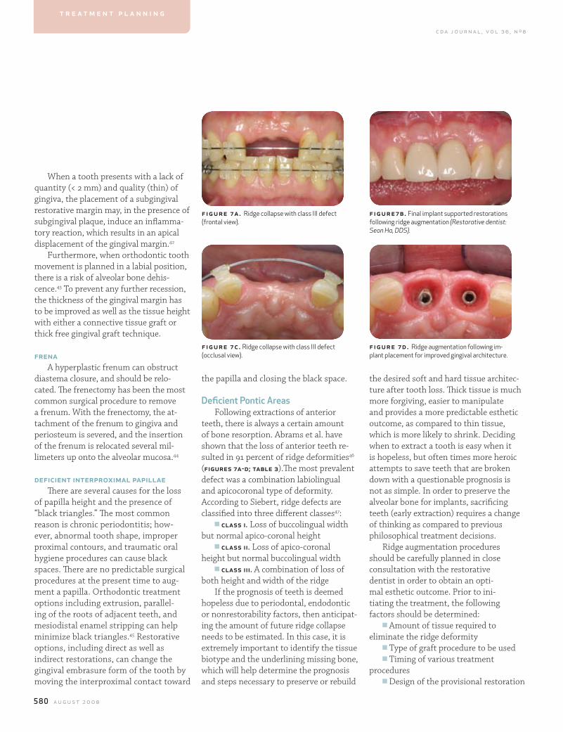
580 a u g u s t 2 0 0 8
c d a j o u r n a l , v o l 3 6 , n º 8
the desired soft and hard tissue architec-ture after tooth loss. Thick tissue is much more forgiving, easier to manipulate and provides a more predictable esthetic outcome, as compared to thin tissue, which is more likely to shrink. Deciding when to extract a tooth is easy when it is hopeless, but often times more heroic attempts to save teeth that are broken down with a questionable prognosis is not as simple. In order to preserve the alveolar bone for implants, sacrificing teeth (early extraction) requires a change of thinking as compared to previous philosophical treatment decisions.
Ridge augmentation procedures should be carefully planned in close consultation with the restorative dentist in order to obtain an opti-mal esthetic outcome. Prior to ini-tiating the treatment, the following factors should be determined:
n Amount of tissue required to eliminate the ridge deformity
n Type of graft procedure to be usedn Timing of various treatment
proceduresn Design of the provisional restoration
the papilla and closing the black space.
Deficient Pontic areasFollowing extractions of anterior
teeth, there is always a certain amount of bone resorption. Abrams et al. have shown that the loss of anterior teeth re-sulted in 91 percent of ridge deformities46 (figures 7a-d; table 3).The most prevalent defect was a combination labiolingual and apicocoronal type of deformity. According to Siebert, ridge defects are classified into three different classes47:
n class i. Loss of buccolingual width but normal apico-coronal height
n class ii. Loss of apico-coronal height but normal buccolingual width
n class iii. A combination of loss of both height and width of the ridge
If the prognosis of teeth is deemed hopeless due to periodontal, endodontic or nonrestorability factors, then anticipat-ing the amount of future ridge collapse needs to be estimated. In this case, it is extremely important to identify the tissue biotype and the underlining missing bone, which will help determine the prognosis and steps necessary to preserve or rebuild
When a tooth presents with a lack of quantity (< 2 mm) and quality (thin) of gingiva, the placement of a subgingival restorative margin may, in the presence of subgingival plaque, induce an inflamma-tory reaction, which results in an apical displacement of the gingival margin.42
Furthermore, when orthodontic tooth movement is planned in a labial position, there is a risk of alveolar bone dehis-cence.43 To prevent any further recession, the thickness of the gingival margin has to be improved as well as the tissue height with either a connective tissue graft or thick free gingival graft technique.
frenaA hyperplastic frenum can obstruct
diastema closure, and should be relo-cated. The frenectomy has been the most common surgical procedure to remove a frenum. With the frenectomy, the at-tachment of the frenum to gingiva and periosteum is severed, and the insertion of the frenum is relocated several mil-limeters up onto the alveolar mucosa.44
deficient interproximal papillaeThere are several causes for the loss
of papilla height and the presence of “black triangles.” The most common reason is chronic periodontitis; how-ever, abnormal tooth shape, improper proximal contours, and traumatic oral hygiene procedures can cause black spaces. There are no predictable surgical procedures at the present time to aug-ment a papilla. Orthodontic treatment options including extrusion, parallel-ing of the roots of adjacent teeth, and mesiodistal enamel stripping can help minimize black triangles.45 Restorative options, including direct as well as indirect restorations, can change the gingival embrasure form of the tooth by moving the interproximal contact toward
figure 7a. Ridge collapse with class III defect (frontal view).
figure 7c. Ridge collapse with class III defect (occlusal view).
fig ur e7 b. Final implant supported restorations following ridge augmentation (Restorative dentist: Seon Ha, DDS).
fig ur e 7 d . Ridge augmentation following im-plant placement for improved gingival architecture.
t r e a t m e n t p l a n n i n g
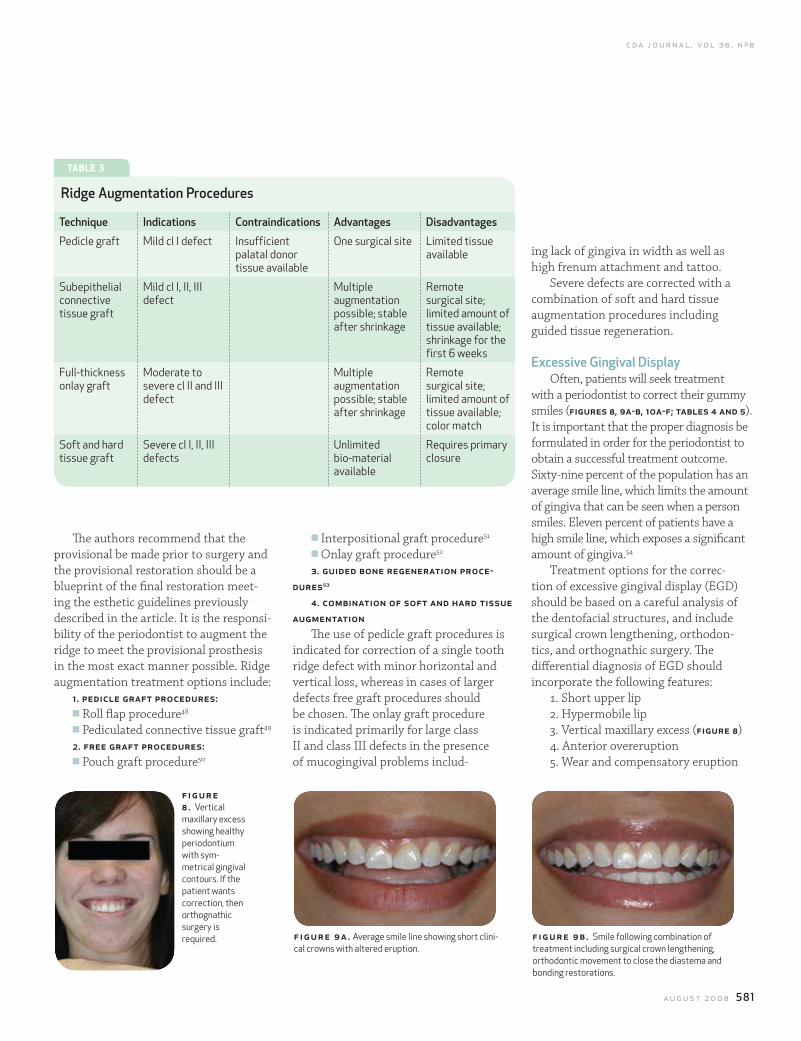
c d a j o u r n a l , v o l 3 6 , n º 8
a u g u s t 2 0 0 8 581
ing lack of gingiva in width as well as high frenum attachment and tattoo.
Severe defects are corrected with a combination of soft and hard tissue augmentation procedures including guided tissue regeneration.
excessive gingival DisplayOften, patients will seek treatment
with a periodontist to correct their gummy smiles (figures 8, 9a-b, 10a-f; tables 4 and 5). It is important that the proper diagnosis be formulated in order for the periodontist to obtain a successful treatment outcome. Sixty-nine percent of the population has an average smile line, which limits the amount of gingiva that can be seen when a person smiles. Eleven percent of patients have a high smile line, which exposes a significant amount of gingiva.54
Treatment options for the correc-tion of excessive gingival display (EGD) should be based on a careful analysis of the dentofacial structures, and include surgical crown lengthening, orthodon-tics, and orthognathic surgery. The differential diagnosis of EGD should incorporate the following features:
1. Short upper lip2. Hypermobile lip3. Vertical maxillary excess (figure 8)4. Anterior overeruption5. Wear and compensatory eruption
n Interpositional graft procedure51
n Onlay graft procedure52
3. guided bone regeneration proce-dures53
4. combination of soft and hard tissue augmentation
The use of pedicle graft procedures is indicated for correction of a single tooth ridge defect with minor horizontal and vertical loss, whereas in cases of larger defects free graft procedures should be chosen. The onlay graft procedure is indicated primarily for large class II and class III defects in the presence of mucogingival problems includ-
The authors recommend that the provisional be made prior to surgery and the provisional restoration should be a blueprint of the final restoration meet-ing the esthetic guidelines previously described in the article. It is the responsi-bility of the periodontist to augment the ridge to meet the provisional prosthesis in the most exact manner possible. Ridge augmentation treatment options include:
1. pedicle graft procedures:n Roll flap procedure48
n Pediculated connective tissue graft49
2. free graft procedures:n Pouch graft procedure50
figure 8 . Vertical maxillary excess showing healthy periodontium with sym-metrical gingival contours. If the patient wants correction, then orthognathic surgery is required. figure 9a. Average smile line showing short clini-
cal crowns with altered eruption.fig ur e 9 b. Smile following combination of treatment including surgical crown lengthening, orthodontic movement to close the diastema and bonding restorations.
tABle 3
ridge augmentation Procedures
Technique Indications Contraindications advantages Disadvantages
Pedicle graft Mild cl I defect Insufficient palatal donor tissue available
One surgical site Limited tissue available
Subepithelial connective tissue graft
Mild cl I, II, III defect
Multiple augmentation possible; stable after shrinkage
Remote surgical site; limited amount of tissue available; shrinkage for the first 6 weeks
Full-thickness onlay graft
Moderate to severe cl II and III defect
Multiple augmentation possible; stable after shrinkage
Remote surgical site; limited amount of tissue available; color match
Soft and hard tissue graft
Severe cl I, II, III defects
Unlimited bio-material available
Requires primary closure
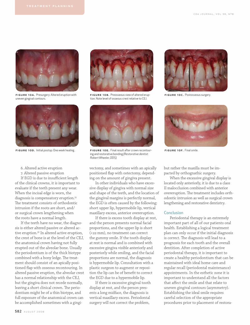
582 a u g u s t 2 0 0 8
c d a j o u r n a l , v o l 3 6 , n º 8
but rather the maxilla must be im-pacted by orthognathic surgery.
When the excessive gingival display is located only anteriorly, it is due to a class II malocclusion combined with anterior overeruption. The treatment includes orth-odontic intrusion as well as surgical crown lengthening and restorative dentistry.
Conclusion Periodontal therapy is an extremely
important part of all of our patients oral health. Establishing a logical treatment plan can only occur if the initial diagnosis is correct. The diagnosis will lead to a prognosis for each tooth and the overall dentition. After completion of active periodontal therapy, it is important to create a healthy periodontium that can be maintained with ideal home care and regular recall (periodontal maintenance) appointments. In the esthetic zone it is important to understand all the factors that affect the smile and that relate to uneven gingival contours (asymmetry). Establishing the ideal smile requires careful selection of the appropriate procedures prior to placement of restora-
vectomy, and sometimes with an apically positioned flap with ostectomy, depend-ing on the amount of gingiva present.
In other individuals who have exces-sive display of gingiva with normal size and shape of the teeth, and the location of the gingival margins is perfectly normal, the EGD is often caused by the following: short upper lip, hypermobile lip, vertical maxillary excess, anterior overeruption.
If there is excess tooth display at rest, and the person presents normal facial proportions, and the upper lip is short (<22 mm), no treatment can correct the gummy smile. If the tooth display at rest is normal and is combined with excessive gingiva visible anteriorly and posteriorly while smiling, and the facial proportions are normal, the diagnosis is hypermobile lip. Consultation with a plastic surgeon to augment or reposi-tion the lip can be of benefit to correct the EGD due to a hypermobile lip.
If there is excessive gingival tooth display at rest, and the person pres-ents a long midface, the diagnosis is vertical maxillary excess. Periodontal surgery will not correct the problem,
6. Altered active eruption 7. Altered passive eruptionIf EGD is due to insufficient length
of the clinical crowns, it is important to evaluate if the teeth present any wear. When the incisal edge is worn, the diagnosis is compensatory eruption.55 The treatment consists of orthodontic intrusion if the roots are short, and/or surgical crown lengthening when the roots have a normal length.
If the teeth have no wear, the diagno-sis is either altered passive or altered ac-tive eruption.56 In altered active eruption, the crest of bone is at the level of the CEJ, the anatomical crown having not fully erupted out of the alveolar bone. Usually the periodontium is of the thick biotype combined with a bony ledge. The treat-ment should consist of an apically posi-tioned flap with osseous recontouring. In altered passive eruption, the alveolar crest has a normal relationship with the CEJ, but the gingiva does not recede normally, leaving a short clinical crown. The perio-dontium might be of a thin biotype, and full exposure of the anatomical crown can be accomplished sometimes with a gingi-
f igur e 10a. Presurgery: Altered eruption with uneven gingival contours.
f igur e 10d. Initial postop: One week healing .
figure 10b. Preosseous view of altered erup-tion. Note level of osseous crest relative to CEJ.
figure 10e. Final result after crown recontour-ing and restorative bonding (Restorative dentist: Robert Wheeler, DDS).
fig ur e 10c. Postosseous surgery.
fig ur e 10f. Final smile.
t r e a t m e n t p l a n n i n g
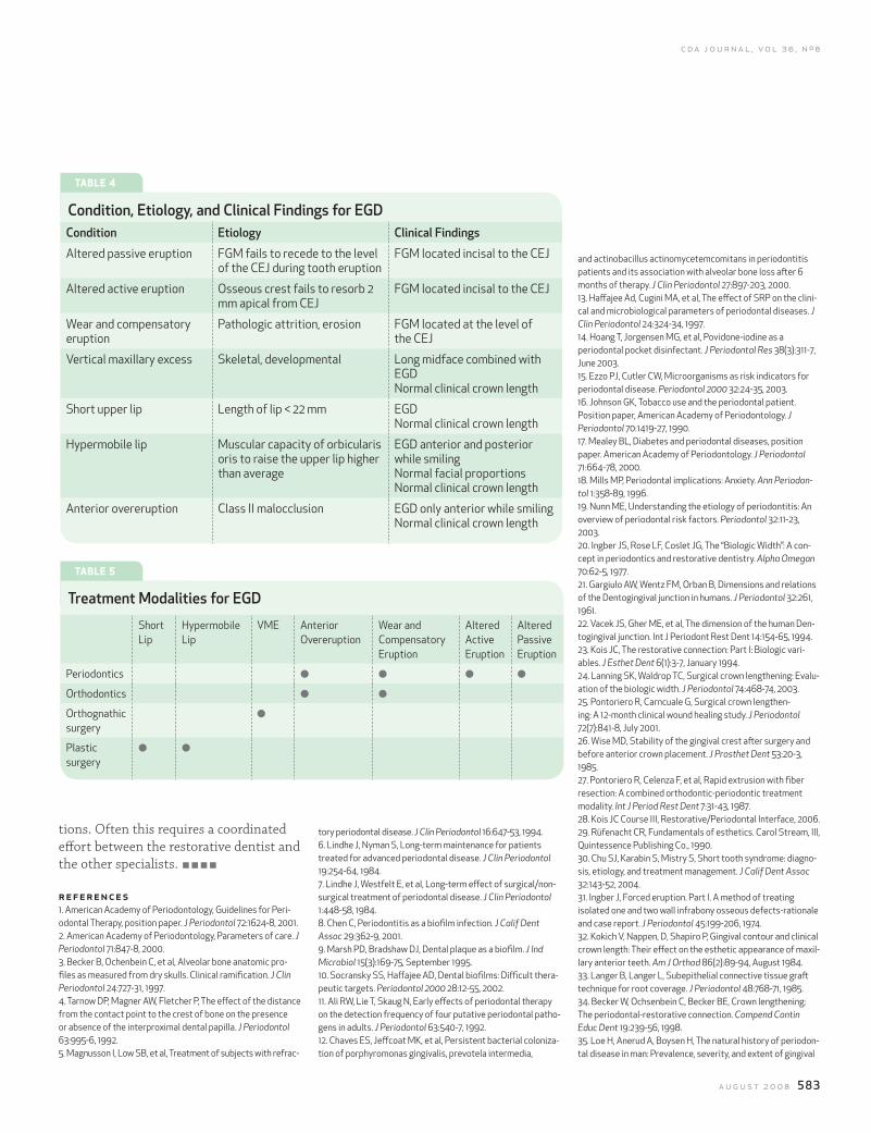
c d a j o u r n a l , v o l 3 6 , n º 8
a u g u s t 2 0 0 8 583
and actinobacillus actinomycetemcomitans in periodontitis patients and its association with alveolar bone loss after 6 months of therapy. J Clin Periodontol 27:897-203, 2000.13. Haffajee Ad, Cugini MA, et al, The effect of SRP on the clini-cal and microbiological parameters of periodontal diseases. J Clin Periodontol 24:324-34, 1997.14. Hoang T, Jorgensen MG, et al, Povidone-iodine as a periodontal pocket disinfectant. J Periodontal Res 38(3):311-7, June 2003.15. Ezzo PJ, Cutler CW, Microorganisms as risk indicators for periodontal disease. Periodontol 2000 32:24-35, 2003.16. Johnson GK, Tobacco use and the periodontal patient. Position paper, American Academy of Periodontology. J Periodontol 70:1419-27, 1990.17. Mealey BL, Diabetes and periodontal diseases, position paper. American Academy of Periodontology. J Periodontol 71:664-78, 2000.18. Mills MP, Periodontal implications: Anxiety. Ann Periodon-tol 1:358-89, 1996.19. Nunn ME, Understanding the etiology of periodontitis: An overview of periodontal risk factors. Periodontol 32:11-23, 2003.20. Ingber JS, Rose LF, Coslet JG, The “Biologic Width”: A con-cept in periodontics and restorative dentistry. Alpha Omegan 70:62-5, 1977.21. Gargiulo AW, Wentz FM, Orban B, Dimensions and relations of the Dentogingival junction in humans. J Periodontol 32:261, 1961.22. Vacek JS, Gher ME, et al, The dimension of the human Den-togingival junction. Int J Periodont Rest Dent 14:154-65, 1994.23. Kois JC, The restorative connection: Part I: Biologic vari-ables. J Esthet Dent 6(1):3-7, January 1994.24. Lanning SK, Waldrop TC, Surgical crown lengthening: Evalu-ation of the biologic width. J Periodontol 74:468-74, 2003.25. Pontoriero R, Carncuale G, Surgical crown lengthen-ing: A 12-month clinical wound healing study. J Periodontol 72(7):841-8, July 2001.26. Wise MD, Stability of the gingival crest after surgery and before anterior crown placement. J Prosthet Dent 53:20-3, 1985.27. Pontoriero R, Celenza F, et al, Rapid extrusion with fiber resection: A combined orthodontic-periodontic treatment modality. Int J Period Rest Dent 7:31-43, 1987.28. Kois JC Course III, Restorative/Periodontal Interface, 2006.29. Rüfenacht CR, Fundamentals of esthetics. Carol Stream, Ill, Quintessence Publishing Co., 1990.30. Chu SJ, Karabin S, Mistry S, Short tooth syndrome: diagno-sis, etiology, and treatment management. J Calif Dent Assoc 32:143-52, 2004.31. Ingber J, Forced eruption. Part I. A method of treating isolated one and two wall infrabony osseous defects-rationale and case report. J Periodontol 45:199-206, 1974.32. Kokich V, Nappen, D, Shapiro P, Gingival contour and clinical crown length: Their effect on the esthetic appearance of maxil-lary anterior teeth. Am J Orthod 86(2):89-94, August 1984.33. Langer B, Langer L, Subepithelial connective tissue graft technique for root coverage. J Periodontol 48:768-71, 1985.34. Becker W, Ochsenbein C, Becker BE, Crown lengthening: The periodontal-restorative connection. Compend Contin Educ Dent 19:239-56, 1998.35. Loe H, Anerud A, Boysen H, The natural history of periodon-tal disease in man: Prevalence, severity, and extent of gingival
tory periodontal disease. J Clin Periodontol 16:647-53, 1994.6. Lindhe J, Nyman S, Long-term maintenance for patients treated for advanced periodontal disease. J Clin Periodontol 19:254-64, 1984.7. Lindhe J, Westfelt E, et al, Long-term effect of surgical/non-surgical treatment of periodontal disease. J Clin Periodontol 1:448-58, 1984.8. Chen C, Periodontitis as a biofilm infection. J Calif Dent Assoc 29:362-9, 2001.9. Marsh PD, Bradshaw DJ, Dental plaque as a biofilm. J Ind Microbiol 15(3):169-75, September 1995.10. Socransky SS, Haffajee AD, Dental biofilms: Difficult thera-peutic targets. Periodontol 2000 28:12-55, 2002.11. Ali RW, Lie T, Skaug N, Early effects of periodontal therapy on the detection frequency of four putative periodontal patho-gens in adults. J Periodontol 63:540-7, 1992.12. Chaves ES, Jeffcoat MK, et al, Persistent bacterial coloniza-tion of porphyromonas gingivalis, prevotela intermedia,
tions. Often this requires a coordinated effort between the restorative dentist and the other specialists.
r ef erences1. American Academy of Periodontology, Guidelines for Peri-odontal Therapy, position paper. J Periodontol 72:1624-8, 2001.2. American Academy of Periodontology, Parameters of care. J Periodontol 71:847-8, 2000.3. Becker B, Ochenbein C, et al, Alveolar bone anatomic pro-files as measured from dry skulls. Clinical ramification. J Clin Periodontol 24:727-31, 1997.4. Tarnow DP, Magner AW, Fletcher P, The effect of the distance from the contact point to the crest of bone on the presence or absence of the interproximal dental papilla. J Periodontol 63:995-6, 1992.5. Magnusson I, Low SB, et al, Treatment of subjects with refrac-
tABle 4
Condition, etiology, and Clinical findings for egDCondition etiology Clinical findings
Altered passive eruption FGM fails to recede to the level of the CEJ during tooth eruption
FGM located incisal to the CEJ
Altered active eruption Osseous crest fails to resorb 2 mm apical from CEJ
FGM located incisal to the CEJ
Wear and compensatory eruption
Pathologic attrition, erosion FGM located at the level of the CEJ
Vertical maxillary excess Skeletal, developmental Long midface combined with EGDNormal clinical crown length
Short upper lip Length of lip < 22 mm EGDNormal clinical crown length
Hypermobile lip Muscular capacity of orbicularis oris to raise the upper lip higher than average
EGD anterior and posterior while smilingNormal facial proportionsNormal clinical crown length
Anterior overeruption Class II malocclusion EGD only anterior while smilingNormal clinical crown length
tABle 5
Treatment modalities for egD
Short Lip
Hypermobile Lip
VME Anterior Overeruption
Wear and Compensatory Eruption
Altered Active Eruption
Altered Passive Eruption
Periodontics l l l l
Orthodontics l l
Orthognathic surgery
l
Plastic surgery
l l

584 a u g u s t 2 0 0 8
c d a j o u r n a l , v o l 3 6 , n º 8
52. Siebert JS, Louis JV, Soft tissue ridge augmentation utiliz-ing a combination onlay-interpositional graft procedure: A case report. Int J Perio Rest Dent 16:311-21, 1996.53. Buser D, Bragger U, Regeneration and enlargement of jaw bone using guided tissue regeneration. Clin Oral Imp Res 1:22-32, 1990.54. Tjan AH, Miller GD, et al Some esthetic factors in a smile. J Prosthet Dent 51:24-8, 1984.55. Chu SJ, Karabin S, Mistry S, Short tooth syndrome: diagno-sis, etiology, and treatment management. J Calif Dent Assoc 32:143-52, 2004.56. Evian CI, Cutler SA, Rosenberg ES, Altered passive erup-tion: The undiagnosed entity. J Am Dent Assoc 124:107-10, 1993.
to request a printed copy of this article, please con-tact Nicolas Ravon, DDS, MSD, 3808 Riverside Drive, Suite 305, Burbank, Calif., 91505.
reactions to orthodontic tooth movement in monkeys. J Clin Periodontol 14:121-9, 1987.44. Edwards JG, The diastema, the frenum, the frenectomy: A clinical study. Am J Orthod 71:489-508, 1987.45. Tuverson DL, Anterior interocclusal relations. Am J Orthod 78, 361-93, 1980.46. Abrams H, Kopczyk RA, Kaplan Al, Incidence of anterior ridge deformities in partially edentulous patients. J Prosthet Dent 57:191-4, 1987.47. Siebert JS, Reconstruction of deformed, partially edentu-lous ridges, using full thickness onlay grafts: I. Technique and wound healing. Comp Cont Edu Gen Dent 4:437-53, 1983.48. Abrams L, Augmentation of the deformed residual eden-tulous ridge for fixed prosthesis. Comp Cont Educ Gen Dent 3:205-14, 1980.49. Mathews DP, The pediculated connective tissue graft: a technique for improving unaesthetic implant restorations. Pract Proced Aesth Dent 9, 719-24, 2002.50. Garber D, Rosenberg E, The edentulous ridge in fixed prosthodontics. Comp Cont Ed Gen Dent 2:212, 1981.51. Langer B, Calagna LJ, The subepithelial connective tissue graft: a new approach to the enhancement of anterior cosmet-ics. Int J Perio Rest Dent 2:23-33, 1982.
recession. J Periodontol 63:489-95, 1992.36. American Academy of Periodontology Report. Oral recon-structive and corrective considerations in periodontal therapy. J Periodontol 76:1588-600, 2005.37. Miller PD Jr., A classification of marginal tissue recession. Int J Periodontics Restorative Dent 5:8-13, 1985.38. Oates TW, Robinson M, Gunsolley JC, Surgical therapies for the treatment of gingival recession. A systematic review. Ann Periodontol 8:303-20, 2003.39. Wennstrom J, Pini-Prato G, Mucogingival therapy. Root coverage. Clin Periodontol Implant Dent pages 550-696; Jan Lindhe, Thorkild Karring, Niklaus P, Copenhagen: Munksgaard, 1997.40. Langer B, Calagna LJ, The subepithelial connective tissue graft. A new approach to the enhancement of anterior cosmet-ics. Int J Periodontics Restorative Dent 2:22-33, 1982.41. Allen AL, Use of the supraperiosteal envelope in soft tissue grafting for root coverage. I. Rationale and technique. Int J Periodontics Restorative Dent 14:216-27, 1994.42. Ericsson I, Lindhe J, Recession in sites with inadequate width of the keratinized gingiva. An experimental study in the dog. J Clin Periodontol 11:95-103, 1984.43. Wennström JL, Lindhe J, et al, Some periodontal tissue
t r e a t m e n t p l a n n i n g
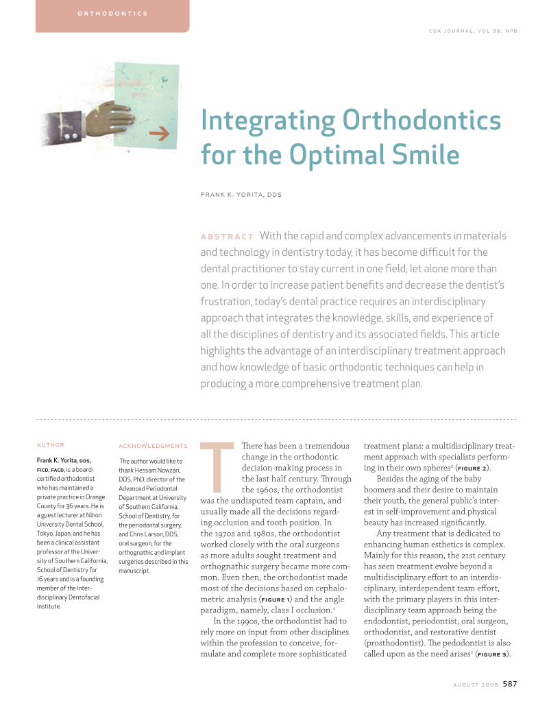
c d a j o u r n a l , v o l 3 6 , n º 8
a u g u s t 2 0 0 8 587
Integrating orthodontics for the optimal Smilefrank k. yorita, dds
abstract With the rapid and complex advancements in materials and technology in dentistry today, it has become difficult for the dental practitioner to stay current in one field, let alone more than one. In order to increase patient benefits and decrease the dentist’s frustration, today’s dental practice requires an interdisciplinary approach that integrates the knowledge, skills, and experience of all the disciplines of dentistry and its associated fields. This article highlights the advantage of an interdisciplinary treatment approach and how knowledge of basic orthodontic techniques can help in producing a more comprehensive treatment plan.
author
frank K. yorita, dds, ficd, facd, is a board-certified orthodontist who has maintained a private practice in Orange County for 36 years. He is a guest lecturer at Nihon University Dental School, Tokyo, Japan, and he has been a clinical assistant professor at the Univer-sity of Southern California, School of Dentistry for 16 years and is a founding member of the Inter-disciplinary Dentofacial Institute.
There has been a tremendous change in the orthodontic decision-making process in the last half century. Through the 1960s, the orthodontist
was the undisputed team captain, and usually made all the decisions regard-ing occlusion and tooth position. In the 1970s and 1980s, the orthodontist worked closely with the oral surgeons as more adults sought treatment and orthognathic surgery became more com-mon. Even then, the orthodontist made most of the decisions based on cephalo-metric analysis (figure 1) and the angle paradigm, namely, class I occlusion.1
In the 1990s, the orthodontist had to rely more on input from other disciplines within the profession to conceive, for-mulate and complete more sophisticated
o r t h o d o n t i c s
treatment plans: a multidisciplinary treat-ment approach with specialists perform-ing in their own spheres2 (figure 2).
Besides the aging of the baby boomers and their desire to maintain their youth, the general public’s inter-est in self-improvement and physical beauty has increased significantly.
Any treatment that is dedicated to enhancing human esthetics is complex. Mainly for this reason, the 21st century has seen treatment evolve beyond a multidisciplinary effort to an interdis-ciplinary, interdependent team effort, with the primary players in this inter-disciplinary team approach being the endodontist, periodontist, oral surgeon, orthodontist, and restorative dentist (prosthodontist). The pedodontist is also called upon as the need arises2 (figure 3).
Tacknowledgments
The author would like to thank Hessam Nowzari, DDS, PhD, director of the Advanced Periodontal Department at University of Southern California, School of Dentistry, for the periodontal surgery, and Chris Larson, DDS, oral surgeon, for the orthognathic and implant surgeries described in this manuscript.
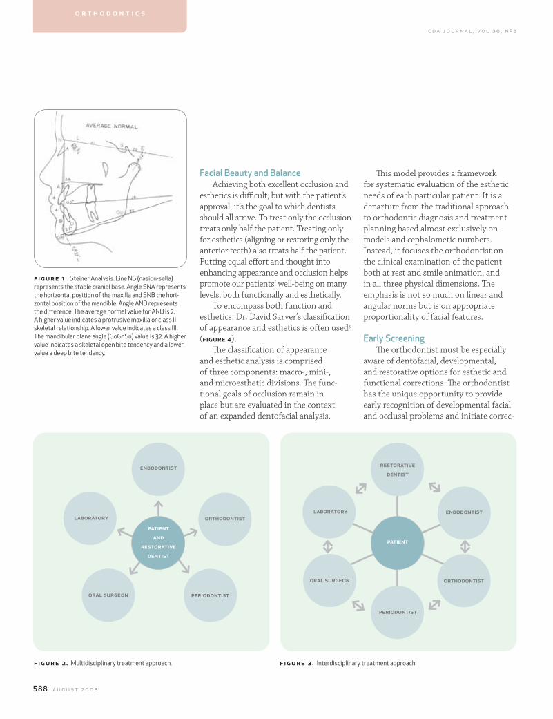
588 a u g u s t 2 0 0 8
c d a j o u r n a l , v o l 3 6 , n º 8
f igur e 1 . Steiner Analysis. Line NS (nasion-sella) represents the stable cranial base. Angle SNA represents the horizontal position of the maxilla and SNB the hori-zontal position of the mandible. Angle ANB represents the difference. The average normal value for ANB is 2. A higher value indicates a protrusive maxilla or class II skeletal relationship. A lower value indicates a class III. The mandibular plane angle (GoGnSn) value is 32. A higher value indicates a skeletal open bite tendency and a lower value a deep bite tendency.
f igur e 2 . Multidisciplinary treatment approach. fig ur e 3 . Interdisciplinary treatment approach.
facial Beauty and BalanceAchieving both excellent occlusion and
esthetics is difficult, but with the patient’s approval, it’s the goal to which dentists should all strive. To treat only the occlusion treats only half the patient. Treating only for esthetics (aligning or restoring only the anterior teeth) also treats half the patient. Putting equal effort and thought into enhancing appearance and occlusion helps promote our patients’ well-being on many levels, both functionally and esthetically.
To encompass both function and esthetics, Dr. David Sarver’s classification of appearance and esthetics is often used3 (figure 4).
The classification of appearance and esthetic analysis is comprised of three components: macro-, mini-, and microesthetic divisions. The func-tional goals of occlusion remain in place but are evaluated in the context of an expanded dentofacial analysis.
This model provides a framework for systematic evaluation of the esthetic needs of each particular patient. It is a departure from the traditional approach to orthodontic diagnosis and treatment planning based almost exclusively on models and cephalometic numbers. Instead, it focuses the orthodontist on the clinical examination of the patient both at rest and smile animation, and in all three physical dimensions. The emphasis is not so much on linear and angular norms but is on appropriate proportionality of facial features.
early ScreeningThe orthodontist must be especially
aware of dentofacial, developmental, and restorative options for esthetic and functional corrections. The orthodontist has the unique opportunity to provide early recognition of developmental facial and occlusal problems and initiate correc-
o r t h o d o n t i c s
patient and
restorative dentist
endodontist
laboratory
oral surgeon
orthodontist
periodontist
patient
restorative dentist
periodontist
laboratory
oral surgeon
endodontist
orthodontist
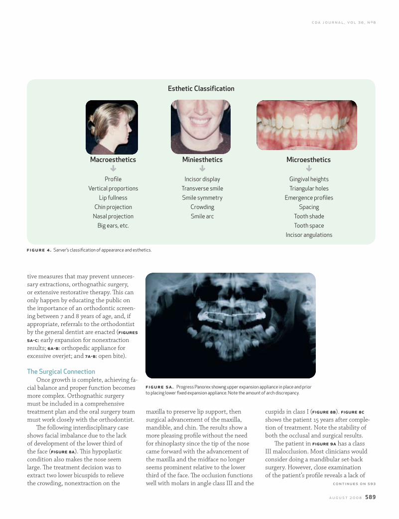
c d a j o u r n a l , v o l 3 6 , n º 8
a u g u s t 2 0 0 8 589
f igur e 4 . Sarver’s classification of appearance and esthetics.
tive measures that may prevent unneces-sary extractions, orthognathic surgery, or extensive restorative therapy. This can only happen by educating the public on the importance of an orthodontic screen-ing between 7 and 8 years of age, and, if appropriate, referrals to the orthodontist by the general dentist are enacted (figures 5a-c: early expansion for nonextraction results; 6a-b: orthopedic appliance for excessive overjet; and 7a-b: open bite).
The Surgical ConnectionOnce growth is complete, achieving fa-
cial balance and proper function becomes more complex. Orthognathic surgery must be included in a comprehensive treatment plan and the oral surgery team must work closely with the orthodontist.
The following interdisciplinary case shows facial imbalance due to the lack of development of the lower third of the face (figure 8a). This hypoplastic condition also makes the nose seem large. The treatment decision was to extract two lower bicuspids to relieve the crowding, nonextraction on the
figure 5a . Progress Panorex showng upper expansion appliance in place and prior to placing lower fixed expansion appliance. Note the amount of arch discrepancy.
maxilla to preserve lip support, then surgical advancement of the maxilla, mandible, and chin. The results show a more pleasing profile without the need for rhinoplasty since the tip of the nose came forward with the advancement of the maxilla and the midface no longer seems prominent relative to the lower third of the face. The occlusion functions well with molars in angle class III and the
cuspids in class I (figure 8b). figure 8c shows the patient 15 years after comple-tion of treatment. Note the stability of both the occlusal and surgical results.
The patient in figure 9a has a class III malocclusion. Most clinicians would consider doing a mandibular set-back surgery. However, close examination of the patient’s profile reveals a lack of
esthetic Classification
GProfile
Vertical proportionsLip fullness
Chin projectionNasal projection
Big ears, etc.
GIncisor display
Transverse smileSmile symmetry
CrowdingSmile arc
GGingival heightsTriangular holes
Emergence profilesSpacing
Tooth shadeTooth space
Incisor angulations
macroesthetics miniesthetics microesthetics
continues on 593

590 a u g u s t 2 0 0 8
c d a j o u r n a l , v o l 3 6 , n º 8
f igur e 5b. Initial banding after expansion appliances removed. fig ur e 5c. Case completed after phase I expansion and full banded phase II treatment. What would extractions have done to her profile?
f igur e 6a. Mixed dentition case with a 12 mm overjet and retrusive profile. fig ur e 6 b. Final results after phase I treatment with a fixed orthopedic appliance (Herbst) and phase II full banding.
o r t h o d o n t i c s
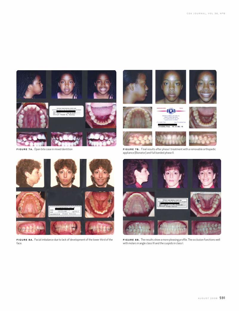
c d a j o u r n a l , v o l 3 6 , n º 8
a u g u s t 2 0 0 8 591
f igure 7a. Open bite case in mixed dentition
f igure 8a. Facial imbalance due to lack of development of the lower third of the face.
fig ur e 7 b. Final results after phase I treatment with a removable orthopedic appliance (Bionator) and full banded phase II.
fig ur e 8 b. The results show a more pleasing profile. The occlusion functions well with molars in angle class III and the cuspids in class I.
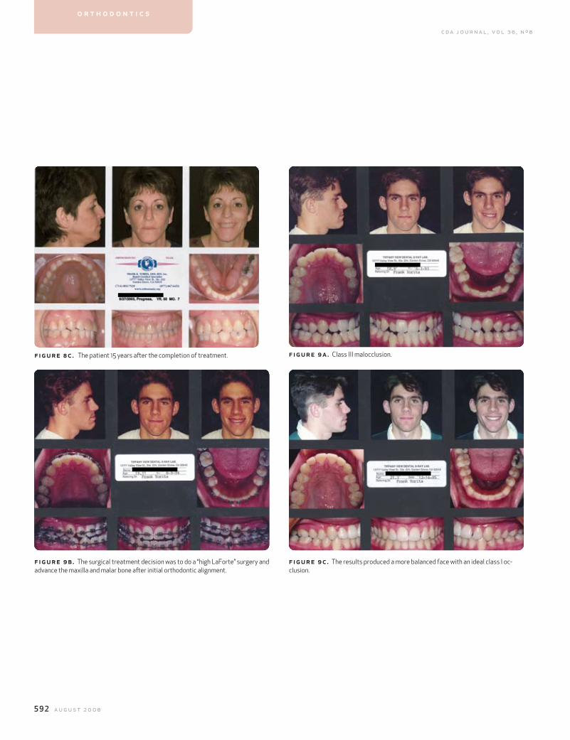
592 a u g u s t 2 0 0 8
c d a j o u r n a l , v o l 3 6 , n º 8
f igur e 8c. The patient 15 years after the completion of treatment.
f igur e 9b. The surgical treatment decision was to do a “high LaForte” surgery and advance the maxilla and malar bone after initial orthodontic alignment.
fig ur e 9 a. Class III malocclusion.
fig ur e 9 c. The results produced a more balanced face with an ideal class I oc-clusion.
o r t h o d o n t i c s

c d a j o u r n a l , v o l 3 6 , n º 8
a u g u s t 2 0 0 8 593
f igure 10a-1 .
f ig ur e 10a- 2.
f igure 10b-1 .
f igures 10c-1 .
f igure 10b- 2.
f igure 10c- 2.
development of the midface and weak “cheek” bones (malar eminences). The surgical treatment decision was to do a “high LaForte” surgery and advance the maxilla and malar bone after initial orthodontic alignment (figure 9b). The results produced a more balanced face with an ideal class I occlusion (figure 9c).
The restorative ConnectionThe orthodontist’s contribution
to the esthetic team effort is often in tooth positioning and site preparation. Teeth may need to be aligned in the ideal position for the final restoration. The following are some case examples of prerestorative orthodontic treat-ment: forced eruption: figures 10a-c; molar uprighting: figures 11a-c; root position for implant: figures 12a-b.
Comprehensive Treatment InteractionsThe following is a summary of what
orthodontic treatment can do to help provide a more comprehensive treatment result:
n Incisor crowding elimination to harmonize interarch alignment style, with or without upper anterior veneers
n Optimal teeth positioning before veneer placement
n Creating better papilla form through space closure and root alignment
n Nonsurgical leveling of gingival scallops
continued from 589fig ur e 10a. Forced eruption: This patient had labial caries on No. 8 that extended to the alveolar crest. In order to preserve the biological width and follow the rule of ferrule, No. 8 had to be extruded by at least 4 mm.
f igure 10b. Incisal edge was reduced as tooth was extruded. Periodontal surgery (crown lengthening) was done after eight weeks of stabilization and maturing of the bone.
f igu re 10c. No. 8 is restored after waiting eight weeks for the gingiva to completely heal. Gingival con-tour is re-established. Note amount of extrusion verified in the radiographs.
continues on 595
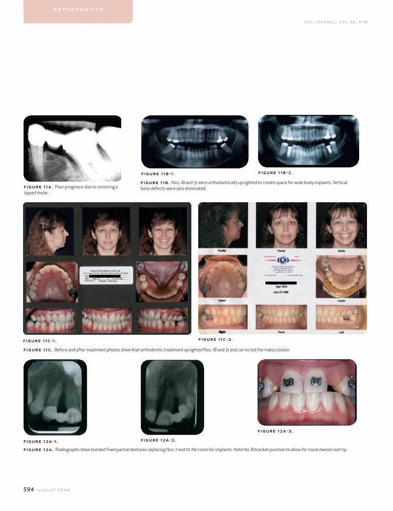
594 a u g u s t 2 0 0 8
c d a j o u r n a l , v o l 3 6 , n º 8
f igur e 11a. Poor prognosis due to restoring a tipped molar.
f igur e 12a-1 .
f igure 11b- 1 .
f igure 12a - 2.
f ig ur e 12a- 3 .
f ig ur e 11b- 2 .
f igur e 11c-1 . f ig ur e 11c- 2 .
o r t h o d o n t i c s
figure 11b . Nos. 18 and 31 were orthodontically uprighted to create space for wide body implants. Vertical bony defects were also eliminated.
f igur e 11c . Before and after treatment photos show that orthodontic treatment uprighted Nos. 18 and 31 and corrected the malocclusion.
f igur e 12a. Radiographs show bonded fixed partial dentures replacing Nos. 7 and 10. No room for implants. Note No. 8 bracket position to allow for more mesial root tip.
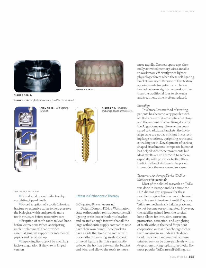
c d a j o u r n a l , v o l 3 6 , n º 8
a u g u s t 2 0 0 8 595
f igu re 12b-1 .
f igure 13 . Self-ligating bracket.
fig ur e 14 . Temporary anchorage device or miniscrew.
figure 12b- 2 .
n Periodontal pocket reduction by uprighting tipped teeth
n Forced eruption of a tooth following fracture or extensive caries to help preserve the biological width and provide more tooth structure before restorative care
n Eruption of tooth roots to level bone before extractions (when anticipating implant placement) that provides essential gingival support for interdental papilla and facial scallop
n Improving lip support by maxillary incisor angulation if they are in lingual version
latest in orthodontic Therapy
Self-ligating Braces (figure 13)Dwight Damon, DDS, a Washington
state orthodontist, reintroduced the self-ligating or tie-less orthodontic bracket and created enough interest that all the large orthodontic supply companies now have their own brand. These brackets have a slide that holds the arch wire in place rather than using an elastomeric or metal ligature tie. This significantly reduces the friction between the bracket and wire, and allows the teeth to move
more rapidly. The new space-age, ther-mally activated memory wires are able to work more efficiently with lighter physiologic forces when these self-ligating brackets are used. Because of this feature, appointments for patients can be ex-tended between eight to 10 weeks rather than the traditional four to six weeks and treatment time is often reduced.
InvisalignThis brace-less method of treating
patients has become very popular with adults because of its cosmetic advantage and the amount of advertising done by the Align Company. However, as com-pared to traditional brackets, the Invis-align trays are not as efficient in correct-ing large rotations, uprighting roots, and extruding teeth. Development of various-shaped attachments (composite buttons) has helped with these movements but ideal results are still difficult to achieve, especially with posterior teeth. Often, traditional brackets have to be placed to complete the more complex cases.
Temporary Anchorage Device (TAD or Miniscrew) (figure 14)4
Most of the clinical research on TADs was done in Europe and Asia since the FDA did not give approval for these modified surgical bone screws to be used in orthodontic treatment until May 2005. TADs are mechanically held in place and do not become osseointegrated. However, the stability gained from the cortical bone allows for intrusion, extrusion, protraction, retraction, and uprighting of teeth without the need for patient cooperation or loss of anchorage (other teeth moving in an undesirable direc-tion). Placement and removal of these mini-screws can be done painlessly with a deeply penetrating topical anesthetic. The most popular TADs are self-drilling, 1.5
contin u ed fr o m 5 93
f igu re 12b. Implants are restored, and No. 8 is veneered.
596 a u g u s t 2 0 0 8
c d a j o u r n a l , v o l 3 6 , n º 8
millimeters in diameter and 8 millimeters in length. Various sizes and head shapes are available depending on the need.
Accelerated Osteogenic Orthodontics (AOO)Currently this technique is not widely
accepted or used; however, clinical studies are being conducted in the dental schools at St. Louis University, Boston University, and University of Southern California.5-7
Accelerated osteogenic orthodon-tics is a revived treatment method that combines selective alveolar decorticating, alveolar augmentation, and orthodontic treatment. Prior to starting orthodontic mechanotherapy, gingival flaps are laid and corticotomy surgery is performed by the periodontist. The surgical wounding of the osseous tissue causes a cascade of physiologic healing events called regional acceleratory phenomenon.8 RAP is a pro-cess that involves accelerated bone turn-over and decrease in regional bone den-sity, resulting in rapid orthodontic tooth movement and decrease in treatment time by half or more.5 This method of ac-celerated osteogenic orthodontics was de-veloped by Drs. William and Tom Wilcko.6
ConclusionThis article is based on the author’s 38
years of orthodontic practice (including residency) and sixteen years of teaching, and is primarily experience-based rather than evidence-based. According to the editor of the Journal of Clinical Orthodon-tics, Dr. Robert Keim, “there is not a divi-sion or debate between “evidence-based” and “experience-based” practice, but rather a mutually beneficial continuum between the two philosophies … the clinical judgment of a skilled practitioner and the patient’s individual preferences and values are given equal weight with scientific evidence in the decision-making (treatment planning) process.”9
Dr. Vince Kokich said “to provide
excellent esthetic care for your patients who have difficult dental problems, it is important to be part of an interdis-ciplinary treatment group.”10 Working together and combining skills to achieve the optimal results for your patients can enhance patient and professional satisfaction as well as stimulate mutual referral and treatment acceptance.
to request a printed copy of this article, please contact Frank K. Yorita, DDS, 12777 Valley View St., Suite 222, Garden Grove, Calif., 92845-2522.
references1. Salzman JA, Practice of Orthodontics, Philadelphia, JB Lip-pincott, vol. 1, 1966.2. Murphy MT, Collaborative interdisciplinary agreements. J Am Dent Assoc 137:1164-7, 2006.3. Sarver DM Ackerman MB, Dynamic smile visualization and quantification. Am J Orthod Dentofac Orthop 124:4-12, 2003.4. Maino BH, Bednar J, Pagin P and Mura P: The Spider screw for skeletal anchorage. J Clin Orthod 37:90-7, 2003.5. Hajji SS, The influence of accelerated osteogenic response on mandibular decrowding (thesis). St. Louis, St. Louis University, 2000.6. Wilcko MW, Wilcko MT, et al, Rapid orthodontics with alveolar reshaping: Two case reports of decrowding. Int J Periodontics Restorative Dent 21:9-19, 2001.7. Nowzari H, Yorita F, Chang H: Periodontally accelerated osteogenic orthodontics combined with autogenous bone grafting. Compendium 39:200-7, 2008.8. Frost HM, The biology of fracture healing: An overview for clinicians. Clinical Orthop Rel Res 248:283-93, 1989.9. Keim RG, The Power of the Pyramid. J Clinic Ortho 10:587-8, 2007.10. Kokich VG, Spear FM, Guidelines for managing the orthodontic-restorative patient. Sem Ortho 3:3-20, 1997.
o r t h o d o n t i c s
c d a j o u r n a l , v o l 3 6 , n º 8
a u g u s t 2 0 0 8 599
Transitioning Patients from Teeth to Implants utilizing fixed restorationssajid jivraj, dds, msed; mamaly reshad, dds, msc; and winston w.l. chee, dds
abstract The provisional phase of treatment can be the most challenging in implant dentistry. The choice of provisional restoration should be based on esthetic demands, functional requirements, duration, and ease of fabrication. One major obstacle to treatment with implants is the transitional phase between a tooth-supported occlusion and an implant-supported occlusion. This is of particular concern when a patient with a failing dentition has not worn a removable prosthesis before and is planned to have a reconstruction supported by implants.
authors
mamaly reshad, dds, msc, is an assistant profes-sor of clinical dentistry, and co-chair for fixed prosthodontics and opera-tive dentistry, Division of Primary Oral Healthcare, University of Southern California, School of Dentistry, Los Angeles.
Winston W.l. Chee, dds, facp, is the Ralph and Jean Bleak Professor of Restorative Dentistry; director, implant dentistry; co-director, advanced prosthodontics at the University of Southern California, School Of Dentistry, and in private practice in Glendale, Calif.
mplants were originally designed with the edentulous patients in mind, as well as to compensate for the poor support and reten-tion available from the mucosa.1,2
With conventional delayed load-ing protocols, patients receiving im-plants are sometimes required to wear a removable transitional prosthesis. For many patients this is unaccept-able both socially and esthetically.
Achieving osseointegration is not the challenge that it seemed to be 30 years ago. Today, implant integration is a given and providing an implant-supported res-toration that is in harmony with the sur-rounding hard and soft tissues is the goal. Bone and gingival grafting procedures are routinely used to optimize the site prior to implant placement. Removable pros-theses can hamper these procedures as
t r a n s i t i o n i n g
pressure from removable prostheses can interfere with the proper healing of grafts.
The purpose of this article is to describe options other than remov-able prostheses in transitioning the patient from teeth to implant-supported restorations. Emphasis is placed on fixed transitional prostheses for the reasons described above.
Transitioning Patients from Teeth to Implants
Today’s patients have high expecta-tions regarding esthetics and providing functional restorations alone may not be sufficient to satisfy many of them. With heightened esthetic expectations, it becomes imperative the restorative dentist understand the patient’s desires and expectations prior to embarking upon any irreversible therapy. Moreover,
ISajid Jivraj, dds, msed, is an associate clinical professor, former chair-man section of fixed prosthodontics and opera-tive dentistry University of Southern California, School of Dentistry, Los Angeles, and in private practice in Oxnard, Calif.
acknowledgments
The authors would like to thank Dr. Delaram Hanookai for the surgical procedures completed in the cases included in this paper. The authors would also like to thank the British Dental Journal for allowing re-production of parts of the article that was originally published, 201 (11): 699-708, Dec. 9, 2006.
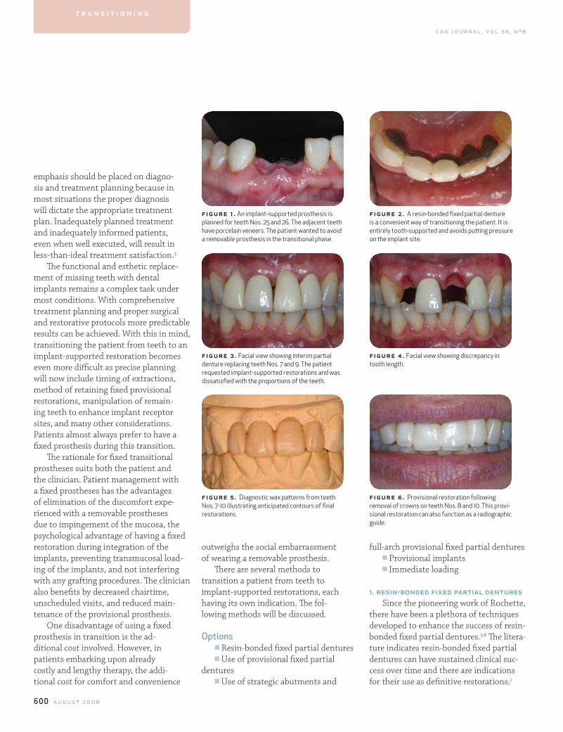
600 a u g u s t 2 0 0 8
c d a j o u r n a l , v o l 3 6 , n º 8
full-arch provisional fixed partial denturesn Provisional implantsn Immediate loading
1. resin-bonded fixed partial denturesSince the pioneering work of Rochette,
there have been a plethora of techniques developed to enhance the success of resin-bonded fixed partial dentures.5,6 The litera-ture indicates resin-bonded fixed partial dentures can have sustained clinical suc-cess over time and there are indications for their use as definitive restorations.7
emphasis should be placed on diagno-sis and treatment planning because in most situations the proper diagnosis will dictate the appropriate treatment plan. Inadequately planned treatment and inadequately informed patients, even when well executed, will result in less-than-ideal treatment satisfaction.3
The functional and esthetic replace-ment of missing teeth with dental implants remains a complex task under most conditions. With comprehensive treatment planning and proper surgical and restorative protocols more predictable results can be achieved. With this in mind, transitioning the patient from teeth to an implant-supported restoration becomes even more difficult as precise planning will now include timing of extractions, method of retaining fixed provisional restorations, manipulation of remain-ing teeth to enhance implant receptor sites, and many other considerations. Patients almost always prefer to have a fixed prosthesis during this transition.
The rationale for fixed transitional prostheses suits both the patient and the clinician. Patient management with a fixed prostheses has the advantages of elimination of the discomfort expe-rienced with a removable prostheses due to impingement of the mucosa, the psychological advantage of having a fixed restoration during integration of the implants, preventing transmucosal load-ing of the implants, and not interfering with any grafting procedures. The clinician also benefits by decreased chairtime, unscheduled visits, and reduced main-tenance of the provisional prosthesis.
One disadvantage of using a fixed prosthesis in transition is the ad-ditional cost involved. However, in patients embarking upon already costly and lengthy therapy, the addi-tional cost for comfort and convenience
outweighs the social embarrassment of wearing a removable prosthesis.
There are several methods to transition a patient from teeth to implant-supported restorations, each having its own indication. The fol-lowing methods will be discussed.
optionsn Resin-bonded fixed partial denturesn Use of provisional fixed partial
denturesn Use of strategic abutments and
figure 1 . An implant-supported prosthesis is planned for teeth Nos. 25 and 26. The adjacent teeth have porcelain veneers. The patient wanted to avoid a removable prosthesis in the transitional phase.
figure 3 . Facial view showing interim partial denture replacing teeth Nos. 7 and 9. The patient requested implant-supported restorations and was dissatisfied with the proportions of the teeth.
figure 5 . Diagnostic wax patterns from teeth Nos. 7-10 illustrating anticipated contours of final restorations.
fig ur e 2 . A resin-bonded fixed partial denture is a convenient way of transitioning the patient. It is entirely tooth-supported and avoids putting pressure on the implant site.
fig ur e 4 . Facial view showing discrepancy in tooth length.
fig ur e 6 . Provisional restoration following removal of crowns on teeth Nos. 8 and 10. This provi-sional restoration can also function as a radiographic guide.
t r a n s i t i o n i n g
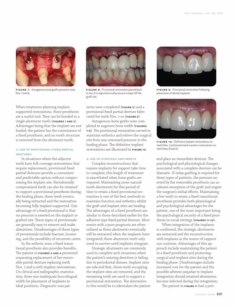
c d a j o u r n a l , v o l 3 6 , n º 8
a u g u s t 2 0 0 8 601
When treatment planning implant-supported restorations, these prostheses are a useful tool. They can be bonded to a single abutment tooth (figures 1 and 2). Advantages being that the implant are not loaded, the patient has the convenience of a fixed prosthesis, and no tooth structure is removed from the abutment tooth.
2. use of provisional-fixed partial dentures
In situations where the adjacent teeth have full-coverage restorations that require replacement, provisional fixed partial dentures provide a convenient and predictable option without compro-mising the implant site. Periodontally compromised teeth can also be retained to support a provisional prosthesis during the healing phase, these teeth eventu-ally being extracted and the restoration becoming fully implant-supported. One advantage of a fixed provisional is that no pressure is exerted on the implant or grafted site. These types of provisionals are generally easy to remove and make alterations. Disadvantages of these types of provisionals include fracture, loosen-ing, and the possibility of recurrent caries.
In the esthetic zone a fixed transi-tional prosthesis also provides benefits. The patient in figures 3 and 4 presented requesting replacement of her remov-able partial denture replacing teeth Nos. 7 and 9 with implant restorations. On clinical and radiographic examina-tion, there was inadequate buccolingual width for placement of implants in ideal positions. Diagnostic wax pat-
terns were completed (figure 5) and a provisional fixed partial denture fabri-cated for teeth Nos. 7-10 (figure 6).
Autogenous bone grafts were com-pleted to augment bone width (figures 7-9). The provisional restoration served to maintain esthetics and relieve the surgical site from any untoward pressure in the healing phase. The definitive implant restorations are illustrated in figure 10.
3. use of strategic abutmentsComplex reconstructions that
require implants for support take time to complete; this length of treatment is exacerbated when bone grafts are required. Maintaining some natural tooth abutments for this period of time to retain a fixed provisional res-toration is one of the best methods to maintain function and esthetics whilst the graft and implant sites are healing. The advantages of a fixed prosthesis are similar to those described earlier for the adhesive type fixed partial denture. Abut-ments with a poor prognosis are often utilized as these abutments eventually will be extracted when the implants have integrated; these abutment teeth only need to survive until implants integrate.
Strategic abutments are commonly used in complete arch situations when the patient’s existing dentition is failing due to periodontal disease. Implant sites are selected first, those teeth occupying the implant sites are removed, and the remaining teeth are used to support a provisional restoration. The alternative to this would be to edentulate the patient
and place an immediate denture. The psychological and physiological changes associated with a complete denture can be dramatic. If onlay grafting is required for these types of patients, the pressure ex-erted by the removable prosthesis can ac-celerate resorption of the graft and negate the surgeon’s initial efforts. Maintaining a few teeth to retain a fixed transitional prosthesis provides both physiological and psychological advantages for the patient, one of the most important being the psychological security of a fixed pros-thesis in social settings (figures 11-24).
When integration of the implants is confirmed, the strategic abutments are extracted and the reconstruction with implants as the source of support can continue. Advantages of this ap-proach include maintaining the patient in a fixed prosthesis and protecting surgical and implant sites during the healing phase. Disadvantages include increased cost for the patient and the possible adverse sequelae to implant integration should retained abutments become infected during the integration.
The patient in figure 11 had a peri-
f igur e 7 . Autogenous bone grafts placed in sites Nos. 7 and 9.
figure 8 . Provisional restoration placed back in situ. It is adjusted so all pressure is kept off the graft site.
fig ur e 9 . Provisional restoration following placement of dental implants.
fig ur e 10. Definitive implant restorations on teeth Nos. 7 and 9 and metal ceramic restorations on teeth Nos. 8 and 10.
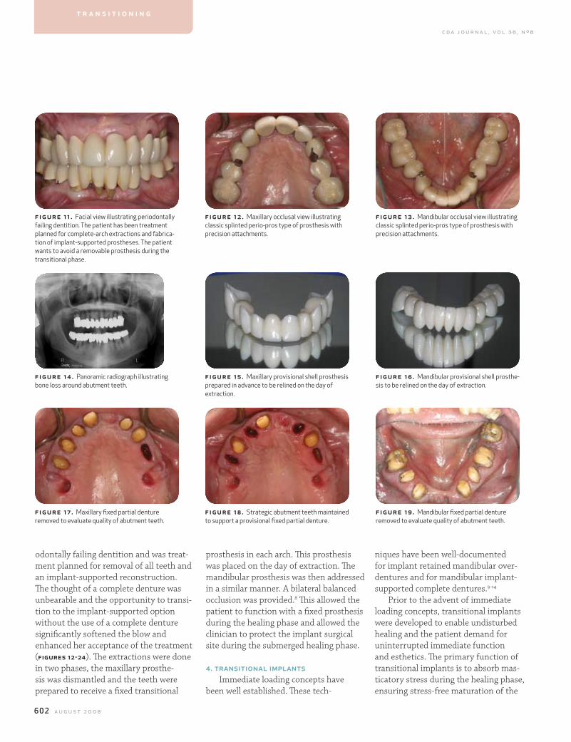
602 a u g u s t 2 0 0 8
c d a j o u r n a l , v o l 3 6 , n º 8
odontally failing dentition and was treat-ment planned for removal of all teeth and an implant-supported reconstruction. The thought of a complete denture was unbearable and the opportunity to transi-tion to the implant-supported option without the use of a complete denture significantly softened the blow and enhanced her acceptance of the treatment (figures 12-24). The extractions were done in two phases, the maxillary prosthe-sis was dismantled and the teeth were prepared to receive a fixed transitional
prosthesis in each arch. This prosthesis was placed on the day of extraction. The mandibular prosthesis was then addressed in a similar manner. A bilateral balanced occlusion was provided.8 This allowed the patient to function with a fixed prosthesis during the healing phase and allowed the clinician to protect the implant surgical site during the submerged healing phase.
4. transitional implantsImmediate loading concepts have
been well established. These tech-
niques have been well-documented for implant retained mandibular over-dentures and for mandibular implant-supported complete dentures.9-14
Prior to the advent of immediate loading concepts, transitional implants were developed to enable undisturbed healing and the patient demand for uninterrupted immediate function and esthetics. The primary function of transitional implants is to absorb mas-ticatory stress during the healing phase, ensuring stress-free maturation of the
f igur e 11 . Facial view illustrating periodontally failing dentition. The patient has been treatment planned for complete-arch extractions and fabrica-tion of implant-supported prostheses. The patient wants to avoid a removable prosthesis during the transitional phase.
f igur e 14 . Panoramic radiograph illustrating bone loss around abutment teeth.
f igur e 17 . Maxillary fixed partial denture removed to evaluate quality of abutment teeth.
figure 12 . Maxillary occlusal view illustrating classic splinted perio-pros type of prosthesis with precision attachments.
figure 15 . Maxillary provisional shell prosthesis prepared in advance to be relined on the day of extraction.
figure 18 . Strategic abutment teeth maintained to support a provisional fixed partial denture.
fig ur e 13 . Mandibular occlusal view illustrating classic splinted perio-pros type of prosthesis with precision attachments.
fig ur e 16 . Mandibular provisional shell prosthe-sis to be relined on the day of extraction.
fig ur e 19 . Mandibular fixed partial denture removed to evaluate quality of abutment teeth.
t r a n s i t i o n i n g
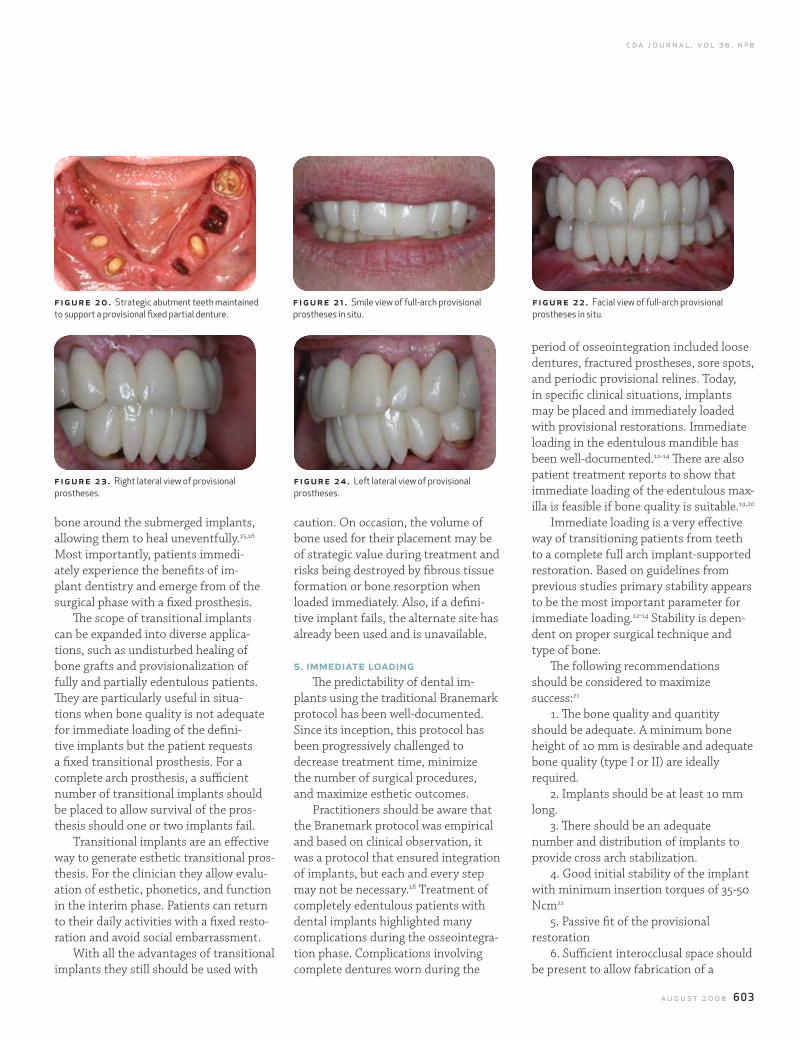
c d a j o u r n a l , v o l 3 6 , n º 8
a u g u s t 2 0 0 8 603
f igure 23. Right lateral view of provisional prostheses.
figure 24. Left lateral view of provisional prostheses.
f igure 20. Strategic abutment teeth maintained to support a provisional fixed partial denture.
figure 21 . Smile view of full-arch provisional prostheses in situ.
fig ur e 22. Facial view of full-arch provisional prostheses in situ.
bone around the submerged implants, allowing them to heal uneventfully.15,16 Most importantly, patients immedi-ately experience the benefits of im-plant dentistry and emerge from of the surgical phase with a fixed prosthesis.
The scope of transitional implants can be expanded into diverse applica-tions, such as undisturbed healing of bone grafts and provisionalization of fully and partially edentulous patients. They are particularly useful in situa-tions when bone quality is not adequate for immediate loading of the defini-tive implants but the patient requests a fixed transitional prosthesis. For a complete arch prosthesis, a sufficient number of transitional implants should be placed to allow survival of the pros-thesis should one or two implants fail.
Transitional implants are an effective way to generate esthetic transitional pros-thesis. For the clinician they allow evalu-ation of esthetic, phonetics, and function in the interim phase. Patients can return to their daily activities with a fixed resto-ration and avoid social embarrassment.
With all the advantages of transitional implants they still should be used with
caution. On occasion, the volume of bone used for their placement may be of strategic value during treatment and risks being destroyed by fibrous tissue formation or bone resorption when loaded immediately. Also, if a defini-tive implant fails, the alternate site has already been used and is unavailable.
5. immediate loadingThe predictability of dental im-
plants using the traditional Branemark protocol has been well-documented. Since its inception, this protocol has been progressively challenged to decrease treatment time, minimize the number of surgical procedures, and maximize esthetic outcomes.
Practitioners should be aware that the Branemark protocol was empirical and based on clinical observation, it was a protocol that ensured integration of implants, but each and every step may not be necessary.18 Treatment of completely edentulous patients with dental implants highlighted many complications during the osseointegra-tion phase. Complications involving complete dentures worn during the
period of osseointegration included loose dentures, fractured prostheses, sore spots, and periodic provisional relines. Today, in specific clinical situations, implants may be placed and immediately loaded with provisional restorations. Immediate loading in the edentulous mandible has been well-documented.12-14 There are also patient treatment reports to show that immediate loading of the edentulous max-illa is feasible if bone quality is suitable.19,20
Immediate loading is a very effective way of transitioning patients from teeth to a complete full arch implant-supported restoration. Based on guidelines from previous studies primary stability appears to be the most important parameter for immediate loading.12-14 Stability is depen-dent on proper surgical technique and type of bone.
The following recommendations should be considered to maximize success:21
1. The bone quality and quantity should be adequate. A minimum bone height of 10 mm is desirable and adequate bone quality (type I or II) are ideally required.
2. Implants should be at least 10 mm long.
3. There should be an adequate number and distribution of implants to provide cross arch stabilization.
4. Good initial stability of the implant with minimum insertion torques of 35-50 Ncm22
5. Passive fit of the provisional restoration
6. Sufficient interocclusal space should be present to allow fabrication of a
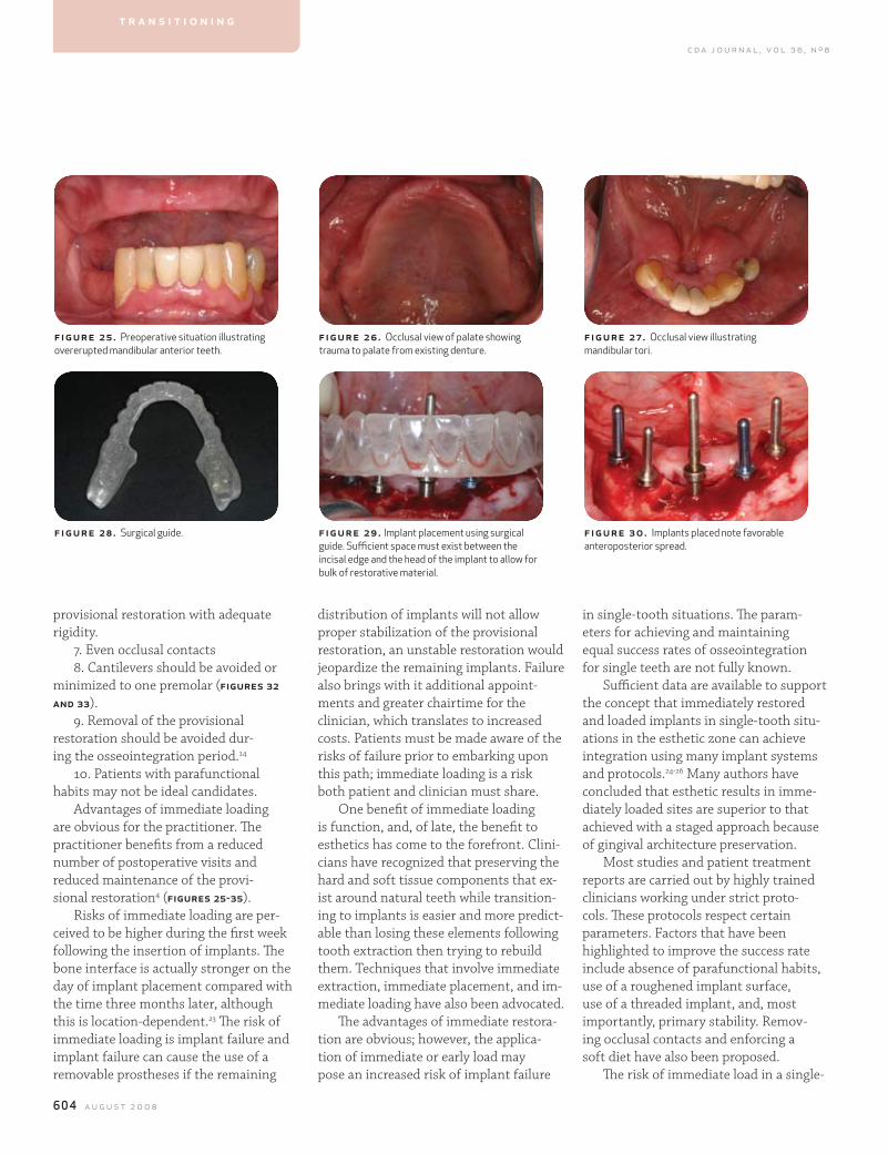
604 a u g u s t 2 0 0 8
c d a j o u r n a l , v o l 3 6 , n º 8
in single-tooth situations. The param-eters for achieving and maintaining equal success rates of osseointegration for single teeth are not fully known.
Sufficient data are available to support the concept that immediately restored and loaded implants in single-tooth situ-ations in the esthetic zone can achieve integration using many implant systems and protocols.24-26 Many authors have concluded that esthetic results in imme-diately loaded sites are superior to that achieved with a staged approach because of gingival architecture preservation.
Most studies and patient treatment reports are carried out by highly trained clinicians working under strict proto-cols. These protocols respect certain parameters. Factors that have been highlighted to improve the success rate include absence of parafunctional habits, use of a roughened implant surface, use of a threaded implant, and, most importantly, primary stability. Remov-ing occlusal contacts and enforcing a soft diet have also been proposed.
The risk of immediate load in a single-
distribution of implants will not allow proper stabilization of the provisional restoration, an unstable restoration would jeopardize the remaining implants. Failure also brings with it additional appoint-ments and greater chairtime for the clinician, which translates to increased costs. Patients must be made aware of the risks of failure prior to embarking upon this path; immediate loading is a risk both patient and clinician must share.
One benefit of immediate loading is function, and, of late, the benefit to esthetics has come to the forefront. Clini-cians have recognized that preserving the hard and soft tissue components that ex-ist around natural teeth while transition-ing to implants is easier and more predict-able than losing these elements following tooth extraction then trying to rebuild them. Techniques that involve immediate extraction, immediate placement, and im-mediate loading have also been advocated.
The advantages of immediate restora-tion are obvious; however, the applica-tion of immediate or early load may pose an increased risk of implant failure
provisional restoration with adequate rigidity.
7. Even occlusal contacts8. Cantilevers should be avoided or
minimized to one premolar (figures 32 and 33).
9. Removal of the provisional restoration should be avoided dur-ing the osseointegration period.14
10. Patients with parafunctional habits may not be ideal candidates.
Advantages of immediate loading are obvious for the practitioner. The practitioner benefits from a reduced number of postoperative visits and reduced maintenance of the provi-sional restoration4 (figures 25-35).
Risks of immediate loading are per-ceived to be higher during the first week following the insertion of implants. The bone interface is actually stronger on the day of implant placement compared with the time three months later, although this is location-dependent.23 The risk of immediate loading is implant failure and implant failure can cause the use of a removable prostheses if the remaining
f igur e 25. Preoperative situation illustrating overerupted mandibular anterior teeth.
f igur e 28. Surgical guide.
figure 26. Occlusal view of palate showing trauma to palate from existing denture.
figure 29. Implant placement using surgical guide. Sufficient space must exist between the incisal edge and the head of the implant to allow for bulk of restorative material.
fig ur e 27 . Occlusal view illustrating mandibular tori.
fig ur e 3 0. Implants placed note favorable anteroposterior spread.
t r a n s i t i o n i n g
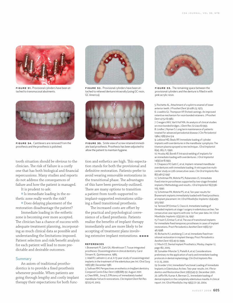
c d a j o u r n a l , v o l 3 6 , n º 8
a u g u s t 2 0 0 8 605
tooth situation should be obvious to the clinician. The risk of failure is a costly one that has both biological and financial repercussions. Many studies and reports do not address the consequences of failure and how the patient is managed.
It is prudent to ask:n Is immediate loading in the es-
thetic zone really worth the risk?n Does delaying placement of the
restoration disadvantage the patient?Immediate loading in the esthetic
zone is becoming ever more accepted. The clinician has a chance of success with adequate treatment planning, incorporat-ing as much clinical data as possible and understanding the limitations imposed. Patient selection and risk/benefit analysis for each patient will lead to more pre-dictable and desirable outcomes.
SummaryAn axiom of traditional prostho-
dontics is to provide a fixed prosthesis whenever possible. When patients are going through lengthy and costly implant therapy their expectations for both func-
tion and esthetics are high. This expecta-tion stands for both the provisional and definitive restoration. Patients prefer to avoid wearing removable restorations in the transitional phase. The advantages of this have been previously outlined. There are many options to transition a patient from tooth-supported to implant-supported restorations utiliz-ing a fixed transitional prosthesis.
The increased costs are offset by the practical and psychological conve-nience of a fixed prosthesis. Patients realize the benefits of implant therapy immediately and are more likely to be accepting of treatment plans involv-ing extensive implant restorations.
referenc es1. Branemark PI, zarb GA, Albrektsson T, Tissue integrated prostheses: Osseointegration in clinical dentistry. Carol Stream Ill., Quintessence, 1985.2. Adell R, Lekholm U, et al, A 15-year study of osseointegrated implants in the treatment of the edentulous jaw. Int J Oral Surg 10(6):387, December 1981.3. Sullivan RM, Perspectives on esthetics in Imp[lant dentistry. Compend Contin Educ Dent 22(8):685-92, August 2001.4. Chee WWL, Jivraj S, Efficiency of immediately loaded mandibular full arch restorations. Clin Implant Dent Rel Res 5(1):52-6, 2003.
5. Rochette AL, Attachment of a splint to enamel of lower anterior teeth. J Prosthet Dent 30:418-23, 1973.6. Livaditis GJ, Thompson VP, Etched castings. An improved retentive mechanism for resin bonded retainers. J Prosthet Dent 47:52-8,1982.7. Creugers NHJ, Van’t Hof MA. An analysis of clinical studies on resin bonded bridges. J Dent Res 72:1244-8,1993.8. Lindhe J, Nyman S. Long term maintenance of patients treated for advanced periodontal disease J Clin Periodontol 1984 ;11(8):504-5149. Leklove MD, Beals RP, Immediate loading of cylinder implants with overdentures in the mandibular symphysis: The titanium plasma sprayed screw technique. J Oral Implantol 6(4), 265-71, 1990.10. Hruska AB, Borelli P, Intraoral welding of implants for an immediate loading with overdentures. J Oral Implantol 14(9):34-8,1993.11. Chiapasco M, Gatti C, et al, Implant-retained mandibular overdentures with immediate loading. A retrospective multi-center study on 226 consecutive cases. Clin Oral Implants Res 8(1):48-57,199712. Schnitman PA, Wohrle PS, Rubenstein JS, Immediate fixed interim porostheses supported by two-stage threaded implants: Methodology and results. J Oral Implantol 16(2):96-105, 1990.13. Schnitman PA, Wohrle PS, et al, Ten-year results for Branemark implants immediately loaded with fixed prosthesis at implant placement. Int J Oral Maxillofac Implants 12(4):495-503,1997.14. Tarnow DP, Emtiaz S, Classi A, Immediate loading of threaded implants at stage 1 surgery in edentulous arches: Ten consecutive case reports with one- to five-year data. Int J Oral Maxillofac Implants 12(3):310-24, 1997.15. Froum S, Emtiaz S, et al, The use of transitional implants for immediate fixed temporary prostheses in cases of implant restorations. Pract Periodontics Aesthet Dent 10(6):737-46,1998.16. Bichacho N, Landsberg CJ, et al, Immediate fixed tran-sitional restoration in implant therapy. Pract Periodontics Aesthet Dent 11(1):45-51,1999.17. Misch CE, Dental Implant Prosthetics, Mosby, chapter 12, page 182, 2005.18. Szmukler-Moncler S, Piatelli A, et al, Considerations preliminary to the application of early and immediate loading protocols in dental implantology. Clin Oral Implants Res 11:12-25, 2000.19. Grunder U Int, Immediate Functional Loading of Immediate Implants in Edentulous Arches: Two-year results. Int J Perio-dontics and Restorative Dent 21(6):545-51, December 2001.20. Jaffin RA, Kumar A, Berman CL, Immediate loading of dental implants in the completely edentulous maxilla: A clinical report. Int J Oral Maxillofac Imp 19(5):721-30, 2004.
f igure 34. Cantilevers are removed from the prosthesis and the prosthesis is polished.
figure 35. Smile view of screw retained immedi-ate load prosthesis. Prosthesis has been adjusted to allow the patient to maintain hygiene.
f igure 31 . Provisional cylinders have been at-tached to transmucosal abutments.
figure 32. Provisional cylinders have been at-tached to relieved denture intraorally (using GC resin, GC America).
fig ur e 3 3 . The remaining space between the provisional cylinders and the denture is filled in with pink acrylic resin.
606 a u g u s t 2 0 0 8
c d a j o u r n a l , v o l 3 6 , n º 8
21. Derbabian K, Simonian K, Immediate loading of dental implants overview and rationale. J Calif Dent Assoc 33(4):337-41, April 2005.22. Brunski JB, Avoid pitfalls of overloading and micromotion of intraosseous implants. Dent Implantol Update 4(10):77-81, 1993.23. Strid KG, Radiographic results, in Branemark PI, zarb GA, Albrektsson T (eds): Tissue integrated prosthesis. Chicago, Quintessence, 1985.24. Malo P, Rangert B, Dvarsater L, Immediate function of Branemark implants in the esthetic zone: A retrospective clinical study with six months to four years of follow-up. Clin Implant Dent Relat Res 2:138-46, 2000.25. Rocci A, Martignoni M, et al, Immediate function of single and partial reconstructions in the maxilla using Mk IV fixtures. A retrospective analysis. Applied Osseointegration Res 2(1):22-43, 2001.26. Wohrle PS, Single-tooth replacement in the aesthetic zone with immediate provisionalization: Fourteen consecutive case reports. Pract Periodontics Aesthet Dent 10:1107-14, 1998.
to request a printed copy of this article, please contact Sajid Jivraj, DDS, MSEd, 300 Esplanande Drive, Suite 1600, Oxnard, Calif., 93036.
c d a j o u r n a l , v o l 3 6 , n º 8
a u g u s t 2 0 0 8 609
The Team approach: Simplifying Complex Caredavid hochwald, dds; simona arcan, dmd, md; and fariborz farnad, dds
abstract In today’s oral and maxillofacial surgery practice, the interaction between the oral and maxillofacial surgeon, the restorative dentist, and the rest of the dental and medical community is the foundation of daily patient care and management. The combination of the talents from each medical and dental discipline results in the highest quality of patient care. The following will illustrate the power of the comprehensive team approach.
authors
David hochwald, dds, is a board-certified oral and maxillofacial surgeon in Bellflower and Huntington Beach, Calif. He is a clinical professor at the Univer-sity of Southern California, School of Dentistry, and director of the Surgical Implant Program.
Simona arcan, dmd, md, is a board-certified oral and maxillofacial surgeon in Bellflower and Huntington Beach, Calif. She also is a member of the Long Beach Memorial Medical Center’s cleft palate team.
ral and maxillofacial surgery has always been an exciting, discipline and specialty of dentistry. The scope of treatment and
expertise of the specialty continues to evolve and improve. Today’s materials and techniques allow us to routinely provide care on a daily basis that could only be dreamed about in the past. This high level of care continues to advance as we push toward the future.
The interaction between the oral and maxillofacial surgeon, the restorative den-tist, and the rest of the dental and medical community is the foundation of today’s daily patient care and management. Den-tists must strive to improve this interac-tion as the outcome of treatment is based on how well we all work and communicate with each other. Combining the talents of more than one medical and dental disci-pline is indicated and necessary in a large number of patient treatment plans and re-
w o r k i n g t o g e t h e r
sults in the highest quality of patient care.The best diagnosis and treatment
for the patient occurs when dentists are aware of the services each discipline is capable of providing and their own limita-tions. This article will illustrate treatment planning and care highlighting a few of today’s common oral and maxillofacial surgery services as examples.
Dentoalveolar procedures including extractions and alveolectomy, exci-sion of pathologic lesions, treatment of trauma, and reconstruction have historically been the foundation of an oral and maxillofacial surgeon’s daily patient care. The specialty’s unique training also provides oral surgeons with the ability to safely administer outpatient general anesthesia and IV sedation, along with local anesthesia, and makes it possible to provide daily office procedures virtually pain-free.
The introduction of osseointegration, the first highly predictable long-term
ofariborz farnad, dmd, is an oral and maxillofacial surgeon in Bellflower and Huntington Beach, Calif. He also is a faculty member of the University of Southern California, School of Dentistry.
acknowledgement
The authors would like to recognize other treating team members Ting-Wey Yen, DDS; Tom Rose, DDS; and Phillip Melnick, DDS.
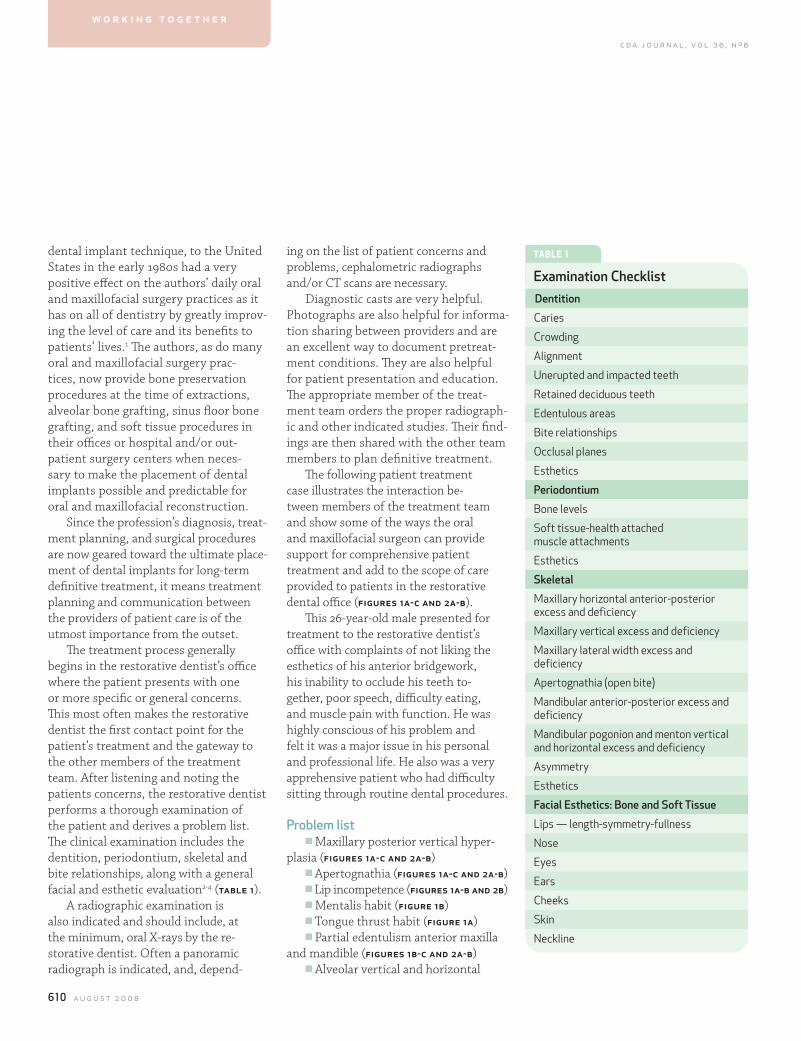
610 a u g u s t 2 0 0 8
c d a j o u r n a l , v o l 3 6 , n º 8
dental implant technique, to the United States in the early 1980s had a very positive effect on the authors’ daily oral and maxillofacial surgery practices as it has on all of dentistry by greatly improv-ing the level of care and its benefits to patients’ lives.1 The authors, as do many oral and maxillofacial surgery prac-tices, now provide bone preservation procedures at the time of extractions, alveolar bone grafting, sinus floor bone grafting, and soft tissue procedures in their offices or hospital and/or out-patient surgery centers when neces-sary to make the placement of dental implants possible and predictable for oral and maxillofacial reconstruction.
Since the profession’s diagnosis, treat-ment planning, and surgical procedures are now geared toward the ultimate place-ment of dental implants for long-term definitive treatment, it means treatment planning and communication between the providers of patient care is of the utmost importance from the outset.
The treatment process generally begins in the restorative dentist’s office where the patient presents with one or more specific or general concerns. This most often makes the restorative dentist the first contact point for the patient’s treatment and the gateway to the other members of the treatment team. After listening and noting the patients concerns, the restorative dentist performs a thorough examination of the patient and derives a problem list. The clinical examination includes the dentition, periodontium, skeletal and bite relationships, along with a general facial and esthetic evaluation2-4 (table 1).
A radiographic examination is also indicated and should include, at the minimum, oral X-rays by the re-storative dentist. Often a panoramic radiograph is indicated, and, depend-
ing on the list of patient concerns and problems, cephalometric radiographs and/or CT scans are necessary.
Diagnostic casts are very helpful. Photographs are also helpful for informa-tion sharing between providers and are an excellent way to document pretreat-ment conditions. They are also helpful for patient presentation and education. The appropriate member of the treat-ment team orders the proper radiograph-ic and other indicated studies. Their find-ings are then shared with the other team members to plan definitive treatment.
The following patient treatment case illustrates the interaction be-tween members of the treatment team and show some of the ways the oral and maxillofacial surgeon can provide support for comprehensive patient treatment and add to the scope of care provided to patients in the restorative dental office (figures 1a-c and 2a-b).
This 26-year-old male presented for treatment to the restorative dentist’s office with complaints of not liking the esthetics of his anterior bridgework, his inability to occlude his teeth to-gether, poor speech, difficulty eating, and muscle pain with function. He was highly conscious of his problem and felt it was a major issue in his personal and professional life. He also was a very apprehensive patient who had difficulty sitting through routine dental procedures.
Problem listn Maxillary posterior vertical hyper-
plasia (figures 1a-c and 2a-b)n Apertognathia (figures 1a-c and 2a-b)n Lip incompetence (figures 1a-b and 2b)n Mentalis habit (figure 1b)n Tongue thrust habit (figure 1a)n Partial edentulism anterior maxilla
and mandible (figures 1b-c and 2a-b)n Alveolar vertical and horizontal
w o r k i n g t o g e t h e r
tABle 1
examination ChecklistDentition
Caries
Crowding
Alignment
Unerupted and impacted teeth
Retained deciduous teeth
Edentulous areas
Bite relationships
Occlusal planes
Esthetics
Periodontium
Bone levels
Soft tissue-health attached muscle attachments
Esthetics
Skeletal
Maxillary horizontal anterior-posterior excess and deficiency
Maxillary vertical excess and deficiency
Maxillary lateral width excess and deficiency
Apertognathia (open bite)
Mandibular anterior-posterior excess and deficiency
Mandibular pogonion and menton vertical and horizontal excess and deficiency
Asymmetry
Esthetics
facial esthetics: Bone and Soft Tissue
Lips — length-symmetry-fullness
Nose
Eyes
Ears
Cheeks
Skin
Neckline
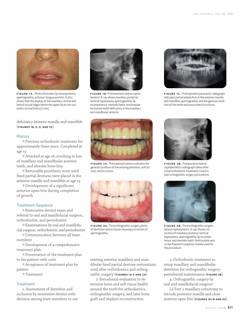
c d a j o u r n a l , v o l 3 6 , n º 8
a u g u s t 2 0 0 8 611
3. Orthodontic treatment to setup maxillary and mandibular dentition for orthognathic surgery-periodontal maintenance (figure 2b)
4. Orthognathic surgery by oral and maxillofacial surgeon5
Le Fort 1 maxillary osteotomy to intrude posterior maxilla and close anterior open bite (figures 3a-b and 4a)
existing anterior maxillary and man-dibular fixed partial denture restorations until after orthodontics and orthog-nathic surgery (figures 1a-c and 2a)
2. Periodontal evaluation to de-termine bone and soft tissue health around the teeth for orthodontics, orthognathic surgery, and later bone graft and implant reconstruction
deficiency anterior maxilla and mandible (figures 1b, 5, 9, and 10)
historyn Previous orthodontic treatment for
approximately three years. Completed at age 13
n Attacked at age 16, resulting in loss of maxillary and mandibular anterior teeth, and alveolar bone loss
n Removable prosthesis worn until fixed partial dentures were placed in the anterior maxilla and mandible at age 19
n Development of a significant anterior open bite during completion of growth
Treatment Sequencen Restorative dentist exam and
referral to oral and maxillofacial surgeon, orthodontist, and periodontist
n Examinations by oral and maxillofa-cial surgeon, orthodontist, and periodontist
n Communication between all team members
n Development of a comprehensive treatment plan
n Presentation of the treatment plan to the patient with costs
n Acceptance of treatment plan by patient
n Treatment
Treatment 1. Assessment of dentition and
occlusion by restorative dentist with decision among team members to use
f igur e 1a. Photo illustrates lip incompetence, apertognathia, and poor tongue position. It also shows that the display of the maxillary central and lateral incisal edges below the upper lip at rest are within normal limits (2 mm).
figure 2a. Pretreatment photo indicates the general condition of the existing dentition, soft tis-sues, and occlusion.
figure 3a. Postorthognathic surgery photo of dentition and occlusion showing correction of apertognathia.
figure 1b . Pretreatment lateral cepha-lometric X-ray shows maxillary posterior vertical hyperplasia, apertognathia, lip incompetence, mentalis habit, and alveolar horizontal width deficiency in the maxillary and mandibular anterior.
fig ur e 2b. Preoperative lateral cephalometric radiograph taken after initial orthodontic treatment. Used to plan orthognathic surgery procedures.
fig ur e 3 b. Postorthognathic surgery lateral cephalometric X-ray. Shows cor-rection of maxillary posterior vertical hyperplasia, apertognathia, lip incompe-tence, and mentalis habit. Notice plate and screw fixation in superior maxilla used for the procedure.
fig ur e 1c . Pretreatment panoramic radiograph indicates partial edentulism in the anterior maxilla and mandible, apertognathia, and the general condi-tion of the teeth and associated structures.
612 a u g u s t 2 0 0 8
c d a j o u r n a l , v o l 3 6 , n º 8
5. Completion of orthodontics after initial healing of orthognathic surgery (figures 3a-b and 4a)
6. Removal of the original maxil-lary and mandibular anterior fixed partial dentures with fabrication of new maxillary and mandibular fixed provi-sional restorations by the restorative dentist in preparation for bone graft-ing and implant treatment (figure 4b)
7. Fabrication of maxillary and man-dibular surgical guides (duplication of the new provisionals) by the restorative dentist to be used by oral and maxil-lofacial surgeon for bone grafting and implant placement (figures 5-8, 10 and 12)
8. Bone graft to reconstruct the alveolar vertical and horizontal deficiencies in the
fig ur e 5 . Photograph during bone graft surgery showing the vertical and horizontal deficiency of the anterior maxilla in relation to ideal tooth position indicated with the prefabricated maxillary surgical guide indexed on the posterior maxillary teeth.
fig ur e 8 . Photograph taken immediately after bone graft surgery with surgical guide in place for reference. Occlusal view to confirm vertical and horizontal correction in area.
f igur e 6 . Photograph during surgery of the cortico-cancellous blocks harvested from the iliac crest fixed into position to reconstruct the anterior maxillary alveolar deficiency. Cortico-cancellous particulate bone from the ilium was packed around the blocks to address the vertical component of the deficiency. The bone graft was cov-ered with a pericardium membrane before tension-free closure of the soft tissue was accomplished.9,10
figure 9 . Photograph during bone graft surgery showing horizontal alveolar defect in the anterior mandible.
figure 7 . Photograph taken immediately after bone graft surgery with surgical guide in place for reference. Anterior-posterior view to confirm verti-cal and horizontal correction in area.
fig ur e 10. Photograph during bone graft surgery with surgical guide in place to illustrate the extent of the anterior mandibular alveolar deficiency requiring bone grafting.
f igur e 4a. Photograph of anterior occlusal relationship just prior to removal of orthodontic appliances following orthognathic surgery and postsurgical orthodontics. Notice the normal vertical overlap (overbite) and horizontal overlap (overjet).
figure 4b. Panoramic radiograph made prior to bone grafting. Notice the original maxillary and mandibular fixed partial dentures have been re-placed with new provisional restorations to facilitate prosthetic management through the grafting and implant treatment phases.
sible.8 Healing abutments were con-nected for single surgery approach and to help manage soft tissue (figure 15)
11. Adjustment and replacement of the anterior maxillary and mandibular provisional restorations were done by the restorative dentist after surgery to remain in place until implants were healed (four months) (figure 15)
12. Placement of the fixed implant-supported provisionals that were made during implant healing by the restor-
w o r k i n g t o g e t h e r
anterior maxilla and mandible by the oral and maxillofacial surgeon using bone har-vested from the left iliac crest in block and particulate fashion6,7 (figures 6, 11 and 13)
9. Management of patient’s provi-sional restorations during graft healing by the restorative dentist (approxi-mately four to six months) (figure 13)
10. Implant placement was done by the oral and maxillofacial surgeon using previously fabricated surgical guides with immediate indexing of implant positions at surgery to make fabrication of fixed implant-supported provisional restorations during implant healing pos-
c d a j o u r n a l , v o l 3 6 , n º 8
a u g u s t 2 0 0 8 613
f igur e 11 . Photograph during bone graft surgery showing cortico-cancellous block grafts to both the labial and lingual of the anterior mandible. Use of the surgical guide for reference during surgery indicated this approach was necessary. Particulate cortico-cancellous bone from the ilium was packed around the blocks and the entire graft was covered with a pericardium membrane before tension-free closure of the soft tissue.
f igur e 14a. Occlusal photograph of the maxil-lary anterior area with the provisional restoration removed to show the result after bone graft healing prior to implant placement.
figure 12 . Photograph taken immediately after bone graft surgery with surgical guide in place for reference. Confirms vertical and horizontal correc-tion in the area.
figure 14b. Photograph of the mandibular anterior with the provisional restoration removed to show the result after bone graft healing prior to implant placement.
fig ur e 13 . Panoramic radiograph made im-mediately following bone graft surgery with adjusted maxillary and mandibular provisional restorations in place. Bone graft fixation screws evident in the maxil-lary and mandibular anterior areas.
fig ur e 14c. Photograph of the anterior maxil-lary and mandibular edentulous areas after bone graft healing with the provisional restorations removed. Notice the relapse of the vertical correc-tion, which occurred during healing, particularly in the maxillary anterior.
f igure 15 . Postoperative panoramic radiograph made immediately following implant placement to the maxillary and mandibular anterior edentulous areas. Provisional restorations were adjusted and repositioned immediately after surgery by the restorative dentist.
figure 16 . Photograph of the maxillary and mandibular provisional restorations modified as indi-cated following implant placement. Notice the added gingival material to cover the remaining vertical soft tissue defects in the maxillary anterior.
fig ur e 17 . Photograph of the final anterior mandibular implant supported fixed restoration. The patient is very happy with the functional and esthetic result. The photo also shows the need for soft tissue surgery around tooth No. 21 to create healthy fixed esthetic mucosa.
ative dentist and lab to manage soft tissue and initial esthetics (figure 16)
13. Adjustment of the provi-sional restoration by the restorative dentist to meet the esthetic de-sires of the patient as well as to en-sure proper function (figure 16)
14. Final implant restorations by the restorative dentist for the anterior maxilla and mandible (figures 17 and 18)
15. Periodontal plastic soft tissue procedures by the periodontist for tissue coverage to mucogingival defects pres-ent on the mandibular premolar teeth
(original abutment teeth) (figure 17)16. Final restorations for the mandibu-
lar premolar teeth by the restorative dentist to complete treatment
17. Long-term maintenance by the restorative dentist with help from other disciplines as needed
614 a u g u s t 2 0 0 8
c d a j o u r n a l , v o l 3 6 , n º 8
ConclusionChoosing the right team and work-
ing closely together results in the high-est quality of treatment for the patient, expands the range of care we can offer, and makes the practice of dentistry more fun and rewarding for all involved.
references1. Adell R, Lekholm U, et al, A 15-year study of osseointegrated implants in the treatment of the edentulous jaw. Int J Oral Surg 10:387-416, 1981.2. Peterson LJ, Indresano AT, et al, (eds) Principles of Oral and Maxillofacial Surgery, JP Lippincott, Philadelphia 3:chapters 49-50, 1992. 3. Poswillo DE, The relationship between oral and plastic surgery. Brit J Plast Surg 30(1):74-80, 1977.4. Arcan SC, Overview of Facial Cosmetic Surgery. J Calif Dent
f igur e 18 . Photograph of the final anterior maxillary and mandibular fixed, implant-supported restorations. Notice the pink porcelain abutting the maxillary gingival tissue. It is designed to provide op-timum esthetics by hiding the remaining soft tissue deficiencies in the maxillary anterior. It is also helpful during function to block air escape that can occur during speech. It is important to design it to provide good access to the patient for hygiene.
w o r k i n g t o g e t h e r
Assoc 32(10):849-853, October 2004.5. Belinfante L, Combining cosmetic soft tissue and skeletal surgeries to achieve greater total facial harmony than either surgery could alone. Am J Cosmet Surg 21:77-87, June 2004.6. Keller EE, VanRoekel NB, et al, Prosthetic surgical recon-struction of the severely resorbed maxilla with iliac bone grafting and tissue integrated prosthesis. Int J Oral Maxillofac Implants 3:155-65, 1987.7. Collins TA, Onlay bone grafting in combination with Brane-mark implants. Oral Maxillofac Surg Clin N Am 3(4):893-902, 1991.8. Hochwald DA, Surgical template impression during stage 1 surgery for fabrication of a provisional restoration to be placed at stage 2 surgery. J Pros Dent 66(6):796-8, 1991.9. Buser D, Dula K, et al, Lateral ridge augmentation using autografts and barrier membranes: A clinical study with 40 partially edentulous patients. J Oral Maxillofac Surg 54:420-32, April 1996.10. Buser D, Dula K, et al, Long-term stability of osseointe-grated implants in bone regenerated with the membrane technique, five-year study. Clin Oral Implants Res 7(2):175-83, June 1996.
to request a printed copy of this article, please contact David A. Hochwald, DDS, 14343 Bellflower Blvd., Bellflower, Calif., 90706.
638 a u g u s t 2 0 0 8
c d a j o u r n a l , v o l 3 6 , n º 8Dr. Bob
To increase the value of gold, have it handled by a dentist.
— AnonymousGold is currently enjoying a world-
wide bull market with the price hovering around $1,000 an ounce. This phenom-enon that places gold right up there with the per-ounce cost of women’s antiaging petroleum-based cosmetics and the Olsen Twins’ Halloween eye enhancements, is causing some concern in the dental profession.
Patient: All this gold you placed in my mouth 30 years ago? I want it replaced with that white stuff. Just put the gold in this little strong box here.
Dentist: No! Not a chance, forget it! Unh-uh. How about I replace it with white gold?
Patient: Nope, white gold is like white chocolate — it’s not real! Upon my demise, I am requesting cremation. Who gets the gold then, huh?
Dentist: I’ve often wondered, but have been afraid to ask. Never see ads of
cremation societies offering attractive urns for your noncombustibles including whatever nuggets of precious metals that might show up, do you?
With interest in cashing in on the auriferous bull market at fever pitch and dental gold having no sentimental value compared to grandma’s wedding ring, dentists experience very little salvaging themselves.
“I could never part with my gold crown. I just love the angulation of the buccal cusps, the fine detail of the occlusal anatomy and the undetectable margins. It and I have been through too many gour-met meals together. I’m having it made into a nose ring.” Heart-warming, but unheard of. Instead, the smallest inlay, the saddest-looking old bridge or crown is offered to, and immediately accepted by, patients who believe they’ve got the makings of a Mercedes down payment transferred from their mouths to the little Ziploc bag in their hands.
Robert E. Horseman, DDS
illustration by charlie o. hayward
,
continues on 637
Gold Fever
The saddest-looking old bridge or crown is offered to, and immediately accepted by, patients who believe they’ve got the makings of a Mercedes down payment.
c d a j o u r n a l , v o l 3 6 , n º 8
a u g u s t 2 0 0 8 637
Pawn shops are doing a gold rush business. Small knots of people will-ing to undergo a soft diet indefinitely, gather outside debating on how to divest themselves of their gold mines painlessly before the inevitable bear market returns. Members of gold restoration study clubs gear up with protest signs prepared to picket the insanity of such shenanigans.
The dental profession is not caught napping. The plan is to offer those pa-tients who value the properties of gold as a restorative material a measure of protec-tion. People are understandably paranoid walking about at night in dimly lit places where gold muggers are likely to lurk. Taking a leaf from General Motors that of-fers LoJack protection in most of its cars, dental suppliers are feverously working to provide patients with a dental equivalent.
It is supposed to work like this: Insert-ed into the matrix of all gold restorations from now on will be a small chip made of whatever small chips are usually made of, only smaller. You suddenly awaken at 3 a.m., the time nowadays when bad things are apparently scheduled. Heart pound-ing, stomach churning as if in response to bad clam dip, you discover a dark figure armed with a pair of Ford pliers and a small ball-peen hammer standing at the foot of the bed.
“Open wide!” Darth Vader demands, pretending to be a dentist with an au-thoritative chairside manner. Two things happen immediately — three, counting sphincter malfunction. The motion sensor in the gold, activated by the bad person’s demand and your own 180-decibel reac-tion, sends a message to LoJack whose
a u g u s t 0 8 d r . b o b
dr. bob, c o n tin u ed fr o m 638
response mechanism will be conveniently outsourced in Bombay.
The company in turn, assuming you’ve paid your monthly subscription fee, alerts your local police department where a dispatcher familiar with Indian patois stands by. Should the perp make good his liberation of your gold, the intelligent chip obliges, transmitting a location finder that will appear automatically on all global positioning devices within 5,000 miles.
Your crown/onlay should be returned to you within 24 hours, ready for recemen-tation unless you elect to join the queue at the pawn shop, realizing that some pos-sessions are not really worth the hassle. In which case, we can offer a nice selection of porcelain or resin replacements in 26 shades of white with no salvageable value whatsoever.