Embed Size (px)
Citation preview
Jagged-1-mediated activation of notch signalling induces adipogenesis ofadipose-derived stem cellsK. Ba*, X. Yang*, L. Wu*, X. Wei*, N. Fu*, Y. Fu*, X. Cai*, Y. Yao*, Y. Ge* and Y. Lin*
*State Key Laboratory of Oral Diseases, West China School of Stomatology, Sichuan University, Chengdu, China
Received 17 April 2012; revision accepted 19 July 2012
AbstractObjectives: Notch signalling plays an importantrole in many cell activities, involving proliferation,migration, differentiation and cell death. The aimof this study was to investigate effects of such sig-nalling on adipogenesis of mouse adipose-derivedstem cells (mASCs).Materials and methods: Jagged1 (50 and 100 ng/ml) was added to mASCs to activate Notch signal-ling, 2 days before adipogenic induction. At 5 and7 days after induction, oil red-O staining was per-formed to evaluate lipid accumulation. Then real-time PCR was performed to examine expression ofNotch downstream genes (Notch-1, -2, Hes-1 andHey-1) and adipogenic transcription factor (PPAR-c).Expressions of Hes-1 and PPAR-c at protein levelwere confirmed by immunofluorescence staining.Results: Our data indicated that Jagged1 promotedadipogenic differentiation of mASCs. Moreover,Jagged1 also increased expression of Notch down-stream genes and PPAR-c. Expressions of Hes-1and PPAR-c were found to be enhanced in Jagged1pre-treated mASCs when compared to controls.Discussion: The results led to the conclusion thatactivation of Notch signalling had stimulated adipo-genesis of mASCs in the presence of adipogenicmedium by promoting expression of PPAR-c.
Introduction
Mesenchymal stem cells (MSCs) compose a group ofadult stem cells that can differentiate into a variety of
cell types, such as osteoblasts, chondrocytes, adipocytes,myocytes and beta-pancreatic islet cells. MSCs can beisolated from many organs and tissues includingmarrow, umbilical cord blood, adipose tissue, adult mus-cle and dental pulp of deciduous teeth (1–3). In recentyears, research interest has grown rapidly on adiposetissue as a stem-cell source from which MSCs can beisolated. Because of the relatively high frequency ofclonogenic cells and easy isolation from adipose tissue,adipose-derived stem cells (ASCs) are thought to holdpromise for a wide range of therapeutic applications.
Notch signalling is a highly conserved pathway ofmulticellular organisms that orchestrates cell fate deter-mination, including cell proliferation, migration, differ-entiation and cell death (4–6). Notch signalling isactivated as a result of cell/cell contact through interac-tions of Notch receptors and their DSL (Delta andSerrate for Drososphila and LAG-2 of Caenorhabditiselegans) ligands. Mammals possess four Notch recep-tors: Notch1–Notch4 and five ligands: Dll1, Dll3, Dll4(homologues of Delta) and Jagged1 & Jagged2 (homo-logues of Serrate) (4). The Notch receptor is activatedwhen bound to members of the Delta and Jagged/Serratefamilies of ligands, leading to metalloprotease TNF-aconverting enzyme and c-secretase complex-dependentcleavage of the Notch intracellular domain (NICD) (7,8).NICD then translocates into the cell nucleus, where itinteracts with the CSL family of transcriptional regula-tors and forms part of a Notch target gene-activatingcomplex (9–11). Notch activation through NICD-CSLcomplex can in turn activate transcription of varioustarget genes, including Hes (Hairy⁄ Enhancer of Split)(12,13), Hes-related repressor protein (HERP) (14, 15),nuclear factor-jB (NF-jB) (16) and peroxisomeproliferator-activated receptor-c (PPAR-c) (17).
Notch-1 has been related to regulation of differentia-tion programmes in several vertebrate cell types such asin adipocytes (18), erythrocytes (19), myeloid cells(20), T cells (21,22), keratinocytes (23,24) and more.
Correspondence: Y. Lin, State Key Laboratory of Oral Diseases, WestChina School of Stomatology, Sichuan University, No. 14., 3rd Sec,Ren Min Nan Road, Chengdu 610041, China. Tel.: +86-28-85503487;Fax: +86-28-85582167; E-mail: [email protected]
© 2012 Blackwell Publishing Ltd538
Cell Prolif., 2012, 45, 538–544 doi: 10.1111/j.1365-2184.2012.00850.x
Moreover, constitutively activating Notch-1 inducesexpression of early differentiation markers and preventsexpression of late differentiation markers. However,roles of Notch signalling in adipogenic differentiation ofpre-adipocytes remain controversial. It has been arguedthat Notch is dispensable in cell fate specification ofadipocytes (25). However, further studies indicate thatactivation of the Notch signalling pathway or expressionof its target genes could either promote or inhibit differ-entiation of pre-adipocytes (18,26,27). The vital linkbetween Notch signalling and adipogenesis is transcrip-tion factor PPAR-c master of the complex process ofadipogenic differentiation, involving many transcriptionregulators and extracellular signals (28). PPAR-c hasbeen suggested to interact with the Notch signalling net-work. For instance, Notch-1 is necessary for expressionof PPAR-c in murine preadipocytes (18). However,interactions between Notch signalling and PPAR-c inASCs has never been investigated.
In this study, we aimed to investigate effects ofNotch signalling on adipogenesis of mouse adipose-derived mesenchymal stem cells. Between Jagged1 pre-treated groups and the control group, we compared (i)accumulation of lipid droplets; (ii) activation of theNotch signalling pathway and expression of PPAR-c,after adipogenic induction; (iii) Hes-1 and PPAR-c pro-tein expression after adipogenic induction. Our dataindicate that activation of Notch signalling enhancedadipogenesis of mouse adipose-derived mesenchymalstem cells by overexpression of PPAR-c.
Materials and methods
Isolation and culture of mASCs
Three-week-old Kunming mice from the SichuanUniversity Animal Center were used in this study, inaccordance with the International Guiding Principles forAnimal Research (1985). All surgical procedures wereperformed under approved anaesthetic methods usingNembutal at 35 mg/kg. Inguinal fat pads were dissectedfrom the mice, chopped and washed extensively withsterile PBS to remove contaminating debris. Then theywere incubated in 0.075% type 1 collagenase (Sigma-Aldrich, St. Louis, MO, USA) for 60 min at 37 °C withagitation. Cells released from adipose tissues were fil-tered and collected by centrifugation at 1200 g for10 min. Resulting pellets were resuspended, washedthree times in medium and cells were seeded in tissueculture-treated flasks in basic medium (a-MEM plus10% FBS). Cultures were maintained in a humidifiedatmosphere of 5% CO2 at 37 °C and mASCs werepassaged three times prior to all assays.
Jagged1 treatment, adipogenic induction and oil red-Ostaining
Fourth passage mASCs were seeded into six-well platesat 1 9 105 cells/well. Once 60% confluency was reached,all wells were divided into three experimental groups:one control and two Jagged1 pre-treated groups, with atleast three parallel wells in each group. Recombinant RatJagged 1 Fc Chimera (R&D Systems, Minneapolis, MN,USA) was dissolved in sterile PBS to obtain stock solu-tion of 200 lg/ml, which was then diluted with basicmedium to desired concentrations. Cells cultured in basicmedium alone was set as control. In Jagged1 groups,mASCs were incubated in Jagged1 solution in gradientconcentrations (50 and 100 ng/ml) for 2 days. Cells incontrol and Jagged1 pre-treated groups were then cul-tured in adipogenic differentiation medium containinga-MEM, 10% FBS, dexamethasone (1 lM), insulin(10 lM), indomethacin (200 lM) and 3-isobutyl-1-meth-ylxanthine (0.5 mM) (Sigma-Aldrich, Oakville, ON, Can-ada). Lipid droplets of differentiated mASCs wereanalysed using oil red-O (ORO) staining as follows: Cellsin each well were fixed in 10% paraformaldehyde solu-tion for 20 min, washed in PBS and stained in ORO(Amresco, Solon, OH, USA) solution (in 60% isopropa-nol) for 20 min. To quantify adipogenic progress, 10 ran-dom microscopic fields (amplification time: 10 9 10)were observed using an Olympus IX 710 microscope(Olympus, Tokyo, Japan) for each well. Images werecaptured for each field and image analysis was carriedout using Image-Pro Plus 6.0.0.260 (Media Cybernetics,Silver Spring, MD, USA). After microscopic study, quan-tification of lipid accumulation was measured by OROstaining extraction assay. Four hundred microlitre extrac-tion solution (100% isopropanol) was added to each well,gently mixed for 15 min, then extract was transferred toa 96-well plate; absorbance was recorded at 510 nmusing a Varioskan Flash spectral scanning multimodereader (Thermo Scientific, Waltham, MA, USA).
Extraction of total RNA, RT-PCR and real-time PCR
We assessed expression of Notch-1, Hes-1, Hey-1,PPAR-c1, -c2 at transcriptional levels by real-time PCR.Initially, total RNA was extracted from fresh cells usingSimply P total RNA extraction kit (BioFlux, Hangzhou,China) and was reverse transcribed into cDNA using aPrimeScript RT reagent Kit with gDNA Eraser (TakaraBio, Tokyo, Japan) according to manufacturer’s instruc-tions. Total RNA and cDNA of each sample were exam-ined using agarose electrophoresis according to theprotocol outlined in Molecular Cloning: A LaboratoryManual (2001, 3rd edition). To establish the standard
© 2012 Blackwell Publishing Ltd Cell Proliferation, 45, 538–544
Notch activation induces adipogenesis of ASCs 539
curve for a certain gene, cDNA samples were amplifiedusing an RT-PCR kit (Tiangen, China) with primers asdisplayed in Table 1. Expression of certain genes wasthen quantified with real-time PCR, utilizing SYBR®
Premix ExTaqTM(Perfect Real Time) kit (Takara, Japan).Reactions were carried out on an ABI 7300 system(ABI, Foster City, CA, USA). For each reaction, amelting curve was generated to test primer dimmerformation and false priming. Then relative expression ofmRNA levels was carried out by means of the doublestandard curve method. GAPDH was used for normali-zation of real-time PCR results.
Immunofluorescence staining of Hes-1 and PPAR-c
To demonstrate distribution of Hes-1 and PPAR-c pro-teins, cells were seeded on glass coverslips for immu-nofluorescence (IF) staining. Before staining, cells weretreated with 100 ng/ml Jagged1 in basic medium, for2 days. Controls were cells cultured in basic medium.Cells were next induced to adipocytes in DM for6 days. They were then washed briefly in PBS, fixed incold paraformaldehyde for 20 min at room temperature,then blocked in 0.5% bovine serum albumin for20 min. Coverslips were subsequently incubated over-night at 4 °C in rabbit polyclonal antibody against Hes-1 (1:100) (Abcam, Cambridge, UK), rabbit polyclonalantibody against PPAR-c (1:100) (Abcam) and rabbitpolyclonal antibody against PPAR-c with phosphoserineat residue 84 (1:100) (Santa Cruz Biotechnology, SantaCruz, CA, USA). Sequentially, slides were incubatedwith secondary antibodies conjugated to fluoresceinisothiocyanate (Invitrogen, Carlsbad, CA, USA) for 1 hat room temperature, and nuclei were counterstainedwith DAPI (Molecular Probes, Eugene, OR, USA) for
5 min. After being rinsed in PBS, cells were observedand photomicrographs were taken using the OlympusIX 710 microscope. Images were analysed usingImage-Pro Plus 6.0.0.260 and integral optical density(IOD) was measured to evaluate Hes-1 and PPAR-cconcentration.
Data analysis
All experiments were repeated a minimum of threetimes and representative data are presented asmean ± SD. ANOVA was used to analyse differenceswithin groups in all assays. To specify significant differ-ence between experimental groups and controls, theDunnett t-test was conducted. To determine effective-ness of different Jagged1 concentrations, data were alsoanalysed using the LSD t-test. P-values <0.05 were con-sidered significantly different in all t-tests.
Results
Jagged1 promoted accumulation of lipid droplets
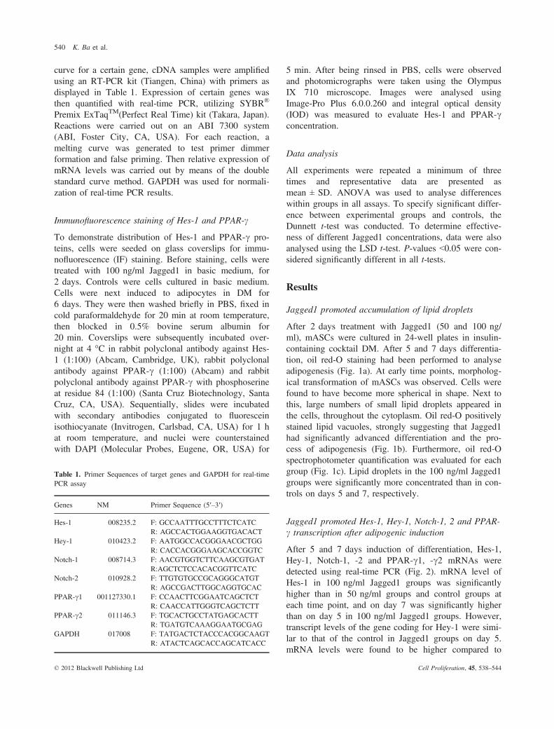
After 2 days treatment with Jagged1 (50 and 100 ng/ml), mASCs were cultured in 24-well plates in insulin-containing cocktail DM. After 5 and 7 days differentia-tion, oil red-O staining had been performed to analyseadipogenesis (Fig. 1a). At early time points, morpholog-ical transformation of mASCs was observed. Cells werefound to have become more spherical in shape. Next tothis, large numbers of small lipid droplets appeared inthe cells, throughout the cytoplasm. Oil red-O positivelystained lipid vacuoles, strongly suggesting that Jagged1had significantly advanced differentiation and the pro-cess of adipogenesis (Fig. 1b). Furthermore, oil red-Ospectrophotometer quantification was evaluated for eachgroup (Fig. 1c). Lipid droplets in the 100 ng/ml Jagged1groups were significantly more concentrated than in con-trols on days 5 and 7, respectively.
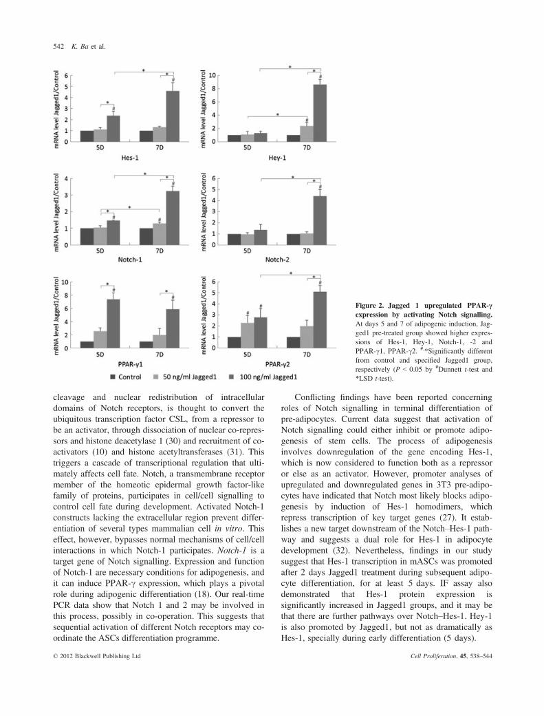
Jagged1 promoted Hes-1, Hey-1, Notch-1, 2 and PPAR-c transcription after adipogenic induction
After 5 and 7 days induction of differentiation, Hes-1,Hey-1, Notch-1, -2 and PPAR-c1, -c2 mRNAs weredetected using real-time PCR (Fig. 2). mRNA level ofHes-1 in 100 ng/ml Jagged1 groups was significantlyhigher than in 50 ng/ml groups and control groups ateach time point, and on day 7 was significantly higherthan on day 5 in 100 ng/ml Jagged1 groups. However,transcript levels of the gene coding for Hey-1 were simi-lar to that of the control in Jagged1 groups on day 5.mRNA levels were found to be higher compared to
Table 1. Primer Sequences of target genes and GAPDH for real-timePCR assay
Genes NM Primer Sequence (5′–3′)
Hes-1 008235.2 F: GCCAATTTGCCTTTCTCATCR: AGCCACTGGAAGGTGACACT
Hey-1 010423.2 F: AATGGCCACGGGAACGCTGGR: CACCACGGGAAGCACCGGTC
Notch-1 008714.3 F: AACGTGGTCTTCAAGCGTGATR:AGCTCTCCACACGGTTCATC
Notch-2 010928.2 F: TTGTGTGCCGCAGGGCATGTR: AGCCGACTTGGCAGGTGCAC
PPAR-c1 001127330.1 F: CCAACTTCGGAATCAGCTCTR: CAACCATTGGGTCAGCTCTT
PPAR-c2 011146.3 F: TGCACTGCCTATGAGCACTTR: TGATGTCAAAGGAATGCGAG
GAPDH 017008 F: TATGACTCTACCCACGGCAAGTR: ATACTCAGCACCAGCATCACC
© 2012 Blackwell Publishing Ltd Cell Proliferation, 45, 538–544
540 K. Ba et al.
controls and with significant difference between the twoJagged1 groups on day 7. Also, it was significantlyhigher than on day 5 in both Jagged1-treated groups.Notch-1 transcript levels in 100 ng/ml Jagged1 groupswas significantly higher than the 50 ng/ml Jagged1 andcontrol groups at each time point, and on day 7, it was
significantly higher than on day 5 in both Jagged1-treated groups. Transcript levels of the gene coding forNotch-2 were similar to those of controls in both Jag-ged1 groups on day 5. On day 7, however, in the100 ng/ml Jagged1 groups, it was significantly higherthan in 50 ng/ml Jagged1 and control groups, and alsohigher than in 100 ng/ml Jagged1 groups on day 5respectively. PPAR-c1 transcript levels in 100 ng/mlJagged1 groups were found to be significantly higherthan in 50 ng/ml Jagged1 and control groups at eachtime point. On day 5, mRNA level of PPAR-c2 in bothJagged1 groups was significantly higher than in controlgroups, but without significant difference between thetwo treated groups. In the 100 ng/ml Jagged1 groups, itwas significantly higher than in 50 ng/ml Jagged1 andcontrol groups on day 7, and also higher than the100 ng/ml Jagged1 groups on day 5.
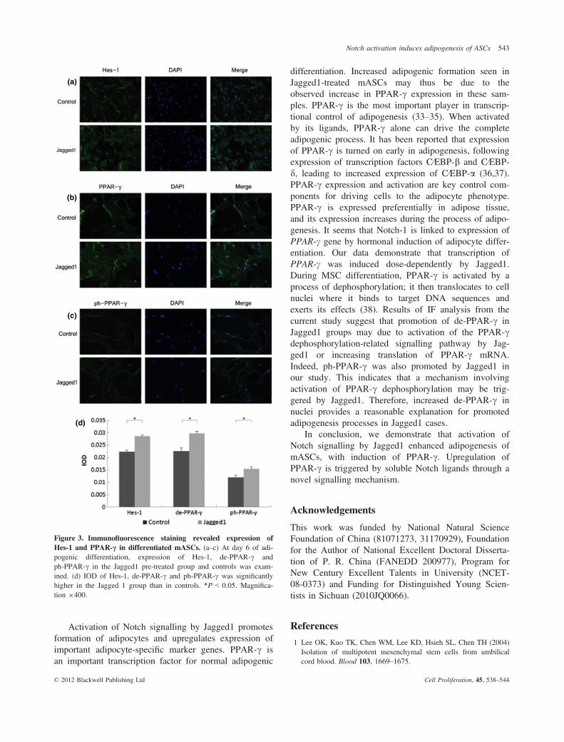
Jagged1 promoted Hes-1 and PPAR-c proteinexpression after 6 days adipogenic induction
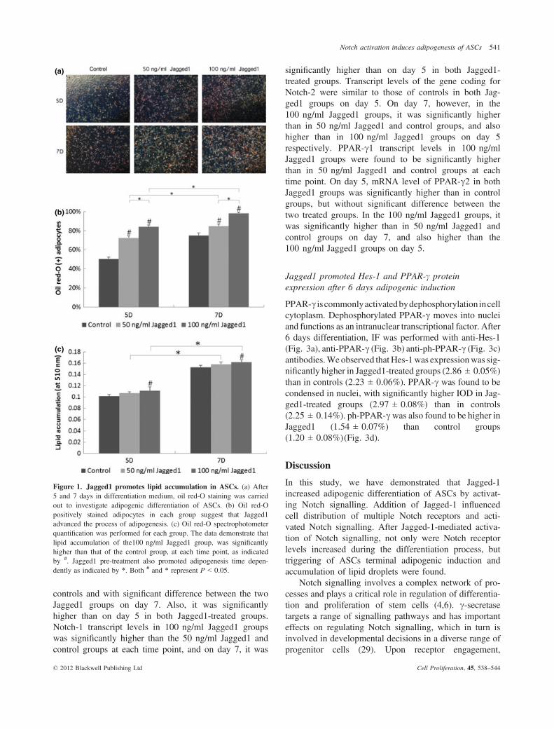
PPAR-c iscommonlyactivatedbydephosphorylation incellcytoplasm. Dephosphorylated PPAR-c moves into nucleiand functions as an intranuclear transcriptional factor. After6 days differentiation, IF was performed with anti-Hes-1(Fig. 3a), anti-PPAR-c (Fig. 3b) anti-ph-PPAR-c (Fig. 3c)antibodies.We observed that Hes-1was expressionwas sig-nificantly higher in Jagged1-treated groups (2.86 ± 0.05%)than in controls (2.23 ± 0.06%). PPAR-c was found to becondensed in nuclei, with significantly higher IOD in Jag-ged1-treated groups (2.97 ± 0.08%) than in controls(2.25 ± 0.14%). ph-PPAR-cwas also found to be higher inJagged1 (1.54 ± 0.07%) than control groups(1.20 ± 0.08%)(Fig. 3d).
Discussion
In this study, we have demonstrated that Jagged-1increased adipogenic differentiation of ASCs by activat-ing Notch signalling. Addition of Jagged-1 influencedcell distribution of multiple Notch receptors and acti-vated Notch signalling. After Jagged-1-mediated activa-tion of Notch signalling, not only were Notch receptorlevels increased during the differentiation process, buttriggering of ASCs terminal adipogenic induction andaccumulation of lipid droplets were found.
Notch signalling involves a complex network of pro-cesses and plays a critical role in regulation of differentia-tion and proliferation of stem cells (4,6). c-secretasetargets a range of signalling pathways and has importanteffects on regulating Notch signalling, which in turn isinvolved in developmental decisions in a diverse range ofprogenitor cells (29). Upon receptor engagement,
(a)
(b)
(c)
Figure 1. Jagged1 promotes lipid accumulation in ASCs. (a) After5 and 7 days in differentiation medium, oil red-O staining was carriedout to investigate adipogenic differentiation of ASCs. (b) Oil red-Opositively stained adipocytes in each group suggest that Jagged1advanced the process of adipogenesis. (c) Oil red-O spectrophotometerquantification was performed for each group. The data demonstrate thatlipid accumulation of the100 ng/ml Jagged1 group, was significantlyhigher than that of the control group, at each time point, as indicatedby #. Jagged1 pre-treatment also promoted adipogenesis time depen-dently as indicated by *. Both # and * represent P < 0.05.
© 2012 Blackwell Publishing Ltd Cell Proliferation, 45, 538–544
Notch activation induces adipogenesis of ASCs 541
cleavage and nuclear redistribution of intracellulardomains of Notch receptors, is thought to convert theubiquitous transcription factor CSL, from a repressor tobe an activator, through dissociation of nuclear co-repres-sors and histone deacetylase 1 (30) and recruitment of co-activators (10) and histone acetyltransferases (31). Thistriggers a cascade of transcriptional regulation that ulti-mately affects cell fate. Notch, a transmembrane receptormember of the homeotic epidermal growth factor-likefamily of proteins, participates in cell/cell signalling tocontrol cell fate during development. Activated Notch-1constructs lacking the extracellular region prevent differ-entiation of several types mammalian cell in vitro. Thiseffect, however, bypasses normal mechanisms of cell/cellinteractions in which Notch-1 participates. Notch-1 is atarget gene of Notch signalling. Expression and functionof Notch-1 are necessary conditions for adipogenesis, andit can induce PPAR-c expression, which plays a pivotalrole during adipogenic differentiation (18). Our real-timePCR data show that Notch 1 and 2 may be involved inthis process, possibly in co-operation. This suggests thatsequential activation of different Notch receptors may co-ordinate the ASCs differentiation programme.
Conflicting findings have been reported concerningroles of Notch signalling in terminal differentiation ofpre-adipocytes. Current data suggest that activation ofNotch signalling could either inhibit or promote adipo-genesis of stem cells. The process of adipogenesisinvolves downregulation of the gene encoding Hes-1,which is now considered to function both as a repressoror else as an activator. However, promoter analyses ofupregulated and downregulated genes in 3T3 pre-adipo-cytes have indicated that Notch most likely blocks adipo-genesis by induction of Hes-1 homodimers, whichrepress transcription of key target genes (27). It estab-lishes a new target downstream of the Notch–Hes-1 path-way and suggests a dual role for Hes-1 in adipocytedevelopment (32). Nevertheless, findings in our studysuggest that Hes-1 transcription in mASCs was promotedafter 2 days Jagged1 treatment during subsequent adipo-cyte differentiation, for at least 5 days. IF assay alsodemonstrated that Hes-1 protein expression issignificantly increased in Jagged1 groups, and it may bethat there are further pathways over Notch–Hes-1. Hey-1is also promoted by Jagged1, but not as dramatically asHes-1, specially during early differentiation (5 days).
Figure 2. Jagged 1 upregulated PPAR-cexpression by activating Notch signalling.At days 5 and 7 of adipogenic induction, Jag-ged1 pre-treated group showed higher expres-sions of Hes-1, Hey-1, Notch-1, -2 andPPAR-c1, PPAR-c2. #,*Significantly differentfrom control and specified Jagged1 group,respectively (P < 0.05 by #Dunnett t-test and*LSD t-test).
© 2012 Blackwell Publishing Ltd Cell Proliferation, 45, 538–544
542 K. Ba et al.
Activation of Notch signalling by Jagged1 promotesformation of adipocytes and upregulates expression ofimportant adipocyte-specific marker genes. PPAR-c isan important transcription factor for normal adipogenic
differentiation. Increased adipogenic formation seen inJagged1-treated mASCs may thus be due to theobserved increase in PPAR-c expression in these sam-ples. PPAR-c is the most important player in transcrip-tional control of adipogenesis (33–35). When activatedby its ligands, PPAR-c alone can drive the completeadipogenic process. It has been reported that expressionof PPAR-c is turned on early in adipogenesis, followingexpression of transcription factors C⁄EBP-b and C⁄EBP-d, leading to increased expression of C⁄EBP-a (36,37).PPAR-c expression and activation are key control com-ponents for driving cells to the adipocyte phenotype.PPAR-c is expressed preferentially in adipose tissue,and its expression increases during the process of adipo-genesis. It seems that Notch-1 is linked to expression ofPPAR-c gene by hormonal induction of adipocyte differ-entiation. Our data demonstrate that transcription ofPPAR-c was induced dose-dependently by Jagged1.During MSC differentiation, PPAR-c is activated by aprocess of dephosphorylation; it then translocates to cellnuclei where it binds to target DNA sequences andexerts its effects (38). Results of IF analysis from thecurrent study suggest that promotion of de-PPAR-c inJagged1 groups may due to activation of the PPAR-cdephosphorylation-related signalling pathway by Jag-ged1 or increasing translation of PPAR-c mRNA.Indeed, ph-PPAR-c was also promoted by Jagged1 inour study. This indicates that a mechanism involvingactivation of PPAR-c dephosphorylation may be trig-gered by Jagged1. Therefore, increased de-PPAR-c innuclei provides a reasonable explanation for promotedadipogenesis processes in Jagged1 cases.
In conclusion, we demonstrate that activation ofNotch signalling by Jagged1 enhanced adipogenesis ofmASCs, with induction of PPAR-c. Upregulation ofPPAR-c is triggered by soluble Notch ligands through anovel signalling mechanism.
Acknowledgements
This work was funded by National Natural ScienceFoundation of China (81071273, 31170929), Foundationfor the Author of National Excellent Doctoral Disserta-tion of P. R. China (FANEDD 200977), Program forNew Century Excellent Talents in University (NCET-08-0373) and Funding for Distinguished Young Scien-tists in Sichuan (2010JQ0066).
References
1 Lee OK, Kuo TK, Chen WM, Lee KD, Hsieh SL, Chen TH (2004)Isolation of multipotent mesenchymal stem cells from umbilicalcord blood. Blood 103, 1669–1675.
(a)
(b)
(c)
(d)
Figure 3. Immunofluorescence staining revealed expression ofHes-1 and PPAR-c in differentiated mASCs. (a–c) At day 6 of adi-pogenic differentiation, expression of Hes-1, de-PPAR-c andph-PPAR-c in the Jagged1 pre-treated group and controls was exam-ined. (d) IOD of Hes-1, de-PPAR-c and ph-PPAR-c was significantlyhigher in the Jagged 1 group than in controls. *P < 0.05. Magnifica-tion 9400.
© 2012 Blackwell Publishing Ltd Cell Proliferation, 45, 538–544
Notch activation induces adipogenesis of ASCs 543
2 Kern S, Eichler H, Stoeve J, Kluter H, Bieback K (2006) Compara-tive analysis of mesenchymal stem cells from bone marrow, umbili-cal cord blood, or adipose tissue. Stem Cells 24, 1294–1301.
3 Phinney DG, Kopen G, Isaacson RL, Prockop DJ (1999) Plasticadherent stromal cells from the bone marrow of commonly usedstrains of inbred mice: variations in yield, growth, and differentia-tion. J. Cell. Biochem. 72, 570–585.
4 Miele L, Osborne B (1999) Arbiter of differentiation and death:Notch signaling meets apoptosis. J. Cell. Physiol. 181, 393–409.
5 Li L, Milner LA, Deng Y, Iwata M, Banta A, Graf L et al. (1998)The human homolog of rat Jagged1 expressed by marrow stromainhibits differentiation of 32D cells through interaction withNotch1. Immunity 8, 43–55.
6 Artavanis-Tsakonas S, Rand MD, Lake RJ (1999) Notch signaling:cell fate control and signal integration in development. Science284, 770–776.
7 Brou C, Logeat F, Gupta N, Bessia C, LeBail O, Doedens JR et al.(2000) A novel proteolytic cleavage involved in Notch signaling: therole of the disintegrin-metalloprotease TACE.Mol. Cell 5, 207–216.
8 Mumm JS, Schroeter EH, Saxena MT, Griesemer A, Tian X, Pan DJet al. (2000) A ligand-induced extracellular cleavage regulates gamma-secretase-like proteolytic activation of Notch1. Mol. Cell 5, 197–206.
9 Aster JC, Robertson ES, Hasserjian RP, Turner JR, Kieff E, Sklar J(1997) Oncogenic forms of NOTCH1 lacking either the primarybinding site for RBP-Jkappa or nuclear localization sequencesretain the ability to associate with RBP-Jkappa and activate tran-scription. J. Biol. Chem. 272, 11336–11343.
10 Zhou S, Fujimuro M, Hsieh JJ, Chen L, Miyamoto A, Weinmaster Get al. (2000) SKIP, a CBF1-associated protein, interacts with theankyrin repeat domain of NotchIC to facilitate NotchIC function.Mol. Cell. Biol. 20, 2400–2410.
11 Wu L, Aster JC, Blacklow SC, Lake R, Artavanis-Tsakonas S,Griffin JD (2000) MAML1, a human homologue of Drosophilamastermind, is a transcriptional co-activator for NOTCH receptors.Nat. Genet. 26, 484–489.
12 Wu L, Griffin JD (2004) Modulation of Notch signaling by master-mind-like (MAML) transcriptional co-activators and their involve-ment in tumorigenesis. Semin. Cancer Biol. 14, 348–356.
13 Grottkau BE, Chen XR, Friedrich CC, Yang XM, Jing W, Wu Yet al. (2009) DAPT enhances the apoptosis of human tongue carci-noma cells. Int. J. Oral Sci. 1, 81–89.
14 Iso T, Chung G, Hamamori Y, Kedes L (2002) HERP1 is a cell type-specific primary target of Notch. J. Biol. Chem. 277, 6598–6607.
15 Iso T, Kedes L, Hamamori Y (2003) HES and HERP families:multiple effectors of the Notch signaling pathway. J. Cell. Physiol.194, 237–255.
16 Ohazama A, Hu Y, Schmidt-Ullrich R, Cao Y, Scheidereit C,Karin M et al. (2004) A dual role for Ikk alpha in tooth develop-ment. Dev. Cell 6, 219–227.
17 Nickoloff BJ, Qin JZ, Chaturvedi V, Denning MF, Bonish B,Miele L (2002) Jagged-1 mediated activation of notch signalinginduces complete maturation of human keratinocytes throughNF-kappaB and PPARgamma. Cell Death Differ. 9, 842–855.
18 Garces C, Ruiz-Hidalgo MJ, Font de Mora J, Park C, Miele L,Goldstein J et al. (1997) Notch-1 controls the expression of fattyacid-activated transcription factors and is required for adipogenesis.J. Biol. Chem. 272, 29729–29734.
19 Shelly LL, Fuchs C, Miele L (1999) Notch-1 inhibits apoptosis inmurine erythroleukemia cells and is necessary for differentiationinduced by hybrid polar compounds. J. Cell. Biochem. 73, 164–175.
20 Milner LA, Bigas A (1999) Notch as a mediator of cell fate deter-mination in hematopoiesis: evidence and speculation. Blood 93,2431–2448.
21 Yasutomo K, Doyle C, Miele L, Fuchs C, Germain RN (2000) Theduration of antigen receptor signalling determines CD4+ versusCD8+ T-cell lineage fate. Nature 404, 506–510.
22 Pui JC, Allman D, Xu L, DeRocco S, Karnell FG, Bakkour Set al. (1999) Notch1 expression in early lymphopoiesis influencesB versus T lineage determination. Immunity 11, 299–308.
23 Rangarajan A, Talora C, Okuyama R, Nicolas M, Mammucari C, OhH et al. (2001) Notch signaling is a direct determinant of keratinocytegrowth arrest and entry into differentiation. EMBO J. 20, 3427–3436.
24 Lowell S, Jones P, Le Roux I, Dunne J, Watt FM (2000) Stimula-tion of human epidermal differentiation by delta-notch signalling atthe boundaries of stem-cell clusters. Curr. Biol. 10, 491–500.
25 Nichols AM, Pan Y, Herreman A, Hadland BK, De Strooper B,Kopan R et al. (2004) Notch pathway is dispensable for adipocytespecification. Genesis 40, 40–44.
26 Ross DA, Rao PK, Kadesch T (2004) Dual roles for the Notch tar-get gene Hes-1 in the differentiation of 3T3-L1 preadipocytes. Mol.Cell. Biol. 24, 3505–3513.
27 Ross DA, Hannenhalli S, Tobias JW, Cooch N, Shiekhattar R,Kadesch T (2006) Functional analysis of Hes-1 in preadipocytes.Mol. Endocrinol. 20, 698–705.
28 Rosen ED, MacDougald OA (2006) Adipocyte differentiation fromthe inside out. Nat. Rev. Mol. Cell Biol. 7, 885–896.
29 Sarmento LM, Huang H, Limon A, Gordon W, Fernandes J, Tav-ares MJ et al. (2005) Notch1 modulates timing of G1-S progres-sion by inducing SKP2 transcription and p27 Kip1 degradation.J. Exp. Med. 202, 157–168.
30 Kao HY, Ordentlich P, Koyano-Nakagawa N, Tang Z, Downes M,Kintner CR et al. (1998) A histone deacetylase corepressor com-plex regulates the Notch signal transduction pathway. Genes Dev.12, 2269–2277.
31 Kurooka H, Honjo T (2000) Functional interaction between themouse notch1 intracellular region and histone acetyltransferasesPCAF and GCN5. J. Biol. Chem. 275, 17211–17220.
32 Huang Y, Yang X, Wu Y, Jing W, Cai X, Tang W et al. (2010)Gamma-secretase inhibitor induces adipogenesis of adipose-derivedstem cells by regulation of Notch and PPAR-gamma. Cell Prolif.43, 147–156.
33 Moerman EJ, Teng K, Lipschitz DA, Lecka-Czernik B (2004)Aging activates adipogenic and suppresses osteogenic programs inmesenchymal marrow stroma/stem cells: the role of PPAR-gamma2transcription factor and TGF-beta/BMP signaling pathways. AgingCell 3, 379–389.
34 Fink T, Abildtrup L, Fogd K, Abdallah BM, Kassem M, Ebbesen Pet al. (2004) Induction of adipocyte-like phenotype in human mes-enchymal stem cells by hypoxia. Stem Cells 22, 1346–1355.
35 Schadinger SE, Bucher NL, Schreiber BM, Farmer SR (2005)PPARgamma2 regulates lipogenesis and lipid accumulation in stea-totic hepatocytes. Am. J. Physiol. Endocrinol. Metab. 288, E1195–E1205.
36 Burgermeister E, Schnoebelen A, Flament A, Benz J, Stihle M,Gsell B et al. (2006) A novel partial agonist of peroxisome prolifer-ator-activated receptor-gamma (PPARgamma) recruits PPAR-gamma-coactivator-1alpha, prevents triglyceride accumulation, andpotentiates insulin signaling in vitro. Mol. Endocrinol. 20, 809–830.
37 Astudillo P, Rios S, Pastenes L, Pino AM, Rodriguez JP (2008)Increased adipogenesis of osteoporotic human-mesenchymal stemcells (MSCs) characterizes by impaired leptin action. J. Cell. Bio-chem. 103, 1054–1065.
38 Wu L, Cai X, Dong H, Jing W, Huang Y, Yang X et al. (2010)Serum regulates adipogenesis of mesenchymal stem cellsvia MEK/ERK-dependent PPARgamma expression andphosphorylation. J. Cell Mol. Med. 14, 922–932.
© 2012 Blackwell Publishing Ltd Cell Proliferation, 45, 538–544
544 K. Ba et al.


