Embed Size (px)
Citation preview
Citation: Wang, C.; Zhang, X.; Luo,
L.; Luo, Y.; Wu, D.; Spilca, D.; Le, Q.;
Yang, X.; Alvarez, K.; Hines, W.C.;
et al. COX-2 Deficiency Promotes
White Adipogenesis via
PGE2-Mediated Paracrine
Mechanism and Exacerbates
Diet-Induced Obesity. Cells 2022, 11,
1819. https://doi.org/10.3390/
cells11111819
Academic Editor: Victoriano
Baladrón
Received: 29 April 2022
Accepted: 31 May 2022
Published: 2 June 2022
Publisher’s Note: MDPI stays neutral
with regard to jurisdictional claims in
published maps and institutional affil-
iations.
Copyright: © 2022 by the authors.
Licensee MDPI, Basel, Switzerland.
This article is an open access article
distributed under the terms and
conditions of the Creative Commons
Attribution (CC BY) license (https://
creativecommons.org/licenses/by/
4.0/).
cells
Article
COX-2 Deficiency Promotes White Adipogenesis viaPGE2-Mediated Paracrine Mechanism and ExacerbatesDiet-Induced ObesityChunqing Wang 1 , Xing Zhang 1, Liping Luo 1 , Yan Luo 1, Dandan Wu 2 , Dianna Spilca 1, Que Le 1,Xin Yang 1, Katelyn Alvarez 1, William Curtis Hines 1, Xuexian O. Yang 2,3 and Meilian Liu 1,3,*
1 Department of Biochemistry and Molecular Biology, University of New Mexico Health Sciences Center,Albuquerque, NM 87131, USA; [email protected] (C.W.); [email protected] (X.Z.);[email protected] (L.L.); [email protected] (Y.L.); [email protected] (D.S.); [email protected] (Q.L.);[email protected] (X.Y.); [email protected] (K.A.); [email protected] (W.C.H.)
2 Department of Molecular Genetics and Microbiology, University of New Mexico Health Sciences Center,Albuquerque, NM 87131, USA; [email protected] (D.W.); [email protected] (X.O.Y.)
3 Autophagy Inflammation and Metabolism Center for Biomedical Research Excellence, School of Medicine,University of New Mexico Health Sciences Center, Albuquerque, NM 87131, USA
* Correspondence: [email protected]; Tel.: +1-(505)-272-4036
Abstract: Cyclooxygenase-2 (COX-2) plays a critical role in regulating innate immunity and metabolismby producing prostaglandins (PGs) and other lipid mediators. However, the implication of adiposeCOX-2 in obesity remains largely unknown. Using adipocyte-specific COX-2 knockout (KO) mice, weshowed that depleting COX-2 in adipocytes promoted white adipose tissue development accompa-nied with increased size and number of adipocytes and predisposed diet-induced adiposity, obesity,and insulin resistance. The increased size and number of adipocytes by COX-2 KO were reversed bythe treatment of prostaglandin E2 (PGE2) but not PGI2 and PGD2 during adipocyte differentiation.PGE2 suppresses PPARγ expression through the PKA pathway at the early phase of adipogenesis,and treatment of PGE2 or PKA activator isoproterenol diminished the increased lipid droplets insize and number in COX-2 KO primary adipocytes. Administration of PGE2 attenuated increasedfat mass and fat percentage in COX-2 deficient mice. Taken together, our study demonstrated thesuppressing effect of adipocyte COX-2 on adipogenesis and reveals that COX-2 restrains adiposetissue expansion via the PGE2-mediated paracrine mechanism and prevents the development ofobesity and related metabolic disorders.
Keywords: COX-2; PGE2; white adipogenesis; adipocyte hypertrophy; obesity; PPARγ
1. Introduction
Obesity, a disorder characterized by excess adiposity, has become a primary cause ofinsulin resistance, type 2 diabetes, and cardiovascular diseases. Pathological expansionof adipose tissue (AT) is accompanied by adipose hypoxia, angiogenesis, remodeling,and inflammation, thereby causing systemic insulin resistance and type 2 diabetes [1,2].However, the mechanisms regulating adipocyte differentiation, maturation, and turn overare incompletely understood.
The nuclear hormone receptor peroxisome proliferator-activated receptor gamma(PPARγ) is a mast regulator of adipocyte differentiation [3,4]. Upon its activation bybinding to its endogenous ligands or synthetic ligands such as thiazolidinediones (TZDs),PPARγ promotes adipogenesis and insulin sensitivity [3,4]. It is primarily expressed inadipocytes [4] and present in other cell types such as regulatory T cells with a lesser ex-tent [5,6]. Activated PPARγ binds to its responsive elements in the promoter and inducesadipogenesis- and lipogenesis-related genes, including Cd36, lipoprotein lipase (Lpl), fattyacid-binding protein (aP2), CCAAT/enhancer-binding protein alpha (C/ebpα) and other
Cells 2022, 11, 1819. https://doi.org/10.3390/cells11111819 https://www.mdpi.com/journal/cells
Cells 2022, 11, 1819 2 of 16
adipogenesis markers such as adiponectin [4,7,8]. In addition to ligand stimulation, PPARγtranscriptional activity is controlled by its protein modification, including phosphoryla-tion (Ser273) [9–11], acetylation (Lys268 and Lys293) [12], sumoylation [13–17], glycosyla-tion [18] and ubiquitination [19–22]. Such modifications of PPARγ are critical for its action,stability, and selective targeting and are functionally connected [9–12,19,20]. For instance,ligand binding not only increases the transcriptional function of PPARγ but also inducesits ubiquitination and subsequent degradation by the proteasome [19]. Sirt1-mediateddeacetylation of PPARγ at Lys268 and Lys293 is critical for its stabilization and has beenlinked to its adipogenesis-inducing effect (unpresented data in the referred study) [12].Additional factors, including interferon γ (IFNγ) and tumor necrosis factor alpha (TNFα),have been shown to regulate PPARγ degradation via a proteasome-dependent mechanismin adipocytes [23–25], indicative of the significance of PPARγ stabilization in its function.However, while fatty acid metabolites are considered potential endogenous ligands ofPPARγ, the mechanisms underlying PPARγ regulation are incompletely understood.
Cyclooxygenase (COX), a rate-limiting enzyme responsible for the biosynthesis ofprostaglandins (PGs), exists in three isoforms; COX-1, the constitutive form, COX-2, the in-ducible form, and COX-3, the splicing variant [26]. COX-2 oxygenates arachidonic acid andconverts it into a number of PGs, including PGD2, PGE2, PGF2α, and prostacyclin (PGI2),all of which exert diverse hormone-like effects via autocrine or paracrine mechanisms [26].The accumulated evidence shows that PGs modulate adipogenesis by acting as an agonistor analog of Peroxisome Proliferator-Activated Receptor γ (PPARγ) [27]. It has also beensuggested that the COX-2/PG axis plays a critical role in regulating AT inflammation andobesity-induced insulin resistance [28–33]. In addition, the COX-2/PG pathway has beenshown to promote thermogenic programming and white adipose tissue (WAT) browning,although it is still controversial whether COX-2 is induced by cold and is correlated withobesity [28,34–37]. On the other hand, while COX-2 overexpression in adipocytes leads toprotection against diet-induced obesity and inflammation, and COX-2 in adipocytes me-diates the anti-inflammatory benefits of intermittent fasting [38,39], whether endogenousCOX-2 in adipocytes is implicated in adiposity and obesity is unclear.
We recently showed that adipose COX-2 is inversely correlated with obesity in humansand in rodents, and that intermittent fasting (IF) restores the expression level of COX-2in adipose tissue of obese mice [39]. In addition, COX-2 in adipocytes mediates the anti-inflammatory effects of intermittent fasting in adipose tissue despite similar anti-obesityeffects of IF in adipocyte-specific COX-2 KO and control mice [39]. In the present study, wespecifically characterized the role of adipose COX-2 in regulating adipogenesis and foundthat COX-2 in adipocytes exerts an anti-obesity effect by suppressing white adipogenesis.PGE2 but not PGI2 and PGD2 attenuates the increased adipogenesis of COX-2 KO primarystromal cells via PKA-mediated suppression of PPARγ function. Moreover, adipocyte-specific COX-2 deficient mice are prone to diet-induced obesity and insulin resistancewhich was ameliorated by PGE2 treatment. Taken together, our study reveals that COX-2in adipocytes plays a critical role in the control of adipocyte differentiation by the PGE2-mediated paracrine mechanism.
2. Materials and Methods2.1. Materials
Antibodies against COX-2 (12282), PPARγ (2443), PGC1α (2178), and PKA (9621) werefrom Cell Signaling Technology (Danvers, MA, USA). Polyclonal antibodies to adiponectinand anti-tubulin were kindly provided by Drs. Lily Dong and Feng Liu at UT Health SanAntonio (San Antonio, TX, USA) as described previously [40]. The anti-UCP1 (ab23841)and anti-Plin1 (ab61682) were purchased from Abcam (Discovery Drive, Cambridge, CB20AX, UK). BODIPY 493/503 (D3922) and DAPI (D3571) were from Thermo Fisher Scientific(Waltham, MA, USA). PGD2 (41598-07-6), PGE2 (14010), PGI2 (69552-46-1), L-161,982(10011565), TG6-10-1 (23444), SC-51089 (10011561), and Rosiglitazone (71740) were obtainedfrom Cayman Chemical Company (Ann Arbor, MI, USA). KT5720 (K3761) and Isoproterenol
Cells 2022, 11, 1819 3 of 16
(I6504) were from Sigma-Aldrich (St. Louis, MO, USA). H89 (371963) was purchased fromMillipore (Burlington, MA, USA).
2.2. Animals
The COX-2 floxed mice were generously provided by Dr. Harvey R. Herschman at theUniversity of California (Los Angeles, CA, USA). Adipocyte-COX-2 specific knockout (KO)mice were generated by crossing COX-2 floxed mice with adiponectin cre mice (JacksonLaboratory, Bar Harbor, ME, USA. Stock number 10803). COX-2 floxed littermates wereused as controls. The knockout efficiency was confirmed in adipose tissue and othertissues by Western blot analysis and Real-Time PCR shown in our previous work. Unlessotherwise noted, male mice were used for all experiments. Animals were housed in aspecific pathogen-free barrier facility with a 12-h light/12-h dark cycle with free access tofood and water. For the high-fat diet challenge study, 6-week-old mice were fed with ahigh fat diet (HFD) (45% kcal fat) from Research Diets Inc. (D12451; New Brunswick, NJ,USA) for 8 or 16 weeks unless otherwise specified, and normal chow diet (NCD) providedby the animal facility at the UNMHSC. All animal experimental protocols were reviewedand approved by the Animal Care Committee of the University of New Mexico HealthSciences Center.
2.3. Administration of PGE2
PGE2 was dissolved in 100% dimethyl sulfoxide (DMSO) and diluted in 0.9% SodiumChloride to reach the concentration of 5 µg/mL. Four-month-old male COX-2 KO, COX-2 floxed, or wild type C57BL/6 mice were administered with PGE2 or vehicle throughintraperitoneal injection at 50 µg/kg every other day for 2 weeks.
2.4. Primary Culture and Differentiation of Adipocytes
Primary stromal vascular fractions (SVFs) from iWAT of 3-week-old COX-2 knockoutand control mice were isolated, cultured, and differentiated into adipocytes accordingto the procedure as described previously [41]. In brief, the confluent stromal cells werecultured in the induction medium (DMEM with 10% Fetal Bovine Serum, plus 8 µg/mLinsulin, 0.1 µg/mL Dexamethesine, and 112 µg/mL Isobutylmethylxanthine) for 4 daysfollowed with differentiation medium (DMEM with 10% Fetal Bovine Serum, plus 8 µg/mLinsulin) for 2 days, and treated with or without PGD2, PGE2 or PGI2 for 6 days during thedifferentiation. On day 6, cells were harvested for Western blot analysis, Real-Time PCR,or staining. Alternatively, day-6 differentiated primary COX-2 KO and control adipocyteswere cultured with a fresh differentiation medium for 24 h. The media was collected andused to treat wild-type primary preadipocytes isolated from inguinal fat that underwentdifferentiation to adipocytes. This treatment was started on day 1 and lasted for 6 daysduring differentiation.
2.5. Treatment of PGE2 Receptor Antagonists or PKA Activator/Inhibitors during Differentiation
Primary stromal cells isolated from inguinal fat of COX-2 KO and control mice weredifferentiated into adipocytes. Starting from day 1 of differentiation, cells were treatedwith β-adrenoceptor agonist isoproterenol (10 µM) for 6 days. For the PGE2 study, primarystromal cells from C57BL/6 mice were differentiated and treated with PGE2 with cotreat-ment of EP1 antagonist SC-51089 (1 µM), EP2 antagonist TG6-10-1 (1 µM), EP4 antagonistL-161,982 (1 µM), or PKA specific inhibitor KT5720 (10 µM) or H89 (10 µM) for 6 days fromday 1 of differentiation.
2.6. Hematoxylin and Eosin (H&E) and Oil Red O Staining
For the H&E staining, tissues were fixed with 10% formalin for 24 h and embeddedin paraffin. Tissue sections (6-µm thick) were stained with H&E according to standardprotocols and analyzed using the NIH Image J software (Bethesda, MD, USA). To analyzethe adipocyte size in adipose tissue, stained tissues were visualized with NanoZoomer
Cells 2022, 11, 1819 4 of 16
Slide Scanner (Leica Biosystems, Buffalo Grove, IL, USA). Pick eight fields per animalgenotype were analyzed and each field randomly count 30 cells’ size by Image J. For thequantification of total adipocyte numbers in adipose tissue, it was estimated by manualcounting of cells on H&E slides from at least three 20× fields of three mice per genotype andapproximated assuming cubic packing as a previous study described [42]. Adipocyte celldiameters of adipose tissue were measured to determine the mean adipocyte diameter. Themean adipocyte density (cells/unit volume) was next calculated from the mean adipocytediameter assuming cubic closest packing of adipocytes. The total number of adipocytesin each epididymal fat pad during normal feeding or HFD feeding was then calculatedfrom the density of adipocytes (cells/unit volume) and the measured total volume of eachepididymal fat pad. For the Oil Red O staining, differentiated primary adipocytes werefixed with 4% formalin for 10 min and stained with Oil Red O in 60% isopropanol for20 min [43], and images were taken by EVOS FL Cell Imaging System.
2.7. Glucose Tolerance Test (GTT) and Insulin Tolerance Test (ITT)
For the GTT, mice were fasted for 12 h, and fasted glucose was measured using aGlucometer (GE, Boston, MA, USA) by tail bleeds. Then mice were intraperitoneallyinjected with 2 g glucose/kg of body weight, and blood glucose was measured at the timeof 15 min, 30 min, 60 min, and 120 min after glucose injection. For the ITT, mice werefasted for 4 h, and fasted glucose was measured using a Glucometer (GE) by tail bleeds.Then mice were intraperitoneally injected with 0.75(U/mL) insulin/kg of body weight,and blood glucose was measured at the time of 15 min, 30 min, 60 min, and 90 min afterinsulin injection.
2.8. DEXA Scanning
To check the body composition, mice were anesthetized through intraperitoneal injec-tion with 0.1 mL/10 g animal body weight Ketamine/Xylazine (10 mg/mL Ketamine and1 mg/mL Xylazine). Bone mineral density, lean mass, fat mass, total body weight, and fatpercentage were determined by dual-energy X-ray absorptiometry (DEXA) (GE MedicalSystems, Madison, WI, USA).
2.9. Immunofluorescence Staining
Adipocytes in 24-well plates were fixed in 4% paraformaldehyde for 10 min, andthen permeabilized with 0.1% saponin in 3% bovine serum albumin (BSA) for 30 min.Incubation with anti-Plin1 for an hour and followed by secondary antibodies as well asanti-Bodipy together for half an hour. Cell Nuclei were stained with DAPI (blue). Confocalimages were taken using Cellomics Image System (Thermo Fisher Scientific) as previouslydescribed [44].
2.10. Real-Time PCR and Western Blot
Total RNA was extracted from mice tissues or primary cultural cells using the RNeasyLipid Tissue Mini Kit (Qiagen, Germantown, MD, USA). The purity and concentration oftotal RNA were determined by a NanoDrop spectrophotometer (Thermo Fisher Scientific).One µg of total RNA was reverse-transcribed using a cDNA kit (AB Applied Biosystems,Waltham, MA, USA). Real-time PCR amplification was detected using SYBR Green PCRmaster mixture (Qiagen) on a Roche 480 Real-time PCR system (Basel, Switzerland). Theprimer sequences used in real-time PCR are shown in Supplementary Table S1. Westernblot analysis was performed following the procedures described previously [45].
2.11. Statistics
Statistical analysis of the data was performed using a two-tailed Student’s t-test betweentwo groups or one-way ANOVA among three different groups. All the results were presentedas the mean ± S.E.M., and a p-value of <0.05 was considered to be statistically significant.
Cells 2022, 11, 1819 5 of 16
3. Results3.1. Adipocyte-Specific Depletion of COX-2 Predisposes Diet-Induced Adiposity and Insulin Resistance
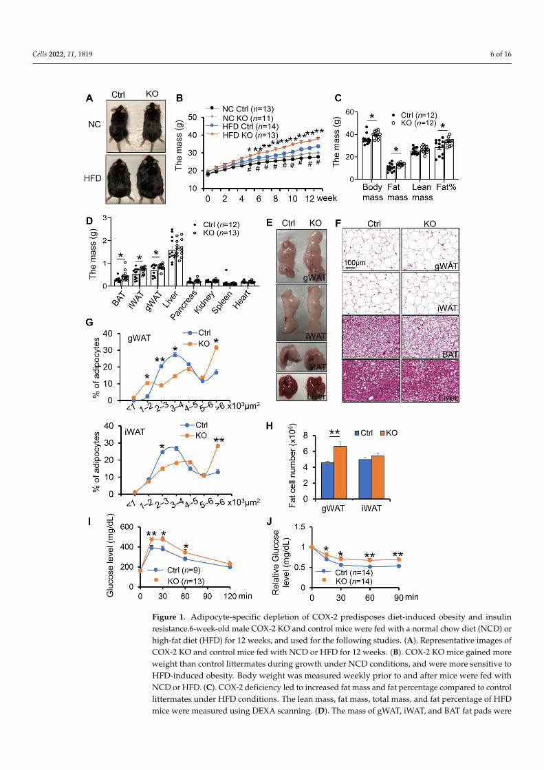
COX-2 has been shown to play an important role in regulating inflammation andenergy homeostasis [28,35,46,47]. However, little is known about the physiological roleof COX-2 in adipocytes. To this end, we generated adipocyte-specific COX-2 knockout(KO) mice by crossing COX-2 floxed mice and adiponectin cre mice [39]. To investigatethe potential effect of COX-2 on diet-induced obesity and insulin resistance in vivo, wefed the COX-2 KO mice and control littermates with HFD for 16-weeks. No significantdifference in food intake was observed between COX-2 KO mice and their control miceduring HFD feeding (Figure S1A). However, the differences in body size and weightwere more pronounced between HFD-fed COX-2 KO mice and the control littermates(Figure 1A,B). The fat mass and fat percentage of COX-2 KO mice were also significantlygreater compared with control littermates on HFD (Figure 1C–E). Consistent with thisfinding, the HFD-fed COX-2 KO mice showed larger fat cell size and increased fat cellnumber in gonadal white adipose tissue (gWAT) and inguinal WAT (iWAT) as well as severehepatosteatosis compared with HFD-fed wild-type littermates (Figure 1F–H), leading tosevere glucose and insulin intolerance after 8 weeks of HFD feeding (Figure 1I,J). Theseresults suggest that COX-2 KO exacerbates diet-induced obesity and insulin resistance.
COX-2 deficiency had little effect on the food intake during HFD feeding (Figure S1A).Surprisingly, the decreased insulin tolerance via COX-2 deficiency was not pronounced despitea dramatic increase in body weight gain under 16 weeks of HFD feedings (Figure S1B,C), im-plying a distinct effect of adipocyte COX-2 on insulin sensitivity in the early and late stagesof obesity. The anti-obesity property of COX-2 was restricted to males given the modesteffects of COX-2 KO on body weight and insulin sensitivity in female mice (Figure S1D–F).Despite little effect on energy expenditure, food intake, activity, and cold-induced thermo-genic gene expression under normal chow diet conditions (Figure S2), adipocyte-specificdepletion of COX-2 reduced the basal expression of UCP1 and PGC1α (Figure S2D,F). Inaddition, COX-2 deficiency in adipocytes improves anti-inflammatory response underNCD conditions, while this improving effect was not observed post-16-week HFD feeding(Figure S3).
3.2. COX-2 Deficiency Enhances White Adipogenesis
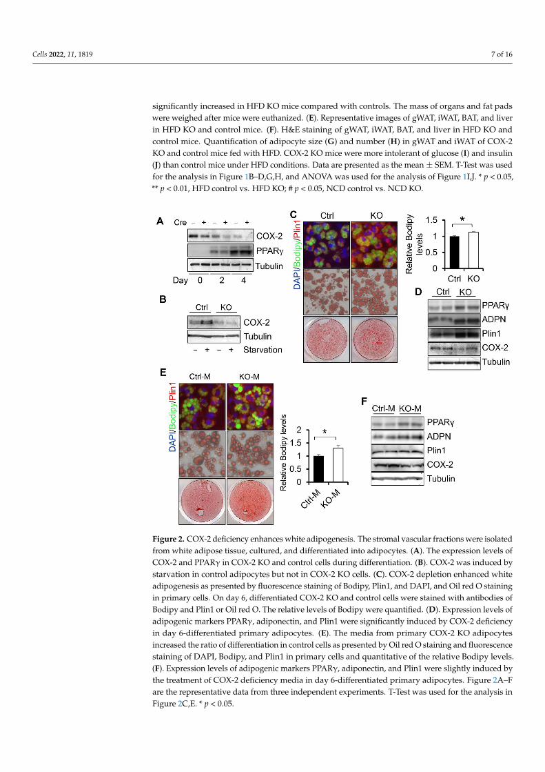
To gain insight into the role of COX-2 in regulating adipogenesis, we performeddifferentiation of COX-2 KO primary preadipocytes. COX-2 protein levels were markedlydecreased in COX-2 KO adipocytes compared to control cells during adipogenesis and uponstarvation treatment (Figure 2A,B), indicating that COX-2 was successfully suppressedby COX-2 KO in adipocytes. The protein levels of COX-2 were suppressed during thedifferentiation and were notably induced by starvation in primary adipocytes (Figure 2A,B),suggesting a potential role of COX-2 in adipogenesis. Fluorescence and Red Oil O stainingresults showed that COX-2 deficiency promoted adipocyte differentiation indicated by theincreased staining of lipid droplet marker Bodipy and upregulated expression levels ofadipogenesis markers PPARγ and Plin1 (Figure 2C,D). Consistent with this, the media fromCOX-2 KO adipocytes enhanced adipocyte differentiation compared to the media from thecontrol cells (Figure 2E,F), indicative of an autocrine or paracrine mechanism mediatingthe suppressing effect of COX-2 on adipocyte differentiation.
Cells 2022, 11, 1819 6 of 16Cells 2022, 11, x FOR PEER REVIEW 6 of 17
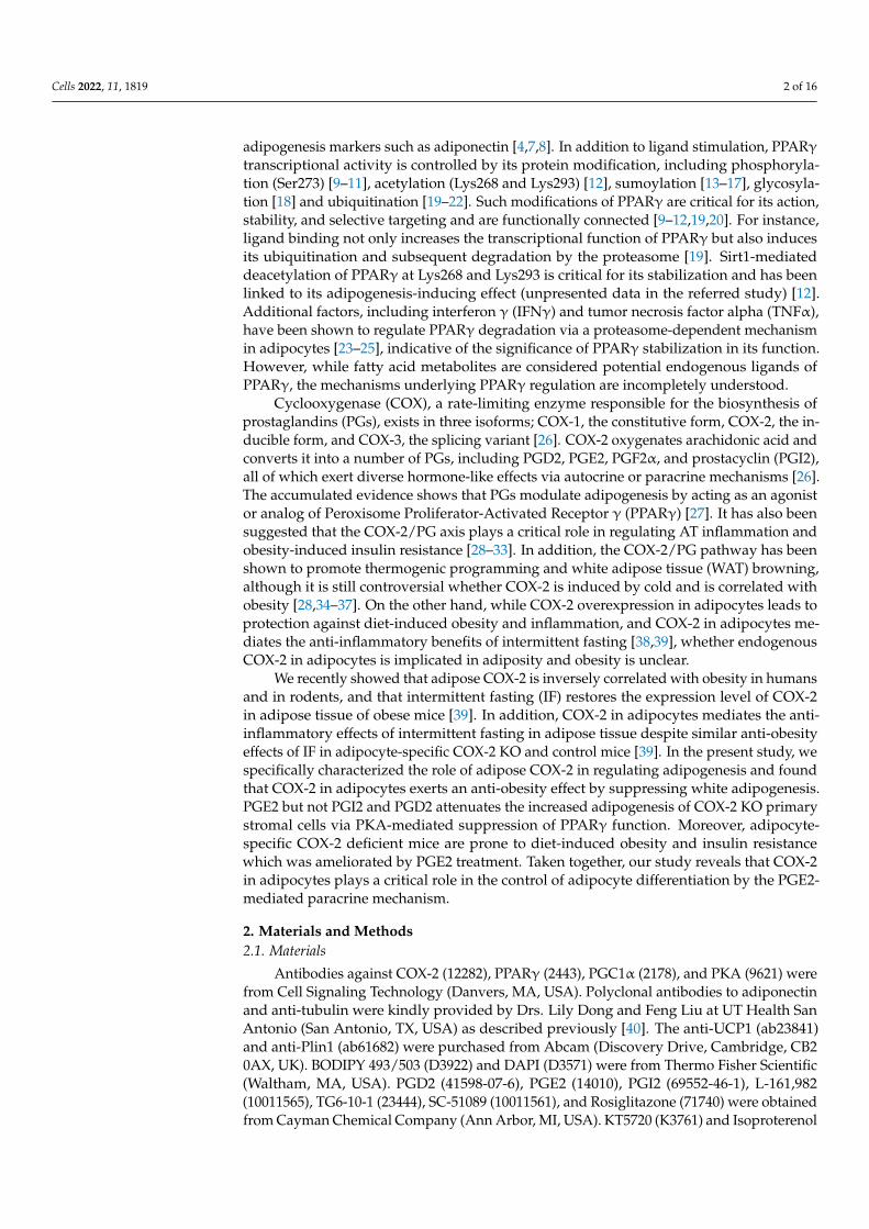
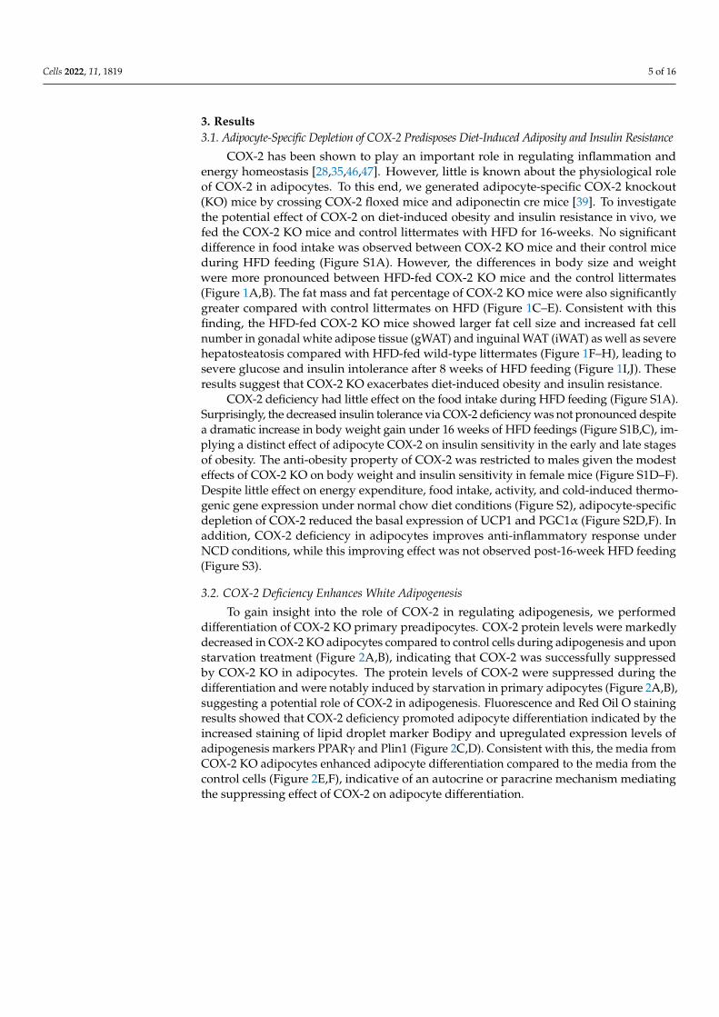
Figure 1. Adipocyte-specific depletion of COX-2 predisposes diet-induced obesity and insulin re-sistance.6-week-old male COX-2 KO and control mice were fed with a normal chow diet (NCD) or high-fat diet (HFD) for 12 weeks, and used for the following studies. (A). Representative images of COX-2 KO and control mice fed with NCD or HFD for 12 weeks. (B). COX-2 KO mice gained more weight than control littermates during growth under NCD conditions, and were more sensitive to HFD-induced obesity. Body weight was measured weekly prior to and after mice were fed with NCD or HFD. (C). COX-2 deficiency led to increased fat mass and fat percentage compared to con-trol littermates under HFD conditions. The lean mass, fat mass, total mass, and fat percentage of HFD mice were measured using DEXA scanning. (D). The mass of gWAT, iWAT, and BAT fat pads were significantly increased in HFD KO mice compared with controls. The mass of organs and fat pads were weighed after mice were euthanized. (E). Representative images of gWAT, iWAT, BAT, and liver in HFD KO and control mice. (F). H&E staining of gWAT, iWAT, BAT, and liver in HFD KO and control mice. Quantification of adipocyte size (G) and number (H) in gWAT and iWAT of
Figure 1. Adipocyte-specific depletion of COX-2 predisposes diet-induced obesity and insulinresistance.6-week-old male COX-2 KO and control mice were fed with a normal chow diet (NCD) orhigh-fat diet (HFD) for 12 weeks, and used for the following studies. (A). Representative images ofCOX-2 KO and control mice fed with NCD or HFD for 12 weeks. (B). COX-2 KO mice gained moreweight than control littermates during growth under NCD conditions, and were more sensitive toHFD-induced obesity. Body weight was measured weekly prior to and after mice were fed withNCD or HFD. (C). COX-2 deficiency led to increased fat mass and fat percentage compared to controllittermates under HFD conditions. The lean mass, fat mass, total mass, and fat percentage of HFDmice were measured using DEXA scanning. (D). The mass of gWAT, iWAT, and BAT fat pads were
Cells 2022, 11, 1819 7 of 16
significantly increased in HFD KO mice compared with controls. The mass of organs and fat padswere weighed after mice were euthanized. (E). Representative images of gWAT, iWAT, BAT, and liverin HFD KO and control mice. (F). H&E staining of gWAT, iWAT, BAT, and liver in HFD KO andcontrol mice. Quantification of adipocyte size (G) and number (H) in gWAT and iWAT of COX-2KO and control mice fed with HFD. COX-2 KO mice were more intolerant of glucose (I) and insulin(J) than control mice under HFD conditions. Data are presented as the mean ± SEM. T-Test was usedfor the analysis in Figure 1B–D,G,H, and ANOVA was used for the analysis of Figure 1I,J. * p < 0.05,** p < 0.01, HFD control vs. HFD KO; # p < 0.05, NCD control vs. NCD KO.
Cells 2022, 11, x FOR PEER REVIEW 7 of 17
COX-2 KO and control mice fed with HFD. COX-2 KO mice were more intolerant of glucose (I) and insulin (J) than control mice under HFD conditions. Data are presented as the mean ± SEM. T-Test was used for the analysis in Figure 1B–D,G,H, and ANOVA was used for the analysis of Figure 1I,J. * p < 0.05, ** p < 0.01, HFD control vs. HFD KO; # p < 0.05, NCD control vs. NCD KO.
3.2. COX-2 Deficiency Enhances White Adipogenesis To gain insight into the role of COX-2 in regulating adipogenesis, we performed dif-
ferentiation of COX-2 KO primary preadipocytes. COX-2 protein levels were markedly decreased in COX-2 KO adipocytes compared to control cells during adipogenesis and upon starvation treatment (Figure 2A,B), indicating that COX-2 was successfully sup-pressed by COX-2 KO in adipocytes. The protein levels of COX-2 were suppressed during the differentiation and were notably induced by starvation in primary adipocytes (Figure 2A,B), suggesting a potential role of COX-2 in adipogenesis. Fluorescence and Red Oil O staining results showed that COX-2 deficiency promoted adipocyte differentiation indi-cated by the increased staining of lipid droplet marker Bodipy and upregulated expres-sion levels of adipogenesis markers PPARγ and Plin1 (Figure 2C,D). Consistent with this, the media from COX-2 KO adipocytes enhanced adipocyte differentiation compared to the media from the control cells (Figure 2E,F), indicative of an autocrine or paracrine mechanism mediating the suppressing effect of COX-2 on adipocyte differentiation.
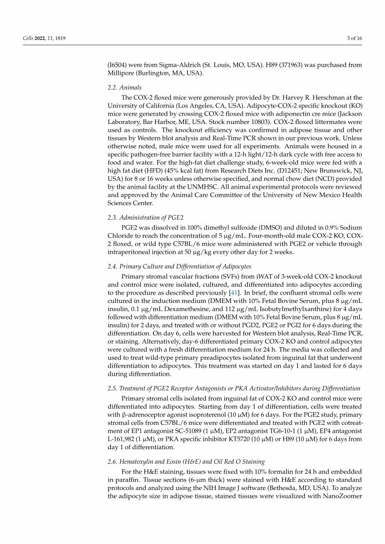
Figure 2. COX-2 deficiency enhances white adipogenesis. The stromal vascular fractions were iso-lated from white adipose tissue, cultured, and differentiated into adipocytes. (A). The expression levels of COX-2 and PPARγ in COX-2 KO and control cells during differentiation. (B). COX-2 was induced by starvation in control adipocytes but not in COX-2 KO cells. (C). COX-2 depletion en-hanced white adipogenesis as presented by fluorescence staining of Bodipy, Plin1, and DAPI, and Oil red O staining in primary cells. On day 6, differentiated COX-2 KO and control cells were stained with antibodies of Bodipy and Plin1 or Oil red O. The relative levels of Bodipy were quantified. (D).
Figure 2. COX-2 deficiency enhances white adipogenesis. The stromal vascular fractions were isolatedfrom white adipose tissue, cultured, and differentiated into adipocytes. (A). The expression levels ofCOX-2 and PPARγ in COX-2 KO and control cells during differentiation. (B). COX-2 was induced bystarvation in control adipocytes but not in COX-2 KO cells. (C). COX-2 depletion enhanced whiteadipogenesis as presented by fluorescence staining of Bodipy, Plin1, and DAPI, and Oil red O stainingin primary cells. On day 6, differentiated COX-2 KO and control cells were stained with antibodies ofBodipy and Plin1 or Oil red O. The relative levels of Bodipy were quantified. (D). Expression levels ofadipogenic markers PPARγ, adiponectin, and Plin1 were significantly induced by COX-2 deficiencyin day 6-differentiated primary adipocytes. (E). The media from primary COX-2 KO adipocytesincreased the ratio of differentiation in control cells as presented by Oil red O staining and fluorescencestaining of DAPI, Bodipy, and Plin1 in primary cells and quantitative of the relative Bodipy levels.(F). Expression levels of adipogenic markers PPARγ, adiponectin, and Plin1 were slightly induced bythe treatment of COX-2 deficiency media in day 6-differentiated primary adipocytes. Figure 2A–Fare the representative data from three independent experiments. T-Test was used for the analysis inFigure 2C,E. * p < 0.05.
Cells 2022, 11, 1819 8 of 16
3.3. Depletion of COX-2 Promotes Adipocyte Maturation via PGE2-Mediated Paracrine Mechanism
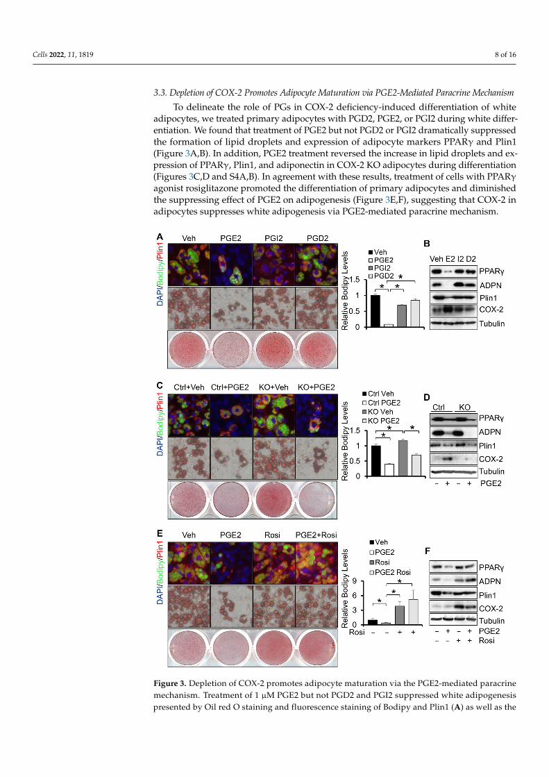
To delineate the role of PGs in COX-2 deficiency-induced differentiation of whiteadipocytes, we treated primary adipocytes with PGD2, PGE2, or PGI2 during white differ-entiation. We found that treatment of PGE2 but not PGD2 or PGI2 dramatically suppressedthe formation of lipid droplets and expression of adipocyte markers PPARγ and Plin1(Figure 3A,B). In addition, PGE2 treatment reversed the increase in lipid droplets and ex-pression of PPARγ, Plin1, and adiponectin in COX-2 KO adipocytes during differentiation(Figures 3C,D and S4A,B). In agreement with these results, treatment of cells with PPARγagonist rosiglitazone promoted the differentiation of primary adipocytes and diminishedthe suppressing effect of PGE2 on adipogenesis (Figure 3E,F), suggesting that COX-2 inadipocytes suppresses white adipogenesis via PGE2-mediated paracrine mechanism.
Cells 2022, 11, x FOR PEER REVIEW 9 of 17
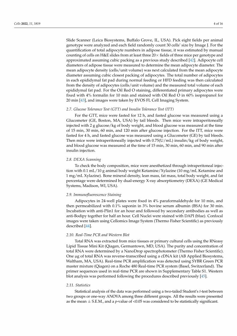
Figure 3. Depletion of COX-2 promotes adipocyte maturation via the PGE2-mediated paracrine mechanism. Treatment of 1 µM PGE2 but not PGD2 and PGI2 suppressed white adipogenesis pre-sented by Oil red O staining and fluorescence staining of Bodipy and Plin1 (A) as well as the ex-pression levels of adipogenic markers PPARγ, adiponectin, and Plin1 (B). Starting from differenti-ation, primary preadipocytes were treated with 1 µM PGE2, PGI2, and PGD2 for 6 days. Treatment of 1 µM PGE2 reversed COX-2 deficiency-induced white adipogenesis presented by Oil red O stain-ing and fluorescence staining of Bodipy and Plin1 (C) as well as the expression levels of adipogenic markers PPARγ, adiponectin, and Plin1 (D) in primary adipocytes. Treatment of 10 µM PPARγ agonist Rosiglitazone restored PGE2 treatment-suppressed white adipogenesis presented by Oil red O staining and fluorescence staining of Bodipy and Plin1 (E) as well as the expression levels of adi-pogenic markers PPARγ, adiponectin, and Plin1 (F) in primary adipocytes. Data in Figure 3A,C,E are presented with means ± S.E.M. * p < 0.05.
Figure 3. Depletion of COX-2 promotes adipocyte maturation via the PGE2-mediated paracrinemechanism. Treatment of 1 µM PGE2 but not PGD2 and PGI2 suppressed white adipogenesispresented by Oil red O staining and fluorescence staining of Bodipy and Plin1 (A) as well as the
Cells 2022, 11, 1819 9 of 16
expression levels of adipogenic markers PPARγ, adiponectin, and Plin1 (B). Starting from differentia-tion, primary preadipocytes were treated with 1 µM PGE2, PGI2, and PGD2 for 6 days. Treatment of1 µM PGE2 reversed COX-2 deficiency-induced white adipogenesis presented by Oil red O stainingand fluorescence staining of Bodipy and Plin1 (C) as well as the expression levels of adipogenicmarkers PPARγ, adiponectin, and Plin1 (D) in primary adipocytes. Treatment of 10 µM PPARγagonist Rosiglitazone restored PGE2 treatment-suppressed white adipogenesis presented by Oilred O staining and fluorescence staining of Bodipy and Plin1 (E) as well as the expression levels ofadipogenic markers PPARγ, adiponectin, and Plin1 (F) in primary adipocytes. Data in Figure 3A,C,Eare presented with means ± S.E.M. * p < 0.05.
3.4. PGE2 Suppresses PPARγ Expression and Adipogenesis through PKA Signaling
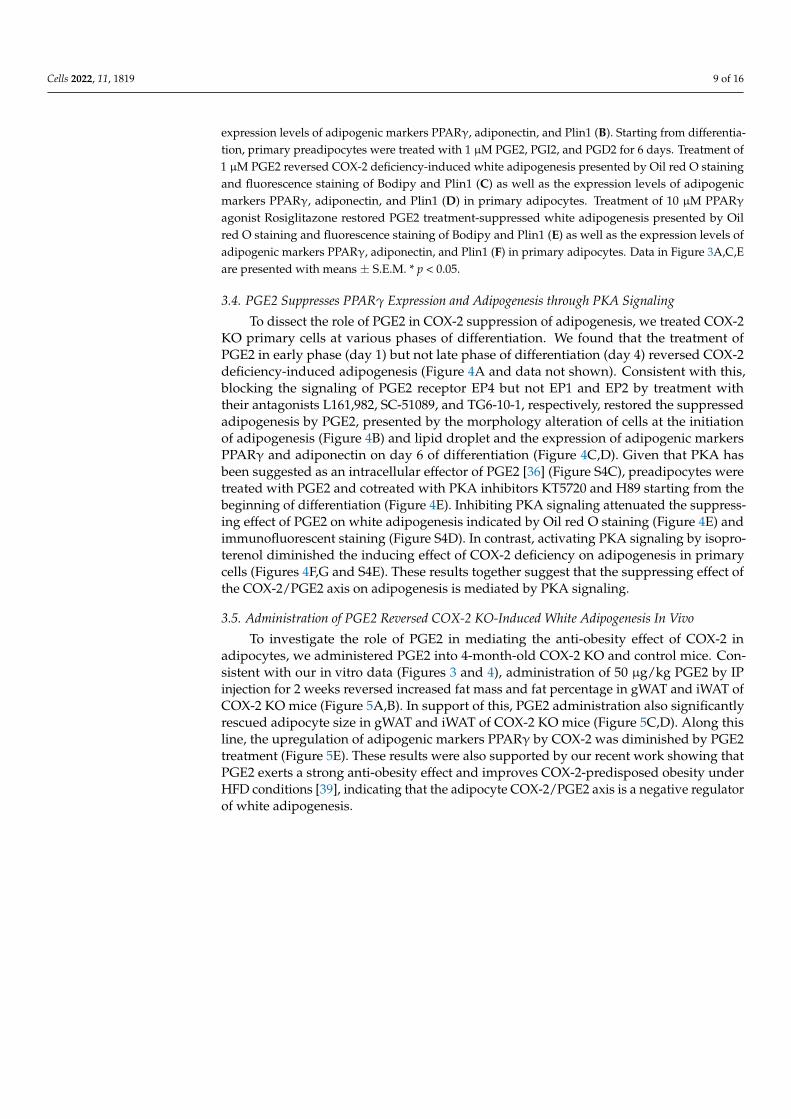
To dissect the role of PGE2 in COX-2 suppression of adipogenesis, we treated COX-2KO primary cells at various phases of differentiation. We found that the treatment ofPGE2 in early phase (day 1) but not late phase of differentiation (day 4) reversed COX-2deficiency-induced adipogenesis (Figure 4A and data not shown). Consistent with this,blocking the signaling of PGE2 receptor EP4 but not EP1 and EP2 by treatment withtheir antagonists L161,982, SC-51089, and TG6-10-1, respectively, restored the suppressedadipogenesis by PGE2, presented by the morphology alteration of cells at the initiationof adipogenesis (Figure 4B) and lipid droplet and the expression of adipogenic markersPPARγ and adiponectin on day 6 of differentiation (Figure 4C,D). Given that PKA hasbeen suggested as an intracellular effector of PGE2 [36] (Figure S4C), preadipocytes weretreated with PGE2 and cotreated with PKA inhibitors KT5720 and H89 starting from thebeginning of differentiation (Figure 4E). Inhibiting PKA signaling attenuated the suppress-ing effect of PGE2 on white adipogenesis indicated by Oil red O staining (Figure 4E) andimmunofluorescent staining (Figure S4D). In contrast, activating PKA signaling by isopro-terenol diminished the inducing effect of COX-2 deficiency on adipogenesis in primarycells (Figures 4F,G and S4E). These results together suggest that the suppressing effect ofthe COX-2/PGE2 axis on adipogenesis is mediated by PKA signaling.
3.5. Administration of PGE2 Reversed COX-2 KO-Induced White Adipogenesis In Vivo
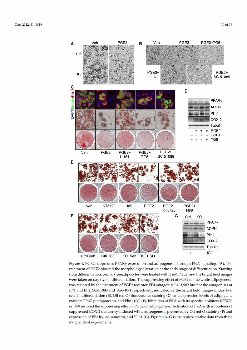
To investigate the role of PGE2 in mediating the anti-obesity effect of COX-2 inadipocytes, we administered PGE2 into 4-month-old COX-2 KO and control mice. Con-sistent with our in vitro data (Figures 3 and 4), administration of 50 µg/kg PGE2 by IPinjection for 2 weeks reversed increased fat mass and fat percentage in gWAT and iWAT ofCOX-2 KO mice (Figure 5A,B). In support of this, PGE2 administration also significantlyrescued adipocyte size in gWAT and iWAT of COX-2 KO mice (Figure 5C,D). Along thisline, the upregulation of adipogenic markers PPARγ by COX-2 was diminished by PGE2treatment (Figure 5E). These results were also supported by our recent work showing thatPGE2 exerts a strong anti-obesity effect and improves COX-2-predisposed obesity underHFD conditions [39], indicating that the adipocyte COX-2/PGE2 axis is a negative regulatorof white adipogenesis.
Cells 2022, 11, 1819 10 of 16Cells 2022, 11, x FOR PEER REVIEW 11 of 17
Figure 4. PGE2 suppresses PPARγ expression and adipogenesis through PKA signaling. (A). The treatment of PGE2 blocked the morphology alteration at the early stage of differentiation. Starting from differentiation, primary preadipocytes were treated with 1 µM PGE2, and the bright field im-ages were taken on day two of differentiation. The suppressing effect of PGE2 on the white adipo-genesis was restored by the treatment of PGE2 receptor EP4 antagonist L161,982 but not the antag-onists of EP1 and EP2, SC-51089 and TG6-10-1 respectively, indicated by the bright field images of day two cells in differentiation (B), Oil red O/fluorescence staining (C), and expression levels of adipogenic markers PPARγ, adiponectin, and Plin1 (D). (E). Inhibition of PKA with its specific in-hibitors KT5720 or H89 restored the suppressing effect of PGE2 on adipogenesis. Activation of PKA with isoproterenol suppressed COX-2 deficiency-induced white adipogenesis presented by Oil red O staining (F) and expression of PPARγ, adiponectin, and Plin1 (G). Figure 4A–G is the representa-tive data from three independent experiments.
Figure 4. PGE2 suppresses PPARγ expression and adipogenesis through PKA signaling. (A). Thetreatment of PGE2 blocked the morphology alteration at the early stage of differentiation. Startingfrom differentiation, primary preadipocytes were treated with 1 µM PGE2, and the bright field imageswere taken on day two of differentiation. The suppressing effect of PGE2 on the white adipogenesiswas restored by the treatment of PGE2 receptor EP4 antagonist L161,982 but not the antagonists ofEP1 and EP2, SC-51089 and TG6-10-1 respectively, indicated by the bright field images of day twocells in differentiation (B), Oil red O/fluorescence staining (C), and expression levels of adipogenicmarkers PPARγ, adiponectin, and Plin1 (D). (E). Inhibition of PKA with its specific inhibitors KT5720or H89 restored the suppressing effect of PGE2 on adipogenesis. Activation of PKA with isoproterenolsuppressed COX-2 deficiency-induced white adipogenesis presented by Oil red O staining (F) andexpression of PPARγ, adiponectin, and Plin1 (G). Figure 4A–G is the representative data from threeindependent experiments.
Cells 2022, 11, 1819 11 of 16
Cells 2022, 11, x FOR PEER REVIEW 12 of 17
3.5. Administration of PGE2 Reversed COX-2 KO-Induced White Adipogenesis In Vivo To investigate the role of PGE2 in mediating the anti-obesity effect of COX-2 in adi-
pocytes, we administered PGE2 into 4-month-old COX-2 KO and control mice. Consistent with our in vitro data (Figures 3 and 4), administration of 50 µg/kg PGE2 by IP injection for 2 weeks reversed increased fat mass and fat percentage in gWAT and iWAT of COX-2 KO mice (Figure 5A,B). In support of this, PGE2 administration also significantly res-cued adipocyte size in gWAT and iWAT of COX-2 KO mice (Figure 5C,D). Along this line, the upregulation of adipogenic markers PPARγ by COX-2 was diminished by PGE2 treat-ment (Figure 5E). These results were also supported by our recent work showing that PGE2 exerts a strong anti-obesity effect and improves COX-2-predisposed obesity under HFD conditions [39], indicating that the adipocyte COX-2/PGE2 axis is a negative regula-tor of white adipogenesis.
Figure 5. Administration of PGE2 reversed COX-2 KO-induced white adipogenesis in vivo. Six-month-old COX-2 KO mice were injected with PGE2 or vehicle for two weeks. (A). PGE2
Figure 5. Administration of PGE2 reversed COX-2 KO-induced white adipogenesis in vivo. Six-month-old COX-2 KO mice were injected with PGE2 or vehicle for two weeks. (A). PGE2 ad-ministration significantly decreased body mass, fat mass, and fat percentage of COX-2 KO mice.n = 5–7/group. (B). Treatment of PGE2 suppressed the mass of three fat pads, inguinal, gonadal, andbrown fat, and pancreas with no significant effect on liver, spleen, and kidney. (C). PGE2 administra-tion decreased the size of adipocytes in gWAT and iWAT despite a little effect on BAT and liver inCOX-2 KO mice. (D). Quantification of adipocyte size in figure (C) showing that a shift of large tosmall adipocytes by PGE2 treatment. (E). PGE2 administration suppressed the expression of PPARγ,adiponectin, and Plin1, although the reduction of adiponectin by PGE2 did not reach significance inCOX-2 KO mice. Data in Figure 5A,B,E are presented with means ± S.E.M. * p < 0.05, ** p < 0.01. Datain Figure 5D are presented with means ± S.E.M. * p < 0.05 and ** p < 0.01 for Ctrl Veh vs. Ctrl PGE2;# p < 0.05 and ## p < 0.01 for Ctrl Veh vs. KO Veh; $ p < 0.05 and $$ p < 0.01 for KO Veh vs. KO PGE2.
Cells 2022, 11, 1819 12 of 16
4. Discussion
Cyclooxygenase-2 (COX-2) is an integral component of inflammation, promoting an im-mune response to infection and injury by producing arachidonic acid-derived prostaglandins(PGs) and other lipokines in immune cells [48]. Interestingly, COX-2 is also expressed inadipocytes, converts arachidonic acid to a variety of PGs in response to nutritional stress,and mediates the metabolic benefits elicited by intermittent fasting (IF) [31,39]. However,the implication of adipose COX-2 in obesity is incompletely understood. Our study showedthat COX-2 in adipocytes limits whit adipogenesis and suppresses pathological expansionof adipose tissue, thereby exerting an anti-obesity property. In addition, COX-2 in adipocytesuppresses progenitor cells’ commitment to differentiation via PGE2-mediated paracrinemechanisms that require PGE2 receptor EP4 and downstream PKA signaling.
COX-2 expression is restricted under basal conditions in adipocytes [31,37], and ismarkedly induced by a variety of physiological conditions such as fasting, adrenocep-tor activation, and mTORC1 inhibition in adipocytes [30,31,36,39,44]. Along this line,COX-2 expression is tightly associated with lipolysis, a cellular process controlled by adren-ergic signaling in adipocytes [28,31,49]. Additionally, IF promotes the production andrelease of COX-2 products PGs from adipocytes, including PGE2, PGD2, and PGI2, allof which communicate directly with progenitors and promote beige adipogenesis in theearly phase [36,39]. COX-2 in adipocytes mediates IF-induced anti-inflammatory effectand -improved insulin sensitivity despite no significant difference in IF-elicited anti-obesityeffect [39]. Given that COX-2 expression in adipose tissue is also suppressed by obesity inhumans and in rodents [39], the present study addresses the anti-obesity effect of COX-2in adipocytes and demonstrates that adipocyte-derived PGE2 inhibits progenitor cell dif-ferentiation into white adipocyte and prevents the development of adiposity and obesity.Consistent with this, overexpressing COX-2 in adipocytes prevents the development ofobesity and adipose tissue inflammation [38]. In addition, the anti-obesity effect of COX-2in adipocytes is selective in male mice given that adipocyte COX-2 deficiency has no sig-nificant effect on diet-induced body weight gain and insulin resistance in female mice(Figure S1). These results support that distinct from male mice, female mice are geneti-cally protected against diet-induced insulin resistance as showed in other models, againsuggesting the protective effect of the sex hormone estrogen in female mice [50–52].
Enzyme immunoassay (ELISA) and Mass Spec analyses have been widely used toassess the production and release of a variety of PGs, including PGE2, PGI2, and PGD2,in adipocytes [30,31,53]. Of note, the short half-lives of PGE2 and PGI2 are approximately5 min and 10 min, respectively, as reported [54–57]. The metabolites of PGE2 such as15-keto-13,14 dihydro-PGE2 appear to be more stable than PGE2 itself [56]. PGE2 secretionlevels were about 50% lower in COX-2 KO adipocytes compared to the controls in 2-hrconditional media, and a similar effect was found in the 4-hr conditional media despiteno significant difference [39]. The reducing effect of COX-2 KO on PGE2 secretion wasretained in the 8- and 18-hr media, albeit to a lesser extent [39], indicative of the short half-life of adipocyte-derived PGE2 as well. The dynamic contribution of adipocytes vs stromalvascular cells to the total levels of PGs in adipose tissue microenvironment remains to beexplored under various pathophysiological conditions. As the beneficial effects of COX-2in adipocytes are abrogated under the later stage of obesity, it is possible that stromalvascular cells become a dominant source of PGs under such conditions. In agreementwith this, the expression levels of COX-2 declined during adipogenesis (Figure 2), and theprotective effect of COX-2 on adipose tissue inflammation only occurred at the early stageof obesity (Figure S3). Of note, PGE2 is a product of COX-2, while feedforward activatesthe Cox-2 gene in primary adipocytes, likely via an autocrine or paracrine mechanism(Figure 2). PGE2-mediated feedforward mechanism magnifies nutritional stress-inducedCOX-2 expression and prostaglandin production in adipocytes. It is possible that PGE2binds to its own receptor EP4 in this case through which activates PKA signaling andsubstantial Cox-2 gene. This also suggests that PGE2-mediated feed-forward regulation ofCOX-2 may have an important impact on physiology, considering that the basal expression
Cells 2022, 11, 1819 13 of 16
level of COX-2 is low, whereas the expression level can be markedly upregulated undercertain circumstances such as fasting or mTORC1 inhibition conditions.
The mechanisms underlying the anti-obesity effect of COX-2 are complicated but likelymediated by its product’s PGs-mediated paracrine mechanisms. The media collected fromCOX-2 KO adipocytes contains lower levels of PGE2 and PGI2 with no significant effect onPGD2 [39]. Despite the thermogenesis-inducing effect of COX-2 [28,34,35,37], depletion ofCOX-2 in adipocytes did not significantly affect the thermogenic gene expression and en-ergy expenditure under both basal and cold stress conditions [39] (Figure S2). In agreementwith this, COX-2 expression levels in adipose tissue are relatively low and do not respondto cold stress [36]. Whereas adipose COX-2 is induced by IF and mediates IF-induced ther-mogenesis [39]. On the other hand, whether COX-2 plays a role in regulating adipogenesisis still controversial. The pharmaceutical inhibition of COX-2 and antisense COX-2 expres-sion enhanced lipid droplet accumulation during adipogenesis [46,58]. Whereas globaldeficiency of COX-2 suppressed adipogenic marker expression and decreased fat mass andbody mass [29]. In addition, COX-2 has been shown to favor brown or beige adipocytedevelopment [28,59]. The present study used an adipocyte-specific COX-2 KO mousemodel and demonstrates that COX-2 in adipocytes plays a negative role in regulating whiteadipogenesis via PGE2-mediated paracrine mechanisms. It is likely that COX-2/PGE2 sig-naling has a distinct role in various types of adipogenesis. Rather than PGE2 receptors EP1and EP2, EP4 mediates the suppressing effect of PGE2 on white adipocyte differentiationby targeting the early stage of differentiation [60] (Figure 4).
In summary, our data show that adipocyte-derived PGE2 serves as a paracrine signalthat limits white adipogenesis. PGE2 suppresses adipogenesis through the PKA/PPARγpathway in preadipocytes. As a result, adipocyte-specific COX-2 deficient mice displayedexacerbated diet-induced adiposity, obesity, and insulin resistance, a phenotype that wasreversed by PGE2 administration. Our study uncovers that the COX-2/PGE2 axis inadipocytes is a key regulator of adipose tissue expansion in obesity.
Supplementary Materials: The following supporting information can be downloaded at: https://www.mdpi.com/article/10.3390/cells11111819/s1, Figure S1: COX-2 KO mice displays no differencesin food intake and insulin sensitivity after 16 week-HFD feeding and female COX-2 KO mice did notexhibit similar phenotype as males; Figure S2: Deficiency of COX-2 in adipocytes has little effect onenergy expenditure under normal chow diet condition; Figure S3: Deficiency of COX-2 in adipocytesimproves type 2 inflammation at NCD but not 16-week HFD conditions; Figure S4: PGE2 suppressesPPARγ expression and adipogenesis through PKA signaling; Table S1: List of primer sequences usedin real-time PCR.
Author Contributions: M.L. designed the project. X.O.Y. serves as a consultant on this project andjoined the discussion. K.A. and W.C.H. contributed to the immunofluorescence staining on thisproject. C.W., X.Z., L.L., Y.L., D.W., D.S., Q.L. and X.Y. conducted the experiments. C.W., X.Z. and D.S.analyzed the results. C.W. drafted the Sections of Methods and Figure Legends. M.L. is the guarantorof this study, and wrote the manuscript. All authors have read and agreed to the published version ofthe manuscript.
Funding: This work is supported by R01 Awards (DK110439 and DK132643, PI: M.L.) from NationalInstitute of Diabetes and Digestive and Kidney Diseases; P20 Award (GM121176, PD: Vojo Deretic,mPIs: M.L. and X.O.Y.) from National Institute of General Medical Sciences; Grant in Aid Award(15GRNT24940018 PI: M.L.) from American Heart Association; Innovative Basic Science Award(1-17-IBS-261 PI: M.L.) from the American Diabetes Association; R01 Award (HL148337 to X.O.Y.)from National Heart Lung and Blood Institute; CoBRE pilot award associated with P30 (P30GM103400,PI: J. Liu, mPI: M.L.); Department of Health and Human Services Secretaries of National institutionof Health: UL1TR001449; National Cancer Institute: CA118100. This project was supported in part bythe Dedicated Health Research Funds from the University of New Mexico School of Medicine.
Institutional Review Board Statement: The animal study protocol was approved by the InstitutionalAnimal Care and Use Committee (IACUC) of the University of New Mexico Health Sciences Center(protocol code: 22-201273-HSC, 22 April 2022).
Cells 2022, 11, 1819 14 of 16
Informed Consent Statement: Not applicable.
Data Availability Statement: Not applicable.
Acknowledgments: We would like to acknowledge the National Institute of Diabetes and DigestiveKidney Diseases (NIDDK), the National Institute of General Medical Science (NIGMS), the AmericanDiabetes Association (ADA), the American Heart Association (AHA), and the University of NewMexico Health Sciences Center (UNMHSC) for funding support. We thank the Autophagy, Inflamma-tion, and Metabolism Center at UNMHSC for providing the Cellomics HCS scanner for our presentstudy and technical support from Sharina Desai.
Conflicts of Interest: The authors have no financial or commercial conflicts of interest to disclose.
AbbreviationsBAT, brown adipose tissue; COX-2, Cyclooxygenase 2; COX-1, Cyclooxygenase 1;
gWAT, gonadal WAT; iWAT, inguinal WAT; PG, prostaglandin; PGs, prostaglandins; PGE2,prostaglandin E2; PGI2, prostacyclin; PGD2, prostaglandin D2; PKA, cAMP-dependent pro-tein kinase; WAT, white adipose tissue; PPARγ, Peroxisome Proliferator Activated Receptorγ; PGC1α, peroxisome proliferator-activated receptor gamma coactivator 1-alpha; UCP1,uncoupling protein 1; C/ebpα, CCAAT/enhancer-binding protein alpha; IFNγ, interferon γ;TNFα, tumor necrosis factor alpha; Plin1, lipid droplet-associated protein 1; IF, intermittentfasting; H&E staining, hematoxylin and eosin staining.
References1. Luo, L.; Liu, M. Adipose tissue in control of metabolism. J. Endocrinol. 2016, 231, R77–R99. [CrossRef] [PubMed]2. Luo, Y.; Liu, B.; Yang, X.; Ma, X.; Zhang, X.; Bragin, D.; Yang, X.O.; Huang, W.; Liu, M. Myeloid adrenergic signaling via CaMKII
forms a feedforward loop of catecholamine biosynthesis. J. Mol. Cell Biol. 2017, 9, 422–434. [CrossRef] [PubMed]3. Tontonoz, P.; Graves, R.A.; Budavari, A.I.; Erdjument-Bromage, H.; Lui, M.; Hu, E.; Tempst, P.; Spiegelman, B.M. Adipocyte-
specific transcription factor ARF6 is a heterodimeric complex of two nuclear hormone receptors, PPARγ and RXR alpha. NucleicAcids Res. 1994, 22, 5628–5634. [CrossRef] [PubMed]
4. Tontonoz, P.; Spiegelman, B.M. Fat and Beyond: The Diverse Biology of PPARγ. Annu. Rev. Biochem. 2008, 77, 289–312. [CrossRef][PubMed]
5. Lehrke, M.; Lazar, M.A. The Many Faces of PPARγ. Cell 2005, 123, 993–999. [CrossRef] [PubMed]6. Cipolletta, D.; Feuerer, M.; Li, A.; Kamei, N.; Lee, J.; Shoelson, S.E.; Benoist, C.; Mathis, D. PPAR-γ is a major driver of the
accumulation and phenotype of adipose tissue Treg cells. Nature 2012, 486, 549–553. [CrossRef]7. Akune, T.; Ohba, S.; Kamekura, S.; Yamaguchi, M.; Chung, U.I.; Kubota, N.; Terauchi, Y.; Harada, Y.; Azuma, Y.;
Nakamura, K.; et al. PPARγ insufficiency enhances osteogenesis through osteoblast formation from bone marrow progenitors.J. Clin. Investig. 2004, 113, 846–855. [CrossRef]
8. Farmer, S.R. Transcriptional control of adipocyte formation. Cell Metab. 2006, 4, 263–273. [CrossRef]9. Choi, J.H.; Banks, A.; Estall, J.; Kajimura, S.; Boström, P.; Laznik, D.; Ruas, J.; Chalmers, M.J.; Kamenecka, T.M.; Blüher, M.; et al.
Anti-diabetic drugs inhibit obesity-linked phosphorylation of PPARγ by Cdk5. Nature 2010, 466, 451–456. [CrossRef]10. Choi, J.H.; Banks, A.; Kamenecka, T.M.; Busby, S.A.; Chalmers, M.J.; Kumar, N.; Kuruvilla, D.S.; Shin, Y.; He, Y.; Bruning, J.; et al.
Antidiabetic actions of a non-agonist PPARγ ligand blocking Cdk5-mediated phosphorylation. Nature 2011, 477, 477–481.[CrossRef]
11. Banks, A.S.; McAllister, F.E.; Camporez, J.P.G.; Zushin, P.-J.H.; Jurczak, M.; Laznik-Bogoslavski, D.; Shulman, G.; Gygi, S.P.;Spiegelman, B.M. An ERK/Cdk5 axis controls the diabetogenic actions of PPARγ. Nature 2015, 517, 391–395. [CrossRef] [PubMed]
12. Qiang, L.; Wang, L.; Kon, N.; Zhao, W.; Lee, S.; Zhang, Y.; Rosenbaum, M.; Zhao, Y.; Gu, W.; Farmer, S.R.; et al. Brown Remodelingof White Adipose Tissue by SirT1-Dependent Deacetylation of PPARγ. Cell 2012, 150, 620–632. [CrossRef] [PubMed]
13. Eifler, K.; Vertegaal, A.C. SUMOylation-Mediated Regulation of Cell Cycle Progression and Cancer. Trends Biochem. Sci. 2015, 40,779–793. [CrossRef] [PubMed]
14. Zelcer, N.; Tontonoz, P. SUMOylation and PPARγ: Wrestling with inflammatory signaling. Cell Metab. 2005, 2, 273–275. [CrossRef]15. Pascual, G.; Fong, A.L.; Ogawa, S.; Gamliel, A.; Li, A.C.; Perissi, V.; Rose, D.W.; Willson, T.M.; Rosenfeld, M.G.; Glass, C.K.
A SUMOylation-dependent pathway mediates transrepression of inflammatory response genes by PPAR-γ. Nature 2005, 437,759–763. [CrossRef]
16. Bailey, S.T.; Ghosh, S. ‘PPAR’ting ways with inflammation. Nat. Immunol. 2005, 6, 966–967. [CrossRef]17. Jennewein, C.; Kuhn, A.-M.; Schmidt, M.V.; Meilladec-Jullig, V.; von Knethen, A.; Gonzalez, F.J.; Brüne, B. Sumoylation of
Peroxisome Proliferator-Activated Receptor γ by Apoptotic Cells Prevents Lipopolysaccharide-Induced NCoR Removal from κBBinding Sites Mediating Transrepression of Proinflammatory Cytokines. J. Immunol. 2008, 181, 5646–5652. [CrossRef]
Cells 2022, 11, 1819 15 of 16
18. Ji, S.; Park, S.Y.; Roth, J.; Kim, H.S.; Cho, J.W. O-GlcNAc modification of PPARγ reduces its transcriptional activity. Biochem.Biophys. Res. Commun. 2012, 417, 1158–1163. [CrossRef]
19. Hauser, S.; Adelmant, G.; Sarraf, P.; Wright, H.M.; Mueller, E.; Spiegelman, B.M. Degradation of the Peroxisome Proliferator-activated Receptor γ Is Linked to Ligand-dependent Activation. J. Biol. Chem. 2000, 275, 18527–18533. [CrossRef]
20. Dutchak, P.A.; Katafuchi, T.; Bookout, A.L.; Choi, J.H.; Yu, R.T.; Mangelsdorf, D.J.; Kliewer, S.A. Fibroblast Growth Factor-21Regulates PPARγ Activity and the Antidiabetic Actions of Thiazolidinediones. Cell 2012, 148, 556–567. [CrossRef]
21. Watanabe, M.; Takahashi, H.; Saeki, Y.; Ozaki, T.; Itoh, S.; Suzuki, M.; Mizushima, W.; Tanaka, K.; Hatakeyama, S. The E3 ubiquitinligase TRIM23 regulates adipocyte differentiation via stabilization of the adipogenic activator PPARγ. eLife 2015, 4, e05615.[CrossRef] [PubMed]
22. Li, J.J.; Wang, R.; Lama, R.; Wang, X.; Floyd, Z.E.; Park, E.A.; Liao, F.-F. Ubiquitin Ligase NEDD4 Regulates PPARγ Stability andAdipocyte Differentiation in 3T3-L1 Cells. Sci. Rep. 2016, 6, 38550. [CrossRef] [PubMed]
23. He, Y.-H.; He, Y.; Liao, X.-L.; Niu, Y.-C.; Wang, G.; Zhao, C.; Wang, L.; Tian, M.-J.; Li, Y.; Sun, C.-H. The calcium-sensing receptorpromotes adipocyte differentiation and adipogenesis through PPARγ pathway. Mol. Cell. Biochem. 2012, 361, 321–328. [CrossRef][PubMed]
24. Noh, K.H.; Kang, H.M.; Yoo, W.; Min, Y.; Kim, D.; Kim, M.; Wang, S.; Lim, J.H.; Jung, C.-R. Ubiquitination of PPAR-gammaby pVHL inhibits ACLY expression and lipid metabolism, is implicated in tumor progression. Metabolism 2020, 110, 154302.[CrossRef] [PubMed]
25. Kim, J.-H.; Park, K.W.; Lee, E.-W.; Jang, W.-S.; Seo, J.; Shin, S.; Hwang, K.-A.; Song, J. Suppression of PPARγ through MKRN1-mediated ubiquitination and degradation prevents adipocyte differentiation. Cell Death Differ. 2014, 21, 594–603. [CrossRef]
26. Marnett, L.J.; Rowlinson, S.W.; Goodwin, D.; Kalgutkar, A.S.; Lanzo, C.A. Arachidonic Acid Oxygenation by COX-1 and COX-2.Mechanisms of catalysis and inhibition. J. Biol. Chem. 1999, 274, 22903–22906. [CrossRef]
27. Fujimori, K. Prostaglandins as PPARγ Modulators in Adipogenesis. PPAR Res. 2012, 2012, 527607. [CrossRef]28. Vegiopoulos, A.; Müller-Decker, K.; Strzoda, D.; Schmitt, I.; Chichelnitskiy, E.; Ostertag, A.; Diaz, M.B.; Rozman, J.;
de Angelis, M.H.; Nüsing, R.M.; et al. Cyclooxygenase-2 Controls Energy Homeostasis in Mice by de Novo Recruitment ofBrown Adipocytes. Science 2010, 328, 1158–1161. [CrossRef]
29. Ghoshal, S.; Trivedi, D.B.; Graf, G.; Loftin, C.D. Cyclooxygenase-2 Deficiency Attenuates Adipose Tissue Differentiation andInflammation in Mice. J. Biol. Chem. 2011, 286, 889–898. [CrossRef]
30. Hu, X.; Cifarelli, V.; Sun, S.; Kuda, O.; Abumrad, N.A.; Su, X. Major role of adipocyte prostaglandin E2 in lipolysis-inducedmacrophage recruitment. J. Lipid Res. 2016, 57, 663–673. [CrossRef]
31. Gartung, A.; Zhao, J.; Chen, S.; Mottillo, E.; VanHecke, G.C.; Ahn, Y.-H.; Maddipati, K.R.; Sorokin, A.; Granneman, J.; Lee, M.-J.Characterization of Eicosanoids Produced by Adipocyte Lipolysis: Implication of Cyclooxygenase-2 in adipose inflammation.J. Biol. Chem. 2016, 291, 16001–16010. [CrossRef]
32. Hsieh, P.-S.; Lu, K.-C.; Chiang, C.-F.; Chen, C.-H. Suppressive effect of COX2 inhibitor on the progression of adipose inflammationin high-fat-induced obese rats. Eur. J. Clin. Investig. 2010, 40, 164–171. [CrossRef]
33. Alcivar, A.A.; Hake, L.E.; Hardy, M.P.; Hecht, N.B. Increased levels of junB and c-jun mRNAs in male germ cells followingtesticular cell dissociation. Maximal stimulation in prepuberal animals. J. Biol. Chem. 1990, 265, 20160–20165. [CrossRef]
34. Madsen, L.; Pedersen, L.M.; Lillefosse, H.H.; Fjære, E.; Bronstad, I.; Hao, Q.; Petersen, R.K.; Hallenborg, P.; Ma, T.;De Matteis, R.; et al. UCP1 Induction during Recruitment of Brown Adipocytes in White Adipose Tissue Is Dependent onCyclooxygenase Activity. PLoS ONE 2010, 5, e11391. [CrossRef] [PubMed]
35. Bayindir, I.; Babaeikelishomi, R.; Kocanova, S.; Sousa, I.S.; Lerch, S.; Hardt, O.; Wild, S.; Bosio, A.; Bystricky, K.; Herzig, S.; et al.Transcriptional Pathways in cPGI2-Induced Adipocyte Progenitor Activation for Browning. Front. Endocrinol. 2015, 6, 129.[CrossRef] [PubMed]
36. Zhang, X.; Luo, Y.; Wang, C.; Ding, X.; Yang, X.; Wu, D.; Silva, F.; Yang, Z.; Zhou, Q.; Wang, L.; et al. Adipose mTORC1 SuppressesProstaglandin Signaling and Beige Adipogenesis via the CRTC2-COX-2 Pathway. Cell Rep. 2018, 24, 3180–3193. [CrossRef][PubMed]
37. Paschos, G.K.; Tang, S.Y.; Theken, K.N.; Li, X.; Verginadis, I.; Lekkas, D.; Herman, L.; Yan, W.; Lawson, J.; FitzGerald, G.A.Cold-Induced Browning of Inguinal White Adipose Tissue Is Independent of Adipose Tissue Cyclooxygenase-2. Cell Rep. 2018,24, 809–814. [CrossRef] [PubMed]
38. Danneskiold-Samsøe, N.B.; Sonne, S.B.; Larsen, J.M.; Hansen, A.N.; Fjære, E.; Isidor, M.S.L.; Petersen, S.; Henningsen, J.; Severi, I.;Sartini, L.; et al. Overexpression of cyclooxygenase-2 in adipocytes reduces fat accumulation in inguinal white adipose tissue andhepatic steatosis in high-fat fed mice. Sci. Rep. 2019, 9, 8979. [CrossRef]
39. Wang, C.; Zhang, X.; Luo, L.; Luo, Y.; Yang, X.; Ding, X.; Wang, L.; Le, H.; Feldman, L.E.R.; Men, X.; et al. Adipocyte-derivedPGE2 is required for intermittent fasting–induced Treg proliferation and improvement of insulin sensitivity. JCI Insight 2022, 7.[CrossRef]
40. Liu, M.; Zhou, L.; Xu, A.; Lam, K.S.L.; Wetzel, M.D.; Xiang, R.; Zhang, J.; Xin, X.; Dong, L.Q.; Liu, F. A disulfide-bond Aoxidoreductase-like protein (DsbA-L) regulates adiponectin multimerization. Proc. Natl. Acad. Sci. USA 2008, 105, 18302–18307.[CrossRef]
Cells 2022, 11, 1819 16 of 16
41. Liu, M.; Bai, J.; He, S.; Villarreal, R.; Hu, D.; Zhang, C.; Yang, X.; Liang, H.; Slaga, T.J.; Yu, Y.; et al. Grb10 Promotes Lipolysisand Thermogenesis by Phosphorylation-Dependent Feedback Inhibition of mTORC1. Cell Metab. 2014, 19, 967–980. [CrossRef][PubMed]
42. Mancuso, D.J.; Sims, H.F.; Yang, K.; Kiebish, M.A.; Su, X.; Jenkins, C.M.; Guan, S.; Moon, S.H.; Pietka, T.; Nassir, F.; et al. GeneticAblation of Calcium-independent Phospholipase A2γ Prevents Obesity and Insulin Resistance during High Fat Feeding byMitochondrial Uncoupling and Increased Adipocyte Fatty Acid Oxidation. J. Biol. Chem. 2010, 285, 36495–36510. [CrossRef][PubMed]
43. Liu, M.; Xiang, R.; Wilk, S.A.; Zhang, N.; Sloane, L.B.; Azarnoush, K.; Zhou, L.; Chen, H.; Xiang, G.; Walter, C.A.; et al. Fat-SpecificDsbA-L Overexpression Promotes Adiponectin Multimerization and Protects Mice from Diet-Induced Obesity and InsulinResistance. Diabetes 2012, 61, 2776–2786. [CrossRef] [PubMed]
44. Zhang, X.; Wu, D.; Wang, C.; Luo, Y.; Ding, X.; Yang, X.; Silva, F.; Arenas, S.; Weaver, J.M.; Mandell, M.; et al. Sustained activationof autophagy suppresses adipocyte maturation via a lipolysis-dependent mechanism. Autophagy 2020, 16, 1668–1682. [CrossRef]
45. Wang, L.; Luo, Y.; Luo, L.; Wu, D.; Ding, X.; Zheng, H.; Wu, H.; Liu, B.; Yang, X.; Silva, F.; et al. Adiponectin restrains ILC2activation by AMPK-mediated feedback inhibition of IL-33 signaling. J. Exp. Med. 2021, 218, e20191054. [CrossRef]
46. Yan, H.; Kermouni, A.; Abdel-Hafez, M.; Lau, D.C. Role of cyclooxygenases COX-1 and COX-2 in modulating adipogenesis in3T3-L1 cells. J. Lipid Res. 2003, 44, 424–429. [CrossRef]
47. Cha, M.-H.; Kim, I.-C.; Lee, B.-H.; Yoon, Y. Baicalein Inhibits Adipocyte Differentiation by Enhancing COX-2 Expression. J. Med.Food 2006, 9, 145–153. [CrossRef]
48. Dennis, E.A.; Norris, P.C. Eicosanoid storm in infection and inflammation. Nat. Rev. Immunol. 2015, 15, 511–523. [CrossRef]49. Klein, T.; Shephard, P.; Kleinert, H.; Komhoff, M. Regulation of cyclooxygenase-2 expression by cyclic AMP. Biochim. et Biophys.
Acta 2007, 1773, 1605–1618. [CrossRef]50. Riant, E.; Waget, A.; Cogo, H.; Arnal, J.-F.; Burcelin, R.; Gourdy, P. Estrogens Protect against High-Fat Diet-Induced Insulin
Resistance and Glucose Intolerance in Mice. Endocrinology 2009, 150, 2109–2117. [CrossRef]51. Pettersson, U.S.; Waldén, T.B.; Carlsson, P.-O.; Jansson, L.; Phillipson, M. Female Mice are Protected against High-Fat Diet Induced
Metabolic Syndrome and Increase the Regulatory T Cell Population in Adipose Tissue. PLoS ONE 2012, 7, e46057. [CrossRef][PubMed]
52. Templeman, N.M.; Clee, S.; Johnson, J.D. Suppression of hyperinsulinaemia in growing female mice provides long-term protectionagainst obesity. Diabetologia 2015, 58, 2392–2402. [CrossRef] [PubMed]
53. Berthou, F.; Ceppo, F.; Dumas, K.; Massa, F.; Vergoni, B.; Alemany, S.; Cormont, M.; Tanti, J.-F. The Tpl2 Kinase Regulates theCOX-2/Prostaglandin E2 Axis in Adipocytes in Inflammatory Conditions. Mol. Endocrinol. 2015, 29, 1025–1036. [CrossRef][PubMed]
54. Ishihara, O.; Sullivan, M.H.; Elder, M.G. Differences of metabolism of prostaglandin E2 and F2 alpha by decidual stromal cellsand macrophages in culture. Eicosanoids 1991, 4, 203–207. [PubMed]
55. Bygdeman, M. Pharmacokinetics of prostaglandins. Best Pract. Res. Clin. Obstet. Gynaecol. 2003, 17, 707–716. [CrossRef]56. Hamberg, M.; Samuelsson, B. On the Metabolism of Prostaglandins E1 and E2 in Man. J. Biol. Chem. 1971, 246, 6713–6721.
[CrossRef]57. Lucas, F.V.; Skrinska, V.A.; Chisolm, G.M.; Hesse, B.L. Stability of prostacyclin in human and rabbit whole blood and plasma.
Thromb. Res. 1986, 43, 379–387. [CrossRef]58. Chu, X.; Nishimura, K.; Jisaka, M.; Nagaya, T.; Shono, F.; Yokota, K. Up-regulation of adipogenesis in adipocytes expressing
stably cyclooxygenase-2 in the antisense direction. Prostaglandins Other Lipid Mediat. 2010, 91, 1–9. [CrossRef]59. Fajas, L.; Miard, S.; Briggs, M.R.; Auwerx, J. Selective cyclo-oxygenase-2 inhibitors impair adipocyte differentiation through
inhibition of the clonal expansion phase. J. Lipid Res. 2003, 44, 1652–1659. [CrossRef]60. Fujimori, K.; Yano, M.; Ueno, T. Synergistic Suppression of Early Phase of Adipogenesis by Microsomal PGE Synthase-1
(PTGES1)-Produced PGE2 and Aldo-Keto Reductase 1B3-Produced PGF2α. PLoS ONE 2012, 7, e44698. [CrossRef]


