Embed Size (px)
Citation preview
Igor Garkavtsev and Rakesh K. JainDai Fukumura, Akira Ushiyama, Dan G. Duda, Lei Xu, Joshua Tam, V. Krishna, K. Chatterjee,
AdipogenesisParacrine Regulation of Angiogenesis and Adipocyte Differentiation During In Vivo
Print ISSN: 0009-7330. Online ISSN: 1524-4571 Copyright © 2003 American Heart Association, Inc. All rights reserved.is published by the American Heart Association, 7272 Greenville Avenue, Dallas, TX 75231Circulation Research
doi: 10.1161/01.RES.0000099243.20096.FA2003;93:e88-e97; originally published online October 2, 2003;Circ Res.
http://circres.ahajournals.org/content/93/9/e88World Wide Web at:
The online version of this article, along with updated information and services, is located on the
http://circres.ahajournals.org/content/96/9/e76.full.pdf http://circres.ahajournals.org/content/94/1/e16.full.pdf
An erratum has been published regarding this article. Please see the attached page for:
http://circres.ahajournals.org/content/suppl/2003/10/29/93.9.e88.DC1.htmlData Supplement (unedited) at:
http://circres.ahajournals.org//subscriptions/
is online at: Circulation Research Information about subscribing to Subscriptions:
http://www.lww.com/reprints Information about reprints can be found online at: Reprints:
document. Permissions and Rights Question and Answer about this process is available in the
located, click Request Permissions in the middle column of the Web page under Services. Further informationEditorial Office. Once the online version of the published article for which permission is being requested is
can be obtained via RightsLink, a service of the Copyright Clearance Center, not theCirculation Researchin Requests for permissions to reproduce figures, tables, or portions of articles originally publishedPermissions:
by guest on September 13, 2014http://circres.ahajournals.org/Downloaded from by guest on September 13, 2014http://circres.ahajournals.org/Downloaded from by guest on September 13, 2014http://circres.ahajournals.org/Downloaded from by guest on September 13, 2014http://circres.ahajournals.org/Downloaded from by guest on September 13, 2014http://circres.ahajournals.org/Downloaded from by guest on September 13, 2014http://circres.ahajournals.org/Downloaded from by guest on September 13, 2014http://circres.ahajournals.org/Downloaded from by guest on September 13, 2014http://circres.ahajournals.org/Downloaded from
Paracrine Regulation of Angiogenesis and AdipocyteDifferentiation During In Vivo Adipogenesis
Dai Fukumura,* Akira Ushiyama,* Dan G. Duda,* Lei Xu, Joshua Tam, V. Krishna K. Chatterjee,Igor Garkavtsev, Rakesh K. Jain
Abstract—With an increasing incidence of obesity worldwide, rational strategies are needed to control adipogenesis.Growth of any tissue requires the formation of a functional and mature vasculature. To gain mechanistic insight into thelink between active adipogenesis and angiogenesis, we developed a model to visualize noninvasively and in real timeboth angiogenesis and adipogenesis using intravital microscopy. Implanted murine preadipocytes induced vigorousangiogenesis and formed fat pads in a mouse dorsal skin-fold chamber. The newly formed vessels subsequentlyremodeled into a mature network consisting of arterioles, capillaries, and venules, whereas the preadipocytesdifferentiated into adipocytes as confirmed by increased aP2 expression. Inhibition of adipocyte differentiation bytransfection of preadipocytes with a peroxisome proliferator-activated receptor � dominant-negative construct not onlyabrogated fat tissue formation but also reduced angiogenesis. Surprisingly, inhibition of angiogenesis by vascularendothelial growth factor receptor-2 (VEGFR2) blocking antibody not only reduced angiogenesis and tissue growth butalso inhibited preadipocyte differentiation. We found that part of this inhibition stems from the paracrine interactionbetween endothelial cells and preadipocytes and that VEGF–VEGFR2 signaling in endothelial cells, but notpreadipocytes, mediates this process. These findings reveal a reciprocal regulation of adipogenesis and angiogenesis,and suggest that blockade of VEGF signaling can inhibit in vivo adipose tissue formation. The full text of this articleis available online at http://www.circresaha.org. (Circ Res. 2003;93:e88-e97.)
Key Words: obesity � adipogenesis � angiogenesis � vascular endothelial growth factor � peroxisomeproliferator-activated receptor �
Adipose tissue exhibits angiogenic activity.1,2 A recentreport demonstrated that established adipose tissue mass
can actually be regulated through the vasculature.3 Impor-tantly, the potential to acquire new fat cells from fat cellprecursors throughout one’s lifespan is now undisputed.4
Thus, understanding the mechanistic relationship betweenadipocyte differentiation and neovascularization during denovo fat tissue formation is critical and has importantimplications. We used intravital microscopy5 along withappropriate animal models and optical probes to monitor andquantify dynamic biological events, including angiogenesis,noninvasively and in real time. To investigate the relationshipbetween adipogenesis and angiogenesis, we adapted the invivo fat pad formation model by implanting 3T3-F442Amurine preadipocytes4,6,7 to a mouse dorsal skin chamber5 insevere combined immunodeficient (SCID) mice. For geneexpression analyses, we implanted 3T3-F442A preadipocytessubcutaneously and recovered the fad pads at different timepoints. This experimental design enabled us to monitor, in
parallel, both the kinetics of angiogenesis and adipogenesisand the gene expression patterns during fat tissue formation.
During the early phase of adipocyte differentiation, twofamilies of transcriptional factors, CCAAT/enhancer bindingprotein (C/EBP) and peroxisome proliferator-activated recep-tor (PPAR) are induced.4,8,9 Activation of PPAR� in adipo-genesis is well characterized and is considered an absoluterequirement for adipocyte differentiation.10–12 To elucidatethe causal link between adipocyte differentiation and angio-genesis, we introduced a dominant-negative PPAR� mutantconstruct into 3T3-F442A cells before implantation using anadenoviral vector, as described previously.13 Mature, termi-nally differentiated adipocytes express multiple genes andproteins including aP2 (an adipocyte-specific fatty acid bind-ing protein, originally identified as 422), FAT/CD36, perili-pin, adipsin, stearoyl-CoA desaturase (SCD1), glucose trans-porter (GLUT4), phosphenolpyruvate carboxykinase(PEPCK), and leptin.4,8,9 Among these, the adipocyte-specificaP2 gene is a downstream target of PPAR� activation and is
Original received August 7, 2003; revision received September 23, 2003; accepted September 24, 2003.From the Edwin L. Steele Laboratory (D.F., A.U., D.G.D., L.X., J.T., I.G., R.K.J.), Department of Radiation Oncology, Massachusetts General Hospital
and Harvard Medical School, Boston, Mass; Department of Medicine (K.K.C.), University of Cambridge, Cambridge, UK. Present address of A.U. is theDepartment of Environmental Health, National Institute of Public Health, Tokyo, Japan.
*These authors contributed equally to this work.Correspondence to Rakesh K. Jain, Department of Radiation Oncology, Massachusetts General Hospital, 100 Blossom St, COX-7, Boston, MA 02114.
E-mail [email protected]© 2003 American Heart Association, Inc.
Circulation Research is available at http://www.circresaha.org DOI: 10.1161/01.RES.0000099243.20096.FA
1
UltraRapid Communication
by guest on September 13, 2014http://circres.ahajournals.org/Downloaded from
the most widely used adipocyte differentiation marker.14,15
Thus, in this study, the kinetics of aP2 expression was used toconfirm the differentiation of 3T3-F442A cells.
Angiogenesis often precedes adipose tissue formation indeveloping tissue, which indicates the requirement of bloodvessels for tissue formation and hints at a potential direct linkbetween angiogenesis and adipogenesis.16 Vascular endothe-lial growth factor receptor 2 (VEGFR2) is expressed onvascular endothelial cells and its signaling is critical in bothphysiological and pathological angiogenesis.17 Among itsligands, VEGF-A is highly expressed in adipose tissue and itsexpression increases significantly during adipocyte differen-tiation.18–21 To assess the importance of VEGFR2 signalingin angiogenesis, vessel remodeling, and adipocyte differenti-ation during fat tissue formation, we determined the effect ofa VEGFR2 blocking antibody22 on angiogenesis, tissue for-mation, 3T3-F442A cell morphology, and changes in aP2expression.
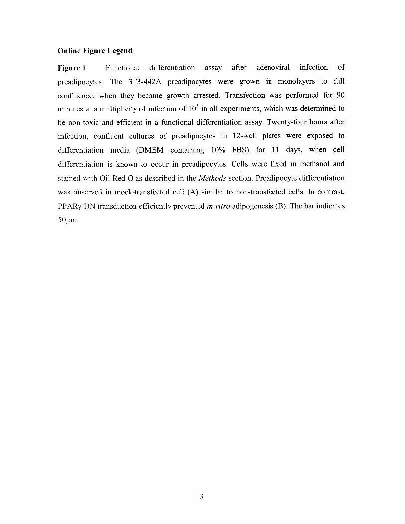
Materials and MethodsCell Lines and AnimalsMale SCID mice, 8 to 12 weeks old, were bred and maintained in ourdefined flora facility and used in all experiments. All procedureswere carried out according to the Public Health Service Policy onHumane Care of Laboratory Animals and approved by the Institu-tional Animal Care and Use Committee. The 3T3-F442A preadipo-cytes (a generous gift from Dr Bruce Spiegelman, Dana-FarberCancer Institute, Boston, Mass) and NIH 3T3 fibroblasts weremaintained in Dulbecco’s Minimum Essential Medium (DMEM,Gibco BRL), supplemented with 10% calf serum, glucose,L-glutamine, penicillin, and streptomycin. A murine endothelial cellline (MECs, CRL-1927) was obtained from ATCC (Manassas, Va)and cultured as recommended by the provider. For cell identificationin vivo, preadipocytes were transfected by the calcium phosphatemethod with the green fluorescent protein (GFP) gene under thecontrol of the EF1� promoter.23 For adipogenesis inhibition, prea-dipocytes were transduced with a recombinant adenovirus encodinga PPAR�–dominant-negative (PPAR�-DN) mutant receptor, ormock adenovirus, as previously described.13 Briefly, a multiplicity ofinfection of 103 for 90 minutes was used for transfection of confluent(growth arrested) preadipocytes. The efficiency of the PPAR�-DNconstruct was assessed functionally, ie, by evaluating the celldifferentiation (online Figure, available in the online data supple-ment at http://www.circresaha.org). For in vivo experiments, trans-fected cells were implanted 2 days after infection.
In Vivo Microscopy Using the TransparentChamber ModelDorsal skinfold chambers were implanted in mice, as describedelsewhere.24 Cell pellets containing 2�105 preadipocytes were im-planted in the center of the chamber. In vivo microscopy usingepi-fluorescence and multiphoton techniques was performed 1 to 2times a week up to 4 weeks after the implantation and was followedby off-line analysis of vascular parameters as described.24 Fiverandom locations in the area of implantation were observed for eachanimal and time point. The number of nonbranching blood vesselsegments (number of segments per unit area), functional vasculardensity (total length of perfused blood vessels per unit area), vesseldiameter, and vessel volume density (calculated blood vessel vol-ume, based on length and diameter of each segment, per unit area)were determined as described elsewhere.24 Angiogenesis and subse-quent vessel remodeling were analyzed in three different experimen-tal settings: after implantation of NIH 3T3 or 3T3-F442A; afterimplantation of GFP/3T3-F442A infected with PPAR�-DN or mockadenovirus; and after implantation of GFP/3T3-F442A in mice
treated with DC101 or rat IgG. In vivo observation using multipho-ton laser-scanning microscopy was used as previously described.25
Reagents and DosageFor VEGFR2 signaling blockade experiments, preadipocytes wereimplanted in dorsal skinfold chambers, and the mice were dividedinto two groups. In one group, rat anti-mouse VEGFR2 monoclonalantibody administration (DC101, ImClone Systems Inc, New York,NY, a generous gift from Drs D. Hicklin and P. Bohlen) was startedon the day of implantation and continued every 3 days for 4 weeks.DC101 was delivered intraperitoneally at a dose of 40 mg/kg bodyweight, which was demonstrated previously to have blocking effectsin vivo.26–28 The control group received 40 mg/kg body weight ofnonspecific isotype-matched rat IgG intraperitoneally on the sameschedule.
For in vitro studies, recombinant mouse VEGF was obtained fromR&D Systems and used at concentrations of 0 to 100 ng/mL. Rat IgGand DC101 were used at doses previously shown to have blockingeffects in vitro.29
Subcutaneous Fat Pad FormationCell suspensions containing 1.5�107 cells in 100 �L of PBS wereinjected into the flank of SCID mice, as described.7 For theantiadipogenesis studies, mice were divided into 3 groups with thefollowing cell implants: GFP/3T3-F442A, GFP/3T3-F442A express-ing PPAR�-DN, and GFP/3T3-F442A mock-transfected. For theantiangiogenesis experiments, GFP/3T3-F442A cells were implantedin 3 groups of mice. These mice received DC101, nonspecific IgG,or vehicle (PBS), respectively. Fat pad formation was allowed toproceed for 4 weeks, after which mice were euthanized and tissuewas harvested. Tissue formed by the implanted preadipocytes wasrecovered using microscissors and fluorescence microscope-guideddissection. Tissue samples were snap-frozen for subsequent RNAextraction.
HistologyTissue samples were harvested and fixed at 4 weeks after implanta-tion. Frozen sections were used for fluorescence immunostainingusing a Phyco-Erythrin anti–�-smooth muscle actin antibody (Sig-ma), whereas adipose cells were identified by their constitutive GFPexpression. DAPI diacetate (Molecular Probes) staining of cellnuclei was used for counterstaining. Quantitation of the GFP-positive tissue area was performed in 5 nonsequential transversesections of the fat tissue generated in the skin chambers, bycalculating the area of tissue inside the perimeter of the GFP-positivetissue (3 spots/section, n�5 mice). The area occupied by nuclei wasconsidered a measure of the number of cells within the tissue. Imageswere acquired using the OpenLab software (Improvision Inc) from afluorescence microscope (Olympus Co, Ltd, Japan, �20 objective)equipped with a CCD camera. After processing images with AdobePhotoshop software (Adobe Systems Inc), binary images werequantified for tissue/cell area using a macro designed by Dr L.L.Munn (Massachusetts General Hospital, Boston, Mass) in the NIHImage software. Resin embedding with routine toluidine-blue coun-terstaining was used for morphological analyses.
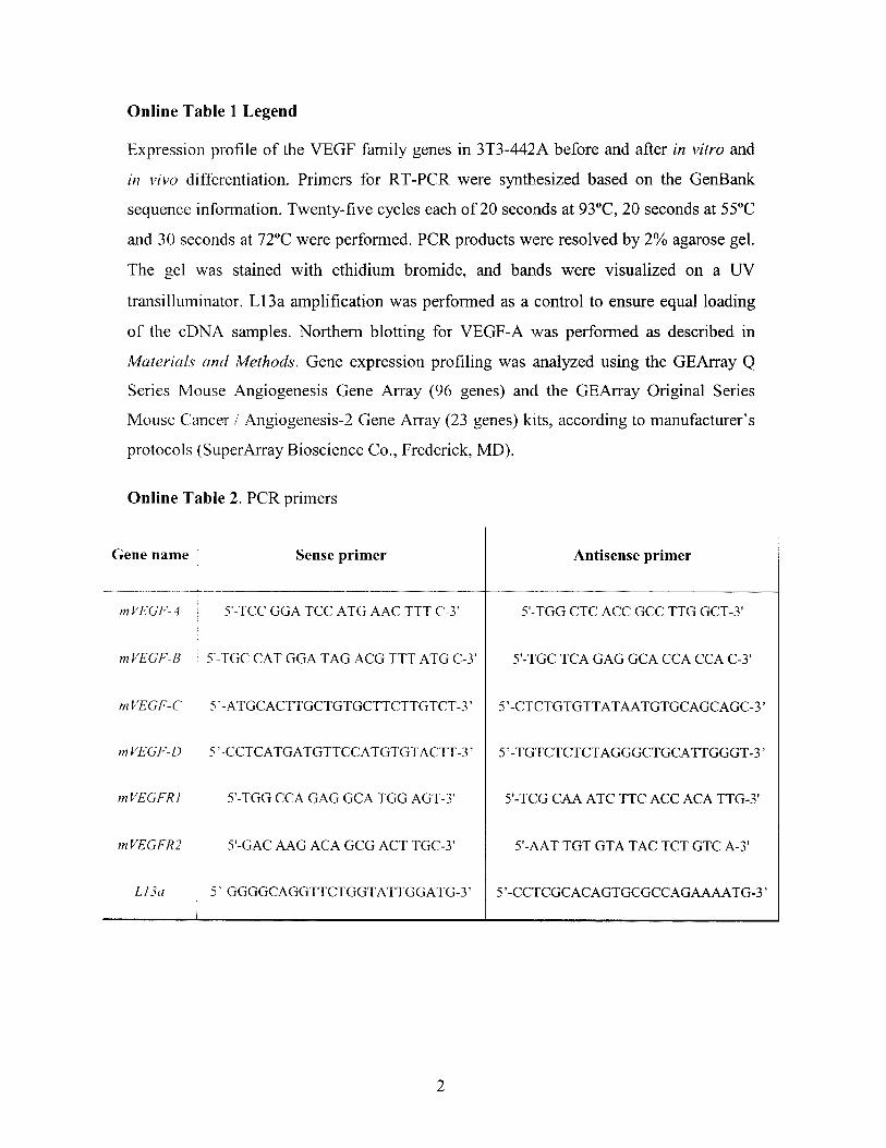
Analyses of Gene ExpressionTotal RNA was extracted from cells and tissue samples using Triazol(Gibco BRL) following the protocol recommended by the manufac-turer. Ten micrograms of total RNA were separated on an agarosegel and transferred to nylon membranes. Northern blots were probedwith PCR-generated cDNA fragments. Nested primers were used togenerate specific amplification products. Primers for PCR (sense,5�-CTGGAAGACAGCTCCTCCTCGAAG-3� and 5�-ATGTGTGATGCCTTTGTGGGAAC-3�; antisense, 5�-TAATCAACATAACCATATCCAAT-3�) were synthesized basedon the mouse aP2 gene sequence (GenBank No. NM_024406).
2 Circulation Research October 31, 2003
by guest on September 13, 2014http://circres.ahajournals.org/Downloaded from
In Vitro Preadipocyte Differentiation AssayTo investigate the effect of VEGF on in vitro differentiation ofpreadipocytes, 3T3-F442A cells were grown to confluence in mediasupplemented with calf serum (CS, maintenance media) and exposedto increasing concentrations of murine recombinant VEGF (R&DSystems) from 0 to 100 ng/mL. In addition to VEGF, in someexperiments, the culture medium was conditioned with blockingconcentrations of DC101 or rat IgG both in the maintenance media(10% CS) and in the differentiation media (containing 10% FBS). Toinvestigate the paracrine effects of VEGF, murine endothelial cellswere cultured with the addition of recombinant murine VEGF and invitro blocking concentration of DC101 (5 �g/mL). For controls,isotype-matched IgG antibody was added at similar concentrations.Twenty-four-hour–conditioned media from the endothelial cells wasadded to confluent cultures of preadipocytes and changed every otherday. Cells were harvested at day 11 (when cell differentiation startedto become apparent morphologically) to analyze the difference inaP2 expression between groups.
MTT AssayFive hundred preadipocytes or fibroblasts were plated in 96-wellplates and mouse recombinant VEGF (50 ng/mL) was added alongwith PBS, DC101 (1 �g/mL), or rat IgG (1 �g/mL). The MTT assaywas performed at day 4, when the cells were still subconfluent in allwells. Before the assay, culture media were removed and replacedwith 100 �L of fresh media and 10 �L of sterile tetrazolium salt;MTT (3[4,5-dimethylthiazol-2-yl]-2,5-diphenyl-tetrazolium bro-mide, Sigma) was then added to each well and incubated for 4 hours
at 37°C. Finally, 100 �L of 10% SDS was added, and afterincubation at 37°C overnight, the plate was read at 490 nm. Theoptical density values were normalized to that of the PBS-treatedcells and were used as a measure of cell viability. To assess theparacrine effect of VEGF on preadipocyte proliferation, cells wereplated in a similar fashion, with conditioned media from EC culturedin the presence of VEGF and blocking concentrations of DC101 orIgG. At day 4, the assay was performed as described, and the opticaldensity value was normalized to the optical density of cells culturedin nonconditioned media.
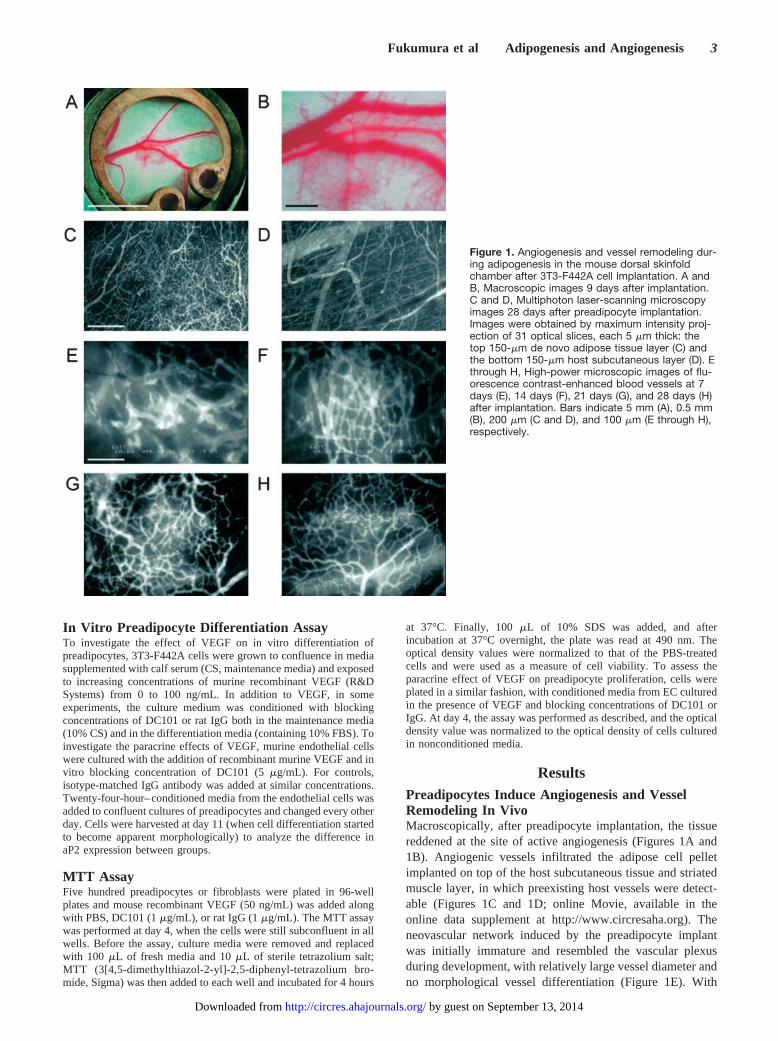
ResultsPreadipocytes Induce Angiogenesis and VesselRemodeling In VivoMacroscopically, after preadipocyte implantation, the tissuereddened at the site of active angiogenesis (Figures 1A and1B). Angiogenic vessels infiltrated the adipose cell pelletimplanted on top of the host subcutaneous tissue and striatedmuscle layer, in which preexisting host vessels were detect-able (Figures 1C and 1D; online Movie, available in theonline data supplement at http://www.circresaha.org). Theneovascular network induced by the preadipocyte implantwas initially immature and resembled the vascular plexusduring development, with relatively large vessel diameter andno morphological vessel differentiation (Figure 1E). With
Figure 1. Angiogenesis and vessel remodeling dur-ing adipogenesis in the mouse dorsal skinfoldchamber after 3T3-F442A cell implantation. A andB, Macroscopic images 9 days after implantation.C and D, Multiphoton laser-scanning microscopyimages 28 days after preadipocyte implantation.Images were obtained by maximum intensity proj-ection of 31 optical slices, each 5 �m thick: thetop 150-�m de novo adipose tissue layer (C) andthe bottom 150-�m host subcutaneous layer (D). Ethrough H, High-power microscopic images of flu-orescence contrast-enhanced blood vessels at 7days (E), 14 days (F), 21 days (G), and 28 days (H)after implantation. Bars indicate 5 mm (A), 0.5 mm(B), 200 �m (C and D), and 100 �m (E through H),respectively.
Fukumura et al Adipogenesis and Angiogenesis 3
by guest on September 13, 2014http://circres.ahajournals.org/Downloaded from
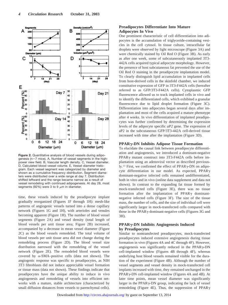
time, these vessels induced by the preadipocyte implantgradually reorganized (Figures 1F through 1H): mesh-likepatterns of angiogenic vessels turned into a dense capillarynetwork (Figures 1G and 1H), with arterioles and venulesbecoming apparent (Figure 1H). The number of blood vesselsegments (Figure 2A) and vessel density (total length ofblood vessels per unit tissue area; Figure 2B) increased,accompanied by a decrease in mean vessel diameter (Figure2C) as the blood vessels remodeled. The total volume ofblood vessels per unit tissue area did not change during theremodeling process (Figure 2D). The blood vessel sizedistribution narrowed with the remodeling of the vesselnetwork (Figure 2E). The remodeled blood vessels werecovered by �-SMA–positive cells (data not shown). Theangiogenic response was specific to preadipocytes, as NIH3T3 fibroblasts did not induce appreciable vessel formationor tissue mass (data not shown). These findings indicate thatpreadipocytes have the unique ability to induce in vivoangiogenesis and remodeling of vessels into efficient net-works with a mature, stable architecture (characterized bysmall diffusion distances from vessels to parenchymal cells).
Preadipocytes Differentiate Into MatureAdipocytes In VivoOne prominent characteristic of cell differentiation into adi-pocytes is the accumulation of triglyceride-containing vesi-cles in the cell cytosol. In tissue culture, intracellular fatdroplets were observed by light microscope (Figure 3A) andwere chemically stained by Oil Red O (Figure 3B). As earlyas after one week, some of subcutaneously implanted 3T3-442A cells acquired typical adipocyte morphology. However,the presence of host subcutaneous fat prevented the use of theOil Red O staining in the preadipocyte implantation model.To clearly distinguish lipid accumulation in implanted cellsfrom host-derived cells in the skinfold chamber, we inducedconstitutive expression of GFP in 3T3-F442A cells (hereafterreferred to as GFP/3T3-F442A cells). Cytoplasmic GFPfluorescence allowed us to track implanted cells in vivo andto identify the differentiated cells, which exhibited a granularfluorescence due to lipid droplet formation (Figure 3C).Differentiation into adipocytes began several days after im-plantation and most of the cells acquired a mature phenotypeafter 4 weeks. In vivo differentiation of implanted preadipo-cytes was further confirmed by determining the expressionlevels of the adipocyte specific aP2 gene. The expression ofaP2 in the subcutaneous GFP/3T3-442A cell-derived tissueincreased with time after the implantation (Figure 3D).
PPAR�-DN Inhibits Adipose Tissue FormationTo elucidate the causal link between preadipocyte differenti-ation and angiogenesis, we introduced a dominant-negativePPAR� mutant construct into 3T3-F442A cells before im-plantation using an adenoviral vector as described previous-ly.13 First, we confirmed the effect of PPAR�-DN on adipo-cyte differentiation in our model. As expected, PPAR�dominant-negative infected cells remained undifferentiated,both in vitro and in vivo (online data supplement and data notshown). In contrast to the expanding fat tissue formed bymock-transfected cells (Figure 3E), there was no tissueformation after the implantation of PPAR� dominant-negative infected cells (Figure 3F). The size of the tissuemass, the number of cells, and the size of individual cell weresignificantly larger in mock-transfected cells compared withthose in the PPAR� dominant-negative cells (Figures 3G and3H).
PPAR�-DN Inhibits Angiogenesis Inducedby PreadipocytesSimilar to nontransfected preadipocytes, mock-transfectedpreadipocytes induced extensive angiogenesis during tissueformation in vivo (Figures 4A and 4C through 4F). However,angiogenesis was significantly reduced in the PPAR�-DNcell-implanted window (Figures 4B through 4F), whereasunderlying host blood vessels remained visible for the dura-tion of the experiment (Figure 4B). Although the number ofvessel segments and vessel density in mock-transfected cellimplants increased with time, they remained unchanged in thePPAR�-DN cell-implanted window (Figures 4A and 4B). Atlater time points, mean vessel diameter was significantlylarger in the PPAR�-DN group, indicating the lack of vesselremodeling (Figure 4E). Thus, the suppression of PPAR�
Figure 2. Quantitative analysis of blood vessels during adipo-genesis (n�7 mice). A, Number of vessel segments in the high-power view field. B, Vascular length density. C, Vessel diameter.D, Calculated blood vessel volume. E, Vessel diameter histo-gram. Each vessel segment was categorized by diameter andshown as a cumulative frequency distribution. Segment diame-ters were distributed over a wide range at day 7. Distributionshifted leftward and the range became narrow as a result ofvessel remodeling with continued adipogenesis. At day 28, mostsegments (92%) were 3 to 9 �m in diameter.
4 Circulation Research October 31, 2003
by guest on September 13, 2014http://circres.ahajournals.org/Downloaded from
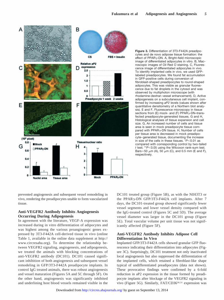
prevented angiogenesis and subsequent vessel remodeling invivo, rendering the preadipocytes unable to form vascularizedtissue.
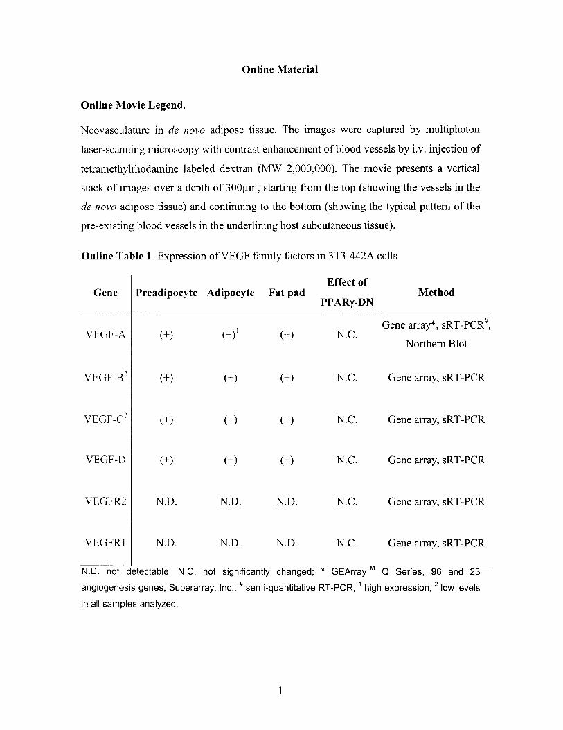
Anti-VEGFR2 Antibody Inhibits AngiogenesisOccurring During AdipogenesisIn agreement with the literature, VEGF-A expression wasincreased during in vitro differentiation of adipocytes andwas highest among the various proangiogenic genes ex-pressed by 3T3-F442A cell-derived tissue in vivo (onlineTable 1, available in the online data supplement at http://www.circresaha.org). To determine the relationship be-tween VEGFR2 signaling, angiogenesis, and adipogenesis,we treated the animals with blocking concentrations ofanti-VEGFR2 antibody (DC101). DC101 caused signifi-cant inhibition of both angiogenesis and subsequent vesselremodeling in GFP/3T3-F442A preadipocyte implants. Incontrol IgG treated animals, there was robust angiogenesisand vessel maturation (Figures 5A and 5C through 5F). Onthe other hand, angiogenesis was significantly inhibitedand underlining host blood vessels remained visible in the
DC101 treated group (Figure 5B), as with the NIH3T3 orthe PPAR�-DN GFP/3T3-F442A cell implants. After 7days, the DC101-treated group showed significantly fewervessel segments and lower vessel density compared withthe IgG-treated control (Figures 5C and 5D). The averagevessel diameter was larger in the DC101 group (Figure5E). Total vascular volume per unit tissue was not signif-icantly affected (Figure 5F).
Anti-VEGFR2 Antibody Inhibits Adipose CellDifferentiation In VivoImplanted GFP/3T3-F442A cells showed granular GFP fluo-rescence indicating their differentiation into adipocytes (Fig-ure 3C). Surprisingly, DC101 treatment not only inactivatedlocal angiogenesis but also suppressed the differentiation ofthe implanted cells, which retained a fibroblast-like shapetypical of undifferentiated preadipocytes (data not shown).These provocative findings were confirmed by a 6-foldreduction in aP2 expression in the tissue formed by preadi-pocyte implants after blockage of the VEGFR2 signaling invivo (Figure 5G). Similarly, FAT/CD3630,31 expression was
Figure 3. Differentiation of 3T3-F442A preadipo-cytes and de novo adipose tissue formation: theeffect of PPAR�-DN. A, Bright-field microscopicimage of differentiated adipocytes in vitro. B, Mac-roscopic images of Oil Red O staining. C, Fluores-cence image of differentiated adipocytes in vivo.To identify implanted cells in vivo, we used GFP-labeled preadipocytes. We found fat accumulationin GFP-positive cells during conversion offibroblast-shaped preadipocytes to round-shapedadipocytes. This was visible as granular fluores-cence due to fat droplets in the cytosol and wasobserved by multiphoton microscope (withrhodamine-dextran vessel enhancement). D, Activeadipogenesis on a subcutaneous cell implant, con-firmed by increasing aP2 levels (values shown afterquantitative densitometry of a Northern blot analy-sis). E and F, Fluorescence microscopy in tissuesections from (E) mock- and (F) PPAR�-DN–trans-fected preadipocyte-generated tissues. G and H,Histological analyses of tissue expansion and cellsize. G, An increased number of cells and tissuearea is seen in mock preadipocyte tissue com-pared with PPAR�-DN tissue. H, Number of cellsper tissue area is decreased in mock preadipo-cyte–generated tissue, documenting the increasein size of the cells in these tissues. *P�0.01 ascompared with corresponding control by two-tailedt test; **P�0.05 using the Wilcoxon rank-sum test.Bars�50 �m (A), 50 �m (C), and 0.5 mm (E and F),respectively.
Fukumura et al Adipogenesis and Angiogenesis 5
by guest on September 13, 2014http://circres.ahajournals.org/Downloaded from
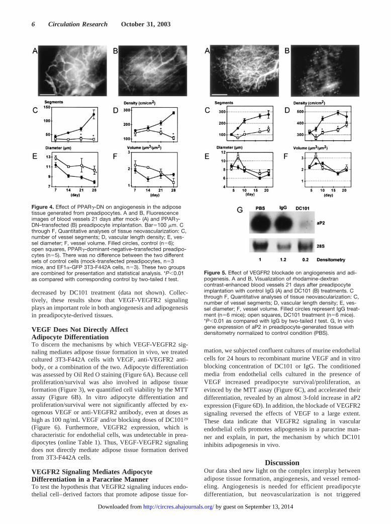
decreased by DC101 treatment (data not shown). Collec-tively, these results show that VEGF-VEGFR2 signalingplays an important role in both angiogenesis and adipogenesisin preadipocyte-derived tissues.
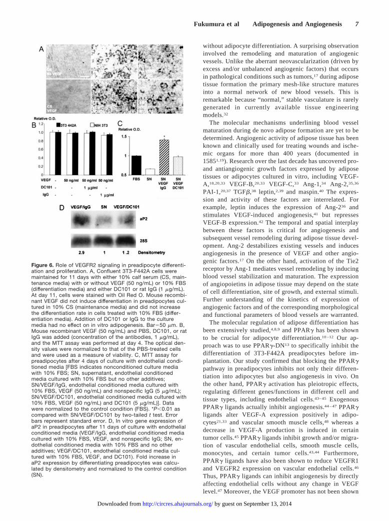
VEGF Does Not Directly AffectAdipocyte DifferentiationTo discern the mechanisms by which VEGF-VEGFR2 sig-naling mediates adipose tissue formation in vivo, we treatedcultured 3T3-F442A cells with VEGF, anti-VEGFR2 anti-body, or a combination of the two. Adipocyte differentiationwas assessed by Oil Red O staining (Figure 6A). Because cellproliferation/survival was also involved in adipose tissueformation (Figure 3), we quantified cell viability by the MTTassay (Figure 6B). In vitro adipocyte differentiation andproliferation/survival were not significantly affected by ex-ogenous VEGF or anti-VEGFR2 antibody, even at doses ashigh as 100 ng/mL VEGF and/or blocking doses of DC10129
(Figure 6). Furthermore, VEGFR2 expression, which ischaracteristic for endothelial cells, was undetectable in prea-dipocytes (online Table 1). Thus, VEGF-VEGFR2 signalingdoes not directly mediate adipose tissue formation derivedfrom 3T3-F442A cells.
VEGFR2 Signaling Mediates AdipocyteDifferentiation in a Paracrine MannerTo test the hypothesis that VEGFR2 signaling induces endo-thelial cell–derived factors that promote adipose tissue for-
mation, we subjected confluent cultures of murine endothelialcells for 24 hours to recombinant murine VEGF and in vitroblocking concentration of DC101 or IgG. The conditionedmedia from endothelial cells cultured in the presence ofVEGF increased preadipocyte survival/proliferation, asevinced by the MTT assay (Figure 6C), and accelerated theirdifferentiation, revealed by an almost 3-fold increase in aP2expression (Figure 6D). In addition, the blockade of VEGFR2signaling reversed the effects of VEGF to a large extent.These data indicate that VEGFR2 signaling in vascularendothelial cells promotes adipogenesis in a paracrine man-ner and explain, in part, the mechanism by which DC101inhibits adipogenesis in vivo.
DiscussionOur data shed new light on the complex interplay betweenadipose tissue formation, angiogenesis, and vessel remod-eling. Angiogenesis is needed for efficient preadipocytedifferentiation, but neovascularization is not triggered
Figure 4. Effect of PPAR�-DN on angiogenesis in the adiposetissue generated from preadipocytes. A and B, Fluorescenceimages of blood vessels 21 days after mock- (A) and PPAR�-DN–transfected (B) preadipocyte implantation. Bar�100 �m. Cthrough F, Quantitative analyses of tissue neovascularization: C,number of vessel segments; D, vascular length density; E, ves-sel diameter; F, vessel volume. Filled circles, control (n�6);open squares, PPAR�-dominant-negative–transfected preadipo-cytes (n�5). There was no difference between the two differentsets of control cells (mock-transfected preadipocytes, n�3mice, and EF1�-GFP 3T3-F442A cells, n�3). These two groupsare combined for presentation and statistical analysis. *P�0.01as compared with corresponding control by two-tailed t test.
Figure 5. Effect of VEGFR2 blockade on angiogenesis and adi-pogenesis. A and B, Visualization of rhodamine-dextrancontrast-enhanced blood vessels 21 days after preadipocyteimplantation with control IgG (A) and DC101 (B) treatments. Cthrough F, Quantitative analyses of tissue neovascularization: C,number of vessel segments; D, vascular length density; E, ves-sel diameter; F, vessel volume. Filled circles represent IgG treat-ment (n�6 mice); open squares, DC101 treatment (n�6 mice).*P�0.01 as compared with IgG by two-tailed t test. G, In vivogene expression of aP2 in preadipocyte-generated tissue withdensitometry normalized to control condition (PBS).
6 Circulation Research October 31, 2003
by guest on September 13, 2014http://circres.ahajournals.org/Downloaded from
without adipocyte differentiation. A surprising observationinvolved the remodeling and maturation of angiogenicvessels. Unlike the aberrant neovascularization (driven byexcess and/or unbalanced angiogenic factors) that occursin pathological conditions such as tumors,17 during adiposetissue formation the primary mesh-like structure maturesinto a normal network of new blood vessels. This isremarkable because “normal,” stable vasculature is rarelygenerated in currently available tissue engineeringmodels.32
The molecular mechanisms underlining blood vesselmaturation during de novo adipose formation are yet to bedetermined. Angiogenic activity of adipose tissue has beenknown and clinically used for treating wounds and ische-mic organs for more than 400 years (documented in15851,19). Research over the last decade has uncovered pro-and antiangiogenic growth factors expressed by adiposetissues or adipocytes cultured in vitro, including VEGF-A,18,20,33 VEGF-B,20,33 VEGF-C,33 Ang-1,34 Ang-2,35,36
PAI-1,20,37 TGF�,38 leptin,2,39 and maspin.40 The expres-sion and activity of these factors are interrelated. Forexample, leptin induces the expression of Ang-236 andstimulates VEGF-induced angiogenesis,41 but repressesVEGF-B expression.42 The temporal and spatial interplaybetween these factors is critical for angiogenesis andsubsequent vessel remodeling during adipose tissue devel-opment. Ang-2 destabilizes existing vessels and inducesangiogenesis in the presence of VEGF and other angio-genic factors.17 On the other hand, activation of the Tie2receptor by Ang-1 mediates vessel remodeling by inducingblood vessel stabilization and maturation. The expressionof angiopoietins in adipose tissue may depend on the stateof cell differentiation, site of growth, and external stimuli.Further understanding of the kinetics of expression ofangiogenic factors and of the corresponding morphologicaland functional parameters of blood vessels are warranted.
The molecular regulation of adipose differentiation hasbeen extensively studied,4,8,9 and PPAR� has been shownto be crucial for adipocyte differentiation.10 –12 Our ap-proach was to use PPAR�-DN13 to specifically inhibit thedifferentiation of 3T3-F442A preadipocytes before im-plantation. Our study confirmed that blocking the PPAR�pathway in preadipocytes inhibits not only their differen-tiation into adipocytes but also angiogenesis in vivo. Onthe other hand, PPAR� activation has pleiotropic effects,regulating different genes/functions in different cell andtissue types, including endothelial cells.43– 45 ExogenousPPAR� ligands actually inhibit angiogenesis.44 – 47 PPAR�ligands alter VEGF-A expression positively in adipo-cytes21,33 and vascular smooth muscle cells,48 whereas adecrease in VEGF-A production is induced in certaintumor cells.45 PPAR� ligands inhibit growth and/or migra-tion of vascular endothelial cells, smooth muscle cells,monocytes, and certain tumor cells.43,44 Furthermore,PPAR� ligands have also been shown to reduce VEGFR1and VEGFR2 expression on vascular endothelial cells.46
Thus, PPAR� ligands can inhibit angiogenesis by directlyaffecting endothelial cells without any change in VEGFlevel.47 Moreover, the VEGF promoter has not been shown
Figure 6. Role of VEGFR2 signaling in preadipocyte differenti-ation and proliferation. A, Confluent 3T3-F442A cells weremaintained for 11 days with either 10% calf serum (CS, main-tenance media) with or without VEGF (50 ng/mL) or 10% FBS(differentiation media) and either DC101 or rat IgG (1 �g/mL).At day 11, cells were stained with Oil Red O. Mouse recombi-nant VEGF did not induce differentiation in preadipocytes cul-tured in 10% CS (maintenance media) and did not increasethe differentiation rate in cells treated with 10% FBS (differ-entiation media). Addition of DC101 or IgG to the culturemedia had no effect on in vitro adipogenesis. Bar�50 �m. B,Mouse recombinant VEGF (50 ng/mL) and PBS, DC101, or ratIgG was added (concentration of the antibodies, 1 �g/mL),and the MTT assay was performed at day 4. The optical den-sity values were normalized to that of the PBS-treated cellsand were used as a measure of viability. C, MTT assay forpreadipocytes after 4 days of culture with endothelial condi-tioned media [FBS indicates nonconditioned culture mediawith 10% FBS; SN, supernatant, endothelial conditionedmedia cultured with 10% FBS but no other additives;SN/VEGF/IgG, endothelial conditioned media cultured with10% FBS, VEGF (50 ng/mL) and nonspecific IgG (5 �g/mL);SN/VEGF/DC101, endothelial conditioned media cultured with10% FBS, VEGF (50 ng/mL) and DC101 (5 �g/mL)]. Datawere normalized to the control condition (FBS). *P�0.01 ascompared with SN/VEGF/DC101 by two-tailed t test. Errorbars represent standard error. D, In vitro gene expression ofaP2 in preadipocytes after 11 days of culture with endothelialconditioned media (VEGF/IgG, endothelial conditioned mediacultured with 10% FBS, VEGF, and nonspecific IgG; SN, en-dothelial conditioned media with 10% FBS and no otheradditives; VEGF/DC101, endothelial conditioned media cul-tured with 10% FBS, VEGF, and DC101). Fold increase inaP2 expression by differentiating preadipocytes was calcu-lated by densitometry and normalized to the control condition(SN).
Fukumura et al Adipogenesis and Angiogenesis 7
by guest on September 13, 2014http://circres.ahajournals.org/Downloaded from
to possess peroxisome proliferators response elements,49
and by inhibiting PPAR-� activity, we could not detect anysignificant change in VEGF-A expression by adipose cells(online Table 1).
A growing body of evidence shows that blood vesselsare more than just carriers of nutrients and passive filtersof blood in tissues. Angiogenesis precedes the develop-ment16,50,51 and repair52 of organs. Secreted factors fromvascular endothelial cells induce proliferation and differ-entiation of preadipocytes,53–55 liver organogenesis,50 pan-creas differentiation,56 and liver protection.57 VEGF is acritical factor in both pathological and physiological an-giogenesis. Our findings show that VEGFR2 signaling inphenotypically normal, immortalized vascular endothelialcells58 mediates the survival/proliferation and differentia-tion of preadipocytes, demonstrating that endothelial cellscan control adipogenesis. This is in agreement with thefindings reported for liver development, where VEGFR2signaling in the endothelial cells is critical for hepatogeniccell multiplication and migration.50 However, stimulationof mature hepatocytes is predominantly mediated throughVEGFR1 signaling in liver sinusoidal endothelial cells.57 Itis conceivable that the maintenance of established adiposetissue is mediated through additional signaling pathways inthe vascular endothelial cells.
Interactions between the extracellular matrix associatedwith angiogenic vessels and preadipocytes may also me-diate adipogenesis. When Matrigel supplemented withbFGF is implanted, angiogenesis is induced followed byadipose precursor cells recruitment and fat pad forma-tion.59 Microvascular endothelial cells have been shown tosecrete extracellular matrix components that promote prea-dipocyte differentiation.55 Remodeling of extracellular ma-trix organization is important for both angiogenesis17 andadipogenesis.60 Expression of metalloproteinases such asMMP 2 and MMP 9 is increased during adipocyte differ-entiation61 and both endogenous and exogenous metallo-proteases induce adipogenesis.61,62 Of particular interest,MMP-9 has been shown to increase the availability ofmatrix-bound VEGF in the tissue.63 On the other hand,tissue inhibitor of metalloproteinases-3 (TIMP-3)– defi-cient mice exhibit increased adipose reconstitution duringmammary involution.64 Recently, TIMP-3 has been shownto inhibit angiogenesis by blocking VEGF binding toVEGFR2.65 Inhibition of adipogenesis by TIMP-3 could bemediated by a paracrine mechanism involving endothelialVEGFR2 signaling, in agreement with our findings.
Finally, improved perfusion by newly formed vessels isrequired for adipocyte differentiation and tissue formation.Accordingly, the HIF-1–regulated gene DEC1/Stra13 inhibitsPPAR� gene expression under hypoxia.66 Tissue oxygenationby angiogenesis and/or vessel remodeling might accelerateadipogenesis by increasing HIF-1 degradation, thereby poten-tiating PPAR� activation.
Taken together, our data show that the molecular andmetabolic microenvironment associated with functional, ma-ture blood vessels promotes preadipocyte differentiation andadipose tissue formation. This observation confirms that thegeneration of normal microcirculatory units is indispensable
for organogenesis. It also raises a provocative questioncorresponding to the observation that most tissues haveadipocytes interspersed with the organ cells: can preadipo-cytes be used in tissue engineering, organogenesis, andtherapeutic angiogenesis? The new adipogenesis-organogenesis model described in this study is ideal toaddress the mechanisms of normalization and maturation ofblood vessels, and to develop and test novel strategies fortissue engineering, organogenesis, and therapeutic angiogen-esis.67 In turn, interfering with this process may providevaluable information for the identification of new targets fortreating various diseases including obesity and solid tumors.Because anti-angiogenic compounds have already been dem-onstrated to decrease established adipose mass,3 anti-VEGFR2 signaling agents undergoing clinical trials for can-cer treatment may be useful candidates for controllingadipogenesis.
AcknowledgmentsThis study was supported by the NIH (Bioengineering ResearchPartnership Grant R24-CA85140). D.G. Duda is a Cancer ResearchInstitute fellow. L. Xu is an NIH postdoctoral fellow (T32-CA73479). J. Tam is a Whitaker Foundation graduate student fellow.V.K. Chatterjee is supported by the Wellcome Trust. We thank DrL.L. Munn for help in data analysis and thoughtful input; J. Kahn andS. Roberge for outstanding technical support; and Drs M.F. Booth,Y. Boucher, L.E. Gerweck, T.P. Padera, and B.R. Stoll for helpfulsuggestions.
References1. Silverman KJ, Lund DP, Zetter BR, Lainey LL, Shahood JA, Freiman
DG, Folkman J, Barger AC. Angiogenic activity of adipose tissue.Biochem Biophys Res Commun. 1988;153:347–352.
2. Sierra-Honigmann MR, Nath AK, Murakami C, Garcia-Cardena G, Papa-petropoulos A, Sessa WC, Madge LA, Schechner JS, Schwabb MB,Polverini PJ, Flores-Riveros JR. Biological action of leptin as anangiogenic factor. Science. 1998;281:1683–1686.
3. Rupnick MA, Panigrahy D, Zhang CY, Dallabrida SM, Lowell BB,Langer R, Folkman J. Adipose tissue mass can be regulated through thevasculature. Proc Natl Acad Sci U S A. 2002;99:10730–10735.
4. Gregoire FM, Smas CM, Sul HS. Understanding adipocyte differen-tiation. Physiol Rev. 1998;78:783–809.
5. Jain RK, Munn LL, Fukumura D. Dissecting tumor pathophysiologyusing intravital microscopy. Nat Rev Cancer. 2002;2:266–276.
6. Green H, Kehinde O. Formation of normally differentiated subcutaneousfat pads by an established preadipose cell line. J Cell Physiol. 1979;101:169–171.
7. Mandrup S, Loftus TM, MacDougald OA, Kuhajda FP, Lane MD. Obesegene expression at in vivo levels by fat pads derived from s.c. implanted3T3-F442A preadipocytes. Proc Natl Acad Sci U S A. 1997;94:4300–4305.
8. Hwang CS, Loftus TM, Mandrup S, Lane MD. Adipocyte differentiationand leptin expression. Annu Rev Cell Dev Biol. 1997;13:231–259.
9. Rosen ED, Spiegelman BM. Molecular regulation of adipogenesis. AnnuRev Cell Dev Biol. 2000;16:145–171.
10. Rosen ED, Sarraf P, Troy AE, Bradwin G, Moore K, Milstone DS,Spiegelman BM, Mortensen RM. PPAR� is required for the differen-tiation of adipose tissue in vivo and in vitro. Mol Cell. 1999;4:611–617.
11. Ren D, Collingwood TN, Rebar EJ, Wolffe AP, Camp HS. PPAR�knockdown by engineered transcription factors: exogenous PPAR�2 butnot PPAR�1 reactivates adipogenesis. Genes Dev. 2002;16:27–32.
12. Rosen ED, Hsu CH, Wang X, Sakai S, Freeman MW, Gonzalez FJ,Spiegelman BM. C/EBP� induces adipogenesis through PPAR�: aunified pathway. Genes Dev. 2002;16:22–26.
13. Gurnell M, Wentworth JM, Agostini M, Adams M, Collingwood TN,Provenzano C, Browne PO, Rajanayagam O, Burris TP, Schwabe JW,Lazar MA, Chatterjee VK. A dominant-negative peroxisome proliferator-activated receptor gamma (PPAR�) mutant is a constitutive repressor and
8 Circulation Research October 31, 2003
by guest on September 13, 2014http://circres.ahajournals.org/Downloaded from
inhibits PPAR�-mediated adipogenesis. J Biol Chem. 2000;275:5754–5759.
14. Bernlohr DA, Doering TL, Kelly TJ Jr, Lane MD. Tissue specificexpression of p422 protein, a putative lipid carrier, in mouse adipocytes.Biochem Biophys Res Commun. 1985;132:850–855.
15. Spiegelman BM, Frank M, Green H. Molecular cloning of mRNA from3T3 adipocytes: regulation of mRNA content for glycerophosphate dehy-drogenase and other differentiation-dependent proteins during adipocytedevelopment. J Biol Chem. 1983;258:10083–10089.
16. Crandall DL, Hausman GJ, Kral JG. A review of the microcirculation ofadipose tissue: anatomic, metabolic, and angiogenic perspectives. Micro-circulation. 1997;4:211–232.
17. Carmeliet P, Jain RK. Angiogenesis in cancer and other diseases: fromgenes to function to therapy. Nature. 2000;407:249–257.
18. Claffey KP, Wikison WO, Spiegelman BM. Vascular endothelial growthfactor: regulation by cell differentiation and activated second messengerpathways. J Biol Chem. 1992;267:16317–16322.
19. Zhang QX, Magovern CJ, Mack CA, Budenbender KT, Ko W, RosengartTK. Vascular endothelial growth factor is the major angiogenic factor inomentum: mechanism of the omentum-mediated angiogenesis. J SurgRes. 1997;67:147–154.
20. Soukas A, Socci ND, Saatkamp BD, Novelli S, Friedman JM. Distincttranscriptional profiles of adipogenesis in vivo and in vitro. J Biol Chem.2001;276:34167–34174.
21. Emoto M, Anno T, Sato Y, Tanabe K, Okuya S, Tanizawa Y, MatsutaniA, Oka Y. Troglitazone treatment increases plasma vascular endothelialgrowth factor in diabetic patients and its mRNA in 3T3-L1 adipocytes.Diabetes. 2001;50:1166–1170.
22. Witte L, Hicklin DJ, Zhu Z, Pytowski B, Kotanides H, Rockwell P,Bohlen P. Monoclonal antibodies targeting the VEGF receptor-2(Flk/KDR) as an anti-angiogenic therapeutic strategy. Cancer MetastasisRev. 1998;17:155–161.
23. Chang YS, diTomasso E, McDonald DM, Jones R, Jain RK, Munn LL.Mosaic blood vessels in tumors: frequency of cancer cells in contact withflowing blood. Proc Natl Acad Sci U S A. 2000;97:14608–14613.
24. Leunig M, Yuan F, Menger MD, Boucher Y, Goetz AE, Messmer K, JainRK. Angiogenesis, microvascular architecture, microhemodynamics, andinterstitial fluid pressure during early growth of human adenocarcinomaLS174T in SCID mice. Cancer Res. 1992;52:6553–6560.
25. Brown EB, Campbell RB, Tsuzuki Y, Xu L, Carmeliet P, Fukumura D,Jain RK. In vivo measurement of gene expression, angiogenesis, andphysiological function in tumors using multiphoton laser scanningmicroscopy. Nat Med. 2001;7:864–868.
26. Prewett M, Huber J, Li Y, Santiago A, O’Connor W, King K, OverholserJ, Hooper A, Pytowski B, Witte L, Bohlen P, Hicklin DJ. Antivascularendothelial growth factor receptor (fetal liver kinase 1) monoclonalantibody inhibits tumor angiogenesis and growth of several mouse andhuman tumors. Cancer Res. 1999;59:5209–5218.
27. Kozin SV, Boucher Y, Hicklin DJ, Bohlen P, Jain RK, Suit HD. Vascularendothelial growth factor receptor-2-blocking antibody potentiatesradiation-induced long-term control of human tumor xenografts. CancerRes. 2001;61:39–44.
28. Izumi Y, Tomaso Ed, Hooper A, Huang P, Huber J, Hicklin DJ,Fukumura D, Jain RK, Suit HD. Responses to anti-angiogenesis treatmentof spontaneous autochthonous tumors and their isografts. Cancer Res.2003;63:747–751.
29. Rockwell P, Neufeld G, Glassman A, Caron D, Goldstein N. In vitroneutralization of vascular endothelial growth factor activation by a mono-clonal antibody. Mol Cell Differ. 1994;3:91–109.
30. Teboul L, Febbraio M, Gaillard D, Amri EZ, Silverstein R, Grimaldi PA.Structural and functional characterization of the mouse fatty acid trans-locase promoter: activation during adipose differentiation. Biochem J.2001;360:305–312.
31. Sato O, Kuriki C, Fukui Y, Motojima K. Dual promoter structure ofmouse and human fatty acid translocase/CD36 genes and unique tran-scriptional activation by peroxisome proliferator-activated receptor � and� ligands. J Biol Chem. 2002;277:15703–15711.
32. Griffith LG, Naughton G. Tissue engineering: current challenges andexpanding opportunities. Science. 2002;295:1009–1014.
33. Asano A, Irie Y, Saito M. Isoform-specific regulation of vascular endo-thelial growth factor (VEGF) family mRNA expression in cultured mousebrown adipocytes. Mol Cell Endocrinol. 2001;174:71–76.
34. Stacker SA, Runting AS, Caesar C, Vitali A, Lackmann M, Chang J,Ward L, Wilks AF. The 3T3-L1 fibroblast to adipocyte conversion is
accompanied by increased expression of angiopoietin-1, a ligand for tie2.Growth Factors. 2000;18:177–191.
35. Guo X, Liao K. Analysis of gene expression profile during 3T3-L1preadipocyte differentiation. Gene. 2000;251:45–53.
36. Cohen B, Barkan D, Levy Y, Goldberg I, Fridman E, Kopolovic J,Rubinstein M. Leptin induces angiopoietin-2 expression in adiposetissues. J Biol Chem. 2001;276:7697–7700.
37. Samad F, Yamamoto K, Loskutoff DJ. Distribution and regulation ofplasminogen activator inhibitor-1 in murine adipose tissue in vivo:induction by tumor necrosis factor-� and lipopolysaccharide. J ClinInvest. 1996;97:37–46.
38. Samad F, Yamamoto K, Pandey M, Loskutoff DJ. Elevated expression oftransforming growth factor-� in adipose tissue from obese mice. MolMed. 1997;3:37–48.
39. Bouloumie A, Drexler HC, Lafontan M, Busse R. Leptin, the product ofOb gene, promotes angiogenesis. Circ Res. 1998;83:1059–1066.
40. Zhang M, Volpert O, Shi YH, Bouck N. Maspin is an angiogenesisinhibitor. Nat Med. 2000;6:196–199.
41. Cao R, Brakenhielm E, Wahlestedt C, Thyberg J, Cao Y. Leptin inducesvascular permeability and synergistically stimulates angiogenesis withFGF-2 and VEGF. Proc Natl Acad Sci U S A. 2001;98:6390–6395.
42. Soukas A, Cohen P, Socci ND, Friedman JM. Leptin-specific patterns ofgene expression in white adipose tissue. Genes Dev. 2000;14:963–980.
43. Willson TM, Lambert MH, Kliewer SA. Peroxisome proliferator-acti-vated receptor � and metabolic disease. Annu Rev Biochem. 2001;70:341–367.
44. Hsueh WA, Law RE. PPAR� and atherosclerosis: effects on cell growthand movement. Arterioscler Thromb Vasc Biol. 2001;21:1891–1895.
45. Panigrahy D, Singer S, Shen LQ, Butterfield CE, Freedman DA, Chen EJ,Moses MA, Kilroy S, Duensing S, Fletcher C, Fletcher JA, Hlatky L,Hahnfeldt P, Folkman J, Kaipainen A. PPAR� ligands inhibit primarytumor growth and metastasis by inhibiting angiogenesis. J Clin Invest.2002;110:923–932.
46. Xin X, Yang S, Kowalski J, Gerritsen ME. Peroxisome proliferator-ac-tivated receptor � ligands are potent inhibitors of angiogenesis in vitroand in vivo. J Biol Chem. 1999;274:9116–9121.
47. Murata T, Hata Y, Ishibashi T, Kim S, Hsueh WA, Law RE, Hinton DR.Response of experimental retinal neovascularization to thiazo-lidinediones. Arch Ophthalmol. 2001;119:709–717.
48. Yamakawa K, Hosoi M, Koyama H, Tanaka S, Fukumoto S, Morii H,Nishizawa Y. Peroxisome proliferator-activated receptor-� agonistsincrease vascular endothelial growth factor expression in humanvascular smooth muscle cells. Biochem Biophys Res Commun. 2000;271:571–574.
49. Fauconnet S, Lascombe I, Chabannes E, Adessi G-L, Desvergne B, WahliW, Bittard H. Differential regulation of vascular endothelial growth factorexpression by peroxisome proliferator-activated receptors in bladdercancer cells. J Biol Chem. 2002;277:23534–23543.
50. Matsumoto K, Yoshitomi H, Rossant J, Zaret KS. Liver organogenesispromoted by endothelial cells prior to vascular function. Science. 2001;294:559–563.
51. Cleaver O, Melton DA. Endothelial signaling during development. NatMed. 2003;9:661–668.
52. Franck-Lissbrant I, Haggstrom S, Damber J-E, Bergh A. Testosteronestimulates angiogenesis and vascular regrowth in the ventral prostate incastrated adult rats. Endocrinology. 1998;139:451–456.
53. Aoki S, Toda S, Sakemi T, Sugihara H. Coculture of endothelial cells andmature adipocytes actively promotes immature preadipocyte developmentin vitro. Cell Struct Funct. 2003;28:55–60.
54. Hutley LJ, Herington AC, Shurerty W, Cheung C, Vesey DA, CameronDP, Prins JB. Human adipose tissue endothelial cells promote preadi-pocyte proliferation. Am J Physiol Endocrinol Metab. 2001;281:E1037–E1044.
55. Varzaneh FE, Shillabeer G, Wong KL, Lau DCW. Extracellular matrixcomponents secreted by microvascular endothelial cells stimulate prea-dipocyte differentiation in vitro. Metabolism. 1994;43:906–912.
56. Lammert E, Cleaver O, Melton D. Induction of pancreatic differentiationby signals from blood vessels. Science. 2001;294:564–567.
57. LeCouter J, Moritz DR, Li B, Phillips GL, Liang XH, Gerber H-P, HillanKJ, Ferrara N. Angiogenesis-independent endothelial protection of liver:role of VEGFR-1. Science. 2003;299:890–893.
58. MacKay K, Striker LJ, Elliot S, Pinkert CA, Brinster RL, Striker GE.Glomerular epithelial, mesangial, and endothelial cell lines fromtransgenic mice. Kidney Int. 1988;33:677–684.
Fukumura et al Adipogenesis and Angiogenesis 9
by guest on September 13, 2014http://circres.ahajournals.org/Downloaded from

59. Kawaguchi N, Toriyama K, Nicodemou-Lena E, Inou K, Torii S,Kitagawa Y. De novo adipogenesis in mice at the site of injection ofbasement membrane and basic fibroblast growth factor. Proc Natl AcadSci U S A. 1998;95:1062–1066.
60. Lilla J, Stickens D, Werb Z. Metalloproteases and adipogenesis: aweighty subject. Am J Pathol. 2002;160:1551–1554.
61. Bouloumie A, Marumo T, Lafontan M, Busse R. Adipocytes producesmatrix metalloproteinases 2 and 9: involvement in adipose differentiation.Diabetes. 2001;50:2080–2086.
62. Kawaguchi N, Xiufeng X, Tajima R, Kronqvist P, Sunberg C, Loechel F,Albrechtsen R, Wewer UM. ADAM 12 protease induces adipogenesis intransgenic mice. Am J Pathol. 2002;160:1895–1903.
63. Bergers G, Brekken R, McMahon G, Vu TH, Itoh T, Tamaki K, TanzawaK, Thorpe P, Itohara S, Werb Z, Hanahan D. Matrix metalloproteinase-9
triggers the angiogenic switch during carcinogenesis. Nat Cell Biol.2000;2:737–744.
64. Fata JE, Leco KJ, Voura EB, Yu HE, Waterhouse P, Murphy G,Moorehead RA, Khokha R. Accelerated apoptosis in the Timp-3-deficientmammary gland. J Clin Invest. 2001;108:831–841.
65. Qi JH, Ebrahem Q, Moore N, Murphy G, Claesson-Welsh L, Bond M,Baker A, Anand-Apte B. A novel function for tissue inhibitor ofmetalloproteinases-3 (TIMP3): inhibition of angiogenesis by blockage ofVEGF binding to VEGF receptor-2. Nat Med. 2003;9:407–415.
66. Yun Z, Maecker HL, Johnson RS, Giaccia AJ. Inhibition of PPAR�2gene expression by the HIF-1-regulated gene DEC1/Stra13: a mechanismfor regulation of adipogenesis by hypoxia. Dev Cell. 2002;2:331–341.
67. Jain RK. Molecular regulation of vessel maturation. Nat Med. 2003;9:685–693.
10 Circulation Research October 31, 2003
by guest on September 13, 2014http://circres.ahajournals.org/Downloaded from
In an article by Fukumura et al (Circ Res. 2003;93:e88–e97), “Paracrine Regulation ofAngiogenesis and Adipocyte Differentiation During In Vivo Adipogenesis,” a catalogue cellnumber was listed incorrectly as CRL-1927 in the Materials and Methods section on Cell Linesand Animals. The correct cell number is CRL-2279.
(Circ Res. 2004;94:e16.)© 2004 American Heart Association, Inc.
Circulation Research is available at http://www.circresaha.org DOI: 10.1161/01.RES.0000113472.89285.38
1
Correction
In an article by Fukumura et al in a previous issue of the journal (Circ Res. 2003;93:e88–e97),the authors did not cite National Institutes of Health grant P01-CA80124 in the Acknowledgmentsection. The corrected Acknowledgment section should read:
This study was supported by the NIH (Program Project Grant P01-CA80124 and Bioengineer-ing Research Partnership Grant R24-CA85140). D.G. Duda is a Cancer Research Institute fellow.L. Xu is an NIH postdoctoral fellow (T32-CA73479). J. Tam is a Whitaker Foundation graduatestudent fellow. V.K. Chatterjee is supported by the Wellcome Trust. We thank Dr L.L. Munn forhelp in data analysis and thoughtful input; J. Kahn and S. Roberge for outstanding technicalsupport; and Drs M.F. Booth, Y. Boucher, L.E. Gerweck, T.P. Padera, and B.R. Stoll for helpfulsuggestions.
e76
Correction