Embed Size (px)
Citation preview
Review
Immunogenicity of anthracyclines:moving towards more personalizedmedicineLionel Apetoh1*, Gregoire Mignot1*, Theocharis Panaretakis2, Guido Kroemer2
and Laurence Zitvogel1
1 INSERM U805, Institut Gustave Roussy, 39 rue Camille Desmoulins, F-94805 Villejuif, France2 INSERM U848, Institut Gustave Roussy, 39 rue Camille Desmoulins, F-94805 Villejuif, France
The current method of cancer management takes intoaccount tumor-related factors to predict therapeutic out-come. However, recent evidence indicates that the hostimmune system also contributes to therapeutic out-come. Here, we highlight anthracyclines, which havebeen used to treat a broad range of cancers since the1960s, as an example of an anticancer treatment that canboost the host’s immune system to improve the efficacyof chemotherapy. It has recently been revealed that thetranslocation of calreticulin to the plasma membrane intumor cells and the release of high-mobility-group box 1(HMGB1) by tumor cells are two key post-transcriptionalevents required for the immunogenicity of anthracy-clines. These discoveries represent a conceptual advancein the understanding of the mechanisms underlying theimmunogenicity of anthracyclines. We review the effectsof anthracyclines on the host immune system and dis-cuss how this knowledge can be exploited for anticancertherapy.
IntroductionThe clinical use of anthracyclines can be viewed as adouble-edged sword. On the one hand, anthracyclines have– for >40 years – had a major role in the management ofpediatric, sarcoma, leukemia, lymphoma, Kaposi, uterine,ovarian and breast malignancies (Table 1). On the otherhand, chronic administration of anthracyclines inducesand/or contributes to cardiomyopathy, congestive heartfailure and myelodysplasia [1]. Moreover, observed drug-resistance mechanisms in tumor cells have also fueled thedebate that surrounds these compounds. Despite theirjanus face, anthracyclines continue to raise interestregarding their mechanism of action, the pathophysiologyof their cardiotoxic effects, the development of third-gener-ation pharmaceutical formulations and their effects on thehost immune system.
Initially described in 1939 as antibiotics recognized fortheir anti-bacterial properties, anthracyclines became oftherapeutic value in the treatment of cancer when the firstmember of the family, daunorubicin, was isolated in 1963.In vitro studies demonstrated that daunorubicin exhibitsantiproliferative effects on leukemia cell lines [2], raisingoncologists’ interest to further investigate this family of
Corresponding author: Zitvogel, L. ([email protected]).* These authors contributed equally to the work.
1471-4914/$ – see front matter � 2008 Elsevier Ltd. All rights reserved. doi:10.1016/j.molmed.2
cytotoxic compounds. Doxorubicin (previously calledadriamycin) was later introduced (Box 1) and becameone of the most widely used anthracyclines because ofits lower toxicity and potent anti-tumor activity againstsolid tumors compared with daunorubicin. The first in vivostudies performed in 1973, which compared the actions ofanthracyclines in immuno-competent and immuno-com-promised mice, indicated that part of the anti-tumoractivity of anthracyclines could be attributable to theimmune system of the host [3]. These findings were latercorroborated in various experimentalmodels (Table 2), andanthracyclines were shown to enhance innate and cognateimmune functions in vivo (Table 3). These data promptedinvestigators to combine anthracyclines with immunomo-dulators [4–6] and to analyze the cellular and molecularevents that account for the immunogenicity of anthracy-clines [7–10].
To comprehensively study the contribution of theimmune system of the host to the tumoricidal activity ofa drug, the following premises should be taken intoaccount. Briefly, following a spontaneous or drug-inducedinsult (i.e. stress, damage or distress), tumor cells inducelocal recruitment and activation of early effectors (such asneutrophils, eosinophils and mastocytes) to establish aninflammatory microenvironment, and myeloid precursorsof macrophages and dendritic cells (DC) will also berecruited, which uptake and process dying tumor cells.DC loaded with their phagocytic cargo can respond to avariety of inflammatory signals and will migrate to sec-ondary lymphoid organs (i.e. lymph nodes, LN) to primenaıve T lymphocytes that subsequently differentiate intoeffector andmemory tumor-specific T-cells. Effector T-cellsmight have acquired the chemokine receptor pattern toreturn to the inflammatory tumor bed and eliminate tumorcells in a major histocompatibility (MHC) class I-depend-ent manner (for CD8+ T-cells) or to modulate the tumormicroenvironment through cytokine release (for CD4+
T-cells). Moreover, DC will also activate LN natural killer(NK) cells that either participate in the polarization ofT-cell responses or might leave the LN to reach the per-ipheral damaged tissue (tumor), where they have a scaven-ging role through recognition of stress molecules that areoverexpressed in the tumor. It is therefore conceivable thatT- and NK (cytotoxic lymphocyte) cells act in concert withearly players of inflammation to destroy the residual living
008.02.002 Available online 18 March 2008 141

Table 1. Current clinical uses of anthracyclines
Compound +/S regimen or
carrier and conjugate
Investigational or
approved cancer
indication
Refs
Epirubicin (CEF versus CMF)
or CMF +/� epirubicin
Premenopausal N+
breast cancer (adjuvant)
[89,90]
Epirubicin and taxanes
sequentially
N+ HR+/� HER2+/� breast
cancer (adjuvant)
[91]
Epirubicin (CEF regimen) HER2+++ breast cancer
(adjuvant)
[92]
Epirubicin (and Herceptin1) HER2+++ and TOPO IIa
gene amplified breast
cancer (adjuvant)
[93]
Epirubicin High-throughput gene
expression profiling in
advanced breast cancer
(pre-operative)
[94]
Epirubicin Metastatic breast cancer [95]
Uncoated citrate-containing
liposomal doxorubicin
Metastatic breast cancer [96,97]
Daunorubicin, idarubicin
(and Ara-C) or mitoxantrone
Acute myeloid leukemia [98,99,100]
Idarubicin, daunorubicin Acute lymphoblastic
leukemia
[101]
Doxorubicin (vincristine and
dexamethasone)
Multiple myeloma [102]
Doxorubicin-based regimen Advanced non Hodgkin
lymphoma (Follicular,
ATL)
[103,104]
Doxorubicin-based regimen Hodgkin lymphoma [105]
PEG-conjugated liposomal
doxorubicin, liposomal
daunorubicin
Kaposi’s sarcoma [106,107]
Doxorubicin (platinum and
cyclophosphamide)
Non-small-cell lung
cancer
[108]
Amrubicin Refractory small cell
lung cancer
[109]
PEG liposomal doxorubicin
(platinum and taxanes)
Ovarian cancer [110]
Single agent doxorubicin Advanced soft tissue
sarcoma
[111]
PEG liposomal doxorubicin
and carboplatin
Endometrial cancer [112]
Doxorubicin-based regimen Neuroblastoma [113]
Mitoxantrone Advanced prostate cancer [114]
Nemorubicin,
morpholinyldoxorubicin
Hepatocellular carcinoma [115,116]
PEG liposomal doxorubicin Glioblastomas [117]
PEG liposomal doxorubicin Head and neck tumors [118]
Abbreviations: ARA-c, cytarabine; ATL, adult T cell lymphoma; CEF, cyclopho-
sphamide, epirubicin plus 5-fluorouracil; CMF, cyclophosphamide, methotrexate
plus fluorouracil; PEG, poly-ethylene glycol.
Box 1. Anthracyclines: a family of anticancer agents
Anthracyclines were initially described in 1963 as antibiotics
derived from Streptomyces sp. [73]. Daunorubicin was the
first anthracycline isolated and was quickly introduced as a
treatment against leukemia [74]. Doxorubicin (first called adria-
mycin) was later produced in 1969 and was shown to act against
solid tumors [75]. Doxorubicin exhibits stronger efficacy against
sarcomas and carcinomas and decreased toxicity compared with
daunorubicin. The family of anthracyclines rapidly expanded with
the discoveries of idarubicin in 1979 [76] and epirubicin in 1980
[77] (the chemical structures of these anthracyclines are shown in
Figure I and Table I). Mitoxantrone, the structure of which is
closely compound to anthracyclines, was also synthesized in 1980
[78].
The mechanism of action of the anthracyclines is not com-
pletely understood. However, characterization of their binding
sites in tumor cells provided several clues as to their
enormous tumoricidal potential. Nuclear DNA is the primary
target for these drugs, as anthracyclines are intercalators that
bind to nuclear DNA by inserting their planar chromophores
(Figure I, Table I) between DNA bases. They also interact with
chromatin, creating unfolding transitions, leading to chromatin
aggregation. Binding of anthracyclines to chromosomal proteins
(such as histone H1, topoisomerases I and II, and HMGB1)
accounts for their interference with both RNA synthesis and
DNA repair. Covalent modification of the DNA by daunorubicin
(involving the ion complex of the drug), generation of reactive
oxygen species (resulting from the chelation of ions by daunor-
ubicin), the inhibition of topoisomerase II (caused by distortion of
the DNA conformation induced by the intercalation process) and
the drug-induced intercalation itself, all function in concert to
promote chromatin aggregation and fragmentation, leading to
apoptosis [79]. However, such cytotoxic mechanisms and tar-
geted modes of action occur in both tumor cells and healthy
tissues.
The major limitation to the clinical implementation of anthra-
cyclines is a cumulative cardiac toxicity, leading to discontinua-
tion of treatment even when therapy is successful. At present,
liposomal formulations (whether pegylated or uncoated) remain
the best-known alternatives that have been found for improving
the therapeutic index and spectrum of activity daunorubicin in
clinical trials [80,81]. Combination therapies with anthracyclines
and other chemotherapeutic agents have been tested and are still
employed in the daily management of malignancies. Treatment
with anthracyclines in association with platinium was tested as
early as 1976 [82], and is still the reference therapy for uterine
[83] and ovarian cancer [84]. In addition, anthracycline in
combination with paclitaxel or cyclophosphamide is now ap-
proved for the treatment of breast cancer [85] (Table 1 of main
text). Multiple combination therapies including anthracyclines are
also used to treat cancer, such as ‘CHOP’ (cyclophosphamide,
hydroxydaunorubicin, oncovin and prednisone) for lymphoma
[86], ECF (epirubicin, cisplatinum, 5-fluorouracil) for gastric
carcinoma [87] and FEC (5-fluorouracil, epirubicin, cyclopho-
sphamide) for breast cancer [85].
Figure I. Basic molecular structure of the anthracyclines [88].
Review Trends in Molecular Medicine Vol.14 No.4
cells that are spared by chemotherapy or cytotoxic agents.To test this hypothesis, investigators aimed to compare theanti-tumor efficacy of a given compound in mice bearingeither an intact (‘immunocompetent’) or a compromisedimmune system [at the level of, for example, NK, T-cells,macrophages and toll-like receptors (TLR) using depletingor neutralizing antibodies or mice carrying gene defects].
Indeed, the relevance of the type of cell demise triggeredby cytotoxic drugs [initially classified as ‘necrosis’ versus‘apoptosis’ (see Box 2)] in the reactivity of the host immunesystem has been addressed since the seminal work ofAlbert et al. [11,12] highlighting the remarkable capacityof DC to cross-present antigens derived from apoptotic cellsbut not necrotic counterparts.
Here, we review the historical and recent findings thatsupport the possibility that anthracyclines should be
142

Table I. Molecular structures of different anthracyclines
Molecule R1 R2 R3
Daunorubicin -H -OCH3
Doxorubicin -OH -OCH3
Epirubicin -OH -OCH3
Idarubicin -H -H
Review Trends in Molecular Medicine Vol.14 No.4
considered not only as direct cytotoxic agents but also aspotent immunomodulators that harness the host immunesystem. The story of an old drug, doxorubicin, pioneers anovel view of modern oncology, supporting the contentionthat cancer should not be considered as simply a disease ofa tissue but, rather, a disease of the entire host.
Immune-mediated action of anthracyclines: thepremisesThe anti-proliferative activity of anthracyclines on tumorcells was characterized in the 1960s [2]. It was well knownat that time that most cytotoxic drugs also exert deleter-ious side effects on non-cancerous host cells, includinghematopoietic cells of the immune system [13]. However,a counterintuitive finding came from a report published inthe late 1970s showing that the cytotoxic activity of sple-nocytes against tumor cells was greater in adriamycin-than in saline-treated mice after mixed lymphocyte tumorcultures [14]. The pioneering studies from Schwartz andGringley [3] revealed the first evidence of the immune-system contribution to doxorubicin-mediated anti-tumoreffects. The researchers aimed to compare the tumoricidal
Table 2. Mouse studies that investigated the immunological actio
Model Cellular target Description
BALB/c x DBA/2F1 mice,
MBL-2
Macrophages DX-activated macropha
C57BL/6 x A Ft, HeLa Macrophages Macrophages, having
in vitro
RDM4 CTL and LAK DX can enhance lysabi
BALB/c, RENCA LAK Chemoimmunotherapy
C57BL/6, P815 Macrophages and NK DX can induce augmen
decrease NK activity
C57BL/6, EL-4 NK and LAK DX can induce diminu
C57BL/6, EL-4 CTL, LAK and
macrophages
DX can function via th
tumors can be DX-sen
C57BL/6, EL-4 CTL DX + IL-2 combined tre
C57BL/6, EL-4 TIL DX enhances cytotoxic
C57BL/6, EL-4 CTL DX and TNF-a can fun
C57BL/6 Macrophages and NK DX enhances cytokines
BALB/c, CT26 APC and CTL Phagocytosis of DX-tre
BALB/c, CT26 APC Phagocytosis of DX-tre
dying cells
BALB/c, CT26 APC and CTL Processing of DX-indu
Abbreviations: APC, antigen presenting cell; CTL, cytotoxic T lymphocyte; DX, doxorubici
lymphokine-activated killer cells; NK, natural killer; PGE2, prostaglandin E2; TIL, tumor
activity of two anthracycline compounds, doxorubicin (DX)and daunorubicin (DN), against the transplantablemurinelymphocytic leukemia P-288 in immunocompetent DBA/2mice [3]. Although biological and pharmacological analysesindicated that DN is the most effective anti-tumor com-pound in vitro, DX featured the strongest activity in vivo[3]. Importantly, when the host immune defenses werecompromised by whole-body irradiation, or by adminis-tration of high doses of cyclophosphamide or methotrexate(both of which ablate lymphocytes), the tumoricidalactivity of DX decreased. These data led to the thought-provoking hypothesis that the anti-tumor effects ofDX are critically dependent on an intact immune system.Some studies also pointed to a role of DX in enhancingthe growth inhibitory potential of macrophages againsttumor lines in vitro [15,16]. Twenty years later, such aphenomenology was corroborated in a variety experimen-tal settings (Table 2). Maccubbin et al. [17] first showedthat DX is an effective immunomodulator capable of boost-ing cytotoxic T lymphocyte (CTL) responses, the tumor-icidal activity of macrophages and the Fc-dependentphagocytosis in spleens while depressing NK andlymphokine activated killer (LAK) effector functions[17]. Subsequently, the authors isolated from the EL-4parental thymoma cell line an anthracycline-resistantEL-4 variant cell line that could not be killed by adriamycinin vitro, yet was sensitive to the same drug in immuno-competent C57BL/6 mice [18]. Furthermore, pharmacoki-netic parameters indicated that, although DX is taken upby CD3+ T cells and accumulates in tumor-infiltratinglymphocytes (TIL), mature T cells are insensitive to DX-mediated cell death and, instead, are boosted in theircytolytic functions against tumors [19]. Interestingly,DX-resistant tumor cells exhibit enhanced MHC class Iexpression, rendering tumor cells more sensitive to CTLattack [20]. Other findings corroborated the direct effects ofDX on immune cells [21,22].
The notion that, in addition to direct cytotoxic effects,anthracyclines could also exert a host-dependent anti-tumor activity participating in tumor shrinkage prevailed
ns of doxorubicin
Refs
ges can inhibit tumor growth [15]
phagocytosed DX-treated debris, can inhibit tumor growth [16]
lity of tumor cells by CTL and LAK [119]
using DX + LAK can cure advanced renal cell carcinoma [23]
tation of macrophage lytic activity ex vivo, and transiently [22]
tion of NK activity ex vivo, depending on PGE2 production [17]
e host immune system in vivo because in vitro DX-resistant
sitive in vivo
[18]
atment efficacy relies mainly on CD8+ T cells [4]
ity of TIL ex vivo, but TIL are not sensitive to the drug in vivo [19]
ction synergistically; survival correlates with CTL activity. [5,6]
(IFNg, IL-1, TNF-a) production by macrophages and NK [21]
ated tumor cells can promote an immune response [8]
ated tumor cells is dependent on calreticulin exposure by [9]
ced apoptotic bodies is dependent on HMGB1 release [10]
n; HMGB1, high-mobility-group box 1 protein; IFNg, interferon-g; IL, interleukin; LAK,
-infiltrating lymphocytes; TNF-a, tumor necrosis factor-a.
143

Table 3. Anthracyclines as immunogenic drugs, from premisesto demonstration
Model Treatment Effect on
tumor
Refs
P288
(leukemia)
DX Shrinkage [3]
+ Cyclophosphamide Growth [3]
+ TBI Growth [3]
RENCA
(kidney
cancer)
DX Growth [23]
LAK Growth [23]
DX + LAK Shrinkage [23]
EL-4
(thymoma)
DX Shrinkage [4]
+ Anti-CD8 Growth [4]
+ Anti-CD4 Moderate
growth
[4]
+ Anti-NK1.1 Shrinkage [4]
CT26 (colon
cancer) in
wild type
DX Shrinkage [8]
+ z-VAD-fmk Growth [8]
CT26 in nude DX Growth [8]
CT26 in
CD11c-DTR
DX Shrinkage [8]
+ DT Growth [8]
CT26 DX Shrinkage [9]
+ siRNA CRT Growth [9]
Mito-
mycin C
Growth [9]
+ CRT Shrinkage [9]
+ inh PP1/GADD34 Shrinkage [9]
CT26 in tlr4�/� DX Growth [10]
CT26 in wild-
type MCA-205
(fibro-
sarcoma)
DX Shrinkage [10]
+ siRNA HMGB1 Growth [10]
+ anti-HMGB1 Growth [10]
Abbreviations: CRT, calreticulin; DT, diphtheria toxin; DTR, DT receptor; DX, dox-
orubicin; HMGB1, high-mobility-group box 1; inh PP1/GADD34, inhibitors of protein
phosphatase 1/growth arrest and DNA-damage inducible protein 34; LAK, lympho-
kine-activated killer; siRNA, small interfering ribonucleic acid; TBI, total body
irradiation; TLR4, Toll-like receptor 4; z-VAD-fmk, benzyloxycarbonyl-Val-Ala-Asp-
fluoromethylketone.
Review Trends in Molecular Medicine Vol.14 No.4
in the 1990s (Table 2). This concept paved the way forcombination therapies whereby DXwas administered withadoptive cell transfer of LAK [23] or recombinant inter-leukin (IL)-2 [4,24,25], tumor necrosis factor (TNF)-a [5,6]or IL-12 [26]. In these experiments, a role for CD8+ T cells(but not NK cells) and/or a correlation between anti-tumorCTL activities and tumor regression or long-term survivalwas demonstrated. Interestingly, tumor cells had to besensitive to DX-induced cell death but not necessarily toTNF-a-induced cell death in vitro for an immune responseto take place [5].
Some clinical trials also reported encouraging resultsagainst soft-tissue sarcoma and non-Hodgkin’s lymphomawhen DX was used in combination with IL-2 and inter-feron-a (IFNa), respectively [27–29]. Epirubicin combinedwith IFNa2b or bacille Calmette-Guerin (BCG; a myco-bacteriumwith anti-tumor effects) could be valuable in thetreatment of transitional cell carcinoma of the urinarybladder [30–32]. However, these treatments have oftenbeen limited by the toxicity of these immunomodulators[27] and/or the poor therapeutic index [33]. These con-clusions prompted a search for the mechanisms under-lying the immunogenicity of anthracyclines to amelioratethe design of combinatorial immunochemotherapies ofcancer.
144
The cellular and molecular components ofanthracycline immunogenicityImmunogenic versus non-immunogenic cell death
Millions of cells die every day within our body throughapoptosis without eliciting an immune response, indicat-ing that apoptosis is a non-immunogenic cell-death pro-cess. By contrast, necrosis, an unprogrammed form of celldeath, leads to the release of cellular content into theextracellular space, which probably elicits an immuneresponse [34–37]. This sharp dichotomy regardingthe immunogenicity of apoptosis and necrosis was ques-tioned by some studies that pointed out that, rather thanthe type of cell death, the tumor cells themselves or thenature of the death-inducing stimulus influences theimmunogenicity of dying cells [38,39] (Box 2).
The knowledge that some cytotoxic agents can selec-tively exert an immunomodulatory effect [34,40] led to thehypothesis that the type of cellular demise induced bychemotherapeutic agents could account for their immuno-genicity. Thus, Casares and colleagues [8] undertook acomparison of the immunogenicity of different classes ofcell-death inducers using a vaccination setting in themouse colon cancer CT26 model. Various compounds weretested such as those targeting endoplasmic reticulum (ER)(i.e. thapsigargin, tunicamycin and brefeldin), lysosomes(i.e. bafilomycin A1), mitochondria (i.e. arsenite, betulinicacid andC2 ceramide), proteasome (i.e.MG132, lactacystinand ALLN), nuclear factor-kB (i.e. Bay 11–7082), or thosecausingDNAdamage (Hoechst 33342). None of these drugswas found to elicit an immunogenic cell death in thismodel. However, anthracyclines such as DX or idarubicin(but notmitomycin C or etoposide, which are as cytotoxic asDX or idarubicin in vitro) could elicit immunogenic celldeath in vivo [8].
The mechanisms underlying the immunogenicity ofanthracyclines in CT26 bearing hosts were then investi-gated. First, using athymic nude mice (devoid of T lympho-cytes) and diphtheria toxin receptor (DTR) transgenicanimals expressing DTR under the control of the CD11cpromoter (which permits DT-induced ablation of DC), thecrucial role of T cells andDCs in the immunogenicity of DX-treated CT26 [41] was demonstrated. Second, by pre-treat-ing CT26 with the pan-caspase inhibitor Z-VAD-fmkduring the exposure to DX, it was reported that caspasesare crucial for an anti-tumor immune response to takeplace. Surprisingly, caspases are required not for thematuration of DC following contact with DX-treatedCT26 tumor cells but for the uptake of the apoptotic bodiesby DC. Indeed, DX-treated CT26, but not CT26 exposed tomitomycin C or etoposide, could be efficiently phagocytosedby myeloid DC both in vitro and in vivo. These dataindicated that phagocytosis of dying tumor cells by DCsconstitutes a first checkpoint that accounts for the immu-nogenicity of anthracyclines.
Calreticulin exposure discriminates immunogenic
from non-immunogenic cell death
Phagocytosis of DX-treated CT26 cells occurs within onehour following anthracycline exposure [9], indicatingthat the molecular events involved in the immunogenicityof DX are unlikely to result from transcription regulatory

Figure 1. Visualizing calreticulin (CRT) and HMGB1 molecules before and after
exposure to an anthracycline. (a) Confocal microscopy showing endoplasmic
reticulum (ER) localization of CRT (red) in permeabilized Jurkat cells prior to
mitoxantrone (MTX) treatment. (b) CRT translocation to cell surface. After
exposure of this T-cell lymphoma cell line to MTX, CRT translocates from the ER
to the plasma membrane in small patches. Chromatin appears in blue (DAPI)
staining the nucleus (CRT is visualized with mouse monoclonal, clone fmc75,
ab22683, AbCam, and AlexaFluor 568, Molecular Probes, Invitrogen,
www.invitrogen.com). (c) Nuclear localization of HMGB1 (green) in
permeabilized EL4 cells prior to MTX treatment. Nuclei are stained with DAPI
(blue). Co-localization of DAPI staining and MHGB1 is represented by lighter-blue
staining. (d) Nuclear exodus of HMGB1 after MTX treatment. (HMGB1 is visualized
with ab18256, AbCam and Alexa Fluor 488, Molecular Probes, Invitrogen).
Box 2. Immunogenic versus non-immunogenic cell death
Cancer cells react to cytotoxic agents either by differentiation
(leading to proliferative arrest and apoptotic removal), proliferative
arrest (which, in the case of senescence, is indefinite) or cell death.
Tumor-cell demise often occurs through apoptosis, a stereotyped
pattern of morphological changes that involves chromatin con-
densation (pyknosis), nuclear fragmentation (karyorrhexis), shrink-
age of the cytoplasm, blebbing of the plasma membrane and final
disintegration of the cell into membrane-surrounded apoptotic
bodies. These alterations cannot be monitored easily in vivo
because dying tumor cells are engulfed by scavengers before they
enter the late stages of the apoptotic process. In some cases, tumors
can also die by necrosis (characterized by swelling of the cell and the
cytoplasmic organelles before the plasma membrane ruptures and
the cellular content is spilled extracellularly), by autophagy
(sequestration of large portions of the cytoplasm in autophagic
vacuoles, often before the cells undergo apoptosis) or as a result of
mitotic catastrophe (defined as a multi- or micro-nucleation and/or
mitotic arrest before apoptosis).
Although there are teleological arguments to consider that
apoptosis must be non-immunogenic or even tolerogenic (to
prevent autoimmunity) and although necrosis (a pathological
feature) has been condemned as pro-inflammatory, there is no
experimental verification that apoptosis = non-immunogenic, and
necrosis = immunogenic. Conversely, it seems that apoptosis is
non-uniform in biochemical terms. The apoptotic execution phase
can involve a variable degree of caspase-dependent and caspase-
independent catabolic reactions, meaning that similar morpholo-
gies might have been acquired through distinct biochemical routes.
Various pathways can induce stimulus-specific changes (eventually
leading to cell death) that can be identified by the composition of the
proteome associated with the plasma membrane. Therefore, we
anticipate that the classification of cell-death modalities will move
from the mere description of morphological changes to biochemical
and/or metabolic criteria. These parameters are awaited before
unambiguously correlating immunogenicity with distinct cell-death
pathways.
Review Trends in Molecular Medicine Vol.14 No.4
processes. Therefore, Obeid and colleagues studied themodulations of protein expression on plasma membranesof tumor cells that were untreated or treated with DX, withor without Z-VAD-fmk, using 2-dimensional gel electro-phoresis coupled to mass spectrometry analyses. Calreti-culin (CRT) was identified as the major protein componentthat is selectively translocated to the plasma-membranesurface within hours following exposure of a variety ofdifferent tumor cells to DX [9] (Figure 1). CRT is a Ca2+-binding lectin chaperone that is mostly present in the ERlumen. Indeed, CRT is frequently used as an ER-specificmarker in subcellular localization studies [42,43]. How-ever, CRT can also appear on the surface of dying cells(Figure 1a,b), serving as an ‘eat me’ signal for adjacentphagocytes [44]. In contrast to the standard kinetics ofCRT translocation to the plasma membrane, which paral-lels that of phosphatidylserine present on the cell surface[44], Obeid et al. [9] found that anthracyclines elicit CRTexposure with very rapid kinetics. Indeed, CRT exposureoccurred within minutes after addition of anthracyclines,whereas phosphatidylserine exposure occurred only afterseveral hours of treatment.
This result was validated using functional analyses.First, Obeid et al. [9] correlated the surface expression ofCRT (as assessed by flow cytometry or confocal microscopy)to the capacity ofmyeloidDC to uptake dying tumor cells invitro, and subsequently to the ability of DX (or otherdrugs)-induced CT26 to mediate a protective anti-tumor
immune response. Second, tumor cells transfected with asmall interfering RNA (siRNA) targeting CRT and exposedto DX failed to elicit T-cell priming in vivo, but still died.Finally, tumor cells killed by a non-immunogenic cell-death inducer (such as mitomycin C or etoposide; i.e. anagent that fails to induce CRT translocation to the tumorplasma membrane) were rendered immunogenic via theadsorption of recombinant CRT onto the tumor-cell surface[9].
Further experiments revealed that enucleated cells(cytoplasts) treated with anthracyclines translocatedCRT to the cell surface as well as intact cells, indicatingthat the target of anthracyclines is cytoplasmic [9]. Anthra-cyclines were then shown to induce the phosphorylation ofthe eukaryotic translation initiation factor eIF2a [9]. Inhi-bition of the phosphatase that dephosphorylates eIF2a
[which is composed of a catalytic subunit, protein phos-phatase 1 (PP1) and the adaptor protein GADD34] led tothe hyperphosphorylation of eIF2a and efficiently inducedthe surface exposure of CRT. Chemical inhibitors of PP1(such as tautomycin or calyculin A) or of the PP1–GADD34complex (such as salubrinal [9]) induced CRT exposure onthe cell surface without any major cytotoxic effects [9].
Because increased CRT expression on the plasma mem-brane might elicit phagocytosis of tumor cells by DC andthus render tumor-cell death immunogenic, inhibitors ofthe PP1–GADD34 complex have been implemented in the
145
Review Trends in Molecular Medicine Vol.14 No.4
treatment of established tumors. When these inhibitorsare injected into day six established CT26 together with anon-immunogenic cell-death inducer (i.e. etoposide ormitomycin C) administered intravenously, synergisticanti-tumor effects are observed only in immunocompetentanimals (not in nude littermates) [9]. Similar results havealso been reported in the fibrosarcomamodelMCA205 [45].CRTmust be presented on the dying tumor cells and not onnearby living tumor cells to mediate its immunogenicity[45,46]. Therefore, CRT expressed on dying tumor cells isthe first checkpoint of anthracycline-mediated immuno-genicity in vivo, through elicitation of efficient phagocyto-sis of dying tumor cells by host DC (Figure 2). Similarly, ithas been discovered that bortezomib-induced heat shockprotein HSP90 on myeloma cells is a prerequisite for DC-mediated T-cell activation [47].
HMGB1–TLR4 interaction: a second checkpoint to
yield immunogenic cell death
In the aforementioned CT26 tumor model, the additionof recombinant CRT fails to enhance the immunogenicity
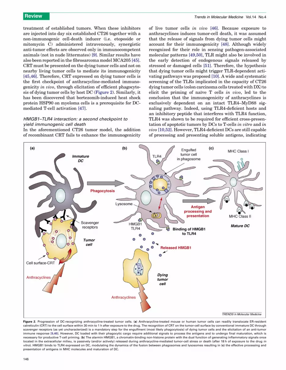
Figure 2. Progression of DC-recognizing anthracycline-treated tumor cells. (a) Anthra
calreticulin (CRT) to the cell surface within 30 min to 1 h after exposure to the drug. The
scavenger receptors (as yet uncharacterized) is a mandatory step for the engulfment (m
immune response [9,46]. However, DC loaded with their phagocytic cargo require add
necessary for productive T-cell priming. (b) The alarmin HMGB1, a chromatin-binding n
located in the extracellular milieu, is passively (and/or actively) released during anthra
vitro). HMGB1 binds to TLR4 expressed on DC, modulating the dynamics of the fusion
presentation of antigens in MHC molecules and maturation of DC.
146
of live tumor cells in vivo [46]. Because exposure toanthracyclines induces tumor-cell death, it was assumedthat the release of signals from dying tumor cells mightaccount for their immunogenicity [48]. Although widelyrecognized for their role in sensing pathogen-associatedmolecular patterns [49,50], TLR might also be involved inthe early detection of endogenous signals released bystressed or damaged cells [51]. Therefore, the hypothesisthat dying tumor cells might trigger TLR-dependent acti-vating pathways was proposed [10]. A wide and systematicscreening of the TLRs implicated in the capacity of CT26dying tumor cells (colon carcinoma cells treatedwithDX) toelicit the priming of naıve T cells in vivo, led to theconclusion that the immunogenicity of anthracyclines isexclusively dependent on an intact TLR4–MyD88 sig-naling pathway. Indeed, using TLR4-deficient hosts andan inhibitory peptide that interferes with TLR4 function,TLR4 was shown to be required for efficient cross-presen-tation of apoptotic tumors by DCs to T-cells in vitro and invivo [10,52]. However, TLR4-deficient DCs are still capableof processing and presenting soluble antigens, indicating
cycline-treated mouse or human tumor cells can readily translocate ER-resident
recognition of CRT on the tumor-cell surface by conventional immature DC through
ost likely phagocytosis) of dying tumor cells and the elicitation of an anti-tumor
itional signals to process the antigens and to undergo final maturation, which is
on-histone protein with the dual function of generating inflammatory signals once
cycline-mediated tumor-cell stress or death (after 18 h of exposure to the drug in
between phagosomes and lysosomes resulting in (c) the effective processing and
Review Trends in Molecular Medicine Vol.14 No.4
that TLR4 is involved in the processing machinery ofmembrane-associated antigens [10].
Based on a previous report claiming that TLR4 couldinhibit the lysosome-dependent degradation of phago-somes in macrophages [53], Apetoh et al. compared thekinetics of fusion between phagosomes and endo-lysosomesin wild-type DC with that in TLR4-deficient DC afteraddition of dying tumor cells [10]. Under these conditions,TLR4 seems to prevent the accelerated routing of thephagocytic cargo to the lysosomes. In accordance withthese data, antigen presentation by TLR4-deficient DCsin vitro could be fully restored using either chloroquine (alysosomotropic alkaline) or bafilomycin A1 (a specificinhibitor of the vacuolar ATPase responsible for lysosomalacidification). Moreover, the concomitant treatment oftumor-bearing Tlr4–/– mice with a cytotoxic compoundalong with chloroquine could ameliorate the anti-tumoreffects of the chemotherapy [10], a result that was notachieved in wild-type littermates.
These findings indicate that an endogenous ligandreleased from dying tumor cells binds to TLR4 on hostDCs and mediates the immunogenicity of DX-exposedtumor cells (Figure 2). Different putative ligands have beenreported forTLR4 [54,55], includingbdefensin2,heat shockproteins, fibrinogen, fibronectin and high-mobility-groupBox 1 (HMGB1). HMGB1 is a non-histone chromatin-bind-ingprotein that functionsasa transcription factor (but it canalso be released upon cellular demise – necrosis rather thanapoptosis) and that stimulates antigen-presenting cells[56,57]. Therefore, HMGB1 has been considered to be analarmin or a damage-associatedmolecular pattern (DAMP)[58] molecule. In line with this concept, it was shown thattreatment of CT26 cells by DX selectively induces therelease of HMGB1 (Figure 1c,d) in vitro but not that ofalternate TLR4 ligands [10]. Similar findingswere obtainedduring the treatment ofMCA205fibrosarcomawithDX. It isnoteworthy that HMGB1 release occurred within 18 h fol-lowingexposure toanthracyclines (or othercytotoxicagents)[10]. The results of immunoprecipitation experiments con-firmed that HMGB1 effectively binds to TLR4 exposed onrat mastocyte Raw cells [10].
The role of HMGB1 in the TLR4-dependent immuno-genicity of DX-treated CT26 (DX-CT26) has been investi-gated by reducing HMGB1 levels via specific siRNAs. Theresults revealed that HMGB1 ismandatory for the primingof naıve T cells by dying DX-CT26 cells and for conferringanti-tumor protective immunity against tumor rechallenge[10]. Moreover, anti-HMGB1 neutralizing antibody com-pletely abrogates both DX-induced immunogenicity of celldeath in vivo and cross-presentation of dying tumor cells toT-cell hybridoma in vitro. Intriguingly, neither CRT norHMGB1 can account for the DC maturation phenotypeinduced by apoptotic tumor cells. This conclusion promptsthe current search for additional signals emitted or ferriedby tumor cells to DCs that could contribute to the polar-ization of the T-cell priming.
Prospects for clinical use of immunomodulatoryproperties of anthracyclinesChallenging the notion that chemotherapy or radiotherapynegatively affects the immune system, accumulating
evidence (from the 1970s to date) indicates that cell deathcan induce an immunological cascade that contributes tothe anti-tumor effects of conventional cytotoxic agents. Theimmune system effectors are elicited in three differentways by conventional therapies. Some therapeutic regi-mens (such as DX, but also oxaliplatin and X-rays [10]) canelicit specific cellular responses that render tumor-celldeath immunogenic. Other drugs might have side effectsthat stimulate the immune system, by transient lympho-depletion (such as high dosing of alkylating agents andfludarabine), by the subversion of immunosuppressivemechanisms (such as metronomic cyclophosphamide,which inhibits regulatory T cells [59], or all trans retinoicacid [60] or gemcitabine [61], both of which modulatemyeloid suppressor cells), or by direct or indirect stimu-latory effects on immune effectors (such as flavonoids,which modify chemokine release [62], or androgen depri-vation, which induces B-cell activation and thymopoiesisleading to increased T-cell repertoire, i.e. an augmentednumber of anti-tumor CTL clones [63]). Moreover, sometreatments (such as fluorouracil or X-rays) can sensitizetumor cells to CTL and NK cell attack by modulatingthe surface expression of tumor-associated antigens [64],Fas [65] and/or NKG2D (natural-killer group 2, member D)ligands [66]. Vaccination against cancer-specific antigenscould also sensitize the glioblastoma tumors to subsequentchemotherapeutic treatment [40].
The data described in this review enable formulation ofthe following predictions for optimization of the clinical useof anthracyclines. First, the potential immunosuppressiveeffects of some current clinical methods of cancer man-agement should be considered. Systematic ablation oflymph nodes for staging might be discussed when consid-ering that tumor-draining lymph nodes are the primarysites of T-cell education. The dose-intensity scheduling ofmost chemotherapy is associated with iterative lymphode-pletion and might compromise the establishment of T-cellpriming. Thus, for instance in breast cancer, low doses ofanthracyclines on a weekly basis in the absence of gluco-corticoids (used as anti-vomiting agents but notoriouslyimmunosuppressive) could be assessed in randomizedtrials aimed at monitoring the ensuing immune response.
Second, neoadjuvant chemotherapy (as opposed to adju-vant chemotherapy) of early (non-advanced) cancers wouldoffer the advantage thatmore tumor antigenwould becomeavailable for the priming of T cells.
Third, prospective trials should be designed so thatserial tumor biopsies, performed before and after che-motherapy, are evaluated for macrophage-, DC-, T- andNK-cell responses as putative prognostic factors. Shouldthe anti-tumor immune responses dictate long-term sur-vival, then local signs of antigen priming (presence ofmature DC in a pseudolymphoid architecture) or effectorcells with a Th1 or Tc1 phenotype might correlate withfavorable responses. Preferably, such data should bematched to gene-expression profiling in tumor beds (eitherstatic before therapy or dynamic following one cycle ofchemotherapy) that might indicate immune signaturesassociated with favorable clinical outcome or response totreatment (as shown in follicular lymphoma [67]).Along these lines, a correlation between pathological
147
Box 3. Outstanding questions
� Which of the current drugs used in the clinical armamentarium of
oncology can be considered ‘immunogenic’? And what are the
molecular mechanisms involved in the immunogenicity of cell
death induced by such drugs?
� If a drug turns out to mediate immune responses, how should the
scheduling and dosing of the current regimen be modified to
avoid compromising this immunity? How might immunotherapy
synergize (or antagonize) with the ongoing immune response?
� Which tumors lack the molecular machinery leading to CRT
exposure and/or HMGB1 secretion? Is there a routine assay that
could test these relevant molecular pathways before and during
treatment?
� How should the impact of development of an immune response
on clinical outcome be demonstrated during the course of
conventional cancer therapy?
� How should the prognostic value of host polymorphisms in
immune-response genes (i.e. TLR4) be shown in the response to
anthracyclines, oxaliplatin and X rays?
� Is there any increased risk in developing autoimmune disorders
when optimizing the immunogenicity of anthracyclines, specifi-
cally in increasing cardiotoxicity through auto-immune reactiv-
ities?
Review Trends in Molecular Medicine Vol.14 No.4
complete response to neoadjuvant chemotherapy withtherapy-induced high CD8+ effector T lymphocyte andlow regulatory T-cell infiltrates has recently been reportedin a series of 56 advanced breast cancers [68].
Last, there are direct diagnostic and therapeutic con-sequences of ‘immunogenic cell death’. From a biotechno-logical point of view, designing of a drug-screeningprogram aimed at selecting products capable of translocat-ing CRT to the plasma membrane of tumor cells could beimagined. Indeed, some, but not all, chemotherapeuticregimens can induce immunogenic cell death with earlyCRT expression, thus eliciting a therapy-associated anti-cancer immune response that determines disease outcome.Furthermore, clinicians could perform an early detection ofCRT exposure on circulating tumor cells or cytospins fromtumor beds one or two days post-chemo- or radio-therapy topredict whether recombinant CRT or peptides inhibitingthe PP1–GADD34 phosphatase complex should be com-bined with the therapeutic regimen to augment thera-peutic success. As a result, it might be interesting tomonitor not only CRT exposure but also the presence ofall factors that are mandatory for the signal-transductionmachinery that leads to CRT exposure in tumor cells, withthe hope of establishing new prognostic or predictive bio-markers. Therapeutic regimens designed to re-establishCRT exposure (for instance, by inhibiting PP1–GADD34)should enhance the immunogenicity of cell death and,hence, boost the therapeutic efficacy of non-immunogenicregimens per se.
In addition, hosts bearing an inherited defect in the tlr4gene or an acquired TLR4 dysfunction [69] might notrespond to conventional ‘immunogenic-prone’ regimen.Indeed, a polymorphism in human TLR4 (rs4986790)has been associated with decreased responses to inhaledlipopolysaccharide [70]. This single-nucleotide exchange(A896G) in the TLR4 gene results in an amino acid sub-stitution (Asp299! Gly) in the extracellular domain ofTLR4. It was also demonstrated that this substitution notonly decreases the binding of HMGB1 to TLR4 but alsoresults in weaker activation of the transcription factornuclear factor-kB (L.A., unpublished) and in a profoundalteration of the capacity of monocyte-derived-human DCto cross-present melanoma tumor antigens from dyingmelanoma cell lines [10]. In a retrospective study, timeto metastases was analyzed in a cohort of 280 patients thathad been treated for breast cancer presenting with lymph-node involvement following local radiotherapy and ananthracycline-based adjuvant chemotherapy. Patientsbearing the TLR4 Asp299! Gly allele featured an accel-erated disease course compared with patients bearing thenormal TLR4 allele, establishing TLR4 Asp299! Gly asan independent predictive factor of early disease pro-gression [10]. These data await further confirmation invarious cancer models that have been treated with specificcytotoxic combinations before envisioning treating TLR4-deficient patients with chloroquine in prospective trials.
Taking into account the considerations discussed here,the ideal management of patients with advanced cancermight integrate the following steps: (i) inhibition of tumor-induced immunosuppression [71]; (ii) priming of naıve Tcells with vaccines; (iii) induction of tumor-cell death
148
with conventional or more recent therapies (i.e.chemo-, radio- or hormone-antibody therapies, or tyrosinekinase inhibitors); (iv) turning non-immunogenic intoimmunogenic cell death [i.e. using recombinant calreticu-lin (rCRT) or inhibition of the PP1–GADD34 complex]; (v)compensation (i.e. using chloroquine treatment) for TLR4defects or host defects (i.e. polymorphisms in molecularmechanisms that elicit immune responses); (vi) boostingongoing effector or memory immune responses [e.g. treat-ment with glycolipids, cytokines, chemokines, anti-KIR(killer inhibitory receptor) antibody, sTRAIL (solubleTNF-related apoptosis inducing ligand), IL-18BP (inter-leukin-18 binding protein)] [40,72]. This integratedstrategy should be evaluated in sizeable randomized trialsin the near future.
Concluding remarksAnthracyclines are thought to mediate their tumoricidalactivity by exerting a direct effect on tumor cells, but recentevidence indicates that the host immune system contrib-utes to the anti-tumor activity greatly (Box 3). Indeed,anthracyclines (e.g. daunorubicin, idarubicin and mitox-antrone) promote cross-talk between the dying tumor cellsand DC, leading to DC-mediated T-cell priming and thecontribution of T cells to long-term survival in experimen-tal tumor models. We have shown that anthracyclines, andalso oxaliplatin and X-rays, induce a series of molecularevents in tumor cells that can be sensed by DC [10]. First,by inducing the phosphorylation of eIF2a, anthracyclinestrigger the rapid translocation of the ER-resident CRT tothe plasma membrane of tumor cells, thereby facilitatingthe uptake of the dying cell by DC. This first step can beelicited by administration of recombinant CRT or by inhi-biting the phosphatase complex PP1–GADD34 whentumors and/or treatment fail to promote membrane CRTexpression. Second, anthracycline-mediated cell death(apoptosis and necrosis at later stages) induces the releaseof the chromatin-binding protein HMGB1 18 h after thedrug-induced stress in vitro. HMGB1 is mandatory to
Review Trends in Molecular Medicine Vol.14 No.4
mediate the immunogenicity of anthracyclines by bindingto host TLR4. DC are required for the response to HMGB1and, upon TLR4 triggering, they are endowed with anti-gen-processing capacities by regulating vesiculardynamics of the compartments involved in antigen pres-entation. We have shown that the lysosomotropic chloro-quine can convert a TLR4-deficient DC (mouse and human)into an efficient antigen-presenting cell capable of cross-presentation to CTL. TLR4 loss-of-function animals areunable to mount an efficient anti-tumor immune responsefollowing treatment with anthracyclines unless chloro-quine is co-administered in the schedule. The relevanceof these findings culminates in the validation of a prog-nostic role of the Asp299! Gly TLR4 polymorphism inbreast cancer patients treatedwithadjuvant anthracyclines[FEC (5-fluorouracil, epirubicin, cyclophosphamide) proto-col; see Box 1]. The Asp299! Gly TLR4 loss-of-functionmutation, which is carried by 12% of all Caucasians, pre-vents the effective binding of HMGB1 to TLR4, and mightaccount for the shorterdisease-free survival observed in thissubset of women after adjuvant FEC. Prospective studieswill be launchedat the InstitutGustaveRoussy (www.igr.fr)to (i) show the prognostic value of CRT exposure in thepathological response to neoadjuvant anthracycline-basedtherapy in breast cancer and (ii) demonstrate the beneficialeffects of combining chloroquine with anthracyclines inAsp299! Gly TLR4 carriers. These findings lead the waytowards development of new therapeutics that combinechemo-and immuno-therapy, taking into account thetumor-host-drug relationship to tailor-make optimal thera-peutic regimen for cancer treatment.
AcknowledgementsL.A. received grants from Ligue contre le cancer and Fondation pour larecherche medicale (FRM) and G.M. from Association pour la Recherchesur le Cancer. L.Z. is supported by grants from Institut National contre leCancer (INCa) and from European DC THERA, and G.K. is supported bya special grant from Ligue contre le Cancer (equipe labellisee), and bygrants from the European Commission (Active p53, RIGHT, Trans-Death,Death-Train, ChemoRes, INCa, Canceropole Ile-de-France and theAssociation for International Cancer Research).
References1 Shapiro, C.L. and Recht, A. (2001) Side effects of adjuvant treatment
of breast cancer. N. Engl. J. Med. 344, 1997–20082 Silvestrini, R. and Gaetani, M. (1963) Action of daunomycin on the
nucleic metabolism of Ehrlich ascites tumor. Tumori 49, 389–3973 Schwartz, H.S. and Grindey, G.B. (1973) Adriamycin and
daunorubicin: a comparison of antitumor activities and tissueuptake in mice following immunosuppression. Cancer Res. 33,1837–1844
4 Ho, R.L. et al. (1993) Development of a safe and effective adriamycinplus interleukin 2 therapy against both adriamycin-sensitive and -resistant lymphomas. Oncol. Res. 5, 373–381
5 Ehrke, M.J. et al. (1998) Doxorubicin plus tumor necrosis factor alphacombination treatments in EL4-lymphoma-bearing C57BL/6 mice.Cancer Immunol. Immunother. 45, 287–298
6 Ehrke, M.J. et al. (2000) Protective specific immunity induced bydoxorubicin plus TNF-a combination treatment of EL4 lymphoma-bearing C57BL/6 mice. Int. J. Cancer 87, 101–109
7 Lake, R.A. and van derMost, R.G. (2006) A better way for a cancer cellto die. N. Engl. J. Med. 354, 2503–2504
8 Casares, N. et al. (2005) Caspase-dependent immunogenicity ofdoxorubicin-induced tumor cell death. J. Exp. Med. 202, 1691–1701
9 Obeid, M. et al. (2007) Calreticulin exposure dictates theimmunogenicity of cancer cell death. Nat. Med. 13, 54–61
10 Apetoh, L. et al. (2007) Toll-like receptor 4-dependent contribution ofthe immune system to anticancer chemotherapy and radiotherapy.Nat. Med. 13, 1050–1059
11 Albert, M.L. et al. (1998) Dendritic cells acquire antigen fromapoptotic cells and induce class I-restricted CTLs. Nature 392, 86–89
12 Albert, M.L. et al. (1998) Tumor-specific killer cells in paraneoplasticcerebellar degeneration. Nat. Med. 4, 1321–1324
13 Shapiro, D.M. et al. (1957) Quantitative biochemical differencesbetween tumor and host as a basis for cancer chemotherapy. V.Niacin and 6-aminonicotinamide. Cancer Res. 17, 600–604
14 Orsini, F. et al. (1977) Increased primary cell-mediated immunity inculture subsequent to adriamycin or daunorubicin treatment ofspleen donor mice. Cancer Res. 37, 1719–1726
15 Stoychkov, J.N. et al. (1979) Effects of adriamycin andcyclophosphamide treatment on induction of macrophage cytotoxicfunction in mice. Cancer Res. 39, 3014–3017
16 Haskill, J.S. (1981) Adriamycin-activated macrophages as tumorgrowth inhibitors. Cancer Res. 41, 3852–3856
17 Maccubbin, D.L. et al. (1990) Indomethacinmodulation of adriamycin-induced effects on multiple cytolytic effector functions. CancerImmunol. Immunother. 31, 373–380
18 Maccubbin, D.L. et al. (1992) Adriamycin-induced modulation of hostdefenses in tumor-bearing mice. Cancer Res. 52, 3572–3576
19 Zaleskis, G. et al. (1994) Intracellular doxorubicin kinetics inlymphoma cells and lymphocytes infiltrating the tumor area invivo: a flow cytometric study. Oncol. Res. 6, 183–194
20 Fisk, B. and Ioannides, C.G. (1998) Increased sensitivity ofadriamycin-selected tumor lines to CTL-mediated lysis results inenhanced drug sensitivity. Cancer Res. 58, 4790–4793
21 Ujhazy, P. et al. (2003) Doxorubicin induces specific immune functionsand cytokine expression in peritoneal cells. Cancer Immunol.Immunother. 52, 463–472
22 Mace, K. et al. (1988) Alterations in murine host defense functions byadriamycin or liposome-encapsulated adriamycin. Cancer Res. 48,130–136
23 Salup, R.R. et al. (1987) Successful treatment of advanced murinerenal cell cancer by bicompartmental adoptivechemoimmunotherapy. J. Immunol. 138, 641–647
24 Lumsden, A.J. et al. (1996) Immunohistochemical characterisation ofimmunological changes at the tumour site after chemo-immunotherapy with doxorubicin, interleukin-2 and interferon-g.Anticancer Res. 16, 1145–1154
25 Lumsden, A.J. et al. (1992) Improved efficacy of doxorubicin bysimultaneous treatment with interferon-g and interleukin-2. InVivo 6, 553–558
26 Zagozdzon, R. et al. (1998) Effective chemo-immunotherapy of L1210leukemia in vivo using interleukin-12 combined with doxorubicin butnot with cyclophosphamide, paclitaxel or cisplatin. Int. J. Cancer 77,720–727
27 Le Cesne, A. et al. (1999) Combination interleukin-2 anddoxorubicin in advanced adult solid tumors: circumvention ofdoxorubicin resistance in soft-tissue sarcoma? J. Immunother. 22,268–277
28 Smalley, R.V. et al. (1992) Interferon a combined with cytotoxicchemotherapy for patients with non-Hodgkin’s lymphoma. N. Engl.J. Med. 327, 1336–1341
29 Solal-Celigny, P. et al. (1993) Recombinant interferon a-2b combinedwith a regimen containing doxorubicin in patients with advancedfollicular lymphoma. Groupe d’Etude des Lymphomes de l’Adulte. N.Engl. J. Med. 329, 1608–1614
30 Raitanen, M.P. and Lukkarinen, O. (1995) A controlled study ofintravesical epirubicin with or without a 2b-interferon asprophylaxis for recurrent superficial transitional cell carcinoma ofthe bladder. Finnish Multicentre Study Group. Br. J. Urol. 76, 697–701
31 Ferrari, P. et al. (1992) Chemoimmunotherapy for prophylaxis ofrecurrence in superficial bladder cancer: interferon-a 2b versusinterferon-a 2b with epirubicin. Anticancer Drugs 3 (Suppl. 1), 25–27
32 Uekado, Y. et al. (1994) The effects of intravesicalchemoimmunotherapy with epirubicin and bacillus Calmette-Guerin for prophylaxis of recurrence of superficial bladder cancer:a preliminary report. Cancer Chemother. Pharmacol. 35 (Suppl.),S65–S68
149
Review Trends in Molecular Medicine Vol.14 No.4
33 Naglieri, E. et al. (1998) Standard interleukin-2 (IL-2) and interferon-a immunotherapy versus an IL-2 and 4-epirubicin immuno-chemotherapeutic association in metastatic renal cell carcinoma.Anticancer Res. 18, 2021–2026
34 Zitvogel, L. et al. (2004) Immune response against dying tumor cells.Adv. Immunol. 84, 131–179
35 Zitvogel, L. et al. (2006) Cancer despite immunosurveillance:immunoselection and immunosubversion. Nat. Rev. Immunol. 6,715–727
36 Ullrich, E. et al. (2008) Tumor stress, cell death and the ensuingimmune response. Cell Death Differ. 15, 21–28
37 Tesniere, A. et al. (2008) Molecular characteristics of immunogeniccancer cell death. Cell Death Differ. 15, 3–12
38 Scheffer, S.R. et al. (2003) Apoptotic, but not necrotic, tumor cellvaccines induce a potent immune response in vivo. Int. J. Cancer 103,205–211
39 Bartholomae, W.C. et al. (2004) T cell immunity induced by live,necrotic, and apoptotic tumor cells. J. Immunol. 173, 1012–1022
40 Zitvogel, L. et al. (2008) Immunological aspects of cancerchemotherapy. Nat. Rev. Immunol. 8, 59–73
41 Jung, S. et al. (2002) In vivo depletion of CD11c+ dendritic cellsabrogates priming of CD8+ T cells by exogenous cell-associatedantigens. Immunity 17, 211–220
42 Williams, D.B. (2006) Beyond lectins: the calnexin/calreticulinchaperone system of the endoplasmic reticulum. J. Cell Sci. 119,615–623
43 Gelebart, P. et al. (2005) Calreticulin, a Ca2+-binding chaperoneof the endoplasmic reticulum. Int. J. Biochem. Cell Biol. 37, 260–266
44 Gardai, S.J. et al. (2005) Cell-surface calreticulin initiates clearance ofviable or apoptotic cells through trans-activation of LRP on thephagocyte. Cell 123, 321–334
45 Obeid, M. et al. (2007) Ecto-calreticulin in immunogenicchemotherapy. Immunol. Rev. 220, 22–34
46 Obeid, M. et al. (2007) Calreticulin exposure is required for theimmunogenicity of g-irradiation and UVC light-induced apoptosis.Cell Death Differ. 14, 1848–1850
47 Spisek, R. et al. (2007) Bortezomib enhances dendritic cell (DC)-mediated induction of immunity to human myeloma via exposureof cell surface heat shock protein 90 on dying tumor cells: therapeuticimplications. Blood 109, 4839–4845
48 Matzinger, P. (1994) Tolerance, danger, and the extended family.Annu. Rev. Immunol. 12, 991–1045
49 Janeway, C.A., Jr (2001) How the immune systemworks to protect thehost from infection: a personal view. Proc. Natl. Acad. Sci. U. S. A. 98,7461–7468
50 Medzhitov, R. (2001) Toll-like receptors and innate immunity. Nat.Rev. Immunol. 1, 135–145
51 Marshak-Rothstein, A. (2006) Toll-like receptors in systemicautoimmune disease. Nat. Rev. Immunol. 6, 823–835
52 Apetoh, L. et al. (2007) The interaction between HMGB1 and TLR4dictates the outcome of anticancer chemotherapy and radiotherapy.Immunol. Rev. 220, 47–59
53 Shiratsuchi, A. et al. (2004) Inhibitory effect of Toll-like receptor 4 onfusion between phagosomes and endosomes/lysosomes inmacrophages. J. Immunol. 172, 2039–2047
54 Tsan, M.F. and Gao, B. (2004) Endogenous ligands of Toll-likereceptors. J. Leukoc. Biol. 76, 514–519
55 Marshak-Rothstein, A. and Rifkin, I.R. (2007) Immunologically activeautoantigens: the role of toll-like receptors in the development ofchronic inflammatory disease. Annu. Rev. Immunol. 25, 419–441
56 Erlandsson Harris, H. and Andersson, U. (2004) Mini-review: Thenuclear protein HMGB1 as a proinflammatory mediator. Eur. J.Immunol. 34, 1503–1512
57 Dumitriu, I.E. et al. (2005) Requirement of HMGB1 and RAGE for thematuration of human plasmacytoid dendritic cells. Eur. J. Immunol.35, 2184–2190
58 Rovere-Querini, P. et al. (2004) HMGB1 is an endogenous immuneadjuvant released by necrotic cells. EMBO Rep. 5, 825–830
59 Ghiringhelli, F. et al. (2007) Metronomic cyclophosphamide regimenselectively depletes CD4+CD25+ regulatory T cells and restores T andNK effector functions in end stage cancer patients. Cancer Immunol.Immunother. 56, 641–648
150
60 Nefedova, Y. et al. (2007)Mechanism of all-trans retinoic acid effect ontumor-associated myeloid-derived suppressor cells. Cancer Res. 67,11021–11028
61 Suzuki, E. et al. (2005) Gemcitabine selectively eliminates splenic Gr-1+/CD11b+ myeloid suppressor cells in tumor-bearing animals andenhances antitumor immune activity. Clin. Cancer Res. 11, 6713–6721
62 Ramiro, E. et al. (2005) Flavonoids from Theobroma cacao down-regulate inflammatory mediators. J. Agric. Food Chem. 53, 8506–8511
63 Aragon-Ching, J.B. et al. (2007) Impact of androgen-deprivationtherapy on the immune system: implications for combinationtherapy of prostate cancer. Front. Biosci. 12, 4957–4971
64 Zusman, I. (2004) Soluble tumor-associated antigens in cancer detection,prevention and therapy. Med. Sci. Monit. 10, RA317–RA324
65 Nakamura, M. et al. (2007) Role of the Fas/FasL pathway incombination therapy with interferon-a and fluorouracil againsthepatocellular carcinoma in vitro. J. Hepatol. 46, 77–88
66 Kim, J.Y. et al. (2006) Increase of NKG2D ligands and sensitivity toNK cell-mediated cytotoxicity of tumor cells by heat shock andionizing radiation. Exp. Mol. Med. 38, 474–484
67 Dave, S.S. et al. (2004) Prediction of survival in follicular lymphomabased on molecular features of tumor-infiltrating immune cells.N. Engl. J. Med. 351, 2159–2169
68 Ladoire, S. et al. Pathological complete response to neoadjuvantchemotherapy of breast carcinoma is associated with thedisappearance of tumor-infiltrating Foxp3+ regulatory T cells. Clin.Cancer Res. (in press)
69 Frenzel, H. et al. (2006) Toll-like receptor interference in myeloiddendritic cells through head and neck cancer. Anticancer Res. 26,4409–4413
70 Arbour, N.C. et al. (2000) TLR4 mutations are associated withendotoxin hyporesponsiveness in humans. Nat. Genet. 25, 187–191
71 Zou, W. (2006) Regulatory T cells, tumour immunity andimmunotherapy. Nat. Rev. Immunol. 6, 295–307
72 Terme, M. et al. NK cell-directed therapies: moving from unexpectedresults to successful strategies. Nat. Immunol. (in press)
73 Dimarco, A. et al. (1963) Experimental studies of the antineoplasticactivity of a new antibiotic, daunomycin. Tumori 49, 203–217
74 Massimo, L. et al. (1967) Preliminary results of the therapeuticeffectiveness in leukemia and malignant tumors in children of anew antineoplastic antibiotic: ‘daunomycin’. Arch. Ital. Patol. Clin.Tumori 10, 3–19
75 Monfardini, S. et al. (1969) Clinical trials with adriamycin in leukemiaand solid tumors. Tumori 55, 197–216
76 Formelli, F. et al. (1979) Fluorescence assay of tissue distribution of 4-demethoxydaunorubicin and 4-demethoxydoxorubicin in micebearing solid tumors. Cancer Chemother. Pharmacol. 3, 261–269
77 Bonfante, V. et al. (1980) Preliminary clinical experience with 4-epidoxorubicin in advanced human neoplasia. Recent ResultsCancer Res. 74, 192–199
78 Alberts, D.S. et al. (1980) Phase I clinical trial f mitoxantrone: a newanthracenedione anticancer drug. Cancer Chemother. Pharmacol. 5,11–15
79 Rabbani, A. et al. (2005) The anthracycline antibiotics: antitumordrugs that alter chromatin structure. Bioessays 27, 50–56
80 Batist, G. et al. (2002) Myocet (liposome-encapsulated doxorubicincitrate): a new approach in breast cancer therapy. Expert Opin.Pharmacother. 3, 1739–1751
81 Gabizon, A. et al. (2003) Pharmacokinetics of pegylated liposomalDoxorubicin: review of animal and human studies. Clin.Pharmacokinet. 42, 419–436
82 Glae, G.R. et al. (1976) Combination chemotherapy of L1210 leukemiawith platinum compounds and cyclophosphamide plus other selectedantineoplastic agents. J. Natl. Cancer Inst. 57, 1363–1366
83 Rose, P.G. (1996) Endometrial carcinoma. N. Engl. J. Med. 335, 640–649
84 Cannistra, S.A. (2004) Cancer of the ovary. N. Engl. J. Med. 351,2519–2529
85 Hortobagyi, G.N. (1998) Treatment of breast cancer. N. Engl. J. Med.339, 974–984
86 Armitage, J.O. (1993) Treatment of non-Hodgkin’s lymphoma.N. Engl. J. Med. 328, 1023–1030
Review Trends in Molecular Medicine Vol.14 No.4
87 Cunningham, D. et al. (2006) Perioperative chemotherapy versussurgery alone for resectable gastroesophageal cancer. N. Engl. J.Med. 355, 11–20
88 Minotti, G. et al. (2004) Anthracyclines: molecular advances andpharmacologic developments in antitumor activity andcardiotoxicity. Pharmacol. Rev. 56, 185–229
89 Levine, M.N. et al. (2005) Randomized trial comparing cyclo-phosphamide, epirubicin, and fluorouracil with cyclophosphamide,methotrexate, and fluorouracil in premenopausal women with node-positive breast cancer: update of National Cancer Institute of CanadaClinical Trials Group Trial MA5. J. Clin. Oncol. 23, 5166–5170
90 Poole, C.J. et al. (2006) Epirubicin and cyclophosphamide,methotrexate, and fluorouracil as adjuvant therapy for early breastcancer. N. Engl. J. Med. 355, 1851–1862
91 Hayes, D.F. et al. (2007) HER2 and response to paclitaxel in node-positive breast cancer. N. Engl. J. Med. 357, 1496–1506
92 Pritchard, K.I. et al. (2006) HER2 and responsiveness of breast cancerto adjuvant chemotherapy. N. Engl. J. Med. 354, 2103–2111
93 Tanner,M. et al. (2006) Topoisomerase IIa gene amplification predictsfavorable treatment response to tailored and dose-escalatedanthracycline-based adjuvant chemotherapy in HER-2/neu-amplified breast cancer: Scandinavian Breast Group Trial 9401.J. Clin. Oncol. 24, 2428–2436
94 Rouzier, R. et al. (2005) Breast cancer molecular subtypes responddifferently to preoperative chemotherapy. Clin. Cancer Res. 11, 5678–5685
95 Habeshaw, T. et al. (1991) Epirubicin at two dose levels withprednisolone as treatment for advanced breast cancer: the resultsof a randomized trial. J. Clin. Oncol. 9, 295–304
96 Harris, L. et al. (2002) Liposome-encapsulated doxorubicin comparedwith conventional doxorubicin in a randomized multicenter trial asfirst-line therapy of metastatic breast carcinoma. Cancer 94, 25–36
97 Batist, G. et al. (2001) Reduced cardiotoxicity and preservedantitumor efficacy of liposome-encapsulated doxorubicin andcyclophosphamide compared with conventional doxorubicin andcyclophosphamide in a randomized, multicenter trial of metastaticbreast cancer. J. Clin. Oncol. 19, 1444–1454
98 Berman, E. et al. (1991) Results of a randomized trial comparingidarubicin and cytosine arabinoside with daunorubicin and cytosinearabinoside in adult patients with newly diagnosed acutemyelogenous leukemia. Blood 77, 1666–1674
99 Vogler, W.R. et al. (1992) A phase III trial comparing idarubicin anddaunorubicin in combination with cytarabine in acute myelogenousleukemia: a Southeastern Cancer Study Group Study. J. Clin. Oncol.10, 1103–1111
100 Arlin, Z. et al. (1990) Randomized multicenter trial of cytosinearabinoside with mitoxantrone or daunorubicin in previouslyuntreated adult patients with acute nonlymphocytic leukemia(ANLL). Lederle Cooperative Group. Leukemia 4, 177–183
101 Bassan, R. et al. (1995) The use of anthracyclines in adult acutelymphoblastic leukemia. Haematologica 80, 280–291
102 Alexanian, R. and Dimopoulos, M. (1994) The treatment of multiplemyeloma. N. Engl. J. Med. 330, 484–489
103 Rigacci, L. et al. (2003) The role of anthracyclines in combinationchemotherapy for the treatment of follicular lymphoma: retrospective
Free journals for dev
In 2002, the WHO and six medical journal publishe
Research Initiative, which enabled nearly 70 of t
reduced-cost access to biomedical literature th
publishers are participating in the program,
Gro Harlem Brundtland, former director-general for
the biggest step ever taken towards reducing the
countrie
For more information, vis
study of the Intergruppo Italiano Linfomi on 761 cases. Leuk.Lymphoma 44, 1911–1917
104 Tsukasaki,K. et al. (2007)VCAP–AMP–VECPcomparedwith biweeklyCHOP for adult T-cell leukemia-lymphoma: Japan Clinical OncologyGroup Study JCOG9801. J. Clin. Oncol. 25, 5458–5464
105 Ferme, C. et al. (2007) Chemotherapy plus involved-field radiation inearly-stage Hodgkin’s disease. N. Engl. J. Med. 357, 1916–1927
106 Northfelt, D.W. et al. (1998) Pegylated-liposomal doxorubicin versusdoxorubicin, bleomycin, and vincristine in the treatment of AIDS-related Kaposi’s sarcoma: results of a randomized phase III clinicaltrial. J. Clin. Oncol. 16, 2445–2451
107 Gill, P.S. et al. (1996) Randomized phase III trial of liposomaldaunorubicin versus doxorubicin, bleomycin, and vincristine inAIDS-related Kaposi’s sarcoma. J. Clin. Oncol. 14, 2353–2364
108 Valdivieso, M. et al. (1984) Increased therapeutic index of weeklydoxorubicin in the therapy of non-small cell lung cancer: aprospective, randomized study. J. Clin. Oncol. 2, 207–214
109 Onoda, S. et al. (2006) Phase II trial of amrubicin for treatment ofrefractory or relapsed small-cell lung cancer: Thoracic OncologyResearch Group Study 0301. J. Clin. Oncol. 24, 5448–5453
110 Gordon, A.N. et al. (2001) Recurrent epithelial ovarian carcinoma: arandomized phase III study of pegylated liposomal doxorubicin versustopotecan. J. Clin. Oncol. 19, 3312–3322
111 Santoro, A. et al. (1995) Doxorubicin versus CYVADIC versusdoxorubicin plus ifosfamide in first-line treatment of advanced softtissue sarcomas: a randomized study of the European Organizationfor Research and Treatment of Cancer Soft Tissue and Bone SarcomaGroup. J. Clin. Oncol. 13, 1537–1545
112 du Bois, A. et al. (2007) Combination therapy with pegylatedliposomal doxorubicin and carboplatin in gynecologic malignancies: aprospective phase II study of the Arbeitsgemeinschaft GynaekologischeOnkologie Studiengruppe Ovarialkarzinom (AGO-OVAR) andKommission Uterus (AGO-K-Ut). Gynecol. Oncol. 107, 518–525
113 Valteau-Couanet, D. et al. (2005) Results of induction chemotherapyin children older than 1 year with a stage 4 neuroblastoma treatedwith the NB 97 French Society of Pediatric Oncology (SFOP) protocol.J. Clin. Oncol. 23, 532–540
114 Tannock, I.F. et al. (2004) Docetaxel plus prednisone or mitoxantroneplus prednisone for advanced prostate cancer. N. Engl. J. Med. 351,1502–1512
115 Sessa, C. et al. (2007) Ongoing phase I and II studies of novelanthracyclines. Cardiovasc. Toxicol. 7, 75–79
116 Quintieri, L. et al. (2005) Formation and antitumor activity of PNU-159682, a major metabolite of nemorubicin in human livermicrosomes. Clin. Cancer Res. 11, 1608–1617
117 Fabel, K. et al. (2001) Long-term stabilization in patients withmalignant glioma after treatment with liposomal doxorubicin.Cancer 92, 1936–1942
118 Harrington, K.J. et al. (2001) Phase II study of pegylated liposomaldoxorubicin (Caelyx) as induction chemotherapy for patients withsquamous cell cancer of the head and neck. Eur. J. Cancer 37, 2015–2022
119 Leroux, J.Y. et al. (1986) Enhancement of murine lymphoma celllysability by CTL and by LAK cells, after treatments with mitomycinC and with adriamycin. Int. J. Immunopharmacol. 8, 369–375
eloping countries
rs launched the Health InterNetwork Access to
he world’s poorest countries to gain free or
rough the internet. Currently more than 70
providing access to over 2000 journals.
the WHO, said that this initiative was ‘‘perhaps
health information gap between rich and poor
s’’.
it www.who.int/hinari
151


