Embed Size (px)
Citation preview
GRAIL: A unique mediator of CD4 T lymphocyteunresponsiveness
Chan C. Whiting1, Leon L. Su1, Jack T. Lin1, and C. Garrison Fathman1,*1Stanford University, Department of Medicine, Division of Rheumatology and Immunology 269Campus Drive West, Stanford, CA 94305
AbstractGRAIL (gene related to anergy in lymphocytes, also known as RNF128), an ubiquitin-proteinligase (E3), utilizes a unique single transmembrane protein with a split function motif, and is animportant gatekeeper of T cell unresponsiveness. While it may play a role in other CD4 T cellfunctions including activation, survival, and differentiation, GRAIL is most well characterized as anegative regulator of TCR responsiveness and cytokine production. Here, we review the recentliterature on this remarkable E3 in the regulation of human and mouse CD4 T cellunresponsiveness.
KeywordsGRAIL; E3; ubiquitin-protein ligase; anergy; T cell unresponsiveness; ubiquitination; de-ubiquitinating enzymes (DUBs)
IntroductionThe ability to distinguish self from non-self is the most important requirement of themammalian immune system. Central (thymic) and peripheral tolerance mechanisms haveevolved to prevent lymphocyte-mediated self-destruction (autoimmunity). Because thymicnegative selection is not foolproof, some autoreactive T cells escape negative selection.Peripheral tolerance mechanisms, therefore, need to be in place to maintain CD4 T cellunresponsiveness to self. One important mechanism of peripheral tolerance that maintainsCD4 T cell unresponsiveness is anergy [1,2]. Anergic CD4 T cells fail to proliferate or toproduce interleukin-2 (IL-2) following immunogenic stimulation. Based on the simplistictwo-signal hypothesis, full T cell activation occurs from the simultaneous engagement of theT cell receptor (TCR)(signal one) and CD4 T cell costimulatory molecules such as CD28(signal two). In the absence of robust activation, (including a variety of extrinsic andintrinsic activation signals), engagement of the CD4 T cell receptor only suboptimallystimulates the T cell (signal one) and, without costimulation, TCR engagement results in aform of CD4 T cell unresponsiveness called anergy [2]. Anergy induction is an activeprocess that is dependent upon tightly controlled biochemical signaling events including up-regulation and degradation of both genes and proteins [3-6]. As demonstrated several yearsago, the development of the anergy phenotype in CD4 T cells could be blocked by inhibitorsof protein synthesis or by calcineurin, which suggested that the induction of anergy activateda unique genetic program [7]. The induced unresponsive state of anergy was relatively longlived in CD4 T cells and could be reversed by the addition of exogenous IL-2, a distinct
*Correspondence to: [email protected].
NIH Public AccessAuthor ManuscriptFEBS J. Author manuscript; available in PMC 2012 January 1.
Published in final edited form as:FEBS J. 2011 January ; 278(1): 47–58. doi:10.1111/j.1742-4658.2010.07922.x.
NIH
-PA Author Manuscript
NIH
-PA Author Manuscript
NIH
-PA Author Manuscript
feature of anergic CD4 T cells. In addition to these molecular events identified earlier, it hasrecently become evident that the post-translational modification of proteins viaubiquitination plays an essential role in the regulatory mechanisms of CD4 T cell anergy.
The balance between ubiquitination and de-ubiquitination of many cellular proteins is wellaccepted as an important mechanism for the maintenance of T cell unresponsiveness andprevention of autoimmunity [3,8,9]. Similar to the well studied phosphorylation inducedpost-translational modification of signaling proteins, ubiquitination is an evolutionarilyconserved and reversible process that is also important in signaling and works by covalentlyattaching monoubiquitin or polyubiquitin chains to target proteins to regulate their stability,activity and localization. Post-translational ubiquitination can result in proteolyticdegradation as well as nonproteolytic outcomes that regulate a broad range of criticalcellular functions, including gene transcription and protein trafficking. Ubiquitinconjugation of target proteins consists of a sequence of steps that require three classes ofmodifying enzymes. The initiation step involves an ATP-dependent attachment of ubiquitinto the ubiquitin-activating enzyme (E1). Next, the thiol ester-linked ubiquitin is transferredfrom the E1 enzyme to a cysteine residue in an ubiquitin-conjugating enzyme (E2). Lastly,the E2 enzyme, together with ubiquitin-protein ligase (E3) transfers ubiquitin to targetproteins, where a stable isopeptide bond is formed between the carboxyl terminus ofubiquitin and the ε-amino group of a lysine residue on the target protein. The E3 determinesthe specificity in the substrate conjugation process; however, it has been a challenge touncover specific target lysine sites or consensus ubiquitination motifs on target proteins.This post-translational process is a reversible reaction where the trimming or removal ofubiquitin linkages is mediated by an equally complex process of de-ubiquitination. Thediverse family of de-ubiquitinating enzymes (DUBs) can be classified into broad categoriesbased on their enzymatic domains; the most common two are the ubiquitin specific proteases(USP/UBPs) and ubiquitin C-terminal hydrolases (UCHs).
The role of ubiquitin ligases as modulators of central and peripheral tolerance has broughtattention to this system as one of the key components of a complex regulatory networkdesigned to maintain an active immune surveillance program [10]. Three ubiquitin-proteinligases, Cbl-b, Itch, and GRAIL have been demonstrated to play a functional role in T cellanergy [1,3,10-13]. Moreover, Itch has been shown to prevent autoimmune activation ofperipheral T cells toward a Th2 bias [14], and Cbl-b attenuates T cell hyper-responsiveactivation absent CD28 costimulation [15-17]. These three E3s function as negativeregulators of the immune response and their expression is induced as part of the geneticprogram tuned by the calcium/calcineurin pathway to help establish and maintain T cellunresponsiveness via setting thresholds for TCR signaling [3,4,14,18-22]. Mechanismsimplicated in the development of anergy associated with these E3s include setting thethreshold for TCR responsiveness, modulation of TCR-specific signals and repression ofcytokine transcription. The induction or function of CD4 regulatory T cells has beensuggested for Cbl-b and GRAIL. Moreover, the defective expression of these E3s has beenlinked to autoimmune or inflammatory diseases, in experimental murine and human models,marking their possible pathogenic roles [11,15,22-24]. While GRAIL is expressed in avariety of tissues including liver and hematopoietic linage cells, only its expression in Tcells has been studied extensively. In this review, we will focus our discussion on recentresearch investigating the biology of GRAIL in T lymphocytes and specifically its role inestablishing and maintaining CD4 T cell unresponsiveness. In this issue of Febs Journal, werefer you to excellent discussions on other members of the RING finger E3s including plantRMR [25] and RFN13 [26,27].
Whiting et al. Page 2
FEBS J. Author manuscript; available in PMC 2012 January 1.
NIH
-PA Author Manuscript
NIH
-PA Author Manuscript
NIH
-PA Author Manuscript
SectionsWhat is GRAIL?
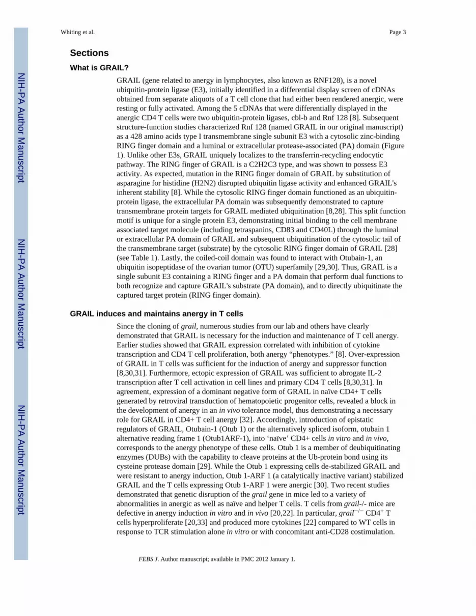
GRAIL (gene related to anergy in lymphocytes, also known as RNF128), is a novelubiquitin-protein ligase (E3), initially identified in a differential display screen of cDNAsobtained from separate aliquots of a T cell clone that had either been rendered anergic, wereresting or fully activated. Among the 5 cDNAs that were differentially displayed in theanergic CD4 T cells were two ubiquitin-protein ligases, cbl-b and Rnf 128 [8]. Subsequentstructure-function studies characterized Rnf 128 (named GRAIL in our original manuscript)as a 428 amino acids type I transmembrane single subunit E3 with a cytosolic zinc-bindingRING finger domain and a luminal or extracellular protease-associated (PA) domain (Figure1). Unlike other E3s, GRAIL uniquely localizes to the transferrin-recycling endocyticpathway. The RING finger of GRAIL is a C2H2C3 type, and was shown to possess E3activity. As expected, mutation in the RING finger domain of GRAIL by substitution ofasparagine for histidine (H2N2) disrupted ubiquitin ligase activity and enhanced GRAIL'sinherent stability [8]. While the cytosolic RING finger domain functioned as an ubiquitin-protein ligase, the extracellular PA domain was subsequently demonstrated to capturetransmembrane protein targets for GRAIL mediated ubiquitination [8,28]. This split functionmotif is unique for a single protein E3, demonstrating initial binding to the cell membraneassociated target molecule (including tetraspanins, CD83 and CD40L) through the luminalor extracellular PA domain of GRAIL and subsequent ubiquitination of the cytosolic tail ofthe transmembrane target (substrate) by the cytosolic RING finger domain of GRAIL [28](see Table 1). Lastly, the coiled-coil domain was found to interact with Otubain-1, anubiquitin isopeptidase of the ovarian tumor (OTU) superfamily [29,30]. Thus, GRAIL is asingle subunit E3 containing a RING finger and a PA domain that perform dual functions toboth recognize and capture GRAIL's substrate (PA domain), and to directly ubiquitinate thecaptured target protein (RING finger domain).
GRAIL induces and maintains anergy in T cellsSince the cloning of grail, numerous studies from our lab and others have clearlydemonstrated that GRAIL is necessary for the induction and maintenance of T cell anergy.Earlier studies showed that GRAIL expression correlated with inhibition of cytokinetranscription and CD4 T cell proliferation, both anergy “phenotypes.” [8]. Over-expressionof GRAIL in T cells was sufficient for the induction of anergy and suppressor function[8,30,31]. Furthermore, ectopic expression of GRAIL was sufficient to abrogate IL-2transcription after T cell activation in cell lines and primary CD4 T cells [8,30,31]. Inagreement, expression of a dominant negative form of GRAIL in naïve CD4+ T cellsgenerated by retroviral transduction of hematopoietic progenitor cells, revealed a block inthe development of anergy in an in vivo tolerance model, thus demonstrating a necessaryrole for GRAIL in CD4+ T cell anergy [32]. Accordingly, introduction of epistaticregulators of GRAIL, Otubain-1 (Otub 1) or the alternatively spliced isoform, otubain 1alternative reading frame 1 (Otub1ARF-1), into ‘naïve’ CD4+ cells in vitro and in vivo,corresponds to the anergy phenotype of these cells. Otub 1 is a member of deubiquitinatingenzymes (DUBs) with the capability to cleave proteins at the Ub-protein bond using itscysteine protease domain [29]. While the Otub 1 expressing cells de-stabilized GRAIL andwere resistant to anergy induction, Otub 1-ARF 1 (a catalytically inactive variant) stabilizedGRAIL and the T cells expressing Otub 1-ARF 1 were anergic [30]. Two recent studiesdemonstrated that genetic disruption of the grail gene in mice led to a variety ofabnormalities in anergic as well as naïve and helper T cells. T cells from grail-/- mice aredefective in anergy induction in vitro and in vivo [20,22]. In particular, grail−/− CD4+ Tcells hyperproliferate [20,33] and produced more cytokines [22] compared to WT cells inresponse to TCR stimulation alone in vitro or with concomitant anti-CD28 costimulation.
Whiting et al. Page 3
FEBS J. Author manuscript; available in PMC 2012 January 1.
NIH
-PA Author Manuscript
NIH
-PA Author Manuscript
NIH
-PA Author Manuscript
Moreover, in vitro differentiated CD4 T cells from grail−/− mice compared to WTlittermates showed significant hypersecretion of IFN-γ in Th1 cells [20,22], lowered IL-4 inTh2 cells [22], and elevated IL-17 and IL-22 in Th17 cells. Consistent with defective anergyin vitro, oral tolerance was abolished in vivo in grail-/- mice using different antigen models.More profound autoimmune symptoms were revealed in aged grail-/- mice compared to WTlittermates including enlarged spleens and mesenteric lymph nodes, massive infiltration ofinflammatory cells in multiple organs, and enhanced susceptibility and severity toexperimental autoimmune encephalitis (EAE) [22]. Furthermore, in the EAE model, CD4+T cell infiltrates from splenocytes and CNS of old grail-/- mice produced significantly higherlevels of IFN-γ and IL-17 when compared to age-matched littermates [33]. Taken together,results from these studies clearly demonstrate that GRAIL is an important gatekeeper forCD4+ T cell anergy. Its role in other T cell functions will be discussed further below.
GRAIL in regulatory T cells (Tregs)Since the thymically derived Foxp3+CD25+ regulatory T cells as well as adaptive Tregulatory cells are special subsets of anergic T cells, we asked whether GRAIL wasexpressed in Tregs and whether their functions are associated with GRAIL expression.Indeed, GRAIL mRNA expression is increased 10-fold in naturally occurring (thymicallyderived) CD4(+) CD25(+) T regulatory cells compared to naive CD25(-) T cells [31,34].Further investigation revealed that CD25(+) Foxp3(+) antigen-specific regulatory T cellswere induced after a “tolerizing-administration” of antigen and that GRAIL expressioncorrelated with the CD25(+) Foxp3(+) antigen-specific subset [31]. Using retroviraltransduction, forced expression of GRAIL in a T cell line was sufficient for conversion ofthese cells to a regulatory phenotype even in the absence of detectable Foxp3 [31]. In a well-characterized, Staphylococcal enterotoxin B (SEB)-mediated model of T cellunresponsiveness in vivo, GRAIL was shown to be up-regulated in the SEB-exposedCD25(+) and CD25(-)FoxP3(+)Vbeta8(+)CD4(+) T cells and FoxP3(-)CD25(-)Vbeta8(+)CD4(+) T cells [35]. Interestingly, a recent study demonstrated that suppressiveand non-proliferative functions of the SEB-expressing FoxP3(+)GRAIL(+) T cells wereindependent of CD25 expression and glucocorticoid-induced tumour necrosis factor R-related protein. This model system reveals a novel paradigm for chronic non-canonical Tcell receptor engagement leading to development of highly suppressiveFoxP3(+)GRAIL(+)CD4(+) T cells. While GRAIL is not required for Treg development, itis required for their suppressive function as grail-/- Tregs exhibited reduced suppressiveactivity on the proliferation of naïve responder cells when compared to WT Tregs [20,22].Interesting, a specific subset of Tregs (CD4+CD62LhighCD25+) do not seem to requireGRAIL for suppressive function even though GRAIL mRNA is highly expressed in thesecells [20]. On the other hand, Nurieva et al demonstrated that grail-/- CD4+CD25+ Tregswere not as effective at suppressing WT CD4 T cells compared to WT Tregs [22]. Takentogether, these data demonstrate that GRAIL is differentially expressed in naturallyoccurring and peripherally induced T regulatory cells and that the expression of GRAIL islinked to their functional regulatory activity.
Regulation of GRAIL expressionGRAIL Transcriptional, Translational and Post-translational regulation
In T lymphocytes, GRAIL RNA message and protein expression are both tightly regulated.Originally, GRAIL was found to be highly up-regulated following anergy induction viaantigen stimulation in the absence of appropriate costimulation, using ionomycin activationin vitro, following peptide stimulation in vitro or administration in a tolerizing fashion invivo [8,32,33]. Consistent with the observation that calcium signaling was required for theanergy induction program [4], the activation of NFAT1 homodimers was responsible for
Whiting et al. Page 4
FEBS J. Author manuscript; available in PMC 2012 January 1.
NIH
-PA Author Manuscript
NIH
-PA Author Manuscript
NIH
-PA Author Manuscript

turning on the expression of GRAIL mRNA [36]. Since the transcription factors earlygrowth response 2(Egr2) and 3 (Egr3), known target genes of NFAT, are involved in theinduction of the anergy program [37], we were intrigued with the idea that Egr2 and Egr3(reported ‘anergy factors’) could regulate GRAIL. Preliminary analysis of the GRAIL 5′promoter region suggests the presence of Egr binding sites (Su et al, unpublished data), butfurther investigations are needed to understand and delineate the mechanism(s) that regulatethe transcription of GRAIL.
In our search of GRAIL interacting proteins, we have revealed an intricate regulatorynetwork of ubiquitination and deubiquination events that are responsible for controlling theexpression of GRAIL protein in anergic T cells (see Figure 2, [28,30] (see table 1)).Specifically, yeast-two hybrid assays identified a GRAIL binding partner, Otubain-1(Otub1) that mediates the degradation of GRAIL [30]. Subsequently, BacterioMatch geneticinteraction assays identified additional control elements including the DUB USP8 [30].GRAIL was found to exist as a trimolecular complex in cells consisting of GRAIL, Otub1,and USP8(mUBPy); the latter two are deubiquitinating enzymes [DUBs] (see Figure 2)[29,38]. Like most ubiquitin-protein ligases, GRAIL is regulated by auto-ubiquitinationlinked through Lys48 of ubiquitin, thus yielding to degradation by the proteasome 26S.Therefore, autoubiquitinated GRAIL must be deubiquitinated to be stabilized to maintainCD4 T cell unresponsiveness. While Otub1 is a de-ubiquitinating enzyme or DUB, whichbinds to autoubiquitinated GRAIL, it does not de-ubiquitinate autoubiquitinated GRAIL[30]. Instead, Otub1 serves an important editing function of GRAIL by mediating thedegradation of autoubiquitinated GRAIL through interactions with the DUB, USP8 thatprevents GRAIL deubiquitination [30]. Indeed, USP8 functions as a chaperone DUB forauto-ubiquitinated GRAIL, removing the ubiquitin attached to GRAIL but leavinguntouched the ubiquitinated target of GRAIL. The DUB function of USP8 is inactivated byOtub1 [30]. Compared to steady-state GRAIL, a dramatic reduction in autoubiquitinatedGRAIL was observed in the presence of USP8. An alternative reading frame of Otub1lacking DUB activity, Otub1ARF-1, can interact with GRAIL and stabilize cellular GRAILprotein levels by stoichiometrically blocking canonical Otub1 binding, thus allowing USP8to deubiquitinate autoubiquitinated GRAIL. Lastly, Otub1ARF-1, in contrast to Otub1,appears to be expressed only in hematopoietic tissues, suggesting its role is limited to thosetissues. Together, these initial studies demonstrate a complex regulation of GRAIL cellularprotein levels via the opposing epistatic regulators, Otub1 and its alternative reading frame,Otub1ARF-1, and their differential effects on USP8 activity.
Our recent studies add further complexity to GRAIL-USP8 reciprocal regulation. Weshowed that the stabilization effect of USP8 on GRAIL was directly dependent on USP8DUB activity, as GRAIL was completely degraded in the presence of an enzymaticallyinactive mutant, C748S USP8 (Su et al, unpublished observations). Furthermore, thepresence of wild-type GRAIL along with USP8 increased the amount of ubiquitinatedUSP8, which was further enhanced when the DUB activity of USP8 was abolished. Thisincreased ubiquitination was dependent on the E3 activity of GRAIL, as no enhancedubiquitination of USP8 was observed in the presence of the H2N2 ligase defective mutant ofGRAIL. These data suggest a reciprocal E3-DUB relationship in which GRAIL canubiquitinate USP8, and ubiquitinated USP8 can de-ubiquitinate GRAIL. Since Otub1waspreviously shown to interact with USP8, we asked whether it had any effect on USP8modulation of GRAIL stability. Interestingly, Otub1 expression completely abolished USP8-mediated stabilization of GRAIL when all 3 proteins were co-expressed. Moreover, thecatalytic inactive C748S USP8 mutant made no difference on Otub1-mediated GRAILstability. Indeed, when the catalytically inactive C748S USP8 mutant was co-expressed withOtub1, a dramatic reduction in USP8 ubiquitination levels was seen, which possibly affectsUSP8 activity on GRAIL stability. Thus, our current working model is that Otub 1 promotes
Whiting et al. Page 5
FEBS J. Author manuscript; available in PMC 2012 January 1.
NIH
-PA Author Manuscript
NIH
-PA Author Manuscript
NIH
-PA Author Manuscript

GRAIL degradation by de-ubiquitination of ubiquitinated USP8, thereby diminishing USP8activity (Figure 2).
How then is the regulator of GRAIL, Otub1 controlled? Recent results from our laboratorydemonstrate that GRAIL is expressed in resting CD4 T cells, whereas Otub1 is not. UponCD4 T cell activation, Otub1 protein translation is enhanced and GRAIL is degraded,allowing for proliferation and cytokine production of the CD4 T cells [24]. Specifically, innaïve CD4 T cells, the loss of GRAIL is mechanistically controlled through a pathwayinvolving CD28 costimulation, IL-2 production and IL-2R signaling, and ultimately, mTOR-dependent translation of select mRNA ([24] and unpublished data) (Figure 3). In particular,IL-2R signaling leads to Akt and mTOR activation, Otub1 translation, de-ubiquitination ofubiquitinated USP8, and subsequent degradation of GRAIL that permits T cell proliferation.In the absence of costimulation (CTLA4-Ig), IL-2R blockade (anti-IL-2), or rapamycintreatment, Otub1 is not translated, and GRAIL expression is maintained. Thus, all threesmall molecule treatments function through the same final common pathway via blockade ofmTOR phosphorylation of S6 with resultant block of Otub1 translation, maintenance of ub-USP8 and resultant GRAIL stability and CD4 T cell unresponsiveness. Accordingly,interference of this pathway using CTLA4-Ig, anti-IL-2, or rapamycin prevents Otub1protein expression, and thus maintains GRAIL expression, which inhibits T cell proliferation[24]. Thus, there is a common mechanism in the maintenance of unresponsiveness: CTLA4-Ig blocks IL-2 production, anti-IL-2 removes IL-2, and rapamycin blocks mTOR activationdownstream of IL-2R signaling; they all inhibit Otub1 translation and maintain functionalub-USP8 and stabilize GRAIL.
Molecular basis of GRAIL mediated T cell unresponsivenessMajor progress was made in the past few years in characterizing the molecular basis bywhich GRAIL regulates functional unresponsiveness in CD4+ T cells. Data from our labsuggest that GRAIL may maintain cells in the unresponsive/anergic state by modulating theexpression of a number of costimulatory molecules including CD40L [39], a criticalcostimulatory molecule required for T cell activation, and a previously unrealizedcostimulator, CD83 (previously described as a cell surface marker for mature dendritic cells)[40]. GRAIL binds to the extracellular portion of CD40L or CD83 via its protease-associated (PA) domain, and facilitates transfer of ubiquitin molecules from the intracellularGRAIL RING finger to the cytoplasmic portion of CD40L or CD83. CD40L and CD83degradation is dependent on the PA domain and a functional RING finger. Downregulationof CD40L occurred following ectopic expression of GRAIL in naïve T cells from CD40-/-
mice, and expression of GRAIL in bone-marrow chimeric mice was associated withdiminished lymphoid follicle formation. Similarly, GRAIL-mediated down-modulation ofCD83 proceeds via the ubiquitin-dependent 26S proteosome pathway. Ubiquitinmodification of lysine residues K168 and K183, but not K192, in the cytoplasmic domain ofCD83 was shown to be necessary for GRAIL-mediated degradation of CD83. ReducedCD83 surface expression levels were seen both on anergized CD4 T cells and followingGRAIL expression by retroviral transduction, whereas GRAIL knock-down by RNAinterference in CD4 T cells resulted in elevated CD83 levels. Furthermore, CD83 expressionon CD4 T cells contributes to T cell activation as a costimulatory molecule. This studysupports the novel mechanism of ubiquitination by GRAIL, identifies CD83 as a substrate ofGRAIL, and ascribes a role for CD83 in CD4 T cell activation. Taken together, these dataprovide a model for intrinsic T cell regulation of co-stimulatory molecules and a molecularframework for the initiation of CD4 T cell anergy.
Additionally, the family of Rho guanine dissociation inhibitors (RhoGDI) has beenidentified as a GRAIL substrate [41] and thus GRAIL, like Cbl-b, can regulate T cellactivation via modulation of the actin cytoskeleton. We demonstrated in Jurkat T cells, that
Whiting et al. Page 6
FEBS J. Author manuscript; available in PMC 2012 January 1.
NIH
-PA Author Manuscript
NIH
-PA Author Manuscript
NIH
-PA Author Manuscript

GRAIL polyubiquinated (via non-K48 on ubiquitin) and stabilized RhoGDI; thus, allowingit to inhibit RhoA GTPase activity, resulting in impaired IL-2 production and proliferation.Since signal transduction of Rho family proteins is critical in regulation of actincytoskeleton reorganization, these data suggest that one mechanism of action for GRAIL'sbiological activity is mediated by alterations in the actin cytoskeleton. Indeed, recent reportsshow GRAIL expression resulted in reduced T/APC conjugation efficiency as assessed byflow cytometry [42]. Moreover, the T/APC conjugates revealed altered polarization ofpolymerized actin and LFA-1 to the T/APC interface that can be restored by knocking downGRAIL expression. These data support the notion that GRAIL is involved in the alterationof actin cytoskeletal rearrangement under anergizing conditions and thus modulates TCRsignaling events in anergic T cells. This is consistent with other published work that anergicT cells demonstrate profound impairment in signaling events upon engagement of their Tcell receptors [43-45]. In contrast to naïve T cells, TCR signaling in anergic T cells exhibitslowered influx of calcium, diminished Ras activation, defective LAT palmitoylationresulting in impairment of PLC-γ phosphorylation and PI3K recruitment to the TCR,diminished ERK and JNK phosphorylation, and impaired translocation of the transcriptionfactor AP-1 to the nucleus. Interestingly, while GRAIL had little impact on proximal TCRsignaling such as calcium flux and Vav phosphorylation, distal signaling eventsdemonstrated significantly decreased JNK phosphorylation [42]. Genetically, naïve grail-/- Tcells show no significant differences of total and phosphorylated levels of ZAP70,phospholipase Cγ1, and MAP kinases p38 and JNK, but elevated baseline levels of MAPkinase ERK1/2 [20,22]. Nurieva et al suggested recently that GRAIL targets endocytosedTCR-CD3 complex via ubiquitination and proteosome-mediated degradation [22]. Thus,unlike Cbl-b that plays a critical role in modulating proximal TCR signal transducersincluding PKCθ, PI3K, PLCγ [46,47], GRAIL appears to affect distal TCR signaling proteinexpression and functions. Clearly, more detailed analysis of GRAIL-mediated TCRsignaling events is still needed.
Other targets of GRAIL thus far identified include tetraspanins CD151 and CD81 [28] (seeTable 1). While the functional relevance of these molecular interactions is currently underinvestigation, the cytosolic NH2 terminal domain of all tetraspanins tested is the target ofGRAIL mediated ubiquitination. These preliminary data supported the possibility that thisfunction allows ubiquitination of other transmembrane proteins with short cytosolic tails. Itis also highly possible that, like RhoGDI, tetraspanins are involved in the regulation of actincytoskeleton reorganization during TCR signaling. This is supported by a report whichshowed that CD81 redistributed to the central zone of the immunological synapse (IS) on theT cell [48] and interestingly, CD81 is also shown to be redistributed in toward the contactarea on the APC. In addition, CD81 interfaces between the plasma membrane and the actincytoskeleton by activating Syk leading to the phosphorylation and mobilization of ezrin, andthus, recruiting F-actin to facilitate cytoskeletal reorganization [49]. Similarly, CD151function has also been linked to cytoskeletal reorganization [50-52]. Consistent with howGRAIL modulates the T/APC interactions and TCR signals as discussed above, it istempting to propose that GRAIL does this by downregulating the expression of tetraspaninsand thus, limits the reorganization of the IS and TCR signaling in anergic T cells.
GRAIL may control the cell cycleWe recently showed that GRAIL may maintain CD4 T cell unresponsiveness by blockingentry into the cell cycle. Specifically, we have shown that GRAIL holds “all” CD4 T cells(SP thymocytes, naïve, memory, and Tregs) in cell cycle arrest at the G1-S inter-phase [24].As discussed above, activation of mTOR via IL-2R signaling allows selective mRNAtranslation, including the epistatic regulator of GRAIL, Otub1, whose expression results inthe degradation of GRAIL and allows T cell proliferation. Indeed, blocking the mTOR
Whiting et al. Page 7
FEBS J. Author manuscript; available in PMC 2012 January 1.
NIH
-PA Author Manuscript
NIH
-PA Author Manuscript
NIH
-PA Author Manuscript

pathway via CTLA-4 Ig, anti-IL-2 or rapamycin results in blockade of Otub1 expression,maintenance of GRAIL stability, and inhibition of CD4 T cell proliferation. Theseobservations provide a mechanistic pathway sequentially linking CD28 costimulation, IL-2Rsignaling, and mTOR activation as important requirements for naive CD4 T cellproliferation through the regulation of Otub1 and GRAIL expression. Our findings alsoextend the role of GRAIL beyond anergy induction and maintenance, suggesting thatendogenous GRAIL regulates entry into cell cycle and proliferation of primary naive CD4 Tcells. Consistent with this proposal is the demonstration that naïve CD4+ grail-/- T cells arehyperproliferative to TCR stimulation in vitro and in vivo. Clearly, the expression of GRAILin T cells significantly alters proliferative capacity, likely by holding the cells in the G1/Stransitional phase, as our earlier studies suggest. Our laboratory is currently conductingvarious screens to search for GRAIL interacting proteins, with focus on candidate substratesthat mediate cell cycle progression in order to provide a mechanistic link between GRAILfunction and T cell unresponsiveness.
Role of GRAIL in controlling T cell activation and proliferation in primary T cellsWhile the role for GRAIL in regulating CD4 T cell proliferation has been demonstrated inclones and in transgenic expression systems, the expression, regulation, and function ofendogenous GRAIL or Otub1 in naive CD4 T cell activation is only at its infancy. In arecent study, we asked how the expression of GRAIL and Otub1 was regulated duringmouse and human naive CD4 T cell activation. We demonstrated that Otub1 was expressedand GRAIL was degraded when naive CD4 T cells were productively activated to undergoproliferation [24]. Our studies revealed that the loss of GRAIL was mechanisticallycontrolled through a pathway involving CD28 costimulation, IL-2 production and IL-2Rsignaling, and ultimately, mTOR-dependent translation of select mRNAs. Blocking mTORby using CTLA4-Ig, anti-IL-2, or rapamycin prevented Otub1 protein expression andmaintained GRAIL expression that inhibits T cell proliferation. This study was the firstdemonstration that endogenous GRAIL protein regulation in primary human and mousenaive CD4 T cells plays an important role in controlling T cell activation and proliferation.A recent study showed that Notch signaling via Jagged-1 during TCR activation in primaryhuman T cells upregulates GRAIL mRNA and induces a novel form of T cellhyporesponsiveness that differs from anergy [53]. While this interesting form ofhyporesponsiveness is not anergy, this study in primary human T cells suggested thatexpression of GRAIL mRNA was associated with hypoproliferation and T cell activation,and not necessarily just anergy. In mice, GRAIL expression can be traced to Qa-2+ CD4single-positive thymocytes poised for export to the periphery [24]; thus, GRAIL expressionmay be an important component of peripheral tolerance in naive CD4 T cells, in addition toits role in CD4 T cell anergy. Qa-2+ CD4 single-positive thymocytes, but not earlier stagethymocytes, respond to TCR ligation in a manner similar to peripheral CD4 T cells [54]. Theobservations of GRAIL expression in Qa-2+ CD4 single-positive thymocytes and expressionin peripheral naive CD4 T cells suggest a possible role for GRAIL in CD4 T cell toleranceto TCR self-peptide/MHC encountered during the transition from the thymus to theperipheral environment. For the naïve CD4 T cell, TCR engagement of self selecting-peptide/MHC needs to remain a nonresponsive event, and yet TCR engagement is necessaryfor maintaining their survival and keeping them poised for potential activation by non-self.When foreign Ag is presented as non-self-peptide in the context of MHC class II, theincreased affinity/avidity of the TCR engagement, as well as the presence of danger-inducedAPC costimulatory signals following B7-CD28 ligation, breaks the GRAIL-maintainedquiescent state of the naive CD4 T. Subsequently, IL-2 signals through the IL-2R on CD4 Tcells via mTOR to ensure GRAIL degradation to allow proliferation. Interestingly, grail-/-mice do not display abnormalities in thymic T cell development; however, their naïveperipheral CD4+ T cells are hyperproliferative upon TCR stimulation in vitro and in vivo
Whiting et al. Page 8
FEBS J. Author manuscript; available in PMC 2012 January 1.
NIH
-PA Author Manuscript
NIH
-PA Author Manuscript
NIH
-PA Author Manuscript

[20,22]. Thus, maintenance of GRAIL serves to preserve quiescence of naive CD4 T cellsand its down-regulation is required to allow activation and proliferation.
Since GRAIL may be a key factor for maintenance of cellular quiescent, it is tempting tohypothesize its involvement in genetic imprinting and mechanisms of epigenetic regulation.In fact, it is well documented that the il-2 locus is methylated in anergic cells (and Tregs)[55-57]. It is entirely possible that an E3 may regulate chromatin structure or histonedeacetylation. How chromatin (nucleosome remodeling), histone deacetylation, DNAhypermethylation all contribute to maintaining T cell quiescence (or ‘anergy’) is stillunclear, but it would not be surprising that these diverse mechanisms are interconnected andthat E3s including GRAIL may somehow have a part in this regulatory process.
Association of GRAIL with autoimmune disorders and other functionsThe significance of GRAIL's role in disease comes from data associating aberrantexpression of GRAIL to a number of autoimmune and infection models. The Non-obesediabetic (NOD) mouse serves as a murine model of human type 1 diabetes that developsincreasing incidence of hyperglycemia with age [58]. The disease process is thought tooccur initially through autoimmune T cell activation, possibly in the pancreatic lymphnodes, followed by inflammation of the islets of Langerhans (insulitis) that, at ∼12 wk ofage, leads to islet β-cell destruction and resultant hyperglycemia [59]. In search of genesdifferentially expressed during disease initiation and progression, we conducted genome-wide analyses of gene expression in pancreatic lymph nodes (PLNs) from NOD and disease-resistant NOD.B10 (H-2b) congenic mice [60]. At certain ages, including 12 wk, grailmRNA was decreased in PLNs of NOD mice compared with NOD.B10 mice. Thisdifferential grail expression was verified by quantitative PCR of pancreatic lymph nodeRNA samples from multiple 12-wk-old NOD and NOD.B10 mice [24]. Our findings suggesta potential peripheral tolerance role for GRAIL on naive CD4 T cells in vivo, which mightbe lost during NOD disease pathogenesis. Consistent with this hypothesis, oral tolerance isabolished in vivo using 2 different models: in OT-II TCR transgenic grail-/- mice fed withovalbumin and in experimental allergic encephalitis, a model of organ-specificautoimmunity, oral tolerization with myelin basic protein [20,22]. Moreover, Nurieva et alrecently reported that grail-/- mice are more prone to develop autoimmune symptomscompared to wild-type mice and exhibit exacerbated EAE [22]. In a study of primate HIVinfection, GRAIL was up-regulated in anergic CD4 T cells isolated from disease-susceptibleSIV-infected rhesus macaques, whereas SIV-resistant sooty mangabey primates showed noincrease in GRAIL [61]. Hyporesponsiveness of Th2 cells in the late phase of Schistosomamansoni infection in mice or chronic antigen restimulation of Th2 cells in vitro correlatedwith elevated GRAIL mRNA expression and the knock down of GRAIL via siRNA blockedrepeated antigen induced hyporesponsiveness [62]. A role for GRAIL in human disease wasrecently demonstrated in patients successfully treated for ulcerative colitis: patients inremission expressed higher levels of GRAIL in CD4 T cells versus patients with ongoingdisease or normal controls [23]. All these findings suggest that regulation of GRAIL playsan important role in peripheral tolerance and its dysregulation contributes to human immunedisorders.
Two recent studies implicate GRAIL's role in other functions besides anergy/toleranceregulation. The first study investigated the role of GRAIL in non-lymphoid development[63]; specifically, the role of GRAIL during hematopoiesis since GRAIL was known to beexpressed in the bone marrow [8]. Their data demonstrated that GRAIL was expressedduring hematopoietic development in the bone marrow and appeared to be differentiallyregulated at the common myeloid progenitor (CMP) developmental branch point. In thesecond study, the potential function of GRAIL in nutrient metabolism was investigated bygenerating mice in which the expression of GRAIL was reduced specifically in the liver
Whiting et al. Page 9
FEBS J. Author manuscript; available in PMC 2012 January 1.
NIH
-PA Author Manuscript
NIH
-PA Author Manuscript
NIH
-PA Author Manuscript

[64], another tissue where GRAIL is abundantly expressed [8]. Adenovirus-mediatedtransfer of a short hairpin RNA specific for GRAIL mRNA markedly reduced the amountsof GRAIL mRNA and protein in the liver. The results of this study demonstrated thatGRAIL in the liver is essential for maintenance of normal glucose and lipid metabolism inliving animals [64]. These studies, together with our data on GRAIL's role in regulating cellcycle, suggest broader functions of GRAIL besides regulation of the immune system.
ConclusionsSince the cloning of GRAIL several years ago, we have seen important advances in ourunderstanding of its molecular basis in the induction and maintenance of CD4 T cell anergyor functional unresponsiveness. The study of GRAIL especially highlights ubiquitinationand de-ubiquitination mechanisms in the regulation of CD4 T cell anergy and proliferation.GRAIL is associated with the CD4 T cell anergy phenotype in vitro and in vivo, and itsexpression in CD4 naïve T cells creates an anergy phenotype. This function of GRAIL istightly regulated by Otub1 and its differentially spliced isoform, Otub-ARF 1, whichstabilizes or destabilizes GRAIL, respectively, by either allowing or preventing auto-ubiquitination and proteasomal degradation of GRAIL protein. To date, GRAIL substratesinclude the family of tetraspanins, RhoGDI proteins, CD83, and CD40L, suggesting themodulation of actin-cytoskeleton and expression of costimulatory molecules and other cellsurface receptors might be critical for the anergy. Our work on primary mouse and humannaïve CD4 T cells revealed that the loss of GRAIL is mechanistically controlled through apathway involving CD28 costimulation, IL-2 production and IL-2R signaling, andultimately, mTOR-dependent translation of select mRNAs including Otub1. BlockingmTOR prevents Otub1 protein expression and maintains GRAIL expression, which inhibitsT cell proliferation. These data suggest that endogenous GRAIL protein regulation inprimary human and mouse naive CD4 T cells plays an important role in controlling T cellactivation and proliferation. The essential contribution of GRAIL to tolerance induction andmaintenance is demonstrated in grail-/- mice and more significantly, GRAIL is linked to anumber of immune dyregulations including autoimmunity (T1D, EAE, IBD) and SIVinfection. One mechanism whereby GRAIL maintains CD4 T cell unresponsiveness may bethrough holding cells in cell cycle arrest. Additionally, GRAIL may play a broader role, asdemonstrated in HSC and glucose and lipid metabolism models as well as other forms of Tcell unresponsiveness such as demonstrated in Jagged-1 mediated Notch signaling duringTCR activation in human T cells. These discoveries hint at the exciting possibility thatGRAIL may be an attractive therapeutic target for a number of different autoimmune andinfectious disease models, and may be involved in proliferative disorders such as cancer.
While these advances have provided a better understanding of GRAIL biology, further workis clearly needed to fully unravel the complex regulation of GRAIL function and tounderstand how GRAIL mediates the unresponsive phenotype in T cells and how itfunctions in other non-immune models. For example, more work is required to characterizethe distribution of the varied isoforms of Otub1 in CD4+ T cell subsets and activationconditions that lead to alterations in the balance between otub1 and Otub1 ARF-1.Additionally, the mechanism by which Otub1 regulates GRAIL expression, in particularidentifying the substrate of its DUB activity still needs to be investigated. Other importantquestions include how Otub1 uses its cysteine protease activity to regulate GRAIL. Clearlythere are other substrates of GRAIL required to help mediate the establishment andmaintenance of anergy. What are these? In addition to the translational and post-translationalregulation of GRAIL protein expression, what are the transcriptional regulators of GRAILbesides NFAT? Current studies in our laboratory include analyzing the molecularpathway(s) in CD4 T cells expressing GRAIL, and in particular, how GRAIL may bemodulating the TCR signaling pathway in anergic T cells. The potential function of GRAIL
Whiting et al. Page 10
FEBS J. Author manuscript; available in PMC 2012 January 1.
NIH
-PA Author Manuscript
NIH
-PA Author Manuscript
NIH
-PA Author Manuscript

in CD4 Tregs is an exciting area to pursue due to the important role of Tregs in immunemodulation. Since GRAIL is widely expressed in non-lymphoid tissues, what's the role ofGRAIL in these tissues? In light of GRAIL's possible function in the regulation of the cellcycle, what are other disease models where GRAIL may play a role (i.e. cancer)? Many ofthese questions are currently under investigation in our laboratory and others. We anticipateexciting discoveries about this remarkable E3 in the near future, and hope that thisinformation will enable us to manipulate the GRAIL pathway for the treatment of variousimmune-and non-immune related disorders.
AcknowledgmentsWe thank Drs. Linda Yip and Jean-Noel Billaud for discussions and critical reading of the manuscript as well asMs. Carol Fernandez for administrative support. Work described in this review was supported by grants from theNIH including RO1 CA65237, U19 AI 082719, and U19 AI70352 (C.G.F.).
References1. Mueller DL. Mechanisms maintaining peripheral tolerance. Nat Immunol. 2010; 11:21–27.
[PubMed: 20016506]2. Schwartz RH. T cell anergy. Annu Rev Immunol. 2003; 21:305–334. [PubMed: 12471050]3. Fathman CG, Lineberry NB. Molecular mechanisms of CD4+ T-cell anergy. Nat Rev Immunol.
2007; 7:599–609. [PubMed: 17612584]4. Heissmeyer V, Macian F, Im SH, Varma R, Feske S, Venuprasad K, Gu H, Liu YC, Dustin ML,
Rao A. Calcineurin imposes T cell unresponsiveness through targeted proteolysis of signalingproteins. Nat Immunol. 2004; 5:255–265. [PubMed: 14973438]
5. Macian F, Garcia-Cozar F, Im SH, Horton HF, Byrne MC, Rao A. Transcriptional mechanismsunderlying lymphocyte tolerance. Cell. 2002; 109:719–731. [PubMed: 12086671]
6. Zheng Y, Zha Y, Gajewski TF. Molecular regulation of T-cell anergy. EMBO Rep. 2008; 9:50–55.[PubMed: 18174897]
7. Quill H, Schwartz RH. Stimulation of normal inducer T cell clones with antigen presented bypurified Ia molecules in planar lipid membranes: specific induction of a long-lived state ofproliferative nonresponsiveness. J Immunol. 1987; 138:3704–3712. [PubMed: 3035012]
8. Anandasabapathy N, Ford GS, Bloom D, Holness C, Paragas V, Seroogy C, Skrenta H, HollenhorstM, Fathman CG, Soares L. GRAIL: an E3 ubiquitin ligase that inhibits cytokine gene transcriptionis expressed in anergic CD4+ T cells. Immunity. 2003; 18:535–547. [PubMed: 12705856]
9. Liu YC, Penninger J, Karin M. Immunity by ubiquitylation: a reversible process of modification.Nat Rev Immunol. 2005; 5:941–952. [PubMed: 16322747]
10. Bhoj VG, Chen ZJ. Ubiquitylation in innate and adaptive immunity. Nature. 2009; 458:430–437.[PubMed: 19325622]
11. Lin AE, Mak TW. The role of E3 ligases in autoimmunity and the regulation of autoreactive Tcells. Curr Opin Immunol. 2007; 19:665–673. [PubMed: 18036806]
12. Mueller DL. E3 ubiquitin ligases as T cell anergy factors. Nat Immunol. 2004; 5:883–890.[PubMed: 15334084]
13. Schartner JM, Fathman CG, Seroogy CM. Preservation of self: an overview of E3 ubiquitin ligasesand T cell tolerance. Semin Immunol. 2007; 19:188–196. [PubMed: 17403607]
14. Fang D, Elly C, Gao B, Fang N, Altman Y, Joazeiro C, Hunter T, Copeland N, Jenkins N, Liu YC.Dysregulation of T lymphocyte function in itchy mice: a role for Itch in TH2 differentiation. NatImmunol. 2002; 3:281–287. [PubMed: 11828324]
15. Bachmaier K, Krawczyk C, Kozieradzki I, Kong YY, Sasaki T, Oliveira-dos-Santos A,Mariathasan S, Bouchard D, Wakeham A, Itie A, Le J, Ohashi PS, Sarosi I, Nishina H, LipkowitzS, Penninger JM. Negative regulation of lymphocyte activation and autoimmunity by themolecular adaptor Cbl-b. Nature. 2000; 403:211–216. [PubMed: 10646608]
Whiting et al. Page 11
FEBS J. Author manuscript; available in PMC 2012 January 1.
NIH
-PA Author Manuscript
NIH
-PA Author Manuscript
NIH
-PA Author Manuscript

16. Huang F, Kitaura Y, Jang I, Naramura M, Kole HH, Liu L, Qin H, Schlissel MS, Gu H.Establishment of the major compatibility complex-dependent development of CD4+ and CD8+ Tcells by the Cbl family proteins. Immunity. 2006; 25:571–581. [PubMed: 17045823]
17. Zhang J, Bardos T, Li D, Gal I, Vermes C, Xu J, Mikecz K, Finnegan A, Lipkowitz S, Glant TT.Cutting edge: regulation of T cell activation threshold by CD28 costimulation through targetingCbl-b for ubiquitination. J Immunol. 2002; 169:2236–2240. [PubMed: 12193687]
18. Baine I, Abe BT, Macian F. Regulation of T-cell tolerance by calcium/NFAT signaling. ImmunolRev. 2009; 231:225–240. [PubMed: 19754900]
19. Chiang YJ, Kole HK, Brown K, Naramura M, Fukuhara S, Hu RJ, Jang IK, Gutkind JS, ShevachE, Gu H. Cbl-b regulates the CD28 dependence of T-cell activation. Nature. 2000; 403:216–220.[PubMed: 10646609]
20. Kriegel MA, Rathinam C, Flavell RA. E3 ubiquitin ligase GRAIL controls primary T cellactivation and oral tolerance. Proc Natl Acad Sci U S A. 2009; 106:16770–16775. [PubMed:19805371]
21. Naramura M, Jang IK, Kole H, Huang F, Haines D, Gu H. c-Cbl and Cbl-b regulate T cellresponsiveness by promoting ligand-induced TCR down-modulation. Nat Immunol. 2002; 3:1192–1199. [PubMed: 12415267]
22. Nurieva RI, Zheng S, Jin W, Chung Y, Zhang Y, Martinez GJ, Reynolds JM, Wang SL, Lin X,Sun SC, Lozano G, Dong C. The E3 ubiquitin ligase GRAIL regulates T cell tolerance andregulatory T cell function by mediating T cell receptor-CD3 degradation. Immunity. 2010;32:670–680. [PubMed: 20493730]
23. Egawa S, Iijima H, Shinzaki S, Nakajima S, Wang J, Kondo J, Ishii S, Yoshio T, Irie T, Nishida T,Kakiuchi Y, Yasumaru M, Yoshihara H, Kanto T, Tsujii M, Tsuji S, Hayashi N. Upregulation ofGRAIL is associated with remission of ulcerative colitis. Am J Physiol Gastrointest Liver Physiol.2008; 295:G163–G169. [PubMed: 18467499]
24. Lin JT, Lineberry NB, Kattah MG, Su LL, Utz PJ, Fathman CG, Wu L. Naive CD4 t cellproliferation is controlled by mammalian target of rapamycin regulation of GRAIL expression. JImmunol. 2009; 182:5919–5928. [PubMed: 19414743]
25. Wang HR, Rogers JC, Jiang L. Plant RMR proteins: Unique vacoular sorting receptors that coupleligand sorting with membrane internalization. FEBS J. 2010 In Press.
26. Bocock JP, Carmicle S, Erickson A. Trafficking and proteolytic processing of RNF13, a modelPA-TM-RING family endosomal membrane ubiquitin ligase. FEBS J. 2010 In Press.
27. Jin X, Cheng H, Zhu D. RNF13: An emerging RING finger E3 ubiquitin ligase important in cellproliferation. FEBS J. 2010 In Press.
28. Lineberry N, Su L, Soares L, Fathman CG. The single subunit transmembrane E3 ligase generelated to anergy in lymphocytes (GRAIL) captures and then ubiquitinates transmembrane proteinsacross the cell membrane. J Biol Chem. 2008; 283:28497–28505. [PubMed: 18713730]
29. Balakirev MY, Tcherniuk SO, Jaquinod M, Chroboczek J. Otubains: a new family of cysteineproteases in the ubiquitin pathway. EMBO Rep. 2003; 4:517–522. [PubMed: 12704427]
30. Soares L, Seroogy C, Skrenta H, Anandasabapathy N, Lovelace P, Chung CD, Engleman E,Fathman CG. Two isoforms of otubain 1 regulate T cell anergy via GRAIL. Nat Immunol. 2004;5:45–54. [PubMed: 14661020]
31. MacKenzie DA, Schartner J, Lin J, Timmel A, Jennens-Clough M, Fathman CG, Seroogy CM.GRAIL is up-regulated in CD4+ CD25+ T regulatory cells and is sufficient for conversion of Tcells to a regulatory phenotype. J Biol Chem. 2007; 282:9696–9702. [PubMed: 17259178]
32. Seroogy CM, Soares L, Ranheim EA, Su L, Holness C, Bloom D, Fathman CG. The gene relatedto anergy in lymphocytes, an E3 ubiquitin ligase, is necessary for anergy induction in CD4 T cells.J Immunol. 2004; 173:79–85. [PubMed: 15210761]
33. Nurieva R, Thomas S, Nguyen T, Martin-Orozco N, Wang Y, Kaja MK, Yu XZ, Dong C. T-celltolerance or function is determined by combinatorial costimulatory signals. EMBO J. 2006;25:2623–2633. [PubMed: 16724117]
34. Ermann J, Szanya V, Ford GS, Paragas V, Fathman CG, Lejon K. CD4(+)CD25(+) T cellsfacilitate the induction of T cell anergy. J Immunol. 2001; 167:4271–4275. [PubMed: 11591749]
Whiting et al. Page 12
FEBS J. Author manuscript; available in PMC 2012 January 1.
NIH
-PA Author Manuscript
NIH
-PA Author Manuscript
NIH
-PA Author Manuscript

35. Schartner JM, Singh AM, Dahlberg PE, Nettenstrom L, Seroogy CM. Recurrent superantigenexposure in vivo leads to highly suppressive CD4+CD25+ and CD4+CD25- T cells with anergicand suppressive genetic signatures. Clin Exp Immunol. 2009; 155:348–356. [PubMed: 19040605]
36. Soto-Nieves N, Puga I, Abe BT, Bandyopadhyay S, Baine I, Rao A, Macian F. Transcriptionalcomplexes formed by NFAT dimers regulate the induction of T cell tolerance. J Exp Med. 2009;206:867–876. [PubMed: 19307325]
37. Safford M, Collins S, Lutz MA, Allen A, Huang CT, Kowalski J, Blackford A, Horton MR, DrakeC, Schwartz RH, Powell JD. Egr-2 and Egr-3 are negative regulators of T cell activation. NatImmunol. 2005; 6:472–480. [PubMed: 15834410]
38. Row PE, Prior IA, McCullough J, Clague MJ, Urbe S. The ubiquitin isopeptidase UBPY regulatesendosomal ubiquitin dynamics and is essential for receptor down-regulation. J Biol Chem. 2006;281:12618–12624. [PubMed: 16520378]
39. Lineberry NB, Su LL, Lin JT, Coffey GP, Seroogy CM, Fathman CG. Cutting edge: Thetransmembrane E3 ligase GRAIL ubiquitinates the costimulatory molecule CD40 ligand duringthe induction of T cell anergy. J Immunol. 2008; 181:1622–1626. [PubMed: 18641297]
40. Su LL, Iwai H, Lin JT, Fathman CG. The transmembrane E3 ligase GRAIL ubiquitinates anddegrades CD83 on CD4 T cells. J Immunol. 2009; 183:438–444. [PubMed: 19542455]
41. Su L, Lineberry N, Huh Y, Soares L, Fathman CG. A novel E3 ubiquitin ligase substrate screenidentifies Rho guanine dissociation inhibitor as a substrate of gene related to anergy inlymphocytes. J Immunol. 2006; 177:7559–7566. [PubMed: 17114425]
42. Schartner JM, Simonson WT, Wernimont SA, Nettenstrom LM, Huttenlocher A, Seroogy CM.Gene related to anergy in lymphocytes (GRAIL) expression in CD4+ T cells impairs actincytoskeletal organization during T cell/antigen-presenting cell interactions. J Biol Chem. 2009;284:34674–34681. [PubMed: 19833735]
43. Gajewski TF, Qian D, Fields P, Fitch FW. Anergic T-lymphocyte clones have altered inositolphosphate, calcium, and tyrosine kinase signaling pathways. Proc Natl Acad Sci U S A. 1994;91:38–42. [PubMed: 7506419]
44. Hundt M, Tabata H, Jeon MS, Hayashi K, Tanaka Y, Krishna R, De Giorgio L, Liu YC, Fukata M,Altman A. Impaired activation and localization of LAT in anergic T cells as a consequence of aselective palmitoylation defect. Immunity. 2006; 24:513–522. [PubMed: 16713970]
45. Li W, Whaley CD, Mondino A, Mueller DL. Blocked signal transduction to the ERK and JNKprotein kinases in anergic CD4+ T cells. Science. 1996; 271:1272–1276. [PubMed: 8638107]
46. Loeser S, Penninger JM. Regulation of peripheral T cell tolerance by the E3 ubiquitin ligase Cbl-b.Semin Immunol. 2007; 19:206–214. [PubMed: 17391982]
47. Schmitz ML. Activation of T cells: releasing the brakes by proteolytic elimination of Cbl-b. SciSignal. 2009; 2:pe38. [PubMed: 19549983]
48. Mittelbrunn M, Yanez-Mo M, Sancho D, Ursa A, Sanchez-Madrid F. Cutting edge: dynamicredistribution of tetraspanin CD81 at the central zone of the immune synapse in both Tlymphocytes and APC. J Immunol. 2002; 169:6691–6695. [PubMed: 12471100]
49. Coffey GP, Rajapaksa R, Liu R, Sharpe O, Kuo CC, Krauss SW, Sagi Y, Davis RE, Staudt LM,Sharman JP, Robinson WH, Levy S. Engagement of CD81 induces ezrin tyrosine phosphorylationand its cellular redistribution with filamentous actin. J Cell Sci. 2009; 122:3137–3144. [PubMed:19654214]
50. Chometon G, Zhang ZG, Rubinstein E, Boucheix C, Mauch C, Aumailley M. Dissociation of thecomplex between CD151 and laminin-binding integrins permits migration of epithelial cells. ExpCell Res. 2006; 312:983–995. [PubMed: 16490193]
51. Johnson JL, Winterwood N, DeMali KA, Stipp CS. Tetraspanin CD151 regulates RhoA activationand the dynamic stability of carcinoma cell-cell contacts. J Cell Sci. 2009; 122:2263–2273.[PubMed: 19509057]
52. Shigeta M, Sanzen N, Ozawa M, Gu J, Hasegawa H, Sekiguchi K. CD151 regulates epithelial cell-cell adhesion through PKC- and Cdc42-dependent actin cytoskeletal reorganization. J Cell Biol.2003; 163:165–176. [PubMed: 14557253]
Whiting et al. Page 13
FEBS J. Author manuscript; available in PMC 2012 January 1.
NIH
-PA Author Manuscript
NIH
-PA Author Manuscript
NIH
-PA Author Manuscript

53. Kostianovsky AM, Maier LM, Baecher-Allan C, Anderson AC, Anderson DE. Up-regulation ofgene related to anergy in lymphocytes is associated with Notch-mediated human T cellsuppression. J Immunol. 2007; 178:6158–6163. [PubMed: 17475842]
54. Ramsdell F, Jenkins M, Dinh Q, Fowlkes BJ. The majority of CD4+8- thymocytes are functionallyimmature. J Immunol. 1991; 147:1779–1785. [PubMed: 1679836]
55. Su L, Creusot RJ, Gallo EM, Chan SM, Utz PJ, Fathman CG, Ermann J. Murine CD4+CD25+regulatory T cells fail to undergo chromatin remodeling across the proximal promoter region of theIL-2 gene. J Immunol. 2004; 173:4994–5001. [PubMed: 15470042]
56. Thomas RM, Gao L, Wells AD. Signals from CD28 induce stable epigenetic modification of theIL-2 promoter. J Immunol. 2005; 174:4639–4646. [PubMed: 15814687]
57. Thomas RM, Saouaf SJ, Wells AD. Superantigen-induced CD4+ T cell tolerance is associated withDNA methylation and histone hypo-acetylation at cytokine gene loci. Genes Immun. 2007; 8:613–618. [PubMed: 17671507]
58. Anderson MS, Bluestone JA. The NOD mouse: a model of immune dysregulation. Annu RevImmunol. 2005; 23:447–485. [PubMed: 15771578]
59. Mathis D, Vence L, Benoist C. beta-Cell death during progression to diabetes. Nature. 2001;414:792–798. [PubMed: 11742411]
60. Kodama K, Butte AJ, Creusot RJ, Su L, Sheng D, Hartnett M, Iwai H, Soares LR, Fathman CG.Tissue- and age-specific changes in gene expression during disease induction and progression inNOD mice. Clin Immunol. 2008; 129:195–201. [PubMed: 18801706]
61. Bostik P, Noble ES, Mayne AE, Gargano L, Villinger F, Ansari AA. Central memory CD4 T cellsare the predominant cell subset resistant to anergy in SIV disease resistant sooty mangabeys.AIDS. 2006; 20:181–188. [PubMed: 16511410]
62. Taylor JJ, Krawczyk CM, Mohrs M, Pearce EJ. Th2 cell hyporesponsiveness during chronicmurine schistosomiasis is cell intrinsic and linked to GRAIL expression. J Clin Invest. 2009;119:1019–1028. [PubMed: 19258704]
63. MacKenzie DA, Seroogy CM. Sustained expression of GRAIL during hematopoiesis results indysregulated differentiation. Acta Haematol. 2009; 122:230–237. [PubMed: 19887782]
64. Nakamichi S, Senga Y, Inoue H, Emi A, Matsuki Y, Watanabe E, Hiramatsu R, Ogawa W, KasugaM. Role of the E3 ubiquitin ligase gene related to anergy in lymphocytes in glucose and lipidmetabolism in the liver. J Mol Endocrinol. 2009; 42:161–169. [PubMed: 19060180]
Whiting et al. Page 14
FEBS J. Author manuscript; available in PMC 2012 January 1.
NIH
-PA Author Manuscript
NIH
-PA Author Manuscript
NIH
-PA Author Manuscript
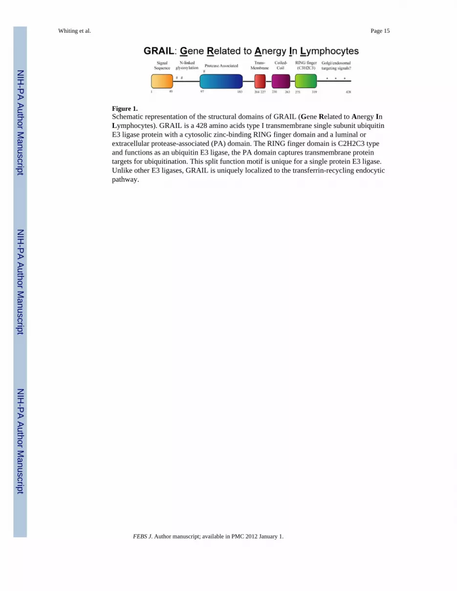
Figure 1.Schematic representation of the structural domains of GRAIL (Gene Related to Anergy InLymphocytes). GRAIL is a 428 amino acids type I transmembrane single subunit ubiquitinE3 ligase protein with a cytosolic zinc-binding RING finger domain and a luminal orextracellular protease-associated (PA) domain. The RING finger domain is C2H2C3 typeand functions as an ubiquitin E3 ligase, the PA domain captures transmembrane proteintargets for ubiquitination. This split function motif is unique for a single protein E3 ligase.Unlike other E3 ligases, GRAIL is uniquely localized to the transferrin-recycling endocyticpathway.
Whiting et al. Page 15
FEBS J. Author manuscript; available in PMC 2012 January 1.
NIH
-PA Author Manuscript
NIH
-PA Author Manuscript
NIH
-PA Author Manuscript

Figure 2.Molecular basis of GRAIL regulation. GRAIL is associated with and regulated by twoisoforms of the ubiquitin-specific protease otubain 1 (Otub1). Otub1, a deubiquitinatingenzyme or DUB, binds to ub-GRAIL but does not deubiquitinate it. USP8, is a DUB thatbinds to GRAIL and to Otub1 in a tri-molecular complex. USP8 can function as a DUB forauto-ubiquitinated GRAIL, however USP8's DUB function is blocked by Otub1, but notcatalytic mutants of Otub1 or it's alternatively spliced isoform, Otub1-ARF1. USP8 must beubiquitinated to function as a DUB for auto-ubiquitinated GRAIL. Otub1, (but not catalyticmutants), de-ubiquitinates ubiquitinated USP8, inactivating it and allowing auto-ubiquitinated. GRAIL to be degraded by the 26S proteosome.
Whiting et al. Page 16
FEBS J. Author manuscript; available in PMC 2012 January 1.
NIH
-PA Author Manuscript
NIH
-PA Author Manuscript
NIH
-PA Author Manuscript
Figure 3.GRAIL and Otub1 regulation by the mTOR pathway controls naïve CD4 T cellproliferation. (left panel) Productive activation of naive CD4 T cells leading to proliferationcomes about through TCR engagement (signal 1) and CD28 costimulation (signal 2); IL-2production, signaling through the IL-2R leading to phosphorylation of Akt and activation ofmTOR, expression of Otub1 protein, and subsequent GRAIL degradation, allowingproliferation to occur. (Right panel) Three independent mechanisms that block mTORactivation result in inhibition of naïve T cell proliferation. CTLA4-Ig blocks CD28costimulation, does not allow IL-2 production, thus prevents Akt phosphorylation, mTOR isinactive, and Otub1 protein is absent, leading to the maintenance of GRAIL, inhibitingproliferation. Anti-IL-2 blocks IL-2R engagement, thus preventing Akt phosphorylation,mTOR is inactive, and Otub1 protein is absent, leading to the maintenance of GRAIL,inhibiting proliferation. Rapamycin blocks the activity of mTOR, prevents proteinexpression of Otub1, leading to the maintenance of GRAIL, inhibiting proliferation.
Whiting et al. Page 17
FEBS J. Author manuscript; available in PMC 2012 January 1.
NIH
-PA Author Manuscript
NIH
-PA Author Manuscript
NIH
-PA Author Manuscript
NIH
-PA Author Manuscript
NIH
-PA Author Manuscript
NIH
-PA Author Manuscript
Whiting et al. Page 18
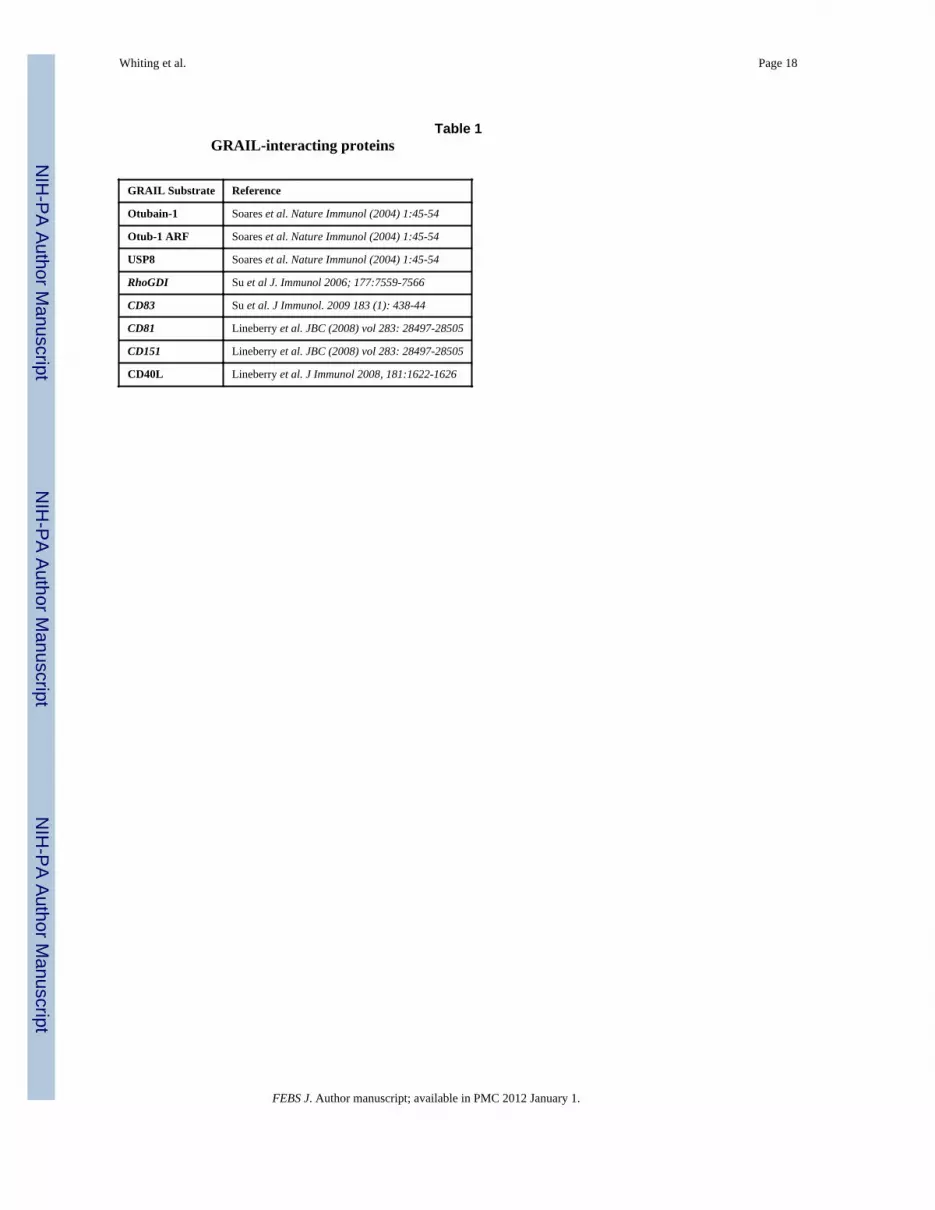
Table 1GRAIL-interacting proteins
GRAIL Substrate Reference
Otubain-1 Soares et al. Nature Immunol (2004) 1:45-54
Otub-1 ARF Soares et al. Nature Immunol (2004) 1:45-54
USP8 Soares et al. Nature Immunol (2004) 1:45-54
RhoGDI Su et al J. Immunol 2006; 177:7559-7566
CD83 Su et al. J Immunol. 2009 183 (1): 438-44
CD81 Lineberry et al. JBC (2008) vol 283: 28497-28505
CD151 Lineberry et al. JBC (2008) vol 283: 28497-28505
CD40L Lineberry et al. J Immunol 2008, 181:1622-1626
FEBS J. Author manuscript; available in PMC 2012 January 1.