Embed Size (px)
Citation preview
letters
NATURE CELL BIOLOGY VOL 5 MAY 2003 www.nature.com/naturecellbiology 433
Translokin is an intracellular mediator ofFGF-2 traffickingCarine Bossard, Henrik Laurell, Loïc Van den Berghe, Sylvain Meunier, Catherine Zanibellato and Hervé Prats*
INSERM U589, IFR 31, Institut Louis Bugnard, CHU Rangueil, Bat L3, 31403 Toulouse, Cedex 04, France*e-mail: [email protected]
Published online: 22 April 2003; DOI: 10.1038/ncb979
Basic fibroblast growth factor (bFGF or FGF-2) exerts itspleiotropic activities both as an exogenous and an intra-cellular factor. FGF-1 and FGF-2 are prototypes for thisdual signalling, but the mechanisms of their intracellularactions remain unknown. Here we show that Translokin, acytoplasmic protein of relative molecular mass 55,000(Mr 55K), interacts specifically with the 18K form ofFGF-2. Translokin is ubiquitously expressed and colocal-izes with the microtubular network. As Translokin does notinteract with FGF-1, we used a strategy based onFGF-1–FGF-2 chimaeras to map the interacting regions inFGF-2 and to generate Nb1a2, a non-interacting variantof FGF-2. Although most of the FGF-2 properties are pre-served in Nb1a2, this variant is defective in intracellulartranslocation and in stimulating proliferation. The fusionof a nuclear localization signal to Nb1a2 restores itsmitogenic activity and its nuclear association. InhibitingTranslokin expression by RNA interference reduces thetranslocation of FGF-2 without affecting the intracellulartrafficking of FGF-1. Our data show that the nuclear asso-ciation of internalized FGF-2 is essential for its mitogenicactivity and that Translokin is important in this transloca-tion pathway.
FGF-2, one of the first described members of the FGF family1, isinvolved in developmental processes, wound healing, angio-genesis and tumour progression2. Five FGF-2 forms of 18K,
22K, 22.5K, 24K and 34K are synthesized through an alternativetranslational initiation process3,4. These isoforms differ only in theiramino terminus, which contains a nuclear localization signal (NLS)in the four high molecular mass (HMM) CUG-initiated forms5, butnot in the smaller AUG-initiated protein of 18K, which is predom-inantly cytoplasmic or excreted and stored in the extracellularmatrix.
Extracellular FGF-2 binds to both high-affinity transmembranetyrosine kinase FGF receptors (FGFRs) and low-affinity receptors(comprising heparan-sulphate-containing proteoglycans)6, therebypromoting the activation of intracellular signalling pathways inwhich mitogen-activated protein kinases (MAPKs) and/or phos-pholipase C are important mediators7. Extracellular FGF-2 caninternalize into the cytoplasm with both types of receptor8,9 andtranslocates into the nucleus during the G1 phase of the cell cycle10
by an unknown mechanism that is distinct from that used byHMM FGF-2 (ref. 11). We previously showed that the three HMMforms of FGF-2 and the intracellular 18K form of FGF-2 are com-ponents of protein complexes of 320K and 130K, respectively12. Tounderstand better the molecular mechanisms by which FGFs exerttheir intracellular effects, here we have used the yeast two-hybridmethod to identify intracellular proteins that interact with FGF-2.
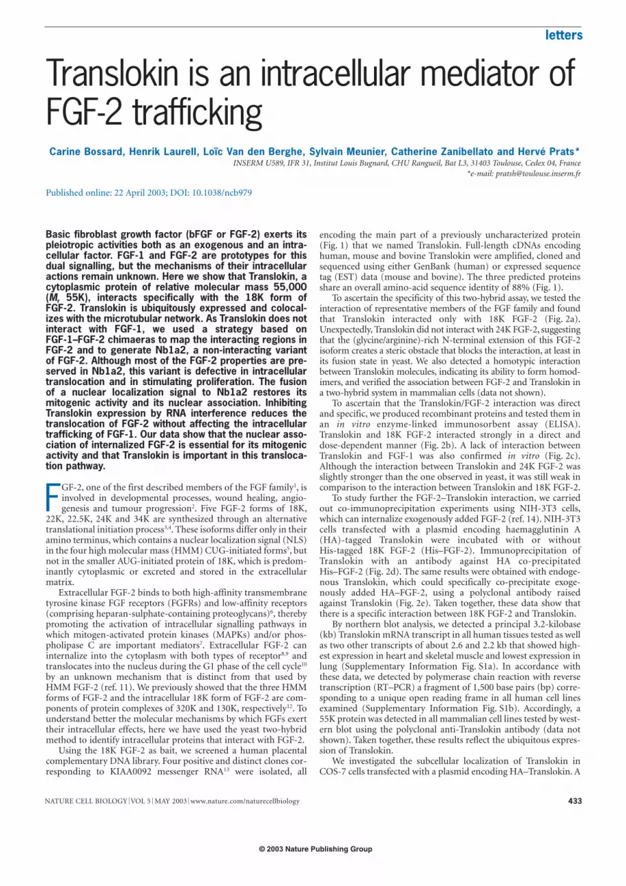
Using the 18K FGF-2 as bait, we screened a human placentalcomplementary DNA library. Four positive and distinct clones cor-responding to KIAA0092 messenger RNA13 were isolated, all
encoding the main part of a previously uncharacterized protein(Fig. 1) that we named Translokin. Full-length cDNAs encodinghuman, mouse and bovine Translokin were amplified, cloned andsequenced using either GenBank (human) or expressed sequencetag (EST) data (mouse and bovine). The three predicted proteinsshare an overall amino-acid sequence identity of 88% (Fig. 1).
To ascertain the specificity of this two-hybrid assay, we tested theinteraction of representative members of the FGF family and foundthat Translokin interacted only with 18K FGF-2 (Fig. 2a).Unexpectedly, Translokin did not interact with 24K FGF-2, suggestingthat the (glycine/arginine)-rich N-terminal extension of this FGF-2isoform creates a steric obstacle that blocks the interaction, at least inits fusion state in yeast. We also detected a homotypic interactionbetween Translokin molecules, indicating its ability to form homod-imers, and verified the association between FGF-2 and Translokin ina two-hybrid system in mammalian cells (data not shown).
To ascertain that the Translokin/FGF-2 interaction was directand specific, we produced recombinant proteins and tested them inan in vitro enzyme-linked immunosorbent assay (ELISA).Translokin and 18K FGF-2 interacted strongly in a direct anddose-dependent manner (Fig. 2b). A lack of interaction betweenTranslokin and FGF-1 was also confirmed in vitro (Fig. 2c).Although the interaction between Translokin and 24K FGF-2 wasslightly stronger than the one observed in yeast, it was still weak incomparison to the interaction between Translokin and 18K FGF-2.
To study further the FGF-2–Translokin interaction, we carriedout co-immunoprecipitation experiments using NIH-3T3 cells,which can internalize exogenously added FGF-2 (ref. 14). NIH-3T3cells transfected with a plasmid encoding haemagglutinin A(HA)-tagged Translokin were incubated with or withoutHis-tagged 18K FGF-2 (His–FGF-2). Immunoprecipitation ofTranslokin with an antibody against HA co-precipitatedHis–FGF-2 (Fig. 2d). The same results were obtained with endoge-nous Translokin, which could specifically co-precipitate exoge-nously added HA–FGF-2, using a polyclonal antibody raisedagainst Translokin (Fig. 2e). Taken together, these data show thatthere is a specific interaction between 18K FGF-2 and Translokin.
By northern blot analysis, we detected a principal 3.2-kilobase(kb) Translokin mRNA transcript in all human tissues tested as wellas two other transcripts of about 2.6 and 2.2 kb that showed high-est expression in heart and skeletal muscle and lowest expression inlung (Supplementary Information Fig. S1a). In accordance withthese data, we detected by polymerase chain reaction with reversetranscription (RT–PCR) a fragment of 1,500 base pairs (bp) corre-sponding to a unique open reading frame in all human cell linesexamined (Supplementary Information Fig. S1b). Accordingly, a55K protein was detected in all mammalian cell lines tested by west-ern blot using the polyclonal anti-Translokin antibody (data notshown). Taken together, these results reflect the ubiquitous expres-sion of Translokin.
We investigated the subcellular localization of Translokin inCOS-7 cells transfected with a plasmid encoding HA–Translokin. A
© 2003 Nature Publishing Group
letters
NATURE CELL BIOLOGY VOL 5 MAY 2003 www.nature.com/naturecellbiology434
monoclonal antibody against HA or a polyclonal antibody againstTranslokin detected a cytoplasmic distribution that completelyoverlapped with the staining of microtubules obtained with anantibody against β-tubulin (Supplementary Information Fig. S1c).
To determine the structural elements in FGF-2 responsible forits interaction with Translokin, we generated a non-interactingmutant of FGF-2 that would also function as a tool for evaluatingthe physiological role of the FGF-2–Translokin interaction. Ourstrategy was based on the fact that Translokin does not interactwith FGF-1, despite the high degree of sequence identity (53%)between FGF-1 and FGF-2 and the close resemblance of theirthree-dimensional structures15. We used a cassette-shufflingapproach to preserve the overall structure of FGF. The chimericFGF-1–FGF-2 proteins were fused with the Gal4 DNA-bindingdomain (DBD), and the ability of the fusion proteins to interactwith Translokin in vivo was examined in the yeast two-hybrid sys-tem (Fig. 3a). We verified the expression of the chimeric proteins bywestern blotting (Fig. 3b).
We found that two separate regions of FGF-2 are required tomaintain the interaction. The first region is located betweenβ-sheets 2 and 5 (compare Na1 with Na2, or Na1b4 with Na2b4).The replacement in FGF-2 of the equivalent amino acids fromFGF-1 (Nb1a2) and the reverse chimaera (Na1b2) showed, respec-tively, the necessity and also the insufficiency of this region. Indeed,a second region positioned between β-sheets 8 and 10 is importantto maintain the interaction (compare Nb2a4 with Nb2a3, Nb3 withNb4, or Na1b3 with Na1b4). The replacement of these two FGF-2regions in FGF-1 generated a chimaera (Na1b2a3b4) that had thesame capacity to interact with Translokin as FGF-2. In the nonin-teracting FGF-2 mutant Nb1a2, the FGF-1-derived sequence wasrestricted to 12 amino acids in the first region. As expected, therecombinant Nb1a2 did not interact in vitro with Translokin in anELISA-based assay (Fig. 3c).
As FGF-2 is a potent mitogen in endothelial cells, we assessedthe mitogenicity of Nb1a2 by proliferation assays on adult bovine
aortic endothelial (ABAE) cells. Nb1a2 showed a very low mito-genic activity that was reduced by roughly 90% as compared withFGF-2 (Fig. 3c). By contrast, Nb1a2 was as efficient as FGF-2 instimulating both tube formation by endothelial cells in Matrigel(Fig. 3c) and cell migration (data not shown). FGF-mediated path-ways require the binding of FGF to both its high-affinity FGFRsand low-affinity heparan-sulphate-containing proteoglycans. Inaddition, phosphorylation and activation of the MAPKs extracellu-lar signal-regulated kinase 1 (ERK1) and ERK2 are required for themitogenic response induced by FGF-2 (refs 7, 16). Activation ofp70S6 kinase is also required for FGF-stimulated cell proliferationbut not for endothelial cell differentiation17. We therefore comparedFGF-2 and Nb1a2 for each of these properties and found no differ-ences between the two proteins (Fig. 3c and data not shown). Takentogether, these results show that the mitogenic and differentiationactivities of FGF-2 are uncoupled by mutations that suppress theFGF-2–Translokin interaction, even though the main signallingpathways involved in the FGF-2–dependent mitogenic response areunaffected.
Exogenously added FGF-2 can translocate to the nucleus of pro-liferating cells10,18,19, and this nuclear translocation is associatedwith a proliferative state of the treated cells10,20. We therefore testedwhether exogenously added Nb1a2 retained the ability of FGF-2 tobe internalized from outside the cell into the cytoplasm and thenfurther translocated to the nucleus. To discriminate betweenendogenous and exogenous FGF-2, we used HA-tagged proteins.After cell collection and fractionation, both HA–FGF-2 andHA–Nb1a2 were detected in the cytoplasmic fraction, whereas onlyHA–FGF-2 was found in the nuclear fraction (Fig. 4a). Takentogether, these results indicate that the loss of mitogenicity owingto mutations disturbing the interaction between FGF-2 andTranslokin is correlated with the inability of the mutant Nb1a2 tobe translocated to the nucleus and/or its vicinity.
To confirm this correlation, we carried out immunofluorescencestudies and confocal analysis of the intracellular movement of
A B
C D
Figure 1 Characterization of Translokin, an FGF-2-interacting protein.Alignment of the predicted amino-acid sequences of human, mouse and bovineTranslokin. The cDNAs were cloned from a human placenta two-hybrid library,
mouse embryos and ABAE (bovine) cells, respectively. The beginning of the arrowsindicate the 5′ end of each clone (clones A–D) isolated by the two-hybrid system.
© 2003 Nature Publishing Group

letters
NATURE CELL BIOLOGY VOL 5 MAY 2003 www.nature.com/naturecellbiology 435
Tran
slokin
a
FGF-
1
FGF-
2/18
K
FGF-
3
FGF-
12
FGF-
6
Translokin
ADDB
FGF-2 (18K)
Contro
l
FGF-
2/24
K
5 2.5 1.25
FGF-2
BSA
20 100
0.5
1
1.5
2
2.5
A49
0
A49
0
b
0
0.5
1
1.5
2
2.5c
62
51
38
26
20
15
HA–Translokind
His–FGF-2
HA–FGF-2
eControl Translokin
HA–FGF-2
His–Translokin (µg ml–1)
FGF-
2/18
KFG
F-1
FGF-
2/24
K
+
+
+ –
– + –
–
– – – – – –
–
+
+ + +
+
+
His–FGF-2
HA–Translokin
IP antibody
Blot: anti-HA
Mr(K)
Figure 2 Specific and direct interaction between Translokin and FGF-2. a, Different FGFs and Translokin were fused to the DNA-binding (DB) or activatingdomains (AD) of Gal4 and subjected to a yeast two-hybrid assay. b, FGF-2 andTranslokin interact directly. FGF-2 and BSA were coated on 96-well plates and incu-bated with different concentrations of His–Translokin. The association was detectedusing an HRP-conjugated antibody against His and read at 490 nm with an ELISAmicroplate reader. Data are the mean ± s.d. of three samples. c, FGF-2 andTranslokin interact specifically. Translokin was coated on 96-well plates and incubat-ed with HA-tagged 18K FGF-2, FGF-1 or 24K FGF-2. The association was detectedusing an antibody against HA and an HRP-conjugated antibody against mouse IgGand determined by ELISA as in b. Data are the mean ± s.d. of three samples. d,Co-immunoprecipitation of HA–Translokin and FGF-2. Mouse NIH-3T3 cells weretransfected or not (−) with a plasmid encoding HA–Translokin and incubated or notwith His-tagged 18K FGF-2. Cell extracts were subjected to immunoprecipitationwith antibodies against HA, followed by immunoblot analysis with antibodies againstHA and His. e, Co-immunoprecipitation of Translokin and FGF-2. Extracts fromNIH-3T3 cells stimulated (+) or not (−) with HA-tagged 18K FGF-2 were subjected toimmunoprecipitation with antibodies against Translokin or a control antibody, fol-lowed by immunoblot analysis with antibodies against HA.
0 15050 100
a
b
c
Nb2a4
Nb1a2
Nb2a3
Nb1a4
Na1b4
Na1b3
Nb1a3
Na1b2
Nb3FGF-1
FGF-
2
Nb1Na1 Nb2 Nb4
Na2 Na2b4
Na1b2
a3b4
Contro
l
0
25
50
75
100
β10β8β5β2
FGF-2
FGF-1
Nb1a4
Na1b4
Na1b3
Nb1a3
Na1b2
Na2
Na1
Nb2a3
Nb2a4
Nb2
Nb3
Nb4
Nb1
Na2b4
Na1b2-a3b4
Nb1a2
0 15050 100
26
38
6150
85
Mr(K)
%
FGF-2
Nb1a2
Control
β-gal activity(percentage of
FGF-2–Translokininteraction)
Prol
ifera
tion
activ
ity
Tubu
loge
nesi
sac
tivity
Rec
epto
rbi
ndin
g
Tran
slok
inin
tera
ctio
n
Figure 3 Characterization of FGF-2–FGF-1 chimaeras. a, Two-hybrid system.The relative intensity of the interactions between the FGF-1–FGF-2 chimaeras andTranslokin was measured by β-galactosidase activity in yeast extracts. Four junc-tions were chosen in β-strands 2, 5, 8 and 10. FGF-1 regions are shown in whiteand FGF-2 regions in black. Nomenclature is based on the origin of the N-terminalextremity: ‘a’ for acidic FGF (FGF-1) and ‘b’ for basic FGF (FGF-2). Numbers indicatethe junctions between FGF-1 and FGF-2. Data are the mean ± s.e.m from at leastthree independent transformations. β-Galactosidase assays were done in duplicate.b, Analysis of the expression of chimaeras fused to the Gal4 DBD. Yeast extractswere subjected to immunoblotting and detected with a monoclonal antibody againstthe Gal4 DBD. c, Comparison of the properties of FGF-2 and Nb1a2. The two pro-teins were compared for their in vitro interaction with Translokin, proliferation activi-ty, differentiation activity and capacity to compete for binding to the FGFR(Methods). Results are expressed as the percentage of FGF-2 activity.
© 2003 Nature Publishing Group

letters
NATURE CELL BIOLOGY VOL 5 MAY 2003 www.nature.com/naturecellbiology436
Nucleus
Cytoplasm
100 0
HA–FGF-2 20
20(ng ml–1)
HA–Nb1a2
a
b Merge
10µm
0
0
0 0
β-actin orβ-tubulin Anti-HA
ChromomycinA3
Control
FGF-2
Nb1a2
FGF-2+ nocodazole
FGF-2(β-actin)
FGF-2+ cyto B(β-actin)
© 2003 Nature Publishing Group
letters
NATURE CELL BIOLOGY VOL 5 MAY 2003 www.nature.com/naturecellbiology 437
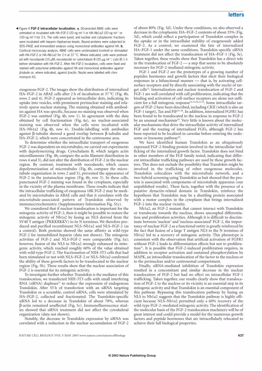
exogenous FGF-2. The images show the distribution of internalizedHA–FGF-2 in ABAE cells after 2 h of incubation at 37 °C (Fig. 4b,rows 2 and 4). FGF-2 appeared as intracellular dots, indicating itsuptake into vesicles, with prominent perinuclear staining and rela-tively sparse nuclear staining. The staining obtained with antibod-ies against HA was specific, because no staining was observed whenFGF-2 was omitted (Fig. 4b, row 1). In agreement with the dataobtained by cell fractionation (Fig. 4a), no nuclear-associatedstaining was observed when the cells were incubated withHA–Nb1a2 (Fig. 4b, row 6). Double-labelling with antibodiesagainst β-tubulin showed a good overlap between β-tubulin andHA–FGF-2, which were concentrated in the centrosome area.
To determine whether the intracellular transport of exogenousFGF-2 was dependent on microtubules, we carried out experimentswith depolymerizing drugs. Cytochalasin B, which targets actinmicrofilaments (Fig. 4b, compare the actin filament distribution inrows 4 and 5), did not alter the distribution of FGF-2 in the nuclearregion. By contrast, treatment with nocodazole, which causesdepolymerization of microtubules (Fig. 4b, compare the micro-tubule organization in rows 2 and 5), prevented the appearance ofFGF-2 in the juxtanuclear region (Fig. 4b, row 3). In these cells,punctuated FGF-2 staining was found in the cytoplasm, especiallyin the vicinity of the plasma membrane. These results indicate thatthe intracellular trafficking of exogenous 18K FGF-2 may be medi-ated by microtubules in ABAE cells and that it correlates with themicrotubule-associated pattern of Translokin observed byimmunocytochemistry (Supplementary Information Fig. S1c).
We considered that if nuclear translocation is important for themitogenic activity of FGF-2, then it might be possible to restore themitogenic activity of Nb1a2 by fusing an NLS derived from theSV40 T antigen (PKKKRKKV) to its N terminus. We therefore pro-duced and purified recombinant NLS–Nb1a2 and NLS–FGF-2 (asa control). Both proteins showed the same affinity as wild-typeFGF-2 for immobilized heparin (data not shown). The mitogenicactivities of FGF-2 and NLS-FGF-2 were comparable (Fig. 5a);however, fusion of the NLS to Nb1a2 strongly enhanced its mito-genic activity, which reached roughly 60% of the value obtainedwith wild-type FGF-2. The fractionation of NIH-3T3 cells that hadbeen stimulated or not with NLS–FGF-2 or NLS–Nb1a2 confirmedthe ability of these growth factors to be translocated to the nuclearregion (Fig. 5b). These results show that the nuclear association ofFGF-2 is essential for its mitogenic activity.
To investigate further whether Translokin is the mediator of thistranslocation, we transfected NIH-3T3 cells with small interferingRNA (siRNA) duplexes21 to reduce the expression of endogenousTranslokin. After 55 h of transfection with an siRNA targetingTranslokin or a scramble, control siRNA, cells were stimulated byHA–FGF-2, collected and fractionated. The Translokin-specificsiRNA led to a decrease in Translokin of about 78%, whereasβ-actin remained unaffected (Fig. 5c). Immunofluorescence stud-ies showed that siRNA treatment did not affect the cytoskeletalorganization (data not shown).
Notably, the decrease in Translokin expression by siRNA wascorrelated with a reduction in the nuclear accumulation of FGF-2
of about 80% (Fig. 5d). Under these conditions, we also observed adecrease in the cytoplasmic HA–FGF-2 contents of about 35% (Fig.5d), which could reflect a participation of Translokin complex inthe uptake or in the intracellular stability of exogenously addedFGF-2. As a control, we examined the fate of internalizedHA–FGF-1 under the same conditions. Translokin-specific siRNAtreatment did not affect the translocation of HA–FGF-1 (Fig. 5e).Taken together, these results show that Translokin has a direct rolein the translocation of FGF-2 a step that seems to be absolutelynecessary for FGF-2-mediated mitogenic activity.
FGF-1 and FGF-2 are the prototypes of a growing number ofpeptides hormones and growth factors that elicit their biologicalresponses in a bifunctional manner that is, by activating cell-surface receptors and by directly associating with the nuclei of tar-get cells22. Internalization and nuclear translocation of FGF-2 andFGF-1 are well correlated with cell proliferation, indicating that thebinding and activation of cell-surface receptors are alone not suffi-cient for a full mitogenic response10,14,20,23,24. Some intracellular tar-gets of FGF-2 have been described, including CKII (which is also anFGF-1 target), Tax and FIF25–28. In addition, internalized FGFR1 hasbeen found to be translocated to the nucleus in response to FGF-2by an unusual mechanism29. Very little is known about the molec-ular mechanisms that drive the intracellular activity of internalizedFGF and the routing of internalized FGFs, although FGF-2 hasbeen reported to be localized in caveolae before entering the endo-some or lysosome pathway30.
We have identified human Translokin as an ubiquitouslyexpressed FGF-2-binding protein involved in the intracellular traf-ficking of the internalized growth factor. Translokin does not bindto other members of the FGF family tested, indicating that differ-ent intracellular trafficking pathways are used by these growth fac-tors. This does not exclude the possibility that Translokin might beinvolved in the trafficking of other exogenous molecules.Translokin colocalizes with the microtubule network, and atwo-hybrid screening using Translokin as bait showed that the pro-tein is associated with components of microtubule apparatus (ourunpublished results). These facts, together with the presence of aputative dynactin-related domain in Translokin, reinforce thehypothesis that Translokin may be a shuttling protein associatedwith a motor complex in the cytoplasm that brings internalizedFGF-2 into the nuclear vicinity.
Nb1a2, an FGF-2 mutant that cannot interact with Translokinor translocate towards the nucleus, shows uncoupled differentia-tion and proliferation activities. Although it is difficult to discrim-inate between ‘nuclear’ and ‘nucleus-associated’ FGF-2, the impor-tance of nuclear FGF-2 as a functional entity is greatly reinforced bythe fact that fusion of a large T antigen NLS to the N terminus ofNb1a2 led to a recovery of mitogenic activity. This phenotype isconsistent with the observation that artificial activation of FGFR1without FGF-2 leads to differentiation effects but not to prolifera-tion31. It is possible that FGF-2-induced proliferation requires, inaddition to receptor activation and sustained phosphorylation byMAPK, an intracellular translocation of the factor to the nucleus orto the perinuclear and/or centrosomal compartment.
Finally, siRNA-mediated inhibition of Translokin expressionresulted in a concomitant and similar decrease in the nucleartranslocation of FGF-2 but had no effect on intracellular FGF-1trafficking. Taken together, our results clearly show that transloca-tion of FGF-2 to the nucleus or its vicinity is an essential step in itsmitogenic activity and that Translokin is an essential component ofthis pathway. Bypassing this translocation pathway by fusing anNLS to Nb1a2 suggests that the Translokin pathway is highly effi-cient because NLS–Nb1a2 permitted only a 60% recovery of thewild-type FGF-2–mediated mitogenic activity. The identification ofthe molecular basis of the FGF-2 translocation machinery will be ofgreat interest and could provide a model for the numerous growthfactors and peptide hormones that are intracellularly relocated toachieve their full biological properties.
Figure 4 FGF-2 intracellular localization. a, G0-arrested ABAE cells wereuntreated or incubated with HA–FGF-2 (20 ng ml−1) or HA–Nb1a2 (20 ng ml−1 or100 ng ml−1) for 2 h. The cells were lysed, and nuclear and cytoplasmic fractionswere incubated with heparin–Sepharose beads. Bound proteins were subjected toSDS–PAGE and immunoblot analysis using monoclonal antibodies against HA. b,Confocal microscopy analysis. ABAE cells were unstimulated (control) or stimulatedwith HA–FGF-2 or HA–Nb1a2 for 2 h at 37 °C. Where indicated, cells were pretreat-ed with nocodazole (10 µM; nocodazole) or cytochalasin B (20 µg ml−1; cyto B) 1 hbefore stimulation with HA–FGF-2. After HA–FGF-2 incubation, cells were fixed andstained with polyclonal antibodies against HA and monoclonal antibodies againstβ-tubulin or, where indicated, against β-actin. Nuclei were labelled with chro-momycin A3.
�
© 2003 Nature Publishing Group

letters
NATURE CELL BIOLOGY VOL 5 MAY 2003 www.nature.com/naturecellbiology438
MethodsYeast two-hybrid systemThe two-hybrid expression-cloning system was done as described28. In brief, the 18K form of FGF-2
was used as a bait, and the corresponding cDNA was inserted in the pAS2 vector. We screened a
human placenta library (Clontech, Palo Alto, CA) cloned into the pACT2 vector in Saccharomyces cere-
visiae strain Y190. β-Galactosidase activity in yeast extracts from at least ten pooled colonies, grown
overnight in 4 ml of selection medium (DOB-Trp/-Leu), was measured with Galacto-Light (TROPIX,
Bedford, MA) in accordance with the manufacturer’s instructions.
RT–PCRTotal RNA was extracted from culture cells or mouse organs with RNAble (Eurobio, Les Ulis,
France) and reverse-transcribed with SuperScript II (Invitrogen, Groningen, The Netherlands) in
accordance with the manufacturer’s instructions. Translokin-specific oligonucleotides were used
for both RT and PCR. We carried out automatic nucleotide sequencing using AmpliTaq FS poly-
merase (Applied Biosystems, Warrington, UK) and an ABI 373A sequencer (Perkin Elmer, Foster
City, CA).
Protein production and purificationWe purified tagged and untagged FGF-2 and Nb1a2, HA–FGF-1, NLS–FGF-2 and NLS–Nb1a2 on a
heparin–Sepharose column by fast protein liquid chromatography (FPLC; Amersham Pharmacia
Biotech, Orsay, France) as described11. HA–FGF-2 was a gift from G. Bouche (IPBS, Toulouse, France).
His–Translokin was produced using the isopropylthiogalactoside (IPTG)-inducible pET expression
system (pET-15b, Novagen, Madison, WI) in Escherichia coli BL21 (DE3) pLysS and purified on a nick-
el–agarose column (Qiagen, Chatsworth, CA). We used 20 mM and 200 mM imidazole for equilibra-
tion and elution, respectively. Protein concentrations were determined by bicinchoninic assay (Pierce,
Rockford, IL). We obtained 35S-labelled FGF-2 by using an in vitro translation system. A plasmid
encoding FGF-2 was linearized, transcribed in vitro and translated in a rabbit reticulocyte lysate system
(Promega, Madison, WI) in the presence of 35S-labelled methionine (Amersham Pharmacia Biotech) as
described32. After translation, the lysate containing 35S-labelled FGF-2 was dialysed against dialysis
buffer (20 mM HEPES (pH 7.0), 140 mM NaCl, 2 mM CaCl2) to remove free 35S-labelled methionine
and reducing agents.
AntibodiesPolyclonal antibodies against Translokin were generated through the immunization of two rabbits
injected three times over 2 weeks and then 1 month later with 200 µg of recombinant His–Translokin
that had been electroeluted from a polyacrylamide gel and diluted twice in Freund’s adjuvant
(Sigma-Aldrich, St Louis, MO). We purified the antiserum with His–Translokin linked to Affigel-10
(Bio-Rad, Hercules, CA) as described28. Polyclonal and monoclonal antibodies against HA (HA.11)
were from Babco (Eurogentec, Herstal, Belgium); monoclonal antibodies against β-actin (AC-15) and
β-tubulin (TUB 2.1), and peroxidase-labelled antibodies against His were from Sigma; monoclonal
antibodies against FGF-2 (Ab3) were from Oncogene Science (Boston, MA); horseradish peroxidase
(HRP)-conjugated antibodies were from Amersham; and the fluorescein isothiocyanate (FITC) or
Cy3-conjugated secondary antibodies were from Sigma-Aldrich. The monoclonal antibody against the
Gal4 DBD were from Clontech. Chromomycin A3 was from Sigma-Aldrich.
In vitro interactionA 96-well plate was coated overnight at 4 °C with 10 µg ml−1 FGF-2, Nb1a2 or bovine serum albumin
(BSA). The plate was washed with PBS containing 0.2% Tween and 100 µl per well of blocking solu-
tion was added for 1 h at 37 °C. We discarded the blocking solution and washed the plates, before
adding different concentrations of His–Translokin for 2 h at 37 °C. After three washings, HRP-conju-
gated antibody against His (1:1,000 dilution) was added for 1 h at 37 °C. We washed the plates three
times and detected the complexes with 100 µl per well of o-phenylenediamine (4 mg ml−1) containing
0.03% (v/v) hydrogen peroxide in 0.1 M citrate buffer (pH 5.0). After 20 min, the reactions were
stopped with 50 µl of 2 M H2SO4 and the absorbance was measured at 490 nm. For the reverse experi-
ment, we coated the plate with Translokin and added HA-tagged proteins. Incubation with monoclon-
al antibodies against HA (dilution 1:1,000), followed by incubation with an HRP-conjugated antibody
against mouse IgG (dilution 1:1,000), was used to detect the interactions.
ImmunoprecipitationNIH-3T3 cells were transiently transfected with a plasmid encoding full-length Translokin with an HA
tag. After 48 h, the cells were incubated for 2 h with 60 ng ml−1 His–FGF-2. The cells were subsequently
rinsed in cold PBS, scraped, pelleted and lysed on ice in 50 mM Tris (pH 7.4), 150 mM NaCl, 1%
Nonidet P-40 and protease inhibitors for 30 min. We removed cell debris by centrifugation at 10,000g
for 10 min at 4 °C. The extracts were incubated with a monoclonal antibody against HA coupled to
a
30
00 2 4 6 8 10
5
10
15
20
25 FGF-2
b
HA–FGF1
e
d
c
siRNA:
HA–FGF-2
Cel
l num
ber ×
10–
4
GF concentration (ng ml–1)
NLS–FGF-2
NLS–Nb1a2
Nb1a2
Scra
mbl
e
Tran
slok
in
Translokin
β-actin
Nuclear
Cytoplasmic
0
20
40
60
80
Nuclear
Cytoplasmic
Tran
slok
inRes
idua
l pro
tein
con
tent
s(p
erce
ntag
e)
FGF-
2
HA–NLS–FGF-2HA–NLS–Nb1a2
–
– –
– –
–
–––+
+ +
+
+
+
Cytoplasm
Nucleus
siRNA Scramble
siRNA Translokin
Nuclear HA–FGF-1
Cytoplasmic HA–FGF-1
Figure 5 Recovery of Nb1a2 mitogenic activity and inhibition of FGF-2. a, Proliferation assay. ABAE cells were stimulated by the indicated concentrationsof FGF-2, NLS–FGF-2, Nb1a2 or NLS–Nb1a2 on days 1 and 3. On day 5, cells weretrypsinized and counted. The experiment was repeated at least three times withsimilar results, using recombinant proteins from two independent productions. b,HA-tagged NLS–FGF-2 and NLS–Nb1a2 are translocated to the nucleus. After stimu-lation with (+) or without (−) 60 ng ml−1 NLS-tagged growth factors, NIH-3T3 cellswere treated and fractionated as in Fig. 4a. Fractions were subjected to SDS–PAGEand immunoblot analysis with antibodies against HA. c, Translokin expression inNIH-3T3 cells transfected with the indicated siRNAs was detected by immunoblotanalysis with antibodies against Translokin (top). β-Actin was monitored as a loading
control (bottom). d, Translokin siRNA inhibits FGF-2 translocation. NIH-3T3 cells,transfected with the indicated siRNAs, were stimulated with 20 ng ml−1 HA–FGF-2for 2 h. The cells were treated and fractionated as in Fig. 4a. Fractions were sub-jected to SDS–PAGE and immunoblot analysis with antibodies against HA (left).Protein was quantified by densitometry and is normalized to expression in cellstransfected with control siRNA (right). Values are the mean ± s.d. of three experi-ments. e, Translokin siRNA does not affect FGF-1 translocation. NIH-3T3 cells trans-fected with the indicated siRNAs were unstimulated or stimulated with 60 ng ml−1
HA–FGF-1 and 10 U ml−1 of heparin for 5 h. The cells were treated and analysed asin d.
© 2003 Nature Publishing Group

letters
NATURE CELL BIOLOGY VOL 5 MAY 2003 www.nature.com/naturecellbiology 439
protein-G–Sepharose beads (Sigma) for 2 h at 4 °C. The beads were washed three times in lysis buffer
and immobilized proteins were released by boiling in Laemmli buffer and analysed by SDS–PAGE.
Cell cultureCells were grown in Dulbecco’s modified Eagle’s medium (DMEM; Invitrogen) containing antibiotics,
1% glutamine and serum. For COS-7 cells, the medium contained 1 g l−1 glucose and 5% fetal calf
serum (FCS). We grew NIH-3T3 and ABAE cells in DMEM (4 g l−1 glucose) with 10% FCS or 10% calf
serum, respectively. All cell transfections with plasmid DNA were made using Fugene 6 (Roche
Diagnostics, Mannheim, Germany) in accordance with the manufacturer’s recommendations.
Transfection of mammalian cells with siRNA duplexesThe 21-nucleotide siRNA duplexes were synthesized and purified by Dharmacon Research (Boulder,
CO). The siRNA sequence targeting mouse Translokin corresponded to the coding region 463–483 rel-
ative to the first nucleotide of the start codon. The control was a scramble siRNA (Dharmacon
Research). Transfection was done with 0.1 µM of siRNA and oligofectamine reagent (Invitrogen) in
accordance with the manufacturer’s instructions.
Because the translocation of FGF-2 is dependent on the cell cycle and on the confluence state of the
cells, we took great care when seeding the cells. In accordance with the putative role of Translokin in
FGF-2-induced proliferation, NIH-3T3 cells transfected with Translokin-targeted siRNA grew more
slowly than cells transfected with control siRNA (data not shown) and were therefore seeded at a
slightly higher density to obtain the same number of cells in both conditions at the time the
HA–FGF-2 was added.
In situ immunocytochemistryCOS-7 cells, grown on coverslips, were transfected and prepared for immunofluorescence
microscopy as described4. ABAE cells, grown on coverslips, were serum-starved for 48 h and stimu-
lated with 20 ng ml−1 HA–FGF or HA–Nb1a2 for 2 h. In some experiments, cells were treated before
stimulation with 10 µM nocodazole or 20 µg ml−1 cytochalasin B (Sigma) for 1 h.
Immunocytochemistry was done as described10 using polyclonal antibodies against HA and mono-
clonal antibodies against β-tubulin as primary antibodies, and Cy3-conjugated antibodies against
rabbit IgG (dilution 1:250) and FITC-conjugated antibodies against mouse IgG (dilution 1:50) as
secondary antibodies (Sigma). We labelled nuclei with chromomycin A3 (0.1 mg ml−1). Antibody
complexes were visualized under a Leica fluorescence microscope or a Zeiss LSM510 confocal micro-
scope with a 63× oil objective lens and aperture of 1.4.
Tubulogenesis (differentiation assay)To measure ABAE cell organization on a basement membrane, 24-well plates were coated with 300 µl
per well of Matrigel (B&D, Bedford, MA), which was allowed to polymerize for 1 h at 37 °C. ABAE
cells (2 × 10⁵ cells per ml) suspended in 500 µl of culture medium were then added to each well. We
added FGF-2 or Nb1a2 to the wells at 0.5 ng ml−1 and incubated the plates overnight at 37 °C. After
removing the medium, we fixed the culture and quantified the length of the tube network with the Q
Win Leica system (Lecia Microsystems, Rueil-Malmaison, France).
ABAE fractionationWe incubated 50% confluent cells, rendered quiescent after 48 h in serum-free conditions, for 2 h with
20 or 60 ng ml−1 of HA-tagged proteins. The cells were collected and fractionated as described10. The
purity and integrity of preparations of nuclei (before sonication) were monitored by phase contrast
microscopy and by the quantitative analysis of the cytoplasmic lactate dehydrogenase content.
Comparison between FGF-2 and Nb1a2For the proliferation assay, ABAE cells were stimulated without (control) or with 2 ng ml−1 FGF-2 or
Nb1a2 added on day 1 and 3. On day 5 cells were trypsinized and counted with a Coulter counter ZM
(Beckman Coulter, Villepinte, France). To measure the relative receptor binding affinity, confluent
ABAE cells were incubated for 2 h at 4 °C with 35S-labelled FGF-2 and 10 µg of unlabelled FGF-2 or
Nb1a2. Cells were then treated as described33. Reported values represent the relative capacity of FGF-2
or Nb1a2 to compete with the specific binding of 35S-labelled FGF-2 to FGFRs. A heat-denatured com-
petitor was used as a control.
GenBank accession numbersHuman Translokin, AY225092; mouse Translokin, AY225093; bovine Translokin, AY225094.
RECEIVED 19 JULY 2002; RECEIVED 26 DECEMBER 2002; ACCEPTED 10 FEBRUARY 2003,PUBLISHED 22 APRIL 2003.
1. Ornitz, D. M. & Itoh, N. Fibroblast growth factors. Genome Biol. 2, 3005.1–3005.12 (2001).
http://genomebiology.com/2001/2/3/reviews/3005
2. Bikfalvi, A., Klein, S., Pintucci, G. & Rifkin, D. B. Biological roles of fibroblast growth factor-2.
Endocr. Rev. 18, 26–45 (1997).
3. Prats, H. et al. High molecular mass forms of basic fibroblast growth factor are initiated by alterna-
tive CUG codons. Proc. Natl Acad. Sci. USA 86, 1836–1840 (1989).
4. Arnaud, E. et al. A new 34-kilodalton isoform of human fibroblast growth factor 2 is cap depend-
ently synthesized by using a non-AUG start codon and behaves as a survival factor. Mol. Cell. Biol.
19, 505–514 (1999).
5. Bugler, B., Amalric, F. & Prats, H. Alternative initiation of translation determines cytoplasmic or
nuclear localization of basic fibroblast growth factor. Mol. Cell. Biol. 11, 573–577 (1991).
6. Johnson, D. E. & Williams, L. T. Structural and functional diversity in the FGF receptor multigene
family. Adv. Cancer Res. 60, 1–41 (1993).
7. Cross, M. J. & Claesson-Welsh, L. FGF and VEGF function in angiogenesis: signalling pathways,
biological responses and therapeutic inhibition. Trends Pharmacol. Sci. 22, 201–207 (2001).
8. Reiland, J. & Rapraeger, A. C. Heparan sulfate proteoglycan and FGF receptor target basic FGF to
different intracellular destinations. J. Cell Sci. 105, 1085–1093 (1993).
9. Roghani, M. & Moscatelli, D. Basic fibroblast growth factor is internalized through both
receptor-mediated and heparan sulfate-mediated mechanisms. J. Biol. Chem. 267, 22156–22162 (1992).
10. Baldin, V., Roman, A. M., Bosc-Bierne, I., Amalric, F. & Bouche, G. Translocation of bFGF to the
nucleus is G1 phase cell cycle specific in bovine aortic endothelial cells. EMBO J. 9, 1511–1517
(1990).
11. Patry, V., Arnaud, E., Amalric, F. & Prats, H. Involvement of basic fibroblast growth factor NH2 ter-
minus in nuclear accumulation. Growth Factors 11, 163–174 (1994).
12. Patry, V., Bugler, B., Maret, A., Potier, M. & Prats, H. Endogenous basic fibroblast growth factor iso-
forms involved in different intracellular protein complexes. Biochem. J. 326, 259–264 (1997).
13. Nagase, T. et al. Prediction of the coding sequences of unidentified human genes. III. The coding
sequences of 40 new genes (KIAA0081–KIAA0120) deduced by analysis of cDNA clones from
human cell line KG-1. DNA Res. 2, 37–43 (1995).
14. Bailly, K., Soulet, F., Leroy, D., Amalric, F. & Bouche, G. Uncoupling of cell proliferation and differ-
entiation activities of basic fibroblast growth factor. FASEB J. 14, 333–344 (2000).
15. Zhu, X. et al. Three-dimensional structures of acidic and basic fibroblast growth factors. Science
251, 90–93 (1991).
16. Pages, G. et al. Mitogen-activated protein kinases p42mapk and p44mapk are required for fibroblast
proliferation. Proc. Natl Acad. Sci. USA 90, 8319–8323 (1993).
17. Kanda, S. et al. Phosphatidylinositol 3′-kinase-independent p70S6 kinase activation by fibroblast
growth factor receptor-1 is important for proliferation but not differentiation of endothelial cells. J.
Biol. Chem. 272, 23347–23353 (1997).
18. Bouche, G. et al. Basic fibroblast growth factor enters the nucleolus and stimulates the transcrip-
tion of ribosomal genes in ABAE cells undergoing G0–G1 transition. Proc. Natl Acad. Sci. USA 84,
6770–6774 (1987).
19. Clarke, W. E., Berry, M., Smith, C., Kent, A. & Logan, A. Coordination of fibroblast growth factor
receptor 1 (FGFR1) and fibroblast growth factor-2 (FGF-2) trafficking to nuclei of reactive astro-
cytes around cerebral lesions in adult rats. Mol. Cell. Neurosci. 17, 17–30 (2001).
20. Joy, A. et al. Nuclear accumulation of FGF-2 is associated with proliferation of human astrocytes
and glioma cells. Oncogene 14, 171–183 (1997).
21. Elbashir, S. M. et al. Duplexes of 21-nucleotide RNAs mediate RNA interference in cultured mam-
malian cells. Nature 411, 494–498 (2001).
22. Keresztes, M. & Boonstra, J. Import(ance) of growth factors in(to) the nucleus. J. Cell Biol. 145,
421–424 (1999).
23. Imamura, T. et al. Recovery of mitogenic activity of a growth factor mutant with a nuclear translo-
cation sequence. Science 249, 1567–1570 (1990).
24. Wiedlocha, A., Falnes, P. O., Madshus, I. H., Sandvig, K. & Olsnes, S. Dual mode of signal transduc-
tion by externally added acidic fibroblast growth factor. Cell 76, 1039–1051 (1994).
25. Bonnet, H. et al. Fibroblast growth factor-2 binds to the regulatory β subunit of CK2 and directly
stimulates CK2 activity toward nucleolin. J. Biol. Chem. 271, 24781–24787 (1996).
26. Skjerpen, C. S., Nilsen, T., Wesche, J. & Olsnes, S. Binding of FGF-1 variants to protein kinase CK2
correlates with mitogenicity. EMBO J. 21, 4058–4069 (2002).
27. Shen, B., Arese, M., Gualandris, A. & Rifkin, D. B. Intracellular association of FGF-2 with the ribo-
somal protein L6/TAXREB107. Biochem. Biophys. Res. Commun. 252, 524–528 (1998).
28. Van den Berghe, L. et al. FIF [fibroblast growth factor-2 (FGF-2)-interacting-factor], a nuclear
putatively antiapoptotic factor, interacts specifically with FGF-2. Mol. Endocrinol. 14, 1709–1724
(2000).
29. Reilly, J. F. & Maher, P. A. Importin beta-mediated nuclear import of fibroblast growth factor recep-
tor: role in cell proliferation. J. Cell Biol. 152, 1307–1312 (2001).
30. Gleizes, P. E., Noaillac-Depeyre, J., Dupont, M. A. & Gas, N. Basic fibroblast growth factor (FGF-2)
is addressed to caveolae after binding to the plasma membrane of BHK cells. Eur. J. Cell Biol. 71,
144–153 (1996).
31. Kudla, A. J. et al. The FGF receptor-1 tyrosine kinase domain regulates myogenesis but is not suffi-
cient to stimulate proliferation. J. Cell Biol. 142, 241–250 (1998).
32. Huez, I., Bornes, S., Bresson, D., Creancier, L. & Prats, H. New vascular endothelial growth factor
isoform generated by internal ribosome entry site-driven CUG translation initiation. Mol.
Endocrinol. 15, 2197–2210 (2001).
33. Estival, A. et al. Differential regulation of fibroblast growth factor (FGF) receptor-1 mRNA and
protein by two molecular forms of basic FGF. Modulation of FGFR-1 mRNA stability. J. Biol.
Chem. 271, 5663–5670 (1996).
ACKNOWLEDGEMENTS
We thank G. Bouche for HA–FGF-2 plasmid; D. Bresson and B. Knibiehler for technical advice; and B.
Bugler, C. Touriol, A. C. Prats and F. Bayard for discussions and critically reading the manuscript. C.B.
was supported by the Association pour la Recherche contre le Cancer; H.L. was supported by an
INSERM fellowship. This work was supported by the Association pour la Recherche contre le Cancer
and the Ligue Nationale contre le Cancer.
Correspondence and requests for materials should be addressed to H.P.
COMPETING FINANCIAL INTERESTS
The authors declare that they have no competing financial interests.
© 2003 Nature Publishing Group


![FIF [Fibroblast Growth Factor - 2 (FGF-2)-Interacting-Factor], a Nuclear Putatively Antiapoptotic Factor, Interacts Specifically with FGF-2](https://img.dokumen.tips/doc/110x75/634d13ddb5aff40b380edb23/fif-fibroblast-growth-factor-2-fgf-2-interacting-factor-a-nuclear-putatively.jpg)