Embed Size (px)
Citation preview
UNIVERSITÉ DE STRASBOURG
ÉCOLE DOCTORALE DES SCIENCES DE LA VIE ET DE LA SANTÉ
IGBMC-CNRS UMR 7104-INSERM U 964
THÈSE présentée par :
Baptiste BIDON
soutenue le : 03 Novembre 2017
pour obtenir le grade de : Docteur de l’université de Strasbourg Discipline/ Spécialité : Biologie/Aspects moléculaires et cellulaires de la biologie
Mediator and NER factors in transcription initiation
Médiateur et facteurs NER lors de l’initiation de la transcription
THÈSE dirigée par :
Dr EGLY Jean-Marc DR INSERM, IGBMC, Illkirch Dr COIN Frédéric DR INSERM, IGBMC, Illkirch
RAPPORTEURS :
Dr DURR Alexandra PUPH, Hôpital Pitié Salpêtrière, Paris Dr BULAVIN Dmitry DR INSERM, IRCAN, Nice
AUTRES MEMBRES DU JURY : Dr WEIXLBAUMER Albert CR INSERM, IGBMC, Illkirch Dr BORG Jean-Paul PUPH, Institut Paoli-Calmettes, Marseille

1
Acknowledgments
I am grateful to Mrs Alexandra Durr, Mr Dmitry Bulavin, Mr Albert Weixlbaumer
and Mr Jean-Paul Borg for examining and reviewing my thesis.
I am thankful to Mr Jean-Marc Egly and Mr Frédéric Coin to have given me the
opportunity to work on such exciting projects.
I am largely obliged to Mr Jean-Marc Egly and Mr Nicolas Le May for all the
scientific work and experiments (functioning or not) that have been achieved and for
discussions we had. Thank you…
I do have a special thought to 4-0-2-5 members where I spend the last four
years. Not only science is about sharing and this PhD would not have been possible
without Lise-Marie, Cathy, Philippe, Emmanuel and Carlos.
I am also thankful to all members of Egly/Coin’s lab for technical support,
conversation, time or just the beer we shared. Grazie mille…
Thank you to my parents to have raised me as a free spirit.
2
Summary
Acknowledgments............................................................................................................1
Summary...............................................................................................................................2
Tables and Figures...........................................................................................................5
Abbreviations.....................................................................................................................6
INTRODUCTION.................................................................................................................9I- RNApolymeraseIImediatedtranscription............................................................101) TheRNApolymeraseII..............................................................................................................102) Promoter..........................................................................................................................................121. Organisation................................................................................................................................................122. Structure.......................................................................................................................................................132.a. Initiator(Inr).....................................................................................................................................132.b. Xgenecorepromoterelement(XCPE1/2)..........................................................................142.c. TATAbox.............................................................................................................................................142.d. TFIIBRecognitionElements(BRE).........................................................................................142.e. DownstreamPromoterElement(DPE).................................................................................152.f. MotifTenElement(MTE).............................................................................................................152.g. DownstreamCoreElement(DCE)............................................................................................15
3) Pre-initiationcomplex................................................................................................................161. Generaltranscriptionfactors..............................................................................................................161.a. TFIIA......................................................................................................................................................171.b. TFIIB.....................................................................................................................................................171.c. TFIID......................................................................................................................................................171.d. TFIIE.....................................................................................................................................................171.e. TFIIF......................................................................................................................................................181.f. TFIIH......................................................................................................................................................18
2. PICassemblyandtranscriptionprocess........................................................................................192.a. PICformation....................................................................................................................................202.b. Initiation..............................................................................................................................................202.c. Proximalpausing/Elongation....................................................................................................202.d. Termination/poly-adenylation.................................................................................................22
4) Specifictranscriptionfactors..................................................................................................221. E2Ftranscriptionfactorfamily..........................................................................................................232. Nuclearreceptors.....................................................................................................................................242.a. Mechanisticclasses.........................................................................................................................24
3
2.b. Retinoicacidreceptor...................................................................................................................253. Immediateearlygenes...........................................................................................................................263.a. Primaryresponse............................................................................................................................263.b. Secondaryresponse.......................................................................................................................27
5) Chromatinregulationoftranscription................................................................................271. DNAmethylation.......................................................................................................................................272. Chromatinorganisation.........................................................................................................................282.a. Nucleosome........................................................................................................................................282.b. Histonevariants...............................................................................................................................302.c. Histonepost-translationalmodifications.............................................................................312.d. Regulationcross-talk.....................................................................................................................34
3. Chromatinrearrangement....................................................................................................................35II- DNAdamageresponse..................................................................................................361) Nucleotideexcisionrepair........................................................................................................361. GG-NER..........................................................................................................................................................361.a. Lesionsrecognition........................................................................................................................371.b. DNAopening.....................................................................................................................................371.c. DNAincision......................................................................................................................................391.d. Synthesis.............................................................................................................................................391.e. Ligation................................................................................................................................................39
2. TC-NER..........................................................................................................................................................392.a. StalledPolII.......................................................................................................................................392.b. Re-initiation.......................................................................................................................................402.c. PolIIroleinTC-NER.......................................................................................................................40
2) NERassociateddiseases............................................................................................................401. Xerodermapigmentosum.....................................................................................................................402. Cockainesyndrome.................................................................................................................................413. Trichothiodystrophy...............................................................................................................................414. Others............................................................................................................................................................425. Aetiology.......................................................................................................................................................42
3) NERintranscription....................................................................................................................42III- Mediatorcomplex.........................................................................................................45
Results.................................................................................................................................73I- ThenucleotideexcisionrepairfactorXPC..............................................................74II- TheMediatorsubunitMED12..................................................................................111
Conclusion......................................................................................................................134I- TheNERfactorXPCregulatestranscription.........................................................135II- TheMediatorsubunitMED12inneurodevelopmentaldisorders..............136
4
Résumé en français....................................................................................................138I- INTRODUCTION..............................................................................................................139II- RÉSULTAS........................................................................................................................1421) Lefacteurderéparationdel’ADNXPC.............................................................................1422) Lasous-unitéduMédiateurMED12..................................................................................144
III- CONCLUSION.................................................................................................................146
Bibliography...................................................................................................................147
5
Tables and Figures
Table 1: RNA Polymerase II ...................................................................................... 11
Table 2: Core promoter elements .............................................................................. 13
Table 3: General transcription factors ....................................................................... 19
Table 4: NER factors and associated pathologies ..................................................... 42
Table 5: Facteurs NER et maladies associées ........................................................ 141
Figure 1: Core promoter elements and respective binding GTF ............................... 16
Figure 2: Transcription mechanism by Pol II ............................................................. 21
Figure 3: Nuclear receptor classes (adapted from Mangelsdorf et al., 1995) ............ 24
Figure 4: Cytosine methylation and demethylation (from Kang and Hyun, 2017) ..... 28
Figure 5: Nucleosome core particle (from Luger et al., 1997) ................................... 29
Figure 6: Histones post-translational modifications (from Kouzarides, 2007) ........... 31
Figure 7: Lysine acetylation ....................................................................................... 33
Figure 8: Arginine and Lysine methylation ................................................................ 34
Figure 9: The two NER pathways .............................................................................. 38
Figure 10: Initiation de la transcription ..................................................................... 139
Figure 11: complexe Médiateur et maladies associées ........................................... 142
Figure 12: XPC contrôle l’acétylation de H3K9 et la tri-méthylation de H3K4 ......... 143
Figure 13: expression des gènes JUN, FOS et EGR1 ............................................ 144
Figure 14: Mutations identifiées dans le gène de la protéine MED12 ..................... 145
6
Abbreviations
6-4 PP 6-4 pyrimidine pyrimidone photoproducts AP-1 Activator Protein-1 AR Androgen Receptor ATAC Ada Two A-Containing ATP Adenosine TriPhosphate ATRA All-Trans Retinoic Acid bp base pairs ATAC Ada Two A-Containing BER Base Excision Repair BRE TFIIB Recognition Elements CAK CDK-Activating Kinase CBP CCNC/H
CREB-Binding Protein Cyclin C/H
CDK Cyclin-Dependent Kinase CETN2 Centrin2 COFS cerebello-oculo-facio-skeletal syndrome CPD cyclobutane-pyrimidine dimers CPDD CTCF-Pair-Defined Domain CPE Core Promoter Elements CPSF Cleavage and Polyadenylation Specificity Factor CS Cockaine syndrome CSA/B Cockaine Syndrome complementation group A/B protein CstF Cleavage stimulatory Factor CTCF CCCTC-binding protein CTD C-Terminal Domain DBD DNA Binding Domain DCE Downstream Core Element DDB Damage specific DNA Binding protein DNA Deoxy-riboNucleic Acid DnmT DNA methyltranferase DP1-3 Dimerization Partner 1-3 DSIF DBR Sensitivity-Inducing Factor DPE Downstream Promoter Element E2F Andenovirus E2 gene Factor EGR-1 Early Growth Response-1 protein ELL Eleven-nineteen Lysine-rich Leukaemia protein ER oEstrogen Receptor ES GCN5 GG-NER
Embryonic Stem General Control of Nutrition protein 5 Global-Genome NER
GNAT GCN5-related N-Acetyl Transferase GR Glucocorticoid Receptor GTF General Transcription Factors HAT Histone Acetyl Transferase HDAC Histone De-ACetylases HMGN1 High Mobility Group Nucleosome-biding domain-containing protein 1 HMT Histone Methyl Transferase
7
HNF-4 Hepatocyte Nuclear Factor-4 HPTM Histones Post-Translational Modification HR Homologous Repair HR23B Homologue Rad 23 B HRE Hormone Response Element HSP Heat Shock Proteins IEG Immediate Early response Genes Inr Initiator KAT2A LBD
Lysine (K) AcetylTransferase 2A Ligand Binding Factor
LMR Low Methylated Regions LRG Late Response Genes mRNA messenger RNA MAT1 Ménage-À-Trois 1 protein MMR Mis Match Repair MR Mineralocorticoid Receptor MTE Motif Ten Element MYST MOF - Ybf2/Sas3 - Sas2 - Tip60 NCOR Nuclear receptor Co-Repressor NELF Nasal Embryonic LHRH Factor NER Nucleotide Excision Repair NHEJ Non-Homologous End Joining PCAF PCNA
p300/CBP-Associated Factor Proliferating Cell Nuclear Antigen
PIC Pre-Initiation Complex PR Progesterone Receptor Pol Polymerase PPAR Peroxisome Proliferator-Activated-Receptor PRMT aRginine Methyl Transferases P-TEFβ Positive Transcription Elongation Factor β PTM Post-Translational Modification RNA RiboNucleic Acid RAR Retinoic Acid Receptor RARE Retinoic Acid Response Element Rpb RNA Polymerase B RPA Replication Factor A RXR Retinoid X Receptor SAGA Spt-Ada-Gcn5 Acetyltransferase Sen1 Senatoxin1 Ser Serine Sirt Sirtuin SMRT Silencing Mediator of Retinoic acid and Thyroid hormone receptor snRNA small nuclear RNA snoRNA small nucleolar RNA SCC Stem Cell Coactivator T3R Thyroid hormone Receptor TAD Trans-Activation Domain TAF TBP Associated Factors TBP TATA Binding Protein TC-NER Transcription-Coupled NER TFIIA-H/S general Transcription Factor of Pol II A-H/S Thr Threonine
8
TDG TPA
Thymine DNA Glycosylase TetradecanoylPhorbol Acetate
TR2 Testicular Receptor TRE TPA Responsive Element TSS Transcription Start Site TTD Trichothiodystrophy Tyr Tyrosine UV Ultra-Violet VAD Vitamin A Deficiency syndrome VDR Vitamin D Receptor XCPE1/2 X Core Promoter Element 1/2 XP Xeroderma Pigmentosum XPA-G Xeroderma Pigmentosum group A-G protein

9
INTRODUCTION
10
Transcription is a fundamental process of living cell. It allows the genetic
information present in Deoxy-riboNucleic Acid (DNA) to be duplicated on a
RiboNucleic Acid (RNA) molecule. A recent study, performed In human cells, has
shown that nearly three quarters of the genome can potentially be transcribed(Djebali
et al., 2012).
For protein coding genes, transcription is first of two processes that allow
protein synthesis. In such case, the molecule is called messenger RNA (mRNA) and
is subsequently translated into a functional protein by the ribosomal machinery.
In living cells, a dedicated enzyme called RNA polymerase (Pol) is
accomplishing the process of transcription. While there is only one RNA Polymerase
in Archea and Bacteria, distinct ones have been isolated and characterised in
Eucharyota(Pikaard et al., 2008; Roeder and Rutter, 1970). Pol I is mostly dedicated
to the synthesis of ribosomal RNA while Pol III is responsible for the synthesis of
transfer RNA and other non-coding RNAs like snRNA, snoRNA or microRNA. Pol IV
and V had only been identified in Plantae Kingdom and are dedicated to the
synthesis of small interfering RNA. Finally, Pol II is the polymerase eliciting mRNA
synthesis. In this manuscript, I will describe the mechanisms of transcription by Pol II
and the results we obtained when we investigated it.
I - RNA polymerase II mediated transcription
1) The RNA polymerase II
RNA polymerase II was first isolated by Roeder and colleagues(Roeder and
Rutter, 1970). RNA polymerase II also called RNA Polymerase B (Rpb) is a large
protein complex composed of twelve subunits named from Rpb1 to Rpb 12 and
collectively weighting more than 500 kDa (Table 1). The five subunits Rpb 5, 6, 8, 10
and 12 are in common with the RNA polymerase I and III(Woychik and Young, 1990).
Some of them are also conserved with their yeast counterparts(McKune et al., 1995)
and to a certain extent with their bacterial counterparts.
Pol II consists of a ten subunits core sub-complex associated with the stalk
sub-complex(Armache et al., 2003). The whole complex is first assembled in the
cellular cytoplasm with the help of several Heat Shock Proteins (HSP) before being

11
transferred in the nucleus through the nuclear import machinery(Boulon et al., 2010).
This assembly starts with the formation of several sub-complexes including Rpb1-
Rbp8-HSP90, before the full complex to be assembled. During transcription, the two
largest subunits Rpb1 and Rpb 2 are forming a positively charged “cleft” where
negatively charged DNA can bind. Rpb1 also forms a mobile “clamp”, which is then
close around the DNA-RNA duplex, building a tunnel like structure. Rpb2 forms the
“wall” that delineates the extremity of the tunnel(Armache et al., 2003).
Table 1: RNA Polymerase II
Sub-complexes Sub-assemblies Subunits Features Size (kDa)
Core Rpb1 sub-assembly Rpb1 Phosphorylation sites 191.6
Rpb5 Common in Pol I, Pol II, Pol III 25.1
Rpb6 Common in Pol I, Pol II, Pol III 17.9
Rpb8 Common in Pol I, Pol II, Pol III 16;5
Rpb2 sub-assembly Rpb2 NTP binding site 138.8
Rpb9 14.3
Rpb3 sub-assembly Rpb3 Promoter recognition 35.3
Rpb10 Common in Pol I, Pol II, Pol III 8.3
Rpb11 13.6
Rpb12 Common in Pol I, Pol II, Pol III 7.7
Stalk Rpb4 25.4
Rpb7 Unique to Pol II 19.1
The C-Terminal Domain (CTD) of Rpb1 subunit forms a special domain of the
polymerase not necessary for its catalytic activity in vitro but allowing the specific
control of its function in vivo(Serizawa et al., 1993). It is composed of a hepta-peptide
repeated 26 times in yeast and 52 times in human with almost regular sequence of
YSPTSPS. Regulation occurs through phosphorylation and dephosphorylation of the
tyrosine, threonine and serine residues Tyr1-Ser2-Thr4-Ser5-Ser7, numbered
relative to their position in the hepta-peptide. The Pre-Initiation Complex (PIC),
including the general transcription factors, has been shown to preferentially associate
with the dephosphorylated form of Pol II(Lu et al., 1991; Robinson et al., 2012). Ser5
and Ser7 are phosphorylated by the Cyclin-Dependent Kinase 7 (CDK7), part of the
general transcription factor TFIIH, leading to the initiation of transcription and to the
escape of the polymerase from the promoter(Wong et al., 2014; Zhou et al., 2000).
12
Ser 5 is also implicated in the recruitment of the capping machinery at the 5’
extremity of the transcribed gene(Schroeder et al., 2000). Both marks are removed
shortly after transcription initiation. Ser2 phosphorylation allows the promoter–
proximal pause release(Jonkers and Lis, 2015). This mark is retained on the CTD
during elongation and is required for splicing and cleavage machinery to be co-
recruited(Gu et al., 2013). In addition to Ser2, Tyr1 and Thr4 phosphorylation are
needed to allow the arrival of the poly-adenylation machinery(Harlen et al., 2016;
Mayer et al., 2012).
2) Promoter
1. Organisation
Promoter is a region of the gene where the transcription machinery
assembled. This region includes the Transcription Start Site (TSS) where the
transcription actually initiates.
Two major types of promoter organisation have been described in the past
decade, focused and dispersed(Carninci et al., 2006). The first one is characterised
by the presence of one TSS, positioned at a specific nucleotide or eventually some
contiguous nucleotides. On the other hand, the second promoter type corresponds to
several weakly actionable TSS that are spread on a 50 to 100 bp region. Aside from
those two main types, a few peculiar promoters have been described to contain a
principal TSS surrounded by several weak ones.
If the focused type seems the more frequent in simpler organisms, it
represents only a third of human genes. Analysing them has led to the discovery of
major Core Promoter Elements (CPE) like TATA box, BRE, XPCE1 or DPE. Focused
promoters are mostly present at developmental and highly regulated genes.
Dispersed promoters have been less studied even though they represent at least two
third of human promoters. They are typically found in CpG regions and are controlling
housekeeping or constitutive genes. The manuscript will concentrate on focused
promoters.
13
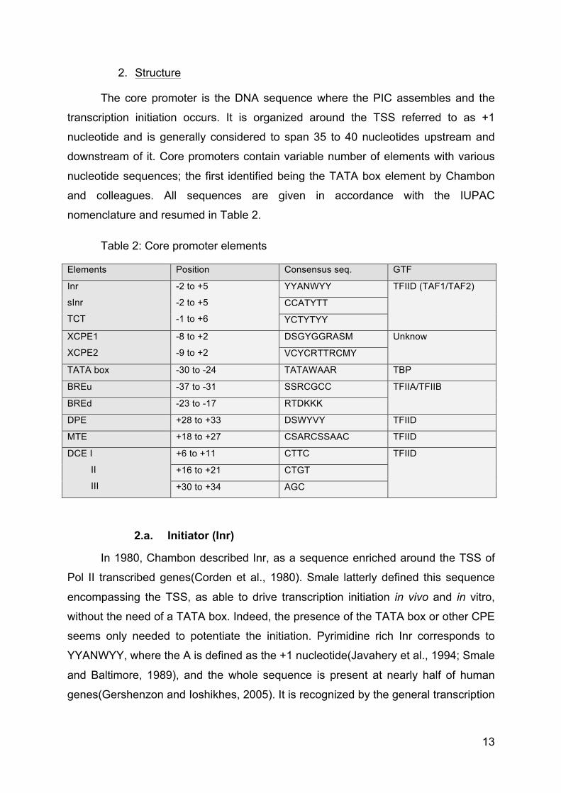
2. Structure
The core promoter is the DNA sequence where the PIC assembles and the
transcription initiation occurs. It is organized around the TSS referred to as +1
nucleotide and is generally considered to span 35 to 40 nucleotides upstream and
downstream of it. Core promoters contain variable number of elements with various
nucleotide sequences; the first identified being the TATA box element by Chambon
and colleagues. All sequences are given in accordance with the IUPAC
nomenclature and resumed in Table 2.
Table 2: Core promoter elements
Elements Position Consensus seq. GTF
Inr
sInr
TCT
-2 to +5
-2 to +5
-1 to +6
YYANWYY TFIID (TAF1/TAF2)
CCATYTT
YCTYTYY
XCPE1
XCPE2
-8 to +2
-9 to +2
DSGYGGRASM Unknow
VCYCRTTRCMY
TATA box -30 to -24 TATAWAAR TBP
BREu -37 to -31 SSRCGCC TFIIA/TFIIB
BREd -23 to -17 RTDKKK
DPE +28 to +33 DSWYVY TFIID
MTE +18 to +27 CSARCSSAAC TFIID
DCE I
II
III
+6 to +11 CTTC TFIID
+16 to +21 CTGT
+30 to +34 AGC
2.a. Initiator (Inr)
In 1980, Chambon described Inr, as a sequence enriched around the TSS of
Pol II transcribed genes(Corden et al., 1980). Smale latterly defined this sequence
encompassing the TSS, as able to drive transcription initiation in vivo and in vitro,
without the need of a TATA box. Indeed, the presence of the TATA box or other CPE
seems only needed to potentiate the initiation. Pyrimidine rich Inr corresponds to
YYANWYY, where the A is defined as the +1 nucleotide(Javahery et al., 1994; Smale
and Baltimore, 1989), and the whole sequence is present at nearly half of human
genes(Gershenzon and Ioshikhes, 2005). It is recognized by the general transcription
14
factor TFIID(Kaufmann and Smale, 1994), especially TAF1 and TAF2(Chalkley and
Verrijzer, 1999).
Recently, a strict Inr (sInr), with a more restrictive sequence has been found in
TATA less promoters where it seems to bypass TATA box by cooperating with the
Sp1 sequence(Yarden et al., 2009). CCATYTT sequence was defined as sInr and is
present in 1,5% of human genes.
Finally, an alternative pyrimidine rich initiator element called TCT has been
described to encompass the TSS of many genes and notably members of the
ribosome gene family. Its sequence is YCTYTYY, where C appears to be the
TSS(Parry et al., 2010).
2.b. X gene core promoter element (XCPE1/2)
The X Core Promoter Element 1 (XCPE1) has been characterised as able to
induce transcription initiation of hepatitis virus X gene and lately found in 1% of
human promoter, especially TATA less ones. It spans from -8 to +2 with the
sequence DSGYGGRASM, where A is the TSS(Tokusumi et al., 2007).
The XCPE2 core promoter element directs transcription initiation of the second
promoter of hepatitis virus X gene and have the sequence VCYCRTTRCMY, where
M is the TSS(Anish et al., 2009). It is found at multiple TATA less human promoters.
2.c. TATA box
The TATA box has also been described by Chambon and named after its
sequence composition. It was first thought to be a general transcription CPE.
Nowadays, it is assumed that it is only present in 10% to 20% of human genes. Its
consensus sequence, TATAWAAR, is present upstream to the TSS at nucleotide -
28. Inr is associated with around 60% of all TATA box (Gershenzon and Ioshikhes,
2005). The TATA box is well conserved from Archea to human.
2.d. TFIIB Recognition Elements (BRE)
The TFIIB Recognition Elements (BRE) are two sequences located directly
upstream (BREu)(Lagrange et al., 1998) and downstream (BREd)(Deng and
Roberts, 2005) of the TATA box, but may also be present in TATA less promoters.
Both sequences are well conserved from Archea to human and their sequences are
15
SSRCGCC and RTDKKK respectively. They are recognised by the general
transcription factors TFIIA and TFIIB.
2.e. Downstream Promoter Element (DPE)
As its name suggests, the Downstream Promoter Element (DPE) is located
downstream the +1 nucleotide between +28 and +33, with the consensus sequence
DSWYVY. Its presence is apparently independent of either TATA box or Inr(Burke
and Kadonaga, 1997; Gershenzon and Ioshikhes, 2005). The DPE element is
notably associated with developmental regulatory genes(Zehavi et al., 2014). It is
recognised and bound by the general transcription factor TFIID.
2.f. Motif Ten Element (MTE)
The Motif Ten Element (MTE), named after computational analysis(Ohler et
al., 2002), is located downstream of the TSS and directly upstream of the DPE. It
spans from +18 to +27 with the sequence CSARCSSAAC. MTE requires association
with Inr sequence. Even though it can act independently of TATA box or DPE, their
presences strongly reinforce its transcription initiation activity(Lim et al., 2004). MTE
is also recognised by TFIID.
More recently, MTE and DPE combination have been found to form a bridge
motif and to constitute an independent core promoter element(Theisen et al., 2010).
2.g. Downstream Core Element (DCE)
First observed in β-globin gene, the Downstream Core Element (DCE) is a
downstream element alternative to DPE, MTE or Bridge motif(Lewis et al., 2000). It is
composed of three sequences, spanning from +6 to +11 (necessary motif CTTC),
from +16 to +21 (necessary motif CTGT) and from +30 to +34 (necessary motif
AGC). It is recognised by TFIID(Lee et al., 2005).
16
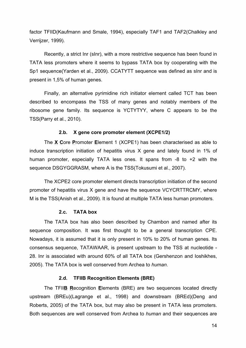
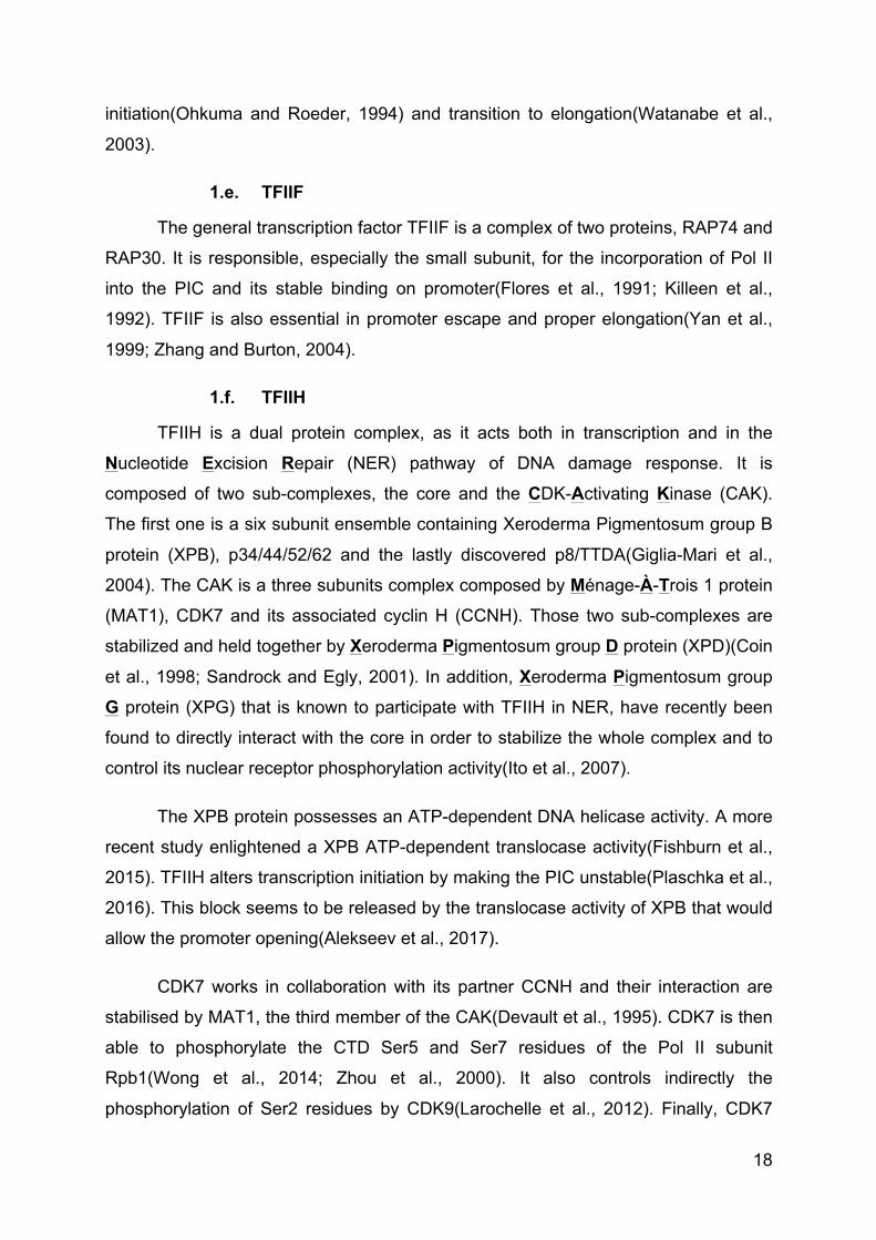
Figure 1: Core promoter elements and respective binding GTF
The diagram shows a promoter with most common CPE and the proteins from
the transcription machinery that contact them.
3) Pre-initiation complex
Even if RNA polymerase II alone is able to transcribe DNA templates in vitro, it
requires the assembly of a large Pre-Initiation Complex (PIC) for transcription to start
at the right nucleotide in a well-coordinated manner in vivo(Weil et al., 1979).
1. General transcription factors
General transcription factors (GTF) have first been purified from human cells
in 1980 (Matsui et al., 1980) and further characterized(Reinberg and Roeder, 1987;
Reinberg et al., 1987; Samuels et al., 1982). They are named TFIIA, TFIIB, TFIID,
TFIIE, TFIIF and TFIIH for basal/general Transcription Factors associated with RNA
Polymerase II; the last letter corresponds to purification fractions. When assembled
with Pol II on the core promoter, they form the Pre-Initiation Complex. They allow the
correct positioning of Pol II on the promoter and the transcription to start at the right
nucleotide.
As well as core promoters largely vary in their sequence compositions;
nowadays it is assumed that PIC composition may vary, especially for TFIID. Some
subunits of general transcription factors seem to be specific to certain set of genes,
cell types or even tissues(Akhtar and Veenstra, 2011). Here, we will describe their
widely observable composition (Table 3).
TBP
TAF1/TAF2
TFIID
TFIIA/TFIIB
DCE
+1
III III
InrTATA
XCPE1
BREdBREu DPEMTE
17
1.a. TFIIA
The general transcription factor TFIIA is a three subunit complex composed of
TFIIAα, TFIIAβ and TFIIAγ. It has been shown to interact with the BRE elements to
stabilize TFIID(Buratowski et al., 1989; Lee et al., 1992).
1.b. TFIIB
The general transcription factor TFIIB is a single protein. It stabilizes the
TFIID-DNA-TFIIA complex and helps the subsequent recruitment of the RNA
polymerase II(Ha et al., 1993; Maldonado et al., 1990) together with TFIIF. TFIIB
participates in determining TSS position(Li et al., 1994) and recognizes BRE
elements. It regulates itself by auto-acetylation(Choi et al., 2003) and participates in
promoter escape(Westover et al., 2004).
1.c. TFIID
The general transcription factor TFIID is the largest complex among all GTF. It
is composed of the TATA Binding Protein (TBP) and 14 TBP Associated Factors
(TAF). As its name indicates, TBP binds the consensus sequence of the TATA
box(Corden et al., 1980; Sawadogo and Roeder, 1985). For either TATA less or
TATA containing promoters, the whole TFIID complex binds the core promoter to
initiate the PIC formation. TAF1/TAF2 dimer binds Inr element(Chalkley and
Verrijzer, 1999). TAF1 is also able to bind DCE(Lee et al., 2005) and TAF6/TAF9 is
able to bind both DPE(Burke and Kadonaga, 1996) and MTE(Shao et al., 2005).
Some of the subunits are also able to interact with certain nuclear receptors(Lavigne
et al., 1999) or to modify histone proteins(Mizzen et al., 1996; Pham and Sauer,
2000), in order to regulate transcription. Furthermore, TFIID interacts with
TFIIF(Dikstein et al., 1996).
1.d. TFIIE
The general transcription factor TFIIE is composed of two copies of each
subunit TFIIEα and TFIIEβ, forming a hetero-tetramer. It directly interacts with Pol II
and DNA promoter as well as TFIID, TFIIF and TFIIH(Maxon et al., 1994; Okuda et
al., 2004; Tanaka et al., 2009). It is mostly implicated in the control of TFIIH
enzymatic activities and required for Pol II transcription, driving both
18
initiation(Ohkuma and Roeder, 1994) and transition to elongation(Watanabe et al.,
2003).
1.e. TFIIF
The general transcription factor TFIIF is a complex of two proteins, RAP74 and
RAP30. It is responsible, especially the small subunit, for the incorporation of Pol II
into the PIC and its stable binding on promoter(Flores et al., 1991; Killeen et al.,
1992). TFIIF is also essential in promoter escape and proper elongation(Yan et al.,
1999; Zhang and Burton, 2004).
1.f. TFIIH
TFIIH is a dual protein complex, as it acts both in transcription and in the
Nucleotide Excision Repair (NER) pathway of DNA damage response. It is
composed of two sub-complexes, the core and the CDK-Activating Kinase (CAK).
The first one is a six subunit ensemble containing Xeroderma Pigmentosum group B
protein (XPB), p34/44/52/62 and the lastly discovered p8/TTDA(Giglia-Mari et al.,
2004). The CAK is a three subunits complex composed by Ménage-À-Trois 1 protein
(MAT1), CDK7 and its associated cyclin H (CCNH). Those two sub-complexes are
stabilized and held together by Xeroderma Pigmentosum group D protein (XPD)(Coin
et al., 1998; Sandrock and Egly, 2001). In addition, Xeroderma Pigmentosum group
G protein (XPG) that is known to participate with TFIIH in NER, have recently been
found to directly interact with the core in order to stabilize the whole complex and to
control its nuclear receptor phosphorylation activity(Ito et al., 2007).
The XPB protein possesses an ATP-dependent DNA helicase activity. A more
recent study enlightened a XPB ATP-dependent translocase activity(Fishburn et al.,
2015). TFIIH alters transcription initiation by making the PIC unstable(Plaschka et al.,
2016). This block seems to be released by the translocase activity of XPB that would
allow the promoter opening(Alekseev et al., 2017).
CDK7 works in collaboration with its partner CCNH and their interaction are
stabilised by MAT1, the third member of the CAK(Devault et al., 1995). CDK7 is then
able to phosphorylate the CTD Ser5 and Ser7 residues of the Pol II subunit
Rpb1(Wong et al., 2014; Zhou et al., 2000). It also controls indirectly the
phosphorylation of Ser2 residues by CDK9(Larochelle et al., 2012). Finally, CDK7
19
also phosphorylates TFIID, TFIIE and TFIIF general transcription factors(Ohkuma
and Roeder, 1994).
CDK7 is also implicated in the cell cycle progression. XPD controls CDK7
activity (Chen et al., 2003). XPD notably control the cellular localisation of CDK7 to
regulate its mitotic kinase activity and the chromosomal segregation(Li et al., 2010).
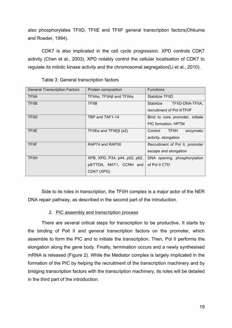
Table 3: General transcription factors
General Transcription Factors Protein composition Functions
TFIIA TFIIAα, TFIIAβ and TFIIAγ Stabilize TFIID
TFIIB TFIIB Stabilize TFIID-DNA-TFIIA,
recruitment of Pol II/TFIIF
TFIID TBP and TAF1-14 Bind to core promoter, initiate
PIC formation, HPTM
TFIIE TFIIEα and TFIIEβ (x2) Control TFIIH enzymatic
activity, elongation
TFIIF RAP74 and RAP30 Recruitment of Pol II, promoter
escape and elongation
TFIIH XPB, XPD, P34, p44, p52, p62,
p8/TTDA, MAT1, CCNH and
CDK7 (XPG)
DNA opening, phosphorylation
of Pol II CTD
Side to its roles in transcription, the TFIIH complex is a major actor of the NER
DNA repair pathway, as described in the second part of the introduction.
2. PIC assembly and transcription process
There are several critical steps for transcription to be productive. It starts by
the binding of Poll II and general transcription factors on the promoter, which
assemble to form the PIC and to initiate the transcription. Then, Pol II performs the
elongation along the gene body. Finally, termination occurs and a newly synthesised
mRNA is released (Figure 2). While the Mediator complex is largely implicated in the
formation of the PIC by helping the recruitment of the transcription machinery and by
bridging transcription factors with the transcription machinery, its roles will be detailed
in the third part of the introduction.
20
2.a. PIC formation
Transcription starts with the assemblage of the PIC around the TSS. It can
happen in two different manners: the stepwise assembly or the holoenzyme
assembly.
The stepwise assembly (aka sequential assembly) starts by the recognition of
the promoter by TFIID. TBP first recognizes the TATA box, if it is present. In any
case, other TFIID subunits recognise Inr and DPE to interact with the promoter. This
interaction is then stabilized and strengthened by the arrival of TFIIA and TFIIB.
Subsequently, Pol II joins the promoter in association with TFIIF. TFIIE and TFIIH are
the last to be recruited to the PIC(He et al., 2013).
The Pol II holoenzyme consists of a large already assembled complex
containing the Pol II, the Mediator complex and most of the general transcription
factors except TFIID/TFIIA. In the holoenzyme pathway, it assembles without DNA
and comes in almost one single step onto the promoter(Koleske and Young, 1995).
2.b. Initiation
From now on, TFIIH is essential for the opening of DNA. This ATP dependent
step allows the transition from a closed conformation to an open conformation PIC.
As mentioned above, various scenarios are possible for TFIIH. The DNA opening
creates an 11-15 bp bubble and the non-coding strand is then inserted in the active
site of Pol II. The first nucleotide synthesis converts the open conformation complex
into an initially transcribing complex(Hantsche and Cramer, 2017). This complex
enters several abortive initiations that only produces short oligonucleotides of less
than 10 bp. If the newly synthetized RNA reaches 10 bp, then Pol II is
phosphorylated by TFIIH and dissociates from the initiation complex, in order to start
elongation. This step is called promoter clearance. After the RNA reaches a length of
25 bp, the capping machinery is recruited and this step allows productive
transcription to start.
2.c. Proximal pausing/Elongation
Pol II encounters a pausing event shortly after starting productive transcription
due to DSIF and NELF. Indeed, they bind Pol II and repress its elongation activity.
The phosphorylation of NELF by P-TEFβ-associated CDK9 leads to its dissociation.
21
Pol II is phosphorylated on Ser2. DSIF is also phosphorylated and turn into an
activator to promote productive elongation(Bernecky et al., 2017; Yamada et al.,
2006).
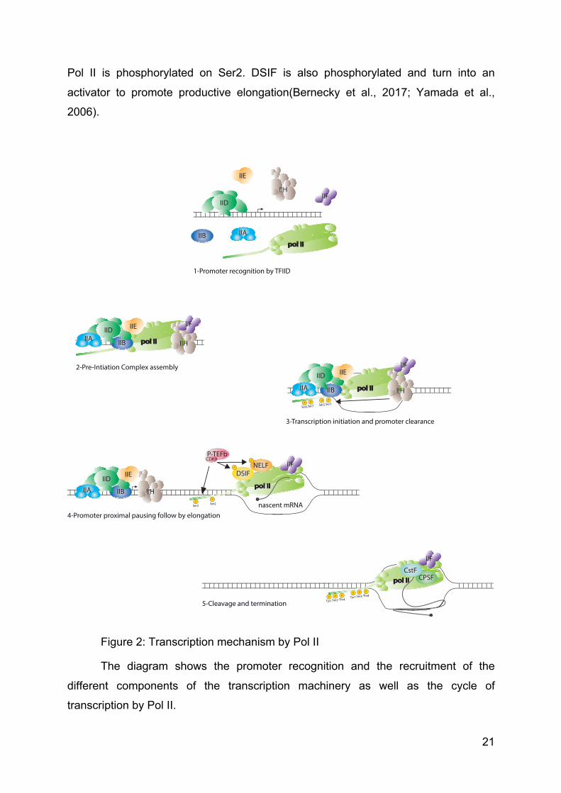
Figure 2: Transcription mechanism by Pol II
The diagram shows the promoter recognition and the recruitment of the
different components of the transcription machinery as well as the cycle of
transcription by Pol II.
pol IIpol II
pol IIpol II
IIB
IIHIIF
IIE
IIDIIA
IIH
IIFIIE
IIAIIB
pol IIpol IIIIB
IID
IIA
IIE
pol IIIIB
IIDIIA IIH
IIFIIE
pol II
IIF
NELFDSIF
CPSFCstF
1-Promoter recognition by TFIID
2-Pre-Intiation Complex assembly
3-Transcription initiation and promoter clearance
4-Promoter proximal pausing follow by elongation
5-Cleavage and termination
IID
IIH
IIF
P-TEFbCDK9
nascent mRNA
P
PP
P
PP
Ser5Ser5
Ser2 Ser2
PSer7
PSer7
PSer2
PTyr1
PThr4
PSer2
PTyr1
PThr4
22
Pol II continues to slide down to the 3’ end of the gene, associated with the
elongation factors TFIIS, ELL, Elongin and P-TEFβ. These factors sustain the
DNA/RNA hybrid, that is necessary for Pol II processivity during elongation(Wind and
Reines, 2000).
2.d. Termination/poly-adenylation
The termination occurs when the elongation complex reaches the 3’ end of the
gene. There are two main ways to end transcription, depending of the gene
sequence(Kuehner et al., 2011).
The first pathway involves the Cleavage and Polyadenylation Specificity
Factor (CPSF), which interacts with the Pol II CTD and recognizes a specific poly A
signal at the 3’ end of the gene. It induces the elongation to stop. Then, the Cleavage
stimulatory Factor (CstF) induces the cleavage of the RNA. Pol II is then released
and the newly synthesised RNA is polyadenylated. The remaining part of the RNA,
downstream of the cleavage site, is therefore degraded by XRN2. Most of the human
genes follow this path.
But for other genes, the termination is achieved through the dissociation of the
RNA/DNA complex by Senataxin 1 protein (Sen1). In such case, the resultant RNA is
not polyadenylated.
4) Specific transcription factors
A large number of actors are required for transcription to occur in the right full
place of the genome, in a well-controlled and coordinated manner. Several proteins
and protein complexes are involved to initiate transcription including specific
transcription factors, coactivators and the Mediator complex. The last one will
extensively be explained in the third part of the introduction.
Specific transcription factors have been selected through evolution by the cells
to answer various stimuli and stresses. Their role is to stimulate (activator) or to
inhibit (repressor) transcription of specific genes to allow cell division, cell growth and
cell death but also cell differentiation, stem cell maintenance or cellular response to
stresses.
23
Specific transcription factors operate through enhancer and silencer regions.
These genomic regions are enriched in specific DNA sequences called Response
Elements (RE), where transcription factors can bind. Transcription factors are all
characterised by a DNA Binding Domain (DBD) to directly contact DNA on specific
RE and a Trans-Activation Domain (TAD) to transfer signal to the transcription
machinery. The sum of activator and repressor signals finally determines the level of
gene activity, ranging from no transcription to high transcribing activity.
Several types of transcription factor have been identified, depending on their
structure, their mode of activation and the biological processes in which they are
implicated.
1. E2F transcription factor family
The first E2F protein has been discovered as a cellular factor required for
adenovirus E2 gene induction(Kovesdi et al., 1986), and lately defined as E2F1.
Height different E2F transcription factors have been identified in this family and
assigned as activators (E2F1-3) or repressors (E2F4-8). They bind DNA as
heterodimer with a Dimerization Partner (DP1-3). This partner is also able to
modulate E2F activity(Bandara et al., 1994; Girling et al., 1993; Ingram et al., 2011;
Ormondroyd et al., 1995). E2F transcription factors are involved in cell cycle
progression and apoptosis, mainly through transcription regulation(DeGregori and
Johnson, 2006; DeGregori et al., 1997).
E2F1 is implicated in G1-S phase progression(Yao et al., 2008), where it
controls an all-or-nothing process. Division has to occur or cell would enter
apoptosis(Wu et al., 2017). E2F1 mainly acts as transcription activator during G1/S
phases(Araki et al., 2003; Burke et al., 2014). Indeed, it activates genes responsible
for DNA synthesis. Moreover, acetylation of histone H3 and H4 accumulates during
G1 phase in an E2F1 dependant manner, in order to open chromatin and allow gene
activation as well as S phase entry(Taubert et al., 2004).
Then, it is made inactive during S/G2 phases(Schulze et al., 1995; Zhang et
al., 2000). Notably, E2F1 is phosphorylated by TFIIH via an interaction with its p62
subunit. This interaction leads to E2F1 degradation during S phase(Vandel and
Kouzarides, 1999).
24
E2F1 also have some roles in the NER pathway that will be presented in the
second part of the introduction.
2. Nuclear receptors
Nuclear receptors are ligand-dependent transcription factors, limited to
Metazoans. They are characterised by a particular Ligand Binding Domain (LBD).
They act either as monomers or dimers but need to bind their specific ligand to be
activated and thus modulate transcription(Escriva et al., 2004).
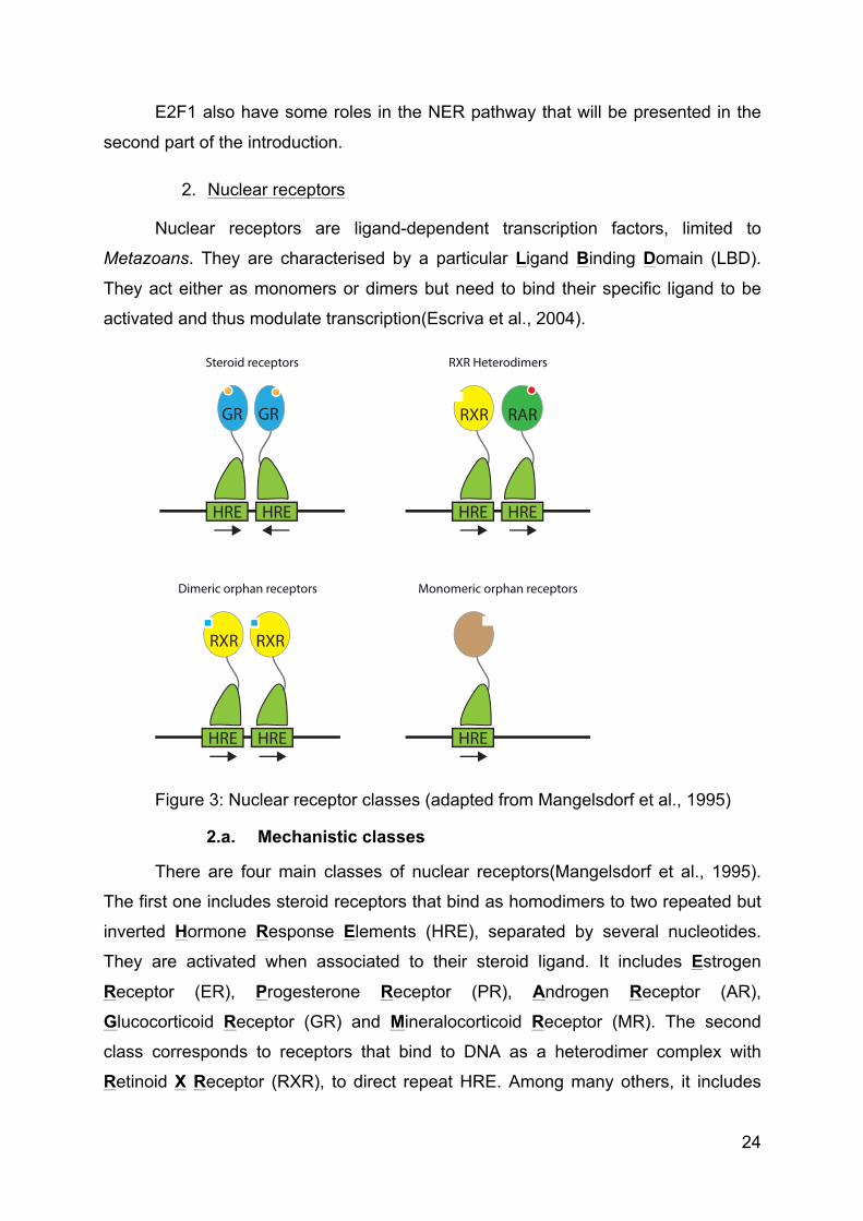
Figure 3: Nuclear receptor classes (adapted from Mangelsdorf et al., 1995)
2.a. Mechanistic classes
There are four main classes of nuclear receptors(Mangelsdorf et al., 1995).
The first one includes steroid receptors that bind as homodimers to two repeated but
inverted Hormone Response Elements (HRE), separated by several nucleotides.
They are activated when associated to their steroid ligand. It includes Estrogen
Receptor (ER), Progesterone Receptor (PR), Androgen Receptor (AR),
Glucocorticoid Receptor (GR) and Mineralocorticoid Receptor (MR). The second
class corresponds to receptors that bind to DNA as a heterodimer complex with
Retinoid X Receptor (RXR), to direct repeat HRE. Among many others, it includes
HREHRE HREHRE
RXR RAR
HREHRE
RXR RXR
HRE
GR GR
Steroid receptors RXR Heterodimers
Dimeric orphan receptors Monomeric orphan receptors
25
Retinoic Acid Receptor (RAR), Thyroid hormone Receptor (T3R), Vitamin D
Receptor (VDR) or Peroxisome Proliferator-Activated-Receptor (PPAR). The third
class corresponds to homodimeric nuclear receptors that bind direct repeat HRE.
RXR, Testicular Receptor (TR2) or Hepatocyte Nuclear Factor-4 (HNF-4) belongs to
this class. Finally, the fourth and last class includes receptors that act either as
monomers or dimers but when only one DNA binding domain contacts one half-site
HRE. Most of the orphan receptors fall in the last two classes.
2.b. Retinoic acid receptor
Vitamin A is essential for several biological processes like embryogenesis,
organ development, homeostasis, immunity and reproduction. Its biological
significance is enlightened by fundamental studies but also by clinical observations.
Indeed, deficiency in vitamin A can lead to growth retardation, intellectual disability
and other symptoms of the Vitamin A Deficiency (VAD) syndrome(Clagett-Dame and
DeLuca, 2002; Zile, 2001).
Vitamin A corresponds to a group of organic compound including retinol,
retinal and All-Trans Retinoic Acid (ATRA), the last one being the most biologically
active form. Its physiological level is tightly regulated(Shannon et al., 2017). The
retinoic acid receptor is a ligand-dependent transcriptional regulator. It includes three
homologous receptors called RARα, RARβ and RARγ. They form a heterodimer with
RXR and activate transcription when they are fixed by their ligand.
The heterodimer RAR/RXR binds to the Retinoic Acid Response Element
(RARE) that corresponds to two successive repeats of RGKTCA separated by
variable number of nucleotides. When no ligand is associated with RAR/RXR dimer,
co-repressors like Nuclear receptor Co-Repressor (NCoR aka NCOR1) or Silencing
Mediator of Retinoic acid and Thyroid hormone receptor (SMRT aka NCOR2)(Perissi
et al., 2010) are bound to it and serve as a platform for the recruitment of Histone De-
ACetylases (HDAC). The latter maintains chromatin in heterochromatin state,
blocking transcription.
Upon ligand binding, RAR encounters conformational changes. This process
leads to co-repressor release and coactivator arrival(Perissi et al., 2004). It is
26
accompanied by the recruitment of several Histone Acetyl Transferase (HAT) and
Histone Methyl Transferase (HMT), which open chromatin and enable transcription.
Retinoic acid seems able to partially modify the genomic location of certain
RAR receptors through a dynamic process and to induce the transcription of
previously unbound but very specific genes(Mahony et al., 2011).
3. Immediate early genes
3.a. Primary response
The transcription of certain genes can be activated very quickly and transiently
by several stimuli without the need of new protein synthesis. These genes are
divided into two classes: the Immediate Early response Genes (IEG) and the delay
primary response genes(Tullai et al., 2007). Some of them code for transcription
factors that subsequently induce the secondary response(Winkles, 1998).
IEG mRNA appear in the cell few minutes after stimulation. Their transcription
is activated by various stimuli like growth factors, mitogens, phorbol esters,
immunological and neurological signals or stresses (Ultra-Violet (UV), toxins)(Fowler
et al., 2011; Greenberg and Ziff, 1984; Herschman, 1991; Morgan and Curran, 1991).
The two most characterised IEG are JUN and FOS(Healy et al., 2013). They
are playing roles in cell proliferation, differentiation and survival. They can be
regulated by Post-Translational Modifications (PTM), which influence their ability to
form a dimer, thus to bind DNA and activate transcription. FOS and JUN form an
active heterodimer complex called Activator Protein-1 (AP-1), via a leucine zipper
motif. When associated, they form a bipartite DBD. The complex binds DNA at
specific responsive element called TetradecanoylPhorbol Acetate (TPA) Responsive
Element (TRE).
Early Growth Response-1 protein (EGR1) is also a transcription factor-coding
IEG, involved in cell growth and differentiation. It binds DNA on specific consensus
sequences and interacts with the transcription machinery(Liu et al., 2001; Zhang et
al., 2003).
27
3.b. Secondary response
The secondary response corresponds to the late induction of a set of genes by the
products of the primary response. They are called Late Response Genes (LRGs), as
well as secondary response genes.
5) Chromatin regulation of transcription
If DNA is bound by a large variety of protein in different dynamic processes,
including transcription, it is rarely naked or freely accessible inside the nucleus.
Indeed, DNA can be chemically modified to alter its accessibility. Moreover, it is
incorporated into chromatin where it is physically compacted.
1. DNA methylation
DNA methylation is well conserved through evolution. It corresponds to the
addition of a methyl group to the 5 position of a cytosine residue (5mC) in the context
of a CpG dinucleotide. In mammals, DNA methylation is implicated in transcription
regulation but also in X-inactivation, genomic imprinting and the silencing of
transposable elements. The genomic pattern of methylation is established during
embryonic life by DNA methyltranferase Dnmt3 and is latterly maintained by Dnmt1
during mitosis.
DNA methylation of promoter regions is associated with repression. The
presence of methyl groups inside the major groove of DNA is supposed to
engendered hindrance that perturbed DNA/TF contacts and block gene
activation(Watt and Molloy, 1988). In the opposite, the binding of TF to DNA can also
influence its methylation status and generate Low Methylated Regions (LMR)(Stadler
et al., 2011).
DNA methylation also participates to gene repression by attracting proteins of
the Methyl CpG Binding Domain (MBD) family that are able to repress
transcription(Bird and Wolffe, 1999).
To actively regulate transcription, DNA methylation is dynamically altered in
cell. Notably, TET deoxygenase is responsible for 5mC oxidation to 5-
hydroxymethylcytosine (5hmC), then to 5-formylcytosine (5fC) and finally to 5-
28
carboxycytosine (5caC). Both 5fC and 5caC can be removed by DNA glycosylase to
be restored to regular cytosine by BER (Figure 4)(Weber et al., 2016).
Figure 4: Cytosine methylation and demethylation (from Kang and Hyun, 2017)
2. Chromatin organisation
To protect it from physical damages but also as a transcription regulation
mechanism, DNA is incorporated into a nucleoprotein complex called chromatin. The
DNA molecule wrap around several histone proteins, in order to form a nucleosome.
The chromatin is composed of a succession of nucleosomes and inter-nucleosome
linker sequences. It appears as beads regularly positioned along a string. This three-
dimensional conformation, called euchromatin, constitutes the first level of a
necessary condensation of the DNA molecule into the nucleus. This is also a way to
regulate its accessibility and thus transcription.
2.a. Nucleosome
The nucleosome also known as nucleosome core particle is composed of 146
bp of DNA wrapped around a core histone octamer in 1.67 turns, for a total of 206
29
kDa (Figure 5). This octamer is composed of two copies of four histone proteins
named H2A, H2B, H3 and H4(Luger et al., 1997). Histones are small proteins
weighting from 11 to 22 kDa and well-conserved trough evolution. They are enriched
in positive amino acids, like lysine and arginine, to interact with negatively charged
DNA. The assembling of the octamer starts by the formation of H3-H4 dimers. Next,
two H3-H4 dimers are assembled in a tetramer. Finally, two H2A-H2B dimers are
added to the tetramer to form a complete octamer(Arents and Moudrianakis,
1995).This “beads on a string” fibre has a diameter of 11 nm and corresponds to the
first level of DNA condensation.
Figure 5: Nucleosome core particle (from Luger et al., 1997)
A ninth “linker” histone named H1 can bind to linker sequences and form a
higher ordered and more stable structure with a diameter of 30 nm(Robinson et al.,
2006). Each nucleosome then encompasses 160 bp(Syed et al., 2010). Two
hypotheses have been proposed for the structure of the 30 nm fibres. The solenoid
model predicts that adjacent nucleosome follow a super-helicoidal path with 6 to 8
nucleosome per turn(Widom and Klug, 1985). The zigzag model postulates two
nucleosomes with straight linker and successively interleaved(Chen and Li, 2010).
Higher structure compactions are less clear but also affect transcription, as it alters
access to DNA. This more compacted chromatin is called heterochromatin and does
not allow transcription. In the cellular context, chromatin seems heterogeneous
30
depending on genomic regions or cell cycles phases. It implies dynamic processes to
pack and unpack the chromatin and control the access to genetic information.
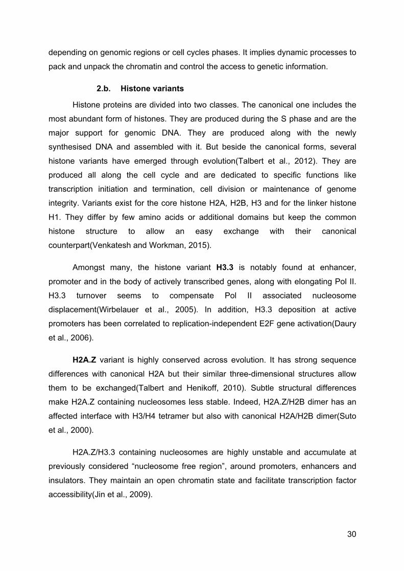
2.b. Histone variants
Histone proteins are divided into two classes. The canonical one includes the
most abundant form of histones. They are produced during the S phase and are the
major support for genomic DNA. They are produced along with the newly
synthesised DNA and assembled with it. But beside the canonical forms, several
histone variants have emerged through evolution(Talbert et al., 2012). They are
produced all along the cell cycle and are dedicated to specific functions like
transcription initiation and termination, cell division or maintenance of genome
integrity. Variants exist for the core histone H2A, H2B, H3 and for the linker histone
H1. They differ by few amino acids or additional domains but keep the common
histone structure to allow an easy exchange with their canonical
counterpart(Venkatesh and Workman, 2015).
Amongst many, the histone variant H3.3 is notably found at enhancer,
promoter and in the body of actively transcribed genes, along with elongating Pol II.
H3.3 turnover seems to compensate Pol II associated nucleosome
displacement(Wirbelauer et al., 2005). In addition, H3.3 deposition at active
promoters has been correlated to replication-independent E2F gene activation(Daury
et al., 2006).
H2A.Z variant is highly conserved across evolution. It has strong sequence
differences with canonical H2A but their similar three-dimensional structures allow
them to be exchanged(Talbert and Henikoff, 2010). Subtle structural differences
make H2A.Z containing nucleosomes less stable. Indeed, H2A.Z/H2B dimer has an
affected interface with H3/H4 tetramer but also with canonical H2A/H2B dimer(Suto
et al., 2000).
H2A.Z/H3.3 containing nucleosomes are highly unstable and accumulate at
previously considered “nucleosome free region”, around promoters, enhancers and
insulators. They maintain an open chromatin state and facilitate transcription factor
accessibility(Jin et al., 2009).
31
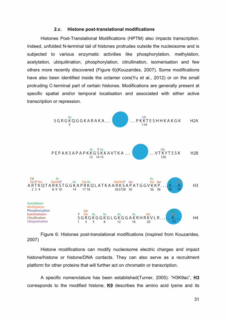
2.c. Histone post-translational modifications
Histones Post-Translational Modifications (HPTM) also impacts transcription.
Indeed, unfolded N-terminal tail of histones protrudes outside the nucleosome and is
subjected to various enzymatic activities like phosphorylation, methylation,
acetylation, ubiquitination, phosphorylation, citrullination, isomerisation and few
others more recently discovered (Figure 6)(Kouzarides, 2007). Some modifications
have also been identified inside the octamer core(Yu et al., 2012) or on the small
protruding C-terminal part of certain histones. Modifications are generally present at
specific spatial and/or temporal localisation and associated with either active
transcription or repression.
Figure 6: Histones post-translational modifications (inspired from Kouzarides, 2007)
Histone modifications can modify nucleosome electric charges and impact
histone/histone or histone/DNA contacts. They can also serve as a recruitment
platform for other proteins that will further act on chromatin or transcription.
A specific nomenclature has been established(Turner, 2005): “H3K9ac”, H3
corresponds to the modified histone, K9 describes the amino acid lysine and its
S G R G K Q G G K A R A K A . . . . . . P K K T E S H H K A K G K H2A
P E P A K S A P A P K K G S K K A V T K A . . . . . . V T K Y T S S K H2B
A R T K Q T A R K S T G G K A P R K Q L A T K A A R K S A P A T G G V K K P . . . H3
S G R G K G G K G L G K G G A K R H R K V L R . . . H4
5 119
12 1201514
2 3 4 8 109 17 1814 26 2827 30 36 38 56 79
1 3 85 12 2016
Ac
AcAcAc
AcAc
AcAcAc
AcAc
Ac
MeMe MeMeMeMe MeMeMe
MeMe
P
P
PP
P Ub
Citrullination
Iso
Ub
Iso
Cit
Cit
AcetylationMethylationPhosphoryationIsomeristaion
Ubiquitination
. K . . . K .
79
AcMe
. . . K . . .
32
position in the protein and ac define the chemical modification (ac: acetylation, me:
mono-methylation, me2/3: di- or –tri-methylation, ub: ubiquitination, ph:
phosphorylation, et caetera).
i. Acetylation
Acetylation is the first described HPTM(Allfrey, 1966), but it also targets other
proteins(Choudhary et al., 2009). Histone Acetyl Transferase (HAT) can mediate N-
acetylation of lysine residues(Kouzarides, 2007). It uses Acetyl-CoA as acetyl donor
to catalyse the reaction. Acetylation suppresses the lysine’s positive charge and
diminishes their interaction with the negatively charged DNA. It leads to chromatin
decondensation and is generally associated with transcriptionally active
genes(Spotswood and Turner, 2002). The reverse reaction is called deacetylation
and is catalysed by histone deacetylase, to restore the lysine’s positive charge.
HAT are usually divided into two classes. Type A HAT are located in the
nucleus, where they acetylate chromatin-associated histones. Type B HAT are
located in the cytoplasm and are responsible for the acetylation of newly synthesised
histones.
Type A HAT are subdivided into three families: i) GCN5-related N-Acetyl
Transferase (GNAT) that includes General Control of Nutrition protein 5 (GCN5) and
p300/CBP-Associated Factor (PCAF), ii) MYST family (MOF - Ybf2/Sas3 - Sas2 -
Tip60) and iii) others like CREB-Binding Protein (CBP) and p300(Kimura et al.,
2005).
GCN5 also know as Lysine (K) AcetylTransferase 2A (KAT2A) is responsible
for H3K14 acetylation alone but it is also included in two different complexes named
Ada Two A-Containing (ATAC) and Spt-Ada-Gcn5 Acetyltransferase (SAGA). Both
complexes stimulate its acetylation activity, especially ATAC(Riss et al., 2015). In
such protein complexes, GCN5 is found to acetylate both H3K9 and H3K14 but on
distinct genomic positions. ATAC associated GCN5 markedly targets enhancer
regions(Krebs et al., 2011; Nagy et al., 2010). Whether incorporated within the SAGA
complex, GCN5 also acetylate H3K18 and H3K23 residues(Rodríguez-Navarro,
2009).
33
In the context of nuclear receptor activation, GCN5/PCAF and CBP/p300 have
been shown to be recruited on the promoter of NR target genes and to be
responsible for H3K9ac and H3K18/27ac respectively(Jin et al., 2011). E2F
Transcription factors also activate transcription by contacting GCN5 and by bringing it
onto promoters. Moreover, E2F1 itself is activated by GCN5/E2F1(Chen et al., 2013;
Lang et al., 2001). GCN5 could also be regulated by E2F1(Yin et al., 2015).
HDAC are the enzymes responsible for the removal of acetyl group from lysine
residues. They are divided in four classes because of their structural differences,
using Zn2+ as cofactor except for the Sirtuin class (Sirt) that uses NAD+(Thaler and
Mercurio, 2014). HDAC have low specificity and need to be regulated. As example,
Sirt1 requires TFIIH to contact and deacetylate the cofactor PGC-1α(Traboulsi et al.,
2014).
Figure 7: Lysine acetylation
ii. Methylation
Histone methylation can have various effects depending on the targeted
residue and its position. Methylation targets both lysine and arginine residues but do
not alters their charge or the histone structure. Methylated residues seem to serve as
a platform for the recruitment of different proteins, able to alter chromatin state or
transcription status. Lysine can be mono-, di-, or tri-methylated with variable
consequences, but arginine can only be mono- or di-methylated(Bedford and Clarke,
2009; Greer and Shi, 2012). The enzymatic reaction is performed by Histone Methyl-
Lysine Acetyl lysine
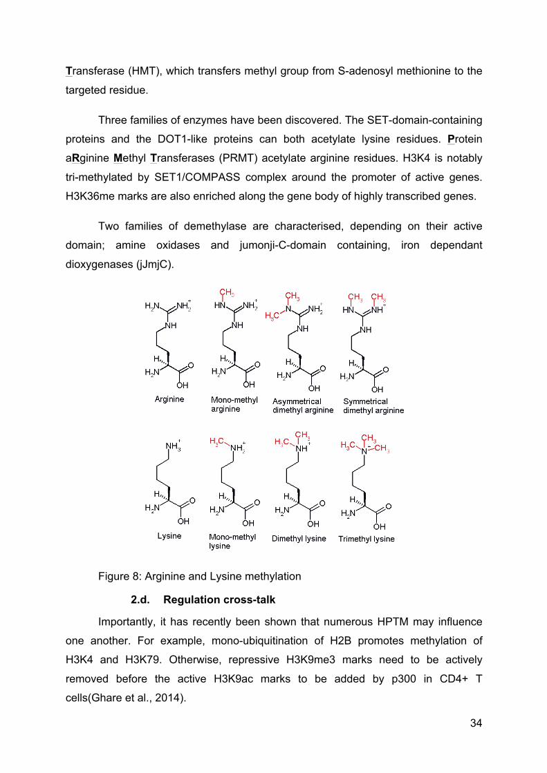
34
Transferase (HMT), which transfers methyl group from S-adenosyl methionine to the
targeted residue.
Three families of enzymes have been discovered. The SET-domain-containing
proteins and the DOT1-like proteins can both acetylate lysine residues. Protein
aRginine Methyl Transferases (PRMT) acetylate arginine residues. H3K4 is notably
tri-methylated by SET1/COMPASS complex around the promoter of active genes.
H3K36me marks are also enriched along the gene body of highly transcribed genes.
Two families of demethylase are characterised, depending on their active
domain; amine oxidases and jumonji-C-domain containing, iron dependant
dioxygenases (jJmjC).
Figure 8: Arginine and Lysine methylation
2.d. Regulation cross-talk
Importantly, it has recently been shown that numerous HPTM may influence
one another. For example, mono-ubiquitination of H2B promotes methylation of
H3K4 and H3K79. Otherwise, repressive H3K9me3 marks need to be actively
removed before the active H3K9ac marks to be added by p300 in CD4+ T
cells(Ghare et al., 2014).
35
Histone variants influence the deposition of histone marks. Indeed, H2A.Z
containing nucleosomes are more prone to H3K9ac and H3K4me3 marks
deposition(Bártfai et al., 2010). DNA methylation status can also influence HPTM.
MBD proteins are notably associated with various HDAC(Bird and Wolffe, 1999).
DNA methylation is also thought to promote H3K9 methylation.
These interrelations between histone post-translational modifications, histone
variants and DNA methylation status constitute a supplemental level of transcription
regulation.
3. Chromatin rearrangement
Gene expression can also be regulated through very large distance.
Rearrangement corresponds to large chromatin loops that allow to bring closer a
specific gene and a distant regulatory element, either upstream or downstream, in
order to regulate transcription. When this element is located on the same DNA
molecule, it is called cis-regulatory element. On the contrary, when the regulatory
element is located on another chromosome, it is called trans-regulatory element.
This has first been enlightened by the discovery of cis-regulation of the β-
globin gene over a 150 kb distance(Vernimmen et al., 2007). Two kinds of loops
have then been described. The first one allows physical rapprochement between
enhancers and promoters(Krivega and Dean, 2012) while the second one brings
close the promoters and the terminators(Lainé et al., 2009). These loops have also
been described to be highly dynamic. For example, they can be induced under
hormonal stimulation(Tan-Wong et al., 2008).
These loop conformations are maintained by a large number of protein.
Among many one, CCCTC-binding protein (CTCF) is the strongest and most
evolutionary conserved. Two copies of the protein can bind CTCF-Pair-Defined
domain (CPD) sequences to gather them. CTCF is implicated in gene activation or
repression but also different functions like enhancer blocking, X-chromosome
inactivation or genome imprinting. Furthermore, it was found to interact with Dnmt1 to
inhibit DNA methylation. CTCF is thought to organise the three-dimensional
conformation of the genome and to direct a large transcription regulation system.
36
II - DNA damage response
All along the life, genomic DNA is exposed to a large number of genotoxic
attacks, either endogenous or exogenous. Indeed, biological by-products, UV rays,
cigarette smoke and environmental chemicals are able to alter the genomic integrity
and to create DNA lesions. These issues have to be properly managed and repaired
in order not to perturb essential processes like DNA transcription and replication,
which are both crucial for cells. The lack of DNA damage response may lead to cell
transformation or death.
Different repair mechanisms have been selected through evolution. They are
differentially activated depending on the lesion type, in order to restore the genomic
integrity. It includes Nucleotide Excision Repair (NER) that is responsible for the
removal of UV-induced lesions and bulky DNA adducts, Base Excision Repair (BER)
that is in charge of single strand breaks, the Non-Homologous End Joining (NHEJ)
and Homologous Repair (HR) pathways, which are responsible for DNA double
strands breaks religation and Mis Match Repair (MMR) that corrects replication
issues.
1) Nucleotide excision repair
NER pathways are engaged in the removal of cyclobutane-pyrimidine dimers
(CPD) and 6-4 pyrimidine pyrimidone photoproducts (6-4) PP that are induced by UV
rays. They are also responsible for the removal of bulky DNA adducts induced by
polycyclic aromatic hydrocarbon or anti-cancerous chemicals(Mocquet et al., 2007).
Two different NER pathways have been discovered, depending on the
activating signal: the Global-Genome NER (GG-NER) and the Transcription-Coupled
NER (TC-NER).
1. GG-NER
As its name indicates, GG-NER acts throughout the genome to search for
lesions in order to initiate their reparation.
37
1.a. Lesions recognition
DNA lesions are recognised all over the genome by the Xeroderma
Pigmentosum complementation group C protein (XPC) in complex with its partners
Homologue Rad 23 B (HR23B) and Centrin2 (CETN2)(Araki et al., 2001; Masutani et
al., 1994). These two proteins are necessary to initiate NER. Indeed, if XPC-HR23B
duplex is sufficient for in vitro NER reaction, CETN2 strongly increases in vivo
reaction(Araújo et al., 2000; Nishi et al., 2005).
Chemical adducts or photoproducts induce DNA distortions that are
recognised by XPC. Nevertheless, only the abnormal DNA structure is recognised by
XPC, the presence of a chemical lesion has to be subsequently confirmed(Sugasawa
et al., 2001, 2002). Along with this recognition mechanism, less distorting-DNA
lesions are not easily identified by XPC/HR23B/CETN2 complex. In such cases, the
damage is first recognised by Damage specific DNA Binding protein 2 (DDB2/XPE)
in complex with DDB1. This DDB complex enhances the DNA disruption and thus
helps to recruit XPC(Fitch et al., 2003). In such case, the DDB-associated Cul4
protein ubiquitinates both DDB2 and XPC to allow NER to initiate(Sugasawa et al.,
2005).
1.b. DNA opening
When bound to the damage, XPC slightly distorts the DNA and recruits the
TFIIH complex(Volker et al., 2001). As mentioned above, TFIIH is a dual complex.
Besides its role in transcription, it is an essential protagonist of NER pathways.
Indeed, both XPB and XPD are required for DNA opening around the lesion. The
mechanism is not yet fully understood but it seems that XPD helicase activity is
responsible for double helix unwinding and opening, when XPB allows the proper
TFIIH anchorage on DNA(Oksenych et al., 2009). The CAK sub-complex seems to
interfere with the overall repair mechanism and needs to be removed from TFIIH
during NER.
When DNA strands are separated around the lesion, the protein RPA binds
the undamaged strand to protect it from nuclease activities and to stabilize the open
structure(Volker et al., 2001). This step allows the arrival of XPA and the release of
the CAK(Coin et al., 2008). RPA is notably acetylated by GCN5/PCAF. This
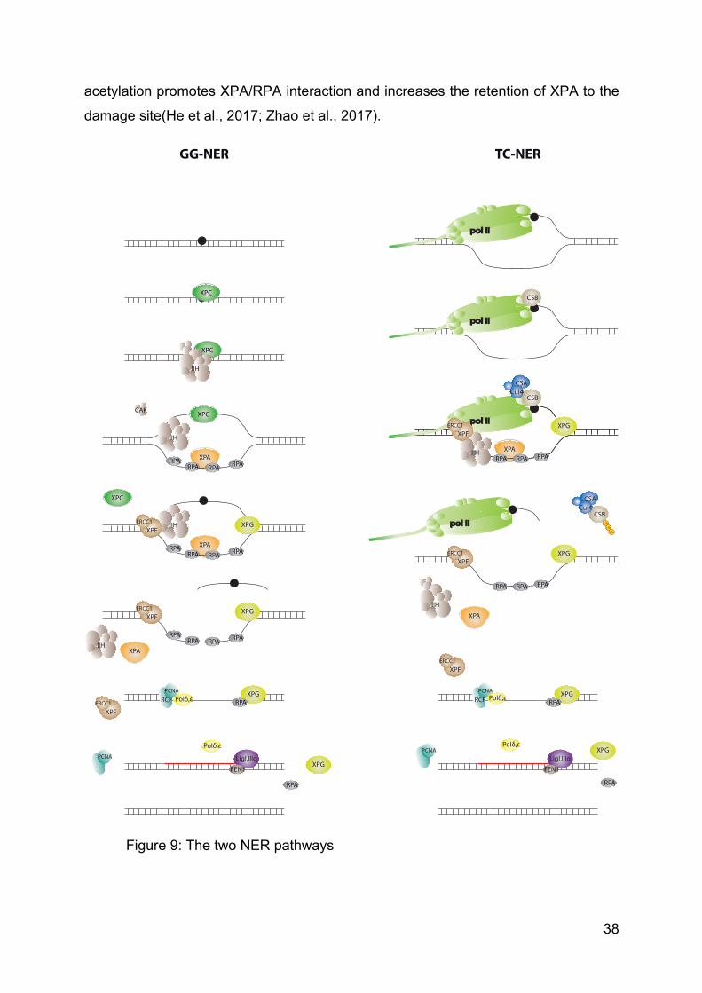
38
acetylation promotes XPA/RPA interaction and increases the retention of XPA to the
damage site(He et al., 2017; Zhao et al., 2017).
Figure 9: The two NER pathways
XPAXPA
XPC
XPC
IIH
XPC
RPA
IIH
RPARPARPA
RPA
IIH
RPARPARPAXPA
XPGXPF
ERCC1
RPA RPARPARPA
XPGXPF
ERCC1
CAK
XPGRPARCF
PCNAPolδ,ε
LigI,IIIαFEN1
pol IIpol II
pol IIpol II
CSB
pol IIpol II
CSB
IIHRPA RPA RPA
XPGXPF
ERCC1
CSACul4
XPA
CSB
XPA
pol IIpol II
CSB
IIHRPA RPA RPA
XPGXPF
ERCC1
CSACul4
IIH
RPA RPA RPA
XPGXPF
ERCC1
CSACul4
UbUb
Ub
pol IIpol II
XPGRPARCF
PCNAPolδ,ε
LigI,IIIαFEN1
IIHXPA
XPC
XPFERCC1
PCNA
Polδ,ε
XPG
RPA
XPFERCC1
PCNAPolδ,ε
XPG
RPA
GG-NER TC-NER
39
1.c. DNA incision
XPA promotes the recruitment of XPF-ERCC1 endonuclease and XPG is
recruited on TFIIH, leading to the removal of XPC. XPG incises the damaged strand
at the 3’ extremity of the lesion bubble and induces the 5’ end cleavage by XPF-
ERCC1. A short 22-30 bp long oligonucleotide is released(Riedl et al., 2003). XPA is
then removed from the lesion.
1.d. Synthesis
After incision, only RPA stays on the undamaged strand to protect it from
nuclease activities. The double incision allows the recruitment of PCNA and RCF.
XPF is then released and the remaining RPA, XPG, RCF and PCNA allow the
recruitment of DNA polymerase δ or ε. After the release of XPG and RPA, synthesis
of the complementary strand is performed using the undamaged strand as
template(Aboussekhra et al., 1995; Moser et al., 2007).
1.e. Ligation
After the synthesis of the complementary strand, a ligase protein performs the
necessary ligation of DNA. The implicated Ligase, as well as the responsible DNA
polymerase, seems to depend on the cell cycle status. Indeed, the couple Pol
δ/Ligase IIIα is in charge of synthesis/ligation throughout the cell cycle while Pol ε
and Ligase I are in charge of synthesis/ligation in quiescent cells(Moser et al., 2007).
2. TC-NER
TC-NER is dedicated to the repair of the transcribed strand(Hu et al., 2015). It
rapidly arises when the RNA polymerase II is stalled on DNA because of a lesion,
during the transcription process. It seems particularly active around promoter and
enhancer. Contrary to GG-NER, it removes a specific lesion to ensure the proper
termination of a crucial ongoing function. In this context, XPC, XPE and DDB proteins
are absolutely dispensable.
2.a. Stalled Pol II
The arrest of Pol II on UV-induced DNA damage strengthens its interaction
with Cockaine Syndrome complementation group B protein (CSB)(van den Boom et
al., 2004). CSB is further stabilized by its de-ubiquitination by USP7(Schwertman et
40
al., 2013). From now on, it strongly binds DNA and alters the Pol II/DNA
interaction(Beerens et al., 2005). CSB then induces the recruitment of the CSA
complex, which notably contains Cul4(Fousteri et al., 2006). CSB also induces the
recruitment of TFIIH and the other NER factors RPA, XPA and the endonucleases
XPF-ERCC1 and XPG.
2.b. Re-initiation
When NER machinery is set up, the regular repair pathway allows the proper
removal of the lesion. After the restoration of genomic integrity, CSA complex re-
ubiquitinate CSB to allow its degradation by the proteasome(Groisman et al., 2006).
This last step is required for normal transcription re-initiation.
2.c. Pol II role in TC-NER
The role of Pol II during the process of TC-NER is not completely understood.
On one hand, the presence of Pol II at the lesion does not prevent dual incision from
occurring in vitro(Tremeau-Bravard et al., 2004). On the other hand, it has been
shown that Pol II can be ubiquitinated, in order to be degraded(Lee et al., 2002).
Finally, CSA also helps the recruitment of chromatin remodelling factors, including
HMGN1, which is responsible for H3K14 acetylation. It has been proposed that local
chromatin remodelling would help Pol II to get back from the lesion, in order to allow
NER(Hanawalt and Spivak, 2008).
2) NER associated diseases
Mutations in NER coding genes are responsible for different syndromes,
associated with photosensitivity. Indeed, the genetic mutations lead to partial or total
impairment of UV-induced NER pathways (Table 4).
1. Xeroderma pigmentosum
Xeroderma pigmentosum (XP) is the first pathology that has been related to
DNA repair deficiency(Cleaver, 1968). Patients were originally described for
photosensitivity and for their high rate of fatal skin cancer, with hereditary
transmission. Indeed, they have a 1000 increased risk rate for skin but also eye and
tongue cancer. Twenty per cent of them also develop progressive neurological
degeneration. Furthermore, they suffer from a slightly higher incidence of internal
41
cancer. There are seven complementation groups, named from XPA to XPG. They
are accountable to mutations in related protein coding genes. XPC and XPE patients
have a deficient GG-NER while the five other complementation groups endure both
TC-NER and GG-NER deficiencies.
More recently, an eighth complementation group has been described, XPV. It
arises from mutations in the gene coding for Pol η. This DNA polymerase is usually
responsible for error-free replication of UV lesions(Johnson et al., 1999). It also
results in photosensitivity and skin cancer, tethering DNA replication.
2. Cockaine syndrome
Patients are photosensitive but show no predisposition to skin cancer.
Cockaine syndrome (CS), also known as progeroid dwarfism, is also characterised
by microcephaly, defective growth and progressive neurological disorder. There are
several levels of severity ranging from the moderate type I CS to the most severely
affected patient showing pre-natal onset(Laugel, 2013). There are two
complementation groups: CSA and CSB.
Furthermore, few mutations in XPB, XPD XPG and ERCC1 have been found
to give rise to dual XP/CS syndrome, associated with a particular propensity to skin
cancer.
3. Trichothiodystrophy
Trichothiodystrophy (TTD) patients exhibit intellectual disability with hereditary
transmission. Scattered and brittle hairs as well as brittle nails mostly characterize
patients. The “tiger-tail” of hair extremity is diagnostic of the disease. They can also
manifest photosensitivity, icthiosis, short stature and decreased fertility. The
photosensitive patients bear mutations in XPB, XPD or p8/TTDA. XPD mutations
have also been reported to be responsible for dual XP/TTD patients. Few TFIIE
mutations was also shown to be responsible for non-photosensitive TTD(Kuschal et
al., 2016).
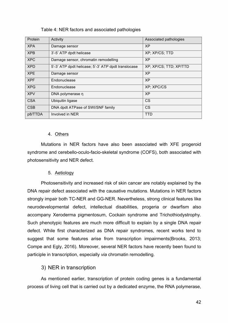
42
Table 4: NER factors and associated pathologies
Protein Activity Associated pathologies
XPA Damage sensor XP
XPB 3’-5’ ATP dpdt helicase XP; XP/CS; TTD
XPC Damage sensor, chromatin remodelling XP
XPD 5’-3’ ATP dpdt helicase; 5’-3’ ATP dpdt translocase XP; XP/CS; TTD; XP/TTD
XPE Damage sensor XP
XPF Endonuclease XP
XPG Endonuclease XP; XPC/CS
XPV DNA polymerase η XP
CSA Ubiquitin ligase CS
CSB DNA dpdt ATPase of SWI/SNF family CS
p8/TTDA Involved in NER TTD
4. Others
Mutations in NER factors have also been associated with XFE progeroid
syndrome and cerebello-oculo-facio-skeletal syndrome (COFS), both associated with
photosensitivity and NER defect.
5. Aetiology
Photosensitivity and increased risk of skin cancer are notably explained by the
DNA repair defect associated with the causative mutations. Mutations in NER factors
strongly impair both TC-NER and GG-NER. Nevertheless, strong clinical features like
neurodevelopmental defect, intellectual disabilities, progeria or dwarfism also
accompany Xeroderma pigmentosum, Cockain syndrome and Trichothiodystrophy.
Such phenotypic features are much more difficult to explain by a single DNA repair
defect. While first characterized as DNA repair syndromes, recent works tend to
suggest that some features arise from transcription impairments(Brooks, 2013;
Compe and Egly, 2016). Moreover, several NER factors have recently been found to
participle in transcription, especially via chromatin remodelling.
3) NER in transcription
As mentioned earlier, transcription of protein coding genes is a fundamental
process of living cell that is carried out by a dedicated enzyme, the RNA polymerase,
43
with the help of several complexes, including chromatin remodellers, transcription
factors, co-factors and general transcription factors.
Thus, the DNA template requires to be carefully maintained as any damage
could alter the process of transcription. Every lesion has to be repaired when
transcription stalled on it. These conclusions suggest a functional link between
transcription and DNA repair.
TFIIH was the first reported complex to be implicated in both transcription and
DNA damage response. It was first identified as part of the general transcription
machinery(Matsui et al., 1980) before to be characterised as the helicase of
nucleotide excision repair(Schaeffer et al., 1993). TFIIH is notably implicated in
transcription initiation, promoter escape and elongation but also in NR
phosphorylation(Drané et al., 2004; Rochette-Egly et al., 1997).
Mediator was recently suggested to allow the switch from transcription to DNA
repair by facilitating the recruitment of repair factors in the context of DNA
lesions(Eyboulet et al., 2013; Kikuchi et al., 2015).
Aside from TFIIH, other NER factors have also been implicated in the process
of transcription. As mentioned earlier, the endonuclease XPG stabilizes the TFIIH
complex in the context of transcription. It is notably required for nuclear receptors-
associated transcription activation(Ito et al., 2007). A recent study also suggests that
a XPG-TFIIH complex could play a role as elongation factor(Narita et al., 2015).
XPG was also shown to be required for the DNA demethylation while
GADD45α induced gene activation(Barreto et al., 2007; Schmitz et al., 2009). Both
XPG and XPF endonucleases have been shown to be required for promoter-
terminator loop formation and optimal transcription. Indeed, XPG promotes DNA
breaks and DNA demethylation at gene promoters to allow the recruitment of CTCF
and gene looping(Le May et al., 2012).
XPC was shown to regulate the stem state of embryonic stem (ES) cells in the
context of a trimeric complex called Stem Cell Coactivator (SCC), together with
HR23B and CETN2. Indeed, Oct4/Sox2 recruits XPC through a direct interaction on
the promoter of Nanog and Oct4 genes as well as most Oct4/Sox2 bound genomic
44
regions, in order to orchestrate the ES cell-specific gene expression(Fong et al.,
2011). In this context, XPC-containing SCC seems required for the maintenance of
either natural or induced pluripotency(Cattoglio et al., 2015).
A recent study showed XPC to regulate TDG-dependent DNA demethylation
in both somatic and stem cells but also in the context of cell reprogramming to
generate induced-pluripotent stem cells(Ho et al., 2017).
Recently, our laboratory uncovered a major role of XPC during transcription(Le
May et al., 2010a), in absence of any genotoxic attack. XPC was found to bind the
promoter of activated gene after the recruitment of the transcription machinery. The
arrival of XPC at the promoter of activated genes engendered the sequential
recruitment of other NER factor including XPA, RPA, XPG and XPF-ERCC1.
Moreover, these NER factors were found to be necessary for optimal
transcription. Indeed, they seem required for DNA demethylation as well as active
histone PTM like H3K9/14 acetylation and H3K4/K9 methylation around the
promoter. Deficiencies in NER factor and notably XPC were found to impair the
recruitment of the other NER factors and to alter gene transactivation. Therefore, the
comprehension of the exact role of XPC and the other NER factors in such
processes is of great interest, both fundamentally and clinically.

45
III - Mediator complex
Manuscript in preparation
46
The Mediator complex and associated human genetic
disorders!
BIDON Baptiste∞, DONNIO Lise-Marie∞, COIN Frederic & EGLY Jean-Marc*
Département de génomique fonctionnelle et Cancer, CNRS, INSERM, Université de
Strasbourg, Institut de Génétique et de Biologie Moléculaire et Cellulaire (IGBMC), 1
rue Laurent Fries, 67404 Illkirch-Graffenstaden, France.
* To whom correspondences should be address:
∞ Should be regarded as joint First Authors
47
ABSTRACT
Mediator (MED) is a multi-subunit complex that plays a central role in the
regulation of protein coding genes transcribed by RNA polymerase II (Pol II). MED
conveys essential information from DNA-bound transcription factors to the basal Pol
II transcription machinery. Although many studies have been engaged on MED, very
few are known concerning the function of each subunit. This last decade, genetic
mutations in MED subunits were shown to cause genetic diseases with common
symptoms like congenital malformation and intellectual disability. After a brief
description of the basic features of MED, the review will describe the mutation-
associated syndromes and discuss their aetiology.
Key words: Mediator, Transcription, Genetic disorders & X-linked Intellectual
Disability.
48
INTRODUCTION
Mediator (MED) is an evolutionarily conserved multi-protein complex that is a
key regulator of protein-coding genes. MED is composed of 25 subunits in yeast and
more than 30 subunits in higher eukaryotes. They are organized into three core
modules (Head, Middle and Tail) and a dissociable Kinase module (Figure 1). MED is
mainly implicated in transcription initiation and functions as a “molecular bridge” that
conveys essential information from transcription factors bound at upstream DNA
responsive elements to the basal transcription machinery, formed by RNA
Polymerase II (Pol II) and the General transcription Factors (GTFs). By means of
this, MED appears to be implicated in enhancer/promoter loop formation but also in
the regulation of other transcriptional events such as elongation(Conaway and
Conaway, 2013), termination(Mukundan and Ansari, 2013), mRNA
processing(Huang et al., 2012) as well as chromatin remodelling(Hsieh et al., 2015;
Kagey et al., 2010; Zhu et al., 2011). From a larger perspective, MED is strongly
implicated in the transcriptional regulation of developmental processes(Yin and
Wang, 2014). These key roles in gene regulation might thus explain why mutations in
human are often associated with developmental disorders or cancers(Schiano et al.,
2014a, 2014b).
Discovery of the Mediator complex
Before 1990, although the basic transcription mechanism was deciphered and
the essential protagonists were identified, very little was known about gene
regulation. Only scarce information was available about co-factors (either activators
or repressors). But unconsciously, people were already working on some MED
subunits, like Gal11 described to be required in galactose responsive genes and later
considered as MED15(Nogi and Fukasawa, 1980) or RGR1 implicated in the
response to glucose and corresponding to MED14(Sakai et al., 1988) as well as Sin4
required for the production of tRNAs which corresponds to MED16(Stadelmann et al.,
1986).
In 1990, the Kornberg lab proposed the term ‘‘Mediator’’ to describe the
recruitment of a yeast protein complex required to “mediate” signal transduction from
Gal4-VP16 chimeric activators to Pol II, in an in vitro transcription assay(Kelleher et
49
al., 1990). In the followings years, they initiate the identification of the MED
composition and delineate its function(Flanagan et al., 1991; Kim et al., 1994).
The human counter-part of yeast Mediator was identified in HeLa cells and
named TRAP (Thyroid hormone receptor-associated protein), for its ability to
associate with the Thyroid hormone Receptor α (TRα) and to potentiate (TRα)-
regulated in vitro transcription(Fondell et al., 1996). Subsequently, different Mediator
related complexes were identified for their abilities to activate transcription, like ARC
(activator-recruited cofactor)/DRIP (vitamin D receptor-interacting proteins)(Näär et
al., 1999; Rachez et al., 1998), CRSP (cofactor required for Sp1 activation)(Ryu et
al., 1999) and PC2 (positive cofactor 2)(Malik et al., 2000). MED was thus considered
as a global regulator of transcription activation.
Multidimensional Protein Identification Technology (MudPID) later stated that
all these proteins are representative of the same Mediator complex, enlightening the
composition of the minimal 26 subunits core complex and the associated 4 subunits
kinase module(Sato et al., 2004). Therefore, a universal nomenclature for each of the
30 MED subunits was admitted(Bourbon et al., 2004). Three of the kinase module
coding genes were found to have undergone duplications that generate the paralogs
pairs MED12/MED12L, MED13/MED13L and CDK8/CDK19. Mass spectrometry
analysis has recently shown that the presence of one paralog of the pair is mutually
exclusive with the other one(Daniels et al., 2013).
Composition and Structure of the Mediator
Considering the various MED-like complexes that have been discovered,
some questions arise. Are there several possible combinations of subunits, as occurs
for SAGA, ATAC or TFIID complexes? Is Mediator composition or architecture
reorganized during various steps of transcription? In the light of the chromatin
environment, we can also question on potential post-translational modifications like
phosphorylation, ubiquitination or acetylation and their effects in the composition and
the structure of MED, as a function of the gene and the response element binding
factors.
However, its size (up to 1.5 MDa), its intrinsic flexibility, and its apparent
composition heterogeneity engender technical difficulties to obtain large amounts of
50
highly purified complex, making the high-resolution structure of the complete MED, a
challenging topic. The first structure obtained at 40Å resolution, showed an extended
protein complex that envelop Pol II. It also showed the complex conservation
between yeast and mice(Asturias et al., 1999). During the following decade, several
low resolution electron microscopy (EM) structures were successively released(Davis
et al., 2002; Dotson et al., 2000; Meyer et al., 2010; Taatjes et al., 2004), establishing
the conservation in eukaryotes and enlightening the modular organisation. They
mostly showed that Head and Middle modules interact with Pol II while Tail module
contacts activators. Each activator was shown to induce a specific MED
conformational change.
In parallel, high-resolution X-ray structures were determined for smaller
entities e.g. Cyclin C (CCNC)(Hoeppner et al., 2005), MED7/MED21(Baumli et al.,
2005; Koschubs et al., 2009; Larivière et al., 2006) or MED8/MED18/MED20. Larger
module structures have later been characterized with high resolution, such as the
crystal structure of S. pombe or S. cerevisiae head module(Imasaki et al., 2011;
Larivière et al., 2012) as well as the partial structure of S. cerevisiae Middle
module(Larivière et al., 2013), showing their internal organisation and their
interactions with the general transcription machinery.
More recently, an accurate electron microscopy structure of the yeast MED
(yMED) has been obtained(Tsai et al., 2014). The localization of head and middle
yMED subunits has been determined in the structure. By comparison, they also
proposed a comprehensive map of the human MED. Cross-linking approach with
endogenous complex have next refined the subunits organisation(Robinson et al.,
2015).
Along with its role in gene activation, Mediator has soon been considered as
part of a larger Pre-Initiation Complex (PIC)(Elmlund et al., 2006; Robinson et al.,
2015). First structures containing both MED and Pol II start to emerge(Cai et al.,
2009). Recently, high resolution structure of the complete PolII/GTFs/Mediator PIC
have been released(Plaschka et al., 2015) and decipher precise MED architecture
and dynamics in the context of transcription initiation(Tsai et al., 2017). Despite its
large number of subunits and contrary to smaller protein complexes like TFIIH, MED
51
harbours only one enzymatic activity. The kinase activity of CDK8 was notably shown
to control MED association with the rest of the PIC(Robinson et al., 2016).
Understanding the global role and functioning of MED is a real challenge that
can only be solved by the combination of two approaches: the fundamental study of
the protein complex and the analysis of the consequences of disease-causing
mutations found in MED genes.
MEDIATOR COMPLEX IN TRANSCRIPTION INITIATION
Structural studies have enabled the emergence of a global view of the
transcription initiation complex with the resolution of the various components such as
Mediator, GTFs (TFIID, TFIIH) and Pol II (see below). The head and middle modules
of MED constitute a compact structure that interacts with Pol II and GTFs, while the
more apart tail module contacts distant transcription factors (TFs). Therefore, MED
requires a constant composition and structure to integrate the basal transcription
machinery, but it also might vary depending on the gene to adapt on specific DNA
binding factors. The subunit MED14 plays the central role to nucleate the three core
modules(Plaschka et al., 2015) and is thus essential for the large MED structural
rearrangement(Tsai et al., 2017). The kinase module transitory binds to middle
module and regulates transcription initiation through its enzymatic activity. All these
events ultimately allow the MED to deliver outputs that range from repression to
maximal activation of genes as well as modulation of the basal transcription.
RNA polymerase II
Early works showed that yMED associates closely with Pol II and some of the
GTF in a build up complex termed Pol II holoenzyme. This complex have been
described to assemble without DNA and to be directly load on the promoter(Kim et
al., 1994; Koleske and Young, 1994; Thompson et al., 1993).
Consistent with these findings, MED subunit deletion or loss-of-function
mutant have been found to compromise both Pol II and MED complex loading on the
promoters of either induced or constitutively active genes(Ansari et al., 2009; Cantin
52
et al., 2003). We have also shown earlier this year that various mutations in MED12
subunit, which segregate with intellectual disability (ID), strongly affect the
recruitment of Pol II and MED12 all over the genome(Donnio et al., 2017).
EM studies suggested that several subunits of Pol II, including Rpb1, Rpb2,
Rpb3, Rpb6, Rpb11 and Rpb12, contact the middle or head modules of MED(Davis
et al., 2002). An in vivo photo-crosslinking approach complemented by genetic
analysis has identified a direct contact between Rpb3 and Med17, an interaction
essential for Pol II genome-wide recruitment(Soutourina et al., 2011). Recent
structural studies have revealed interaction between the head and Rbp4/7 or
between the sub-module MED18/20 and Rbp3/11. The interaction is strengthened by
contacts between the middle module subunits MED4/9 and Rpb1 and strongly
depends on the hinge of the middle module. Indeed, mutation in MED21 does not
alter MED integrity but strongly disturbs its interaction with Pol II(Nozawa et al., 2017;
Plaschka et al., 2015; Sato et al., 2016; Tsai et al., 2017). Finally, the head module of
MED is thought to bind the Pol II C-terminal domain (CTD) to facilitate its
phosphorylation by CDK7(Kim et al., 1994; Robinson et al., 2016, 2012).
The head module of yMED stably associates with a Pol II-TFIIF complex, but
not with the polymerase or TFIIF alone(Takagi et al., 2006). This difference suggests
a combinatorial interaction but may also be the result of the conformational change
induced by TFIIF(Forget et al., 1997). Moreover, a cryo-EM analysis showed that the
presence of an activator (like VP-16) together with Mediator, is required to stably
orient Pol II-TFIIF on the promoter, prior to transcription initiation(Bernecky and
Taatjes, 2012; Bernecky et al., 2011). MED also overcome the inhibition of Pol II
imposed by Gdown1, through TFIIF(Cheng et al., 2012; Hu et al., 2006; Jishage et
al., 2012). Concordant with the MED-Pol II dissociation during transcription initiation,
the head module of yMED was shown not to interact with Pol II-TFIIF when
associated with DNA:RNA duplex(Takagi et al., 2006).
TFIIA, TFIIB and TBP/TFIID
Template commitment experiment first revealed a physical interaction of TFIID
with MED(Koleske et al., 1992). Indeed, TFIID acts in collaboration with MED in the
context of transcription activation(Guermah et al., 1998, 2001). Using immobilized
53
template assays and extract depleted or supplemented with purified factors, MED
was revealed to coordinate TFIID binding to promoter(Black et al., 2006; Johnson et
al., 2002). MED also facilitate TFIIB recruitment during the PIC assembly(Baek et al.,
2006). Moreover, a purified TFIIA/TFIID/MED bound to promoter DNA was found to
serve as a platform that supports active levels of PIC assembly and
transcription(Johnson and Carey, 2003). The Head module of MED is structurally
found to contact TFIIB through its ribbon part, and stabilize the initiation
complex(Plaschka et al., 2015).
TFIIH and TFIIE
Different studies demonstrated that the tail module subunit MED15 binds
stably to TFIIE and TFIIH(Sakurai and Fukasawa, 1997, 2000), whereas the head
module MED11 subunit was found interact with the Rad3/XPD subunit of
TFIIH(Esnault et al., 2008). Mediator has been shown to enhance in vitro
phosphorylation of the Pol II CTD by TFIIH in a yeast reconstituted transcription
system containing Pol II and basal factors, increasing its transcriptional activity(Kim
et al., 1994). On the other hand, the phosphorylation of TFIIH subunit Cyclin H
(CCNH) by MED subunit CDK8 seems to block CCNH/CDK7 kinase
activity(Akoulitchev et al., 2000). This activity have been established to be a key
event for promoter clearance and disruption of MED-Pol II interaction(Søgaard and
Svejstrup, 2007). We also found few years ago a link between MED23 and TFIIH
subunit CDK7. What is more, non-syndromic ID associated-MED23 mutation was
shown to disturbed the recruitment of both TFIIH-CDK7 and MED-CDK8 on the
promoter of activated genes(Hashimoto et al., 2011), enlightening the strong contact
between TFIIH functions and the Mediator.
DNA binding factors
EM studies of human Mediator complexes revealed marked conformational
changes of MED upon TFs binding. The structural comparison of complexes purified
using either the SREBP-1a activator, the VP16 activation domain or a FLAG-tagged
MED26 showed substantial differences in size and shape between them(Ebmeier
and Taatjes, 2010). Different MED subunits were shown to interact with either the C-
terminus domain or the activation domain of p53, leading to different consequences
54
on the overall PIC structure. Only MED associated with the p53 activation domain
elicits the conformation able to activate a stalled Pol II into a productive Pol II(Meyer
et al., 2010). Beside, CMT2B2 associated-MED25 mutation was shown to disturb its
proline rich interaction domain, thus affecting the range of possible interacting SH3
domain proteins(Leal et al., 2009), with dramatic consequences.
Different Mediator subunits can work in synergy to regulate some genes. For
example, the glucocorticoid receptor (GR) has been reported to use its ligand-
dependent activation domain to target MED1, while its ligand-independent N-terminal
activation domain targets MED14. By consequence, the expression of some GR-
target genes requires MED14 but not MED1, while expression of other genes
requires both(Chen et al., 2006). Similarly, MED23 is essential for expression of Egr1
(Early Growth Response protein 1) gene in mES (mouse Embryonic Stems) cells, but
is dispensable for its expression in MEF (Mouse Embryonic Fibroblast)
cells(Balamotis et al., 2009).
Elongation and termination
MED seems to function as a “molecular bridge” that conveys essential
information from transcription factors bound at upstream responsive elements to Pol
II transcription machinery, suggesting a role in enhancer/promoter loop formation.
Indeed, the role of MED in gene looping is established by the discovery of super-
enhancers, which consist of clusters of enhancers. These domains are strongly
bound by master transcription factors Oct4, Sox2 and Nanog, which recruit MED to
control stem cell identity(Whyte et al., 2013). It is further reinforced by Mediator
interaction with a cohesin complex containing NIPBL, Smc1a and Smc3, but not
CTCF, to facilitate enhancer-promoter loops at stem cell associated-genes like
Nanog, Oct4, Phc1 or Lefty1(Kagey et al., 2010). Aside, MED was also found to
interact with certain ncRNA-a (non-coding RNA-activating) to regulate local gene
expression through chromatin looping(Lai et al., 2013).
In addition to play a central role in PIC assembly, MED contributes to others
steps of transcription. An in vitro study demonstrated that purified Mediator complex
could stimulate transcription elongation by overcoming the DSIF-induced Pol II
proximal pausing(Malik et al., 2007). Furthermore, MED26 interacts first with TFIID in
55
Pol II initiation complex and then exchanges TFIID for elongation complexes
containing ELL/EAF and P-TEFβ, in order to facilitate transition of Pol II from a
stalled state into an elongation state(Takahashi et al., 2011).
Mediator has also been reported to be important for transcription termination.
MED18 was shown to be required for the recruitment of cleavage and
polyadenylation factors while its absence leads to accumulation of Pol II near the 3’
end of genes(Mukundan and Ansari, 2011). It was next demonstrated to facilitate the
recruitment of cleavage factor 1 (CF1) complex at the 3′ end of genes by interacting
with it(Mukundan and Ansari, 2013). These studies also highlight the role of MED in
promoter/terminator loop formation and the link between such loops and transcription
termination. Indeed, MED18 deletion strongly affects the loop formation and the
termination process.
GENETIC DISORDERS ASSOCIATED WITH MEDIATOR
These last years, more and more studies have shown that mutations in MED
subunits are associated with a wide range of human genetic disorders leading to
congenital malformation and/or intellectual disability, enlightening the major role of
Mediator in developmental processes (Table 1 and 2, Figure 2).
Understanding the defects that lead to such disorders at the molecular level
might deepen our knowledge on Mediator as a major regulatory element of
development, in addition to providing an explanation to the disorder itself.
Kinase module subunits
MED12-associated syndromes
Except MED14, MED12 is the only Mediator subunit coding gene to be located
on the sexual chromosome X. Considering the central role of MED14, it is not
surprising that no mutations have been detected in this subunit when only one copy
is available in male individuals. Such mutations would probably be too deleterious.
MED12 is located at Xq13.1 and codes for a 230kDa Mediator subunit part of the
kinase module, along with MED13, CDK8 and CCNC or one of their paralogs. Three
56
independent but closely related syndromes, all presenting mild to severe ID, have
been associated with MED12 variations. With the raise of genetic diagnostic, MED12
mutations have also been found to be responsible for other non-syndromic ID (Figure
3).
Opitz-Kaveggia syndrome (also known as FG syndrome; MIM #305450) was
initially described in 1974 by Opitz and Kaveggia in a family of five males affected by
intellectual disability (ID), macrocephaly, hypotonia and imperforate anus with X-
linked inheritance(Opitz and Kaveggia, 1974). This syndrome has been linked to a
recurrent missense mutation (c.2881C>T, p.R961W) in MED12 gene(Risheg et al.,
2007). Hitherto, this mutation has been found in ten families, all sharing intellectual
disability (ID), macrocephaly and hypotonia with variable digestive or genito-urinary
anomalies (for a total of 23 affected males)(Clark et al., 2009). Another MED12
missense mutation (c.2873G>A, p.G958E) has been reported in a family with three
cousins affected by Opitz-Kaveggia syndrome(Rump et al., 2011).
Few years later, Lujan and Fryns independently described another X-Linked
Intellectual Disability (XLID) syndrome (commonly called Lujan-Fryns or Lujan
syndrome, MIM #309520) also characterized by intellectual disability, macrocephaly,
hypotonia and genito-urinary anomalies. It further includes dysgenesis of the corpus
callosum, characteristic facial anomalies and behavioural disturbance(Fryns and
Buttiens, 1987; Lujan et al., 1984). Later on, Schwartz team discovered a mutation
(c.3020A>G, p.N1007S) in MED12 gene as responsible for Lujan
syndrome(Schwartz et al., 2007).
Ohdo syndrome (MIM #300895) comprises a heterogeneous group of
disorders characterized by intellectual disability and blepharophimosis (narrowing of
the eye opening)(Ohdo et al., 1986). The Maat-Kievit-Brunner type (OSMKB or X-
linked Ohdo syndrome) was initially distinguished from the other types of Ohdo
syndrome due to its X-linked inheritance(Verloes et al., 2006). The facial
characteristics also include prominent cheeks, nose with a rounded tip and a narrow
mouth. As people with the condition get older, these characteristics become more
pronounced and the face becomes more triangular (Table 1). Exome sequencing
performed in two families with the OSMKB type revealed two different MED12
missense mutations segregating with the phenotype (c.3443G>A, p.R1148H, or
57
c.3493T>C p.S1165P). Subsequent analysis of a cohort of nine males with Ohdo
syndrome, revealed another de novo missense mutation (c.5185C>A p.H1729N) in
MED12(Isidor et al., 2014; Vulto-van Silfhout et al., 2013). Certain differences are
noted among patients, even between patients bearing the same mutation (pR1148H).
Finally, Martinez team simultaneously published two studies presenting a
MED12 mutation (c.887G>A, p.R296Q) and enlightening the overlapping phenotypes
of MED12-related patients(Caro-Llopis et al., 2016; Martínez et al., 2017). Indeed,
the affected male was characterized as Ohdo syndrome. However, the authors noted
symptoms not previously reported in other OSMKB patients, further expanding Ohdo
syndrome and overlapping with other MED12-related symptoms.
MED12 Non-syndromic XLID
Along with these syndrome-associated mutations, numerous alterations of
MED12 have been identified, as the sequencing techniques were becoming more
widely used, in non-syndromic X-linked Intellectual Disability (XLID) patients. Indeed,
fourteen genetic alterations, mostly missense mutations, were characterised along
the gene, in patients with intellectual disability (Table 1; Figure 3). Only few of them
perfectly feat with one the three syndromes, but others are sharing most
characteristic symptoms. In addition to ID, they often present delayed development
with speech difficulties, Micro or Macrocephaly, various cranio-facial abnormalities
(long narrow face, high forehead, altered philtrum), genito-urinary malformation and
feeding or digestive troubles, in childhood and sometimes longer. In such conditions,
some authors also defined a fourth and a fifth Med12-related condition(Narayanan
and Phadke, 2017; Prontera et al., 2016).
For the three major syndromes, all reports only presented male patients along
with mutations-carrying females. As MED12 gene is encoded on X-chromosome, the
authors often explained the absence of symptoms in female by the chromosomal
compensation, also supported sometimes by skewed inactivation in favour of the wild
allele. But in 2013, unlike the other MED12-missense mutations previously
described, cognitive impairment was also noted in one heterozygous female with
frameshift mutation. Indeed, sequencing of all X-chromosome exons in a large family
with profound XLID, allows the identification of a frameshift mutation in MED12
58
(c.5898insC frameshift, p.S1967Qfsx84)(Lesca et al., 2013). The truncating mutation
seems to have a more severe effect on MED12 function than previously described
missense mutations. Nevertheless, two affected female-containing families have then
been described with MED12 missense mutations (c.1562G>A, p.R521H and
c.2312T>C, p.I771T)(Fieremans et al., 2016; Prontera et al., 2016). Recently, three
new missense mutations (c.617G>A, p.R206Q; c.2692A>G, p.N898D and
c.3884G>A, p.R1295H) have been found in patients with XLID, bearing redundant
symptoms with both definite syndromes and non-syndromic XLID. Among patients,
women were also found to be partially affected by mutations (Figure 2)(Donnio et al.,
2017).
MED12 amino-acid changes responsible for FG syndrome and Lujan
syndrome are very close (within 50a.a.), thus it is not so surprising to observe such
clinical signs overlay. But although both syndromes show a common X-linked
inheritance and their main symptoms to overlap (Table 1), neither one of them was
considered in the differential diagnosis of the other. The clinical consequences of the
different genetic variations that affect MED12 have first been described through the
overlapping FG, Lujan and Ohdo syndromes, but more and more mutations are
discovered in XLID patients. With regular intra-syndromes variability and observable
resemblance between all described individuals, they could most likely be
reconsidered as affected by a unique “MED12 syndrome” with major common
symptoms and some peculiar signs depending on the position of the mutation (Table
1). While the prevailing approach was to assign a unique variant as the genetic origin
of a disease, it would be meaningful to consider the overall gene sequence to
originate symptoms of seemingly related patients.
MED12 functions
Considering our current knowledge on the cellular functions of MED12, we can
unveil potential origin for certain clinical signs of the patients. The amino acid
sequence of MED12 reveals two different domains in its C-terminal part: the PQL
domain, a domain rich in proline, glutamine and leucine, which is involved in proteins
interaction and an OPA domain, a domain rich in glutamine. MED12 has been shown
to interact via its PQL domain with Amyloid Precursor Protein Intracellular Domain
(AICD)(Xu et al., 2011). AICD translocate into the nucleus and activate different
59
genes implicated in cellular processes relevant to Alzheimer disease. AICD was
shown to recruit the Mediator complex through MED12 interaction on AICD-
responsive promoters. This might be the first suggestion of the role of MED12 in
neuron maintenance.
Sonic hedgehog (Shh) signalling pathway is strongly implicated in neuronal
cell lineage and brain development(Yao et al., 2016), and is controlled by MED(Zhou
et al., 2006). Indeed, MED12 interaction with Gli3 abolishes the repression of Shh
targeted genes by the MED complex. In FG and Lujan patients, it was found that the
MED12 mutations disrupt this MED repression thereby leading to altered Sonic
hedgehog pathway(Zhou et al., 2012).
Mediator is involved in a protein network required for extra neuronal gene
silencing. Indeed, MED12 has been demonstrated to link REST (RE1-silencing
transcription factor) with G9a in order to silence REST-target genes, in non-neuronal
cells. G9a is a histone methyltransferase, which catalyses histone H3K9 repressive
mono- and di-methylation(Ding et al., 2008). Several MED12 mutations have been
shown to compromise its ability to mediate REST-direct recruitment of G9a and to
disturb the expression of neuronal genes(Ding et al., 2008; Vulto-van Silfhout et al.,
2013). The same mutations, plus some others, have also been found to disturb the
expression of Immediate Early Genes (EIGs). These genes are important for brain
development and neuronal plasticity. They also code for transcription factors that
control the expression of REST. MED12 mutations were found to disturb the
expression of REST but also MMP-3, implicated in neuronal and synaptic plasticity
and REST-regulated SYN1, implicated in axonogenesis and synaptogenesis(Donnio
et al., 2017).
The Shh and REST signalling pathways are important for neuronal cells as
well as brain organisation and their abnormal regulation undoubtedly participate to
brain malformation and malfunction in affected patients. The etiological basis of
Med12 associated disorders, while not fully resolved, is also suggested by studies
that implicate MED12 in critical aspects of development, e.g. genitourinary
malformation(Moghal, 2003). Furthermore, Med12-deficient zebra fish embryos show
defects in the development of brain, neural crest and ear, among other organs(Hong
et al., 2005; Rau et al., 2006; Wang et al., 2006). In these models, Med12 has been
60
shown to play an important role in the production of mono-aminergic neurons and
cranial sensory ganglia through selective regulation of neuronal gene
expression(Wang et al., 2006). This may also produce some explanations to the
neurological features observed in the MED12-related patients. Finally, another work
has identified a role for Med12 during endoderm development which may originate
the craniofacial characteristics and the digestive defects observed in Med12-
patients(Shin et al., 2008).
Considering the roles of MED12 in brain development, it is not surprising to
found some polymorphisms to be associated with psychiatric diseases. Indeed, an
increased risk of schizophrenia in people with northern European ancestry has been
associated with a particular polymorphism in the MED12 gene, known as the HOPA
(12bp) polymorphism. This variation is an insertion of four additional amino acid
residues (QQHQ) in the OPA domain of MED12(DeLisi et al., 2000; Philibert et al.,
2007). A second rare deletion polymorphism within the MED12 OPA domain
(HOPA−15 bp) appears to be related with psychosis(Beyer et al., 2002).
Med13L syndrome
Searching for genes associated with congenital heart defects, a MED13L gene
interruption was reported to be responsible for transposition of the great arteries
(TGA; MIM #608808) in patient presenting both ID and TGA(Muncke et al., 2003). In
2011, a new study has found a MED13L missense mutation to be responsible for
mild ID in a consanguineous family with no reported heart defects(Najmabadi et al.,
2011). A MED13L splicing abnormality and a 2 bp deletion was also reported to be
associated with autism disorder(Codina-Solà et al., 2015; Iossifov et al., 2012).
In 2013 first appears the concept of MED13L syndrome with gene dosage
changes. Several patients with chromosomal deletion or duplication combine both ID
and congenital heart defects, with specific morphological features(Asadollahi et al.,
2013). The next years, several studies strengthened the association between
MED13L issues (either a disruption or genetic mutation) and intellectual
disability(Gilissen et al., 2014; Hamdan et al., 2014; Redin et al., 2014; Utami et al.,
2014), with no regards for eventual heart defect. Later, two patients were reported
with ID and other characteristic features but only one of them was affected by heart
61
problem, a persistent foramen ovale(van Haelst et al., 2015), showing that MED13L
is not necessarily associated with cardiac issues.
In 2015, Adegbola et al. published a large patient study with either mutational
or chromosomal issues of MED13L gene that allow to define a new syndrome called
Mental Retardation And Distinctive Facial Features With Or Without Cardiac Defects
(MRFACD; MIM #616789). The syndrome combines mild to severe ID with specific
facial abnormalities such as bulbous nose, irregular nasal bridge and up slanting
palpebral fissure, associated with hypotonia and speech delay in most of the cases.
Some patients also bear congenital heart defects. It can be TGA, tetralogy of Fallot,
ventricular septal defect or persistent foramen ovale(Adegbola et al., 2015;
Asadollahi et al., 2017; Cafiero et al., 2015; Caro-Llopis et al., 2016; Mullegama et
al., 2017; Yamamoto et al., 2017). All MED13L patients are summurized in Table 1 of
Asadollahi et al., 2017.
A seven years old child with moderate intellectual disability and craniofacial
abnormalities has been found to carry a 800-Kb deletion of chromosome 17, notably
encompassing the MED13 gene(Boutry-Kryza et al., 2012). As mentioned earlier,
MED13L is mutually exclusive with its paralog MED13(Daniels et al., 2013). Further
research will be required to explain their respective roles in development.
Nonetheless, MED13/MED13L are implicated in the interaction of the Kinase module
with the core MED(Davis et al., 2013). Considering the role of MED in development
and the specific role of the Kinase module in transcription regulation, one can
speculate that any mutation or abnormal protein level of MED13/MED13L would alter
the highly regulated programme of transcription controlling organismal development.
Although symptoms are not precisely overlapping those related to MED12,
similarities may be considered with MED13/MED13L patients. Indeed, they present
mild to severe ID, often associated with hypotonia and specific facial features like
slanting palpebral fissure or bulbous nose that are found altogether in MED12 related
syndromes.
CDK19 and mild mental retardation
The female probant with a unique combination of symptoms is affected
by bilateral congenital retinal folds, nystagmus, microcephaly and mild intellectual
62
disability. FISH analysis performed on patient lymphocytes revealed chromosomal
inversion between 6q12.1 and 6q21 on one chromosome. The breakpoint 6q21
disrupts the CDK19 gene. qPCR revealed a 50% reduction of the
transcript(Mukhopadhyay et al., 2010). If CDK19 is yet not well characterised, its
paralog CDK8(Daniels et al., 2013) is already know to be implicated in dendritic
branching(Kaufmann and Moser, 2000) and neuronal wiring of the visual system as
well as neuronal dendritic proliferation(Berger et al., 2008; Loncle et al., 2007).
Head module subunits
Med17 and infantile cerebral and cerebellar atrophy
Few years ago, a specific form of microcephaly within the Caucasus Jewish
community has been associated with mutation in MED17(Kaufmann et al., 2010).
This association was discovered through the study of five infants from four unrelated
families who manifested post-natal progressive microcephaly, spasticity, epilepsy
and severe developmental retardation (MIM #613668). Brain scans revealed cerebral
and cerebellar atrophy with severe myelination defect, small thalami and a thin
brainstem. A homozygous missense mutation in MED17 (c.1112T>C, p.L371P) was
found to segregate with the disease state.
In 2016, another family of two siblings have been described(Hirabayashi et al.,
2016). Although both were born at term without complication, the boy encountered
post-natal microcephaly while the girl suffered from cerebellar atrophy associated
with myelination delay. They also quickly develop nystagmus and sudden
opisthotonic posturing and subsequently became hypotonic with choreiform
movement. Whole exome sequencing revealed a combination of two heterozygous
mutations in MED17 (c.1013-5A>G, p.S338Nfs*15 and c.1484T>G, p.L495T),
inherited from their heterozygous father and heterozygous mother respectively.
MED17 belong to the head module and has a central role in Mediator
architecture and function. It is critical for head module assembly(Imasaki et al., 2011;
Takagi et al., 2006) and its contact with the rest of Mediator, by forming the largest
contact between the Head and Middle modules(Tsai et al., 2014). Moreover, as
described in introduction, the interaction of Med17 with Pol II subunit Rpb3 is
essential for genome-wide Pol II recruitment in vivo(Soutourina et al., 2011). This
63
may explain transcription impairment and lethality of Med17 inactivation in yeast and
Drosophila(Boube et al., 2000; Linder et al., 2006; Thompson and Young, 1995).
Additionally, MED17 is an established physical target of the transcription factors p53
and NF-κB(Ito et al., 1999; van Essen et al., 2009) and also of the DNA repair
proteins Rad2/XPG(Eyboulet et al., 2013).
Further studies will be required to understand how Med17 mutations might
impact these critical functions and lead to infantile cerebral and cerebellar atrophy.
L371P missense mutation has a stronger phenotype while the two others mutations
require to be associated, as heterozygous parents behave normally. Considering the
apparent normal prenatal development of patients and the decisive postnatal issues,
the authors proposed that MED17 mutations disrupt a critical function of MED17 in
oligodendrocyte development, a process beginning only after birth in human.
Med20 and infantile-onset neurodegenerative movement disorder
Two sisters have been described for infantile movement disorder associated
syndrome(Vodopiutz et al., 2015). When they were born from uneventful
pregnancies, they rapidly exhibited delayed psychomotor development with
spasticity. They subsequently started to loose acquired skills and their head
circumference decreased to a characterised cerebral and cerebellar atrophy. By
whole exome sequencing, the two siblings were found to carry a mutation in head
subunit MED20 (c.341G>C, p.G114A) that segregate with the disease.
While the specific roles of MED20 subunit are not well described, it appeared
to form a movable jaw during transcription initiation in association with
MED18(Larivière et al., 2012). Importantly, it can be noted that MED20 patients are
shearing similar symptoms with MED17 patients, while both subunits belong to the
head module.
LGS and CDL associated with a large chromosomal deletion including Med30
Comparative genomic hybridization array (aCGH) on a malformed foetus
revealed a large 2.88-Mb deletion on chromosome 8, notably encompassing Med30.
Malformations, especially the cranio-facial features, were recognisable as Langer-
Giedion syndrome and Cornelia de Lange syndrome (LGS/CDL; MIM #150230 and
64
#614701) both associated with intellectual disability and microcephaly(Chen et al.,
2015).
Tail module subunits
Med15 and DiGeorge syndrome
The DiGeorge syndrome (DGS)/velocardiofacial syndrome (VCFS) (MIM
#188400) also know as 22q11.2 deletion syndrome is associated with variable
symptoms which often includes congenital heart defect, developmental delay and
characteristic facial features, notably palpebral abnormalities. A significant number of
patients are affected by intellectual disability. Among several gene, the deleted
region encompassed MED15. A study in mice revealed its presence and possible
roles in the development of structures affected in DGS/VCFS(Berti et al., 2001). It
have also been proposed to be associated with schizophrenia(De Luca et al., 2003).
Med23 and non-syndromic intellectual disability
A recent work in our lab has uncovered a direct link between a non syndromic
intellectual disability and MED23, a tail subunit(Hashimoto et al., 2011). This link was
established through genetic analysis of an Algerian family where five of eight
children, born to healthy consanguineous parents, exhibited inability to read or to
write but no malformations and normal metabolic screening. The variation (c.1850
G>A, p.R617Q) in MED23 gene was revealed to segregate with the disease and was
not found in control chromosomes (MIM #614249). In 2015, a pair of brother was
subsequently discovered to bear heterozygous MED23 mutations (c.3656A>G,
p.H1219R and c.4006C>T, p.R1336X) in a non-consanguineous family(Trehan et al.,
2015). The siblings are affected by profound ID with globally delayed development,
brain anomalies (EEG and myelination), spasticity and congenital heart disease.
Med23 was originally identified as a genetic suppressor of hyperactive ras
phenotype in C. Elegans(Singh and Han, 1995) and to mediates the response of IEG
gene to serum mitogens, notably EGR1(Balamotis et al., 2009; Stevens, 2002; Wang
et al., 2005, 2013). In line with such a function, our group found that the mutation in
MED23 alters the interaction between enhancer-bound transcription factors and
MED, leading to altered expression of mitogen-responsive IEG. These genes,
65
important for brain development and functioning, have been show to be deregulated
by the two other mutations.
In our hands, Mediator was shown to interact with TCF4(Hashimoto et al.,
2011). De novo mutations (deletions, frameshift, nonsense, splice site or missense
mutations) of TFC4 coding gene caused Pitt-Hopkins syndrome (PTHS, MIM #
610954), an autosomal dominant disorder characterized by severe intellectual
disability, distinctive facial features and breathing anomalies. In addition, half of
PTHS patients develop a postnatal microcephaly(Peippo and Ignatius, 2011), like
patients affected by MED23 mutations. TCF4 is strongly implicated in the regulation
of IEGs transcription, which are dysregulated in different Mediator related
neurological disorder(Donnio et al., 2017; Hashimoto et al., 2011).
Med25 and CMT2B2
Charcot-Marie-Tooth (CMT) disease, also known as hereditary motor and
sensory neuropathy, encompasses a group of clinically and genetically related
disorders, affecting the peripheral nervous system. This disease, one of the most
common inherited neurological disorders, is characterized by muscle wasting,
weakness and sensory loss across various parts of the body (Reilly et al., 2011).
Among all forms of CMT, the axonal form is very rare (CMT2B2; MIM #605589) and
at this time three causative genes have been identified: Lamin, GDAP1 and MED25.
The association of the MED25 subunit with ARCMT2 was established through
investigation of an extended Costa Rican family with Spanish and Amerindian
ancestor (Leal et al., 2009). Affected members presented chronic symmetric sensory-
motor neuropathy and primary axonal degenerative process with mild myelin
impairment. A homozygous missense mutation (c.1004C>T, p.A335V) in MED25
gene was identified as the cause of the disease(Leal et al., 2009).
Another MED25 genetic disruption has been associated with ID. Seven adults
from a large consanguineous family in Northeastern Brazil were found to carry a
mutation in MED25 that segregate the disease (c.418C>T, p.R140W). They present
moderate to severe ID accompanied with facial characteristics like tall high foreheads
or prognatism. They are totally dependent for daily life tasks and only able to speak
few words. Behavioural problems are present for some of tem(Figueiredo et al.,
66
2015). Another study presents seven patients from four unrelated families affected by
eye-intellectual disability syndrome. They were found to carry MED25 mutation that
segregates with the disease (c.116A>G, p.T39C). They share various phenotype
such as microcephaly, craniofacial malformations, cardiovascular defects and
different eye issues like hypertelorism, strabismus or cataract (MIM #616449)(Basel-
Vanagaite et al., 2015).
MED25 contains several domains that allow it to interact with multiple proteins,
such as the histone acetyltransferase CBP and RAR (retinoic acid receptor) in a
ligand-dependent manner. These interactions are important for the recruitment of
MED complex to retinoic acid (RA)-responsive genes(Lee et al., 2007), that are
involved in the maintenance of adult neurons(Maden, 2007). If mutations alter the
structure and interactome of MED25, proper regulation could be compromised and
partially explain the axonal degeneration symptom observed in CMT disease and
other neurological issues.
CONCLUSION
Although recent studies have greatly expanded our knowledge on the
Mediator complex, we are only beginning to understand the diversity of its role in
transcription. Further characterization of Mediator will be necessary to improve our
comprehension of the complex mechanisms that regulate the expression of protein
coding genes. Beside to biochemical characterisation, clinical data provided us with
capital insights into Mediator functioning and its roles in development. Considering
the number of neurological and neurodevelopmental disorder related to its subunits,
we could consider the Mediator as a major player of the brain development.
Advancing our comprehension on transcription mechanism will also help us to better
understand the aetiology of patients bearing Mediator mutations.
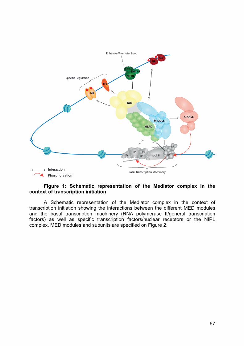
67
Figure 1: Schematic representation of the Mediator complex in the context of transcription initiation
A Schematic representation of the Mediator complex in the context of transcription initiation showing the interactions between the different MED modules and the basal transcription machinery (RNA polymerase II/general transcription factors) as well as specific transcription factors/nuclear receptors or the NIPL complex. MED modules and subunits are specified on Figure 2.
pol IIIID
GR
NIPBL
SMC3SMC1A
IIA IIB IIH
IIF
IIE
TFs
KINASE
TAIL
GR
NR
Specific Regulation
Basal Transcription Machinery
Enhancer/Promoter Loop
Interaction
Phosphoryation
HEAD
MIDDLE
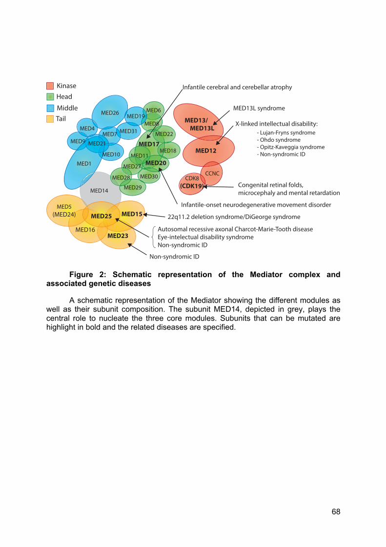
68
Figure 2: Schematic representation of the Mediator complex and associated genetic diseases
A schematic representation of the Mediator showing the different modules as well as their subunit composition. The subunit MED14, depicted in grey, plays the central role to nucleate the three core modules. Subunits that can be mutated are highlight in bold and the related diseases are specified.
MED26
MED17
MED31MED4 MMED7
MED6
MED8MED19
CDK8(CDK19)
MED13/ MED13L
MED12
CCNC
MED5(MED24)
MED16
MED11
MED22
MED14
MED23
MED15MED25
MED28
MED18
MED308
MED29
MEM
MED27
1MED20
KinaseHead
MiddleTail
Infantile cerebral and cerebellar atrophy
X-linked intellectual disability: - Lujan-Fryns syndrome - Ohdo syndrome - Opitz-Kaveggia syndrome - Non-syndromic ID
Non-syndromic ID
Autosomal recessive axonal Charcot-Marie-Tooth diseaseEye-intelectual disability syndromeNon-syndromic ID
MED13L syndrome
22q11.2 deletion syndrome/DiGeorge syndrome
Congenital retinal folds, microcephaly and mental retardation
Infantile-onset neurodegenerative movement disorder
MED1MED10
MED21MED9
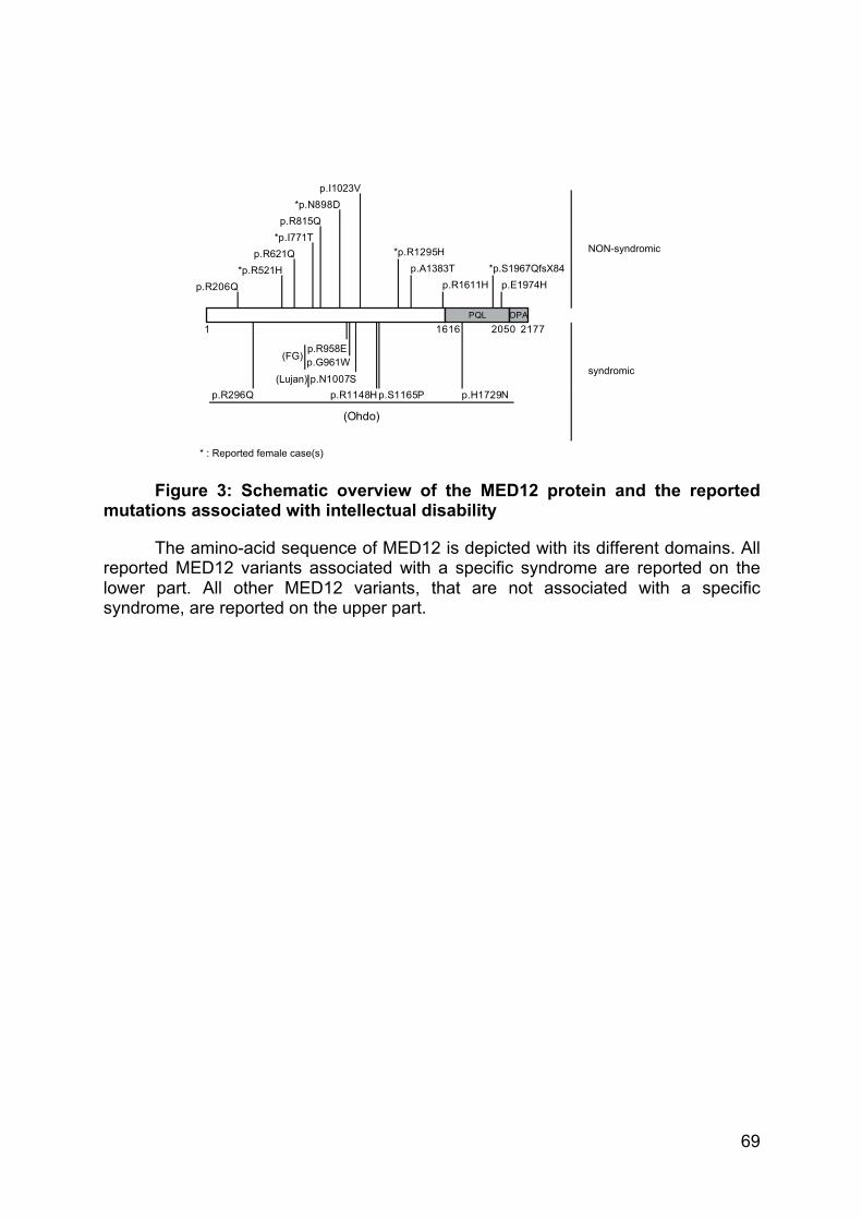
69
Figure 3: Schematic overview of the MED12 protein and the reported mutations associated with intellectual disability
The amino-acid sequence of MED12 is depicted with its different domains. All reported MED12 variants associated with a specific syndrome are reported on the lower part. All other MED12 variants, that are not associated with a specific syndrome, are reported on the upper part.
PQL1616 2050
p.R958E
*p.N898D
(Lujan) p.N1007S
*p.R1295H
p.R206Q
1 2177
(Ohdo)
p.S1165P p.H1729Np.R1148H
OPA
p.G961W(FG)
p.A1383Tp.E1974H
*p.S1967QfsX84*p.R521Hp.R621Q
*p.I771Tp.R815Q
p.I1023V
p.R296Q
p.R1611H
NON-syndromic
syndromic
* : Reported female case(s)
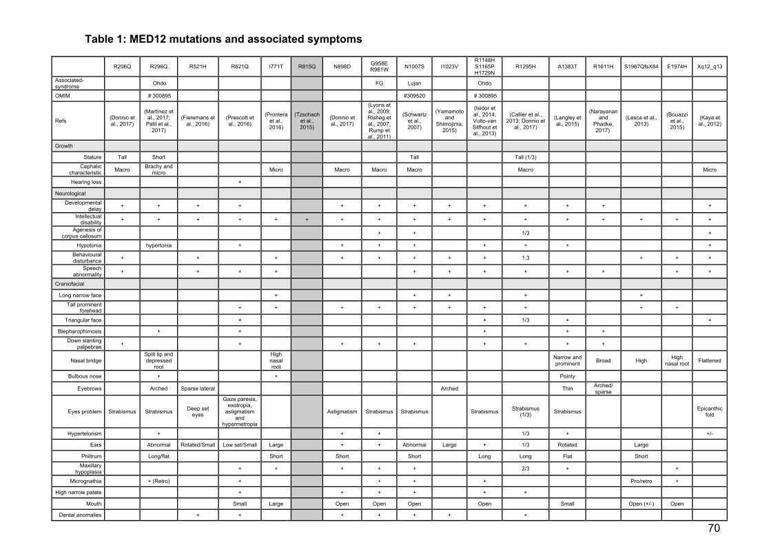
70
Table 1: MED12 mutations and associated symptoms
R206Q R296Q R521H R621Q I771T R815Q N898D G958E R961W N1007S I1023V
R1148H S1165P H1729N
R1295H A1383T R1611H S1967QfsX84 E1974H Xq12_q13
Associated-syndrome Ohdo FG Lujan Ohdo
OMIM # 300895 #309520 # 300895
Refs (Donnio et al., 2017)
(Martínez et al., 2017; Patil et al.,
2017)
(Fieremans et al., 2016)
(Prescott et al., 2016)
(Prontera et al., 2016)
(Tzschach et al., 2015)
(Donnio et al., 2017)
(Lyons et al., 2009; Risheg et al., 2007; Rump et al., 2011)
(Schwartz et al., 2007)
(Yamamoto and
Shimojima, 2015)
(Isidor et al., 2014; Vulto-van Silfhout et al., 2013)
(Callier et al., 2013; Donnio et
al., 2017)
(Langley et al., 2015)
(Narayanan and
Phadke, 2017)
(Lesca et al., 2013)
(Bouazzi et al., 2015)
(Kaya et al., 2012)
Growth
Stature Tall Short Tall Tall (1/3)
Cephalic characteristic Macro Brachy and
micro Micro Macro Macro Macro Macro Micro
Hearing loss +
Neurological
Developmental delay + + + + + + + + + + + + +
Intellectual disability + + + + + + + + + + + + + + + + +
Agenesis of corpus callosum + + 1/3 +
Hypotonia hypertonia + + + + + + + +
Behavioural disturbance + + + + + + + + 1.3 + + +
Speech abnormality + + + + + + + + + + + +
Craniofacial
Long narrow face + + + + +
Tall prominent forehead + + + + + + + + + +
Triangular face + + 1/3 + +
Blepharophimosis + + + + +
Down slanting palpebrae + + + + + + + + +
Nasal bridge Split tip and depressed
root
High nasal root
Narrow and prominent Broad High High
nasal root Flattened
Bulbous nose + + Pointy
Eyebrows Arched Sparse lateral Arched Thin Arched/ sparse
Eyes problem Strabismus Strabismus Deep set eyes
Gaze paresis, exotropia,
astigmatism and
hypermetropia
Astigmatism Strabismus Strabismus Strabismus Strabismus (1/3) Strabismus Epicanthic
fold
Hypertelorism + + + 1/3 + +/-
Ears Abnormal Rotated/Small Low set/Small Large + + Abnormal Large + 1/3 Rotated Large
Philtrum Long/flat Short Short Short Long Long Flat Short
Maxillary hypoplasia + + + + + 2/3 + +
Micrognathia + (Retro) + + + + Pro/retro +
High narrow palate + + + + + +
Mouth Small Large Open Open Open Open Small Open (+/-) Open
Dental anomalies + + + + + + +
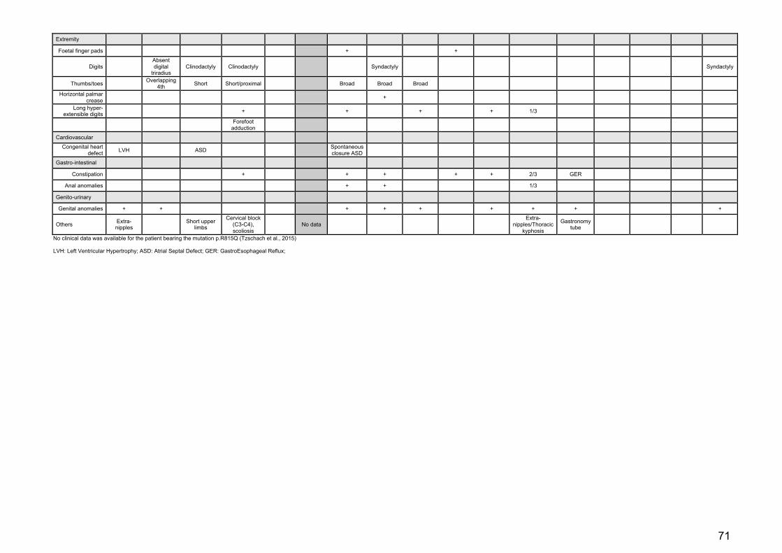
71
Extremity
Foetal finger pads + +
Digits Absent digital
triradius Clinodactyly Clinodactyly Syndactyly Syndactyly
Thumbs/toes Overlapping 4th Short Short/proximal Broad Broad Broad
Horizontal palmar crease +
Long hyper-extensible digits + + + + 1/3
Forefoot adduction
Cardiovascular
Congenital heart defect LVH ASD Spontaneous
closure ASD
Gastro-intestinal
Constipation + + + + + 2/3 GER
Anal anomalies + + 1/3
Genito-urinary
Genital anomalies + + + + + + + + +
Others Extra-nipples Short upper
limbs
Cervical block (C3-C4), scoliosis
No data Extra-
nipples/Thoracic kyphosis
Gastronomy tube
No clinical data was available for the patient bearing the mutation p.R815Q (Tzschach et al., 2015) LVH: Left Ventricular Hypertrophy; ASD: Atrial Septal Defect; GER: GastroEsophageal Reflux;
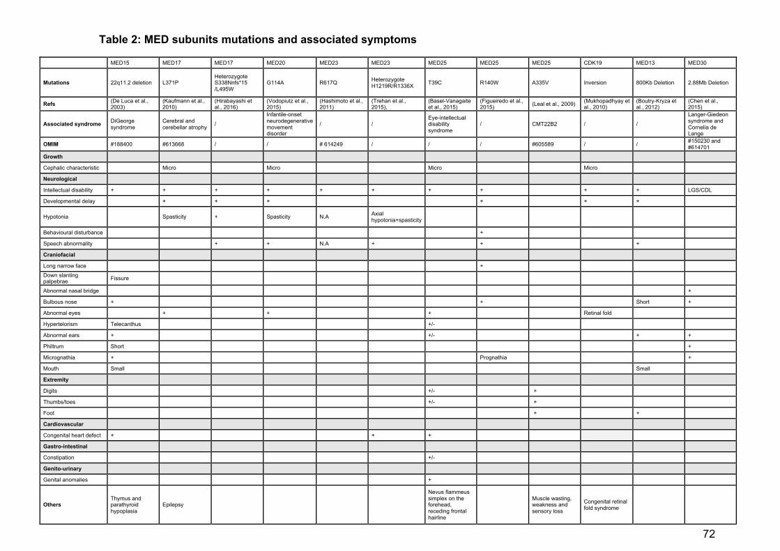
72
Table 2: MED subunits mutations and associated symptoms
MED15 MED17 MED17 MED20 MED23 MED23 MED25 MED25 MED25 CDK19 MED13 MED30
Mutations 22q11.2 deletion L371P Heterozygote S338Nnfs*15 /L495W
G114A R617Q Heterozygote H1219R/R1336X T39C R140W A335V Inversion 800Kb Deletion 2.88Mb Deletion
Refs (De Luca et al., 2003)
(Kaufmann et al., 2010)
(Hirabayashi et al., 2016)
(Vodopiutz et al., 2015)
(Hashimoto et al., 2011)
(Trehan et al., 2015),
(Basel-Vanagaite et al., 2015)
(Figueiredo et al., 2015) (Leal et al., 2009) (Mukhopadhyay et
al., 2010) (Boutry-Kryza et al., 2012)
(Chen et al., 2015)
Associated syndrome DiGeorge syndrome
Cerebral and cerebellar atrophy /
Infantile-onset neurodegenerative movement disorder
/ / Eye-intellectual disability syndrome
/ CMT22B2 / /
Langer-Giedeon syndrome and Cornelia de Lange
OMIM #188400 #613668 / / # 614249 / / / #605589 / / #150230 and #614701
Growth
Cephalic characteristic Micro Micro Micro Micro
Neurological
Intellectual disability + + + + + + + + + + LGS/CDL
Developmental delay + + + + + +
Hypotonia Spasticity + Spasticity N.A Axial hypotonia+spasticity
Behavioural disturbance +
Speech abnormality + + N.A + + +
Craniofacial
Long narrow face +
Down slanting palpebrae Fissure
Abnormal nasal bridge +
Bulbous nose + + Short +
Abnormal eyes + + + Retinal fold
Hypertelorism Telecanthus +/-
Abnormal ears + +/- + +
Philtrum Short +
Micrognathia + Prognathia +
Mouth Small Small
Extremity
Digits +/- +
Thumbs/toes +/- +
Foot + +
Cardiovascular
Congenital heart defect + + +
Gastro-intestinal
Constipation +/-
Genito-urinary
Genital anomalies +
Others Thymus and parathyroid hypoplasia
Epilepsy
Nevus flammeus simplex on the forehead, receding frontal hairline
Muscle wasting, weakness and sensory loss
Congenital retinal fold syndrome

73
Results

74
I - The nucleotide excision repair factor XPC
Manuscript in preparation
75
Cooperation between E2F1 and XPC to regulate H3 histone
acetylation by GCN5 acetylase
BIDON Baptiste∞, ILTIS Izarn∞, SEMER Marissa, NAGY Zita, LARNICOL Annabel,
COIN Fréderic, EGLY Jean-Marc* & LE MAY Nicolas*
Département de génomique fonctionnelle et Cancer, CNRS, INSERM, Université de
Strasbourg, Institut de Génétique et de Biologie Moléculaire et Cellulaire (IGBMC), 1
rue Laurent Fries, 67404 Illkirch-Graffenstaden, France.
* To whom correspondences should be address:
[email protected]; [email protected]
∞ Should be regarded as joint First Authors
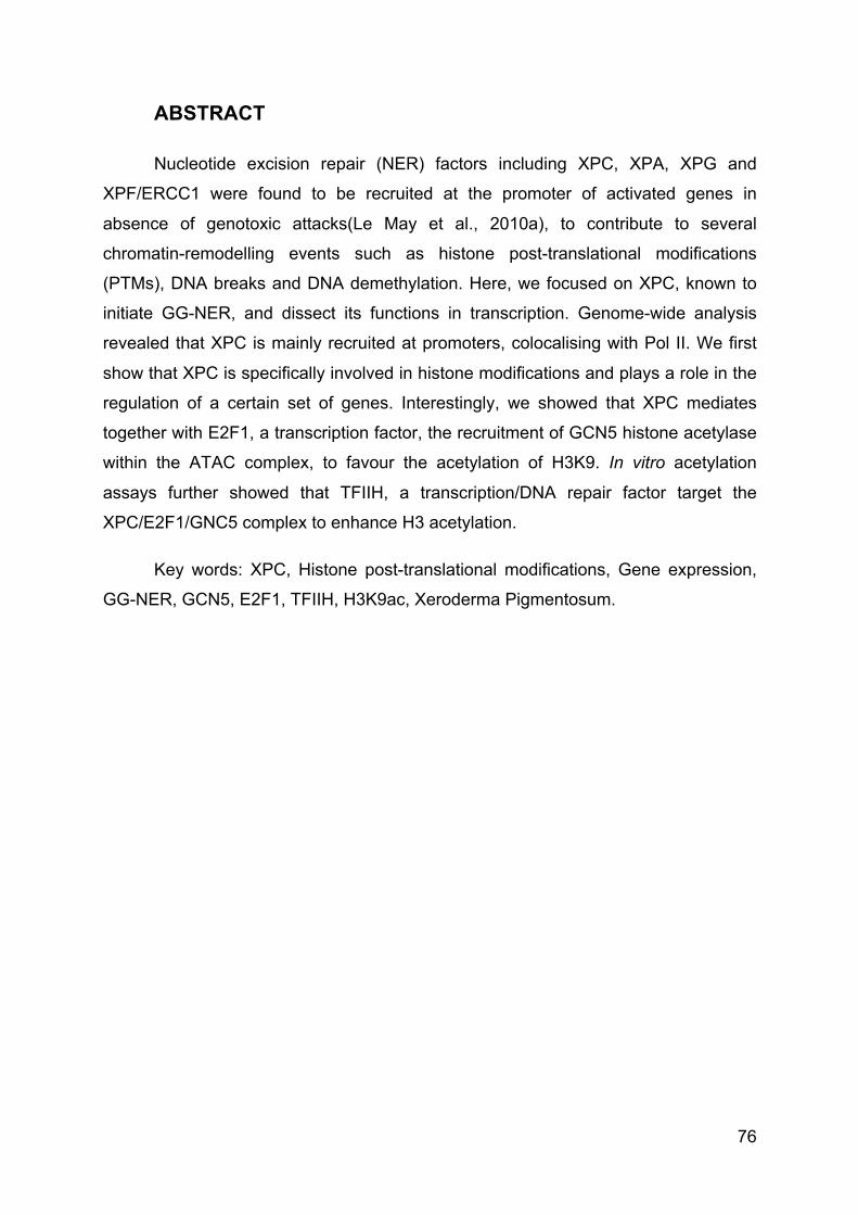
76
ABSTRACT
Nucleotide excision repair (NER) factors including XPC, XPA, XPG and
XPF/ERCC1 were found to be recruited at the promoter of activated genes in
absence of genotoxic attacks(Le May et al., 2010a), to contribute to several
chromatin-remodelling events such as histone post-translational modifications
(PTMs), DNA breaks and DNA demethylation. Here, we focused on XPC, known to
initiate GG-NER, and dissect its functions in transcription. Genome-wide analysis
revealed that XPC is mainly recruited at promoters, colocalising with Pol II. We first
show that XPC is specifically involved in histone modifications and plays a role in the
regulation of a certain set of genes. Interestingly, we showed that XPC mediates
together with E2F1, a transcription factor, the recruitment of GCN5 histone acetylase
within the ATAC complex, to favour the acetylation of H3K9. In vitro acetylation
assays further showed that TFIIH, a transcription/DNA repair factor target the
XPC/E2F1/GNC5 complex to enhance H3 acetylation.
Key words: XPC, Histone post-translational modifications, Gene expression,
GG-NER, GCN5, E2F1, TFIIH, H3K9ac, Xeroderma Pigmentosum.
77
INTRODUCTION
Transcription is a highly orchestrated event that requires several hundreds of
proteins, including the basal transcription machinery, the Mediator, the co-activators
and the chromatin remodelers. Transcription initiation also requires the recruitment of
certain DNA repair proteins. The link between DNA repair and transcription was first
established with the discovery of the multi-protein complex TFIIH, which is both a
basal transcription and a nucleotide-excision repair (NER) factor(Feaver et al., 1993;
Schaeffer et al., 1993). This connection was then strengthened by the discovery that
other NER factors (CSB, XPC, XPA, XPG, XPF/ERCC1) also participate to
transcription(Barreto et al., 2007; Le May et al., 2010a; Schmitz et al., 2009). These
factors were first characterized as part of NER, a DNA repair pathway that can
eliminate a wide variety of DNA lesions, originated by endogenous or exogenous
genotoxic attacks like UV irradiation(Friedberg et al., 2006). While Cockayne
syndrome group A and B (CSA and CSB) proteins target RNA polymerase II (Pol II)
stalled in front of a DNA lesions to engage transcription coupled-NER (TC-NER),
Xeroderma pigmentosum group C (XPC) protein, in complex with HR23B, recognizes
a wide variety of DNA damages located all over the genome, to initiate the global
genome-NER (GG-NER)(Sugasawa, 2010). XPC was also shown to regulate the
DDB2 complex, which mediate chromatin decondensation through ubiquitination
around DNA damages(Luijsterburg et al., 2012; Takedachi et al., 2010).
Our group has shown earlier that NER factors are associated with the RNA
polymerase II (Pol II) transcription machinery and are sequentially recruited
(XPC/CSB followed by RPA/XPA and XPG/XPF) at the promoter of retinoic acid-
activated RARß2(Le May et al., 2010a). In such conditions, the recruitment of the
NER factors is necessary to achieve optimal chromatin remodelling, including histone
post-translational modifications (PTMs) as well as active DNA demethylation, DNA
breaks induction and gene looping(Barreto et al., 2007; Le May et al., 2010a, 2012;
Schmitz et al., 2009). More recently, a DNA repair complex containing XPC has been
characterised to function as a coactivator of Oct4/Sox2 in ES and iPS cells(Cattoglio
et al., 2015; Fong et al., 2011).
Understanding the functions of NER factors is particularly important since
mutations in the genes give rise to the human autosomal recessive disorder
78
Xeroderma pigmentosum (XP). Biochemical defect in XP-C represents the most
frequent NER defective group. Clinically, XPC is characterized by an extreme
photosensitivity and a high susceptibility to develop tumours (melanoma, squamous
cell carcinoma) on sunlight-exposed areas of the skin(Cleaver, 2005), accompanied
by an increased susceptibility for lung, breast and colorectal cancers. However, these
XPC individuals rarely exhibit neurological disorders or developmental defects.
In the present study, we have investigated the roles of XPC in chromatin
remodelling upon transcription. Our genome-wide scale data have revealed that XPC
is mainly recruited with Pol II at promoter regions. A specific set of gene is down
regulated when XPC is absent of their promoter. The absence of XPC coincided with
deregulated enrichment of Pol II and altered euchromatin marks, including H3K9ac,
at the promoter. The absence of H3K9ac on such promoters led us to identify
interaction between XPC and the Histone Acetyl-Transferase (HAT) General Control
of Nutrition 5/K Acetyl-Transferase 2A (GCN5/KAT2A), as part of the ATAC complex.
An E2F1 signature was found to characterize most of the genes regulated by
XPC/GCN5. Finally, we describe the recruitment of a complex including XPC/HR23B,
E2F1 and GCN5 on promoter of activated gene. This complex acts cooperatively with
the general transcription TFIIH to increase the GCN5 HAT activity. Altogether, the
results presented here provide new insights into the transcriptional role of XPC and
the molecular aetiology of XP-C.
RESULTS
XPC deficiency disturbs gene expression
To investigate the involvement of XPC in transcription, we analysed RARß2
transactivation in HeLa cells constitutively expressing shRNA directed against either
XPC (shXPC), XPA (shXPA) or scrambled (shCtrl), after all-trans retinoic acid
(ATRA) treatment. We also used fibroblasts derived from two XP-C patients
presenting severe and mild clinical features and bearing p.R579st (XPC/R579st) and
p.P334H (XPC/P334H) mutations respectively as well as XPC rescued fibroblasts
(XPC/WT)(Bernardes de Jesus et al., 2008; Gozukara et al., 2001; Li et al., 1993). At
79
6h post ATRA treatment, we observed a correlation between RARß2 expression
(Figure 1A1 and 1B1) and the recruitment of the transcriptional machinery (including
Pol II, RAR and TFIIH kinase CDK7) but also the NER factors (XPC, XPA, XPG) at
RARß2 promoter in shCtrl, XPC/WT as well as in XPC/P334H cells (Figure 1A2 and
A5, B2 and B5, B4 and B7). However, such correlation was abolished in shXPC and
XPC/R579st cells, (Figure 1A3 and A6, B3 and B6) both characterized by the
absence of XPC (Figure S1A). Surprisingly, in shXPA cells, we observed a reduced
RARß2 mRNA induction without deregulation of the recruitment of XPC or the
transcriptional machinery (Figure 1A4 and A7).
We next sought to identify the correlation between XPC and Pol II at the
genomic scale. Chromatin-immunoprecipitation (ChIP) followed by high-throughput
DNA sequencing (ChIP-seq) yielded 2,191 and 16,440 binding events for XPC and
Pol II respectively, 6 hours post ATRA treatment in XPC/WT cells (Figure 1C).
Interestingly, we identified 1,797 XPC/Pol II common binding events, representing
82% of XPC peaks and covering 1529 genes. According to HOMER annotation, one
third (32%) of the XPC binding events appeared at promoter region (Figure 1D). By
combination of promoter, 5’-UTR and exon annotated peaks (mostly located in the
first one), the proportion of XPC peaks located close to the transcription start site
(TSS) rises to 44%. Interestingly, 94% of the XPC-bound promoters also showed a
Pol II enrichment (Figure 1D).
Collectively, these findings established an important enrichment of XPC around
promoters also occupied by Pol II in XPC proficient cells, for up to 1500 genes.
XPC specifically controls H3K9 acetylation by recruiting GCN5
Following gene activation, the recruitment of the basal transcription machinery
and NER factors is accompanied by chromatin remodelling events like histones post-
translational modifications (PTMs), DNA demethylation, DNA breaks and gene
looping(Barreto et al., 2007; Fong et al., 2013; Le May et al., 2010a; Schmitz et al.,
2009). We observed that XPC recruitment at the RARß2 promoter was crucial for
preparing accurate transcription: (i) BioChip assay which measures the incorporation
of biotinylated dUTP within broken DNA, evidenced DNA cleavages in XPC/WT and
XPC/P334H but not in shXPC, and XPC/R579st (Figure S1B). (ii) Unmethylated DNA
80
immunoprecipitation (UnMeDIP) showed that in XPC deficient cells, the RARß2
promoter region remained methylated contrary to what occured in wild type cells
(Figure S1C). (iii) Similarly, the q3C assay did not show any stable interaction (loop
formation) between the promoter and the terminator of the RARß2 in shXPC and
XPC/R579st cells (Figure S1D).
We next showed that upon ATRA treatment, an increase of H3K9ac and
H3K4me3 active histone marks was detected around RARß2 promoter in shCtrl,
XPC/WT and XPC/P334H cells, whereas this enrichment was not observed in shXPC
and XPC/R579st cells (Figure 2A). Note that the silencing of XPC and XPA do not
disturbed the steady state level of these histone PTMs (Figure S1E). Interestingly, in
shXPA cells, where the normal recruitment of XPC was detected (Figure 1A), the
H3K9ac as well as H3K4me3 marks were not disturbed (Figure 2A, left panel), while
the DNA breaks, DNA demethylation and DNA loop were indeed absent(Le May et
al., 2010a, 2012). These results suggested that chromatin remodelling events were
specifically related to the presence of XPC, acting independently of the other NER
factors. ChIP-seq analysis next showed that among the 1,797 XPC/Pol II binding
events detected in XPC/WT cells, 98% (1756) were enriched for the H3K9ac mark
and 78% (1385) with both H3K9ac and H3K4me3 marks (Figure 2B). Histones
modifications are regulated by various enzymes such as lysine methyltransferases
(KMT), lysine-demethylases (KDM), histone acetyltransferases (HAT) as well as
histone deacetylases (HDAC), also found around the promoter of activated genes.
Given the role of GCN5 in H3K9 acetylation during NER(Guo et al., 2011), we
investigated whether the variation of H3K9ac at the activated RARß2 promoter could
be mediated by XPC. We detected the presence of GCN5 concomitantly to the
increased acetylation of H3K9 at the activated RARß2 promoter in shCtrl, XPC/WT,
XPC/P334H as well as in shXPA cells but not in XPC deficient cells (Figure 2A). Note
that the steady state level of GCN5 was not disturbed by the absence of XPC or XPA
(Figure S2A). ChIP-reChIP (XPC/GCN5 or GCN5/XPC) experiments next showed
that XPC and GCN5 co-occupied the RARß2 promoter in shCtrl cells but not in
shXPC cells (Figure 2C). ChIP-seq performed in ATRA treated XPC/WT cells yielded
19,758 genomic locations for GCN5 (Figure 2D). Among the 1797 XPC/Pol II co-
occupied positions, 53% (948) were overlapping with GCN5 binding events. As
expected, almost all of the common XPC/Pol II/GCN5 binding events are enriched for
81
the H3K9ac mark (99%, Figure S2B). HOMER annotation further indicated that
almost 50% of them were located close to the TSS (Figure S2C).
All together our results underlined the involvement of XPC in the regulation of
histones modifications at promoters of ATRA activated genes. They also indicated a
significant correlation between XPC and GCN5 recruitment, associated with promoter
specific H3K9 acetylation.
XPC interacts directly with GCN5
We next investigated the sequence of events that made possible GCN5
recruitment. In cells lacking GCN5 (si GCN5) (Figure 3A), RARß2 expression was
decreased compared to cells transfected with scrambled siRNA (si Ctrl) (Figure 3B).
ChIP experiments next showed that silencing GCN5 did not alter the enrichment of
both Pol II and XPC at RARß2 promoter but prevent the acetylation of H3K9
compared to si Ctrl cells (Figure 3C and 3D). It seemed that the H3K9 acetylation at
RARß2 promoter required GCN5. Indeed, this later joined the promoter upon the
recruitment of the basal transcription machinery.
To further investigate the relationship between XPC and GCN5, we designed
several experiments. Using nuclear extracts from XPC/WT cells, we found that XPC
and GCN5 co-immunoprecipitated (Figure 3E, lanes 5 and 7); such complex was not
found in XPC/R579st cells (lanes 6 and 8). We next produced and purified the
recombinant tagged heterodimer XPC/HR23B and GCN5 (Figure 3F, left panel). We
found an interaction between XPC and GCN5 (Figure 3F, right panel). Searching for
XPC/GCN5 interacting domains, we designed and purified several truncated
recombinant proteins (Figure 3G)(Bernardes de Jesus et al., 2008). GCN5 was found
to interact with all the XPC variants except with XPC/Q474st. This further delineates
the 474-579 XPC domain as required for its interaction with GCN5.
Collectively, these data showed that the regulation of H3K9 acetylation by XPC
is mediated by GCN5.
GCN5 as part of the ATAC complex is recruited by XPC
RNA-seq analysis was performed in parallel to ChIP-seq in ATRA treated
XPC/WT and XPC/R579st cells. Among the 1529 genes that were covered by
82
XPC/Pol II binding events (Figure 1C), RNA-seq allowed the identification of 283
genes that were significantly down regulated (Table S1). We also observed that in
our experimental conditions, some of them (around 200) were up regulated (see
below). ChIP-seq comparison between XPC/WT and XPC/R579st cells highlighted a
correlation between the level of PTMS in the TSS surrounding and the level of gene
expression (Figure S3A). Indeed, down-regulated genes failed to be surrounded by
H3K9ac and H3K4me3 in XPC/R579st cells while these PTMs remained higher for
the up-regulated genes. Interestingly, the comparison of GCN5 ChIP-seq data
between these two cell lines showed the presence of GCN5, on the promoters of
down-regulated genes, correlating with increased H3K9ac (Figure S3A). Surprisingly,
GCN5 was almost absent at the promoter of all the up-regulated genes in XPC/WT
cells while H3K9 was acetylated. Expression of representatives of up regulated and
down regulated genes such as the cyclin D1 (CCND1) and the death associated
protein kinase 1 (DAPK1) respectively were analysed. CCDN1 is overexpressed in
XPC/R579st cells compared to XPC/WT and XPC/P334H cells as opposed to what
occurs with DAPK1 in XPC/R579st cells compared to XPC/WT as well as
XPC/P334H (Figure 4A1 and A2). ChIP experiments further showed that the
recruitment of XPC and Pol II (Figure 4A3 and A4) as well as the presence of
acetylated H3K9 and H3K14 paralleled CCDN1 and DAPK1 expression (Figure 4A5
and 4A6) in the corresponding cells.
GCN5 is found as part of two functionally distinct coactivator complexes, SAGA
(Spt Ada GCN5 Acetyltransferase) and ATAC (Ada Two A Containing); both included
GCN5 or its closely related paralog PCAF as well as distinct ADA family proteins. We
thus investigated which GCN5 containing complex (SAGA and/or ATAC) could be
associated with XPC. Immuno-precipitations (IP) from XPC/WT nuclear extracts
showed that XPC co-precipitate with ZZZ3 and WDR5 subunits of ATAC as well as
with TRRAP and SPT7L subunits of SAGA respectively (Figure 4B and 4C, lanes 3).
Further IPs using antibodies directed against either TRRAP or ZZZ3 subunits
showed that XPC could be detected distinctly in both complexes (Figure 3C, lanes 4
and 5). Interestingly, PCAF was not immunoprecipitated by XPC although this
histone acetyl-transferase HAT was detected in both SAGA and ATAC (Figure 4C).
We then were wondering whether the recruitment of such complexes was XPC
dependent. While H3 acetylation parallel gene expression, ChIP experiments detect
83
a significant and specific enrichment of ZZZ3 (ATAC) that parallel the recruitment of
GCN5 at DAPK1 in XPC/WT and XPC/P334H cells (Figure 4A10). No enhancement
of the TRRAP (SAGA complex) was observed in these ATRA treated cells. In
XPC/R579st cells, ATAC as well as SAGA complexes were not recruited. Note that
all the up-regulated genes as exemplified by CCDN1 were unable to recruit GCN5
containing complexes while H3K4 and H3K14 acetylation occurred in XPC deficient
ATRA treated cells (Figure 4A5 and A9, and Fig S3A). Interestingly, we detected the
enrichment of ZZZ3 (but not TRRAP) together with GCN5 at RARß2 promoter in
ATRA treated shCtrl and shXPA cells while this pattern was lost in shXPC cells,
indicating that the ATAC recruitment specifically depended on XPC (Figure S3B).
Knowing the dual role of XPC in transcription and DNA repair, we next
determined whether GCN5 contribution to GG-NER, after UVC irradiation, involved
SAGA or ATAC presence at damage sites. We then followed the coming and going
of factors by ChIP/Western assays upon UV irradiation of Hela cells(Coin et al.,
2008). We observed that XPC presence decreased 5min after UV irradiation (Figure
4C), once having attracted TFIIH (as indicated by the detection of its XPB subunit).
At 15min post UV, we visualized a transient arrival of GCN5 together with WDR5
subunit of ATAC as well as a significant increase of H3K9 acetylation (Figure 4C,
lane 3). Similarly using XPB antibodies, we also observed 15min post UV, the
transient presence of GCN5 together with the ATAC subunit (Figure 4D, lane 3). To
be noted also the arrival of the XPF endonuclease starting at 5min post UV,
indicating that the removal of the DNA damage generated by UV irradiation was on
going (Figure 4D, lanes 2-4).
Altogether, our findings indicated that H3 acetylation resulted from the
recruitment of GCN5 as part of the ATAC complex by XPC, a situation also observed
in DNA repair.
E2F1 cooperate with XPC for recruiting GCN5
We next investigated whether DNA binding elements could be selective in
recruiting the ATAC complex in a XPC dependent manner at specific genomic
locations(Krebs et al., 2011; Spedale et al., 2012). Gene Ontology (GO) indicated
that most of the down-regulated genes (254/283; p-value 1,99.10-12) contained
84
E2F1 binding site around their TSS (Table S2). Moreover, E2F1 ChIP-seq data from
ENCODE (www.encodeproject.org) highlighted a stronger E2F1 binding factor
around TSS from the down regulated genes (Figure 5A).
It has been described that E2F1 can directly interact with GCN5 to stimulate
transcription of its target genes(Lang et al., 2001). Antibodies directed towards E2F1
precipitated both XPC and GCN5 from XPC/WT nuclear extracts (Figure 5B).
Moreover, using purified recombinant proteins (Figure 5C, right panel), we observed
that antibody directed towards GCN5 precipitated XPC and E2F1 (Figure 5C, left
panel, lanes 7, 9 and 10)(Singh and Dagnino, 2016), underlining a partnership
between these three components.
ChIP experiments next showed the recruitment of E2F1 at the promoter of
DAPK1, that possess an E2F1 binding site, in ATRA treated XPC proficient cells
(Figure 5D). Such enrichment was lost in XPC/R579Stp cells (lacking XPC), while it
was still detected in XPC/P334H cells (Figure 5D). E2F1 was not recruited at CCDN1
that did not contain an E2F1 responsive element. We next evaluate the impact of
E2F1 on CCDN1 and DAPK1 by transiently transfecting XPC/WT cells with siRNA
targeting either E2F1 or GCN5 (Figure 5E). Silencing E2F1 abolished DAPK1
transactivation, a situation also observed when silencing GCN5 (Figure 5F). On the
contrary, silencing E2F1 and GCN5 did not affect transactivation of genes such as
CCND1 (Figure 5F). In si E2F1 and si GCN5 cells, Pol II as well as XPC were
recruited at DAPK1 promoter (Figure 5G). In si E2F1, XPC as well as GCN5 were
absent while in si GCN5 cells, E2F1 was present (Figure 5H) suggesting that E2F1
was involved in the positioning of XPC at the DAPK1 promoter. The absence of
either GCN5 or E2F1 did not allow histone acetylation around DAPK1 promoter
(Figure 5I). Silencing either E2F1 or GCN5 did not disturb CCND1 expression (Figure
5F). In such case, as expected, Pol II, TFIIH/XPB as well as XPC were recruited and
H3K9 was acetylated in the three cell lines (Figure 5G, 5H and 5I). Interestingly,
ChIP-seq patterns clearly showed the presence of XPC, Pol II, GCN5, E2F1 as well
as H3K9ac and H3K4me3 around the TSS of DAPK1, the expression of which was
down regulated in XPC/R579st cells (Figure S3C). On the contrary, the absence of
XPC did not prevent the recruitment of Pol II and HPTMs that was even increased
around CCND1; in this later case we noticed that neither GCN5 nor E2F were
present at the promoter (Figure S3C).
85
TFIIH enhances GCN5 enzymatic activity
The above data underlined a connection between XPC and H3K9 acetylation.
To further evaluate the influence of XPC on histone acetylation, we set up an
acetylation assay. Note that a complete in vitro assay including the entire
transcription machinery with the E2F1 binding site in a chromatinized template was
not available. We first observed that GCN5 is able to acetylate H3K9 (Figure 6A,
lanes 1-2); Moreover, neither XPC nor E2F1 that co-localized and interacted with
GCN5 in vivo (Figure 5B) as well as in vitro (Figure 5C, lanes 7, 9 and 10) were able
to improve such H3K9 acetylation (Figure 6A, lanes 6, 7 and 12). Interestingly, the
transcription/DNA repair factor TFIIH stimulates the H3K9 acetylation by GCN5
(Figure 6A, lanes 5 and 8). Moreover, addition of XPC and/or E2F1 did not enhance
GCN5 HAT activity (figure 6A, lanes 13, 14 and 16). In our experimental conditions,
TFIIH fraction that did not contain a HAT activity (Figure 6A, lane 5) was not found to
phosphorylate GCN5 (data not shown).
Using purified recombinant proteins, we observed that antibody directed
towards GCN5 precipitated TFIIH, as shown by XPB and CDK7 (Figure 6B, lanes 8
and 13). Antibodies directed towards GCN5 are also able to precipitate XPC and
E2F1, in addition to TFIIH (Figure 6B, lane 19). Moreover, antibodies directed
towards TFIIH (XPB) precipitated both GCN5 and XPC from XPC/WT nuclear
extracts (Figure 6C, lanes 2). Interestingly, the interaction between GCN5 and XPB
was lost in absence of XPC (Figure S5, right panel). In addition, antibodies against
GCN5 precipitated XPB and XPC while antibodies directed towards XPC precipitated
XPB and GCN5, from XPC/WT nuclear extracts (Figure 6C, lanes 1 ad 3).
ChIP experiments next showed the recruitment of TFIIH at the promoter of
DAPK1 and CCND1, in ATRA treated XPC proficient cells (si Ctrl) (Figure 6D).
Silencing E2F1, which abolished DAPK1 transactivation (Figure 5F), decreases the
recruitment of TFIIH at the promoter of DAPK1; a situation also observed when
silencing GCN5 (Figure 6D). Interestingly, silencing E2F1 and GCN5 did not affect
the recruitment of TFIIH at the promoter of CCND1 (Figure 6D).
Altogether, these results indicated that TFIIH stimulated H3 acetylation by
interacting with GCN5.
86
DISCUSSION
Having shown NER factors together with the transcription machinery targeting
promoter of activated genes for the formation of the preinitiation complex(Le May et
al., 2010a), we were next focusing on their role in the RNA synthesis process.
Previous investigations have indicated the sequential arrival of the NER factors in the
following order: XPC, XPA, and XPG, XPF/ERCC1, once the transcription machinery
was positioned around the promoter of a given gene. Here we document the role of
XPC in regulating histones PTMS at the transcription initiation level.
ChIP-seq experiments demonstrated that in ATRA treated cells, XPC together
with RNA pol II was mainly localized around TSS (representing around 45% of XPC
binding events), of up to 1500 activated genes (Figure 1C and 1D). XPC was also
found located on other DNA regulatory regions (enhancers) such as those involved in
the pluripotency regulation of ES cells(Fong et al., 2011). Focusing on fibroblasts
from patients and rescued ones, we discovered that defect in XPC disrupt the
expression of a certain set of genes. We show that XPC, the first NER factor that
joined the transcription machinery(Le May et al., 2010a), is involved in the chromatin
remodelling process, and particularly in histone post-translational modifications
(Figure 2), as also observed in yeast where the ATP-chromatin remodelling complex
SWI/SNF targeted XPC after UV irradiation(Gong et al., 2006). It seems that part of
the chromatin remodelling process, is conditioned by the presence of XPC. Indeed, in
absence of XPA (as occurred in shXPA cells), that abrogated the recruitment of all
the NER factors except XPC, histone H3 acetylation around the promoter of activated
gene was present(Le May et al., 2010a, 2012). However, DNA breaks, DNA
demethylation and DNA loop, a role devoted to the other NER factors (Le May 2012),
were absent (Figure S1B-1D).
Our data suggest that XPC is specifically required for the chromatin
modification around the promoter of a certain set of genes, such as DAPK1. In this
case, the H3K9/H3K14 acetylation occurs when XPC leads to the GCN5 histone
acetylase recruitment (Figures 4A8 and S3), which in fact is not sufficient. This
recruitment also requires E2F1. Indeed, (i) 89% of the XPC dependent genes
contains E2F1 consensus binding site(s); (ii) the recruitment of both XPC and GCN5
is abrogated in E2F1 silenced cells as shown for DAPK1 (Figure 5F4 and F6); (iii)
87
neither E2F1 nor GCN5 are found at the promoter of activated genes in XPC-
deficient cells (Figures 4A, 5D and S3). Moreover, E2F1 recruit GCN5 within ATAC
complex instead of SAGA although both contain GCN5(Lang et al., 2001),
highlighting a potential role of the ATAC associated proteins in discriminating
expression of specific (or selected) genes. ATAC was shown to be preferentially
recruited to both promoters and enhancers, while SAGA can principally be found at
promoters(Krebs et al., 2011).
In addition to dissect the role of XPC during transcription, this work highlights
the uniqueness of the expression of each gene. We indeed show how components
(or signal) are required to activate a certain set of genes. Transcription is initiated
following (1) ATRA ligand induction; this later promotes the recruitment of the
corresponding nuclear receptor and the co-activator complexes. The general
transcription factors as well as Pol II next targets the promoter; (2) some of these
genes encompass an E2F1 responsive element and likely its cognate factor; (3) XPC
is recruited after the basal transcription machinery(Le May et al., 2010a); (4) both
E2F1 and XPC serve as a platform for the recruitment GCN5 within the ATAC
complex. GCN5 will then proceed to H3 histone acetylation (an essential step that
will allow chromatin opening) and allowed further RNA synthesis. This might explain
how among the 1500 genes targeted by both XPC and Pol II after ATRA treatment,
only 283 genes (as shown for DAPK1 but also for HOXB13 and LRRC11, Figure S6)
were down regulated in XPC deficient cells. Some other genes such as CCND1 were
also recruiting XPC and follow another scenario in which GCN5 is not involved. We
also have observed that in absence of XPC, the H3K4 methylation was defective as
well as the recruitment of its enzyme partner, SET1 methyl transferase (Figure S7).
The SET1 coding gene was one of the genes down regulated in absence of XPC
(Table S1). Whether or not this histone methylation modification is consecutive with
the histone acetylation process will require further investigations. Moreover, it cannot
be excluded that additional players such as components of ATAC, other histone
modification complexes, TBP associated factors (TAFs, know to interact with DNA
repair factors)(Chatzinikolaou et al., 2017; Kamileri et al., 2012) might also participate
in chromatin modification. It should be noted the parallel between transcription and
NER, in which GCN5 as well as E2F1 were shown to be involved in the regulation of
88
H3K9 acetylation(Guo et al., 2010, 2011; Kakumu et al., 2017), underlining how both
processes are connected and is common role of XPC.
It seems that XPC, together with E2F1, are involved in the recruitment of
GCN5 but not in the regulation of its activity. Our minimal in vitro system established
in absence of both chromatinized DNA and components of activated transcription,
showed that XPC and E2F1 either alone or in combination do not modify the level of
H3 acetylation (Fig. 6A). However we observed that H3K9 acetylation was stimulated
by TFIIH, which is not surprising knowing that: (i) intact TFIIH is required for proper
histones PTMs(Singh et al., 2015); (ii) binding of TFIIH to activated genes as well as
DNA lesions is impaired when silencing E2F1 (Figure 6D). Although we knew that the
phosphorylation of E2F1 at DNA lesions was required to promote GCN5 and NER
factors recruitment, we failed to identify phosphorylation of either GCN5 or E2F1 by
the CDK7 kinase of TFIIH. It is also possible that the phosphorylation of GCN5
required for its location at DNA lesions(Guo et al., 2010, 2011), could be performed
by other kinase such as the PKA. Indeed, during fasting, PKA phosphorylates GCN5,
thereby increasing GCN5 acetyl transferase activity(Sakai et al., 2016).
Then arise questions about the clinical features of XPC patients that mainly
display skin sensitivity to sunlight with a 1,000-fold increased susceptibility to
developing skin cancer. Among all the XP patients, XP-C ones rarely exhibit visible
neurological disorders or developmental defects. Interestingly, Gene Ontology point
out that a large proportion of genes was related to chromatin structure regulation and
present oncogenic and immunologic signatures (Table S1). Indeed, it has been
demonstrated that upon genotoxic attack, XPC can induce the expression of cytokine
like interleukin-6 (IL-6), that have pro-inflammatory effects in lung fibroblasts(Schreck
et al., 2016). Moreover, multi-omic analyses had identified factors and pathways
implicated in the cellular response to UV-induced DNA damages in relation with the
immune system (Boeing et al., 2016). For example, DAPK1 is an inhibitor of RIG-I
signalling which is necessary to induce the production of type I interferon(Willemsen
et al., 2017). It was also shown to act as a tumour suppressor in multiple cancer
types(Kissil et al., 1997; Raveh and Kimchi, 2001).
It is clear that further identification and analysis of the deregulated genes
would (i) help to determine relevant markers for an early and specific diagnosis, (ii)
89
anticipate/predict the cancer risk among the different symptoms within XPC patients.
In addition, our work uncover the role of XPC, one of the NER factors, in the
regulation of the expression of a certain set of genes through the regulation of
Histone PTMs.
EXPERIMENTAL PROCEDURE
Cell Culture
HeLa Silencix cells (Tebu-Bio) including shCtrl, shXPC and shXPA cells as
well as XP-C patients derived fibroblasts GM14867 (XPC/R579St) and GM02096
(XPC/P334H) and rescued XP-C (XPC/R579Stp + GFP-XPCwt)(Bernardes de Jesus
et al., 2008) were cultured in appropriate medium and maintain at 37°C in a 5% CO2
environment. 12 hours before ATRA treatment, cells were incubated in phenol red-
free medium with charcoal treated-FCS and 40 mg/ml gentamycin. Cells were treated
with 10µM all-trans retinoic acid (ATRA) (MP).
Antibodies
Antibodies towards His, Flag, RNA Pol II, RAR, TBP, XPA, GCN5, XPG,
ZZZ3, TRRAP and tubulin were produced at the IGBMC. CDK7 (C-19), XPB (S-19),
XPF (H300) and XPC (D-18) antibodies were purchased at Santa-Cruz
Biotechnology. H3k4me3 (ab1012) and CTCF (ab70303) antibodies were purchased
at Abcam and WDR5 (07-706) antibodies from Upstate. Antibodies against H3
(#4620), GCN5 (2676) and Spt7L were obtained from Cell signalling, Epigentek and
Bethyl respectively. Antibodies against H3K9ac (61251) and H3K14ac (39599) were
from Active Motif.
Reverse Transcription and Quantitative PCR
Total RNA was isolated from cells using a GenElute Mammalian Total RNA
Miniprep kit (Sigma) and reverse transcribed with SuperScript II reverse transcriptase
(Invitrogen). The quantitative PCR was done using a Lightcycler 480 (Roche). The
primer sequences used in qPCR are indicated in Table S3. The mRNA expression of
90
a gene represents the ratio between values from treated and untreated cells after
normalization against GAPDH.
Chromatin Immunoprecipitation (ChIP)
Cells were cross-linked at room temperature (RT) for 10 min with 1%
formaldehyde. Chromatin was prepared and sonicated on ice for 30 min using a
Qsonica Q800R as previously described12. Samples were immunoprecipitated with
antibodies at 4°C overnight, and protein G Sepharose beads (Upstate) were added,
incubated for 4 hours at 4°C, and sequentially washed. Protein-DNA complexes were
eluted and decrosslinked. DNA fragments were purified using QIAquick PCR
purification kit (QIAGEN) and analysed by qPCR using primers as indicated in Table
S3.
ChIP/Western blot on UV-irradiated cells
XPC/WT cells were seeded (106 cells per dish) 24h prior to the experiment,
rinsed with PBS and UV-irradiated (20 J/m2). Cells were then cross-linked at room
temperature for 30 min with 1% formaldehyde at indicated times post-UV irradiation
and chromatin was prepared(Fousteri et al., 2006). ChIP/Western blot, using XPB or
GFP antibodies, was performed as previously described(Coin et al., 2008). Briefly,
the chromatin suspension was sonicated in buffer S (10 mM Tris-HCl [pH 8.0], 140
mM NaCl, 1% Triton X-100, 0.1% Na-deoxycholate, 0.1% SDS) using the Qsonica
Q800R in 20s pulse followed by 40 s cooling. Samples were sedimented (13 000
rpm, 15 min), and the supernatant that contained the cross-linked chromatin was
frozen and stored at -80°C. In each assay, 600 µg of protein from cross-linked
chromatin was immunoprecipitated with 1 µg of antibody in buffer S for 4h at 4°C.
The immunocomplexes were collected by adsorption to protein G Sepharose beads
(Upstate, Billerica, MA) overnight at 4°C. The beads were next washed three times
with 5 volumes of buffer S and resuspended in 1 X Laemmlli SDS Buffer. Samples
were incubated at 95°C for 90 min for crosslinking reversal prior electrophoresis.
RNA-seq analysis
Total RNA from XPC/R579St and XPC/WT cells were extracted before or 6
hours after ATRA treatment (10uM) using TRI REAGENT (MRC) and purified by
91
phenol-chloroform extraction. Libraries was prepared with TruSeq Stranded mRNA
Sample Preparation kit following guide instruction and subsequently proceed on an
Illumina Hiseq 4000 as single-end 50 base reads following Illumina’s instructions.
Image analysis and base calling were performed using RTA 2.7.3 and bcl2fastq
2.17.1.14. Reads were mapped onto the hg19 assembly of the human genome.
Reads count was performed with HOMER v4.8.3(65) and differently expression was
estimated with EdgeR. Genome ontology was performed with Genomic Region
Enrichment Analysis Tool 3.0.0 (http://bejerano.stanford.edu/great/public/html/).
ChIP-seq analysis
Purified DNA fragments were prepared with the ChIP-IT High Sensitivity Kit
(Active Motif) and the related antibodies. ChIP-seq was performed on an Illumina
Hiseq 4000 as single-end 50 base reads following Illumina’s instructions. Image
analysis and base calling were performed using RTA 1.17.20 and CASAVA 1.8.2.
Reads were mapped onto the hg19 assembly of the human genome. Peak detection
was performed using MACS14 (http://liulab.dfci.harvard.edu/MACS/) under settings
where the input fraction was used as negative control. Peaks detected were
annotated using HOMER (http://biowhat.ucsd.edu/homer/ngs/annotation.html) as
well as TSS protein enrichment comparison. As reference coordinates, we used the
MACS-determined peaks of human genes as defined by RefSeq database.
Plasmids and purification of recombinant proteins
PCR products for the entire coding sequence XPC and the different variants
were cloned into pDONOR-207 vector using the Gateway system (Invitrogen) and
later cloned in bicistronic plasmid VEAP5317 with hHR23B kindly obtained from A.
Poterszman. For recombinant Flag-GCN5 expression in Sf9 cells, the corresponding
vector was kindly provided by L. Tora. PCR product for the entire coding sequence of
E2F1 was also cloned into pDONOR-207 vector and later sub-cloned in pAC8 vector.
Sf9 cells were infected with baculoviruses expressing a FLAG-tagged GCN5, Strep
XPC/ His-tagged hHR23B or c myc-tagged E2F1 and the harvested recombinant
proteins were purified as previously described(Singh et al., 2015).
GFP-ERCC3 (XPB), Flag-ERCC2 (XPD), GTF2H1 (p62), GTF2H4 (p52),
GTF2H2 (p44), GTF2H3 (p34), Flag-CDK7, CCNH (Cyclin H), MNAT1 (MAT1), and
92
GTF2H5 (p8/TTD-A) subunits of TFIIH were produced as previously
described(Dubaele et al., 2003).
Sf21 insect cells were infected with the different baculoviruses in order to
separately obtain core-IIH, CAK and ERCC2. The different whole-cell extracts were
incubated 4h at 4°C with anti-M2-Flag antibody bound to agarose beads. After
extensive washings, the immunoprecipitated fractions were eluted. The recombinant
TFIIH was made by mixing purified core-IIH, CAK, and ERCC2.
siRNA transfection
ON-TARGET plus smart pool siRNA control or targeting human GCN5 and
E2F1 were purchased from Dharmacon and transfected in HeLa or XPC/WT cells at
a final concentration of 100nM using X-tremeGENE siRNA transfection reagent
(Roche) following manufacturer protocol.
Co-immunoprecipation
For in vivo co-IPs, nuclear extracts from XPC/WT and XPC/R579Spt cells
were prepared as previously described (ref). After GFP-trap or GCN5, ZZZ3 or
TRRAP Immunoprecipitation using the appropriate antibodies conjugated to protein
G coated Dynabeads, followed by extensive washes (150mM NaCl) was carried out
and the different co-precipitated proteins were detected using specific antibodies
after immunoblotting.
For in vitro co-IPs, recombinant purified flag-GCN5 was then incubated with
recombinant purified Hid-hHR23B/Strep-XPC full-length or variants before Flag
immunoprecipitation was carried out. After washes, bound proteins were resolved by
SDS–PAGE and detected by western blot.
Histone acetyltransferase assay
Recombinant purified GCN5 was incubated with recombinant purified proteins
(XPC/hHR23B, E2F1 and TFIIH). The HAT of GCN5 was then measured using
histone acetyltransferase assay as previously described (Di Cerbo et al., 2014). For
the HAT assay, recombinant histones H3.3 and Octamer were incubated with GCN5
HAT and the other putative partners (XPC/HR32B, E2F1, TFIIH complex) in the
93
presence of acetyl-CoA (without in case of mock reaction) in HAT buffer (50 mM Tris-
HCl pH8.0, 7% glycerol, 25 mM NaCl, 0.1 mM EDTA, 5 mM DTT) for 1 hour at 30°C.
The reaction was then analysed by western blotting with specific antibodies (H3 and
H3K9ac) and the activity of the enzyme was checked using antibodies against known
specific targets.
Acknowledgements
We are grateful to Sylvain Daujat (IGBMC) for in vitro HAT assay, to A.
Poterszman (IGBMC) for the VEAP5317 construct, to members of our team and to
Didier Devys (IGBMC) for fruitful discussion. We thank IGBMC antibodies and cell
culture facilities. Sequencing was performed by the IGBMC Microarray and
Sequencing platform, a member of the “France Génomique” consortium (ANR-10-
INBS-0009). This study was supported by l’Association de la Recherche contre le
Cancer (ARC n°SL220130607082) and the National Research Foundation of Korea
for International Collaboration. This study was supported by the Ligue contre Le
cancer (CCIR-GE 2015). This study was supported by the grant ANR-10- LABX-
0030-INRT, a French State fund managed by the Agence Nationale de la Recherche
under the frame program Investissements d’Avenir ANR-10- IDEX-0002-02. B.B was
supported by the fondation pour la recherche médicale (FDT20160736467). M.S was
supported by le prix d'encouragement de la province Sud (Nouvelle Calédonie).
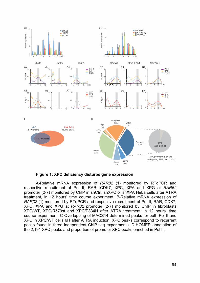
94
Figure 1: XPC deficiency disturbs gene expression
A-Relative mRNA expression of RARβ2 (1) monitored by RTqPCR and respective recruitment of Pol II, RAR, CDK7, XPC, XPA and XPG at RARβ2 promoter (2-7) monitored by ChIP in shCtrl, shXPC or shXPA HeLa cells after ATRA treatment, in 12 hours’ time course experiment. B-Relative mRNA expression of RARβ2 (1) monitored by RTqPCR and respective recruitment of Pol II, RAR, CDK7, XPC, XPA and XPG at RARβ2 promoter (2-7) monitored by ChIP in fibroblasts XPC/WT, XPC/R579st and XPC/P334H after ATRA treatment, in 12 hours’ time course experiment. C-Overlapping of MACS14 determined peaks for both Pol II and XPC in XPC/WT cells 6H after ATRA induction. XPC peaks correspond to recurrent peaks found in three independent ChIP-seq experiments. D-HOMER annotation of the 2,191 XPC peaks and proportion of promoter XPC peaks enriched in Pol II.
RNA Pol IIXPC
C
16,440 peaks2,191 peaks
1,797 peaks
D
TTS2%
3’UTR1%
ncRNA1%
XPC promoters peaks overlapping RNA pol II peaks
94%(658 peaks)
shCtrl
% In
put
% In
put
1.6
0.8
0120 4 8
120 4 8
1.6
0.8
0
shXPC
120 4 8
1.6
0.8
0
120 4 8
1.6
0.8
0
XPC/P334H XPC/R579St
80 2 64
6
3
0
80 2 64
6
3
0
8 (h)0 2 64
6
3
0
8 (h)0 2 64
6
3
0
XPC/WT
Pol IIRARCDK7
XPCXPAXPG
40 2 8
6
3
06
!"
6
3
080 2 64
% In
put
% In
put
A1
shXPA
Pol IIRARCDK7
XPCXPAXPG
12 (h)0 4 8
1.6
0.8
0
12 (h)0 4 8
1.6
0.8
0
0 3 8 12 (h)1 6
3
2
0
mRN
A ex
pres
sion
1
XPC/WTXPC/R579St XPC/P334H
3
2
0
mRN
A ex
pres
sion
1
0 3 8 12 (h)1 6
shCtrlshXPC shXPA
Promoter32%
Intron32%
Intergenic19%
Exon7%
5’UTR5%
B1
A2 A3 A4
A5 A6 A7
B2 B3 B4
B5 B6 B7
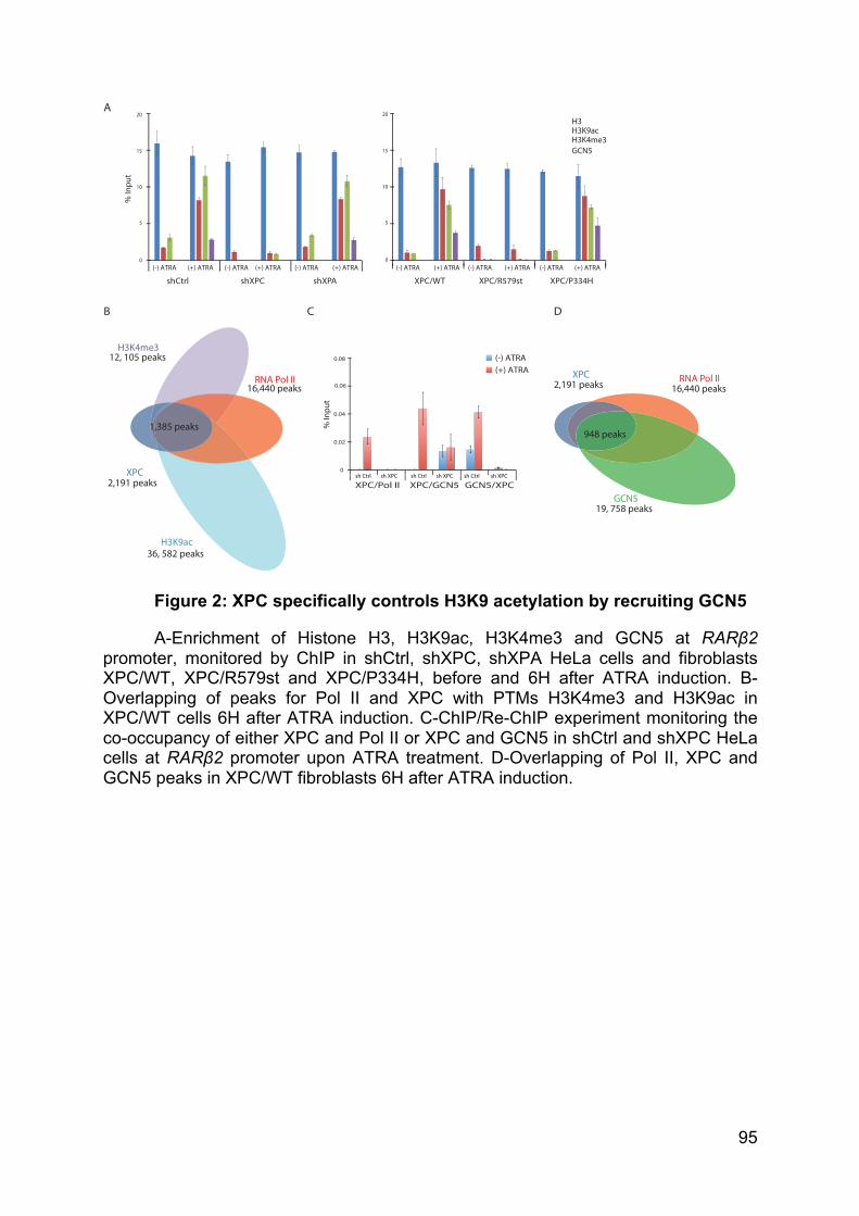
95
Figure 2: XPC specifically controls H3K9 acetylation by recruiting GCN5
A-Enrichment of Histone H3, H3K9ac, H3K4me3 and GCN5 at RARβ2 promoter, monitored by ChIP in shCtrl, shXPC, shXPA HeLa cells and fibroblasts XPC/WT, XPC/R579st and XPC/P334H, before and 6H after ATRA induction. B-Overlapping of peaks for Pol II and XPC with PTMs H3K4me3 and H3K9ac in XPC/WT cells 6H after ATRA induction. C-ChIP/Re-ChIP experiment monitoring the co-occupancy of either XPC and Pol II or XPC and GCN5 in shCtrl and shXPC HeLa cells at RARβ2 promoter upon ATRA treatment. D-Overlapping of Pol II, XPC and GCN5 peaks in XPC/WT fibroblasts 6H after ATRA induction.
A
B
H3K9ac36, 582 peaks
RNA Pol II
XPC
16,440 peaks
2,191 peaks
1,385 peaks
H3K4me312, 105 peaks
RNA Pol IIXPC16,440 peaks2,191 peaks
948 peaks
GCN519, 758 peaks
C
GCN5
0.08
0.06
0.04
0
0.02
sh Ctrl sh XPC sh Ctrl sh XPC sh Ctrl sh XPC
XPC/Pol II XPC/GCN5 GCN5/XPC
(-) ATRA(+) ATRA
% In
put
shCtrl shXPC shXPA XPC/WT XPC/R579st XPC/P334H(-) ATRA (+) ATRA (-) ATRA (+) ATRA (-) ATRA (+) ATRA(-) ATRA (+) ATRA (-) ATRA (+) ATRA (-) ATRA (+) ATRA
% In
put
20
15
5
10
0
20
15
5
10
0
H3H3K9acH3K4me3
D
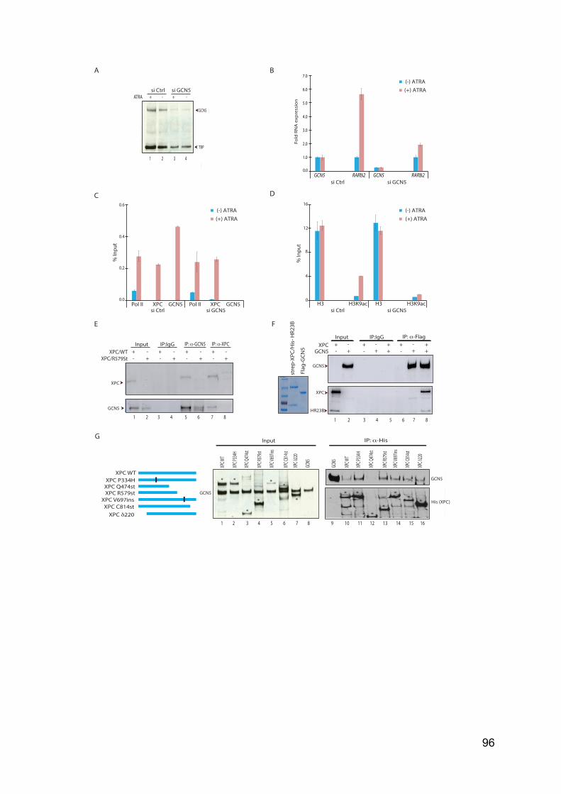
96
A B
XPC/WT
C
0.0
1.0
2.0
3.0
4.0
5.0
6.0
7.0
Fold
RN
A e
xpre
ssio
n
E F
XPCGCN5
+ -
- +
+ + -
- + +
+ + -
- + +
G
XPC WT
XPC δ220
XPC P334HXPC Q474stXPC R579st
XPC V697ins
XPC P
334H
XPC W
T
XPC Q
474st
XPC δ
220
XPC R
579st
XPC V
697in
s
XPC C814st
XPC C
814st
XPC P
334H
XPC W
T
XPC Q
474st
XPC δ
220
XPC R
579st
XPC V
697in
s
XPC C
814st
* *
*
*
**
** *
**
**
*
1 2 3 4 5 6 7 8 9 10 11 12 13 14 15 16
ATRA + - + -
GCN5
TBP
1 2 3 4
si Ctrl si GCN5Pol II XPC GCN5 Pol II XPC GCN5
% In
put
0.6
0.4
0.0
0.2
si Ctrl si GCN5H3 H3K9ac H3 H3K9ac
16
0
12
8
4
D
% In
put
GCN5 RARΒ2 GCN5 RARΒ2
(-) ATRA(+) ATRA
(-) ATRA(+) ATRA
(-) ATRA(+) ATRA
+ - + - + - + -XPC/R579St - + - + - + - +
1 2 3 4 5 6 7 8 1 2 3 4 5 6 7 8
GCN5
GCN5
si Ctrl si GCN5
si Ctrl si GCN5
XPC
GCN5
XPC
GCN5
His (XPC)
GCN5
Input
InputInput
IP:IgGIP:IgG
IP: α-GCN5 IP: α-XPCIP: α-Flag
IP: α-His
stre
p-XP
C/H
is- H
R23B
Flag
-GCN
5
HR23B
GCN5
97
Figure 3: XPC interacts directly with GCN5
A-GCN5 and TBP protein expression monitored by Western Blot in HeLa cells treated with scrambled siRNA (si Ctrl) or siRNA targeting GCN5 (si GCN5), before and 6H after ATRA treatment. B-Relative mRNA expression of RARβ2 and GCN5 in HeLa cells treated with si Ctrl or si GCN5, before and 6H after ATRA treatment. C-D-Occupancy of PolII, XPC and GCN5 as well as H3 and H3K9ac at RARβ2 promoter in HeLa cells treated with si Ctrl or si GCN, before and 6H after ATRA treatment. E-Immunoprecipitation performed on nuclear extract from XPC/WT or XPC/R579st fibroblasts with antibody against XPC, GCN5 or IgG. F-Blue staining of recombinant Flag-GCN5 and duplex strep-XPC/His-HR23B (left panel). In vitro co-immunoprecipitation assay performed by antibodies against Flag tag and IgG with the recombinant protein Flag-GCN5 and the duplex strep-XPC/His-HR23B (right panel). G-Global overview of His-tag XPC full length protein (XPC FL) and several mutated proteins or truncated proteins, from ether the N-terminus or the C-terminus. In vitro co-immuno-precipitation assay performed with antibodies against His-tag with the different recombinant His-XPC and Flag-GCN5. INPUT (left panel) and IP (right panel) are revealed by WB against His and Flag. Asterisk represents the position of various forms of XPC.
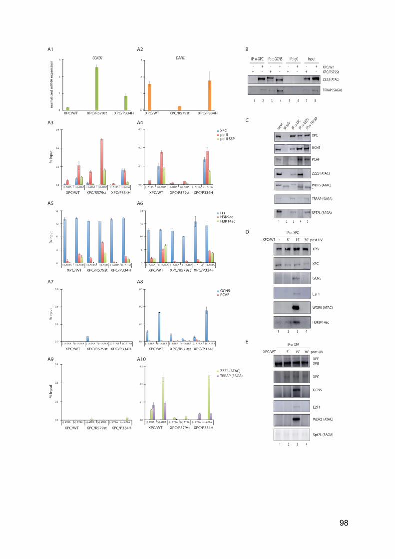
98
CCND1 DAPK1
norm
aliz
ed m
RNA
expr
essio
n%
Inpu
t
0.9
0.6
0.3
0.0
XPC/WT XPC/R579st XPC/P334H(-) ATRA (+) ATRA (-) ATRA (+) ATRA (-) ATRA (+) ATRA
XPC/WT XPC/R579st XPC/P334H(-) ATRA (+) ATRA (-) ATRA (+) ATRA (-) ATRA (+) ATRA
0.2
0.1
0.0
0.3 XPCpol IIpol II S5P
XPC/WT XPC/R579st XPC/P334H
3
2
0
1
!
3
2
0
1
XPC/WT XPC/R579st XPC/P334H
% In
put
0.9
0.6
0.3
0.0
XPC/WT XPC/R579st XPC/P334H(-) ATRA (+) ATRA (-) ATRA (+) ATRA (-) ATRA (+) ATRA
XPC/WT XPC/R579st XPC/P334H(-) ATRA (+) ATRA (-) ATRA (+) ATRA (-) ATRA (+) ATRA
0.3
0.2
0.1
0.0
ZZZ3 (ATAC)TRRAP (SAGA)
% In
put
H3H3K9acH3K14ac
XPC/WT XPC/R579st XPC/P334H(-) ATRA (+) ATRA (-) ATRA (+) ATRA (-) ATRA (+) ATRA
16
0
12
8
4
XPC/WT XPC/R579st XPC/P334H(-) ATRA (+) ATRA (-) ATRA (+) ATRA (-) ATRA (+) ATRA
20
0
15
10
5
% In
put
0.9
0.6
0.3
0.0
XPC/WT XPC/R579st XPC/P334H(-) ATRA (+) ATRA (-) ATRA (+) ATRA (-) ATRA (+) ATRA
XPC/WT XPC/R579st XPC/P334H(-) ATRA (+) ATRA (-) ATRA (+) ATRA (-) ATRA (+) ATRA
0.3
0.2
0.1
0.0
GCN5PCAF
- + - + - + - +
InputIP: IgGIP: α-GCN5IP: α-XPC
1 2 3 4 5 6 7 8
+ - + - + - + -
A1 B
TRRAP (SAGA)
ZZZ3 (ATAC)
C
D
A10 A9
A8 A7
A6 A5
A4 A3
A2
Input
IP: Ig
G
IP: α-ZZZ3
IP: α-TRRAP
1 2 3 4 5
IP: α-XPC
XPC
PCAF
GCN5
TRRAP (SAGA)
SPT7L (SAGA)
WDR5 (ATAC)*
XPBXPF
XPC
GCN5
Spt7L (SAGA)
WDR5 (ATAC)
E2F1
XPC/WT - 5’ 15’ 30’ post-UV
IP: α-XPB
XPB
XPC
GCN5
WDR5 (ATAC)
H3K9/14ac
E2F1
1 2 3 4
1 2 3 4
XPC/WT - 5’ 15’ 30’ post-UV
IP: α-XPC
ZZZ3 (ATAC)
E
XPC/R579StXPC/WT
99
Figure 4: GCN5 as part of the ATAC complex is recruited by XPC
A-Relative mRNA expression of CCND1 (1) and DAPK1 (2) in patient fibroblasts, before and 6H after ATRA treatment as well as the respective recruitment of XPC, Pol II, serine 2 phosphorylated Pol II (3-4), the relative presence of H3, H3K9ac, H3K14ac (5-6), the recruitment of GCN5, PCAF (7-8), ZZZ3 (ATAC) and TRRAP (SAGA) (9-10) monitored by ChIP at CCND1 and DAPK1 promoters. B-Immunoprecipitation performed on nuclear extract from XPC/WT and XPC/R579st fibroblasts with antibody against XPC, GCN5 or IgG. Western Blot was revealed with antibodies directed towards ZZZ3 (ATAC) and TRRAP (SAGA). C-Immunoprecipitation performed on nuclear extract of XPC/WT fibroblasts with antibody against TRRAP, ZZZ3, XPC or IgG. Western Blot was revealed with antibodies directed towards XPC, GCN5, PCAF, ZZZ3, WDR5 (ATAC), TRRAP and SPT7L (SAGA). D-XPC ChIP experiment and E-XPB ChIP experiment coupled to western Blot performed in XPC/WT fibroblasts UV-irradiated and harvested at indicated times. ImmunoBlot was revealed with antibodies directed against XPB, XPC, XPF, GCN5, E2F1, WDR5, SPT7L and H3K9/14ac.
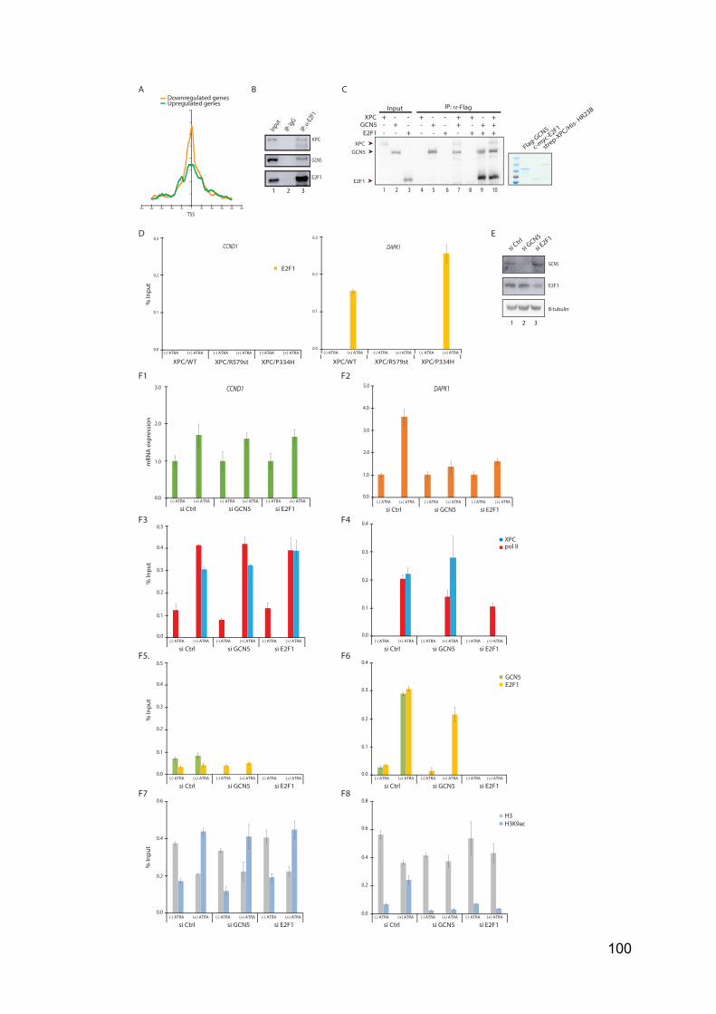
100
B
0
1
2
3
4
5
6
7
8
-2500 -2000 -1500 -1000 -500 0 500 1000 1500 2000 2500
TSS
Downregulated genesUpregulated genes
C
Input
1 2 3
XPC
E2F1
GCN5
D CCND1 DAPK1
A
E2F1
% In
put
(-) ATRA (+) ATRA (-) ATRA (+) ATRA (-) ATRA (+) ATRA
0.3
0.2
0.1
0.0
XPC/WT XPC/R579st XPC/P334H(-) ATRA (+) ATRA (-) ATRA (+) ATRA (-) ATRA (+) ATRA
XPC/WT XPC/R579st XPC/P334H
0.3
0.2
0.1
0.0
E2F1
XPCGCN5
Input + -
- +
E2F1 - + - - - -
+ + - - - +
+ -
+
+ -
- + +
+ - -
+ + +
GCN5XPC
1 2 3 4 5 6 7 8 9 10
strep-XPC/H
is- HR23B
Flag-GCN5
c-myc-E2F1
IP: α-Flag
B-tubulin
E2F1
GCN5
E
si E2F1
si Ctrl
si GCN5
1 2 3
F1
% In
put
0.5
0.4
0.3
0.0
0.2
0.1
0.4
0.3
0.2
0.0
0.1
% In
put
0.6
0.4
0.0
0.2
0.8
0.6
0.0
0.4
0.2
(-) ATRA (+) ATRA (-) ATRA (+) ATRA (-) ATRA (+) ATRA
si Ctrl si GCN5 si E2F1(-) ATRA (+) ATRA (-) ATRA (+) ATRA (-) ATRA (+) ATRA
si Ctrl si GCN5 si E2F1
3.0
2.0
1.0
0.0
3.0
4.0
1.0
0.0
2.0
5.0
mRN
A ex
pres
sion
CCND1 DAPK1
XPCpol II
GCN5
H3H3K9ac
(-) ATRA (+) ATRA (-) ATRA (+) ATRA (-) ATRA (+) ATRA
si Ctrl si GCN5 si E2F1(-) ATRA (+) ATRA (-) ATRA (+) ATRA (-) ATRA (+) ATRA
si Ctrl si GCN5 si E2F1
E2F1
(-) ATRA (+) ATRA (-) ATRA (+) ATRA (-) ATRA (+) ATRA
si Ctrl si GCN5 si E2F1(-) ATRA (+) ATRA (-) ATRA (+) ATRA (-) ATRA (+) ATRA
si Ctrl si GCN5 si E2F1
% In
put
0.5
0.4
0.3
0.0
0.2
0.1
0.4
0.3
0.2
0.0
0.1
(-) ATRA (+) ATRA (-) ATRA (+) ATRA (-) ATRA (+) ATRA
si Ctrl si GCN5 si E2F1(-) ATRA (+) ATRA (-) ATRA (+) ATRA (-) ATRA (+) ATRA
si Ctrl si GCN5 si E2F1
F2
F3 F4
F5. F6
F7 F8
IP: α-
E2F1
IP: Ig
G
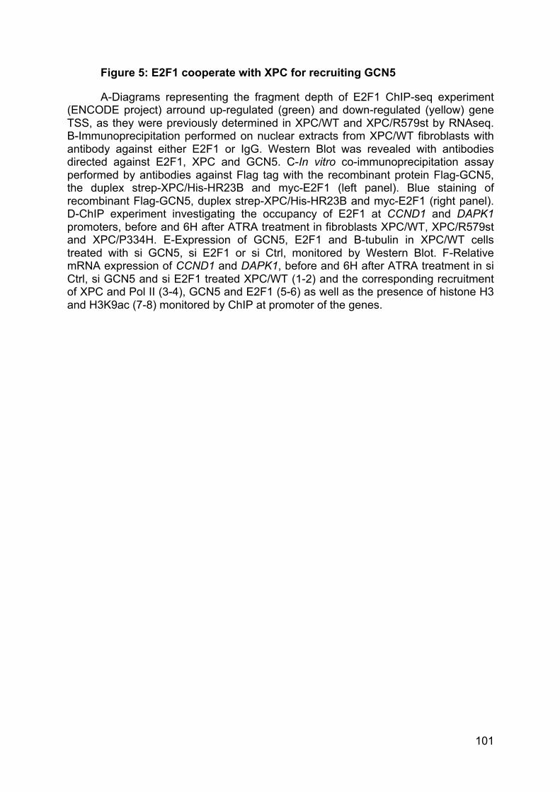
101
Figure 5: E2F1 cooperate with XPC for recruiting GCN5
A-Diagrams representing the fragment depth of E2F1 ChIP-seq experiment (ENCODE project) arround up-regulated (green) and down-regulated (yellow) gene TSS, as they were previously determined in XPC/WT and XPC/R579st by RNAseq. B-Immunoprecipitation performed on nuclear extracts from XPC/WT fibroblasts with antibody against either E2F1 or IgG. Western Blot was revealed with antibodies directed against E2F1, XPC and GCN5. C-In vitro co-immunoprecipitation assay performed by antibodies against Flag tag with the recombinant protein Flag-GCN5, the duplex strep-XPC/His-HR23B and myc-E2F1 (left panel). Blue staining of recombinant Flag-GCN5, duplex strep-XPC/His-HR23B and myc-E2F1 (right panel). D-ChIP experiment investigating the occupancy of E2F1 at CCND1 and DAPK1 promoters, before and 6H after ATRA treatment in fibroblasts XPC/WT, XPC/R579st and XPC/P334H. E-Expression of GCN5, E2F1 and B-tubulin in XPC/WT cells treated with si GCN5, si E2F1 or si Ctrl, monitored by Western Blot. F-Relative mRNA expression of CCND1 and DAPK1, before and 6H after ATRA treatment in si Ctrl, si GCN5 and si E2F1 treated XPC/WT (1-2) and the corresponding recruitment of XPC and Pol II (3-4), GCN5 and E2F1 (5-6) as well as the presence of histone H3 and H3K9ac (7-8) monitored by ChIP at promoter of the genes.
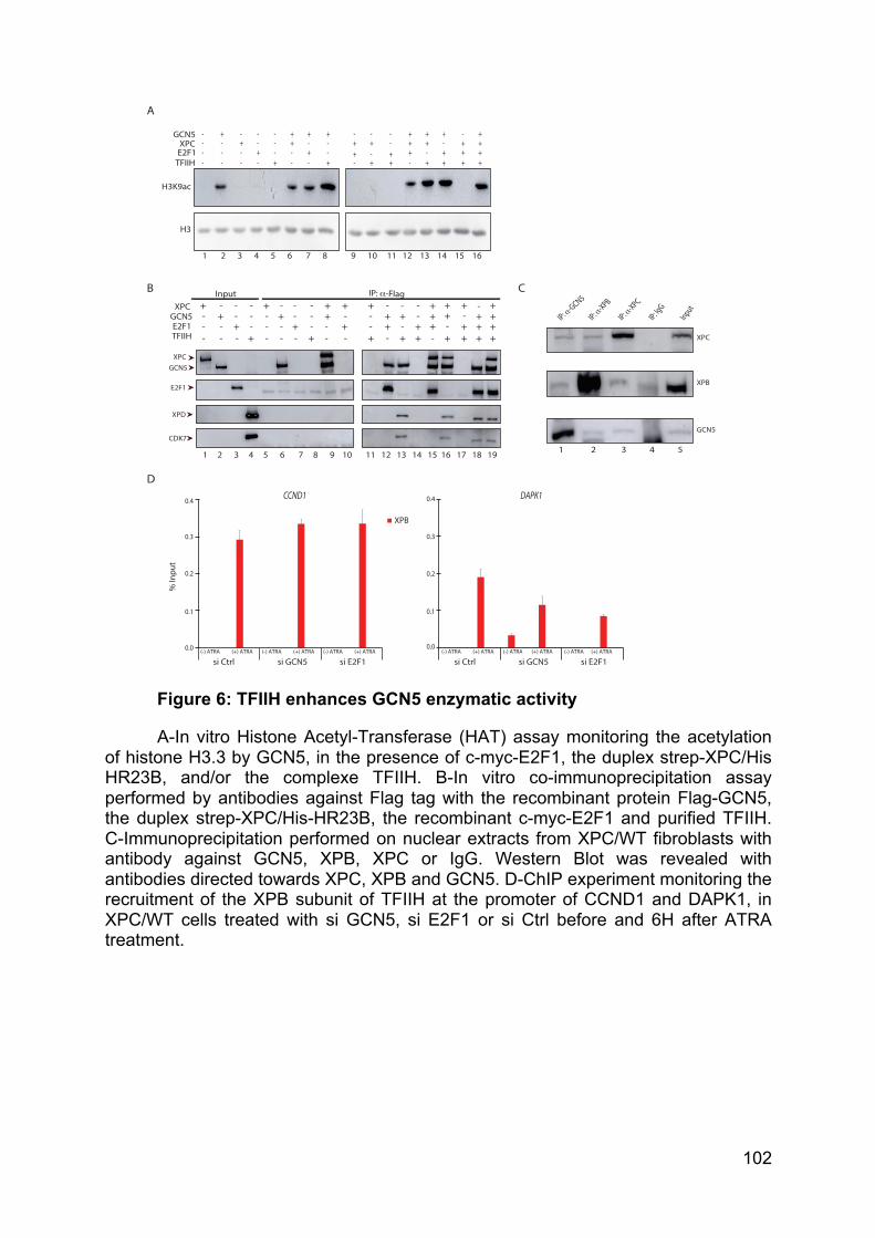
102
Figure 6: TFIIH enhances GCN5 enzymatic activity
A-In vitro Histone Acetyl-Transferase (HAT) assay monitoring the acetylation of histone H3.3 by GCN5, in the presence of c-myc-E2F1, the duplex strep-XPC/His HR23B, and/or the complexe TFIIH. B-In vitro co-immunoprecipitation assay performed by antibodies against Flag tag with the recombinant protein Flag-GCN5, the duplex strep-XPC/His-HR23B, the recombinant c-myc-E2F1 and purified TFIIH. C-Immunoprecipitation performed on nuclear extracts from XPC/WT fibroblasts with antibody against GCN5, XPB, XPC or IgG. Western Blot was revealed with antibodies directed towards XPC, XPB and GCN5. D-ChIP experiment monitoring the recruitment of the XPB subunit of TFIIH at the promoter of CCND1 and DAPK1, in XPC/WT cells treated with si GCN5, si E2F1 or si Ctrl before and 6H after ATRA treatment.
XPC
XPB
GCN5
1 2 3 4 5
C
D
A
Input IP: α-Flag + -
- +
- + - - -
- -
- + - -
- + -
- +
- + - - -
- -
- + - -
- + +
- + -
- -
-
-
- + -
-
- +
+
+ + +
+ + + - - +
-
+ +
+
- +
+
+ - + +
+ -
+ +
+ + +
+
E2F1
XPD
XPCGCN5
CDK7
1 2 3 4 5 6 7 8 9 10 11 12 13 14 15 16 17 18 19
XPCGCN5E2F1TFIIH
CCND1 DAPK1
% In
put
0.4
0.3
0.0
0.2
0.1
0.4
0.3
0.2
0.0
0.1
XPB
(-) ATRA (+) ATRA (-) ATRA (+) ATRA (-) ATRA (+) ATRA
si Ctrl si GCN5 si E2F1(-) ATRA (+) ATRA (-) ATRA (+) ATRA (-) ATRA (+) ATRA
si Ctrl si GCN5 si E2F1
B
- -
+ -
-
-
-
-
- + -
-
- +
+ - + - -
- - -
-
- + +
+ -
-
- + -
-
+ + + +
+ + + -
+ + +
+ + + + +
+ - +
+
- + -
- -
+
- +
+ +
-
H3K9ac
XPCGCN5
E2F1TFIIH
H3
1 2 3 4 5 6 7 8 9 10 11 12 13 14 15 16
Input
IP: IgG
IP: α-XPB
IP: α-GCN5
IP: α-XPC
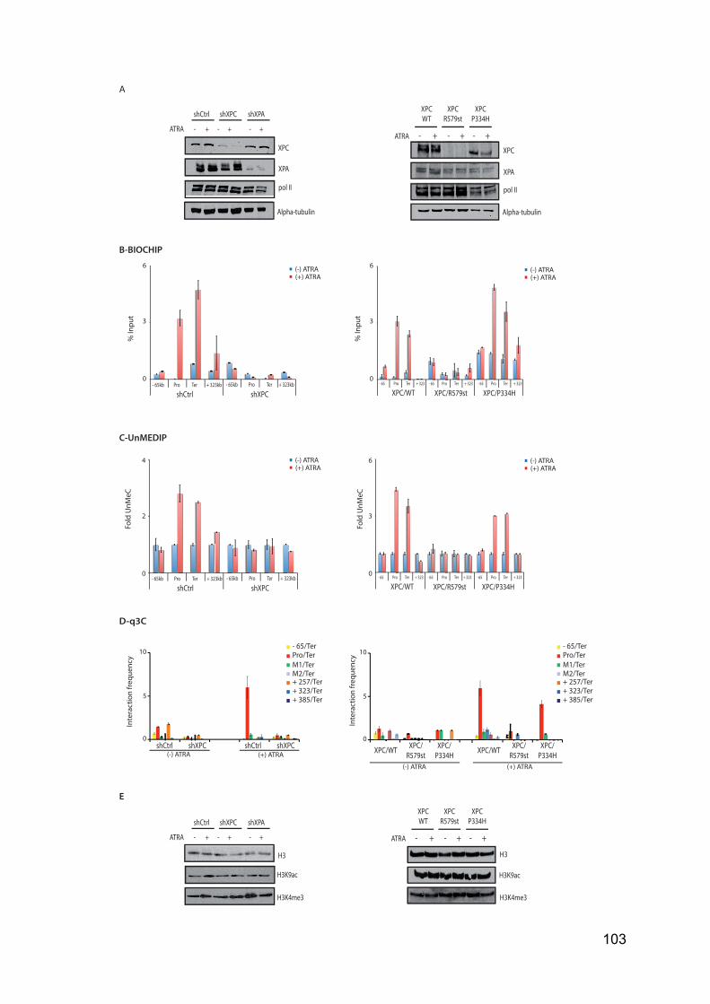
103
- 65/TerPro/TerM1/TerM2/Ter+ 257/Ter+ 323/Ter+ 385/Ter
Inte
ract
ion
frequ
ency
0
5
10
shXPCshCtrl(-) ATRA (+) ATRA
shXPCshCtrl0
5
10
(-) ATRA (+) ATRA
XPC/R579stXPC/WT XPC/
P334HXPC/
R579stXPC/WT XPC/P334H
D-q3C
B-BIOCHIP
% In
put
C-UnMEDIP
(-) ATRA(+) ATRA
shXPCshCtrl
Fold
UnM
eC
4
2
0- 65kb Pro Ter + 323kb - 65kb Pro Ter + 323kb
6
3
0
XPC/R579stXPC/WT XPC/P334H- 65 Pro Ter + 323 - 65 Pro Ter + 323 - 65 Pro Ter + 323
Fold
UnM
eC
6
3
0
shXPCshCtrl- 65kb Pro Ter + 323kb - 65kb Pro Ter + 323kb
% In
put
6
3
0
XPC/R579stXPC/WT XPC/P334H- 65 Pro Ter + 323 - 65 Pro Ter + 323 - 65 Pro Ter + 323
(-) ATRA(+) ATRA
(-) ATRA(+) ATRA
(-) ATRA(+) ATRA
Inte
ract
ion
frequ
ency
- 65/TerPro/TerM1/TerM2/Ter+ 257/Ter+ 323/Ter+ 385/Ter
H3
H3K9ac
H3K4me3
XPCWT
XPCR579st
XPCP334H
H3
H3K9ac
H3K4me3
- + - + - +ATRA
shCtrl shXPC shXPA
ATRA - + - + - +
E
shCtrl shXPC shXPA
ATRA - + - + - +
XPC
XPA
pol II
Alpha-tubulin
XPCWT
XPCR579st
XPCP334H
XPC
XPA
pol II
Alpha-tubulin
- + - + - +ATRA
A
104
Figure S1:
A-Relative protein expression of XPC, XPA, Pol II and alpha-tubulin analysed by Western Blot in whole cell extract from shCtrl, shXPC or shXPA HeLa cells and XPC/WT, XPC/R579st and XPC/P334H fibroblasts, before and 6H after ATRA treatment. B-Detection of DNA breaks at -65kb, pro, ter and +323kb of the RARβ2 locus in shCtrl or shXPC HeLa cells and XPC/WT, XPC/R579st and XPC/P334H fibroblasts, before and 6H after ATRA treatment by BioChIP. DNA breaks are detected through incorporation of Biotin-dUTP by terminal deoxynucleotidyl transferase followed by regular ChIP with Biotin antibodies. C-Analysis of unmethylated DNA by UnMedIP at -65kb, pro, ter and +323kb of the RARβ2 locus in shCtrl or shXPC HeLa cells and XPC/WT, XPC/R579st and XPC/P334H fibroblasts, before and 6H after ATRA treatment. D-q3C assays were performed using crosslinked and HindIII-digested chromatin from shCtrl or shXPC HeLa cells and XPC/WT, XPC/R579st and XPC/P334H fibroblasts, before and 6H after ATRA treatment, as previously defined in Le May et al., 2012. E-Protein expression of histone H3 and presence of the H3K9ac or H4K4me3 forms analysed by Western Blot from whole cell extracts from shCtrl, shXPC or shXPA HeLa cells and XPC/WT, XPC/R579st and XPC/P334H fibroblasts, in before and 6H after ATRA treatment.

105
Figure S2:
A-Relative protein expression of TBP and GCN5 analysed by Western Blot in whole cell extract from shCtrl, shXPC or shXPA HeLa cells and XPC/WT, XPC/R579st and XPC/P334H fibroblasts, before and 6H after ATRA treatment. B-Proportion of XPC/Pol II/GCN5 common peaks enriched in H3K9ac mark. C-HOMER annotation of the XPC/Pol II/GCN5 common peaks.
A
XPCWT
XPCR579st
XPCP334H
TBP
GCN5
ATRA - + - + - +
shCtrl
shXPC
shXPA
TBP
GCN5
ATRA - + - + - +
C
TTS6%
3’UTR1%
ncRNA1%
Promoter42%
Intron26%
Intergenic9%
Exon8% 5’UTR
7%
B
99%(935 peaks)
XPC/PolII/GCN5 peaks overlapp by H3K9ac marks
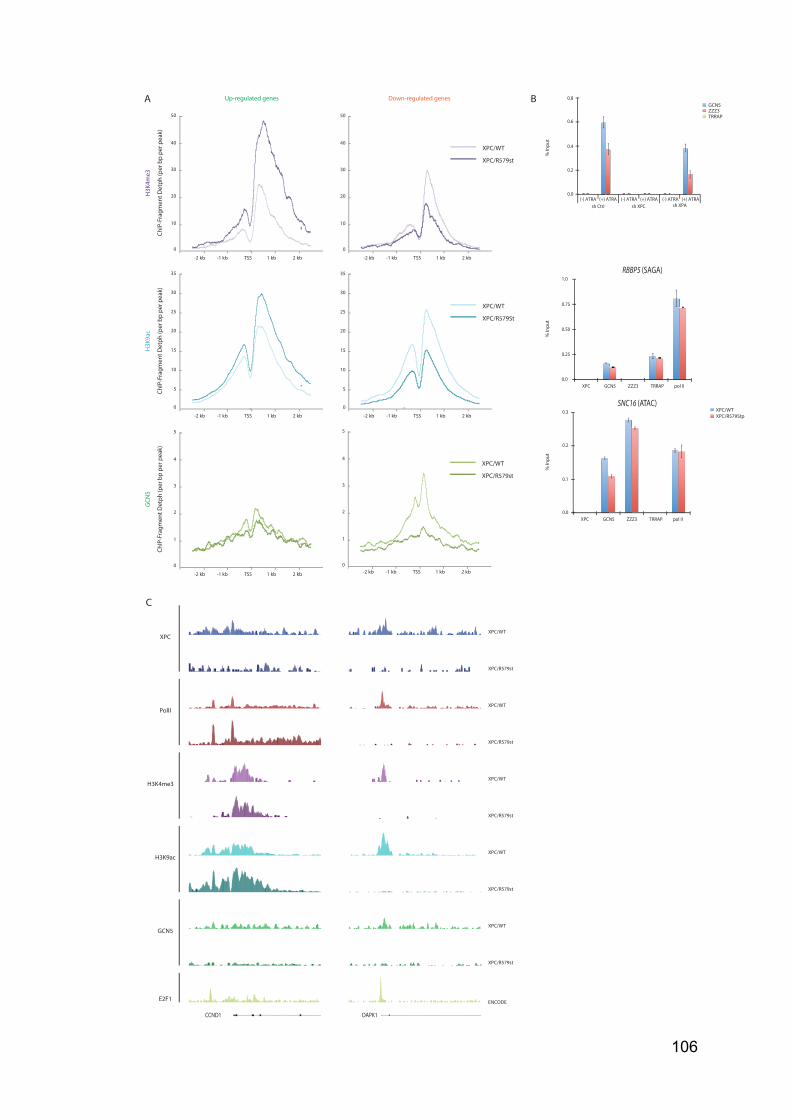
106
A
H3K
9ac
H3K
4me3
TSS 1 kb 2 kb-2 kb -1 kb
ChIP
-Fra
gmen
t Det
ph (p
er b
p pe
r pea
k)
0
5
10
15
20
25
30
TSS 1 kb 2 kb-2 kb -1 kb
35
XPC/WT
XPC/R579St
Up-regulated genes Down-regulated genes
TSS 1 kb 2 kb-2 kb -1 kb
ChIP
-Fra
gmen
t Det
ph (p
er b
p pe
r pea
k)
0
10
40
20
30
TSS 1 kb 2 kb-2 kb -1 kb
50
XPC/WT
XPC/R579st
0
5
10
15
20
25
30
35
0
10
40
20
30
50
TSS 1 kb 2 kb-2 kb -1 kb
0
1
4
2
3
TSS 1 kb 2 kb-2 kb -1 kb
5
XPC/WT
XPC/R579st
0
1
4
2
3
5
GCN
5
ChIP
-Fra
gmen
t Det
ph (p
er b
p pe
r pea
k)
B
sh Ctrl(-) ATRA (+) ATRA (-) ATRA (+) ATRA (-) ATRA (+) ATRA
sh XPC sh XPA
0.8
0.6
0.0
0.4
0.2
% In
put
GCN5ZZZ3TRRAP
XPC/WTXPC/R579Stp
% In
put
GCN5 pol IIZZZ3 TRRAP
0.3
0.2
0.1
0.0XPC
GCN5 pol IIZZZ3 TRRAPXPC
% In
put
1.0
0.75
0.50
0.0
0.25
RBBP5 (SAGA)
SNC16 (ATAC)
C
XPC
GCN5
PolII
H3K4me3
H3K9ac
CCND1 DAPK1
XPC/WT
XPC/WT
XPC/WT
XPC/WT
XPC/WT
XPC/R579st
XPC/R579st
XPC/R579st
XPC/R579st
XPC/R579st
E2F1 ENCODE
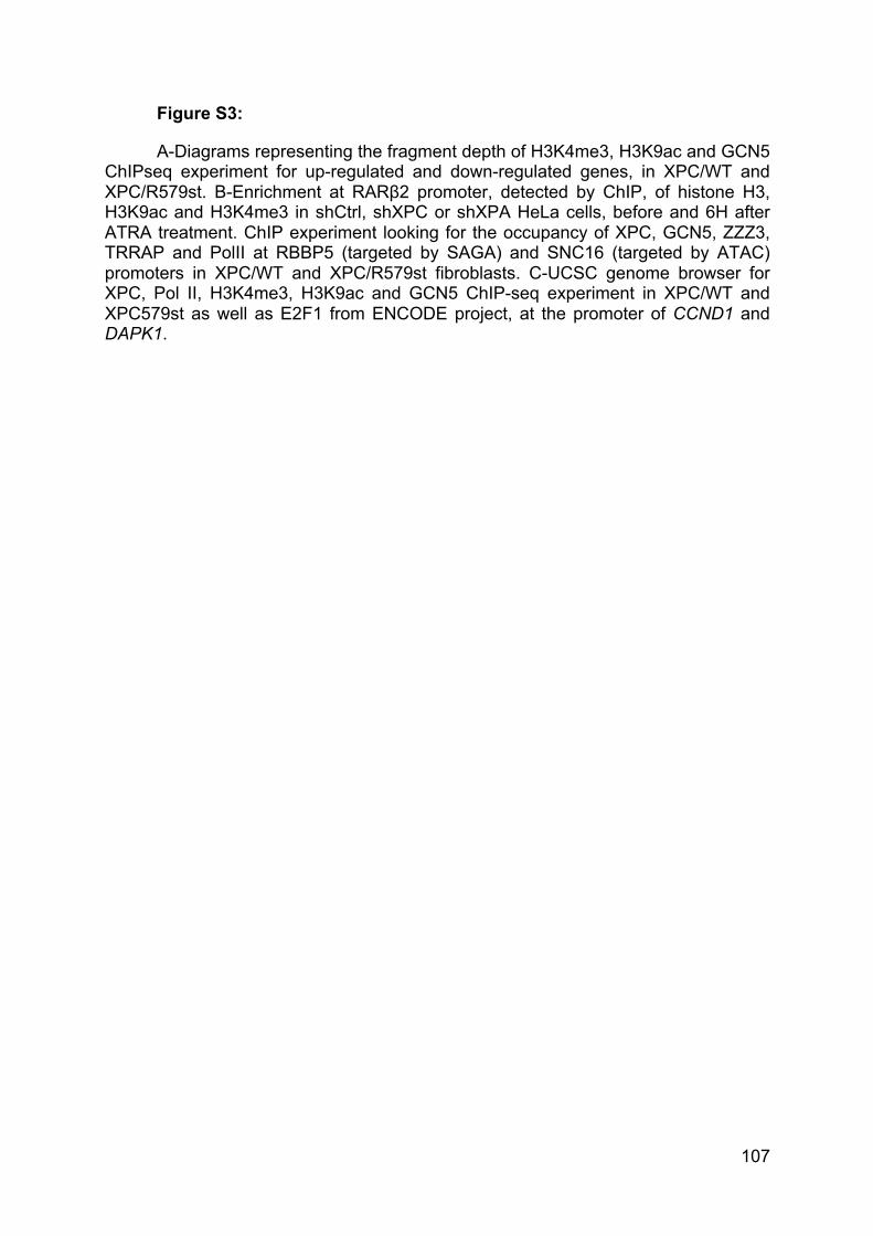
107
Figure S3:
A-Diagrams representing the fragment depth of H3K4me3, H3K9ac and GCN5 ChIPseq experiment for up-regulated and down-regulated genes, in XPC/WT and XPC/R579st. B-Enrichment at RARβ2 promoter, detected by ChIP, of histone H3, H3K9ac and H3K4me3 in shCtrl, shXPC or shXPA HeLa cells, before and 6H after ATRA treatment. ChIP experiment looking for the occupancy of XPC, GCN5, ZZZ3, TRRAP and PolII at RBBP5 (targeted by SAGA) and SNC16 (targeted by ATAC) promoters in XPC/WT and XPC/R579st fibroblasts. C-UCSC genome browser for XPC, Pol II, H3K4me3, H3K9ac and GCN5 ChIP-seq experiment in XPC/WT and XPC579st as well as E2F1 from ENCODE project, at the promoter of CCND1 and DAPK1.
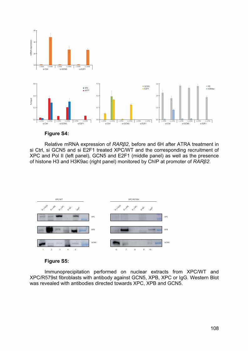
108
Figure S4:
Relative mRNA expression of RARβ2, before and 6H after ATRA treatment in si Ctrl, si GCN5 and si E2F1 treated XPC/WT and the corresponding recruitment of XPC and Pol II (left panel), GCN5 and E2F1 (middle panel) as well as the presence of histone H3 and H3K9ac (right panel) monitored by ChIP at promoter of RARβ2.
Figure S5:
Immunoprecipitation performed on nuclear extracts from XPC/WT and XPC/R579st fibroblasts with antibody against GCN5, XPB, XPC or IgG. Western Blot was revealed with antibodies directed towards XPC, XPB and GCN5.
% In
put
(-) ATRA (+) ATRA (-) ATRA (+) ATRA (-) ATRA (+) ATRA
si Ctrl si GCN5 si E2F1(-) ATRA (+) ATRA (-) ATRA (+) ATRA (-) ATRA (+) ATRA
si Ctrl si GCN5 si E2F1(-) ATRA (+) ATRA (-) ATRA (+) ATRA (-) ATRA (+) ATRA
si Ctrl si GCN5 si E2F1
0.6
0.4
0.0
0.2
0.3
0.2
0.0
0.1
3.0
1.0
0.0
2.0
XPCpol II
GCN5 H3H3K9acE2F1
(-) ATRA (+) ATRA (-) ATRA (+) ATRA (-) ATRA (+) ATRA
si Ctrl si GCN5 si E2F1
60
40
20
0.0
mRN
A ex
pres
sion
XPC
XPB
GCN5
Input
IP: IgG
IP: α-XPB
IP: α-GCN5
IP: α-XPC
XPC/WT
1 2 3 4 5
XPC
XPB
GCN5
Input
IP: IgG
IP: α-XPB
IP: α-GCN5
IP: α-XPC
XPC/R579St
6 7 8 9 10
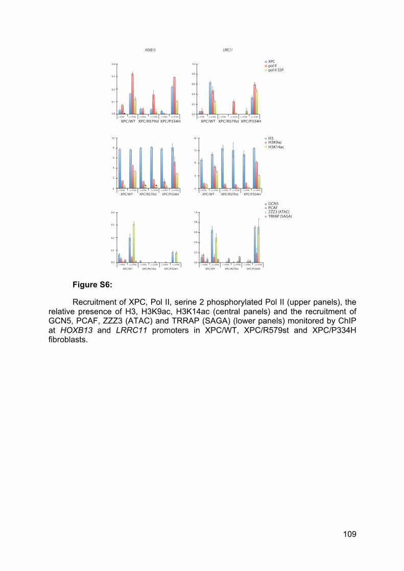
109
Figure S6:
Recruitment of XPC, Pol II, serine 2 phosphorylated Pol II (upper panels), the relative presence of H3, H3K9ac, H3K14ac (central panels) and the recruitment of GCN5, PCAF, ZZZ3 (ATAC) and TRRAP (SAGA) (lower panels) monitored by ChIP at HOXB13 and LRRC11 promoters in XPC/WT, XPC/R579st and XPC/P334H fibroblasts.
XPC/WT XPC/R579st XPC/P334H(-) ATRA (+) ATRA (-) ATRA (+) ATRA (-) ATRA (+) ATRA
10
0
8
4
6
2
XPC/WT XPC/R579st XPC/P334H(-) ATRA (+) ATRA (-) ATRA (+) ATRA (-) ATRA (+) ATRA
16
0
12
8
4
HOXB13 LRRC11
XPC/WT XPC/R579st XPC/P334H(-) ATRA (+) ATRA (-) ATRA (+) ATRA (-) ATRA (+) ATRA
0.3
0.2
0.1
0.4
0.0
XPC/WT XPC/R579st XPC/P334H(-) ATRA (+) ATRA (-) ATRA (+) ATRA (-) ATRA (+) ATRA
0.8
0.6
0.4
0.0
0.2
1.0
0.3
0.2
0.1
0.0
0.4
XPC/WT XPC/R579st XPC/P334H
(-) ATRA (+) ATRA (-) ATRA (+) ATRA (-) ATRA (+) ATRA
XPC/WT XPC/R579st XPC/P334H
(-) ATRA (+) ATRA (-) ATRA (+) ATRA (-) ATRA (+) ATRA
0.8
0.6
0.4
0.0
0.2
1.0
XPCpol IIpol II S5P
H3H3K9acH3K14ac
ZZZ3 (ATAC)TRRAP (SAGA)
GCN5PCAF
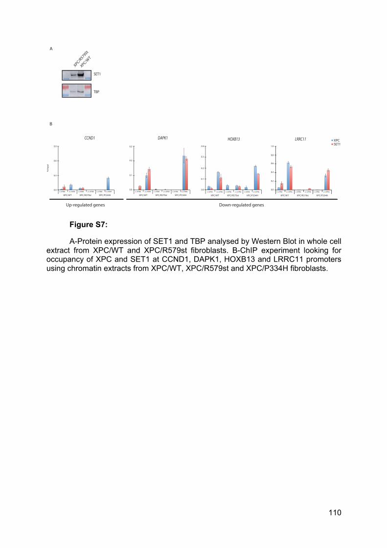
110
Figure S7:
A-Protein expression of SET1 and TBP analysed by Western Blot in whole cell extract from XPC/WT and XPC/R579st fibroblasts. B-ChIP experiment looking for occupancy of XPC and SET1 at CCND1, DAPK1, HOXB13 and LRRC11 promoters using chromatin extracts from XPC/WT, XPC/R579st and XPC/P334H fibroblasts.
XPC/WT
XPC/R579St
TBP
SET1
CCND1 DAPK1 HOXB13 LRRC11
Down-regulated genesUp-regulated genes
A
B
XPCSET1
% In
put
0.9
0.6
0.3
0.0
XPC/WT XPC/R579st XPC/P334H
(-) ATRA (+) ATRA (-) ATRA (+) ATRA (-) ATRA (+) ATRA
XPC/WT XPC/R579st XPC/P334H
(-) ATRA (+) ATRA (-) ATRA (+) ATRA (-) ATRA (+) ATRA
0.2
0.1
0.0
0.3SESESE
XPC/WT XPC/R579st XPC/P334H
(-) ATRA (+) ATRA (-) ATRA (+) ATRA (-) ATRA (+) ATRA
0.3
0.2
0.1
0.4
0.0
XPC/WT XPC/R579st XPC/P334H
(-) ATRA (+) ATRA (-) ATRA (+) ATRA (-) ATRA (+) ATRA
0.8
0.6
0.4
0.0
0.2
1.0

111
II - The Mediator subunit MED12
112
MED12-related XLID disorders are dose-dependent of immediate
early genes (IEGs) expression
DONNIO Lise-MARIE∞, BIDON Baptiste∞, HASHIMOTO Satoru∞, MAY Melanie,
EPANCHINTSEV Alexey, RYAN Colm, ALLEN William, HACKETT Anna, GECZ
Jozef, SKINNER Cindy, STENVENSON Roger E., de BROUWER Arjan P.M.,
COUTTON Charles, FRANCANNET Christine, JOUK Pierre-Simon, SCHWARTZ
Charles E. & EGLY Jean-Marc*
Département de génomique fonctionnelle et Cancer, CNRS, INSERM, Université de
Strasbourg, Institut de Génétique et de Biologie Moléculaire et Cellulaire (IGBMC), 1
rue Laurent Fries, 67404 Illkirch-Graffenstaden, France.
* To whom correspondences should be address:
[email protected]; [email protected]
∞ Should be regarded as joint First Authors
113
O R I G I N A L A R T I C L E
MED12-related XLID disorders are dose-dependent ofimmediate early genes (IEGs) expressionLise-Marie Donnio1,†, Baptiste Bidon1,†, Satoru Hashimoto1,2,†, Melanie May3,Alexey Epanchintsev1, Colm Ryan1, William Allen4, Anna Hackett5,Jozef Gecz6, Cindy Skinner3, Roger E. Stevenson3, Arjan P.M. de Brouwer7,Charles Coutton8, Christine Francannet9, Pierre-Simon Jouk8,Charles E. Schwartz3 and Jean-Marc Egly1,*1Department of Functional Genomics and Cancer biology, IGBMC, CNRS/INSERM/Universite de Strasbourg,67404 Illkirch-Graffenstaden, France, 2Department of Clinical Pharmacology and Therapeutics Oita UniversityFaculty of Medicine, Yufu city, Oita 879-5593, Japan, 3Greenwood Genetic Center, Greenwood, SC 29649, USA,4Fullerton Genetics Center, Asheville, NC 28803, USA, 5Hunter Genetics, Waratah, NSW 2298, Australia,6School of Medicine, and the Robinson Research Institute, The University of Adelaide, and South AustralianHealth and Medical Research Institute, Adelaide, SA 5000, Australia, 7Department of Human Genetics, DondersInstitute for Brain, Cognition and Behaviour, Radboud University Medical Center, Nijmegen, 6525 HP, TheNetherlands, 8Departement de Genetique et Procreation, Centre Hospitalier-Universitaire, Institut AlbertBonniot, CNRS/INSERM/Universite Grenoble Alpes, 38000 Grenoble, France and 9Service de GenetiqueMedicale, Centre Hospitalier-Universitaire, 63003 Clermont-Ferrand, France
*To whom correspondence should be addressed at: Department of Functional Genomics and Cancer Biology, IGBMC, 1 rue Laurent Fries, 67404 Illkirch-Graffenstaden, France. Tel: 0033 388653447; Fax: 0033 388653201; Email: [email protected]
AbstractMediator occupies a key role in protein coding genes expression in mediating the contacts between gene specific factors andthe basal transcription machinery but little is known regarding the role of each Mediator subunits. Mutations in MED12 arelinked with a broad spectrum of genetic disorders with X-linked intellectual disability that are difficult to range as Lujan,Opitz-Kaveggia or Ohdo syndromes. Here, we investigated several MED12 patients mutations (p.R206Q, p.N898D, p.R961W,p.N1007S, p.R1148H, p.S1165P and p.R1295H) and show that each MED12 mutations cause specific expression patterns of JUN,FOS and EGR1 immediate early genes (IEGs), reflected by the presence or absence of MED12 containing complex at their re-spective promoters. Moreover, the effect of MED12 mutations has cell-type specificity on IEG expression. As a consequence,the expression of late responsive genes such as the matrix metalloproteinase-3 and the RE1 silencing transcription factor im-plicated respectively in neural plasticity and the specific expression of neuronal genes is disturbed as documented forMED12/p.R1295H mutation. In such case, JUN and FOS failed to be properly recruited at their AP1-binding site. Our results sug-gest that the differences between MED12-related phenotypes are essentially the result of distinct IEGs expression patterns,
†The authors wish it to be known that, in their opinion, the first three authors should be regarded as joint First Authors.Received: January 23, 2017. Revised: March 3, 2017. Accepted: March 8, 2017
VC The Author 2017. Published by Oxford University Press. All rights reserved. For Permissions, please email: [email protected]
2062
Human Molecular Genetics, 2017, Vol. 26, No. 11 2062–2075
doi: 10.1093/hmg/ddx099Advance Access Publication Date: 24 March 2017Original Article
Downloaded from https://academic.oup.com/hmg/article-abstract/26/11/2062/3084497/MED12-related-XLID-disorders-are-dose-dependent-ofby IRMA Strasbourg useron 04 October 2017
114
the later ones depending on the accurate formation of the transcription initiation complex. This might challenge clinicians torethink the traditional syndromes boundaries and to include genetic criterion in patients’ diagnostic.
IntroductionMammalian mediator (MED) is an evolutionary conservedmulti-protein complex that is a key regulator of gene expressioninvolved in cell growth, homeostasis, development and differ-entiation (1–3). Mediator is composed of more than 30 subunits,arranged in four different modules (Fig. 1A). Upon gene activa-tion, MED conveys essential information from transcriptionalregulatory proteins bound at DNA responsive elements to RNApolymerase II (Pol II) machinery bound at the transcription startsite (TSS) (4–6).
Dysfunction of the transcriptional machinery componentshas been shown to elicit a range of effects on cell states (prolif-eration or differentiation) giving rise to diverse pathologies, in-cluding cancers (7). Mutations in MED subunits are alsoassociated with a wide range of genetic disorders, such as infan-tile cerebral and cerebellar atrophy (MIM: 613668) (MED17, MIM:603810) (8), Charcot-Marie Tooth disease (MIM: 605589) (MED25,MIM: 610197) (9) and non-syndromic intellectual disability (MIM:614249) (MED23, MIM: 605042) (10) and (MED13L, MIM: 608771)(11); most of them exhibiting neurological defects (12). Germline mutations of MED12 (MIM: 300188) have already been foundin several genetic disorders associated with X-linked intellec-tual disability (XLID) (13), such as Opitz-Kaveggia syndrome alsonamed FG syndrome (MIM: 305450) (p.R961W) (14) and (p.G958E)(15), Lujan syndrome (MIM: 309520) (p.N1007S) (16) and Ohdosyndrome (MIM: 249620) (p.S1165P, p.R1148H and H1729N)(17,18). More recently, an important number of mutations havealso been associated with non-syndromic XLID (p.S1967QfsX84)(19), (p.R521H) (20), (p.R621Q) (21), (p.I771T) (22), (p.R815Q) (23),(p.I1023V) (24) (p.A1383T) (25) and (p.E1974H) (26) (Fig. 1B).However, the underlying basis leading to phenotypically dis-tinct syndromes from several genetic mutations of MED12 re-mains unclear.
MED12 gene is located at Xq13.1 and together with MED13,CDK8 and Cyclin C (CCNC) forms the Kinase module, that vari-ably associates with the core Mediator (27). MED12 is requiredfor the stable incorporation of CDK8/CCNC into Mediator andappears to initiate the CDK8 kinase activity (28) which can regu-late transcription (29). For example, CDK8 phosphorylation ofPol II leads to the disruption of Mediator–Pol II interactions thusresulting in transcriptional inhibition (30). Similarly, CDK8phosphorylates Cyclin H, a subunit of the general transcription/DNA repair factor TFIIH, and thus represses both the ability ofTFIIH to activate transcription and its CTD kinase activity (31).MED12 has also been linked with the Notch, Wnt and Sonichedgehog signalling pathways and control key aspects of braindevelopment and function, from initial patterning to neuronalplasticity (32–37).
Here, we first describe a male patient with characteristics in-cluding intellectual and developmental delay (38). He was foundto carry a maternally inherited missense mutation in MED12(MED12/p.R1295H). In an attempt to deepen our knowledgeabout the role of Mediator in the regulation of gene expression,we have deeply investigated the effect of this mutation on thetranscriptional activation of key genes. We discovered that thisMED12 mutation specifically modulates the expression of acti-vated immediate early genes (IEGs) such as JUN (MIM: 165160)by disturbing the formation of the transcription complexes.
Consequently, late response genes (LRGs) that are regulated bythe AP1 complex (FOS/JUN dimer) are also disrupted. We thenenlarged our study to seven different mutations and comeacross distinct deregulation of JUN/FOS(MIM:164810)/EGR1(MIM:164810) expression, possibly explaining the largeclinical spectrum covered by MED12-related patients. Thismight challenge clinicians to rethink the traditional syndromesboundaries and to reconsider patients’ diagnostic through ge-netic criterion.
ResultMED12/p.R1295H mutation disturb IEGs expression
Mediator is known to mediate the response of IEGs, involved indiverse processes such as brain development and neuronalplasticity (10,39). To evaluate the global impact of MED12 onIEGs expression, we used CRISPR/Cas9 system to Knock-DownMED12 in human LAN-1 neuroblastoma cell line (MED12-KD).We first observed that silencing MED12 does not impact thelevel of other Mediator subunits (Fig. 1C). We thus evaluated theexpression of three IEGs: JUN, FOS and EGR1 which code for DNAbinding factors, 30 min after serum addition to wild type (WT)and MED12-KD cells. Quantitative RT-PCR on total mRNA ex-tract clearly showed that JUN, FOS and EGR1 expression is downregulated in MED12-KD cells (Fig. 1D) as confirmed by the de-crease of their protein synthesis (Supplementary Material, Fig.S1).
We next focused on a MED12 patient from whom skin fibro-blasts had been obtained (Table 1; Supplementary Material, Fig.S2A and B; see ‘Materials and Methods’ section ). The R1295Hpatient and his heterozygous mother carried a G-to-A missensesubstitution at nucleotide position c.3884 resulting in an argi-nine to histidine modification (p.R1295H) referred as R1295Hsonand R1295Hmother, respectively (Fig. 1E). This patient is cur-rently monitored for intellectual disability and delayed motorskills. He has a Marfan habitus (MIM: 154700) and was suspectedto have X-linked Lujan-Fryns syndrome. His mother had thesame skeletal and morphologic features as well as a mild intel-lectual disability (38). Sanger sequencing of the MED12 tran-script revealed that !80% of cultivated mother fibroblastsexpress the mutant allele, due to skewed X-inactivation. RT-qPCR and Western blot analysis on both patient and mother fi-broblasts shows that the mutation does not significantly affectmRNA expression of MED12 and MED23 subunits of Mediator aswell as the corresponding protein level (SupplementaryMaterial, Fig. S3). However, we repeatedly observed that CDK8subunit of the Kinase module was slightly less expressed inR1295H fibroblasts (Supplementary Material, Fig. S3).
In response to serum mitogens, JUN activation was defectivein both R1295Hson and R1295Hmother fibroblasts while FOS andEGR1 were similarly expressed when compared with WT cells(Fig. 1F). In order to gain confidence in the consequence ofMED12/p.R1295H mutation in IEGs serum induction response,we performed clonal selection (cs) from the R1295H mother’s fi-broblasts and obtained cells expressing only one allele due toX-inactivation (see ‘Materials and Methods’ section). IncsR1295H clone selected cells, we similarly observed that JUN
2063Human Molecular Genetics, 2017, Vol. 26, No. 11 |
Downloaded from https://academic.oup.com/hmg/article-abstract/26/11/2062/3084497/MED12-related-XLID-disorders-are-dose-dependent-ofby IRMA Strasbourg useron 04 October 2017
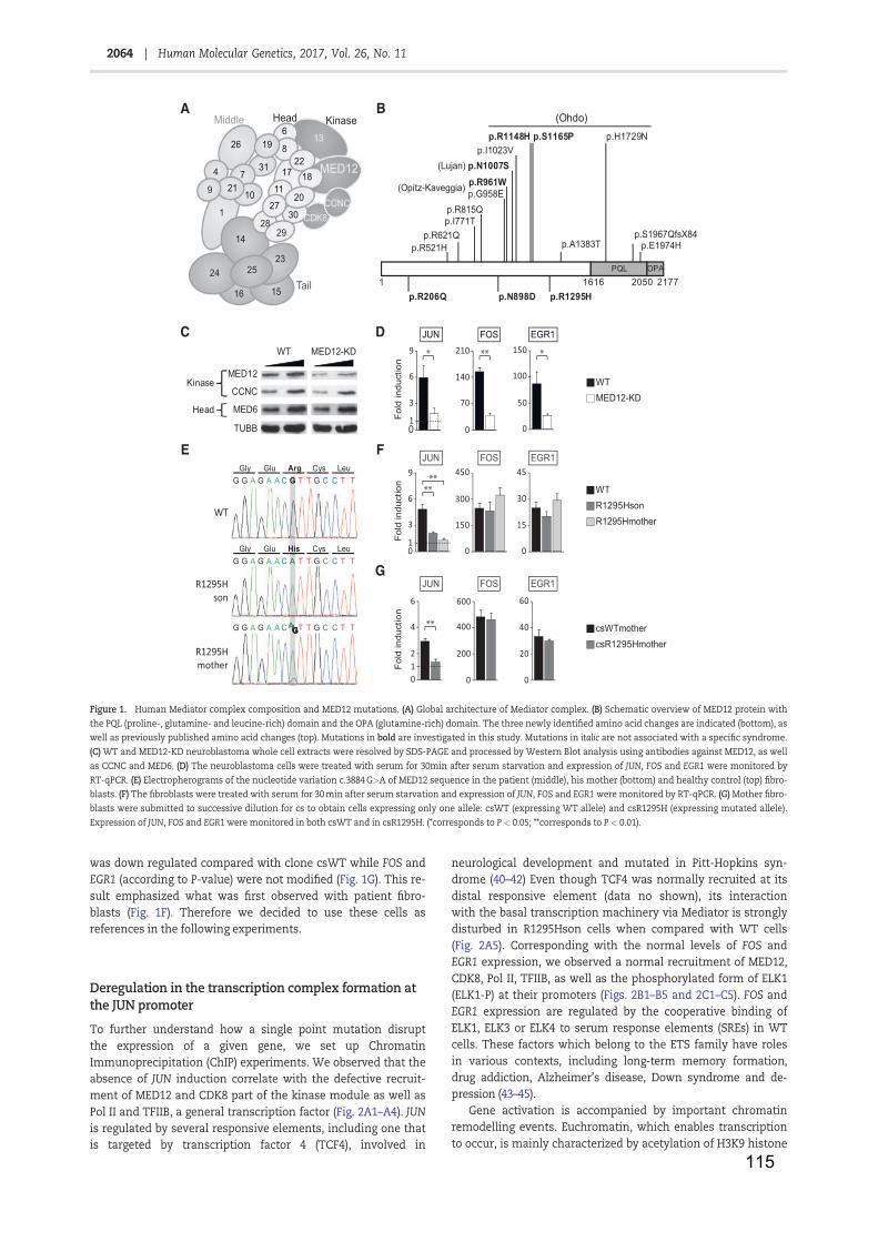
115
was down regulated compared with clone csWT while FOS andEGR1 (according to P-value) were not modified (Fig. 1G). This re-sult emphasized what was first observed with patient fibro-blasts (Fig. 1F). Therefore we decided to use these cells asreferences in the following experiments.
Deregulation in the transcription complex formation atthe JUN promoter
To further understand how a single point mutation disruptthe expression of a given gene, we set up ChromatinImmunoprecipitation (ChIP) experiments. We observed that theabsence of JUN induction correlate with the defective recruit-ment of MED12 and CDK8 part of the kinase module as well asPol II and TFIIB, a general transcription factor (Fig. 2A1–A4). JUNis regulated by several responsive elements, including one thatis targeted by transcription factor 4 (TCF4), involved in
neurological development and mutated in Pitt-Hopkins syn-drome (40–42) Even though TCF4 was normally recruited at itsdistal responsive element (data no shown), its interactionwith the basal transcription machinery via Mediator is stronglydisturbed in R1295Hson cells when compared with WT cells(Fig. 2A5). Corresponding with the normal levels of FOS andEGR1 expression, we observed a normal recruitment of MED12,CDK8, Pol II, TFIIB, as well as the phosphorylated form of ELK1(ELK1-P) at their promoters (Figs. 2B1–B5 and 2C1–C5). FOS andEGR1 expression are regulated by the cooperative binding ofELK1, ELK3 or ELK4 to serum response elements (SREs) in WTcells. These factors which belong to the ETS family have rolesin various contexts, including long-term memory formation,drug addiction, Alzheimer’s disease, Down syndrome and de-pression (43–45).
Gene activation is accompanied by important chromatinremodelling events. Euchromatin, which enables transcriptionto occur, is mainly characterized by acetylation of H3K9 histone
A
C D
E F
G
B
Figure 1. Human Mediator complex composition and MED12 mutations. (A) Global architecture of Mediator complex. (B) Schematic overview of MED12 protein withthe PQL (proline-, glutamine- and leucine-rich) domain and the OPA (glutamine-rich) domain. The three newly identified amino acid changes are indicated (bottom), aswell as previously published amino acid changes (top). Mutations in bold are investigated in this study. Mutations in italic are not associated with a specific syndrome.(C) WT and MED12-KD neuroblastoma whole cell extracts were resolved by SDS-PAGE and processed by Western Blot analysis using antibodies against MED12, as wellas CCNC and MED6. (D) The neuroblastoma cells were treated with serum for 30min after serum starvation and expression of JUN, FOS and EGR1 were monitored byRT-qPCR. (E) Electropherograms of the nucleotide variation c.3884 G>A of MED12 sequence in the patient (middle), his mother (bottom) and healthy control (top) fibro-blasts. (F) The fibroblasts were treated with serum for 30 min after serum starvation and expression of JUN, FOS and EGR1 were monitored by RT-qPCR. (G) Mother fibro-blasts were submitted to successive dilution for cs to obtain cells expressing only one allele: csWT (expressing WT allele) and csR1295H (expressing mutated allele).Expression of JUN, FOS and EGR1 were monitored in both csWT and in csR1295H. (*corresponds to P < 0.05; **corresponds to P < 0.01).
2064 | Human Molecular Genetics, 2017, Vol. 26, No. 11
Downloaded from https://academic.oup.com/hmg/article-abstract/26/11/2062/3084497/MED12-related-XLID-disorders-are-dose-dependent-ofby IRMA Strasbourg useron 04 October 2017
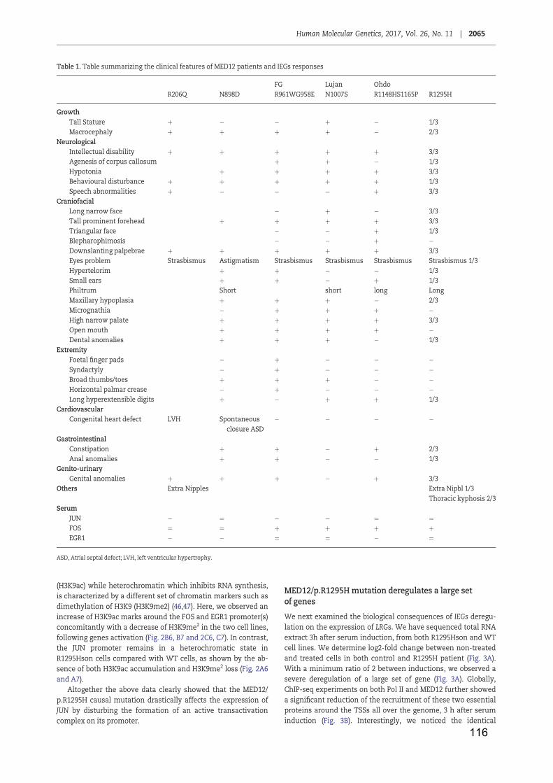
116
(H3K9ac) while heterochromatin which inhibits RNA synthesis,is characterized by a different set of chromatin markers such asdimethylation of H3K9 (H3K9me2) (46,47). Here, we observed anincrease of H3K9ac marks around the FOS and EGR1 promoter(s)concomitantly with a decrease of H3K9me2 in the two cell lines,following genes activation (Fig. 2B6, B7 and 2C6, C7). In contrast,the JUN promoter remains in a heterochromatic state inR1295Hson cells compared with WT cells, as shown by the ab-sence of both H3K9ac accumulation and H3K9me2 loss (Fig. 2A6and A7).
Altogether the above data clearly showed that the MED12/p.R1295H causal mutation drastically affects the expression ofJUN by disturbing the formation of an active transactivationcomplex on its promoter.
MED12/p.R1295H mutation deregulates a large setof genes
We next examined the biological consequences of IEGs deregu-lation on the expression of LRGs. We have sequenced total RNAextract 3h after serum induction, from both R1295Hson and WTcell lines. We determine log2-fold change between non-treatedand treated cells in both control and R1295H patient (Fig. 3A).With a minimum ratio of 2 between inductions, we observed asevere deregulation of a large set of gene (Fig. 3A). Globally,ChIP-seq experiments on both Pol II and MED12 further showeda significant reduction of the recruitment of these two essentialproteins around the TSSs all over the genome, 3 h after seruminduction (Fig. 3B). Interestingly, we noticed the identical
Table 1. Table summarizing the clinical features of MED12 patients and IEGs responses
FG Lujan OhdoR206Q N898D R961WG958E N1007S R1148HS1165P R1295H
GrowthTall Stature þ " " þ " 1/3Macrocephaly þ þ þ þ " 2/3
NeurologicalIntellectual disability þ þ þ þ þ 3/3Agenesis of corpus callosum þ þ " 1/3Hypotonia þ þ þ þ 3/3Behavioural disturbance þ þ þ þ þ 1/3Speech abnormalities þ " " " þ 3/3
CraniofacialLong narrow face " þ " 3/3Tall prominent forehead þ þ þ þ 3/3Triangular face " " þ 1/3Blepharophimosis " " þ "Downslanting palpebrae þ þ þ þ þ 3/3Eyes problem Strasbismus Astigmatism Strasbismus Strasbismus Strasbismus Strasbismus 1/3Hypertelorim þ þ " " 1/3Small ears þ þ " þ 1/3Philtrum Short short long LongMaxillary hypoplasia þ þ þ " 2/3Micrognathia " þ þ þ "High narrow palate þ þ þ þ 3/3Open mouth þ þ þ þ "Dental anomalies þ þ þ " 1/3
ExtremityFoetal finger pads " þ " " "Syndactyly " þ " " "Broad thumbs/toes þ þ þ " "Horizontal palmar crease " þ " " "Long hyperextensible digits þ " þ þ 1/3
CardiovascularCongenital heart defect LVH Spontaneous
closure ASD" " " "
GastrointestinalConstipation þ þ " þ 2/3Anal anomalies þ þ " " 1/3
Genito-urinaryGenital anomalies þ þ þ " þ 3/3
Others Extra Nipples Extra Nipbl 1/3Thoracic kyphosis 2/3
SerumJUN " ¼ " " ¼ ¼FOS ¼ ¼ þ þ þ þEGR1 " " ¼ ¼ " ¼
ASD, Atrial septal defect; LVH, left ventricular hypertrophy.
2065Human Molecular Genetics, 2017, Vol. 26, No. 11 |
Downloaded from https://academic.oup.com/hmg/article-abstract/26/11/2062/3084497/MED12-related-XLID-disorders-are-dose-dependent-ofby IRMA Strasbourg useron 04 October 2017
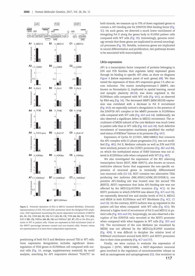
117
positioning of both Pol II and Mediator around TSS in WT cells.Gene expression deregulation includes significant down-regulation of 3550 genes in R1295Hson cell compared with nor-mal cells (Fig. 3A, orange, supplementary material). By motifanalysis, searching for AP1 responsive element ‘TGACTC’ on
both strands, we measure up to 70% of down regulated genes tocontain a AP1-binding site for JUN/FOS DNA binding factor (Fig.3C). On such genes, we detected a much lower enrichment ofelongating Pol II along the genes body in R1295H patient cellscompared with WT cells (Fig. 3D). Interestingly, genome ontol-ogy reveals that these genes are implicated in several neurologi-cal processes (Fig. 3E). Notably, numerous genes are implicatedin neural differentiation and proliferation, two pathways knownto be associated with macrocephaly.
LRGs expression
AP1 is a transcription factor composed of proteins belonging toJUN and FOS families that regulates lately expressed genesthrough its binding to specific AP1 sites, as show on diagramsFigure 4 (below expression panel of each genes) (48). We thentested the expression of three AP1 responsive genes 3 h after se-rum induction. The matrix metalloproteinase-3 (MMP3, alsoknown as Stromelysin-1), implicated in spatial learning, neuraland synaptic plasticity (49,50), was down regulated in theR1295Hson cells compared with WT cells (Fig. 4A1), as observedby RNA-seq (Fig. 3A). The decreased MMP3 (MIM:185250) expres-sion was correlated with a decrease in Pol II recruitment(Fig. 4A2); we especially noticed a deregulation in the presence ofthe JUN/FOS AP1 complex at the MMP3 promoter in R1295Hsoncells compared with WT cells (Fig. 4A3 and A4). Additionally, wealso observed a significant defect in MED12 recruitment. The re-cruitment of MED6 subunit of the core Mediator was much lowerin patient cells than in WT cells (Fig. 4A5 and A6). Such defectiverecruitment of transcription machinery paralleled the methyl-ated status of H3K9me2 histone at its promoter (Fig. 4A7).
Expression of Cyclin D1 (CCND1, MIM:168461) that connectsthe AP1 complex with G1 phase progression (51), was not modi-fied (Fig. 4B1). Pol II, Mediator subunits as well as JUN and FOSwere similarly present at the CCND1 promoter (Fig. 4B2 and B6),on which the methylated status of H3K9 histones was not al-tered in R1295Hson cells when compared with WT (Fig. 4B7).
We also investigated the expression of the RE1 silencingtranscription factor (REST, MIM: 600571), also known as neuronrestrictive silencer factor that suppresses the non-specific ex-pression of neuronal genes in terminally differentiatednon-neuronal cells (52–54). REST contains two alternative TSSsproducing two isoforms (NM_005612.4/NM_001193508.1); oneputative AP1-binding site was located near the second TSS(REST2). REST1 expression that lacks AP1-binding site was notaffected by the MED12/p.R1295H mutation (Fig. 4C1). At theREST1 promoter in which JUN/FOS was absent (Fig. 4C5 and C6),we repeatedly detected low and similar levels of Pol II, MED12and MED6 in both R1295Hson and WT fibroblasts (Fig. 4C2, C5and C6). On the contrary, REST2 isoform was up regulated in thepatient cell line when compared with WT cells (Fig. 4D1). Wedetected a higher level of recruitment of Pol II and MED12 in pa-tient cells (Fig. 4D2 and D5). Surprisingly, we also observed a dis-ruption of the JUN/FOS ratio recruited at the REST2 promoterwhen compared with what happens in WT cells (Fig. 3D3 andD4). The recruitment of the core Mediator (as visualized byMED6) was not affected by the MED12/p.R1295H mutation(Fig. 4D6). It was difficult to decipher the relative level ofH3K9me2 enrichment around both REST1 and REST2 promotersdue to their close proximity (Fig. 4C7 and D7).
Finally, we were curious to evaluate the expression ofSynapsin I (SYN1, MIM:313440), a REST-dependent neuronalgene involved in the regulation of neurotransmitter release aswell as axonogenesis and synaptogenesis (55). One mutation in
JUN
Fold
enr
ichm
ent
Pol II
1
3
6
9
0
A1
TFIIB
012
4
6
A3
FOS EGR1
B1
TFIIB
012
4
6
B3 Pol II
01
3
6
9
C1
C3
Fold
enr
ichm
ent
C4B4A4
MED12
012
4
6 MED12
012
4
6
ELK1-P
01
3
6
9C5B5A5 ELK1-P
01
3
6
9
Fold
enr
ichm
ent
012
4
6 H3K9ac
012
4
6 H3K9ac
012
4
6 H3K9acC6B6A6
C7B7A7 H3K9me² H3K9me²
Fold
enr
ichm
ent
Fold
enr
ichm
ent
Fold
enr
ichm
ent
0
0.5
1
1.5
0
0.5
1
1.5H3K9me²
0
0.5
1
1.5
Pol II
01
3
6
9
TFIIB
012
4
6
012
4
6 TCF4 (pro)
CDK8
0
0.5
1
1.5 2C2A B2
Fold
enr
ichm
ent
-300
SRFSRE
Sp1 Ets
+1+1-400
SRFSRE
Ets AP-1 AP-1
distal proximal
MEF2RSRF
TCF
+1-200-3000
AP-1 AP-1SP1
WT R1295Hson
CDK8 CDK8
MED12
Figure 2. Promoter dynamics of IEGs in MED12 mutated fibroblast. Schematicrepresentations of JUN, FOS and EGR1 promoters show the designed PCR ampli-cons. ChIP experiment monitoring the serum-dependent recruitment of MED12(A1, B1, C1), CDK8 (A2, B2, C2), Pol II (A3, B3, C3), TFIIB (A4, B4, C4), TCF4 (A5),ELK1-P (B5, C5), H3K9ac (A6, B6, C6) and H3K9me2 (A7, B7, C7) on the IEGs pro-moter in WT or patient cells. Values are expressed as fold enrichment (ratio ofthe INPUT percentage between treated and non-treated cells). Present valuesare representative of at least three independent experiments.
2066 | Human Molecular Genetics, 2017, Vol. 26, No. 11
Downloaded from https://academic.oup.com/hmg/article-abstract/26/11/2062/3084497/MED12-related-XLID-disorders-are-dose-dependent-ofby IRMA Strasbourg useron 04 October 2017
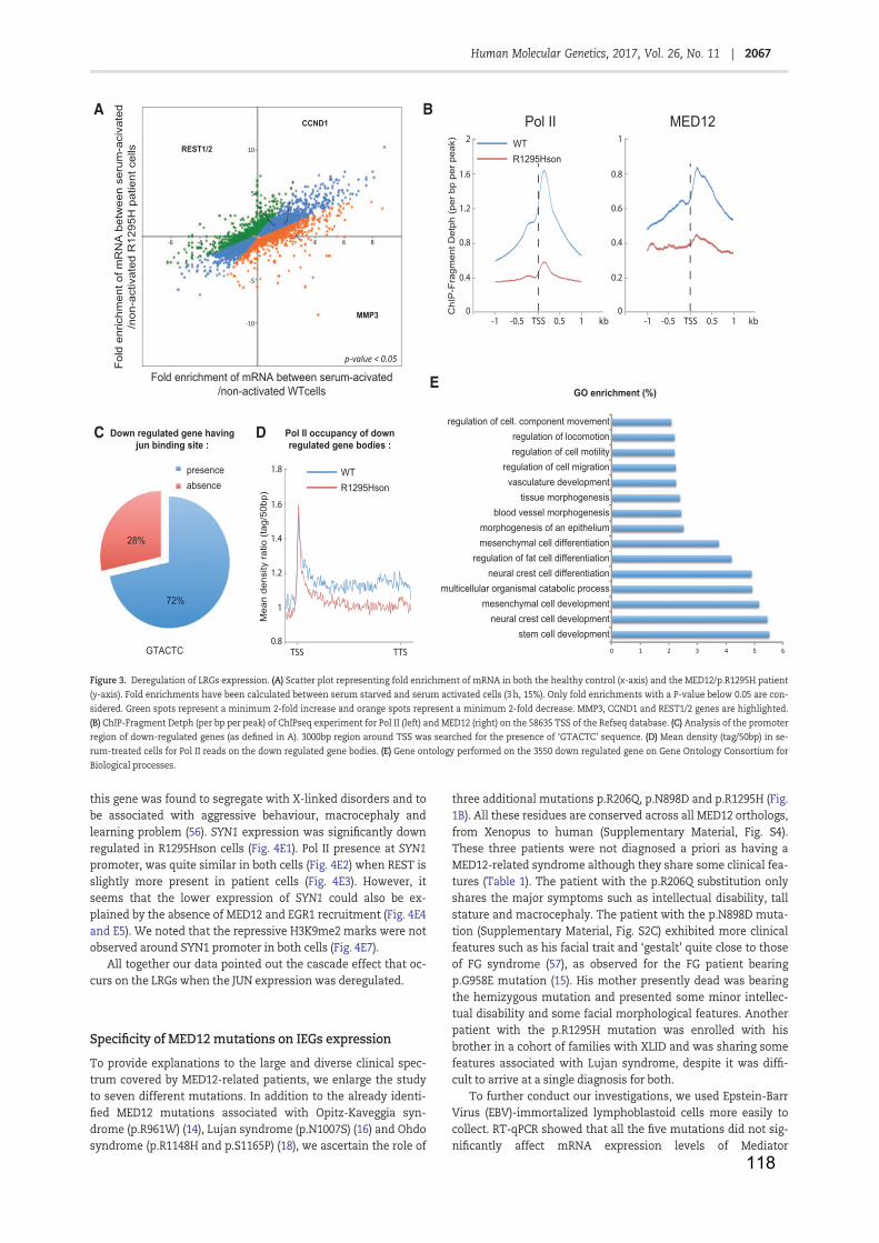
118
this gene was found to segregate with X-linked disorders and tobe associated with aggressive behaviour, macrocephaly andlearning problem (56). SYN1 expression was significantly downregulated in R1295Hson cells (Fig. 4E1). Pol II presence at SYN1promoter, was quite similar in both cells (Fig. 4E2) when REST isslightly more present in patient cells (Fig. 4E3). However, itseems that the lower expression of SYN1 could also be ex-plained by the absence of MED12 and EGR1 recruitment (Fig. 4E4and E5). We noted that the repressive H3K9me2 marks were notobserved around SYN1 promoter in both cells (Fig. 4E7).
All together our data pointed out the cascade effect that oc-curs on the LRGs when the JUN expression was deregulated.
Specificity of MED12 mutations on IEGs expression
To provide explanations to the large and diverse clinical spec-trum covered by MED12-related patients, we enlarge the studyto seven different mutations. In addition to the already identi-fied MED12 mutations associated with Opitz-Kaveggia syn-drome (p.R961W) (14), Lujan syndrome (p.N1007S) (16) and Ohdosyndrome (p.R1148H and p.S1165P) (18), we ascertain the role of
three additional mutations p.R206Q, p.N898D and p.R1295H (Fig.1B). All these residues are conserved across all MED12 orthologs,from Xenopus to human (Supplementary Material, Fig. S4).These three patients were not diagnosed a priori as having aMED12-related syndrome although they share some clinical fea-tures (Table 1). The patient with the p.R206Q substitution onlyshares the major symptoms such as intellectual disability, tallstature and macrocephaly. The patient with the p.N898D muta-tion (Supplementary Material, Fig. S2C) exhibited more clinicalfeatures such as his facial trait and ‘gestalt’ quite close to thoseof FG syndrome (57), as observed for the FG patient bearingp.G958E mutation (15). His mother presently dead was bearingthe hemizygous mutation and presented some minor intellec-tual disability and some facial morphological features. Anotherpatient with the p.R1295H mutation was enrolled with hisbrother in a cohort of families with XLID and was sharing somefeatures associated with Lujan syndrome, despite it was diffi-cult to arrive at a single diagnosis for both.
To further conduct our investigations, we used Epstein-BarrVirus (EBV)-immortalized lymphoblastoid cells more easily tocollect. RT-qPCR showed that all the five mutations did not sig-nificantly affect mRNA expression levels of Mediator
A
REST1/2
CCND1
MMP3
10
5
-6
-10
-5
864- 4
Fold
enr
ichm
ent o
f mR
NA
bet
wee
n se
rum
-aci
vate
d/n
on-a
ctiv
ated
R12
95H
pat
ient
cel
ls
Fold enrichment of mRNA between serum-acivated/non-activated WTcells
p-value < 0.05
CC
hIP
-Fra
gmen
t Det
ph (p
er b
p pe
r pea
k)
0
0.4
1.6
0.8
1.2
TSS 0.5 1-1 -0.5
2
0
0.2
0.8
0.4
0.6
1Pol II MED12
D
TSS 0.5 1-1 -0.5
TSS
Mea
n de
nsity
ratio
(tag
/50b
p)
0.8
1
1.6
1.2
1.4
1.8
TTS
WTR1295Hson
kbkb
WTR1295Hson
60 54321
regulation of cell. component movement
regulation of fat cell differentiationmesenchymal cell differentiationmorphogenesis of an epithelium
blood vessel morphogenesistissue morphogenesis
vasculature developmentregulation of cell migration
regulation of cell motilityregulation of locomotion
stem cell developmentneural crest cell development
mesenchymal cell developmentmulticellular organismal catabolic process
neural crest cell differentiation
GO enrichment (%)E
B
Down regulated gene havingjun binding site :
presenceabsence
GTACTC
72%
28%
Pol II occupancy of downregulated gene bodies :
Figure 3. Deregulation of LRGs expression. (A) Scatter plot representing fold enrichment of mRNA in both the healthy control (x-axis) and the MED12/p.R1295H patient(y-axis). Fold enrichments have been calculated between serum starved and serum activated cells (3 h, 15%). Only fold enrichments with a P-value below 0.05 are con-sidered. Green spots represent a minimum 2-fold increase and orange spots represent a minimum 2-fold decrease. MMP3, CCND1 and REST1/2 genes are highlighted.(B) ChIP-Fragment Detph (per bp per peak) of ChIPseq experiment for Pol II (left) and MED12 (right) on the 58635 TSS of the Refseq database. (C) Analysis of the promoterregion of down-regulated genes (as defined in A). 3000bp region around TSS was searched for the presence of ‘GTACTC’ sequence. (D) Mean density (tag/50bp) in se-rum-treated cells for Pol II reads on the down regulated gene bodies. (E) Gene ontology performed on the 3550 down regulated gene on Gene Ontology Consortium forBiological processes.
2067Human Molecular Genetics, 2017, Vol. 26, No. 11 |
Downloaded from https://academic.oup.com/hmg/article-abstract/26/11/2062/3084497/MED12-related-XLID-disorders-are-dose-dependent-ofby IRMA Strasbourg useron 04 October 2017
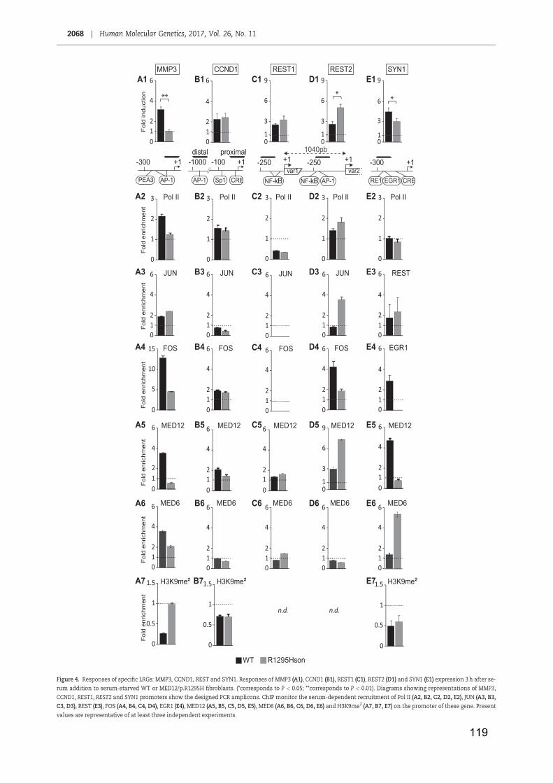
119
012
4
6MMP3
A1 B1SYN1
D1
A3 B3
A4
D3
A7 B7
*
01
3
6
9
***
012
4
6CCND1
E3
E7
REST
D4
E1
013
6
9REST2
4E4B EGR1
JUN
012
4
6
012
4
6
0
0.5
1
1.5
D5
D6
D2
E5
E6
E2
A5
A6
A2
B5
B6
B2
0
0.5
1
1.5
-300 +1
AP-1PEA3
proximaldistal+1-100-1000
AP-1 Sp1 CRE
-300 +1
RE1 CREEGR1
FOS
0
5
10
15 FOS
012
4
6 FOS
012
4
6
JUN
012
4
6JUN
012
4
6
0
0.5
1
1.5
+1
AP-1var2
-250+1var1
1040pb
NF-kB
-250
NF-kB
REST1C1
013
6
9
JUN
012
4
6
FOS
012
4
6C4
C5
C6
C3
C2
n.d. n.d.
Fold
indu
ctio
nFo
ld e
nric
hmen
tFo
ld e
nric
hmen
tFo
ld e
nric
hmen
tFo
ld e
nric
hmen
tFo
ld e
nric
hmen
tFo
ld e
nric
hmen
t
WT R1295Hson
Pol IIPol IIPol II II loPII loP
MED12MED12MED12 21DEM21DEM
MED6MED6MED6 6DEM6DEM
H3K9me²H3K9me²H3K9me²
Figure 4. Responses of specific LRGs: MMP3, CCND1, REST and SYN1. Responses of MMP3 (A1), CCND1 (B1), REST1 (C1), REST2 (D1) and SYN1 (E1) expression 3 h after se-rum addition to serum-starved WT or MED12/p.R1295H fibroblasts. (*corresponds to P < 0.05; **corresponds to P < 0.01). Diagrams showing representations of MMP3,CCND1, REST1, REST2 and SYN1 promoters show the designed PCR amplicons. ChIP monitor the serum-dependent recruitment of Pol II (A2, B2, C2, D2, E2), JUN (A3, B3,C3, D3), REST (E3), FOS (A4, B4, C4, D4), EGR1 (E4), MED12 (A5, B5, C5, D5, E5), MED6 (A6, B6, C6, D6, E6) and H3K9me2 (A7, B7, E7) on the promoter of these gene. Presentvalues are representative of at least three independent experiments.
2068 | Human Molecular Genetics, 2017, Vol. 26, No. 11
Downloaded from https://academic.oup.com/hmg/article-abstract/26/11/2062/3084497/MED12-related-XLID-disorders-are-dose-dependent-ofby IRMA Strasbourg useron 04 October 2017
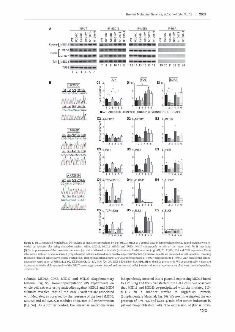
120
subunits MED12, CDK8, MED17 and MED23 (SupplementaryMaterial, Fig. S5). Immunoprecipitation (IP) experiments onwhole cell extracts using antibodies against MED12 and MED6subunits revealed, that all the MED12 variants are associatedwith Mediator, as observed by the presence of the head (MED6,MED22) and tail (MED23) modules at 300 mM KCl concentration(Fig. 5A). As a further control, the missense mutations were
independently inserted into a plasmid expressing MED12 fusedto a B10-tag and then transfected into HeLa cells. We observedthat MED16 and MED23 co-precipitated with the mutated B10-MED12 in a manner similar to tagged-WT protein(Supplementary Material, Fig. S6). We next investigated the ex-pression of JUN, FOS and EGR1 30 min after serum induction inpatient lymphoblastoid cells. The expression of JUN is down
WT1 R206Q2 N898D3 R961W4 N1007S5 R1295H6
A
B
Fold
indu
ctio
n
C1JUN
D1FOS EGR1
E1
Fold
enr
ichm
ent
C2 D2 E2
6C3 D3 E3
Fold
enr
ichm
ent
C4
Fold
enr
ichm
ent
C5
D4 E4
E5D5
Fold
enr
ichm
ent
012
4
6 ELK3
1 2 3 4 5 6
12
0
4
8
1 2 3 4 5 6
6
012
4
1 2 3 4 5 6
12
0
4
8
1 2 3 4 5 6
12
4
6 Pol II
01 2 3 4 5 6
1
3
6
9 Pol II
01 2 3 4 5 6
12
4
Pol II
01 2 3 4 5 6
012
4
6 MED12
1 2 3 4 5 6
12
4
6 MED12
01 2 3 4 5 6
12
4
6 MED12
01 2 3 4 5 6
012
4
6 TCF4 (Prox)
1 2 3 4 5 6
ELK1-P12
0
4
8
1 2 3 4 5 601
3
6
9 ELK1-P
1 2 3 4 5 6
01
3
6
9 ELK3
1 2 3 4 5 6012
4
6 TCF4 (Dis)
1 2 3 4 5 6
**
**
****
WT
R96
1WN
1007
S
N89
8D
R12
95H
R20
6Q
INPUT IP MED12 IP MED6 IP BSA
Kinase
Head
Tail
WT
R96
1WN
1007
S
N89
8D
R12
95H
R20
6Q
WT
R96
1WN
1007
S
N89
8D
R12
95H
R20
6Q
WT
R96
1WN
1007
S
N89
8D
R12
95H
R20
6Q
MED12
MED23
MED6
MED22
TUBB1 2 3 4 5 6
7 8 9 10 11 12 13 14 15 16 17 18 19 20 21 22 23 24
Tyr Pro GlyTyr Argp.R206Q
TACTACCGGCCAGGG
Tyr Pro GlyTyr GlnTACTACCAGCCAGGG
p.N898DLeu Glu LeuLeu AsnCTGCTGAATGAACTG
Leu Glu LeuLeu AspCTGCTGGATGAACTG
p.R1295HGly CysLeuGlu Arg
GGAGAACGTTGCCTT
Gly CysLeuGlu HisGGAGAACATTGCCTT
Figure 5. MED12 mutated lymphoblats. (A) Analysis of Mediator composition by IP of MED12, MED6 or a control (BSA) in lymphoblastoid cells. Bound proteins were re-vealed by Western blot using antibodies against MED6, MED12, MED22, MED23 and TUBB. INPUT corresponds to 20% of the lysate used for IP reactions.(B) Electropherograms of the three new mutations (in bold) of affected individuals (bottom) and healthy control (top). (C1, D1, E1)JUN, FOS and EGR1 expression 30minafter serum addition to serum-starved lymphoblastoid cell lines derived from healthy subject (WT) or MED12 patient. Results are presented as fold induction, meaningthe ratio of treated cells relative to non-treated cells, after normalization against GAPDH. (*corresponds to P < 0.05; **corresponds to P < 0.01). ChIP monitor the serum-dependent recruitment of MED12 (C2, D2, E2), Pol II (C3, D3, E3), TCF4 (C4, C5), ELK1-P (D4, E4) or ELK3 (D5, E5) on the IEGs promoter in WT or patient cells. Values areexpressed as fold enrichment (ratio of the INPUT percentage between treated and non-treated cells). Present values are representative of at least three independentexperiments.
2069Human Molecular Genetics, 2017, Vol. 26, No. 11 |
Downloaded from https://academic.oup.com/hmg/article-abstract/26/11/2062/3084497/MED12-related-XLID-disorders-are-dose-dependent-ofby IRMA Strasbourg useron 04 October 2017
121
regulated in R206Q, R961W and N1007S cells as compared withWT, N898D and R1295H cells (Fig. 5C1, lanes 2, 4 and 5). In con-trast, FOS is up regulated in R961W, N1007S and R1295H cells(Fig. 5D1, lanes 4–6). EGR1 expression is down regulated in bothR206Q and N898D cells compared with WT, R961W, N1007S andR1295H cells (Fig. 5E1, lanes 2 and 3). The two mutations relatedto Ohdo syndrome (R1148H and S1165P) also lead to an up regu-lation of FOS and a down-regulation of EGR1 (SupplementaryMaterial, Fig. S7), illustrating the regulatory role of MED12 inIEGs expression regulation.
ChIP experiments next showed that the recruitment of Pol IIand MED12 at their respective promoter parallels the expressionlevel of JUN, FOS and EGR1 (Fig. 5C2 and C3, D2 and D3 and E2and E3). The recruitment of TCF4, at the responsive element ofJUN promoter is impaired when Pol II and MED12 binding is de-fective (Fig. 5C4 and C5, lanes 2, 4 and 5). In R961W, N1007S andR1295H cells, the up-regulation of FOS parallel the reduced bind-ing of ELK1-P which is compensated by an increase binding ofits paralog ELK3 on the SRE element (Fig. 5D4–D5, lanes 4–6). Wealso observed a higher binding of ELK1-P and a lower binding ofELK3 at the FOS promoter in R206Q cells (Fig. 5D4 and D5, lane2). In N898D cells, neither ELK1-P nor ELK3 were detected at SRE(Fig. 5D4 and D5, lane 3). However, in these two cell lines, FOSseems to be normally expressed (Fig. 5D1, lanes 2 and 3). In bothR206Q and N898D cells, where EGR1 is down regulated, neitherELK1-P nor ELK3 are recruited at SRE (Fig. 5E4 and E5, lanes 2and 3), while in the R961W, N1007S and R1295H cells as well asin WT cells, we observed ELK1-P recruitment (lanes 1 and 4–6).
Knowing that each gene can be under the control of differentstimuli, we also investigated the behaviour of JUN and FOSwhen cells were submitted to another cellular stress such as UVirradiation. Contrary to what was observed under serum mito-gen stress, JUN is not down regulated but even seems to be upregulated in R206Q cells (Supplementary Material, Fig. S8A, lane2). In UV treated R961W and R1295H cells, FOS seemed to besimilarly overexpressed compared with what occurred in seruminduced cells. However, we observed a much lower stimulationin N1007S cells and a significant increase in R206Q cells(Supplementary Material, Fig. S8B lanes 4, 6 and 5, 2, respec-tively). Our data show that MED12 patients might be sensitive toother cellular stress. We also investigated the effect of MED12mutations on the expression of the RARb (MIM: 180220) gene in-duced by all trans retinoic acid (t-RA), that mediates cellular sig-nalling during embryonic morphogenesis, cell growth anddifferentiation (58). Every t-RA treated lymphoblastoid cell lines,except N1007S cells, accumulated RARb mRNA (SupplementaryMaterial, Fig. S8C, lane 5). This demonstrate that JUN and FOSare sensitive to any kind of stress in general.
All together, the above data significantly showed that eachMED12 mutations might lead to a specific pattern of IEGs ex-pression, resulting from impairment in the formation of thetransactivation complex. This will undoubtedly influence theLRGs in a different manner.
DiscussionIn an attempt to dissect the complicated molecular mecha-nisms underlying the control of gene expression, we decided todeeper our knowledge on the large multi-subunit Mediator com-plex. In human, the central role of Mediator was highlighted bythe discovery of mutations in some of its subunits leading to se-vere genetic disorders (8–10,15,16,18). Among MED subunits,MED12 is the part of the Kinase module that plays a crucial rolein gene expression regulation (4,5,31). Our work first revealed
that MED12 knock down in human neuroblastoma cells signifi-cantly reduced the expression of JUN, FOS and EGR1, three genesinvolved in brain development and plasticity (Fig. 1D)(12,39,59,60).
We focused on several missense MED12 mutations responsi-ble for broad and diverse clinical features. Although patientsshare some similar characteristics such as intellectual disabil-ities, several were not diagnosed a priori as having a MED12 as-sociated syndrome, such as Lujan, Opitz-Kaveggia or Ohdosyndromes (Table 1).
As Mediator serves as a link between gene specific-bindingfactors and the basal transcription machinery, we investigatedthe activity of several genes in cells carrying MED12 mutations.We then observed that the MED12 mutations led to the abnormaland specific deregulation of IEGs expression. For example,MED12/p.R206Q,/p.R961W and/p.N1007S disrupt JUN expression(Fig. 5C1). In such cases, TCF4 was not properly recruited to itsbinding element (Fig. 5C5) and was then unable to contact thebasal Pol II transcription machinery on proximal promoter(Fig. 5C4). Similarly, MED12/p.R961W,/pN1007S and/p.R1295H mu-tations up regulated FOS expression (Fig. 5D1), through a changein the correct configuration of the transcription machinery at theIEGs (e.g. ELK3 recruitment instead of ELK1, Fig. 5D2–D5). A thirdexample was provided by the MED12/p.R206Q and/p.N898D thatdown regulated EGR1 expression due to the absence of an ELKproteins in the transactivation complex (Fig. 5E4 and E5).
We also question about the cause that prevent normalMediator function. Although MED12 mutations were shown notto disrupt the formation of the Mediator complex in solution(Fig. 5A), it is likely that they modify in some way the intrinsicstability of the complex, leading to incomplete binding of the ki-nase module within the transcription complex. In vitro investi-gations predicted a role for the Kinase module of MED towardseveral substrates such as TFIIH, Pol II, as well as CDK8 itself inthe context of activated gene expression (28). Moreover, it wasalso shown that the Kinase complex incorporation within MEDwas essential for CDK8 substrate specificity. When MED12 is re-quired for stable incorporation of CDK8/CCNC into MED, it isalso required to activate the CDK8 kinase. Here we found thatthe kinase module (according to ChIP experiments on MED12and CDK8) did not properly target the JUN promoter (Fig. 2A1and A2). Consequently, inaccurate expression of IEGs coding forDNA-binding factors, will further modify LRGs expressionthrough a consecutive sequence of events. Indeed in theR1295Hson cells, the deregulation of MMP3 and REST2 expres-sion might be explained by a modification in the JUN/FOS re-cruitment at their respective promoters (Fig. 4A3–A4). It is likelythat MED12/p.R1295H associated defect in bridging DNA bindingfactor to Pol II machinery could also be responsible at least par-tially, for such deregulation. We indeed observed some changesin the Kinase(MED12)/Core-Mediator(MED6) ratio at the MMP3and/or SYN1 promoter compared with WT cells (Fig. 4A5, A6 andE5, E6). It is clear that MED12 mutations lead to a modificationof the correct configuration of the transcription machinery atthe IEGs and the LRGs promoter, which resulted in the modifica-tion of the transcriptional programme in the patient cells. Thespecificity of individual mutation also depends on the cellularcontext as shown for the FOS expression in MED12/p.R1295H fi-broblasts or for the expression of RARb in N898D fibroblasts ver-sus lymphoblastoids cells (Supplementary Material, Fig. S9).
Our study has shown that the MED12 mutations led to theabnormal expression of IEGs and resulted in changes into LRGsexpression. Ultimately, these results suggest that the differentphenotypes caused by the different mutations are essentially
2070 | Human Molecular Genetics, 2017, Vol. 26, No. 11
Downloaded from https://academic.oup.com/hmg/article-abstract/26/11/2062/3084497/MED12-related-XLID-disorders-are-dose-dependent-ofby IRMA Strasbourg useron 04 October 2017
122
the result of the above to varying degrees. The deregulation ofJUN, regarded as a marker of neural activity and its involvementin partnership with FOS, partially explained the neurologicalphenotypes of the patients as summarized Table 1. Althoughthe gene regulation abnormalities do not precisely explain theclinical phenotypes, we observed significant down-regulation ofgenes containing AP1-binding site targeted by JUN/FOS com-plex. Gene Ontology enrichment then shows that most of thesegenes are involved in development and neurological processes.Further investigations on the expression patterns of certaingenes including MMP3, REST2 and SYN1 in neural cells will helpto decipher the specific consequences of MED12 mutation inneurological context. Future studies should help to elucidatethe genotype/phenotype relationship of the patients withMED12 mutations.
We noticed some heterozygous mother with missense mu-tation to share part of the symptoms (e.g. mild intellectual dis-ability). MED12 is located on chromosome X and one of its twoalleles is inactivated in females. We have observed that most ofcultivated heterozygous fibroblasts (about 80%) from the motherof the MED12/p.R1295H patient express the mutated allele. Thisskewed XCI phenomenon might explain why the MED12/p.R1295H and/p.N898D mothers exhibit similar but milder clini-cal features than their sons. Actually, the partial maternalsymptoms could be explained by a stochastic effect occurring attwo different times: at the early embryonic stage of the X-inacti-vation process and lately during the embryonic organ specifica-tion due to a random redistribution of founder cells. Thisrandom process could be systematically skewed in some casesdue to preferential inactivation of the wild allele. We could notdiscard this hypothesis since MED12 was implicated in the acti-vation of non-coding RNA. In this way, mutated MED12 mighthave a possibility to disturb the balance of XIST and/or TSIXnon-coding RNAs transcription which control the X-inactivationprocess (61). In fact, a defect of XIST in tiny ring X chromosomepatients is already know to result in developmental and cogni-tive disorders (62).
A number of human diseases with IEG expression affectingbrain development and plasticity are caused by mutations ingenes encoding for MED subunits or proteins interacting withMED, such as nucleotide excision repair factors or chromatin re-modellers. Mutations of MED13L, MED17, MED23 or MED25 areresponsible for infantile cerebral and cerebellar atrophy, non-syndromic mental retardation and Charcot-Marie Tooth disease(8–11). Mutations in TCF4, which interact with the Mediator, re-sult in the Pitt-Hopkins syndrome (42). Examples are also pro-vided by xeroderma pigmentosum (XP, MIM:278730) patientsthat have progressive neurological degeneration as well as bothtrichothiodystrophy (MIM:610675) and Cockayne syndrome(MIM:610651) patients that are mentally retarded (63,64). It islikely that more and more mutations in MED and its transcrip-tion partners will be detected in patients with cognitive defi-ciencies as sequencing becomes more commonly used.Therefore, one could speculate that these patients should beclassified on a genomic basis rather than distinct syndromes.
Material and MethodsCell culture
Human lymphoblastoıdes cells were generated by EBV transfor-mation of the peripheral blood lymphocytes isolated from pa-tients. Human primary fibroblasts were isolated from patient’s
skin. Informed consent was obtained from all subjects beforesample collection.
The cs of R1295Hmother fibroblast was done by successivedilution and confirm by Sanger sequencing of total mRNA.
CRISPR/Cas9 system was used to Knock-Down MED12 in neu-roblastoma LAN-1 cell line. A targeting vector was constructed onthe bases of the pPGKNEO-DTA (addgene no. 13443). 50 arm span-ning intron 2/exon 3 (1 kb, HindIII/AgeI) and 30 arm spanningexon 5/intron 5 (960 bp, NheI/XhoI) were PCR amplified from ge-nomic DNA of LAN-1 neuroblastoma cells and sequentiallycloned into the first pPGKNEO-DTA vector using respected en-zymes. Furthermore a GFP was amplified from pEGFPC1 vectorand inserted via restriction digest (AgeI/NcoI) so that exon 3and GFP will be translated in frame. A guiding RNA targetingexon 3/intron 3 junction was designed and cloned into p459 vec-tor (addgene no. 48139). All constructs were verified by sequenc-ing. All together the targeting vector and CRISPR/Cas9 systemtargeting the end of exon 3 were transfected into LAN-1 neuro-blastoma cells using Amaxa nucleofection protocol. Cells werebriefly selected with puromycin for first 2 days and with neomy-cin for next two weeks. Clones were first pre-screened for GFP ex-pression and further verified by genomic PCR for absence of exon4 and Med12-specific integration of targeting vector.
Med12 3 arm fw CCAAGCTAGCTAACTCCTAACACCAGGTGTACTGC
Med12 3 arm rv CCCTCGAGCAAGCTTACACAGCATGCCCTACTCTCTACC
Med12 5 arm fw TCAGATCTCGAGCTCAAGCTTGGTCAGCCTAGGAGGAGGCACTG
Med12 5 arm rvATGGTGGCGACCGGTGGGGCTAGTTGCGTGAGTGGCTTGG
px4_3ex_Med12_fwcaccgAAAAGGTAAGGTACTGTTTCpx4_3ex_Med12_rvaaacGAAACAGTACCTTACCTTTTc
All the cells were cultured in appropriate medium and main-tained at 37 !C in 5% CO2 environment.
For serum inductions, cells were incubated in red phenol-free medium without serum for 24 h before treatment by addi-tion of serum (15% final concentration) directly into the me-dium. For retinoic acid inductions, cells were incubated in redphenol-free medium containing charcoal treated Fetal CalfSerum and antibiotics before treatment with 10 mM tRA (Biomol)into the same medium. For UV inductions, cells were first PBSwash then 4 millions of lymphoblastoid cells in 2 ml of PBS werespreading on a 10-cm dish. Cells were exposed to UV irradiation(20 J/m2) and medium added back.
RNA extraction, reverse transcriptionand real-time qPCR
Total RNA was extracted with RNeasy mini kit (QIAgen) and re-verse transcribed with poly-dT primer using Superscript II re-verse transcriptase (Invitrogen). Real-time quantitative PCR wascarried out on the Lightcycler 480 using SYBR Green I Master(Roche). The primer sequences are available in SupplementaryMaterial. mRNA levels represent the ratio between values ob-tained from treated cells compared with untreated cells normal-ized against the housekeeping Glyceraldehyde 3-phosphatedehydrogenase (GAPDH) mRNA.
Co-immunoprecipitation assays
Cells were harvest in lysis buffer (50mM Tris HCl pH7.5, 150 mMNaCl, 1mM EDTA, 1% Triton) with protease inhibitor cocktail
2071Human Molecular Genetics, 2017, Vol. 26, No. 11 |
Downloaded from https://academic.oup.com/hmg/article-abstract/26/11/2062/3084497/MED12-related-XLID-disorders-are-dose-dependent-ofby IRMA Strasbourg useron 04 October 2017
123
(PIC). 200 mg of whole cell extract were incubated with protein Gmagnetic bead (dynabead, invitrogen) and 2 mg of antibodies.After washes at 300 mM salt, beads were boiled in Laemmli buf-fer and proteins were separated by SDS-PAGE, transferred to ni-trocellulose membrane and probed with antibodies.
Chromatin immunoprecipitation
After treatment, cells were crosslinked at room temperature for15 min with 1% formaldehyde. Cells were lysed in shearing buffer(50 mM Tris-HCl pH8, 10 mM EDTA, 1% SDS) with PIC at 4 !C for15 min. Nucleus were pelleted and resuspended in sonicationbuffer (50 mM Hepes pH7.6, 140 mM NaCl, 1 mM EDTA, 1% TritonX100, 0.1% Na-deoxycholate, 0.1% SDS) with PIC. Chromatin wasshared using 800R sonicator (Qsonica). Samples were immuno-precipitated with antibodies at 4 !C overnight followed by addi-tion of a pre-blocked mix of protein G- and A-Sepharose beads(GE Healthcare Life Sciences) for 3 h at 4 !C. Bound complexeswere sequentially washed with sonication buffer, high salt buffer(sonication buffer at 300 mM NaCl) and TE buffer (10 mM Tris,1mM EDTA). Protein-DNA complexes were eluted, and DNA frag-ments were purified using QIAquick PCR purification kit(QIAGEN). qPCR was performed as described earlier using sets ofprimers available in Supplementary Material. All the results arepresented as ‘fold recruitment’ and represent the ratio of inputpercentage between treated and non-treated cells.
Plasmids
MED12 expression plasmid was obtained using the GatewayInvitrogen cloning method. WT MED12 was amplified from acDNA bank of control lymphoblastoid cells and cloned intopDONR207 (Invitrogen) using standard BP reaction. The clonedsequence was then transferred by LR reaction into pSG5 puroB10 tag vector (N-terminal fusion of the epitope B of the humanestrogen receptor). This vector was constructed by inserting theattL1 and attL2 Gateway linkers (Invitrogen) into the pSG5 vectorbackbone. PCR-based mutagenesis was performed usingPhusion High-Fidelity DNA Polymerase (New England BioLabs,inc.) with primer bearing a point mutation for the aminochanges R206Q, N898D, R961W, N1007S and R1295H.
MED12 expression plasmid was transfected using Jet PEI(Polyplus) in HeLa cells, 48 h before the experiments.
Antibodies
For Western blot, the following antibodies were used: MED6(santacruz, sc-9434), MED12 (Bethyl, A300-774A), MED22 (santa-cruz, sc-393738), MED23 (BD Pharmingen, 550429), CCNC (santa-cruz, sc-1061) and b-tubulin (TUBB) (millipore, MAB3408).
For immunoprecipitation, the following antibodies wereused: monoclonal antibodies against B10 (IGBMC), RNA Pol II(IGBMC, 1BP 7C2), ELK3 (IGBMC, 5NE 2F3A2) and polyclonal anti-bodies against BSA (santacruz, sc50528), MED6 (santacruz, sc-9434), MED12 (Bethyl, A300-774A), TFIIB (santacruz, sc-225),TCF4 (santacruz, sc13027), ELK1-P (santacruz, sc8406), JUN (san-tacruz, sc-45 or sc 822), FOS (santacruz, sc-7202), REST (Bethyl,A300-539A) and H3K9ac (cell signalling, no. 9671 or no. 9649)and H3K9me2 (cell signalling, no. 9753).
Mutation screening of the MED12 gene
Genomic DNA was prepared from lymphocytes according tostandard protocols. Sanger resequencing of 718 X-chromosome
genes was conducted as described previously in Tarpey et al.(2009) for the proband in family K9338. The proband in K9467,was considered to have a phenotype similar to FG syndromeand he was sequenced for all coding exons of MED12. The singlemale in family 8935 was sequenced for all coding exons inMED12 as part of a general screen of a cohort of males with ID.
Sequences alignment
Sequences alignment was performed with BioEdit software(open source). The following sequence of Med12 has been used:Homo sapiens (NP_005111.2), Mus musculus (NP_067496.2), Rattusnorvegicus (NP_001180221.1), Bos Taurus (XP_005228076.1), Pongoabelii (NP_001124553.2), Pan troglodytes (NP_001009019.1), Daniorerio (NP_001034550.1), Xenopus tropicalis (XP_002934949.2).
RNAseq
Total RNA was extracted with TRI REAGENT (MolecularResearch center, inc.) and purified by phenol-chloroform extrac-tion. Libraries was prepared with TruSeq Stranded mRNASample Preparation kit following guide instruction and subse-quently proceed on an Illumina Hiseq 4000 as single-end 50base reads following Illumina’s instructions. Image analysisand base calling were performed using RTA 2.7.3 and bcl2fastq2.17.1.14. Reads were mapped onto the hg38 assembly of the hu-man genome. Reads count was performed with HOMER v4.8.3(65) and differently expression was estimated with EdgeR.Genome ontology was performed on http://geneontology.org,searching for biological processes.
ChIPseq
DNA fragments analysed by ChIP-seq were prepared trough reg-ular ChIP procedure and purified by phenol-chloroform extrac-tion. ChIP-seq was performed on an Illumina Hiseq 4000 assingle-end 50 base reads following Illumina’s instructions.Image analysis and base calling were performed using RTA1.17.20 and CASAVA 1.8.2. Reads were mapped onto the hg38 as-sembly of the human genome. Quantitative comparisons of theChIP-seq data were performed using HOMER v4.8.3 (65). As ref-erence coordinates, we used RefSeq coordinates of humangenes. Tag densities were collected 61 kilobases of the TSS oralong the gene body.
Clinical reports
Patient K8935 (with mutation MED12/p.R206Q): W.D.A. is a 391=2-year old African American male who has been institutionalizedin facilities for those with intellectual disability since age17 years. Psychological testing showed IQ measurements of 52,age 7 years. Vineland social quotient was 52 at age 12 years.Sequencing of the MED12 gene identified c.617G > A, p.R206Qmutation. Clinical findings are given in Table 1.
Patient K9467 (with mutation MED12/p.N898D): A single male isaffected in K9467. The mother had a prior spontaneous abor-tion. The birth weight was 3.4 kg and the length was 50 cm.Imperforate anus and cryptochidism were present and cor-rected surgically. Development was globally delayed with inde-pendent walking and first clear words achieved at age threeyears. Special education was required. Intellectualperformance was mildly impaired with a full scale IQ of 58. Atage 6 years, he had a height of 113.5 cm (35th centile) and
2072 | Human Molecular Genetics, 2017, Vol. 26, No. 11
Downloaded from https://academic.oup.com/hmg/article-abstract/26/11/2062/3084497/MED12-related-XLID-disorders-are-dose-dependent-ofby IRMA Strasbourg useron 04 October 2017
124
occipital frontal circumference (OFC) of 55.3 cm (>97th centile).He has a tall and narrow forehead, downslanting palpebral fis-sures, small posteriorly rotated ears (4.5 cm length), maxillarydental crowding and prognathism. The thumbs were flat withangulation of the distal phalanges and the great toes werebroad. Neurological examination was normal. There was a ten-dency to frustration and psychological lability, intolerance anda short attention span. At age of nineteen years, he has a heightof 176 cm, weight of 61 kg and head circumference of 61 cm(>99th centile). Gross motor function is normal, fine motorfunction is slightly impaired. IQ has not been reevaluated butclinical appreciation is consistent with 6-years old IQ.Behavioural problems are prominent with marked frustrationintolerance. Constipation and easy vomiting still required per-manent medical management (Supplementary Material, Fig.S2C and D).
His mother presents with similar but milder dysmorphic fea-tures. She suffered from obstinate constipation. She had normalintelligence but some choleric behavioural problems.Unfortunately, she suddenly died of intracranial aneurysm rup-ture at the age of 47 years.
Patients K9338 (with mutation MED12/p.R1295H): K9338 hastwo brothers with intellectual disability born to consanguineousparents. They presented with somewhat different phenotypes.One had congenital anal stenosis that was dilated, undescendedtestes, an umbilical hernia and bilateral inguinal hernias Hewas very excitable as a child and this transitioned into tantrumsand restlessness as a teen. IQ testing, age 11 years, gave a scoreof 58. The second male was hypotonic, slow to feed and devel-oped slowly, and did not walk until after age 2 years. He alsohad undescended testes, finger contractures at the PIP joints,hammer toes and a gap between toes 1 and 2. Additional clinicalfindings are listed in Table 1. Sequencing of 718 genes on the Xchromosome identified a c.3884G > A, p.R1295K mutation inone of the males which was subsequently found in his brother.
Patients A.J. (with mutation MED12/p.R1295H from FRANCE). A.J.was the second child of unrelated parents. His two brotherswere in good health. He was born at term (40 WG) after an un-eventful pregnancy. Birth weight was 3.1 kg. The neonatalcourse was uncomplicated but he had delayed developmentalmilestones. At 22 months, it was noted a global hypotony andsome morphologic particularities. Ophthalmological and heartexaminations, as well as abdominal ultrasound, echocardiogra-phy and CT scan findings were unremarkable. He walked at ageof 24 months. Speech was slightly delayed. At age 4 years, hehad an orchiopexy because his left testis was on the inguinal ca-nal. During childhood, he suffered from repetitive strain injury.From the age of 7, he had school difficulties and was enrolled ina school mainstreaming class. He had a nasal speech and a longand thin habitus. At age 20 years, his height was 188 cm (þ2DS),weight was 68.5 (þ1.5DS) and OFC was 57 cm (þ2DS). Arm spanwas 184 cm. Thoraco abdominal examination showed a mildscoliosis, pectus excavatum, two surnumemary nipples and ahypotonic abdominal wall. The facial features included a longand hypotonic face, moderate hypertelorism, bilateral ptosiswith epicanthus, down slanting palpebral fissures, evertedlower eyelids, arched, large and sparse eyebrows, medially flar-ing, malar hypoplasia, a long nose with high and narrow nasalbridge, thin upper lip, teeth malposition with absence of the up-per lateral incisors and first molars. Ears were small and low-setwith thick helices. Joint laxity, long digits, without a positivethumb sign and flat feet were present. Audiometry evaluation,EEG, metabolic screen in blood and urine, endocrinologic andcytogenetic screen (karyotype, comparative genomic
hybridization (CGH) array) were all normal. Ophthalmologicevaluation showed a left divergent strabismus (SupplementaryMaterial, Fig. S2A and B).
His mother had the same skeletal and morphologic featureswith a long and hypotonic face, high arched eyebrows, bilateralptosis, a long filtrum, teeth malposition. She had moderatelearning difficulties but none intellectual disability. Her heightwas 173 cm OFC was 56.5 cm (þ1.5DS). Echocardiographyshowed a mitral valve prolapse requiring simple monitoring.She underwent surgery for left divergent strabismus and suf-fered from repeated ankle sprains and early arthrosis of the hip.As her son, she had a nasal speech.
Patients’ consent. R1295H fibroblasts were obtained at CHUClermont-Ferrand and provided by C.F. Informed and writtenconsent was obtained from the patient and his mother. N898Dcell lines were obtained at CHU Grenobles and provided by C.C.and P.S.J. Informed and written consent was obtained from thepatient and his father. S1165P and R1148H cell lines were ob-tained at Donders Institute for Brain and provided by AdB.Informed consent was obtained from all families as publishedin Vulto-van Silfhout et al. (18). R206Q, R961W, N1007S andR1295H lymphoblastic cell lines were obtained through theX-linked mental retardation (XLMR) study at the GreenwoodGenetic Centre and provided by CES. Informed consent was ob-tained from all families as published for R961W (14), N1007S (16)and presented at the 2014 annual meeting of the AmericanSociety of Human Genetics for R206Q and R1295H. XLMR re-search at the Greenwood Genetic Centre has been approved bythe Institutional Review Board at Self Regional Hospital inGreenwood, SC.
Supplementary MaterialSupplementary Material is available at HMG online.
Acknowledgements
This study is dedicated to all the patients and families, particu-larly to Antoine J. We are gratefull to Annick Toutain, LaurenceFaivre, Han G Brunner and Anneke T Vulto-van Silfhout forhelping in collecting patient cells and clinical data. CindySkinner coordinated sample collection and sharing of materials.We also acknowledge the IGBMC facilities for their technical ex-pertise. We also are grateful to Frederic Coin, Emanuel Compe,Jamel Chelly, Jean-Louis Mandel for fruitful discussion.
Conflict of Interest statement. All authors disclose any financialand personal relationships with other people or organizationsthat could inappropriately influence (bias) their work.
FundingThis study was supported (to J.M.E.) by an ERC Advanced grant;l’Agence Nationale de la Recherche [no. ANR1 08MIEN-022-03];l’Association pour la Recherche contre le Cancer; l’AssociationNationale des Membres de l’Ordre National du MeriteANMONM; la ligue contre le cancer fellowships (to L.M.D.);Fondation pour la Recherche Medicale (to B.B.), young scientistgrant from Inserm (to S.H.); Grant-in-Aid for Scientific Researchfrom MEXT and JSPS Japan (to S.H.); NICHD grant (to C.E.S.) [HD26202] and a grant from the South Carolina Department ofDisabilities and Special Needs.
2073Human Molecular Genetics, 2017, Vol. 26, No. 11 |
Downloaded from https://academic.oup.com/hmg/article-abstract/26/11/2062/3084497/MED12-related-XLID-disorders-are-dose-dependent-ofby IRMA Strasbourg useron 04 October 2017
125
Web ResourcesThe URLs for data presented herein are as follows:OMIM: http://www.omim.orgRefSeq: http://www.ncbi.nlm.nih.gov/RefSeq
References1. Kornberg, R.D. (2005) Mediator and the mechanism of tran-
scriptional activation. Trends Biochem. Sci, 30, 235–239.2. Malik, S. and Roeder, R.G. (2010) The metazoan Mediator co-
activator complex as an integrative hub for transcriptionalregulation. Nat. Rev. Genet., 11, 761–772.
3. Whyte, W.A., Orlando, D.A., Hnisz, D., Abraham, B.J., Lin,C.Y., Kagey, M.H., Rahl, P.B., Lee, T.I. and Young, R.A. (2013)Master transcription factors and mediator establish super-enhancers at key cell identity genes. Cell, 153, 307–319.
4. Allen, B.L. and Taatjes, D.J. (2015) The Mediator complex: acentral integrator of transcription. Nat. Rev. Mol. Cell Biol., 16,155–166.
5. Ansari, S.A. and Morse, R.H. (2013) Mechanisms of Mediatorcomplex action in transcriptional activation. Cell. Mol. LifeSci. CMLS, 70, 2743–2756.
6. Conaway, R.C. and Conaway, J.W. (2011) Function and regu-lation of the Mediator complex. Curr. Opin. Genet. Dev., 21,225–230.
7. Lee, T.I. and Young, R.A. (2013) Transcriptional regulationand its misregulation in disease. Cell, 152, 1237–1251.
8. Kaufmann, R., Straussberg, R., Mandel, H., Fattal-Valevski,A., Ben-Zeev, B., Naamati, A., Shaag, A., Zenvirt, S., Konen,O., Mimouni-Bloch, A. et al. (2010) Infantile cerebral and cere-bellar atrophy is associated with a mutation in the MED17subunit of the transcription preinitiation mediator complex.Am. J. Hum. Genet., 87, 667–670.
9. Leal, A., Huehne, K., Bauer, F., Sticht, H., Berger, P., Suter, U.,Morera, B., Valle, G., Lupski, J.R., Ekici, A. et al. (2009)Identification of the variant Ala335Val of MED25 as responsi-ble for CMT2B2: molecular data, functional studies of theSH3 recognition motif and correlation between wild-typeMED25 and PMP22 RNA levels in CMT1A animal models.Neurogenetics, 10, 275–287.
10. Hashimoto, S., Boissel, S., Zarhrate, M., Rio, M., Munnich, A.,Egly, J.-M. and Colleaux, L. (2011) MED23 mutation links in-tellectual disability to dysregulation of immediate earlygene expression. Science, 333, 1161–1163.
11. Adegbola, A., Musante, L., Callewaert, B., Maciel, P., Hu, H.,Isidor, B., Picker-Minh, S., Le Caignec, C., Delle Chiaie, B.,Vanakker, O. et al. (2015) Redefining the MED13L syndrome.Eur. J. Hum. Genet., 23, 1308–1317.
12. Berk, A.J. (2012) Yin and yang of mediator function revealed byhuman mutants. Proc. Natl. Acad. Sci. U. S. A., 109, 19519–19520.
13. Graham, J.M. and Schwartz, C.E. (2013) MED12 related disor-ders. Am. J. Med. Genet. A, 161A, 2734–2740.
14. Risheg, H., Graham, J.M., Clark, R.D., Rogers, R.C., Opitz, J.M.,Moeschler, J.B., Peiffer, A.P., May, M., Joseph, S.M., Jones, J.R.et al. (2007) A recurrent mutation in MED12 leading to R961Wcauses Opitz-Kaveggia syndrome. Nat. Genet., 39, 451–453.
15. Rump, P., Niessen, R., Verbruggen, K., Brouwer, O., de Raad,M. and Hordijk, R. (2011) A novel mutation in MED12 causesFG syndrome (Opitz-Kaveggia syndrome). Clin. Genet., 79,183–188.
16. Schwartz, C.E., Tarpey, P.S., Lubs, H.A., Verloes, A., May,M.M., Risheg, H., Friez, M.J., Futreal, P.A., Edkins, S., Teague,J. et al. (2007) The original Lujan syndrome family has a novel
missense mutation (p.N1007S) in the MED12 gene. J. Med.Genet., 44, 472–477.
17. Isidor, B., Lefebvre, T., Le Vaillant, C., Caillaud, G., Faivre, L.,Jossic, F., Joubert, M., Winer, N., Le Caignec, C., Borck, G. et al.(2014) Blepharophimosis, short humeri, developmental delayand hirschsprung disease: expanding the phenotypic spectrumof MED12 mutations. Am. J. Med. Genet. A, 164A, 1821–1825.
18. Vulto-van Silfhout, A.T., de Vries, B.B.A., van Bon, B.W.M.,Hoischen, A., Ruiterkamp-Versteeg, M., Gilissen, C., Gao, F.,van Zwam, M., Harteveld, C.L., van Essen, A.J. et al. (2013)Mutations in MED12 cause X-linked Ohdo syndrome. Am. J.Hum. Genet., 92, 401–406.
19. Lesca, G., Moizard, M.-P., Bussy, G., Boggio, D., Hu, H., Haas,S.A., Ropers, H.-H., Kalscheuer, V.M., Des Portes, V., Labalme,A. et al. (2013) Clinical and neurocognitive characterizationof a family with a novel MED12 gene frameshift mutation.Am. J. Med. Genet. A, 161A, 3063–3071.
20. Fieremans, N., Van Esch, H., Holvoet, M., Van Goethem, G.,Devriendt, K., Rosello, M., Mayo, S., Martinez, F., Jhangiani,S., Muzny, D.M. et al. (2016) Identification of intellectual dis-ability genes in female patients with a Skewed X-Inactivation pattern. Hum. Mutat., 37, 804–811.
21. Prescott, T.E., Kulseth, M.A., Heimdal, K.R., Stadheim, B.,Hopp, E., Gambin, T., Coban Akdemir, Z.H., Jhangiani, S.N.,Muzny, D.M., Gibbs, R.A. et al. (2016) Two male sibs with se-vere micrognathia and a missense variant in MED12. Eur. J.Med. Genet., 59, 367–372.
22. Prontera, P., Ottaviani, V., Rogaia, D., Isidori, I., Mencarelli,A., Malerba, N., Cocciadiferro, D., Rolph, P., Stangoni, G.,Vulto-van Silfhout, A. et al. (2016) A novel MED12 mutation:Evidence for a fourth phenotype. Am. J. Med. Genet. A, 170,2377–2382.
23. Tzschach, A., Grasshoff, U., Beck-Woedl, S., Dufke, C., Bauer,C., Kehrer, M., Evers, C., Moog, U., Oehl-Jaschkowitz, B., DiDonato, N. et al. (2015) Next-generation sequencing in X-linked intellectual disability. Eur. J. Hum. Genet., 23, 1513–1518.
24. Yamamoto, T. and Shimojima, K. (2015) A novel MED12 mu-tation associated with non-specific X-linked intellectual dis-ability. Hum. Genome Var., 2, 15018.
25. Langley, K.G., Brown, J., Gerber, R.J., Fox, J., Friez, M.J., Lyons,M. and Schrier Vergano, S.A. (2015) Beyond Ohdo syndrome:a familial missense mutation broadens the MED12 spec-trum. Am. J. Med. Genet. A, 167A, 3180–3185.
26. Bouazzi, H., Lesca, G., Trujillo, C., Alwasiyah, M.K. andMunnich, A. (2015) Nonsyndromic X-linked intellectual defi-ciency in three brothers with a novel MED12 missense muta-tion [c.5922G>T (p.Glu1974His)]. Clin. Case Rep., 3, 604–609.
27. Liu, Y., Ranish, J.A., Aebersold, R. and Hahn, S. (2001) Yeastnuclear extract contains two major forms of RNA polymer-ase II mediator complexes. J. Biol. Chem., 276, 7169–7175.
28. Knuesel, M.T., Meyer, K.D., Donner, A.J., Espinosa, J.M. andTaatjes, D.J. (2009) The human CDK8 subcomplex is a his-tone kinase that requires Med12 for activity and can func-tion independently of mediator. Mol. Cell. Biol., 29, 650–661.
29. Nemet, J., Jelicic, B., Rubelj, I. and Sopta, M. (2014) The twofaces of Cdk8, a positive/negative regulator of transcription.Biochimie, 97, 22–27.
30. Hengartner, C.J., Myer, V.E., Liao, S.M., Wilson, C.J., Koh, S.S.and Young, R.A. (1998) Temporal regulation of RNA polymer-ase II by Srb10 and Kin28 cyclin-dependent kinases. Mol. Cell,2, 43–53.
31. Akoulitchev, S., Chuikov, S. and Reinberg, D. (2000) TFIIH isnegatively regulated by cdk8-containing mediator com-plexes. Nature, 407, 102–106.
2074 | Human Molecular Genetics, 2017, Vol. 26, No. 11
Downloaded from https://academic.oup.com/hmg/article-abstract/26/11/2062/3084497/MED12-related-XLID-disorders-are-dose-dependent-ofby IRMA Strasbourg useron 04 October 2017
126
32. Kim, S., Xu, X., Hecht, A. and Boyer, T.G. (2006) Mediator is atransducer of Wnt/beta-catenin signaling. J. Biol. Chem., 281,14066–14075.
33. Moghal, N. and Sternberg, P.W. (2003) A component of thetranscriptional mediator complex inhibits RAS-dependentvulval fate specification in C. elegans. Development, 130, 57–69.
34. Rau, M.J., Fischer, S. and Neumann, C.J. (2006) ZebrafishTrap230/Med12 is required as a coactivator for Sox9-dependent neural crest, cartilage and ear development. Dev.Biol., 296, 83–93.
35. Treisman, J. (2001) Drosophila homologues of the transcrip-tional coactivation complex subunits TRAP240 and TRAP230are required for identical processes in eye-antennal disc de-velopment. Dev. Camb. Engl., 128, 603–615.
36. Yoda, A., Kouike, H., Okano, H. and Sawa, H. (2005)Components of the transcriptional Mediator complex are re-quired for asymmetric cell division in C. elegans.Development, 132, 1885–1893.
37. Zhou, X., Khan, S.G., Tamura, D., Ueda, T., Boyle, J., Compe,E., Egly, J.-M., DiGiovanna, J.J. and Kraemer, K.H. (2013)Abnormal XPD-induced nuclear receptor transactivation inDNA repair disorders: trichothiodystrophy and xerodermapigmentosum. Eur. J. Hum. Genet., 21, 831–837.
38. Callier, P., Aral, B., Hanna, N., Lambert, S., Dindy, H., Ragon,C., Payet, M., Collod-Beroud, G., Carmignac, V., Delrue, M.A.et al. (2013) Systematic molecular and cytogenetic screeningof 100 patients with marfanoid syndromes and intellectualdisability. Clin. Genet., 84, 507–521.
39. Perez-Cadah!ıa, B., Drobic, B. and Davie, J.R. (2011) Activationand function of immediate-early genes in the nervous sys-tem. Biochem. Cell Biol. Biochim. Biol. Cell, 89, 61–73.
40. Forrest, M.P., Waite, A.J., Martin-Rendon, E. and Blake, D.J.(2013) Knockdown of human TCF4 affects multiple signalingpathways involved in cell survival, epithelial to mesenchy-mal transition and neuronal differentiation. PloS One, 8,e73169.
41. Nateri, A.S., Spencer-Dene, B. and Behrens, A. (2005)Interaction of phosphorylated c-Jun with TCF4 regulates in-testinal cancer development. Nature, 437, 281–285.
42. Peippo, M. and Ignatius, J. (2012) Pitt-Hopkins Syndrome.Mol. Syndromol., 2, 171–180.
43. Besnard, A., Galan-Rodriguez, B., Vanhoutte, P. and Caboche,J. (2011) Elk-1 a transcription factor with multiple facets inthe brain. Front. Neurosci., 5,
44. Buchwalter, G., Gross, C. and Wasylyk, B. (2004) Ets ternarycomplex transcription factors. Gene, 324, 1–14.
45. Gille, H., Kortenjann, M., Thomae, O., Moomaw, C.,Slaughter, C., Cobb, M.H. and Shaw, P.E. (1995) ERK phos-phorylation potentiates Elk-1-mediated ternary complexformation and transactivation. embo J., 14, 951–962.
46. Bannister, A.J. and Kouzarides, T. (2011) Regulation of chro-matin by histone modifications. Cell Res., 21, 381–395.
47. Li, B., Carey, M. and Workman, J.L. (2007) The role of chroma-tin during transcription. Cell, 128, 707–719.
48. Hess, J., Angel, P. and Schorpp-Kistner, M. (2004) AP-1 subu-nits: quarrel and harmony among siblings. J. Cell Sci., 117,5965–5973.
49. Meighan, S.E., Meighan, P.C., Choudhury, P., Davis, C.J.,Olson, M.L., Zornes, P.A., Wright, J.W. and Harding, J.W.(2006) Effects of extracellular matrix-degrading proteasesmatrix metalloproteinases 3 and 9 on spatial learning andsynaptic plasticity. J. Neurochem., 96, 1227–1241.
50. Pauly, T., Ratliff, M., Pietrowski, E., Neugebauer, R., Schlicksupp,A., Kirsch, J. and Kuhse, J. (2008) Activity-dependent shedding
of the NMDA receptor glycine binding site by matrix metallo-proteinase 3: a PUTATIVE mechanism of postsynaptic plastic-ity. PloS One, 3, e2681.
51. Baldin, V., Lukas, J., Marcote, M.J., Pagano, M. and Draetta, G.(1993) Cyclin D1 is a nuclear protein required for cell cycleprogression in G1. Genes Dev., 7, 812–821.
52. Chong, J.A., Tapia-Ram!ırez, J., Kim, S., Toledo-Aral, J.J.,Zheng, Y., Boutros, M.C., Altshuller, Y.M., Frohman, M.A.,Kraner, S.D. and Mandel, G. (1995) REST: a mammalian si-lencer protein that restricts sodium channel gene expres-sion to neurons. Cell, 80, 949–957.
53. Roopra, A., Qazi, R., Schoenike, B., Daley, T.J. and Morrison,J.F. (2004) Localized domains of G9a-mediated histone meth-ylation are required for silencing of neuronal genes. Mol. Cell,14, 727–738.
54. Schoenherr, C.J. and Anderson, D.J. (1995) The neuron-restrictive silencer factor (NRSF): a coordinate repressor ofmultiple neuron-specific genes. Science, 267, 1360–1363.
55. Cesca, F., Baldelli, P., Valtorta, F. and Benfenati, F. (2010) Thesynapsins: key actors of synapse function and plasticity.Prog. Neurobiol., 91, 313–348.
56. Garcia, C.C., Blair, H.J., Seager, M., Coulthard, A., Tennant, S.,Buddles, M., Curtis, A. and Goodship, J.A. (2004)Identification of a mutation in synapsin I, a synaptic vesicleprotein, in a family with epilepsy. J. Med. Genet., 41, 183–186.
57. Clark, R.D., Graham, J.M., Friez, M.J., Hoo, J.J., Jones, K.L.,McKeown, C., Moeschler, J.B., Raymond, F.L., Rogers, R.C.,Schwartz, C.E. et al. (2009) FG syndrome, an X-linked multi-ple congenital anomaly syndrome: the clinical phenotypeand an algorithm for diagnostic testing. Genet. Med. Off. J.Am. Coll. Med. Genet., 11, 769–775.
58. Al Tanoury, Z., Piskunov, A. and Rochette-Egly, C. (2013)Vitamin A and retinoid signaling: genomic and nongenomiceffects. J. Lipid Res., 54, 1761–1775.
59. Flanagan, P.M., Kelleher, R.J., Sayre, M.H., Tschochner, H.and Kornberg, R.D. (1991) A mediator required for activationof RNA polymerase II transcription in vitro. Nature, 350,436–438.
60. Koleske, A.J., Buratowski, S., Nonet, M. and Young, R.A.(1992) A novel transcription factor reveals a functional linkbetween the RNA polymerase II CTD and TFIID. Cell, 69,883–894.
61. Masui, O., Bonnet, I., Le Baccon, P., Brito, I., Pollex, T.,Murphy, N., Hupe, P., Barillot, E., Belmont, A.S. and Heard, E.(2011) Live-cell chromosome dynamics and outcome of Xchromosome pairing events during ES cell differentiation.Cell, 145, 447–458.
62. Migeon, B.R., Luo, S., Stasiowski, B.A., Jani, M., Axelman, J.,Van Dyke, D.L., Weiss, L., Jacobs, P.A., Yang-Feng, T.L. andWiley, J.E. (1993) Deficient transcription of XIST from tinyring X chromosomes in females with severe phenotypes.Proc. Natl. Acad. Sci. U. S. A., 90, 12025–12029.
63. Brooks, P.J. (2013) Blinded by the UV light: How the focus ontranscription-coupled NER has distracted from understand-ing the mechanisms of Cockayne syndrome neurologic dis-ease. DNA Repair, 12, 656–671.
64. Lehmann, A.R. (2003) DNA repair-deficient diseases, xero-derma pigmentosum, Cockayne syndrome and trichothio-dystrophy. Biochimie, 85, 1101–1111.
65. Heinz, S., Benner, C., Spann, N., Bertolino, E., Lin, Y.C., Laslo,P., Cheng, J.X., Murre, C., Singh, H. and Glass, C.K. (2010)Simple combinations of lineage-determining transcriptionfactors prime cis-regulatory elements required for macro-phage and B cell identities. Mol. Cell, 38, 576–589.
2075Human Molecular Genetics, 2017, Vol. 26, No. 11 |
Downloaded from https://academic.oup.com/hmg/article-abstract/26/11/2062/3084497/MED12-related-XLID-disorders-are-dose-dependent-ofby IRMA Strasbourg useron 04 October 2017
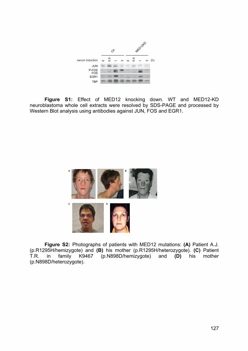
127
C D
A B
Figure S1: Effect of MED12 knocking down. WT and MED12-KD neuroblastoma whole cell extracts were resolved by SDS-PAGE and processed by Western Blot analysis using antibodies against JUN, FOS and EGR1.
Figure S2: Photographs of patients with MED12 mutations: (A) Patient A.J. (p.R1295H/hemizygote) and (B) his mother (p.R1295H/heterozygote). (C) Patient T.R. in family K9467 (p.N898D/hemizygote) and (D) his mother (p.N898D/heterozygote).
0.5
0 0.5
3 110 3
JUN
FOSEGR1
Ctr MED12KD
serum induction
TBP
P-FOS
(h)
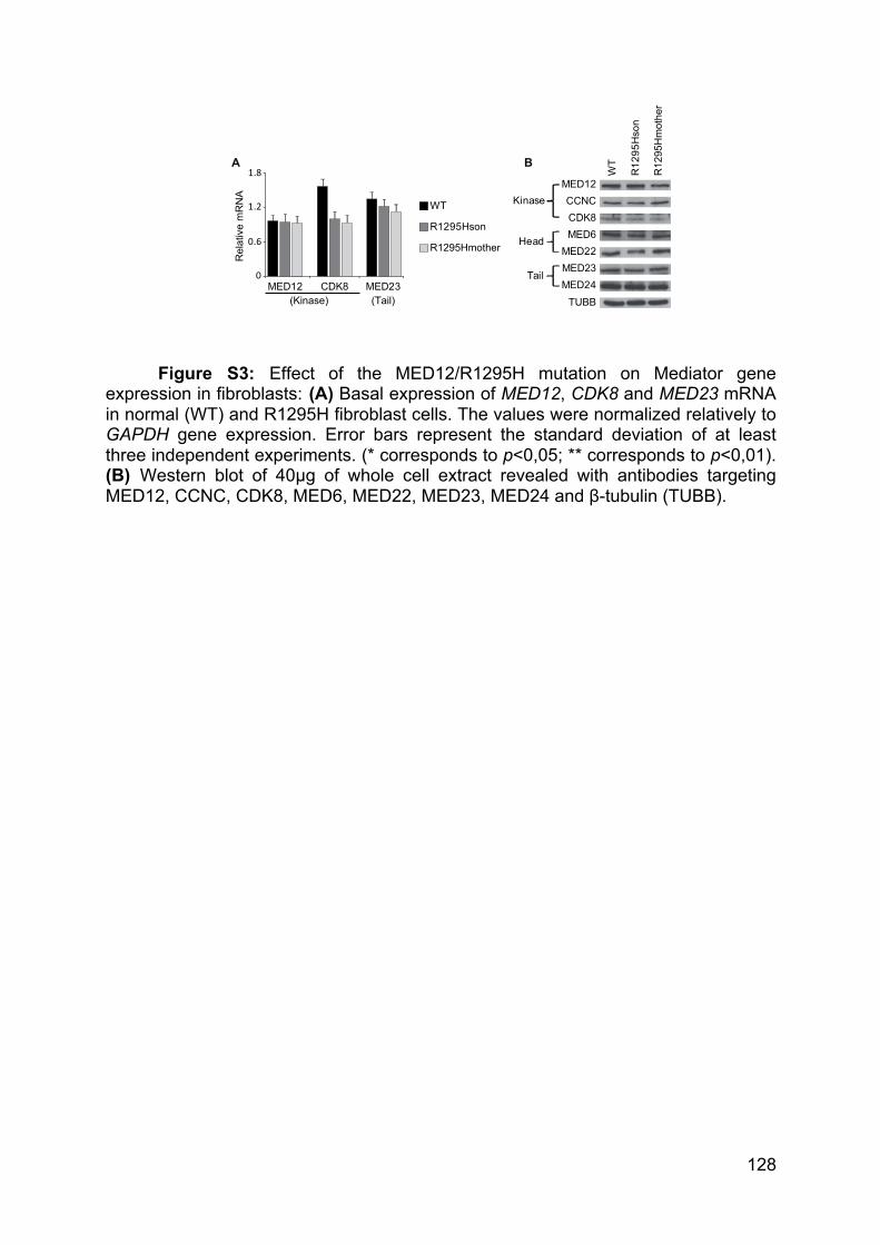
128
Figure S3: Effect of the MED12/R1295H mutation on Mediator gene
expression in fibroblasts: (A) Basal expression of MED12, CDK8 and MED23 mRNA in normal (WT) and R1295H fibroblast cells. The values were normalized relatively to GAPDH gene expression. Error bars represent the standard deviation of at least three independent experiments. (* corresponds to p<0,05; ** corresponds to p<0,01). (B) Western blot of 40µg of whole cell extract revealed with antibodies targeting MED12, CCNC, CDK8, MED6, MED22, MED23, MED24 and β-tubulin (TUBB).
0
0.6
1.2
1.8
MED12 MED23CDK8(Tail)(Kinase)
WT
R1295Hson
R1295Hmother
A B
Rel
ativ
e m
RN
A Kinase
Head
Tail
MED12CCNC
MED6CDK8
WT
R12
95H
son
R12
95H
mot
her
MED24
MED22
TUBB
MED23
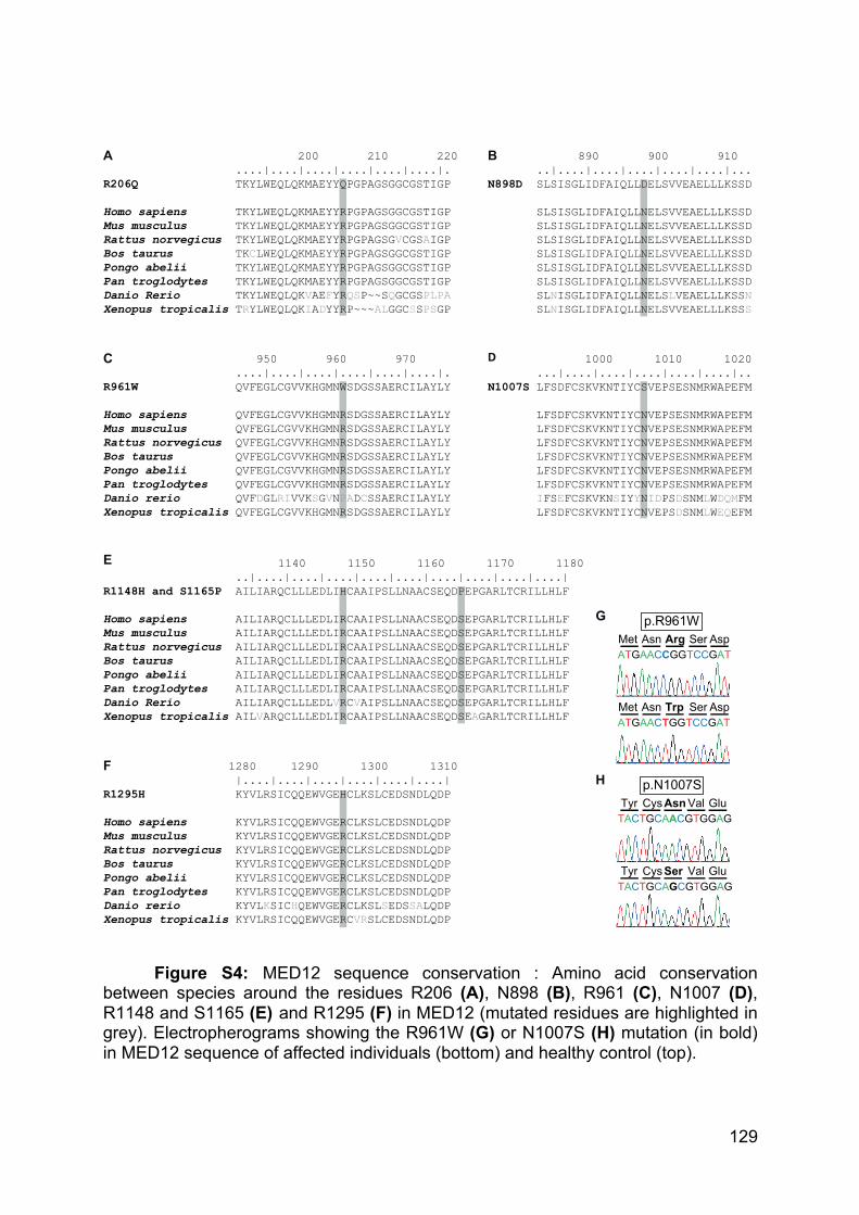
129
Figure S4: MED12 sequence conservation : Amino acid conservation between species around the residues R206 (A), N898 (B), R961 (C), N1007 (D), R1148 and S1165 (E) and R1295 (F) in MED12 (mutated residues are highlighted in grey). Electropherograms showing the R961W (G) or N1007S (H) mutation (in bold) in MED12 sequence of affected individuals (bottom) and healthy control (top).
200 210 220 ....|....|....|....|....|....|.R206Q TKYLWEQLQKMAEYYQPGPAGSGGCGSTIGP Homo sapiens TKYLWEQLQKMAEYYRPGPAGSGGCGSTIGP Mus musculus TKYLWEQLQKMAEYYRPGPAGSGGCGSTIGP Rattus norvegicus TKYLWEQLQKMAEYYRPGPAGSGVCGSAIGP Bos taurus TKCLWEQLQKMAEYYRPGPAGSGGCGSTIGP Pongo abelii TKYLWEQLQKMAEYYRPGPAGSGGCGSTIGP Pan troglodytes TKYLWEQLQKMAEYYRPGPAGSGGCGSTIGP Danio Rerio TKYLWEQLQKVAEFYRQSP~~SQGCGSPLPA Xenopus tropicalis TRYLWEQLQKIADYYRP~~~ALGGCSSPSGP
890 900 910 ..|....|....|....|....|....|...N898D SLSISGLIDFAIQLLDELSVVEAELLLKSSD SLSISGLIDFAIQLLNELSVVEAELLLKSSD SLSISGLIDFAIQLLNELSVVEAELLLKSSD SLSISGLIDFAIQLLNELSVVEAELLLKSSD SLSISGLIDFAIQLLNELSVVEAELLLKSSD SLSISGLIDFAIQLLNELSVVEAELLLKSSD SLSISGLIDFAIQLLNELSVVEAELLLKSSD SLNISGLIDFAIQLLNELSLVEAELLLKSSN SLNISGLIDFAIQLLNELSVVEAELLLKSSS
950 960 970 ....|....|....|....|....|....|.R961W QVFEGLCGVVKHGMNWSDGSSAERCILAYLYHomo sapiens QVFEGLCGVVKHGMNRSDGSSAERCILAYLYMus musculus QVFEGLCGVVKHGMNRSDGSSAERCILAYLYRattus norvegicus QVFEGLCGVVKHGMNRSDGSSAERCILAYLYBos taurus QVFEGLCGVVKHGMNRSDGSSAERCILAYLYPongo abelii QVFEGLCGVVKHGMNRSDGSSAERCILAYLYPan troglodytes QVFEGLCGVVKHGMNRSDGSSAERCILAYLYDanio rerio QVFDGLRIVVKSGVNPADCSSAERCILAYLYXenopus tropicalis QVFEGLCGVVKHGMNRSDGSSAERCILAYLY
A
C
B
E
D
1280 1290 1300 1310 |....|....|....|....|....|....|R1295H KYVLRSICQQEWVGEHCLKSLCEDSNDLQDPHomo sapiens KYVLRSICQQEWVGERCLKSLCEDSNDLQDPMus musculus KYVLRSICQQEWVGERCLKSLCEDSNDLQDPRattus norvegicus KYVLRSICQQEWVGERCLKSLCEDSNDLQDPBos taurus KYVLRSICQQEWVGERCLKSLCEDSNDLQDPPongo abelii KYVLRSICQQEWVGERCLKSLCEDSNDLQDPPan troglodytes KYVLRSICQQEWVGERCLKSLCEDSNDLQDPDanio rerio KYVLKSICHQEWVGERCLKSLSEDSSALQDPXenopus tropicalis KYVLRSICQQEWVGERCVRSLCEDSNDLQDP
Tyr Val GluCysAsnp.N1007S
TACTGCAACGTGGAG
Tyr Val GluCysSerTACTGCAGCGTGGAG
Met Ser AspAsn Argp.R961W
ATGAACCGGTCCGAT
Met Ser AspAsn TrpATGAACTGGTCCGAT
F
G
H
1140 1150 1160 1170 1180 ..|....|....|....|....|....|....|....|....|....|R1148H and S1165P AILIARQCLLLEDLIHCAAIPSLLNAACSEQDPEPGARLTCRILLHLFHomo sapiens AILIARQCLLLEDLIRCAAIPSLLNAACSEQDSEPGARLTCRILLHLFMus musculus AILIARQCLLLEDLIRCAAIPSLLNAACSEQDSEPGARLTCRILLHLFRattus norvegicus AILIARQCLLLEDLIRCAAIPSLLNAACSEQDSEPGARLTCRILLHLFBos taurus AILIARQCLLLEDLIRCAAIPSLLNAACSEQDSEPGARLTCRILLHLFPongo abelii AILIARQCLLLEDLIRCAAIPSLLNAACSEQDSEPGARLTCRILLHLFPan troglodytes AILIARQCLLLEDLIRCAAIPSLLNAACSEQDSEPGARLTCRILLHLFDanio Rerio AILIARQCLLLEDLVRCVAIPSLLNAACSEQDSEPGARLTCRILLHLFXenopus tropicalis AILVARQCLLLEDLIRCAAIPSLLNAACSEQDSEAGARLTCRILLHLF
1000 1010 1020 ...|....|....|....|....|....|..N1007S LFSDFCSKVKNTIYCSVEPSESNMRWAPEFM LFSDFCSKVKNTIYCNVEPSESNMRWAPEFM LFSDFCSKVKNTIYCNVEPSESNMRWAPEFM LFSDFCSKVKNTIYCNVEPSESNMRWAPEFM LFSDFCSKVKNTIYCNVEPSESNMRWAPEFM LFSDFCSKVKNTIYCNVEPSESNMRWAPEFM LFSDFCSKVKNTIYCNVEPSESNMRWAPEFM IFSEFCSKVKNSIYYNIDPSDSNMLWDQMFM LFSDFCSKVKNTIYCNVEPSDSNMLWEQEFM
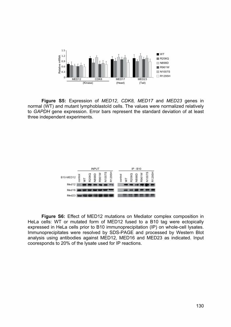
130
Figure S5: Expression of MED12, CDK8, MED17 and MED23 genes in normal (WT) and mutant lymphoblastoïd cells. The values were normalized relatively to GAPDH gene expression. Error bars represent the standard deviation of at least three independent experiments.
Figure S6: Effect of MED12 mutations on Mediator complex composition in HeLa cells: WT or mutated form of MED12 fused to a B10 tag were ectopically expressed in HeLa cells prior to B10 immunoprecipitation (IP) on whole-cell lysates. Immunoprecipitates were resolved by SDS-PAGE and processed by Western Blot analysis using antibodies against MED12, MED16 and MED23 as indicated. Input cooresponds to 20% of the lysate used for IP reactions.
0
0.3
0.6
0.9
1.2
1.5
MED12 MED23CDK8
Rel
ativ
e m
RN
A
MED17(Head) (Tail)(Kinase)
WTR206QN898DR961WN1007SR1295H
WT
R96
1W
N10
07S
N89
8D
R12
95H
R20
6Q
cont
rol
WT
R96
1W
N10
07S
N89
8D
R12
95H
R20
6Q
cont
rol
Med12
Med16
Med23
INPUT IP : B10
B10-MED12
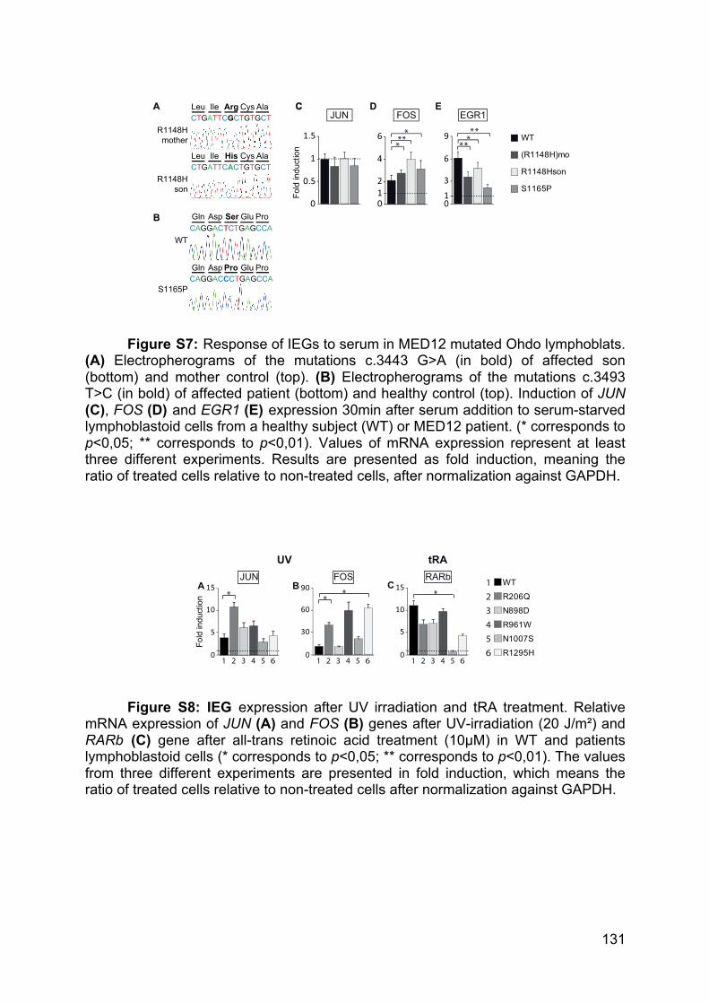
131
Figure S7: Response of IEGs to serum in MED12 mutated Ohdo lymphoblats. (A) Electropherograms of the mutations c.3443 G>A (in bold) of affected son (bottom) and mother control (top). (B) Electropherograms of the mutations c.3493 T>C (in bold) of affected patient (bottom) and healthy control (top). Induction of JUN (C), FOS (D) and EGR1 (E) expression 30min after serum addition to serum-starved lymphoblastoid cells from a healthy subject (WT) or MED12 patient. (* corresponds to p<0,05; ** corresponds to p<0,01). Values of mRNA expression represent at least three different experiments. Results are presented as fold induction, meaning the ratio of treated cells relative to non-treated cells, after normalization against GAPDH.
Figure S8: IEG expression after UV irradiation and tRA treatment. Relative mRNA expression of JUN (A) and FOS (B) genes after UV-irradiation (20 J/m²) and RARb (C) gene after all-trans retinoic acid treatment (10µM) in WT and patients lymphoblastoid cells (* corresponds to p<0,05; ** corresponds to p<0,01). The values from three different experiments are presented in fold induction, which means the ratio of treated cells relative to non-treated cells after normalization against GAPDH.
WT
S1165P
(R1148H)mo
R1148Hson
EGR1CC
FOSA DLeu Cys AlaIle Arg
CTGATTCGCTGTGCT
Leu Cys AlaIle HisCTGATTCACTGTGCT
R1148Hson
R1148Hmother
Fold
indu
ctio
n
0
0.5
1
1.5
JUNE
10
2
4
6
013
6
9***
* ***
**
CAGGACTCTGAGCCAB Gln Glu ProAsp Ser
CAGGACCCTGAGCCAGln Glu ProAspPro
S1165P
WT
0
5
10
15
1 2 3 4 5 6
Fold
indu
ctio
n
JUN FOS WTR206QN898DR961WN1007SR1295H
1234560
30
60
90
1 2 3 4 5 6
A B
UV
CRARb
0
5
10
15
1 2 3 4 5 6
tRA
* ** *
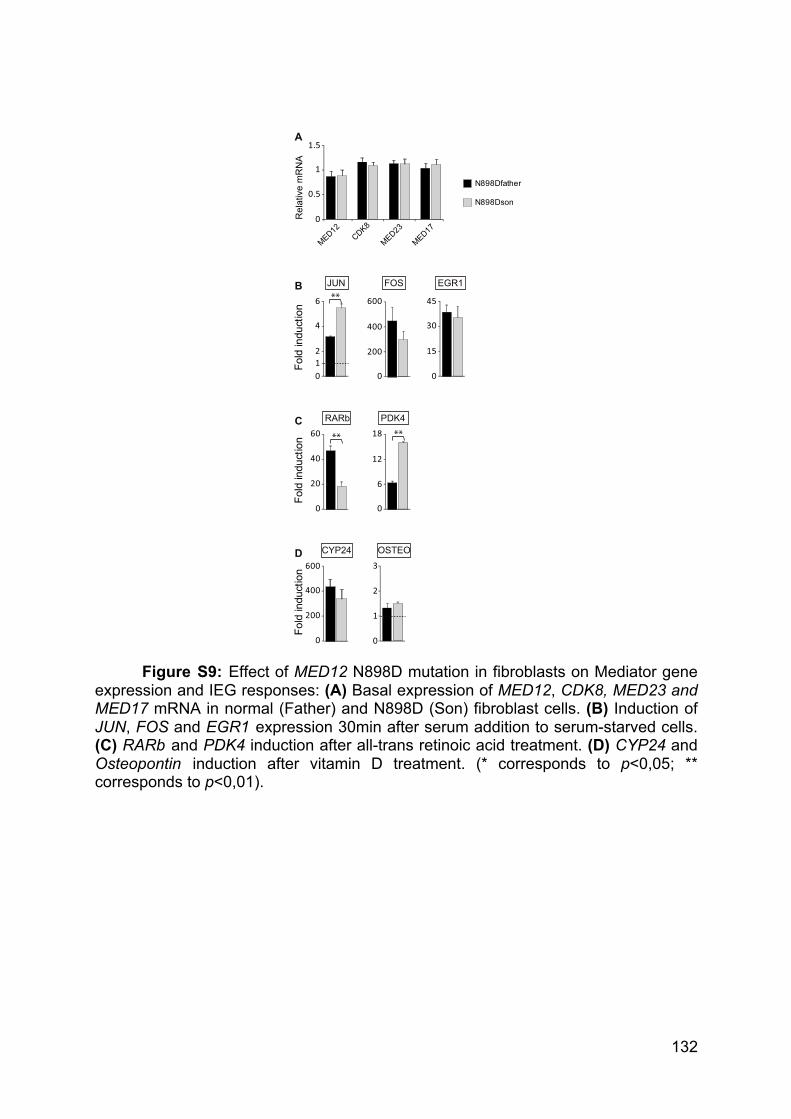
132
Figure S9: Effect of MED12 N898D mutation in fibroblasts on Mediator gene expression and IEG responses: (A) Basal expression of MED12, CDK8, MED23 and MED17 mRNA in normal (Father) and N898D (Son) fibroblast cells. (B) Induction of JUN, FOS and EGR1 expression 30min after serum addition to serum-starved cells. (C) RARb and PDK4 induction after all-trans retinoic acid treatment. (D) CYP24 and Osteopontin induction after vitamin D treatment. (* corresponds to p<0,05; ** corresponds to p<0,01).
N898Dfather
N898Dson
0
0.5
1
1.5
MED12CDK8
MED23
MED17
Rel
ativ
e m
RN
A
A
JUN FOS EGR1
PDK4RARb
CYP24 OSTEO
Fold
indu
ctio
n
0
200
400
600
0
20
40
60
0
6
12
18
012
4
6
0
200
400
600
0
15
30
45B
C
D
Fold
indu
ctio
nFo
ld in
duct
ion
** **
**
0
1
2
3
133
mRNA
primers
Forward Reverse
GAPDH TCGACAGTCAGCCGCATCTTCTTT ACCAAATCCGTTGACTCCGACCTT
RARb CCAGCAAGCCTCACATGTTTCCAA TACACGCTCTGCACCTTTAGCACT
MED12 GCAGAAGAGCATGTCCCTATT TGGCTGTAGAGGGAGGTAAG
MED17 AGTCCAGTGAAGGGCTTCTGGAAA CGGCTTGCTAAGCTGTCAATGGTT
MED23 AATGCGCTATGAATGCACGA GTTTGGAAAGGGACCAGGAGA
CDK8 GGGATCTCTATGTCGGCATGTAG AAATGACGTTTGGATGCTTAAGC
JUN AGCGCCTGATAATCCAGTCC CTGCTCATCTGTCACGTTCTTG
FOS CAAGCGGAGACAGACCAACT AGTCAGATCAAGGGAAGCCA
EGR1 AGCACCTGACCGCAGAGTCTTT CACCAGCACCTTCTCGTTGTT
MMP3 CACTCACAGACCTGACTCGG AGTCAGGGGGAGGTCCATAG
CCND1 GGTGCTGCGGGCCAT CTCGCAGACCTCCAGCAT
REST1 CCCGAAACTCCAGCAACAAAG CCTGGGTGGCCATAACTGTA
REST2
SYN1
CCGGCTGCGCGAATACAG
TGCTCAGCAGTACAACGTACC
CAGGGCCATTCCAATGTTGC
GACACTTGCGATGTCCTGGAA
ChIP primers Forward Reverse
JUN prox CCAGAGAAGAATCTTCTAGG CCCCAAGGCCTTCCCATTGG
JUN distal CCGTCTCACTCTCTTGCTCTTC CAACTGGACAAAATGGCTCTG
FOS pro GAGCAGTTCCCGTCAATCC GCATTTCGCAGTTCCTGTCT
EGR1 pro CTGCCATATTAGGGCTTCCTGCTT TATTTGAAGGGTCTGGAACGGCAC
MMP3 pro TCCTGCTGCCATTTGGATGA GCCTCCTTGTAGGTCCAACC
CCND1 AP1 AACCTTCGGTGGTCTTGTCC AGCTGAGAAACAGTGATCTCCA
CCND1 pro ATTCTCTGCCGGGCTTTGAT TGCAACTTCAACAAAACTCCCC
REST1 pro TGACCTAAGGGCAGGAGTGA CTACCAAGCAAGGAGTGCCC
REST2 pro
SYN1 pro
GGAAGGCGCCGTTGAGT
CATTCCCCAAATTGCGCATCC
TGAAGCGCAGAAATCGCTGT
CGAAGGCACTGTCCGCGGTGC

134
Conclusion
135
In order to decipher the complex mechanisms that regulate the transcription of
protein coding genes by the RNA polymerase II, we need to properly define the
structures, interactions, partnership, enzymatic activities and target genes of the
multiple components of the transcription machinery. For this purposes, it is useful to
combine the fundamental approaches with the clinical one. Indeed, clinical data and
biological samples give fruitful insights to protein specific roles that cannot be gained
by molecular or biochemical approaches. In the same time, fundamental techniques
bring priceless perspectives on the etiology of mutation-associated diseases, to both
physicians and patients.
I - The NER factor XPC regulates transcription
Our lab has previously uncovered the fundamental roles of the DNA repair
factor XPC in the formation of the Pre-Initiation Complex. XPC allows the recruitment
of XPA, RPA, XPG and XPF-ERCC1 to the promoter of the NR activated gene
RARβ2, what is necessary for its proper transactivation(Le May et al., 2010a).
Indeed, they are required for DNA demethylation and active HPTM. XPG and XPF
have also been found to be necessary for the formation of the promoter-terminator
loop, by promoting CTCF recruitment(Le May et al., 2012).
Our current study unveiled new unexpected roles for XPC in transcription.
XPC was found to be recruited at the promoter of a certain set of genes together with
Pol II. Using patient cells constitutively depleted for XPC, we showed that XPC was
necessary for the proper expression of these genes. XPC is strictly required for the
acetylation of H3K9 and the tri-methylation of H3K4 at the promoter of XPC bound
genes. It cooperates with the transcription factor E2F1 to recruit the acetyl
transferase GCN5, as part of the ATAC complex. It is interesting to note that RPA is
also acetylated by GCN5 in the context of nucleotide excision repair, while it is
recruited subsequently to XPC(He et al., 2017; Zhao et al., 2017).
Interestingly, relations between XPC and E2F1 have recently been shown in
the context of global genome NER. E2F1 notably interact with its partner HR23B. It
was also found to localize around UV-induced DNA damages to promote NER
factors recruitment through H3K9 acetylation, acting in a non-transcriptional
process(Guo et al., 2011; Singh and Dagnino, 2016).
136
In the present case, E2F1 seems to be responsible for specificity, as most the
regulated genes do contain an E2F1 response element. The genes that are down-
regulated in absence of XPC are mainly implicate in the regulation of the chromatin
structure and present oncogenic and immunologic ontology. We showed here that
XPC regulated DAPK1 and it was also shown that XPC can induce interleukin-
6(Schreck et al., 2016), while both are implicated in cancer progression. Among the
regulated genes, we also founded the SET1 methyl transferase. Our results suggest
that the absence of XPC is also directly responsible for the decrease of H3K4me3
mark at the promoter XPC-regulated genes.
Our work greatly improves the comprehension of the mechanism of action of
XPC and enlightens the overlapping pathways where XPC engage in the context of
transcription and DNA repair. Therefore, these two fundamental processes of the cell
appear more and more intricate.
II - The Mediator subunit MED12 in neurodevelopmental
disorders
We published earlier the relation between a mutation in MED23 and
intellectual disability in a large Algerian consanguineous family(Hashimoto et al.,
2011). This study unveiled the role of Mediator in the regulation of immediate early
genes and the implication of Mediator in brain development and functioning.
By studying several MED12 mutations, we showed that MED12 is strongly
implicated in the regulation of IEG, especially JUN(Donnio et al., 2017). What is
more, the position of the mutation on MED12 differentially impacted the JUN
expression. Considering the roles of IEG in the cognitive defects(Berk, 2012; Pérez-
Cadahía et al., 2011), it may be possible that the position of the mutation differentially
influence the neurological development, thus explaining the phenotypic differences
among patients. The mediator serves as a bridge between the transcription
machinery and the specific transcription factors. Mutations that affect one of brick of
the bridge undoubtedly perturbed the normal transcription program.
Since the first case, almost twenty MED12 mutations have been published
with closely related symptoms. The affected patients are considered among one of
137
the three definite syndromes (Ohdo syndrome, FG syndrome and Lujan syndrome)
or as non-syndromic ID. They share some phenotypic trait like intellectual disability,
neurodevelopmental delay, speech difficulty and congenital digestive issues, defining
a “MED12 phenotype”. Therefore, syndrome boundaries are becoming vague for
some patients.
Other Mediator subunits have been found to be mutated and to give rise to
several form of intellectual disability associated with various phenotypes/syndromes.
MED13L can easily be compared with MED12 phenotype. This proximity is not
surprising since both MED12 and MED13L belongs to the same Mediator module.
Therefore, some authors have proposed the concept of a MED12/M13L clinical
spectrum(Caro-Llopis et al., 2016).
In a larger perspective, considering the increasing number of patients with a
mutation in one of the Mediator subunit and the relatively close neurodevelopmental
issues, the concept of “Mediatorophaties” is emerging(Caro-Llopis et al., 2016).
Altogether, these results brought a new comprehension on the mechanism of
transcription and demonstrated the substantial benefits that arise from the
combination of clinical and fundamental research.

138
Résumé en français
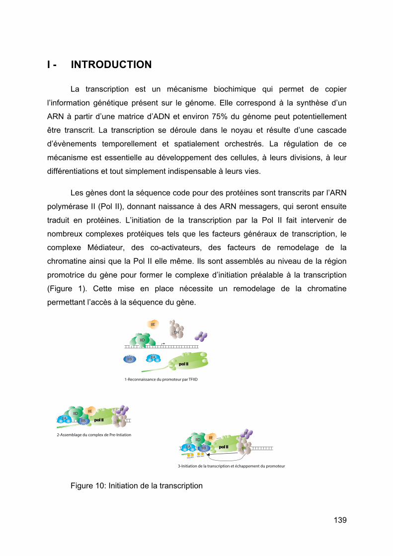
139
I - INTRODUCTION
La transcription est un mécanisme biochimique qui permet de copier
l’information génétique présent sur le génome. Elle correspond à la synthèse d’un
ARN à partir d’une matrice d’ADN et environ 75% du génome peut potentiellement
être transcrit. La transcription se déroule dans le noyau et résulte d’une cascade
d’évènements temporellement et spatialement orchestrés. La régulation de ce
mécanisme est essentielle au développement des cellules, à leurs divisions, à leur
différentiations et tout simplement indispensable à leurs vies.
Les gènes dont la séquence code pour des protéines sont transcrits par l’ARN
polymérase II (Pol II), donnant naissance à des ARN messagers, qui seront ensuite
traduit en protéines. L’initiation de la transcription par la Pol II fait intervenir de
nombreux complexes protéiques tels que les facteurs généraux de transcription, le
complexe Médiateur, des co-activateurs, des facteurs de remodelage de la
chromatine ainsi que la Pol II elle même. Ils sont assemblés au niveau de la région
promotrice du gène pour former le complexe d’initiation préalable à la transcription
(Figure 1). Cette mise en place nécessite un remodelage de la chromatine
permettant l’accès à la séquence du gène.
Figure 10: Initiation de la transcription
pol IIpol II
pol IIpol II
IIB
IIHIIF
IIE
IIDIIA
IIH
IIFIIE
IIAIIB
pol IIpol IIIIB
IID
IIA
IIE
1-Reconnaissance du promoteur par TFIID
2-Assemblage du complex de Pre-Intiation
3-Initiation de la transcription et échappement du promoteur
IID
IIH
IIF
PP
Ser5Ser5
PSer7
PSer7
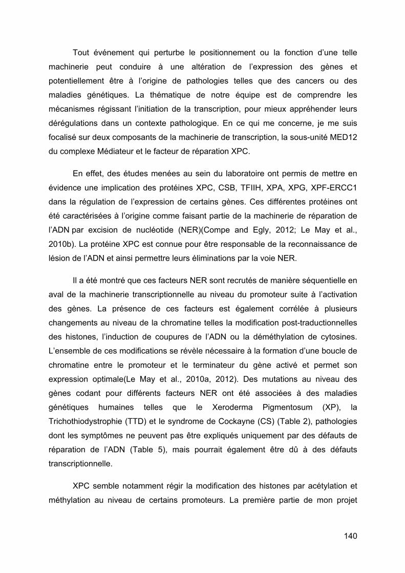
140
Tout événement qui perturbe le positionnement ou la fonction d’une telle
machinerie peut conduire à une altération de l’expression des gènes et
potentiellement être à l’origine de pathologies telles que des cancers ou des
maladies génétiques. La thématique de notre équipe est de comprendre les
mécanismes régissant l’initiation de la transcription, pour mieux appréhender leurs
dérégulations dans un contexte pathologique. En ce qui me concerne, je me suis
focalisé sur deux composants de la machinerie de transcription, la sous-unité MED12
du complexe Médiateur et le facteur de réparation XPC.
En effet, des études menées au sein du laboratoire ont permis de mettre en
évidence une implication des protéines XPC, CSB, TFIIH, XPA, XPG, XPF-ERCC1
dans la régulation de l’expression de certains gènes. Ces différentes protéines ont
été caractérisées à l’origine comme faisant partie de la machinerie de réparation de
l’ADN par excision de nucléotide (NER)(Compe and Egly, 2012; Le May et al.,
2010b). La protéine XPC est connue pour être responsable de la reconnaissance de
lésion de l’ADN et ainsi permettre leurs éliminations par la voie NER.
Il a été montré que ces facteurs NER sont recrutés de manière séquentielle en
aval de la machinerie transcriptionnelle au niveau du promoteur suite à l’activation
des gènes. La présence de ces facteurs est également corrélée à plusieurs
changements au niveau de la chromatine telles la modification post-traductionnelles
des histones, l’induction de coupures de l’ADN ou la déméthylation de cytosines.
L’ensemble de ces modifications se révèle nécessaire à la formation d’une boucle de
chromatine entre le promoteur et le terminateur du gène activé et permet son
expression optimale(Le May et al., 2010a, 2012). Des mutations au niveau des
gènes codant pour différents facteurs NER ont été associées à des maladies
génétiques humaines telles que le Xeroderma Pigmentosum (XP), la
Trichothiodystrophie (TTD) et le syndrome de Cockayne (CS) (Table 2), pathologies
dont les symptômes ne peuvent pas être expliqués uniquement par des défauts de
réparation de l’ADN (Table 5), mais pourrait également être dû à des défauts
transcriptionnelle.
XPC semble notamment régir la modification des histones par acétylation et
méthylation au niveau de certains promoteurs. La première partie de mon projet
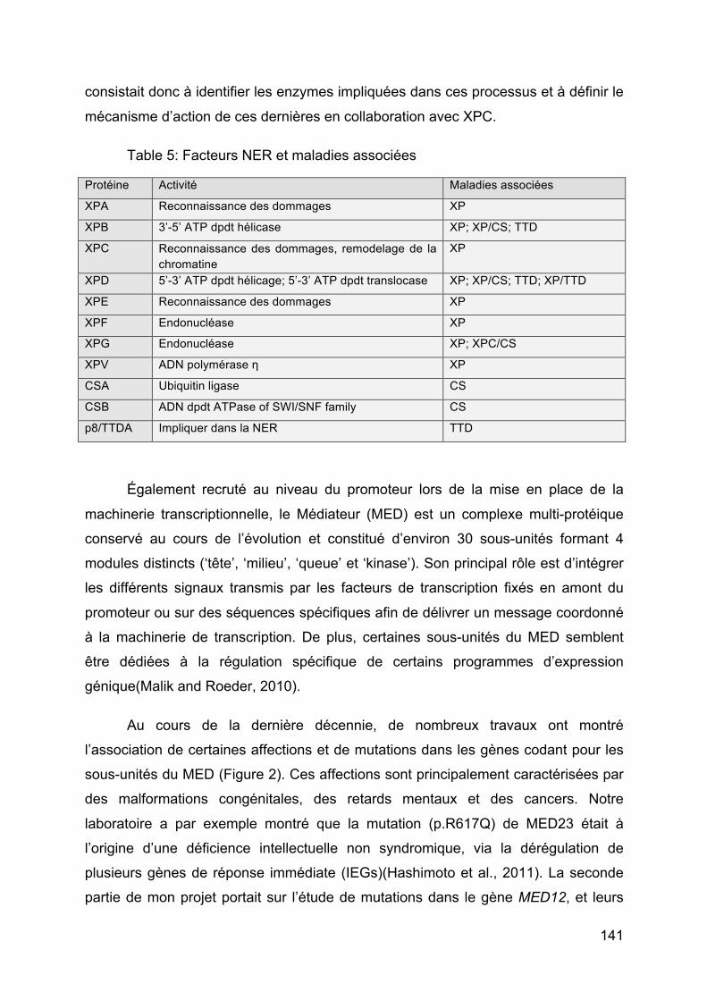
141
consistait donc à identifier les enzymes impliquées dans ces processus et à définir le
mécanisme d’action de ces dernières en collaboration avec XPC.
Table 5: Facteurs NER et maladies associées
Protéine Activité Maladies associées
XPA Reconnaissance des dommages XP
XPB 3’-5’ ATP dpdt hélicase XP; XP/CS; TTD
XPC Reconnaissance des dommages, remodelage de la chromatine
XP
XPD 5’-3’ ATP dpdt hélicage; 5’-3’ ATP dpdt translocase XP; XP/CS; TTD; XP/TTD
XPE Reconnaissance des dommages XP
XPF Endonucléase XP
XPG Endonucléase XP; XPC/CS
XPV ADN polymérase η XP
CSA Ubiquitin ligase CS
CSB ADN dpdt ATPase of SWI/SNF family CS
p8/TTDA Impliquer dans la NER TTD
Également recruté au niveau du promoteur lors de la mise en place de la
machinerie transcriptionnelle, le Médiateur (MED) est un complexe multi-protéique
conservé au cours de l’évolution et constitué d’environ 30 sous-unités formant 4
modules distincts (‘tête’, ‘milieu’, ‘queue’ et ‘kinase’). Son principal rôle est d’intégrer
les différents signaux transmis par les facteurs de transcription fixés en amont du
promoteur ou sur des séquences spécifiques afin de délivrer un message coordonné
à la machinerie de transcription. De plus, certaines sous-unités du MED semblent
être dédiées à la régulation spécifique de certains programmes d’expression
génique(Malik and Roeder, 2010).
Au cours de la dernière décennie, de nombreux travaux ont montré
l’association de certaines affections et de mutations dans les gènes codant pour les
sous-unités du MED (Figure 2). Ces affections sont principalement caractérisées par
des malformations congénitales, des retards mentaux et des cancers. Notre
laboratoire a par exemple montré que la mutation (p.R617Q) de MED23 était à
l’origine d’une déficience intellectuelle non syndromique, via la dérégulation de
plusieurs gènes de réponse immédiate (IEGs)(Hashimoto et al., 2011). La seconde
partie de mon projet portait sur l’étude de mutations dans le gène MED12, et leurs
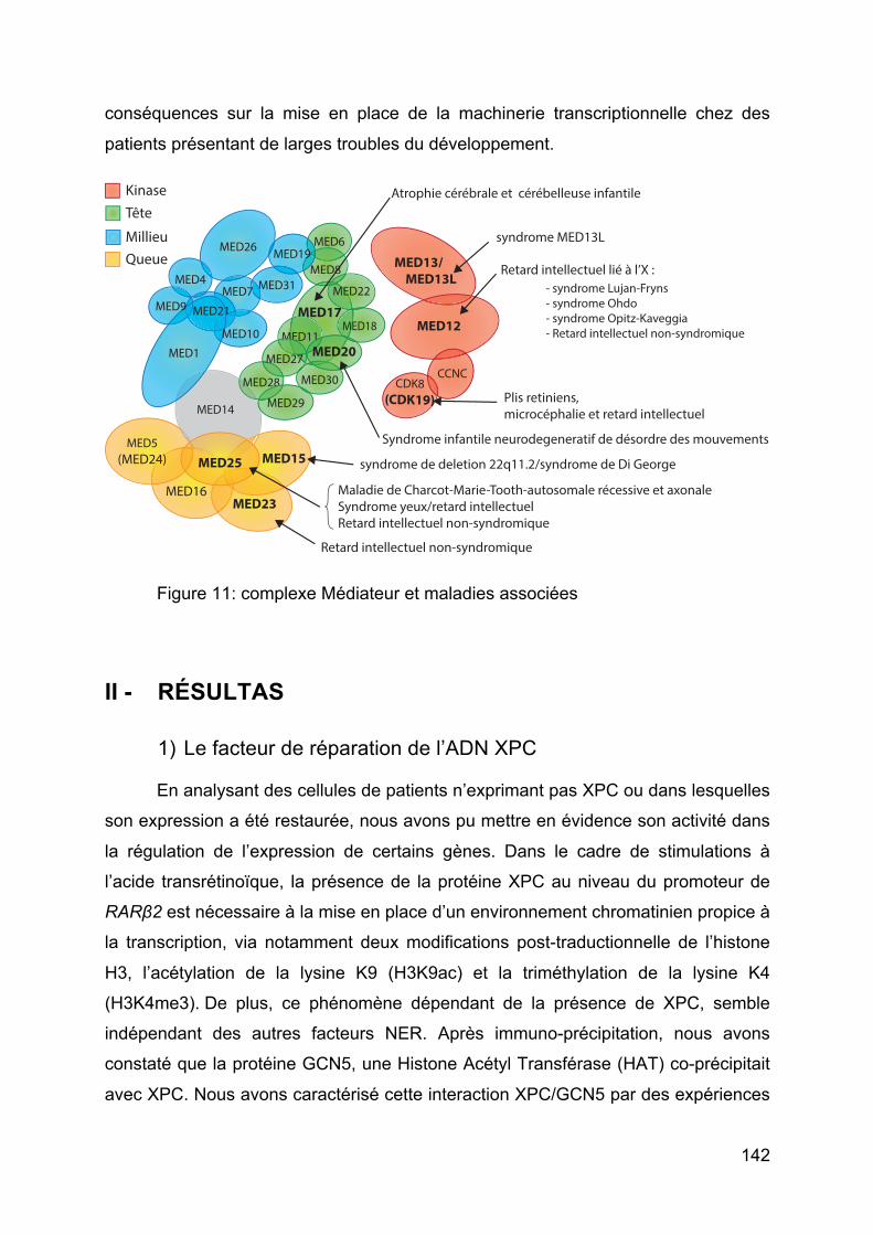
142
conséquences sur la mise en place de la machinerie transcriptionnelle chez des
patients présentant de larges troubles du développement.
Figure 11: complexe Médiateur et maladies associées
II - RÉSULTAS
1) Le facteur de réparation de l’ADN XPC
En analysant des cellules de patients n’exprimant pas XPC ou dans lesquelles
son expression a été restaurée, nous avons pu mettre en évidence son activité dans
la régulation de l’expression de certains gènes. Dans le cadre de stimulations à
l’acide transrétinoïque, la présence de la protéine XPC au niveau du promoteur de
RARβ2 est nécessaire à la mise en place d’un environnement chromatinien propice à
la transcription, via notamment deux modifications post-traductionnelle de l’histone
H3, l’acétylation de la lysine K9 (H3K9ac) et la triméthylation de la lysine K4
(H3K4me3). De plus, ce phénomène dépendant de la présence de XPC, semble
indépendant des autres facteurs NER. Après immuno-précipitation, nous avons
constaté que la protéine GCN5, une Histone Acétyl Transférase (HAT) co-précipitait
avec XPC. Nous avons caractérisé cette interaction XPC/GCN5 par des expériences
MED26
MED17
MED31MED4 MMED7
MED6
MED8MED19
CDK8(CDK19)
MED13/ MED13L
MED12
CCNC
MED5(MED24)
MED16
MED11
MED22
MED14
MED23
MED15MED25
MED28
MED18
MED308
MED29
MEM
MED27
1MED20
KinaseTête
MillieuQueue
Atrophie cérébrale et cérébelleuse infantile
Retard intellectuel lié à l’X : - syndrome Lujan-Fryns - syndrome Ohdo - syndrome Opitz-Kaveggia - Retard intellectuel non-syndromique
Retard intellectuel non-syndromique
Maladie de Charcot-Marie-Tooth-autosomale récessive et axonaleSyndrome yeux/retard intellectuelRetard intellectuel non-syndromique
syndrome MED13L
syndrome de deletion 22q11.2/syndrome de Di George
Plis retiniens, microcéphalie et retard intellectuel
Syndrome infantile neurodegeneratif de désordre des mouvements
MED1MED10
MED21MED9
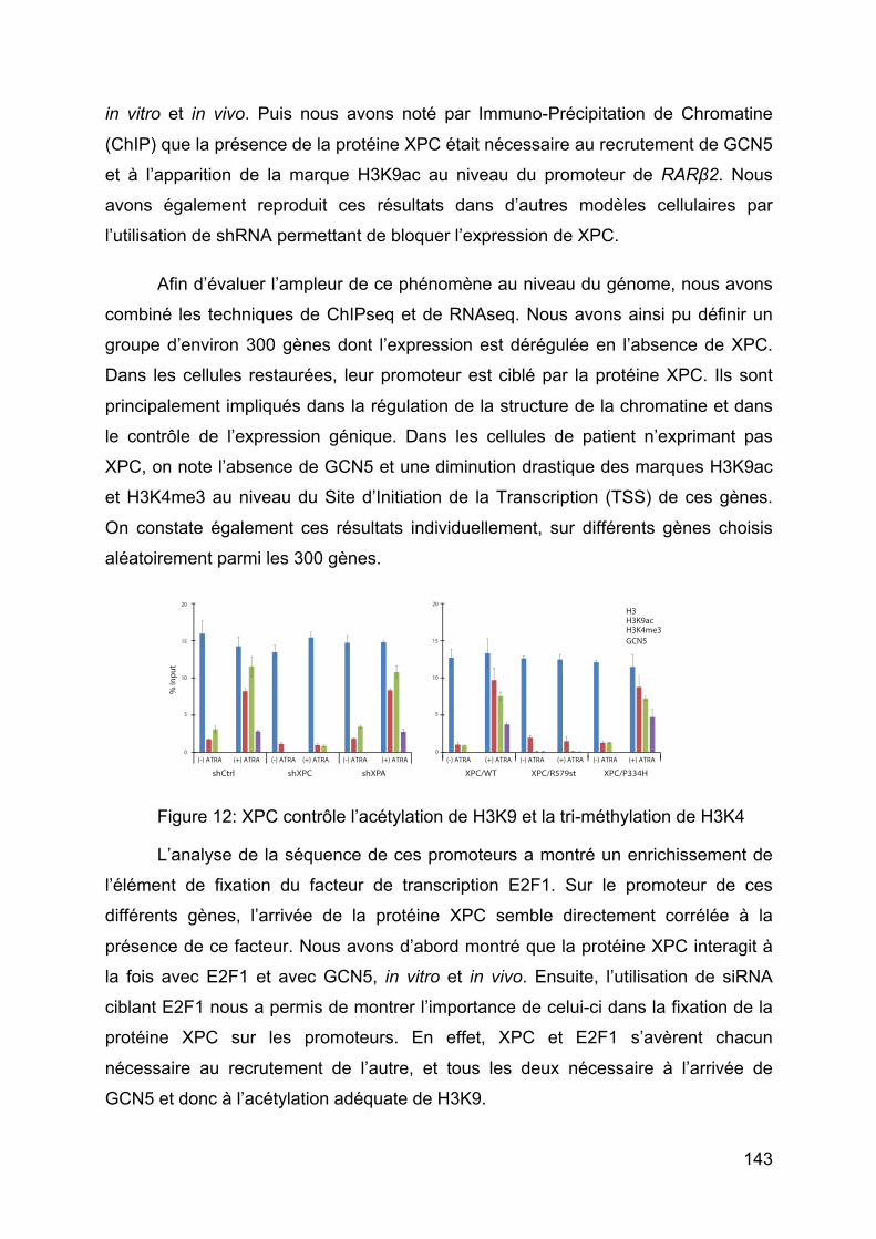
143
in vitro et in vivo. Puis nous avons noté par Immuno-Précipitation de Chromatine
(ChIP) que la présence de la protéine XPC était nécessaire au recrutement de GCN5
et à l’apparition de la marque H3K9ac au niveau du promoteur de RARβ2. Nous
avons également reproduit ces résultats dans d’autres modèles cellulaires par
l’utilisation de shRNA permettant de bloquer l’expression de XPC.
Afin d’évaluer l’ampleur de ce phénomène au niveau du génome, nous avons
combiné les techniques de ChIPseq et de RNAseq. Nous avons ainsi pu définir un
groupe d’environ 300 gènes dont l’expression est dérégulée en l’absence de XPC.
Dans les cellules restaurées, leur promoteur est ciblé par la protéine XPC. Ils sont
principalement impliqués dans la régulation de la structure de la chromatine et dans
le contrôle de l’expression génique. Dans les cellules de patient n’exprimant pas
XPC, on note l’absence de GCN5 et une diminution drastique des marques H3K9ac
et H3K4me3 au niveau du Site d’Initiation de la Transcription (TSS) de ces gènes.
On constate également ces résultats individuellement, sur différents gènes choisis
aléatoirement parmi les 300 gènes.
Figure 12: XPC contrôle l’acétylation de H3K9 et la tri-méthylation de H3K4
L’analyse de la séquence de ces promoteurs a montré un enrichissement de
l’élément de fixation du facteur de transcription E2F1. Sur le promoteur de ces
différents gènes, l’arrivée de la protéine XPC semble directement corrélée à la
présence de ce facteur. Nous avons d’abord montré que la protéine XPC interagit à
la fois avec E2F1 et avec GCN5, in vitro et in vivo. Ensuite, l’utilisation de siRNA
ciblant E2F1 nous a permis de montrer l’importance de celui-ci dans la fixation de la
protéine XPC sur les promoteurs. En effet, XPC et E2F1 s’avèrent chacun
nécessaire au recrutement de l’autre, et tous les deux nécessaire à l’arrivée de
GCN5 et donc à l’acétylation adéquate de H3K9.
GCN5
shCtrl shXPC shXPA XPC/WT XPC/R579st XPC/P334H(-) ATRA (+) ATRA (-) ATRA (+) ATRA (-) ATRA (+) ATRA(-) ATRA (+) ATRA (-) ATRA (+) ATRA (-) ATRA (+) ATRA
% In
put
20
15
5
10
0
20
15
5
10
0
H3H3K9acH3K4me3
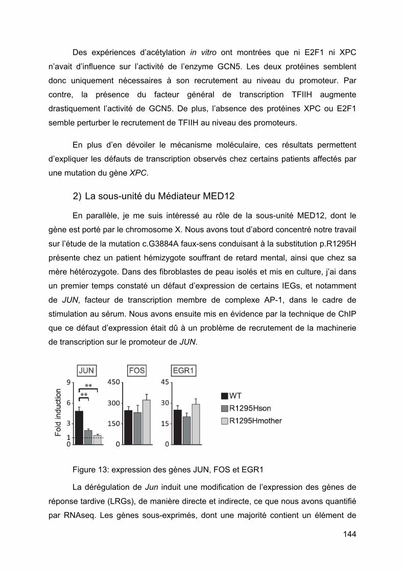
144
Des expériences d’acétylation in vitro ont montrées que ni E2F1 ni XPC
n’avait d’influence sur l’activité de l’enzyme GCN5. Les deux protéines semblent
donc uniquement nécessaires à son recrutement au niveau du promoteur. Par
contre, la présence du facteur général de transcription TFIIH augmente
drastiquement l’activité de GCN5. De plus, l’absence des protéines XPC ou E2F1
semble perturber le recrutement de TFIIH au niveau des promoteurs.
En plus d’en dévoiler le mécanisme moléculaire, ces résultats permettent
d’expliquer les défauts de transcription observés chez certains patients affectés par
une mutation du gène XPC.
2) La sous-unité du Médiateur MED12
En parallèle, je me suis intéressé au rôle de la sous-unité MED12, dont le
gène est porté par le chromosome X. Nous avons tout d’abord concentré notre travail
sur l’étude de la mutation c.G3884A faux-sens conduisant à la substitution p.R1295H
présente chez un patient hémizygote souffrant de retard mental, ainsi que chez sa
mère hétérozygote. Dans des fibroblastes de peau isolés et mis en culture, j’ai dans
un premier temps constaté un défaut d’expression de certains IEGs, et notamment
de JUN, facteur de transcription membre de complexe AP-1, dans le cadre de
stimulation au sérum. Nous avons ensuite mis en évidence par la technique de ChIP
que ce défaut d’expression était dû à un problème de recrutement de la machinerie
de transcription sur le promoteur de JUN.
Figure 13: expression des gènes JUN, FOS et EGR1
La dérégulation de Jun induit une modification de l’expression des gènes de
réponse tardive (LRGs), de manière directe et indirecte, ce que nous avons quantifié
par RNAseq. Les gènes sous-exprimés, dont une majorité contient un élément de
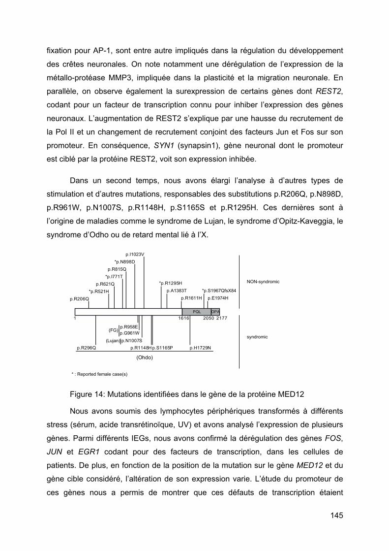
145
fixation pour AP-1, sont entre autre impliqués dans la régulation du développement
des crêtes neuronales. On note notamment une dérégulation de l’expression de la
métallo-protéase MMP3, impliquée dans la plasticité et la migration neuronale. En
parallèle, on observe également la surexpression de certains gènes dont REST2,
codant pour un facteur de transcription connu pour inhiber l’expression des gènes
neuronaux. L’augmentation de REST2 s’explique par une hausse du recrutement de
la Pol II et un changement de recrutement conjoint des facteurs Jun et Fos sur son
promoteur. En conséquence, SYN1 (synapsin1), gène neuronal dont le promoteur
est ciblé par la protéine REST2, voit son expression inhibée.
Dans un second temps, nous avons élargi l’analyse à d’autres types de
stimulation et d’autres mutations, responsables des substitutions p.R206Q, p.N898D,
p.R961W, p.N1007S, p.R1148H, p.S1165S et p.R1295H. Ces dernières sont à
l’origine de maladies comme le syndrome de Lujan, le syndrome d’Opitz-Kaveggia, le
syndrome d’Odho ou de retard mental lié à l’X.
Figure 14: Mutations identifiées dans le gène de la protéine MED12
Nous avons soumis des lymphocytes périphériques transformés à différents
stress (sérum, acide transrétinoïque, UV) et avons analysé l’expression de plusieurs
gènes. Parmi différents IEGs, nous avons confirmé la dérégulation des gènes FOS,
JUN et EGR1 codant pour des facteurs de transcription, dans les cellules de
patients. De plus, en fonction de la position de la mutation sur le gène MED12 et du
gène cible considéré, l’altération de son expression varie. L’étude du promoteur de
ces gènes nous a permis de montrer que ces défauts de transcription étaient
PQL1616 2050
p.R958E
*p.N898D
(Lujan) p.N1007S
*p.R1295H
p.R206Q
1 2177
(Ohdo)
p.S1165P p.H1729Np.R1148H
OPA
p.G961W(FG)
p.A1383Tp.E1974H
*p.S1967QfsX84*p.R521Hp.R621Q
*p.I771Tp.R815Q
p.I1023V
p.R296Q
p.R1611H
NON-syndromic
syndromic
* : Reported female case(s)
146
également corrélés à une modification du recrutement de la machinerie
transcriptionnelle.
De manière intéressante, les différentes mutations de MED12 n’affectent pas
nécessairement l’expression des même gènes de réponse immédiate, ni n’entrainent
les même altérations de ceux-ci. Ces différences que nous notons entre lignées
cellulaires pourraient être une piste intéressante pour permettre d’expliquer la
variabilité des symptômes observés chez les patients.
III - CONCLUSION
Ces résultats nous éclairent sur la position et la fonction de la protéine XPC et
de sous-unité MED12 du complexe Médiateur au moment de l’initiation de la
transcription. Outre l’aspect fondamental, l’étude des mécanismes moléculaires qui
sous-tendent ces maladies permet d’en décrypter l’étiologie.

147
Bibliography
148
Aboussekhra, A., Biggerstaff, M., Shivji, M.K., Vilpo, J.A., Moncollin, V.,
Podust, V.N., Protić, M., Hübscher, U., Egly, J.M., and Wood, R.D. (1995).
Mammalian DNA nucleotide excision repair reconstituted with purified protein
components. Cell 80, 859–868.
Adegbola, A., Musante, L., Callewaert, B., Maciel, P., Hu, H., Isidor, B., Picker-
Minh, S., Le Caignec, C., Delle Chiaie, B., Vanakker, O., et al. (2015). Redefining the
MED13L syndrome. Eur. J. Hum. Genet. EJHG 23, 1308–1317.
Akhtar, W., and Veenstra, G.J.C. (2011). TBP-related factors: a paradigm of
diversity in transcription initiation. Cell Biosci. 1, 23.
Akoulitchev, S., Chuikov, S., and Reinberg, D. (2000). TFIIH is negatively
regulated by cdk8-containing mediator complexes. Nature 407, 102–106.
Alekseev, S., Nagy, Z., Sandoz, J., Weiss, A., Egly, J.-M., Le May, N., and
Coin, F. (2017). Transcription without XPB Establishes a Unified Helicase-
Independent Mechanism of Promoter Opening in Eukaryotic Gene Expression. Mol.
Cell 65, 504–514.e4.
Allfrey, V.G. (1966). Structural modifications of histones and their possible role
in the regulation of ribonucleic acid synthesis. Proc. Can. Cancer Conf. 6, 313–335.
Anish, R., Hossain, M.B., Jacobson, R.H., and Takada, S. (2009).
Characterization of transcription from TATA-less promoters: identification of a new
core promoter element XCPE2 and analysis of factor requirements. PloS One 4,
e5103.
Ansari, S.A., He, Q., and Morse, R.H. (2009). Mediator complex association
with constitutively transcribed genes in yeast. Proc. Natl. Acad. Sci. U. S. A. 106,
16734–16739.
Araki, K., Nakajima, Y., Eto, K., and Ikeda, M.-A. (2003). Distinct recruitment
of E2F family members to specific E2F-binding sites mediates activation and
repression of the E2F1 promoter. Oncogene 22, 7632–7641.
Araki, M., Masutani, C., Takemura, M., Uchida, A., Sugasawa, K., Kondoh, J.,
149
Ohkuma, Y., and Hanaoka, F. (2001). Centrosome protein centrin 2/caltractin 1 is
part of the xeroderma pigmentosum group C complex that initiates global genome
nucleotide excision repair. J. Biol. Chem. 276, 18665–18672.
Araújo, S.J., Tirode, F., Coin, F., Pospiech, H., Syväoja, J.E., Stucki, M.,
Hübscher, U., Egly, J.M., and Wood, R.D. (2000). Nucleotide excision repair of DNA
with recombinant human proteins: definition of the minimal set of factors, active forms
of TFIIH, and modulation by CAK. Genes Dev. 14, 349–359.
Arents, G., and Moudrianakis, E.N. (1995). The histone fold: a ubiquitous
architectural motif utilized in DNA compaction and protein dimerization. Proc. Natl.
Acad. Sci. U. S. A. 92, 11170–11174.
Armache, K.-J., Kettenberger, H., and Cramer, P. (2003). Architecture of
initiation-competent 12-subunit RNA polymerase II. Proc. Natl. Acad. Sci. U. S. A.
100, 6964–6968.
Asadollahi, R., Oneda, B., Sheth, F., Azzarello-Burri, S., Baldinger, R., Joset,
P., Latal, B., Knirsch, W., Desai, S., Baumer, A., et al. (2013). Dosage changes of
MED13L further delineate its role in congenital heart defects and intellectual
disability. Eur. J. Hum. Genet. EJHG 21, 1100–1104.
Asadollahi, R., Zweier, M., Gogoll, L., Schiffmann, R., Sticht, H., Steindl, K.,
and Rauch, A. (2017). Genotype-phenotype evaluation of MED13L defects in the
light of a novel truncating and a recurrent missense mutation. Eur. J. Med. Genet. 60,
451–464.
Asturias, F.J., Jiang, Y.W., Myers, L.C., Gustafsson, C.M., and Kornberg, R.D.
(1999). Conserved structures of mediator and RNA polymerase II holoenzyme.
Science 283, 985–987.
Baek, H.J., Kang, Y.K., and Roeder, R.G. (2006). Human Mediator enhances
basal transcription by facilitating recruitment of transcription factor IIB during
preinitiation complex assembly. J. Biol. Chem. 281, 15172–15181.
Balamotis, M.A., Pennella, M.A., Stevens, J.L., Wasylyk, B., Belmont, A.S.,
and Berk, A.J. (2009). Complexity in transcription control at the activation domain-
150
mediator interface. Sci. Signal. 2, ra20.
Bandara, L.R., Lam, E.W., Sørensen, T.S., Zamanian, M., Girling, R., and La
Thangue, N.B. (1994). DP-1: a cell cycle-regulated and phosphorylated component
of transcription factor DRTF1/E2F which is functionally important for recognition by
pRb and the adenovirus E4 orf 6/7 protein. EMBO J. 13, 3104–3114.
Barreto, G., Schäfer, A., Marhold, J., Stach, D., Swaminathan, S.K., Handa,
V., Döderlein, G., Maltry, N., Wu, W., Lyko, F., et al. (2007). Gadd45a promotes
epigenetic gene activation by repair-mediated DNA demethylation. Nature 445, 671–
675.
Bártfai, R., Hoeijmakers, W.A.M., Salcedo-Amaya, A.M., Smits, A.H., Janssen-
Megens, E., Kaan, A., Treeck, M., Gilberger, T.-W., Françoijs, K.-J., and
Stunnenberg, H.G. (2010). H2A.Z demarcates intergenic regions of the plasmodium
falciparum epigenome that are dynamically marked by H3K9ac and H3K4me3. PLoS
Pathog. 6, e1001223.
Basel-Vanagaite, L., Smirin-Yosef, P., Essakow, J.L., Tzur, S., Lagovsky, I.,
Maya, I., Pasmanik-Chor, M., Yeheskel, A., Konen, O., Orenstein, N., et al. (2015).
Homozygous MED25 mutation implicated in eye-intellectual disability syndrome.
Hum. Genet. 134, 577–587.
Baumli, S., Hoeppner, S., and Cramer, P. (2005). A conserved mediator hinge
revealed in the structure of the MED7.MED21 (Med7.Srb7) heterodimer. J. Biol.
Chem. 280, 18171–18178.
Bedford, M.T., and Clarke, S.G. (2009). Protein arginine methylation in
mammals: who, what, and why. Mol. Cell 33, 1–13.
Beerens, N., Hoeijmakers, J.H.J., Kanaar, R., Vermeulen, W., and Wyman, C.
(2005). The CSB protein actively wraps DNA. J. Biol. Chem. 280, 4722–4729.
Berger, J., Senti, K.-A., Senti, G., Newsome, T.P., Asling, B., Dickson, B.J.,
and Suzuki, T. (2008). Systematic identification of genes that regulate neuronal
wiring in the Drosophila visual system. PLoS Genet. 4, e1000085.
151
Berk, A.J. (2012). Yin and yang of mediator function revealed by human
mutants. Proc. Natl. Acad. Sci. U. S. A. 109, 19519–19520.
Bernardes de Jesus, B.M., Bjørås, M., Coin, F., and Egly, J.M. (2008).
Dissection of the molecular defects caused by pathogenic mutations in the DNA
repair factor XPC. Mol. Cell. Biol. 28, 7225–7235.
Bernecky, C., and Taatjes, D.J. (2012). Activator–Mediator Binding Stabilizes
RNA Polymerase II Orientation within the Human Mediator–RNA Polymerase II–
TFIIF Assembly. J. Mol. Biol. 417, 387–394.
Bernecky, C., Grob, P., Ebmeier, C.C., Nogales, E., and Taatjes, D.J. (2011).
Molecular architecture of the human Mediator-RNA polymerase II-TFIIF assembly.
PLoS Biol. 9, e1000603.
Bernecky, C., Plitzko, J.M., and Cramer, P. (2017). Structure of a transcribing
RNA polymerase II-DSIF complex reveals a multidentate DNA-RNA clamp. Nat.
Struct. Mol. Biol.
Berti, L., Mittler, G., Przemeck, G.K., Stelzer, G., Günzler, B., Amati, F., Conti,
E., Dallapiccola, B., Hrabé de Angelis, M., Novelli, G., et al. (2001). Isolation and
characterization of a novel gene from the DiGeorge chromosomal region that
encodes for a mediator subunit. Genomics 74, 320–332.
Beyer, K.S., Klauck, S.M., Benner, A., Poustka, F., and Poustka, A. (2002).
Association studies of theHOPA dodecamer duplication variant in different subtypes
of autism. Am. J. Med. Genet. 114, 110–115.
Bird, A.P., and Wolffe, A.P. (1999). Methylation-induced repression--belts,
braces, and chromatin. Cell 99, 451–454.
Black, J.C., Choi, J.E., Lombardo, S.R., and Carey, M. (2006). A mechanism
for coordinating chromatin modification and preinitiation complex assembly. Mol. Cell
23, 809–818.
van den Boom, V., Citterio, E., Hoogstraten, D., Zotter, A., Egly, J.-M., van
Cappellen, W.A., Hoeijmakers, J.H.J., Houtsmuller, A.B., and Vermeulen, W. (2004).
152
DNA damage stabilizes interaction of CSB with the transcription elongation
machinery. J. Cell Biol. 166, 27–36.
Bouazzi, H., Lesca, G., Trujillo, C., Alwasiyah, M.K., and Munnich, A. (2015).
Nonsyndromic X-linked intellectual deficiency in three brothers with a novel MED12
missense mutation [c.5922G>T (p.Glu1974His)]. Clin. Case Rep. 3, 604–609.
Boube, M., Faucher, C., Joulia, L., Cribbs, D.L., and Bourbon, H.M. (2000).
Drosophila homologs of transcriptional mediator complex subunits are required for
adult cell and segment identity specification. Genes Dev. 14, 2906–2917.
Boulon, S., Pradet-Balade, B., Verheggen, C., Molle, D., Boireau, S.,
Georgieva, M., Azzag, K., Robert, M.-C., Ahmad, Y., Neel, H., et al. (2010). HSP90
and its R2TP/Prefoldin-like cochaperone are involved in the cytoplasmic assembly of
RNA polymerase II. Mol. Cell 39, 912–924.
Bourbon, H.-M., Aguilera, A., Ansari, A.Z., Asturias, F.J., Berk, A.J., Bjorklund,
S., Blackwell, T.K., Borggrefe, T., Carey, M., Carlson, M., et al. (2004). A unified
nomenclature for protein subunits of mediator complexes linking transcriptional
regulators to RNA polymerase II. Mol. Cell 14, 553–557.
Boutry-Kryza, N., Labalme, A., Till, M., Schluth-Bolard, C., Langue, J.,
Turleau, C., Edery, P., and Sanlaville, D. (2012). An 800 kb deletion at 17q23.2
including the MED13 (THRAP1) gene, revealed by aCGH in a patient with a SMC
17p. Am. J. Med. Genet. A. 158A, 400–405.
Brooks, P.J. (2013). Blinded by the UV light: How the focus on transcription-
coupled NER has distracted from understanding the mechanisms of Cockayne
syndrome neurologic disease. DNA Repair 12, 656–671.
Buratowski, S., Hahn, S., Guarente, L., and Sharp, P.A. (1989). Five
intermediate complexes in transcription initiation by RNA polymerase II. Cell 56, 549–
561.
Burke, T.W., and Kadonaga, J.T. (1996). Drosophila TFIID binds to a
conserved downstream basal promoter element that is present in many TATA-box-
deficient promoters. Genes Dev. 10, 711–724.
153
Burke, T.W., and Kadonaga, J.T. (1997). The downstream core promoter
element, DPE, is conserved from Drosophila to humans and is recognized by
TAFII60 of Drosophila. Genes Dev. 11, 3020–3031.
Burke, J.R., Liban, T.J., Restrepo, T., Lee, H.-W., and Rubin, S.M. (2014).
Multiple mechanisms for E2F binding inhibition by phosphorylation of the
retinoblastoma protein C-terminal domain. J. Mol. Biol. 426, 245–255.
Cafiero, C., Marangi, G., Orteschi, D., Ali, M., Asaro, A., Ponzi, E., Moncada,
A., Ricciardi, S., Murdolo, M., Mancano, G., et al. (2015). Novel de novo
heterozygous loss-of-function variants in MED13L and further delineation of the
MED13L haploinsufficiency syndrome. Eur. J. Hum. Genet. EJHG 23, 1499–1504.
Cai, G., Imasaki, T., Takagi, Y., and Asturias, F.J. (2009). Mediator structural
conservation and implications for the regulation mechanism. Struct. Lond. Engl. 1993
17, 559–567.
Callier, P., Aral, B., Hanna, N., Lambert, S., Dindy, H., Ragon, C., Payet, M.,
Collod-Beroud, G., Carmignac, V., Delrue, M.A., et al. (2013). Systematic molecular
and cytogenetic screening of 100 patients with marfanoid syndromes and intellectual
disability. Clin. Genet. 84, 507–521.
Cantin, G.T., Stevens, J.L., and Berk, A.J. (2003). Activation domain-mediator
interactions promote transcription preinitiation complex assembly on promoter DNA.
Proc. Natl. Acad. Sci. U. S. A. 100, 12003–12008.
Carninci, P., Sandelin, A., Lenhard, B., Katayama, S., Shimokawa, K.,
Ponjavic, J., Semple, C.A.M., Taylor, M.S., Engström, P.G., Frith, M.C., et al. (2006).
Genome-wide analysis of mammalian promoter architecture and evolution. Nat.
Genet. 38, 626–635.
Caro-Llopis, A., Rosello, M., Orellana, C., Oltra, S., Monfort, S., Mayo, S., and
Martinez, F. (2016). De novo mutations in genes of mediator complex causing
syndromic intellectual disability: mediatorpathy or transcriptomopathy? Pediatr. Res.
80, 809–815.
Cattoglio, C., Zhang, E.T., Grubisic, I., Chiba, K., Fong, Y.W., and Tjian, R.
154
(2015). Functional and mechanistic studies of XPC DNA-repair complex as
transcriptional coactivator in embryonic stem cells. Proc. Natl. Acad. Sci. U. S. A.
112, E2317–E2326.
Chalkley, G.E., and Verrijzer, C.P. (1999). DNA binding site selection by RNA
polymerase II TAFs: a TAF(II)250-TAF(II)150 complex recognizes the initiator. EMBO
J. 18, 4835–4845.
Chatzinikolaou, G., Apostolou, Z., Aid-Pavlidis, T., Ioannidou, A., Karakasilioti,
I., Papadopoulos, G.L., Aivaliotis, M., Tsekrekou, M., Strouboulis, J., Kosteas, T., et
al. (2017). ERCC1-XPF cooperates with CTCF and cohesin to facilitate the
developmental silencing of imprinted genes. Nat. Cell Biol. 19, 421–432.
Chen, P., and Li, G. (2010). Dynamics of the higher-order structure of
chromatin. Protein Cell 1, 967–971.
Chen, C.-P., Lin, M.-H., Chen, Y.-Y., Chern, S.-R., Chen, Y.-N., Wu, P.-S.,
Pan, C.-W., Lee, M.-S., and Wang, W. (2015). Prenatal diagnosis and array
comparative genomic hybridization characterization of interstitial deletions of 8q23.3-
q24.11 and 8q24.13 associated with Langer-Giedion syndrome, Cornelia de Lange
syndrome and haploinsufficiency of TRPS1, RAD21 and EXT1. Taiwan. J. Obstet.
Gynecol. 54, 592–596.
Chen, J., Larochelle, S., Li, X., and Suter, B. (2003). Xpd/Ercc2 regulates CAK
activity and mitotic progression. Nature 424, 228–232.
Chen, L., Wei, T., Si, X., Wang, Q., Li, Y., Leng, Y., Deng, A., Chen, J., Wang,
G., Zhu, S., et al. (2013). Lysine acetyltransferase GCN5 potentiates the growth of
non-small cell lung cancer via promotion of E2F1, cyclin D1, and cyclin E1
expression. J. Biol. Chem. 288, 14510–14521.
Chen, W., Rogatsky, I., and Garabedian, M.J. (2006). MED14 and MED1
differentially regulate target-specific gene activation by the glucocorticoid receptor.
Mol. Endocrinol. Baltim. Md 20, 560–572.
Cheng, B., Li, T., Rahl, P.B., Adamson, T.E., Loudas, N.B., Guo, J.,
Varzavand, K., Cooper, J.J., Hu, X., Gnatt, A., et al. (2012). Functional association of
155
Gdown1 with RNA polymerase II poised on human genes. Mol. Cell 45, 38–50.
Choi, C.H., Hiromura, M., and Usheva, A. (2003). Transcription factor IIB
acetylates itself to regulate transcription. Nature 424, 965–969.
Choudhary, C., Kumar, C., Gnad, F., Nielsen, M.L., Rehman, M., Walther,
T.C., Olsen, J.V., and Mann, M. (2009). Lysine acetylation targets protein complexes
and co-regulates major cellular functions. Science 325, 834–840.
Clagett-Dame, M., and DeLuca, H.F. (2002). The role of vitamin A in
mammalian reproduction and embryonic development. Annu. Rev. Nutr. 22, 347–
381.
Clark, R.D., Graham, J.M., Friez, M.J., Hoo, J.J., Jones, K.L., McKeown, C.,
Moeschler, J.B., Raymond, F.L., Rogers, R.C., Schwartz, C.E., et al. (2009). FG
syndrome, an X-linked multiple congenital anomaly syndrome: The clinical phenotype
and an algorithm for diagnostic testing. Genet. Med. 11, 769–775.
Cleaver, J.E. (1968). Defective repair replication of DNA in xeroderma
pigmentosum. Nature 218, 652–656.
Cleaver, J.E. (2005). Cancer in xeroderma pigmentosum and related disorders
of DNA repair. Nat. Rev. Cancer 5, 564–573.
Codina-Solà, M., Rodríguez-Santiago, B., Homs, A., Santoyo, J., Rigau, M.,
Aznar-Laín, G., Del Campo, M., Gener, B., Gabau, E., Botella, M.P., et al. (2015).
Integrated analysis of whole-exome sequencing and transcriptome profiling in males
with autism spectrum disorders. Mol. Autism 6, 21.
Coin, F., Marinoni, J.C., Rodolfo, C., Fribourg, S., Pedrini, A.M., and Egly, J.M.
(1998). Mutations in the XPD helicase gene result in XP and TTD phenotypes,
preventing interaction between XPD and the p44 subunit of TFIIH. Nat. Genet. 20,
184–188.
Coin, F., Oksenych, V., Mocquet, V., Groh, S., Blattner, C., and Egly, J.M.
(2008). Nucleotide excision repair driven by the dissociation of CAK from TFIIH. Mol.
Cell 31, 9–20.
156
Compe, E., and Egly, J.-M. (2012). TFIIH: when transcription met DNA repair.
Nat. Rev. Mol. Cell Biol. 13, 343–354.
Compe, E., and Egly, J.-M. (2016). Nucleotide Excision Repair and
Transcriptional Regulation: TFIIH and Beyond. Annu. Rev. Biochem. 85, 265–290.
Conaway, R.C., and Conaway, J.W. (2013). The Mediator complex and
transcription elongation. Biochim. Biophys. Acta BBA - Gene Regul. Mech. 1829, 69–
75.
Corden, J., Wasylyk, B., Buchwalder, A., Sassone-Corsi, P., Kedinger, C., and
Chambon, P. (1980). Promoter sequences of eukaryotic protein-coding genes.
Science 209, 1406–1414.
Daniels, D.L., Ford, M., Schwinn, M.K., Benink, H., Galbraith, M.D.,
Amunugama, R., Jones, R., Allen, D., Okazaki, N., Yamakawa, H., et al. (2013).
Mutual exclusivity of MED12/MED12L, MED13/13L, and CDK8/19 paralogs revealed
within the CDK-Mediator kinase module. J Proteomics Bioinform S 2, 2.
Daury, L., Chailleux, C., Bonvallet, J., and Trouche, D. (2006). Histone H3.3
deposition at E2F-regulated genes is linked to transcription. EMBO Rep. 7, 66–71.
Davis, J.A., Takagi, Y., Kornberg, R.D., and Asturias, F.A. (2002). Structure of
the yeast RNA polymerase II holoenzyme: Mediator conformation and polymerase
interaction. Mol. Cell 10, 409–415.
Davis, M.A., Larimore, E.A., Fissel, B.M., Swanger, J., Taatjes, D.J., and
Clurman, B.E. (2013). The SCF-Fbw7 ubiquitin ligase degrades MED13 and
MED13L and regulates CDK8 module association with Mediator. Genes Dev. 27,
151–156.
DeGregori, J., and Johnson, D.G. (2006). Distinct and Overlapping Roles for
E2F Family Members in Transcription, Proliferation and Apoptosis. Curr. Mol. Med. 6,
739–748.
DeGregori, J., Leone, G., Miron, A., Jakoi, L., and Nevins, J.R. (1997). Distinct
roles for E2F proteins in cell growth control and apoptosis. Proc. Natl. Acad. Sci. U.
157
S. A. 94, 7245–7250.
DeLisi, L.E., Smith, A.B., Razi, K., Stewart, J., Wang, Z., Sandhu, H.K., and
Philibert, R.A. (2000). Investigation of a candidate gene for schizophrenia on Xq13
previously associated with mental retardation and hypothyroidism. Am. J. Med.
Genet. 96, 398–403.
De Luca, A., Conti, E., Grifone, N., Amati, F., Spalletta, G., Caltagirone, C.,
Bonaviri, G., Pasini, A., Gennarelli, M., Stefano, B., et al. (2003). Association study
between CAG trinucleotide repeats in the PCQAP gene (PC2 glutamine/Q-rich-
associated protein) and schizophrenia. Am. J. Med. Genet. Part B Neuropsychiatr.
Genet. Off. Publ. Int. Soc. Psychiatr. Genet. 116B, 32–35.
Deng, W., and Roberts, S.G.E. (2005). A core promoter element downstream
of the TATA box that is recognized by TFIIB. Genes Dev. 19, 2418–2423.
Devault, A., Martinez, A.M., Fesquet, D., Labbé, J.C., Morin, N., Tassan, J.P.,
Nigg, E.A., Cavadore, J.C., and Dorée, M. (1995). MAT1 (’menage à trois’) a new
RING finger protein subunit stabilizing cyclin H-cdk7 complexes in starfish and
Xenopus CAK. EMBO J. 14, 5027–5036.
Dikstein, R., Ruppert, S., and Tjian, R. (1996). TAFII250 is a bipartite protein
kinase that phosphorylates the base transcription factor RAP74. Cell 84, 781–790.
Ding, N., Zhou, H., Esteve, P.-O., Chin, H.G., Kim, S., Xu, X., Joseph, S.M.,
Friez, M.J., Schwartz, C.E., Pradhan, S., et al. (2008). Mediator Links Epigenetic
Silencing of Neuronal Gene Expression with X-Linked Mental Retardation. Mol. Cell
31, 347–359.
Djebali, S., Davis, C.A., Merkel, A., Dobin, A., Lassmann, T., Mortazavi, A.,
Tanzer, A., Lagarde, J., Lin, W., Schlesinger, F., et al. (2012). Landscape of
transcription in human cells. Nature 489, 101–108.
Donnio, L.-M., Bidon, B., Hashimoto, S., May, M., Epanchintsev, A., Ryan, C.,
Allen, W., Hackett, A., Gecz, J., Skinner, C., et al. (2017). MED12-related XLID
disorders are dose-dependent of immediate early genes (IEGs) expression. Hum.
Mol. Genet. 26, 2062–2075.
158
Dotson, M.R., Yuan, C.X., Roeder, R.G., Myers, L.C., Gustafsson, C.M.,
Jiang, Y.W., Li, Y., Kornberg, R.D., and Asturias, F.J. (2000). Structural organization
of yeast and mammalian mediator complexes. Proc. Natl. Acad. Sci. U. S. A. 97,
14307–14310.
Drané, P., Compe, E., Catez, P., Chymkowitch, P., and Egly, J.-M. (2004).
Selective regulation of vitamin D receptor-responsive genes by TFIIH. Mol. Cell 16,
187–197.
Dubaele, S., Proietti De Santis, L., Bienstock, R.J., Keriel, A., Stefanini, M.,
Van Houten, B., and Egly, J.-M. (2003). Basal transcription defect discriminates
between xeroderma pigmentosum and trichothiodystrophy in XPD patients. Mol. Cell
11, 1635–1646.
Ebmeier, C.C., and Taatjes, D.J. (2010). Activator-Mediator binding regulates
Mediator-cofactor interactions. Proc. Natl. Acad. Sci. U. S. A. 107, 11283–11288.
Elmlund, H., Baraznenok, V., Lindahl, M., Samuelsen, C.O., Koeck, P.J.B.,
Holmberg, S., Hebert, H., and Gustafsson, C.M. (2006). The cyclin-dependent kinase
8 module sterically blocks Mediator interactions with RNA polymerase II. Proc. Natl.
Acad. Sci. U. S. A. 103, 15788–15793.
Escriva, H., Bertrand, S., and Laudet, V. (2004). The evolution of the nuclear
receptor superfamily. Essays Biochem. 40, 11–26.
Esnault, C., Ghavi-Helm, Y., Brun, S., Soutourina, J., Van Berkum, N.,
Boschiero, C., Holstege, F., and Werner, M. (2008). Mediator-Dependent
Recruitment of TFIIH Modules in Preinitiation Complex. Mol. Cell 31, 337–346.
Eyboulet, F., Cibot, C., Eychenne, T., Neil, H., Alibert, O., Werner, M., and
Soutourina, J. (2013). Mediator links transcription and DNA repair by facilitating
Rad2/XPG recruitment. Genes Dev. 27, 2549–2562.
Feaver, W.J., Svejstrup, J.Q., Bardwell, L., Bardwell, A.J., Buratowski, S.,
Gulyas, K.D., Donahue, T.F., Friedberg, E.C., and Kornberg, R.D. (1993). Dual roles
of a multiprotein complex from S. cerevisiae in transcription and DNA repair. Cell 75,
1379–1387.
159
Fieremans, N., Van Esch, H., Holvoet, M., Van Goethem, G., Devriendt, K.,
Rosello, M., Mayo, S., Martinez, F., Jhangiani, S., Muzny, D.M., et al. (2016).
Identification of Intellectual Disability Genes in Female Patients with a Skewed X-
Inactivation Pattern. Hum. Mutat. 37, 804–811.
Figueiredo, T., Melo, U.S., Pessoa, A.L.S., Nobrega, P.R., Kitajima, J.P.,
Correa, I., Zatz, M., Kok, F., and Santos, S. (2015). Homozygous missense mutation
in MED25 segregates with syndromic intellectual disability in a large consanguineous
family. J. Med. Genet. 52, 123–127.
Fishburn, J., Tomko, E., Galburt, E., and Hahn, S. (2015). Double-stranded
DNA translocase activity of transcription factor TFIIH and the mechanism of RNA
polymerase II open complex formation. Proc. Natl. Acad. Sci. U. S. A. 112, 3961–
3966.
Fitch, M.E., Nakajima, S., Yasui, A., and Ford, J.M. (2003). In vivo recruitment
of XPC to UV-induced cyclobutane pyrimidine dimers by the DDB2 gene product. J.
Biol. Chem. 278, 46906–46910.
Flanagan, P.M., Kelleher, R.J., Sayre, M.H., Tschochner, H., and Kornberg,
R.D. (1991). A mediator required for activation of RNA polymerase II transcription in
vitro. Nature 350, 436–438.
Flores, O., Lu, H., Killeen, M., Greenblatt, J., Burton, Z.F., and Reinberg, D.
(1991). The small subunit of transcription factor IIF recruits RNA polymerase II into
the preinitiation complex. Proc. Natl. Acad. Sci. U. S. A. 88, 9999–10003.
Fondell, J.D., Ge, H., and Roeder, R.G. (1996). Ligand induction of a
transcriptionally active thyroid hormone receptor coactivator complex. Proc. Natl.
Acad. Sci. U. S. A. 93, 8329–8333.
Fong, Y.W., Inouye, C., Yamaguchi, T., Cattoglio, C., Grubisic, I., and Tjian, R.
(2011). A DNA repair complex functions as an Oct4/Sox2 coactivator in embryonic
stem cells. Cell 147, 120–131.
Fong, Y.W., Cattoglio, C., and Tjian, R. (2013). The intertwined roles of
transcription and repair proteins. Mol. Cell 52, 291–302.
160
Forget, D., Robert, F., Grondin, G., Burton, Z.F., Greenblatt, J., and
Coulombe, B. (1997). RAP74 induces promoter contacts by RNA polymerase II
upstream and downstream of a DNA bend centered on the TATA box. Proc. Natl.
Acad. Sci. U. S. A. 94, 7150–7155.
Fousteri, M., Vermeulen, W., van Zeeland, A.A., and Mullenders, L.H.F.
(2006). Cockayne syndrome A and B proteins differentially regulate recruitment of
chromatin remodeling and repair factors to stalled RNA polymerase II in vivo. Mol.
Cell 23, 471–482.
Fowler, T., Sen, R., and Roy, A.L. (2011). Regulation of primary response
genes. Mol. Cell 44, 348–360.
Friedberg, E.C., Aguilera, A., Gellert, M., Hanawalt, P.C., Hays, J.B.,
Lehmann, A.R., Lindahl, T., Lowndes, N., Sarasin, A., and Wood, R.D. (2006). DNA
repair: from molecular mechanism to human disease. DNA Repair 5, 986–996.
Fryns, J.P., and Buttiens, M. (1987). X-linked mental retardation with
marfanoid habitus. Am. J. Med. Genet. 28, 267–274.
Gershenzon, N.I., and Ioshikhes, I.P. (2005). Synergy of human Pol II core
promoter elements revealed by statistical sequence analysis. Bioinforma. Oxf. Engl.
21, 1295–1300.
Ghare, S.S., Joshi-Barve, S., Moghe, A., Patil, M., Barker, D.F., Gobejishvili,
L., Brock, G.N., Cave, M., McClain, C.J., and Barve, S.S. (2014). Coordinated
histone H3 methylation and acetylation regulate physiologic and pathologic fas ligand
gene expression in human CD4+ T cells. J. Immunol. Baltim. Md 1950 193, 412–421.
Giglia-Mari, G., Coin, F., Ranish, J.A., Hoogstraten, D., Theil, A., Wijgers, N.,
Jaspers, N.G.J., Raams, A., Argentini, M., van der Spek, P.J., et al. (2004). A new,
tenth subunit of TFIIH is responsible for the DNA repair syndrome trichothiodystrophy
group A. Nat. Genet. 36, 714–719.
Gilissen, C., Hehir-Kwa, J.Y., Thung, D.T., van de Vorst, M., van Bon, B.W.M.,
Willemsen, M.H., Kwint, M., Janssen, I.M., Hoischen, A., Schenck, A., et al. (2014).
Genome sequencing identifies major causes of severe intellectual disability. Nature
161
511, 344–347.
Girling, R., Partridge, J.F., Bandara, L.R., Burden, N., Totty, N.F., Hsuan, J.J.,
and La Thangue, N.B. (1993). A new component of the transcription factor
DRTF1/E2F. Nature 365, 468.
Gong, F., Fahy, D., and Smerdon, M.J. (2006). Rad4-Rad23 interaction with
SWI/SNF links ATP-dependent chromatin remodeling with nucleotide excision repair.
Nat. Struct. Mol. Biol. 13, 902–907.
Gozukara, E.M., Khan, S.G., Metin, A., Emmert, S., Busch, D.B., Shahlavi, T.,
Coleman, D.M., Miller, M., Chinsomboon, N., Stefanini, M., et al. (2001). A stop
codon in xeroderma pigmentosum group C families in Turkey and Italy: molecular
genetic evidence for a common ancestor. J. Invest. Dermatol. 117, 197–204.
Greenberg, M.E., and Ziff, E.B. (1984). Stimulation of 3T3 cells induces
transcription of the c-fos proto-oncogene. Nature 311, 433–438.
Greer, E.L., and Shi, Y. (2012). Histone methylation: a dynamic mark in health,
disease and inheritance. Nat. Rev. Genet. 13, 343–357.
Groisman, R., Kuraoka, I., Chevallier, O., Gaye, N., Magnaldo, T., Tanaka, K.,
Kisselev, A.F., Harel-Bellan, A., and Nakatani, Y. (2006). CSA-dependent
degradation of CSB by the ubiquitin-proteasome pathway establishes a link between
complementation factors of the Cockayne syndrome. Genes Dev. 20, 1429–1434.
Gu, B., Eick, D., and Bensaude, O. (2013). CTD serine-2 plays a critical role in
splicing and termination factor recruitment to RNA polymerase II in vivo. Nucleic
Acids Res. 41, 1591–1603.
Guermah, M., Malik, S., and Roeder, R.G. (1998). Involvement of TFIID and
USA components in transcriptional activation of the human immunodeficiency virus
promoter by NF-kappaB and Sp1. Mol. Cell. Biol. 18, 3234–3244.
Guermah, M., Tao, Y., and Roeder, R.G. (2001). Positive and Negative TAFII
Functions That Suggest a Dynamic TFIID Structure and Elicit Synergy with TRAPs in
Activator-Induced Transcription. Mol. Cell. Biol. 21, 6882–6894.
162
Guo, R., Chen, J., Zhu, F., Biswas, A.K., Berton, T.R., Mitchell, D.L., and
Johnson, D.G. (2010). E2F1 localizes to sites of UV-induced DNA damage to
enhance nucleotide excision repair. J. Biol. Chem. 285, 19308–19315.
Guo, R., Chen, J., Mitchell, D.L., and Johnson, D.G. (2011). GCN5 and E2F1
stimulate nucleotide excision repair by promoting H3K9 acetylation at sites of
damage. Nucleic Acids Res. 39, 1390–1397.
Ha, I., Roberts, S., Maldonado, E., Sun, X., Kim, L.U., Green, M., and
Reinberg, D. (1993). Multiple functional domains of human transcription factor IIB:
distinct interactions with two general transcription factors and RNA polymerase II.
Genes Dev. 7, 1021–1032.
van Haelst, M.M., Monroe, G.R., Duran, K., van Binsbergen, E., Breur, J.M.,
Giltay, J.C., and van Haaften, G. (2015). Further confirmation of the MED13L
haploinsufficiency syndrome. Eur. J. Hum. Genet. EJHG 23, 135–138.
Hamdan, F.F., Srour, M., Capo-Chichi, J.-M., Daoud, H., Nassif, C., Patry, L.,
Massicotte, C., Ambalavanan, A., Spiegelman, D., Diallo, O., et al. (2014). De novo
mutations in moderate or severe intellectual disability. PLoS Genet. 10, e1004772.
Hanawalt, P.C., and Spivak, G. (2008). Transcription-coupled DNA repair: two
decades of progress and surprises. Nat. Rev. Mol. Cell Biol. 9, 958–970.
Hantsche, M., and Cramer, P. (2017). Conserved RNA polymerase II initiation
complex structure. Curr. Opin. Struct. Biol. 47, 17–22.
Harlen, K.M., Trotta, K.L., Smith, E.E., Mosaheb, M.M., Fuchs, S.M., and
Churchman, L.S. (2016). Comprehensive RNA Polymerase II Interactomes Reveal
Distinct and Varied Roles for Each Phospho-CTD Residue. Cell Rep. 15, 2147–2158.
Hashimoto, S., Boissel, S., Zarhrate, M., Rio, M., Munnich, A., Egly, J.-M., and
Colleaux, L. (2011). MED23 mutation links intellectual disability to dysregulation of
immediate early gene expression. Science 333, 1161–1163.
He, H., Wang, J., and Liu, T. (2017). UV-Induced RPA1 Acetylation Promotes
Nucleotide Excision Repair. Cell Rep. 20, 2010–2025.
163
He, Y., Fang, J., Taatjes, D.J., and Nogales, E. (2013). Structural visualization
of key steps in human transcription initiation. Nature 495, 481–486.
Healy, S., Khan, P., and Davie, J.R. (2013). Immediate early response genes
and cell transformation. Pharmacol. Ther. 137, 64–77.
Herschman, H.R. (1991). Primary response genes induced by growth factors
and tumor promoters. Annu. Rev. Biochem. 60, 281–319.
Hirabayashi, S., Saitsu, H., and Matsumoto, N. (2016). Distinct but milder
phenotypes with choreiform movements in siblings with compound heterozygous
mutations in the transcription preinitiation mediator complex subunit 17 (MED17).
Brain Dev. 38, 118–123.
Ho, J.J., Cattoglio, C., McSwiggen, D.T., Tjian, R., and Fong, Y.W. (2017).
Regulation of DNA demethylation by the XPC DNA repair complex in somatic and
pluripotent stem cells. Genes Dev. 31, 830–844.
Hoeppner, S., Baumli, S., and Cramer, P. (2005). Structure of the mediator
subunit cyclin C and its implications for CDK8 function. J. Mol. Biol. 350, 833–842.
Hong, S.-K., Haldin, C.E., Lawson, N.D., Weinstein, B.M., Dawid, I.B., and
Hukriede, N.A. (2005). The zebrafish kohtalo/trap230 gene is required for the
development of the brain, neural crest, and pronephric kidney. Proc. Natl. Acad. Sci.
102, 18473–18478.
Hsieh, T.-H.S., Weiner, A., Lajoie, B., Dekker, J., Friedman, N., and Rando,
O.J. (2015). Mapping Nucleosome Resolution Chromosome Folding in Yeast by
Micro-C. Cell 162, 108–119.
Hsu, L.M. (2002). Promoter clearance and escape in prokaryotes. Biochim.
Biophys. Acta 1577, 191–207.
Hu, J., Adar, S., Selby, C.P., Lieb, J.D., and Sancar, A. (2015). Genome-wide
analysis of human global and transcription-coupled excision repair of UV damage at
single-nucleotide resolution. Genes Dev. 29, 948–960.
Hu, X., Malik, S., Negroiu, C.C., Hubbard, K., Velalar, C.N., Hampton, B.,
164
Grosu, D., Catalano, J., Roeder, R.G., and Gnatt, A. (2006). A Mediator-responsive
form of metazoan RNA polymerase II. Proc. Natl. Acad. Sci. U. S. A. 103, 9506–
9511.
Huang, Y., Li, W., Yao, X., Lin, Q., Yin, J., Liang, Y., Heiner, M., Tian, B., Hui,
J., and Wang, G. (2012). Mediator Complex Regulates Alternative mRNA Processing
via the MED23 Subunit. Mol. Cell 45, 459–469.
Imasaki, T., Calero, G., Cai, G., Tsai, K.-L., Yamada, K., Cardelli, F.,
Erdjument-Bromage, H., Tempst, P., Berger, I., Kornberg, G.L., et al. (2011).
Architecture of the Mediator head module. Nature 475, 240–243.
Ingram, L., Munro, S., Coutts, A.S., and La Thangue, N.B. (2011). E2F-1
regulation by an unusual DNA damage-responsive DP partner subunit. Cell Death
Differ. 18, 122–132.
Iossifov, I., Ronemus, M., Levy, D., Wang, Z., Hakker, I., Rosenbaum, J.,
Yamrom, B., Lee, Y.-H., Narzisi, G., Leotta, A., et al. (2012). De novo gene
disruptions in children on the autistic spectrum. Neuron 74, 285–299.
Isidor, B., Lefebvre, T., Le Vaillant, C., Caillaud, G., Faivre, L., Jossic, F.,
Joubert, M., Winer, N., Le Caignec, C., Borck, G., et al. (2014). Blepharophimosis,
short humeri, developmental delay and hirschsprung disease: expanding the
phenotypic spectrum of MED12 mutations. Am. J. Med. Genet. A. 164A, 1821–1825.
Ito, M., Yuan, C.-X., Malik, S., Gu, W., Fondell, J.D., Yamamura, S., Fu, Z.-Y.,
Zhang, X., Qin, J., and Roeder, R.G. (1999). Identity between TRAP and SMCC
Complexes Indicates Novel Pathways for the Function of Nuclear Receptors and
Diverse Mammalian Activators. Mol. Cell 3, 361–370.
Ito, S., Kuraoka, I., Chymkowitch, P., Compe, E., Takedachi, A., Ishigami, C.,
Coin, F., Egly, J.-M., and Tanaka, K. (2007). XPG stabilizes TFIIH, allowing
transactivation of nuclear receptors: implications for Cockayne syndrome in XP-G/CS
patients. Mol. Cell 26, 231–243.
Javahery, R., Khachi, A., Lo, K., Zenzie-Gregory, B., and Smale, S.T. (1994).
DNA sequence requirements for transcriptional initiator activity in mammalian cells.
165
Mol. Cell. Biol. 14, 116–127.
Jin, C., Zang, C., Wei, G., Cui, K., Peng, W., Zhao, K., and Felsenfeld, G.
(2009). H3.3/H2A.Z double variant-containing nucleosomes mark “nucleosome-free
regions” of active promoters and other regulatory regions. Nat. Genet. 41, 941–945.
Jin, Q., Yu, L.-R., Wang, L., Zhang, Z., Kasper, L.H., Lee, J.-E., Wang, C.,
Brindle, P.K., Dent, S.Y.R., and Ge, K. (2011). Distinct roles of GCN5/PCAF-
mediated H3K9ac and CBP/p300-mediated H3K18/27ac in nuclear receptor
transactivation. EMBO J. 30, 249–262.
Jishage, M., Malik, S., Wagner, U., Uberheide, B., Ishihama, Y., Hu, X., Chait,
B.T., Gnatt, A., Ren, B., and Roeder, R.G. (2012). Transcriptional regulation by Pol
II(G) involving mediator and competitive interactions of Gdown1 and TFIIF with Pol II.
Mol. Cell 45, 51–63.
Johnson, K.M., and Carey, M. (2003). Assembly of a Mediator/TFIID/TFIIA
Complex Bypasses the Need for an Activator. Curr. Biol. 13, 772–777.
Johnson, K.M., Wang, J., Smallwood, A., Arayata, C., and Carey, M. (2002).
TFIID and human mediator coactivator complexes assemble cooperatively on
promoter DNA. Genes Dev. 16, 1852–1863.
Johnson, R.E., Kondratick, C.M., Prakash, S., and Prakash, L. (1999).
hRAD30 mutations in the variant form of xeroderma pigmentosum. Science 285,
263–265.
Jonkers, I., and Lis, J.T. (2015). Getting up to speed with transcription
elongation by RNA polymerase II. Nat. Rev. Mol. Cell Biol. 16, 167–177.
Kagey, M.H., Newman, J.J., Bilodeau, S., Zhan, Y., Orlando, D.A., van
Berkum, N.L., Ebmeier, C.C., Goossens, J., Rahl, P.B., Levine, S.S., et al. (2010).
Mediator and cohesin connect gene expression and chromatin architecture. Nature
467, 430–435.
Kakumu, E., Nakanishi, S., Shiratori, H.M., Kato, A., Kobayashi, W., Machida,
S., Yasuda, T., Adachi, N., Saito, N., Ikura, T., et al. (2017). Xeroderma
166
pigmentosum group C protein interacts with histones: regulation by acetylated states
of histone H3. Genes Cells Devoted Mol. Cell. Mech. 22, 310–327.
Kamileri, I., Karakasilioti, I., Sideri, A., Kosteas, T., Tatarakis, A., Talianidis, I.,
and Garinis, G.A. (2012). Defective transcription initiation causes postnatal growth
failure in a mouse model of nucleotide excision repair (NER) progeria. Proc. Natl.
Acad. Sci. U. S. A. 109, 2995–3000.
Kang, K.A., and Hyun, J.W. (2017). Oxidative Stress, Nrf2, and Epigenetic
Modification Contribute to Anticancer Drug Resistance. Toxicol. Res. 33, 1–5.
Kaufmann, J., and Smale, S.T. (1994). Direct recognition of initiator elements
by a component of the transcription factor IID complex. Genes Dev. 8, 821–829.
Kaufmann, W.E., and Moser, H.W. (2000). Dendritic anomalies in disorders
associated with mental retardation. Cereb. Cortex N. Y. N 1991 10, 981–991.
Kaufmann, R., Straussberg, R., Mandel, H., Fattal-Valevski, A., Ben-Zeev, B.,
Naamati, A., Shaag, A., Zenvirt, S., Konen, O., Mimouni-Bloch, A., et al. (2010).
Infantile cerebral and cerebellar atrophy is associated with a mutation in the MED17
subunit of the transcription preinitiation mediator complex. Am. J. Hum. Genet. 87,
667–670.
Kaya, N., Colak, D., Albakheet, A., Al-Owain, M., Abu-Dheim, N., Al-Younes,
B., Al-Zahrani, J., Mukaddes, N.M., Dervent, A., Al-Dosari, N., et al. (2012). A novel
X-linked disorder with developmental delay and autistic features. Ann. Neurol. 71,
498–508.
Kelleher, R.J., Flanagan, P.M., and Kornberg, R.D. (1990). A novel mediator
between activator proteins and the RNA polymerase II transcription apparatus. Cell
61, 1209–1215.
Kikuchi, Y., Umemura, H., Nishitani, S., Iida, S., Fukasawa, R., Hayashi, H.,
Hirose, Y., Tanaka, A., Sugasawa, K., and Ohkuma, Y. (2015). Human mediator
MED17 subunit plays essential roles in gene regulation by associating with the
transcription and DNA repair machineries. Genes Cells Devoted Mol. Cell. Mech. 20,
191–202.
167
Killeen, M., Coulombe, B., and Greenblatt, J. (1992). Recombinant TBP,
transcription factor IIB, and RAP30 are sufficient for promoter recognition by
mammalian RNA polymerase II. J. Biol. Chem. 267, 9463–9466.
Kim, Y.J., Björklund, S., Li, Y., Sayre, M.H., and Kornberg, R.D. (1994). A
multiprotein mediator of transcriptional activation and its interaction with the C-
terminal repeat domain of RNA polymerase II. Cell 77, 599–608.
Kimura, A., Matsubara, K., and Horikoshi, M. (2005). A decade of histone
acetylation: marking eukaryotic chromosomes with specific codes. J. Biochem.
(Tokyo) 138, 647–662.
Kissil, J.L., Feinstein, E., Cohen, O., Jones, P.A., Tsai, Y.C., Knowles, M.A.,
Eydmann, M.E., and Kimchi, A. (1997). DAP-kinase loss of expression in various
carcinoma and B-cell lymphoma cell lines: possible implications for role as tumor
suppressor gene. Oncogene 15, 403–407.
Koleske, A.J., and Young, R.A. (1994). An RNA polymerase II holoenzyme
responsive to activators. Nature 368, 466–469.
Koleske, A.J., and Young, R.A. (1995). The RNA polymerase II holoenzyme
and its implications for gene regulation. Trends Biochem. Sci. 20, 113–116.
Koleske, A.J., Buratowski, S., Nonet, M., and Young, R.A. (1992). A novel
transcription factor reveals a functional link between the RNA polymerase II CTD and
TFIID. Cell 69, 883–894.
Koschubs, T., Seizl, M., Larivière, L., Kurth, F., Baumli, S., Martin, D.E., and
Cramer, P. (2009). Identification, structure, and functional requirement of the
Mediator submodule Med7N/31. EMBO J. 28, 69–80.
Kouzarides, T. (2007). SnapShot: Histone-modifying enzymes. Cell 131, 822.
Kovesdi, I., Reichel, R., and Nevins, J.R. (1986). Identification of a cellular
transcription factor involved in E1A trans-activation. Cell 45, 219–228.
Krebs, A.R., Karmodiya, K., Lindahl-Allen, M., Struhl, K., and Tora, L. (2011).
SAGA and ATAC histone acetyl transferase complexes regulate distinct sets of
168
genes and ATAC defines a class of p300-independent enhancers. Mol. Cell 44, 410–
423.
Krivega, I., and Dean, A. (2012). Enhancer and promoter interactions-long
distance calls. Curr. Opin. Genet. Dev. 22, 79–85.
Kuehner, J.N., Pearson, E.L., and Moore, C. (2011). Unravelling the means to
an end: RNA polymerase II transcription termination. Nat. Rev. Mol. Cell Biol. 12,
283–294.
Kuschal, C., Botta, E., Orioli, D., Digiovanna, J.J., Seneca, S., Keymolen, K.,
Tamura, D., Heller, E., Khan, S.G., Caligiuri, G., et al. (2016). GTF2E2 Mutations
Destabilize the General Transcription Factor Complex TFIIE in Individuals with DNA
Repair-Proficient Trichothiodystrophy. Am. J. Hum. Genet. 98, 627–642.
Lagrange, T., Kapanidis, A.N., Tang, H., Reinberg, D., and Ebright, R.H.
(1998). New core promoter element in RNA polymerase II-dependent transcription:
sequence-specific DNA binding by transcription factor IIB. Genes Dev. 12, 34–44.
Lai, F., Orom, U.A., Cesaroni, M., Beringer, M., Taatjes, D.J., Blobel, G.A.,
and Shiekhattar, R. (2013). Activating RNAs associate with Mediator to enhance
chromatin architecture and transcription. Nature 494, 497–501.
Lainé, J.-P., Singh, B.N., Krishnamurthy, S., and Hampsey, M. (2009). A
physiological role for gene loops in yeast. Genes Dev. 23, 2604–2609.
Lang, S.E., McMahon, S.B., Cole, M.D., and Hearing, P. (2001). E2F
transcriptional activation requires TRRAP and GCN5 cofactors. J. Biol. Chem. 276,
32627–32634.
Langley, K.G., Brown, J., Gerber, R.J., Fox, J., Friez, M.J., Lyons, M., and
Schrier Vergano, S.A. (2015). Beyond Ohdo syndrome: A familial missense mutation
broadens the MED12 spectrum. Am. J. Med. Genet. A. 167A, 3180–3185.
Larivière, L., Geiger, S., Hoeppner, S., Röther, S., Strässer, K., and Cramer,
P. (2006). Structure and TBP binding of the Mediator head subcomplex Med8-
Med18-Med20. Nat. Struct. Mol. Biol. 13, 895–901.
169
Larivière, L., Plaschka, C., Seizl, M., Wenzeck, L., Kurth, F., and Cramer, P.
(2012). Structure of the Mediator head module. Nature 492, 448–451.
Larivière, L., Plaschka, C., Seizl, M., Petrotchenko, E.V., Wenzeck, L.,
Borchers, C.H., and Cramer, P. (2013). Model of the Mediator middle module based
on protein cross-linking. Nucleic Acids Res. 41, 9266–9273.
Larochelle, S., Amat, R., Glover-Cutter, K., Sansó, M., Zhang, C., Allen, J.J.,
Shokat, K.M., Bentley, D.L., and Fisher, R.P. (2012). Cyclin-dependent kinase control
of the initiation-to-elongation switch of RNA polymerase II. Nat. Struct. Mol. Biol. 19,
1108–1115.
Laugel, V. (2013). Cockayne syndrome: the expanding clinical and mutational
spectrum. Mech. Ageing Dev. 134, 161–170.
Lavigne, A.C., Mengus, G., Gangloff, Y.G., Wurtz, J.M., and Davidson, I.
(1999). Human TAF(II)55 interacts with the vitamin D(3) and thyroid hormone
receptors and with derivatives of the retinoid X receptor that have altered
transactivation properties. Mol. Cell. Biol. 19, 5486–5494.
Leal, A., Huehne, K., Bauer, F., Sticht, H., Berger, P., Suter, U., Morera, B.,
Del Valle, G., Lupski, J.R., Ekici, A., et al. (2009). Identification of the variant
Ala335Val of MED25 as responsible for CMT2B2: molecular data, functional studies
of the SH3 recognition motif and correlation between wild-type MED25 and PMP22
RNA levels in CMT1A animal models. Neurogenetics 10, 275–287.
Lee, D.-H., Gershenzon, N., Gupta, M., Ioshikhes, I.P., Reinberg, D., and
Lewis, B.A. (2005). Functional characterization of core promoter elements: the
downstream core element is recognized by TAF1. Mol. Cell. Biol. 25, 9674–9686.
Lee, D.K., DeJong, J., Hashimoto, S., Horikoshi, M., and Roeder, R.G. (1992).
TFIIA induces conformational changes in TFIID via interactions with the basic repeat.
Mol. Cell. Biol. 12, 5189–5196.
Lee, H.-K., Park, U.-H., Kim, E.-J., and Um, S.-J. (2007). MED25 is distinct
from TRAP220/MED1 in cooperating with CBP for retinoid receptor activation. EMBO
J. 26, 3545–3557.
170
Lee, K.-B., Wang, D., Lippard, S.J., and Sharp, P.A. (2002). Transcription-
coupled and DNA damage-dependent ubiquitination of RNA polymerase II in vitro.
Proc. Natl. Acad. Sci. U. S. A. 99, 4239–4244.
Le May, N., Mota-Fernandes, D., Vélez-Cruz, R., Iltis, I., Biard, D., and Egly,
J.M. (2010a). NER factors are recruited to active promoters and facilitate chromatin
modification for transcription in the absence of exogenous genotoxic attack. Mol. Cell
38, 54–66.
Le May, N., Egly, J.-M., and Coin, F. (2010b). True lies: the double life of the
nucleotide excision repair factors in transcription and DNA repair. J. Nucleic Acids
2010.
Le May, N., Fradin, D., Iltis, I., Bougnères, P., and Egly, J.-M. (2012). XPG
and XPF endonucleases trigger chromatin looping and DNA demethylation for
accurate expression of activated genes. Mol. Cell 47, 622–632.
Lesca, G., Moizard, M.-P., Bussy, G., Boggio, D., Hu, H., Haas, S.A., Ropers,
H.-H., Kalscheuer, V.M., Des Portes, V., Labalme, A., et al. (2013). Clinical and
neurocognitive characterization of a family with a novel MED12 gene frameshift
mutation. Am. J. Med. Genet. A. 161, 3063–3071.
Lewis, B.A., Kim, T.K., and Orkin, S.H. (2000). A downstream element in the
human beta-globin promoter: evidence of extended sequence-specific transcription
factor IID contacts. Proc. Natl. Acad. Sci. U. S. A. 97, 7172–7177.
Li, L., Bales, E.S., Peterson, C.A., and Legerski, R.J. (1993). Characterization
of molecular defects in xeroderma pigmentosum group C. Nat. Genet. 5, 413–417.
Li, X., Urwyler, O., and Suter, B. (2010). Drosophila Xpd regulates Cdk7
localization, mitotic kinase activity, spindle dynamics, and chromosome segregation.
PLoS Genet. 6, e1000876.
Li, Y., Flanagan, P.M., Tschochner, H., and Kornberg, R.D. (1994). RNA
polymerase II initiation factor interactions and transcription start site selection.
Science 263, 805–807.
171
Lim, C.Y., Santoso, B., Boulay, T., Dong, E., Ohler, U., and Kadonaga, J.T.
(2004). The MTE, a new core promoter element for transcription by RNA polymerase
II. Genes Dev. 18, 1606–1617.
Linder, T., Zhu, X., Baraznenok, V., and Gustafsson, C.M. (2006). The
classical srb4-138 mutant allele causes dissociation of yeast Mediator. Biochem.
Biophys. Res. Commun. 349, 948–953.
Liu, J., Grogan, L., Nau, M.M., Allegra, C.J., Chu, E., and Wright, J.J. (2001).
Physical interaction between p53 and primary response gene Egr-1. Int. J. Oncol. 18,
863–870.
Loncle, N., Boube, M., Joulia, L., Boschiero, C., Werner, M., Cribbs, D.L., and
Bourbon, H.-M. (2007). Distinct roles for Mediator Cdk8 module subunits in
Drosophila development. EMBO J. 26, 1045–1054.
Lu, H., Flores, O., Weinmann, R., and Reinberg, D. (1991). The
nonphosphorylated form of RNA polymerase II preferentially associates with the
preinitiation complex. Proc. Natl. Acad. Sci. U. S. A. 88, 10004–10008.
Luger, K., Mäder, A.W., Richmond, R.K., Sargent, D.F., and Richmond, T.J.
(1997). Crystal structure of the nucleosome core particle at 2.8 A resolution. Nature
389, 251–260.
Luijsterburg, M.S., Lindh, M., Acs, K., Vrouwe, M.G., Pines, A., van Attikum,
H., Mullenders, L.H., and Dantuma, N.P. (2012). DDB2 promotes chromatin
decondensation at UV-induced DNA damage. J. Cell Biol. 197, 267–281.
Lujan, J.E., Carlin, M.E., and Lubs, H.A. (1984). A form of X-linked mental
retardation with marfanoid habitus. Am. J. Med. Genet. 17, 311–322.
Lyons, M.J., Graham, J.M., Neri, G., Hunter, A.G.W., Clark, R.D., Rogers,
R.C., Moscarda, M., Boccuto, L., Simensen, R., Dodd, J., et al. (2009). Clinical
experience in the evaluation of 30 patients with a prior diagnosis of FG syndrome. J.
Med. Genet. 46, 9–13.
Maden, M. (2007). Retinoic acid in the development, regeneration and

172
maintenance of the nervous system. Nat. Rev. Neurosci. 8, 755–765.
Mahony, S., Mazzoni, E.O., McCuine, S., Young, R.A., Wichterle, H., and
Gifford, D.K. (2011). Ligand-dependent dynamics of retinoic acid receptor binding
during early neurogenesis. Genome Biol. 12, R2.
Maldonado, E., Ha, I., Cortes, P., Weis, L., and Reinberg, D. (1990). Factors
involved in specific transcription by mammalian RNA polymerase II: role of
transcription factors IIA, IID, and IIB during formation of a transcription-competent
complex. Mol. Cell. Biol. 10, 6335–6347.
Malik, S., and Roeder, R.G. (2010). The metazoan Mediator co-activator
complex as an integrative hub for transcriptional regulation. Nat. Rev. Genet. 11,
761–772.
Malik, S., Gu, W., Wu, W., Qin, J., and Roeder, R.G. (2000). The USA-derived
transcriptional coactivator PC2 is a submodule of TRAP/SMCC and acts
synergistically with other PCs. Mol. Cell 5, 753–760.
Malik, S., Barrero, M.J., and Jones, T. (2007). Identification of a regulator of
transcription elongation as an accessory factor for the human Mediator coactivator.
Proc. Natl. Acad. Sci. U. S. A. 104, 6182–6187.
Mangelsdorf, D.J., Thummel, C., Beato, M., Herrlich, P., Schütz, G., Umesono,
K., Blumberg, B., Kastner, P., Mark, M., Chambon, P., et al. (1995). The nuclear
receptor superfamily: the second decade. Cell 83, 835–839.
Martínez, F., Caro-Llopis, A., Roselló, M., Oltra, S., Mayo, S., Monfort, S., and
Orellana, C. (2017). High diagnostic yield of syndromic intellectual disability by
targeted next-generation sequencing. J. Med. Genet. 54, 87–92.
Masutani, C., Sugasawa, K., Yanagisawa, J., Sonoyama, T., Ui, M., Enomoto,
T., Takio, K., Tanaka, K., van der Spek, P.J., and Bootsma, D. (1994). Purification
and cloning of a nucleotide excision repair complex involving the xeroderma
pigmentosum group C protein and a human homologue of yeast RAD23. EMBO J.
13, 1831–1843.
173
Matsui, T., Segall, J., Weil, P.A., and Roeder, R.G. (1980). Multiple factors
required for accurate initiation of transcription by purified RNA polymerase II. J. Biol.
Chem. 255, 11992–11996.
Maxon, M.E., Goodrich, J.A., and Tjian, R. (1994). Transcription factor IIE
binds preferentially to RNA polymerase IIa and recruits TFIIH: a model for promoter
clearance. Genes Dev. 8, 515–524.
Mayer, A., Heidemann, M., Lidschreiber, M., Schreieck, A., Sun, M.,
Hintermair, C., Kremmer, E., Eick, D., and Cramer, P. (2012). CTD tyrosine
phosphorylation impairs termination factor recruitment to RNA polymerase II. Science
336, 1723–1725.
McKune, K., Moore, P.A., Hull, M.W., and Woychik, N.A. (1995). Six human
RNA polymerase subunits functionally substitute for their yeast counterparts. Mol.
Cell. Biol. 15, 6895–6900.
Meyer, K.D., Lin, S.-C., Bernecky, C., Gao, Y., and Taatjes, D.J. (2010). p53
activates transcription by directing structural shifts in Mediator. Nat. Struct. Mol. Biol.
17, 753–760.
Mizzen, C.A., Yang, X.J., Kokubo, T., Brownell, J.E., Bannister, A.J., Owen-
Hughes, T., Workman, J., Wang, L., Berger, S.L., Kouzarides, T., et al. (1996). The
TAF(II)250 subunit of TFIID has histone acetyltransferase activity. Cell 87, 1261–
1270.
Mocquet, V., Kropachev, K., Kolbanovskiy, M., Kolbanovskiy, A., Tapias, A.,
Cai, Y., Broyde, S., Geacintov, N.E., and Egly, J.-M. (2007). The human DNA repair
factor XPC-HR23B distinguishes stereoisomeric benzo[a]pyrenyl-DNA lesions.
EMBO J. 26, 2923–2932.
Moghal, N. (2003). A component of the transcriptional mediator complex
inhibits RAS-dependent vulval fate specification in C. elegans. Development 130,
57–69.
Morgan, J.I., and Curran, T. (1991). Stimulus-transcription coupling in the
nervous system: involvement of the inducible proto-oncogenes fos and jun. Annu.
174
Rev. Neurosci. 14, 421–451.
Moser, J., Kool, H., Giakzidis, I., Caldecott, K., Mullenders, L.H.F., and
Fousteri, M.I. (2007). Sealing of chromosomal DNA nicks during nucleotide excision
repair requires XRCC1 and DNA ligase III alpha in a cell-cycle-specific manner. Mol.
Cell 27, 311–323.
Mukhopadhyay, A., Kramer, J.M., Merkx, G., Lugtenberg, D., Smeets, D.F.,
Oortveld, M.A.W., Blokland, E.A.W., Agrawal, J., Schenck, A., van Bokhoven, H., et
al. (2010). CDK19 is disrupted in a female patient with bilateral congenital retinal
folds, microcephaly and mild mental retardation. Hum. Genet. 128, 281–291.
Mukundan, B., and Ansari, A. (2011). Novel Role for Mediator Complex
Subunit Srb5/Med18 in Termination of Transcription. J. Biol. Chem. 286, 37053–
37057.
Mukundan, B., and Ansari, A. (2013). Srb5/Med18-mediated Termination of
Transcription Is Dependent on Gene Looping. J. Biol. Chem. 288, 11384–11394.
Mullegama, S.V., Jensik, P., Li, C., Dorrani, N., UCLA Clinical Genomics
Center, Kantarci, S., Blumberg, B., Grody, W.W., and Strom, S.P. (2017). Coupling
clinical exome sequencing with functional characterization studies to diagnose a
patient with familial Mediterranean fever and MED13L haploinsufficiency syndromes.
Clin. Case Rep. 5, 833–840.
Muncke, N., Jung, C., Rüdiger, H., Ulmer, H., Roeth, R., Hubert, A.,
Goldmuntz, E., Driscoll, D., Goodship, J., Schön, K., et al. (2003). Missense
mutations and gene interruption in PROSIT240, a novel TRAP240-like gene, in
patients with congenital heart defect (transposition of the great arteries). Circulation
108, 2843–2850.
Näär, A.M., Beaurang, P.A., Zhou, S., Abraham, S., Solomon, W., and Tjian,
R. (1999). Composite co-activator ARC mediates chromatin-directed transcriptional
activation. Nature 398, 828–832.
Nagy, Z., Riss, A., Fujiyama, S., Krebs, A., Orpinell, M., Jansen, P., Cohen, A.,
Stunnenberg, H.G., Kato, S., and Tora, L. (2010). The metazoan ATAC and SAGA
175
coactivator HAT complexes regulate different sets of inducible target genes. Cell.
Mol. Life Sci. CMLS 67, 611–628.
Najmabadi, H., Hu, H., Garshasbi, M., Zemojtel, T., Abedini, S.S., Chen, W.,
Hosseini, M., Behjati, F., Haas, S., Jamali, P., et al. (2011). Deep sequencing reveals
50 novel genes for recessive cognitive disorders. Nature 478, 57–63.
Narayanan, D.L., and Phadke, S.R. (2017). A novel variant in MED12 gene:
Further delineation of phenotype. Am. J. Med. Genet. A. 173, 2257–2260.
Narita, T., Narita, K., Takedachi, A., Saijo, M., and Tanaka, K. (2015).
Regulation of Transcription Elongation by the XPG-TFIIH Complex Is Implicated in
Cockayne Syndrome. Mol. Cell. Biol. 35, 3178–3188.
Nishi, R., Okuda, Y., Watanabe, E., Mori, T., Iwai, S., Masutani, C.,
Sugasawa, K., and Hanaoka, F. (2005). Centrin 2 stimulates nucleotide excision
repair by interacting with xeroderma pigmentosum group C protein. Mol. Cell. Biol.
25, 5664–5674.
Nogi, Y., and Fukasawa, T. (1980). A novel mutation that affects utilization of
galactose in Saccharomyces cerevisiae. Curr. Genet. 2, 115–120.
Nozawa, K., Schneider, T.R., and Cramer, P. (2017). Core Mediator structure
at 3.4 Å extends model of transcription initiation complex. Nature 545, 248–251.
Ohdo, S., Madokoro, H., Sonoda, T., and Hayakawa, K. (1986). Mental
retardation associated with congenital heart disease, blepharophimosis,
blepharoptosis, and hypoplastic teeth. J. Med. Genet. 23, 242–244.
Ohkuma, Y., and Roeder, R.G. (1994). Regulation of TFIIH ATPase and
kinase activities by TFIIE during active initiation complex formation. Nature 368, 160–
163.
Ohler, U., Liao, G., Niemann, H., and Rubin, G.M. (2002). Computational
analysis of core promoters in the Drosophila genome. Genome Biol. 3,
RESEARCH0087.
Oksenych, V., Bernardes de Jesus, B., Zhovmer, A., Egly, J.-M., and Coin, F.
176
(2009). Molecular insights into the recruitment of TFIIH to sites of DNA damage.
EMBO J. 28, 2971–2980.
Okuda, M., Tanaka, A., Arai, Y., Satoh, M., Okamura, H., Nagadoi, A.,
Hanaoka, F., Ohkuma, Y., and Nishimura, Y. (2004). A novel zinc finger structure in
the large subunit of human general transcription factor TFIIE. J. Biol. Chem. 279,
51395–51403.
Opitz, J.M., and Kaveggia, E.G. (1974). Studies of malformation syndromes of
man 33: the FG syndrome. An X-linked recessive syndrome of multiple congenital
anomalies and mental retardation. Z. Für Kinderheilkd. 117, 1–18.
Ormondroyd, E., de la Luna, S., and La Thangue, N.B. (1995). A new member
of the DP family, DP-3, with distinct protein products suggests a regulatory role for
alternative splicing in the cell cycle transcription factor DRTF1/E2F. Oncogene 11,
1437–1446.
Parry, T.J., Theisen, J.W.M., Hsu, J.-Y., Wang, Y.-L., Corcoran, D.L., Eustice,
M., Ohler, U., and Kadonaga, J.T. (2010). The TCT motif, a key component of an
RNA polymerase II transcription system for the translational machinery. Genes Dev.
24, 2013–2018.
Patil, S.J., Somashekar, P.H., Shukla, A., Siddaiah, S., Bhat, V., Girisha, K.M.,
and Rao, P.N. (2017). Clinical Variability in Familial X-Linked Ohdo Syndrome-Maat-
Kievit-Brunner Type with MED12 Mutation. J. Pediatr. Genet. 6, 198–204.
Peippo, M., and Ignatius, J. (2011). Pitt-Hopkins Syndrome. Mol. Syndromol.
Pérez-Cadahía, B., Drobic, B., and Davie, J.R. (2011). Activation and function
of immediate-early genes in the nervous system. Biochem. Cell Biol. Biochim. Biol.
Cell. 89, 61–73.
Perissi, V., Aggarwal, A., Glass, C.K., Rose, D.W., and Rosenfeld, M.G.
(2004). A corepressor/coactivator exchange complex required for transcriptional
activation by nuclear receptors and other regulated transcription factors. Cell 116,
511–526.
177
Perissi, V., Jepsen, K., Glass, C.K., and Rosenfeld, M.G. (2010).
Deconstructing repression: evolving models of co-repressor action. Nat. Rev. Genet.
11, 109–123.
Pham, A.D., and Sauer, F. (2000). Ubiquitin-activating/conjugating activity of
TAFII250, a mediator of activation of gene expression in Drosophila. Science 289,
2357–2360.
Philibert, R.A., Bohle, P., Secrest, D., Deaderick, J., Sandhu, H., Crowe, R.,
and Black, D.W. (2007). The association of the HOPA12bp polymorphism with
schizophrenia in the NIMH genetics initiative for schizophrenia sample. Am. J. Med.
Genet. B Neuropsychiatr. Genet. 144B, 743–747.
Pikaard, C.S., Haag, J.R., Ream, T., and Wierzbicki, A.T. (2008). Roles of
RNA polymerase IV in gene silencing. Trends Plant Sci. 13, 390–397.
Plaschka, C., Larivière, L., Wenzeck, L., Seizl, M., Hemann, M., Tegunov, D.,
Petrotchenko, E.V., Borchers, C.H., Baumeister, W., Herzog, F., et al. (2015).
Architecture of the RNA polymerase II-Mediator core initiation complex. Nature 518,
376–380.
Plaschka, C., Hantsche, M., Dienemann, C., Burzinski, C., Plitzko, J., and
Cramer, P. (2016). Transcription initiation complex structures elucidate DNA opening.
Nature 533, 353–358.
Prescott, T.E., Kulseth, M.A., Heimdal, K.R., Stadheim, B., Hopp, E., Gambin,
T., Coban Akdemir, Z.H., Jhangiani, S.N., Muzny, D.M., Gibbs, R.A., et al. (2016).
Two male sibs with severe micrognathia and a missense variant in MED12. Eur. J.
Med. Genet. 59, 367–372.
Prontera, P., Ottaviani, V., Rogaia, D., Isidori, I., Mencarelli, A., Malerba, N.,
Cocciadiferro, D., Rolph, P., Stangoni, G., Vulto-van Silfhout, A., et al. (2016). A
novel MED12 mutation: Evidence for a fourth phenotype. Am. J. Med. Genet. A. 170,
2377–2382.
Rachez, C., Suldan, Z., Ward, J., Chang, C.P., Burakov, D., Erdjument-
Bromage, H., Tempst, P., and Freedman, L.P. (1998). A novel protein complex that
178
interacts with the vitamin D3 receptor in a ligand-dependent manner and enhances
VDR transactivation in a cell-free system. Genes Dev. 12, 1787–1800.
Rau, M.J., Fischer, S., and Neumann, C.J. (2006). Zebrafish Trap230/Med12
is required as a coactivator for Sox9-dependent neural crest, cartilage and ear
development. Dev. Biol. 296, 83–93.
Raveh, T., and Kimchi, A. (2001). DAP kinase-a proapoptotic gene that
functions as a tumor suppressor. Exp. Cell Res. 264, 185–192.
Redin, C., Gérard, B., Lauer, J., Herenger, Y., Muller, J., Quartier, A., Masurel-
Paulet, A., Willems, M., Lesca, G., El-Chehadeh, S., et al. (2014). Efficient strategy
for the molecular diagnosis of intellectual disability using targeted high-throughput
sequencing. J. Med. Genet. 51, 724–736.
Reinberg, D., and Roeder, R.G. (1987). Factors involved in specific
transcription by mammalian RNA polymerase II. Purification and functional analysis
of initiation factors IIB and IIE. J. Biol. Chem. 262, 3310–3321.
Reinberg, D., Horikoshi, M., and Roeder, R.G. (1987). Factors involved in
specific transcription in mammalian RNA polymerase II. Functional analysis of
initiation factors IIA and IID and identification of a new factor operating at sequences
downstream of the initiation site. J. Biol. Chem. 262, 3322–3330.
Riedl, T., Hanaoka, F., and Egly, J.-M. (2003). The comings and goings of
nucleotide excision repair factors on damaged DNA. EMBO J. 22, 5293–5303.
Risheg, H., Graham, J.M., Clark, R.D., Rogers, R.C., Opitz, J.M., Moeschler,
J.B., Peiffer, A.P., May, M., Joseph, S.M., Jones, J.R., et al. (2007). A recurrent
mutation in MED12 leading to R961W causes Opitz-Kaveggia syndrome. Nat. Genet.
39, 451–453.
Riss, A., Scheer, E., Joint, M., Trowitzsch, S., Berger, I., and Tora, L. (2015).
Subunits of ADA-two-A-containing (ATAC) or Spt-Ada-Gcn5-acetyltrasferase (SAGA)
Coactivator Complexes Enhance the Acetyltransferase Activity of GCN5. J. Biol.
Chem. 290, 28997–29009.
179
Robinson, P.J., Trnka, M.J., Pellarin, R., Greenberg, C.H., Bushnell, D.A.,
Davis, R., Burlingame, A.L., Sali, A., and Kornberg, R.D. (2015). Molecular
architecture of the yeast Mediator complex. eLife 4.
Robinson, P.J., Trnka, M.J., Bushnell, D.A., Davis, R.E., Mattei, P.-J.,
Burlingame, A.L., and Kornberg, R.D. (2016). Structure of a Complete Mediator-RNA
Polymerase II Pre-Initiation Complex. Cell 166, 1411–1422.e16.
Robinson, P.J.J., Fairall, L., Huynh, V.A.T., and Rhodes, D. (2006). EM
measurements define the dimensions of the “30-nm” chromatin fiber: evidence for a
compact, interdigitated structure. Proc. Natl. Acad. Sci. U. S. A. 103, 6506–6511.
Robinson, P.J.J., Bushnell, D.A., Trnka, M.J., Burlingame, A.L., and Kornberg,
R.D. (2012). Structure of the mediator head module bound to the carboxy-terminal
domain of RNA polymerase II. Proc. Natl. Acad. Sci. U. S. A. 109, 17931–17935.
Rochette-Egly, C., Adam, S., Rossignol, M., Egly, J.M., and Chambon, P.
(1997). Stimulation of RAR alpha activation function AF-1 through binding to the
general transcription factor TFIIH and phosphorylation by CDK7. Cell 90, 97–107.
Rodríguez-Navarro, S. (2009). Insights into SAGA function during gene
expression. EMBO Rep. 10, 843–850.
Roeder, R.G., and Rutter, W.J. (1970). Specific nucleolar and nucleoplasmic
RNA polymerases. Proc. Natl. Acad. Sci. U. S. A. 65, 675–682.
Rump, P., Niessen, R., Verbruggen, K., Brouwer, O., de Raad, M., and
Hordijk, R. (2011). A novel mutation in MED12 causes FG syndrome (Opitz-Kaveggia
syndrome). Clin. Genet. 79, 183–188.
Ryu, S., Zhou, S., Ladurner, A.G., and Tjian, R. (1999). The transcriptional
cofactor complex CRSP is required for activity of the enhancer-binding protein Sp1.
Nature 397, 446–450.
Sakai, A., Shimizu, Y., and Hishinuma, F. (1988). Isolation and
characterization of mutants which show an oversecretion phenotype in
Saccharomyces cerevisiae. Genetics 119, 499–506.
180
Sakai, M., Tujimura-Hayakawa, T., Yagi, T., Yano, H., Mitsushima, M., Unoki-
Kubota, H., Kaburagi, Y., Inoue, H., Kido, Y., Kasuga, M., et al. (2016). The GCN5-
CITED2-PKA signalling module controls hepatic glucose metabolism through a
cAMP-induced substrate switch. Nat. Commun. 7, 13147.
Sakurai, H., and Fukasawa, T. (1997). Yeast Gal11 and Transcription Factor
IIE Function through a Common Pathway in Transcriptional Regulation. J. Biol.
Chem. 272, 32663–32669.
Sakurai, H., and Fukasawa, T. (2000). Functional Connections between
Mediator Components and General Transcription Factors of Saccharomyces
cerevisiae. J. Biol. Chem. 275, 37251–37256.
Samuels, M., Fire, A., and Sharp, P.A. (1982). Separation and characterization
of factors mediating accurate transcription by RNA polymerase II. J. Biol. Chem. 257,
14419–14427.
Sandrock, B., and Egly, J.M. (2001). A yeast four-hybrid system identifies Cdk-
activating kinase as a regulator of the XPD helicase, a subunit of transcription factor
IIH. J. Biol. Chem. 276, 35328–35333.
Sato, S., Tomomori-Sato, C., Parmely, T.J., Florens, L., Zybailov, B.,
Swanson, S.K., Banks, C.A.S., Jin, J., Cai, Y., Washburn, M.P., et al. (2004). A set of
consensus mammalian mediator subunits identified by multidimensional protein
identification technology. Mol. Cell 14, 685–691.
Sato, S., Tomomori-Sato, C., Tsai, K.-L., Yu, X., Sardiu, M., Saraf, A.,
Washburn, M.P., Florens, L., Asturias, F.J., Conaway, R.C., et al. (2016). Role for the
MED21-MED7 Hinge in Assembly of the Mediator-RNA Polymerase II Holoenzyme.
J. Biol. Chem. 291, 26886–26898.
Sawadogo, M., and Roeder, R.G. (1985). Interaction of a gene-specific
transcription factor with the adenovirus major late promoter upstream of the TATA
box region. Cell 43, 165–175.
Schaeffer, L., Roy, R., Humbert, S., Moncollin, V., Vermeulen, W.,
Hoeijmakers, J.H., Chambon, P., and Egly, J.M. (1993). DNA repair helicase: a
181
component of BTF2 (TFIIH) basic transcription factor. Science 260, 58–63.
Schiano, C., Casamassimi, A., Vietri, M.T., Rienzo, M., and Napoli, C.
(2014a). The roles of mediator complex in cardiovascular diseases. Biochim.
Biophys. Acta 1839, 444–451.
Schiano, C., Casamassimi, A., Rienzo, M., de Nigris, F., Sommese, L., and
Napoli, C. (2014b). Involvement of Mediator complex in malignancy. Biochim.
Biophys. Acta 1845, 66–83.
Schmitz, K.-M., Schmitt, N., Hoffmann-Rohrer, U., Schäfer, A., Grummt, I., and
Mayer, C. (2009). TAF12 recruits Gadd45a and the nucleotide excision repair
complex to the promoter of rRNA genes leading to active DNA demethylation. Mol.
Cell 33, 344–353.
Schreck, I., Grico, N., Hansjosten, I., Marquardt, C., Bormann, S., Seidel, A.,
Kvietkova, D.L., Pieniazek, D., Segerbäck, D., Diabaté, S., et al. (2016). The
nucleotide excision repair protein XPC is essential for bulky DNA adducts to promote
interleukin-6 expression via the activation of p38-SAPK. Oncogene 35, 908–918.
Schroeder, S.C., Schwer, B., Shuman, S., and Bentley, D. (2000). Dynamic
association of capping enzymes with transcribing RNA polymerase II. Genes Dev.
14, 2435–2440.
Schulze, A., Zerfass, K., Spitkovsky, D., Middendorp, S., Bergès, J., Helin, K.,
Jansen-Dürr, P., and Henglein, B. (1995). Cell cycle regulation of the cyclin A gene
promoter is mediated by a variant E2F site. Proc. Natl. Acad. Sci. U. S. A. 92,
11264–11268.
Schwartz, C.E., Tarpey, P.S., Lubs, H.A., Verloes, A., May, M.M., Risheg, H.,
Friez, M.J., Futreal, P.A., Edkins, S., Teague, J., et al. (2007). The original Lujan
syndrome family has a novel missense mutation (p.N1007S) in the MED12 gene. J.
Med. Genet. 44, 472–477.
Schwertman, P., Vermeulen, W., and Marteijn, J.A. (2013). UVSSA and USP7,
a new couple in transcription-coupled DNA repair. Chromosoma 122, 275–284.
182
Serizawa, H., Conaway, J.W., and Conaway, R.C. (1993). Phosphorylation of
C-terminal domain of RNA polymerase II is not required in basal transcription. Nature
363, 371–374.
Shannon, S.R., Moise, A.R., and Trainor, P.A. (2017). New insights and
changing paradigms in the regulation of vitamin A metabolism in development. Wiley
Interdiscip. Rev. Dev. Biol. 6.
Shao, H., Revach, M., Moshonov, S., Tzuman, Y., Gazit, K., Albeck, S.,
Unger, T., and Dikstein, R. (2005). Core promoter binding by histone-like TAF
complexes. Mol. Cell. Biol. 25, 206–219.
Shin, C.H., Chung, W.-S., Hong, S.-K., Ober, E.A., Verkade, H., Field, H.A.,
Huisken, J., and Y. R. Stainier, D. (2008). Multiple roles for Med12 in vertebrate
endoderm development. Dev. Biol. 317, 467–479.
Singh, N., and Han, M. (1995). sur-2, a novel gene, functions late in the let-60
ras-mediated signaling pathway during Caenorhabditis elegans vulval induction.
Genes Dev. 9, 2251–2265.
Singh, R.K., and Dagnino, L. (2016). E2F1 interactions with hHR23A inhibit its
degradation and promote DNA repair. Oncotarget 7, 26275–26292.
Singh, A., Compe, E., Le May, N., and Egly, J.-M. (2015). TFIIH subunit
alterations causing xeroderma pigmentosum and trichothiodystrophy specifically
disturb several steps during transcription. Am. J. Hum. Genet. 96, 194–207.
Smale, S.T., and Baltimore, D. (1989). The “initiator” as a transcription control
element. Cell 57, 103–113.
Søgaard, T.M.M., and Svejstrup, J.Q. (2007). Hyperphosphorylation of the C-
terminal repeat domain of RNA polymerase II facilitates dissociation of its complex
with mediator. J. Biol. Chem. 282, 14113–14120.
Soutourina, J., Wydau, S., Ambroise, Y., Boschiero, C., and Werner, M.
(2011). Direct Interaction of RNA Polymerase II and Mediator Required for
Transcription in Vivo. Science 331, 1451–1454.
183
Spedale, G., Timmers, H.T.M., and Pijnappel, W.W.M.P. (2012). ATAC-king
the complexity of SAGA during evolution. Genes Dev. 26, 527–541.
Spotswood, H.T., and Turner, B.M. (2002). An increasingly complex code. J.
Clin. Invest. 110, 577–582.
Stadelmann, B., Heyer, W.D., Thuriaux, P., Kohli, J., Smith, C., Agris, P.F.,
Kuo, K.C., and Gehrke, C. (1986). Antisuppressor mutations and sulfur-carrying
nucleosides in transfer RNAs of Schizosaccharomyces pombe. J. Biol. Chem. 261,
16351–16355.
Stadler, M.B., Murr, R., Burger, L., Ivanek, R., Lienert, F., Schöler, A., van
Nimwegen, E., Wirbelauer, C., Oakeley, E.J., Gaidatzis, D., et al. (2011). DNA-
binding factors shape the mouse methylome at distal regulatory regions. Nature 480,
490–495.
Stevens, J.L. (2002). Transcription Control by E1A and MAP Kinase Pathway
via Sur2 Mediator Subunit. Science 296, 755–758.
Sugasawa, K. (2010). Regulation of damage recognition in mammalian global
genomic nucleotide excision repair. Mutat. Res. 685, 29–37.
Sugasawa, K., Okamoto, T., Shimizu, Y., Masutani, C., Iwai, S., and Hanaoka,
F. (2001). A multistep damage recognition mechanism for global genomic nucleotide
excision repair. Genes Dev. 15, 507–521.
Sugasawa, K., Shimizu, Y., Iwai, S., and Hanaoka, F. (2002). A molecular
mechanism for DNA damage recognition by the xeroderma pigmentosum group C
protein complex. DNA Repair 1, 95–107.
Sugasawa, K., Okuda, Y., Saijo, M., Nishi, R., Matsuda, N., Chu, G., Mori, T.,
Iwai, S., Tanaka, K., Tanaka, K., et al. (2005). UV-induced ubiquitylation of XPC
protein mediated by UV-DDB-ubiquitin ligase complex. Cell 121, 387–400.
Suto, R.K., Clarkson, M.J., Tremethick, D.J., and Luger, K. (2000). Crystal
structure of a nucleosome core particle containing the variant histone H2A.Z. Nat.
Struct. Biol. 7, 1121–1124.
184
Syed, S.H., Goutte-Gattat, D., Becker, N., Meyer, S., Shukla, M.S., Hayes,
J.J., Everaers, R., Angelov, D., Bednar, J., and Dimitrov, S. (2010). Single-base
resolution mapping of H1-nucleosome interactions and 3D organization of the
nucleosome. Proc. Natl. Acad. Sci. U. S. A. 107, 9620–9625.
Taatjes, D.J., Schneider-Poetsch, T., and Tjian, R. (2004). Distinct
conformational states of nuclear receptor-bound CRSP-Med complexes. Nat. Struct.
Mol. Biol. 11, 664–671.
Takagi, Y., Calero, G., Komori, H., Brown, J.A., Ehrensberger, A.H., Hudmon,
A., Asturias, F., and Kornberg, R.D. (2006). Head Module Control of Mediator
Interactions. Mol. Cell 23, 355–364.
Takahashi, H., Parmely, T.J., Sato, S., Tomomori-Sato, C., Banks, C.A.S.,
Kong, S.E., Szutorisz, H., Swanson, S.K., Martin-Brown, S., Washburn, M.P., et al.
(2011). Human mediator subunit MED26 functions as a docking site for transcription
elongation factors. Cell 146, 92–104.
Takedachi, A., Saijo, M., and Tanaka, K. (2010). DDB2 complex-mediated
ubiquitylation around DNA damage is oppositely regulated by XPC and Ku and
contributes to the recruitment of XPA. Mol. Cell. Biol. 30, 2708–2723.
Talbert, P.B., and Henikoff, S. (2010). Histone variants--ancient wrap artists of
the epigenome. Nat. Rev. Mol. Cell Biol. 11, 264–275.
Talbert, P.B., Ahmad, K., Almouzni, G., Ausió, J., Berger, F., Bhalla, P.L.,
Bonner, W.M., Cande, W.Z., Chadwick, B.P., Chan, S.W.L., et al. (2012). A unified
phylogeny-based nomenclature for histone variants. Epigenetics Chromatin 5, 7.
Tanaka, A., Watanabe, T., Iida, Y., Hanaoka, F., and Ohkuma, Y. (2009).
Central forkhead domain of human TFIIE beta plays a primary role in binding double-
stranded DNA at transcription initiation. Genes Cells Devoted Mol. Cell. Mech. 14,
395–405.
Tan-Wong, S.M., French, J.D., Proudfoot, N.J., and Brown, M.A. (2008).
Dynamic interactions between the promoter and terminator regions of the
mammalian BRCA1 gene. Proc. Natl. Acad. Sci. U. S. A. 105, 5160–5165.
185
Taubert, S., Gorrini, C., Frank, S.R., Parisi, T., Fuchs, M., Chan, H.-M.,
Livingston, D.M., and Amati, B. (2004). E2F-dependent histone acetylation and
recruitment of the Tip60 acetyltransferase complex to chromatin in late G1. Mol. Cell.
Biol. 24, 4546–4556.
Thaler, F., and Mercurio, C. (2014). Towards selective inhibition of histone
deacetylase isoforms: what has been achieved, where we are and what will be next.
ChemMedChem 9, 523–526.
Theisen, J.W.M., Lim, C.Y., and Kadonaga, J.T. (2010). Three key subregions
contribute to the function of the downstream RNA polymerase II core promoter. Mol.
Cell. Biol. 30, 3471–3479.
Thompson, C.M., and Young, R.A. (1995). General requirement for RNA
polymerase II holoenzymes in vivo. Proc. Natl. Acad. Sci. U. S. A. 92, 4587–4590.
Thompson, C.M., Koleske, A.J., Chao, D.M., and Young, R.A. (1993). A
multisubunit complex associated with the RNA polymerase II CTD and TATA-binding
protein in yeast. Cell 73, 1361–1375.
Tokusumi, Y., Ma, Y., Song, X., Jacobson, R.H., and Takada, S. (2007). The
new core promoter element XCPE1 (X Core Promoter Element 1) directs activator-,
mediator-, and TATA-binding protein-dependent but TFIID-independent RNA
polymerase II transcription from TATA-less promoters. Mol. Cell. Biol. 27, 1844–
1858.
Traboulsi, H., Davoli, S., Catez, P., Egly, J.-M., and Compe, E. (2014).
Dynamic partnership between TFIIH, PGC-1α and SIRT1 is impaired in
trichothiodystrophy. PLoS Genet. 10, e1004732.
Trehan, A., Brady, J.M., Maduro, V., Bone, W.P., Huang, Y., Golas, G.A.,
Kane, M.S., Lee, P.R., Thurm, A., Gropman, A.L., et al. (2015). MED23-associated
intellectual disability in a non-consanguineous family. Am. J. Med. Genet. A. 167,
1374–1380.
Tremeau-Bravard, A., Riedl, T., Egly, J.-M., and Dahmus, M.E. (2004). Fate of
RNA polymerase II stalled at a cisplatin lesion. J. Biol. Chem. 279, 7751–7759.
186
Tsai, K.-L., Tomomori-Sato, C., Sato, S., Conaway, R.C., Conaway, J.W., and
Asturias, F.J. (2014). Subunit Architecture and Functional Modular Rearrangements
of the Transcriptional Mediator Complex. Cell 157, 1430–1444.
Tsai, K.-L., Yu, X., Gopalan, S., Chao, T.-C., Zhang, Y., Florens, L.,
Washburn, M.P., Murakami, K., Conaway, R.C., Conaway, J.W., et al. (2017).
Mediator structure and rearrangements required for holoenzyme formation. Nature
544, 196–201.
Tullai, J.W., Schaffer, M.E., Mullenbrock, S., Sholder, G., Kasif, S., and
Cooper, G.M. (2007). Immediate-Early and Delayed Primary Response Genes are
Distinct in Function and Genomic Architecture. J. Biol. Chem. 282, 23981–23995.
Turner, B.M. (2005). Reading signals on the nucleosome with a new
nomenclature for modified histones. Nat. Struct. Mol. Biol. 12, 110–112.
Tzschach, A., Grasshoff, U., Beck-Woedl, S., Dufke, C., Bauer, C., Kehrer, M.,
Evers, C., Moog, U., Oehl-Jaschkowitz, B., Di Donato, N., et al. (2015). Next-
generation sequencing in X-linked intellectual disability. Eur. J. Hum. Genet. EJHG
23, 1513–1518.
Utami, K.H., Winata, C.L., Hillmer, A.M., Aksoy, I., Long, H.T., Liany, H.,
Chew, E.G.Y., Mathavan, S., Tay, S.K.H., Korzh, V., et al. (2014). Impaired
development of neural-crest cell-derived organs and intellectual disability caused by
MED13L haploinsufficiency. Hum. Mutat. 35, 1311–1320.
Vandel, L., and Kouzarides, T. (1999). Residues phosphorylated by TFIIH are
required for E2F-1 degradation during S-phase. EMBO J. 18, 4280–4291.
van Essen, D., Engist, B., Natoli, G., and Saccani, S. (2009). Two Modes of
Transcriptional Activation at Native Promoters by NF-κB p65. PLoS Biol. 7, e73.
Venkatesh, S., and Workman, J.L. (2015). Histone exchange, chromatin
structure and the regulation of transcription. Nat. Rev. Mol. Cell Biol. 16, 178–189.
Verloes, A., Bremond-Gignac, D., Isidor, B., David, A., Baumann, C., Leroy,
M.-A., Stevens, R., Gillerot, Y., Héron, D., Héron, B., et al. (2006). Blepharophimosis-
187
mental retardation (BMR) syndromes: A proposed clinical classification of the so-
called Ohdo syndrome, and delineation of two new BMR syndromes, one X-linked
and one autosomal recessive. Am. J. Med. Genet. A. 140A, 1285–1296.
Vernimmen, D., De Gobbi, M., Sloane-Stanley, J.A., Wood, W.G., and Higgs,
D.R. (2007). Long-range chromosomal interactions regulate the timing of the
transition between poised and active gene expression. EMBO J. 26, 2041–2051.
Vodopiutz, J., Schmook, M.T., Konstantopoulou, V., Plecko, B., Greber-
Platzer, S., Creus, M., Seidl, R., and Janecke, A.R. (2015). MED20 mutation
associated with infantile basal ganglia degeneration and brain atrophy. Eur. J.
Pediatr. 174, 113–118.
Volker, M., Moné, M.J., Karmakar, P., van Hoffen, A., Schul, W., Vermeulen,
W., Hoeijmakers, J.H., van Driel, R., van Zeeland, A.A., and Mullenders, L.H. (2001).
Sequential assembly of the nucleotide excision repair factors in vivo. Mol. Cell 8,
213–224.
Vulto-van Silfhout, A.T., de Vries, B.B.A., van Bon, B.W.M., Hoischen, A.,
Ruiterkamp-Versteeg, M., Gilissen, C., Gao, F., van Zwam, M., Harteveld, C.L., van
Essen, A.J., et al. (2013). Mutations in MED12 cause X-linked Ohdo syndrome. Am.
J. Hum. Genet. 92, 401–406.
Wang, G., Balamotis, M.A., Stevens, J.L., Yamaguchi, Y., Handa, H., and
Berk, A.J. (2005). Mediator Requirement for Both Recruitment and Postrecruitment
Steps in Transcription Initiation. Mol. Cell 17, 683–694.
Wang, W., Yao, X., Huang, Y., Hu, X., Liu, R., Hou, D., Chen, R., and Wang,
G. (2013). Mediator MED23 regulates basal transcription in vivo via an interaction
with P-TEFb. Transcription 4, 39–51.
Wang, X., Yang, N., Uno, E., Roeder, R.G., and Guo, S. (2006). A subunit of
the mediator complex regulates vertebrate neuronal development. Proc. Natl. Acad.
Sci. 103, 17284–17289.
Watanabe, T., Hayashi, K., Tanaka, A., Furumoto, T., Hanaoka, F., and
Ohkuma, Y. (2003). The carboxy terminus of the small subunit of TFIIE regulates the
188
transition from transcription initiation to elongation by RNA polymerase II. Mol. Cell.
Biol. 23, 2914–2926.
Watt, F., and Molloy, P.L. (1988). Cytosine methylation prevents binding to
DNA of a HeLa cell transcription factor required for optimal expression of the
adenovirus major late promoter. Genes Dev. 2, 1136–1143.
Weber, A.R., Krawczyk, C., Robertson, A.B., Kuśnierczyk, A., Vågbø, C.B.,
Schuermann, D., Klungland, A., and Schär, P. (2016). Biochemical reconstitution of
TET1-TDG-BER-dependent active DNA demethylation reveals a highly coordinated
mechanism. Nat. Commun. 7, 10806.
Weil, P.A., Luse, D.S., Segall, J., and Roeder, R.G. (1979). Selective and
accurate initiation of transcription at the Ad2 major late promotor in a soluble system
dependent on purified RNA polymerase II and DNA. Cell 18, 469–484.
Westover, K.D., Bushnell, D.A., and Kornberg, R.D. (2004). Structural basis of
transcription: separation of RNA from DNA by RNA polymerase II. Science 303,
1014–1016.
Whyte, W.A., Orlando, D.A., Hnisz, D., Abraham, B.J., Lin, C.Y., Kagey, M.H.,
Rahl, P.B., Lee, T.I., and Young, R.A. (2013). Master Transcription Factors and
Mediator Establish Super-Enhancers at Key Cell Identity Genes. Cell 153, 307–319.
Widom, J., and Klug, A. (1985). Structure of the 300A chromatin filament: X-
ray diffraction from oriented samples. Cell 43, 207–213.
Willemsen, J., Wicht, O., Wolanski, J.C., Baur, N., Bastian, S., Haas, D.A.,
Matula, P., Knapp, B., Meyniel-Schicklin, L., Wang, C., et al. (2017).
Phosphorylation-Dependent Feedback Inhibition of RIG-I by DAPK1 Identified by
Kinome-wide siRNA Screening. Mol. Cell 65, 403–415.e8.
Wind, M., and Reines, D. (2000). Transcription elongation factor SII.
BioEssays News Rev. Mol. Cell. Dev. Biol. 22, 327–336.
Winkles, J.A. (1998). Serum- and polypeptide growth factor-inducible gene
expression in mouse fibroblasts. Prog. Nucleic Acid Res. Mol. Biol. 58, 41–78.
189
Wirbelauer, C., Bell, O., and Schübeler, D. (2005). Variant histone H3.3 is
deposited at sites of nucleosomal displacement throughout transcribed genes while
active histone modifications show a promoter-proximal bias. Genes Dev. 19, 1761–
1766.
Wong, K.H., Jin, Y., and Struhl, K. (2014). TFIIH phosphorylation of the Pol II
CTD stimulates mediator dissociation from the preinitiation complex and promoter
escape. Mol. Cell 54, 601–612.
Woychik, N.A., and Young, R.A. (1990). RNA polymerase II: subunit structure
and function. Trends Biochem. Sci. 15, 347–351.
Wu, Y., Ma, S., Xia, Y., Lu, Y., Xiao, S., Cao, Y., Zhuang, S., Tan, X., Fu, Q.,
Xie, L., et al. (2017). Loss of GCN5 leads to increased neuronal apoptosis by
upregulating E2F1- and Egr-1-dependent BH3-only protein Bim. Cell Death Dis. 8,
e2570.
Xu, X., Zhou, H., and Boyer, T.G. (2011). Mediator is a transducer of amyloid-
precursor-protein-dependent nuclear signalling. EMBO Rep. 12, 216–222.
Yamada, T., Yamaguchi, Y., Inukai, N., Okamoto, S., Mura, T., and Handa, H.
(2006). P-TEFb-mediated phosphorylation of hSpt5 C-terminal repeats is critical for
processive transcription elongation. Mol. Cell 21, 227–237.
Yamamoto, T., and Shimojima, K. (2015). A novel MED12 mutation associated
with non-specific X-linked intellectual disability. Hum. Genome Var. 2, 15018.
Yamamoto, T., Shimojima, K., Ondo, Y., Shimakawa, S., and Okamoto, N.
(2017). MED13L haploinsufficiency syndrome: A de novo frameshift and recurrent
intragenic deletions due to parental mosaicism. Am. J. Med. Genet. A. 173, 1264–
1269.
Yan, Q., Moreland, R.J., Conaway, J.W., and Conaway, R.C. (1999). Dual
roles for transcription factor IIF in promoter escape by RNA polymerase II. J. Biol.
Chem. 274, 35668–35675.
Yao, G., Lee, T.J., Mori, S., Nevins, J.R., and You, L. (2008). A bistable Rb-
190
E2F switch underlies the restriction point. Nat. Cell Biol. 10, 476–482.
Yao, P.J., Petralia, R.S., and Mattson, M.P. (2016). Sonic Hedgehog Signaling
and Hippocampal Neuroplasticity. Trends Neurosci. 39, 840–850.
Yarden, G., Elfakess, R., Gazit, K., and Dikstein, R. (2009). Characterization
of sINR, a strict version of the Initiator core promoter element. Nucleic Acids Res. 37,
4234–4246.
Yin, J., and Wang, G. (2014). The Mediator complex: a master coordinator of
transcription and cell lineage development. Dev. Camb. Engl. 141, 977–987.
Yin, Y.-W., Jin, H.-J., Zhao, W., Gao, B., Fang, J., Wei, J., Zhang, D.D.,
Zhang, J., and Fang, D. (2015). The Histone Acetyltransferase GCN5 Expression Is
Elevated and Regulated by c-Myc and E2F1 Transcription Factors in Human Colon
Cancer. Gene Expr. 16, 187–196.
Yu, Y., Song, C., Zhang, Q., DiMaggio, P.A., Garcia, B.A., York, A., Carey,
M.F., and Grunstein, M. (2012). Histone H3 lysine 56 methylation regulates DNA
replication through its interaction with PCNA. Mol. Cell 46, 7–17.
Zehavi, Y., Sloutskin, A., Kuznetsov, O., and Juven-Gershon, T. (2014). The
core promoter composition establishes a new dimension in developmental gene
networks. Nucl. Austin Tex 5, 298–303.
Zhang, C., and Burton, Z.F. (2004). Transcription factors IIF and IIS and
nucleoside triphosphate substrates as dynamic probes of the human RNA
polymerase II mechanism. J. Mol. Biol. 342, 1085–1099.
Zhang, F., Lin, M., Abidi, P., Thiel, G., and Liu, J. (2003). Specific interaction
of Egr1 and c/EBPbeta leads to the transcriptional activation of the human low
density lipoprotein receptor gene. J. Biol. Chem. 278, 44246–44254.
Zhang, H.S., Gavin, M., Dahiya, A., Postigo, A.A., Ma, D., Luo, R.X., Harbour,
J.W., and Dean, D.C. (2000). Exit from G1 and S phase of the cell cycle is regulated
by repressor complexes containing HDAC-Rb-hSWI/SNF and Rb-hSWI/SNF. Cell
101, 79–89.
191
Zhao, M., Geng, R., Guo, X., Yuan, R., Zhou, X., Zhong, Y., Huo, Y., Zhou, M.,
Shen, Q., Li, Y., et al. (2017). PCAF/GCN5-Mediated Acetylation of RPA1 Promotes
Nucleotide Excision Repair. Cell Rep. 20, 1997–2009.
Zhou, H., Kim, S., Ishii, S., and Boyer, T.G. (2006). Mediator Modulates Gli3-
Dependent Sonic Hedgehog Signaling. Mol. Cell. Biol. 26, 8667–8682.
Zhou, H., Spaeth, J.M., Kim, N.H., Xu, X., Friez, M.J., Schwartz, C.E., and
Boyer, T.G. (2012). MED12 mutations link intellectual disability syndromes with
dysregulated GLI3-dependent Sonic Hedgehog signaling. Proc. Natl. Acad. Sci. 109,
19763–19768.
Zhou, M., Halanski, M.A., Radonovich, M.F., Kashanchi, F., Peng, J., Price,
D.H., and Brady, J.N. (2000). Tat modifies the activity of CDK9 to phosphorylate
serine 5 of the RNA polymerase II carboxyl-terminal domain during human
immunodeficiency virus type 1 transcription. Mol. Cell. Biol. 20, 5077–5086.
Zhu, X., Zhang, Y., Bjornsdottir, G., Liu, Z., Quan, A., Costanzo, M., Davila
Lopez, M., Westholm, J.O., Ronne, H., Boone, C., et al. (2011). Histone
modifications influence mediator interactions with chromatin. Nucleic Acids Res. 39,
8342–8354.
Zile, M.H. (2001). Function of vitamin A in vertebrate embryonic development.
J. Nutr. 131, 705–708.
Baptiste BIDON Médiateur et facteurs NER lors
de l’initiation de la transcription Résumé La synthèse d’ARN messagers résulte d’une cascade d’évènements temporellement et spatialement orchestrée. Au moment de l’initiation de la transcription, divers facteurs tels que les facteurs généraux de transcription, le complexe Médiateur, des co-activateurs, des facteurs de remodelage de la chromatine ainsi que l’ARN polymérase II sont recrutés au niveau de la région promotrice du gène. Certains facteurs de la voie NER de réparation de l’ADN sont également recrutés. En utilisant des cellules de patients porteurs de mutations dans les gènes MED12 (sous-unité du Médiateur) ou XPC (facteur initiant la voie NER), nous avons pu étudier le rôle de ces protéines dans la transcription. Les patients MED12 sont notamment caractérisés par une lourde déficience intellectuelle et des malformations congénitales. Nous avons montré que MED12 est impliqué dans le contrôle de certains gènes de réponse immédiate comme JUN, qui contribue notamment au développent et à la plasticité cérébrale. L’expression de ce dernier est affectée par les mutations de MED12, mais différemment en fonction de la position de la mutation, apportant une possible indication sur l’origine des variations phénotypiques observées chez les patients. En parallèle, les patients XPC se caractérisent par une forte photosensibilité. Nous avons montré que la protéine XPC, en collaboration avec le facteur E2F1, est impliquée dans le recrutement de l’histone acetyl-transférase GCN5 au niveau du promoteur d’un certain nombre de gènes. Cette dernière permet notamment l’a modification de l’environnement chromatinien, en coopération avec le facteur général de transcription TFIIH et participe ainsi à l’initiation de la transcription. En plus d’approfondir la compréhension des mécanismes régissant la transcription, ces résultats ont permis de mieux comprendre l’étiologie des maladies associées aux mutations.
Abstract The synthesis of messenger RNA is a highly regulated process. During transcription initiation, a large number of proteins are recruited to gene promoter, including the RNA polymerase II, general transcription factors, co-activators, chromatin remodellers and the Mediator complex. Some DNA repair factors from the NER pathway are also recruited. Using cells derived from patients bearing mutations in either MED12 gene or XPC gene, we studied the roles of such proteins in transcription. MED12 patients are mostly characterised by intellectual disability and developmental delay. We showed that MED12 is implicated in the transcription regulation of immediate early genes like JUN, known for its role in neurological development and neuronal plasticity. JUN expression is markedly altered by MED12 mutations. We also showed that the position of the mutation influences this alteration, bringing possible explanation for inter-patients symptom variability. Meanwhile, XPC patients are mostly characterized by photosensitivity. We showed that XPC protein, which engages one of the NER pathways, is implicated in chromatin post-translational modification. Together with E2F1, it helps the recruitment of GCN5 acetyl-transferase to promoter of a certain set of genes. On the promoter, GCN5 notably cooperates with TFIIH to modify the chromatin environment during transcription initiation. In addition to help the comprehension of the transcription mechanisms, these results bring knew insight into the aetiology of mutations associated diseases.