Embed Size (px)
Citation preview
www.elsevier.com/locate/ynimg
NeuroImage 24 (2005) 1170–1179
The LONI Debabeler: a mediator for neuroimaging software
Scott C. Neu,a,* Daniel J. Valentino,a,b and Arthur W. Togaa
aDepartment of Neurology, UCLA Laboratory of Neuro Imaging, David Geffen School of Medicine, Room 4-238,
710 Westwood Plaza, Box 951769, Los Angeles, CA 90095-1769, USAbDepartment of Radiological Sciences, UCLA, Los Angeles, CA 90095-1721, USA
Received 11 June 2004; revised 12 September 2004; accepted 28 October 2004
Available online 31 December 2004
Brain image analysis often involves processing neuroimaging data with
different software packages. Using different software packages together
requires exchanging files between them; the output files of one package
are used as input files to the next package in the processing sequence.
File exchanges become problematic when different packages use
different file formats or different conventions within the same file
format. Although comprehensive medical image file formats have been
developed, no one format exists that satisfies the needs of analyses that
involve multiple processing algorithms. The LONI Debabeler acts as a
mediator between neuroimaging software packages by automatically
using an appropriate file translation to convert files between each pair
of linked packages. These translations are built and edited using the
Debabeler graphical interface and compensate for package-dependent
variations that result in intrapackage incompatibilities. The Debabeler
gives neuroimaging processing environments a configurable automaton
for file translation and provides users a flexible application for
developing robust solutions to translation problems.
D 2004 Elsevier Inc. All rights reserved.
Keywords: File format; Translation; Automated analysis; Software
integration
Introduction
Communication between neuroimaging software that was
written by different authors is often achieved through the exchange
of image data files. These files contain volumetric or surface data
along with additional descriptive information (metadata). For
example, consider image data acquired in the coronal plane by a
magnetic resonance scanner and stored using the DICOM1
(Bidgood and Horii, 1992; Bidgood et al., 1997) file format. In
order to perform a particular analysis, it may be necessary to
convert the data to the ANALYZE2 (Robb et al., 1989) file format
1053-8119/$ - see front matter D 2004 Elsevier Inc. All rights reserved.
doi:10.1016/j.neuroimage.2004.10.035
* Corresponding author. Fax: +1 310 206 5518.
E-mail address: [email protected] (S.C. Neu).
Available online on ScienceDirect (www.sciencedirect.com).
2 http://www.mayo.edu/bir/PDF/ANALYZE75.pdf.
1 http://medical.nema.org/dicom/2003.html.
to use it as input for one software package, and then convert the
output to the MINC3 file format for a second package. An entirely
new analysis may require reformatting the image data into the axial
plane and converting the data into other file formats specified by
other image processing and analysis packages (Rex et al., 2003;
Fissell et al., 2003).
Most neuroimaging software packages can be classified as
monolithic or modular environments (Rex et al., 2003). Monolithic
environments offer a complete and unified analysis in one package
whereas modular environments enable users to configure their own
sequences of analysis modules. Monolithic environments usually
ensure that each processing step is compatible with the next by
strictly limiting the structure of the information passed between
steps. Although this improves runtime stability, it inhibits the
integration of the system with software packages that do not adhere
to the same structural restrictions. It also makes it difficult to
perform analyses on neuroimaging data stored in incompatible file
formats and to exchange data between institutions. Because it
seems unlikely that the efforts of the brain mapping community
will standardize on a single file format and set of conventions due
to the different and changing needs of individual researchers, it
ultimately becomes necessary to develop solutions to the inter-
operability problems encountered in modular environments.
Neuroimaging file formats currently use one or more files to
store image data and its associated metadata. The two most
common configurations are illustrated in Fig. 1. In Fig. 1a, the
metadata is stored at the beginning of one file and is followed by
the image data (in some cases, additional metadata may follow the
image data as well). This paradigm is used by the DICOM and
MINC file formats, and is allowed by the Interfile (Todd-Pokropek
et al., 1992) and NIFTI4 file formats. Most often, the DICOM file
format will store each slice of an image volume separately, which
results in many files of this type. The second configuration is
shown in Fig. 1b, which stores the metadata in one file and the
image data in a different file. The ANALYZE and AFNI5 (Cox,
1996) file formats use this two-file paradigm, which is again
3 http://www.bic.mni.mcgill.ca/software/minc/minc.html.4 http://nifti.nimh.nih.gov/dfwg.5 http://afni.nimh.nih.gov/pub/dist/doc/README.attributes.
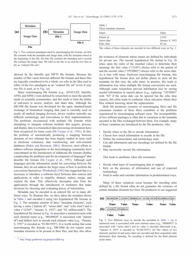
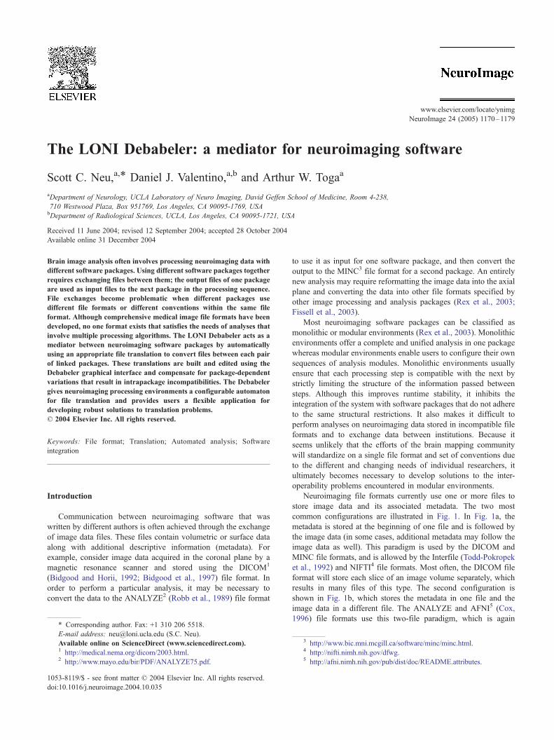
Fig. 1. Two common paradigms used by neuroimaging file formats. (a) One
file contains both the metadata and image data, with the metadata stored at
the beginning of the file. (b) One file contains the metadata and a second
file contains the image data. We refer to the file in (a) and the two files in
(b) as bordered file setsQ.
Table 1
Three metadata elements
Name Value
Patient id 12345
Series date January 8, 1953
Echo time 82 ms
The values of these elements are encoded in two different ways in Fig. 2.
Fig. 2. Two different ways to encode the metadata in Table 1. (a) A
numerical term is associated with each element name (e.g., b00080021Q isassociated with bseries dateQ) and its value is encoded afterwards (e.g.,
bJanuary 8, 1953Q is encoded as b01/08/1953Q). (b) The values of two
S.C. Neu et al. / NeuroImage 24 (2005) 1170–1179 1171
allowed by the Interfile and NIFTI file formats. Because the
number of files varies between different file formats and these files
are logically considered to be a whole, we refer to the files used in
either of the two paradigms as an bordered file setQ (even if only
one file is used, as in Fig. 1a).
Many neuroimaging file formats (e.g., ANALYZE, Interfile,
AFNI, and MINC) were defined by researchers to meet the specific
needs of scientific communities, and this tends to limit the ability
of end-users to access, analyze, and share data. Although the
DICOM file format was developed for the open, standard-based
exchange of biomedical imaging data (and is currently used on
nearly all medical imaging devices), device vendors typically use
different terminology and conventions in their implementations.
The problems encountered with multiple file formats while
attempting to integrate software modules that acquire, process,
and display data in a biomedical data processing environment have
been recognized for many years (De Cuyper et al., 1991). In fact,
the problem of automatically producing a mapping between
elements of two schemas has been largely studied in areas such
as electronic commerce, data warehousing, and web-oriented
databases (Rahm and Bernstein, 2001). However, most efforts to
address software integration in the neuroimaging community have
concentrated on the introduction of additional file formats (further
complicating the problem) and the development of languages6 that
describe file formats (De Cuyper et al., 1991). Although such
languages provide information useful for converting between file
formats, they do not address the larger issue of how to perform the
conversions themselves. Wiederhold (1992) has suggested that it is
necessary to introduce a software layer between data sources and
applications in order to simplify, abstract, reduce, merge, and
explain the data. This effectively decouples data from the
applications through the introduction of mediators that make
decisions by choosing and evaluating pieces of information.
Metadata may be encoded in an ordered file set in many dif-
ferent ways. To illustrate this, we have taken the metadata shown
in Table 1 and encoded it using two hypothetical file formats in
Fig. 2. The metadata consists of three bmetadata elementsQ, eachhaving a name (bpatient idQ, bseries dateQ, and becho timeQ) and a
value (b12345Q, bJanuary 8, 1953Q, and b82 millisecondsQ). Thehypothetical file format in Fig. 2a associates a numerical term with
each element name (e.g., b00100020Q is associated with bpatientidQ) and defines how to encode each element value (e.g., bJanuary8, 1953Q is encoded as b01/08/1953Q). It is worth noting that some
neuroimaging file formats (e.g., DICOM) do not require some
metadata elements to be present in their files, and they also allow
6 http://forge.gridforum.org/projects/dfdl-wg.
the existence of elements whose names are defined by individuals
for private use. The second hypothetical file format in Fig. 2b
relies upon the order of the encoded values to determine their
meaning; the first value (b12345Q) always refer to the patient id
and the second value (b19530801Q) always refer to the series date.
As is true with many fixed-size neuroimaging file formats, this
hypothetical file format does not define places to store all the
metadata (in this case, the echo time). In practice, this leads to
information loss when multiple file format conversions are used.
Although some researchers prevent information loss by storing
needed information in unused places (e.g., replacing b19530801Qwith b82Q if the series date can be ignored but the echo time
cannot), this often leads to confusion when end-users obtain their
files without knowing about the replacements.
Both file producers (creators of neuroimaging files) and file
consumers (readers of these files) contribute to the problems
experienced by neuroimaging software users. The incompatibility
of two software packages is often due to variations in the metadata
encoded in the files exchanged between them. For example, many
of these variations are introduced by the file producers, who:
! Decide where in the file to encode information.
! Choose how much information to encode in the file.
! Define the nomenclature and terminology used.
! Can add information and use encodings not defined by the file
format.
! May incorrectly encode file information.
This leads to problems when file consumers:
! Decide what types of neuroimaging data to support.
! Rely on the presence of information and use of expected
terminology.
! Need to order and correlate information in predetermined ways.
Many of these variations occur because the encoding rules
defined by a file format often do not guarantee the existence of
certain metadata elements nor force file producers to use suggested
elements (patient id and series date) are encoded and their sequential order
determines their meaning. No encoding is defined for the third element
(echo time).
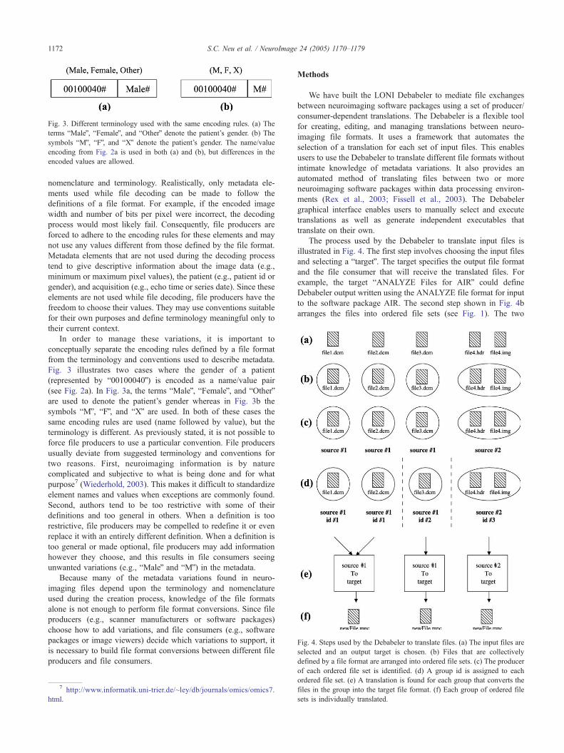
Fig. 3. Different terminology used with the same encoding rules. (a) The
terms bMaleQ, bFemaleQ, and bOtherQ denote the patient’s gender. (b) The
symbols bMQ, bFQ, and bXQ denote the patient’s gender. The name/value
encoding from Fig. 2a is used in both (a) and (b), but differences in the
encoded values are allowed.
Fig. 4. Steps used by the Debabeler to translate files. (a) The input files are
selected and an output target is chosen. (b) Files that are collectively
defined by a file format are arranged into ordered file sets. (c) The producer
S.C. Neu et al. / NeuroImage 24 (2005) 1170–11791172
nomenclature and terminology. Realistically, only metadata ele-
ments used while file decoding can be made to follow the
definitions of a file format. For example, if the encoded image
width and number of bits per pixel were incorrect, the decoding
process would most likely fail. Consequently, file producers are
forced to adhere to the encoding rules for these elements and may
not use any values different from those defined by the file format.
Metadata elements that are not used during the decoding process
tend to give descriptive information about the image data (e.g.,
minimum or maximum pixel values), the patient (e.g., patient id or
gender), and acquisition (e.g., echo time or series date). Since these
elements are not used while file decoding, file producers have the
freedom to choose their values. They may use conventions suitable
for their own purposes and define terminology meaningful only to
their current context.
In order to manage these variations, it is important to
conceptually separate the encoding rules defined by a file format
from the terminology and conventions used to describe metadata.
Fig. 3 illustrates two cases where the gender of a patient
(represented by b00100040Q) is encoded as a name/value pair
(see Fig. 2a). In Fig. 3a, the terms bMaleQ, bFemaleQ, and bOtherQare used to denote the patient’s gender whereas in Fig. 3b the
symbols bMQ, bFQ, and bXQ are used. In both of these cases the
same encoding rules are used (name followed by value), but the
terminology is different. As previously stated, it is not possible to
force file producers to use a particular convention. File producers
usually deviate from suggested terminology and conventions for
two reasons. First, neuroimaging information is by nature
complicated and subjective to what is being done and for what
purpose7 (Wiederhold, 2003). This makes it difficult to standardize
element names and values when exceptions are commonly found.
Second, authors tend to be too restrictive with some of their
definitions and too general in others. When a definition is too
restrictive, file producers may be compelled to redefine it or even
replace it with an entirely different definition. When a definition is
too general or made optional, file producers may add information
however they choose, and this results in file consumers seeing
unwanted variations (e.g., bMaleQ and bMQ) in the metadata.
Because many of the metadata variations found in neuro-
imaging files depend upon the terminology and nomenclature
used during the creation process, knowledge of the file formats
alone is not enough to perform file format conversions. Since file
producers (e.g., scanner manufacturers or software packages)
choose how to add variations, and file consumers (e.g., software
packages or image viewers) decide which variations to support, it
is necessary to build file format conversions between different file
producers and file consumers.
7 http://www.informatik.uni-trier.de/~ley/db/journals/omics/omics7.
html.
Methods
We have built the LONI Debabeler to mediate file exchanges
between neuroimaging software packages using a set of producer/
consumer-dependent translations. The Debabeler is a flexible tool
for creating, editing, and managing translations between neuro-
imaging file formats. It uses a framework that automates the
selection of a translation for each set of input files. This enables
users to use the Debabeler to translate different file formats without
intimate knowledge of metadata variations. It also provides an
automated method of translating files between two or more
neuroimaging software packages within data processing environ-
ments (Rex et al., 2003; Fissell et al., 2003). The Debabeler
graphical interface enables users to manually select and execute
translations as well as generate independent executables that
translate on their own.
The process used by the Debabeler to translate input files is
illustrated in Fig. 4. The first step involves choosing the input files
and selecting a btargetQ. The target specifies the output file format
and the file consumer that will receive the translated files. For
example, the target bANALYZE Files for AIRQ could define
Debabeler output written using the ANALYZE file format for input
to the software package AIR. The second step shown in Fig. 4b
arranges the files into ordered file sets (see Fig. 1). The two
of each ordered file set is identified. (d) A group id is assigned to each
ordered file set. (e) A translation is found for each group that converts the
files in the group into the target file format. (f) Each group of ordered file
sets is individually translated.

8 http://java.sun.com/j2se/1.4.2/docs/guide/imageio/index.html.
Fig. 5. The Debabeler GUI while selecting a new processor. The dialog in the foreground lists the available processors as well as editable information about
each processor’s function, inputs, and outputs. New processors are created in the window behind the dialog, where they can be graphically connected to other
functional modules.
S.C. Neu et al. / NeuroImage 24 (2005) 1170–1179 1173
ANALYZE files belong to one set and each DICOM file has its
own set. The next step identifies the file producer that created each
ordered file set. In Fig. 4c, the first three files are identified as
having been produced by bsource #1Q and the last file set is
identified as having been produced by bsource #2Q. Common file
producers include medical image scanners (e.g., fMRI images
acquired at a Siemens scanner and stored in a mosaic layout using
the DICOM file format) or the output files produced by software
packages. The step shown in Fig. 4d further labels each ordered file
set by assigning each set a group id. These group ids are used to
form groups of ordered file sets that logically belong together. This
can be used to separate proton density MR image files from their
T2 counterparts or to remove scout images from an image series.
Once each ordered file set has been identified and grouped, each
group is translated independently from the others. The Debabeler
uses the group and target names to find the appropriate translation
for each group from its translation registry, and then uses the
translation to convert the files into the target file format. The three
groups shown in Fig. 4d are translated into the files shown in Fig.
4f using the two translations in Fig. 4e.
Visual environment
Solutions to translation problems can be developed within the
Debabeler’s visual environment. Its Java graphical user interface
(Fig. 5) provides access to libraries of functional modules
(bprocessorsQ) that can be graphically connected together between
input and output trees of image data and metadata. This
configurability enables users to provide missing information
(e.g., minimum and maximum pixel values) and correct erroneous
metadata values, as well as expand abbreviations (e.g., change bMQto bMaleQ), decipher enumerations (e.g., change b5Q to bSPGRQ),and convert between units (e.g., Gauss to Tesla). With the
appropriate processors, proprietary metadata may be decoded and
conversions can be applied between different pixel data types (e.g.,
convert 1-byte pixel values to 2-byte pixel values).
Fig. 6a shows an example of two connected modules. The
module on the left has one input and two outputs, with one output
connected to an input of the module on the right. A bmacro-moduleQencapsulates modules and the connections that join them. The two
processors and their connection in Fig. 6b are contained in the
macro-module of Fig. 6a. Every unconnected input or output inside
a macro-module is an input or output of the macro-module itself
unless specified otherwise; for example, the inputs of the macro-
module in Fig. 6a are the inputs of the first processor in Fig. 6b.
The LONI Debabeler uses the Java 1.4 Image I/O architecture8
to read and write neuroimaging files. Because image data and
metadata are organized and managed as trees, Debabeler users are
isolated from the underlying encoding rules of different file
formats. Another benefit of the Java 1.4 Image I/O architecture
is its capability to register plugins that decode and encode files.
Authors who write file decoders and encoders for new neuro-
imaging file formats can add new functionality to the Debabeler by
adding their plugins to the Debabeler runtime classpath. We have
extended the plugin functionality to Debabeler processors so that it
is possible for authors to add processor libraries for new
manipulations of metadata and image data.
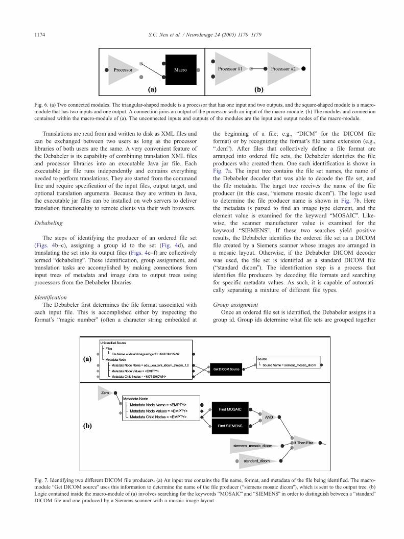
Fig. 6. (a) Two connected modules. The triangular-shaped module is a processor that has one input and two outputs, and the square-shaped module is a macro-
module that has two inputs and one output. A connection joins an output of the processor with an input of the macro-module. (b) The modules and connection
contained within the macro-module of (a). The unconnected inputs and outputs of the modules are the input and output nodes of the macro-module.
S.C. Neu et al. / NeuroImage 24 (2005) 1170–11791174
Translations are read from and written to disk as XML files and
can be exchanged between two users as long as the processor
libraries of both users are the same. A very convenient feature of
the Debabeler is its capability of combining translation XML files
and processor libraries into an executable Java jar file. Each
executable jar file runs independently and contains everything
needed to perform translations. They are started from the command
line and require specification of the input files, output target, and
optional translation arguments. Because they are written in Java,
the executable jar files can be installed on web servers to deliver
translation functionality to remote clients via their web browsers.
Debabeling
The steps of identifying the producer of an ordered file set
(Figs. 4b–c), assigning a group id to the set (Fig. 4d), and
translating the set into its output files (Figs. 4e–f) are collectively
termed bdebabelingQ. These identification, group assignment, and
translation tasks are accomplished by making connections from
input trees of metadata and image data to output trees using
processors from the Debabeler libraries.
Identification
The Debabeler first determines the file format associated with
each input file. This is accomplished either by inspecting the
format’s bmagic numberQ (often a character string embedded at
Fig. 7. Identifying two different DICOM file producers. (a) An input tree contains
module bGet DICOM sourceQ uses this information to determine the name of the f
Logic contained inside the macro-module of (a) involves searching for the keywor
DICOM file and one produced by a Siemens scanner with a mosaic image layou
the beginning of a file; e.g., bDICMQ for the DICOM file
format) or by recognizing the format’s file name extension (e.g.,
b.dcmQ). After files that collectively define a file format are
arranged into ordered file sets, the Debabeler identifies the file
producers who created them. One such identification is shown in
Fig. 7a. The input tree contains the file set names, the name of
the Debabeler decoder that was able to decode the file set, and
the file metadata. The target tree receives the name of the file
producer (in this case, bsiemens mosaic dicomQ). The logic used
to determine the file producer name is shown in Fig. 7b. Here
the metadata is parsed to find an image type element, and the
element value is examined for the keyword bMOSAICQ. Like-
wise, the scanner manufacturer value is examined for the
keyword bSIEMENSQ. If these two searches yield positive
results, the Debabeler identifies the ordered file set as a DICOM
file created by a Siemens scanner whose images are arranged in
a mosaic layout. Otherwise, if the Debabeler DICOM decoder
was used, the file set is identified as a standard DICOM file
(bstandard dicomQ). The identification step is a process that
identifies file producers by decoding file formats and searching
for specific metadata values. As such, it is capable of automati-
cally separating a mixture of different file types.
Group assignment
Once an ordered file set is identified, the Debabeler assigns it a
group id. Group ids determine what file sets are grouped together
the file name, format, and metadata of the file being identified. The macro-
ile producer (bsiemens mosaic dicomQ), which is sent to the output tree. (b)
ds bMOSAICQ and bSIEMENSQ in order to distinguish between a bstandardQt.

Fig. 8. Two types of DICOM group assignments. (a) The group assignment for a Siemens mosaic DICOM file uses the file name as the group id since all the
images of the series are contained inside the file. (b) The group assignment for standard MR DICOM files uses the echo time, unique series id, patient
orientation, image orientation, image width, and image height to generate a group id.
S.C. Neu et al. / NeuroImage 24 (2005) 1170–1179 1175
during translation. The group assignment step allows users to
define how neuroimaging files are organized into image series.
This is advantageous when a file producer imposes an ordering that
needs to be altered (e.g., a scanner that combines scout images with
image slices) or when computational limitations require more
manageable groups of files. A group assignment that assigns group
ids to DICOM files with a mosaic image layout produced by a
Siemens scanner is shown in Fig. 8a. Since all of the images in the
series are contained in one file, the name of the DICOM file is used
as the group id. This ensures that all the images in the DICOM file
are translated as one group. For MR DICOM series that store one
image slice per file, a different group id is needed to group all the
images in the series together. Fig. 8b shows the processors
involved in generating a group id that consists of the echo time
[(00018,0081)], series id [(0020,000E)], patient [(0020,0020)] and
image [(0020,0037)] orientation, and the image width
[(0028,0010)] and height [(0028,0011)]. Inclusion of echo time
separates proton density from T2-weighted images, and slice
orientation separates images that were acquired in different
orthogonal planes. Assigning the same group id to DICOM files
from the same image series groups the files together during the
translation step.
Translation
After the identification and group assignment steps, the
Debabeler translates each group of ordered file sets. Using the
name of the file producer and the output target, an appropriate
translation is found for each group and used for file
conversion. Fig. 9 depicts two translations that convert Siemens
mosaic DICOM files and standard MR DICOM files into
ANALYZE files. In each translation, the image slices are
reformatted to comply with the SPM9 extension rules. In the
9 http://www.fil.ion.ucl.ac.uk/spm/distrib.html.
Siemens mosaic DICOM to ANALYZE translation in Fig. 9a, a
special processor is used to decode Siemens’ private DICOM
element (0029,1020) to obtain information about the mosaic
layout. This information is used to divide the mosaic image into
its components, and the component images are sorted into an
image array. If the image volume is not consistent with the SPM
conventions, it is reformatted into the correct orientation. Similar
operations are performed in the standard MR DICOM to
ANALYZE translation in Fig. 9b. However, since there are many
DICOM files in a single group, the metadata and image data from
all the files must be collected and combined. Fig. 9c shows the
output tree for both translations where the metadata and image data
for the output ANALYZE files are set.
Results
Table 2 lists the encoder and decoder plugins currently supplied
with the Debabeler. Although the Debabeler may be used to create
file format translations from scratch, many users may prefer to edit
the translations (Table 3) that are supplied with the Debabeler
download package. These translations can be used without
modification if they are appropriate, but in most cases users will
want to edit them (e.g., change the output file name) to better
satisfy their needs. We also include the Java source code for our
processor libraries so that expert users may better understand and
fix technical problems.
The results obtained by running a Debabeler executable Java jar
file on a LINUX computer (1 Gb of RAM, one CPU at 1.8 GHz) to
convert different numbers of DICOM files to the NIFTI file format
are summarized in Table 4. Each of the five rows of the table
corresponds to converting unsorted DICOM files into one or more
NIFTI files and recording the total amount of time taken to
perform the conversion. In each case, the Java virtual machine
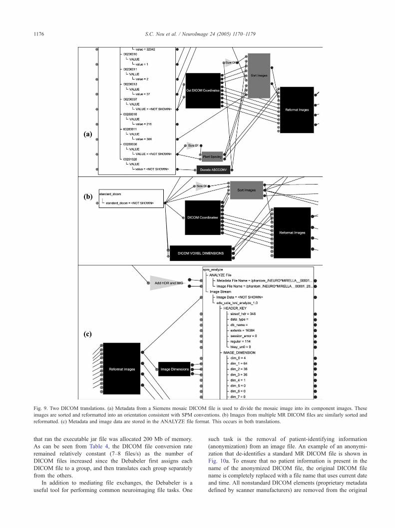
Fig. 9. Two DICOM translations. (a) Metadata from a Siemens mosaic DICOM file is used to divide the mosaic image into its component images. These
images are sorted and reformatted into an orientation consistent with SPM conventions. (b) Images from multiple MR DICOM files are similarly sorted and
reformatted. (c) Metadata and image data are stored in the ANALYZE file format. This occurs in both translations.
S.C. Neu et al. / NeuroImage 24 (2005) 1170–11791176
that ran the executable jar file was allocated 200 Mb of memory.
As can be seen from Table 4, the DICOM file conversion rate
remained relatively constant (7–8 files/s) as the number of
DICOM files increased since the Debabeler first assigns each
DICOM file to a group, and then translates each group separately
from the others.
In addition to mediating file exchanges, the Debabeler is a
useful tool for performing common neuroimaging file tasks. One
such task is the removal of patient-identifying information
(anonymization) from an image file. An example of an anonymi-
zation that de-identifies a standard MR DICOM file is shown in
Fig. 10a. To ensure that no patient information is present in the
name of the anonymized DICOM file, the original DICOM file
name is completely replaced with a file name that uses current date
and time. All nonstandard DICOM elements (proprietary metadata
defined by scanner manufacturers) are removed from the original

Table 4
Time taken by the Debabeler to convert DICOM files to NIFTI files
DICOM files
(input)
Conversion
time (s)
Conversion rate
(file/s)
NIFTI files
(output)
123 20 6.2 1
219 31 7.1 3
354 51 6.9 9
668 87 7.7 14
1207 152 8.0 28
Five sets of DICOM files were converted to NIFTI files on a LINUX
computer (1 Gb of RAM, one CPU at 1.8 GHz).
Table 2
Neuroimaging file formats supported by the Debabeler Java 1.4 Image I/O
plugins
File format Read (decode) Write (encode)
AFNI U UANALYZE U UDICOM U UGE U UMedX UMINC U UNIFTI U URaw U U
S.C. Neu et al. / NeuroImage 24 (2005) 1170–1179 1177
file and all unique ids (possibly containing unique scanner codes)
are one-way encrypted to retain uniqueness. Further, a command
line argument is used to replace the patient id in the anonymized
DICOM file. The Debabeler’s programmable interface allows
users to construct different levels of patient de-identification in
order to satisfy different standards of patient confidentiality (e.g.,
HIPAA). Fig. 10b shows an example of how the Debabeler can be
used to convert a raw (no encoded metadata) neuroimaging file
into a standard neuroimaging file (ANALYZE). In this case, the
Debabeler reads the file as though it were a single image block,
and command line arguments (e.g., image width, height, and color
type) are used to divide the image block into multiple images. This
can also be used to skip past fixed-sized metadata blocks if the
sizes of the blocks are known. As shown in Fig. 10c, the
Debabeler is capable of writing to more than one file format in a
single translation. Here the images decoded from a MINC file are
reformatted and stored as an ANALYZE file. Since the ANALYZE
file format limits encoded metadata to 348 byte and does not
define temporal scanner metadata, the repetition, echo, and
inversion times in the MINC file are instead stored in an XML
file. Using multiple file formats in translations can prevent
important metadata from getting lost, and using XML saves them
as human-readable text.
Discussion
Neuroimaging data are acquired by imaging devices and
processed by software packages, and both store image intensity
values and associated descriptive information to files using a
variety of file formats. Although some file producers create files
using the same file format, many use different terminology and
nomenclature for the descriptive data. This becomes problematic
Table 3
Translations (T) and anonymizations (A) supplied with the Debabeler
Source Target file format
AFNI ANALYZE DICOM MINC NIFTI
ANALYZE T A T T T
DICOM M T A T T
GE M T T T T
MINC M T T A T
Raw M T M M M
An anonymization removes patient-identifying information from a file.
Multiple translations (M) can be used to convert between file formats that
do not have a single translation.
for biomedical image analysis programs that expect either a
specific file format or depend upon particular metadata
conventions. Despite the development of bstandardQ medical
image file formats, no one format has satisfied the needs of
image-based studies involving multiple processing and analysis
algorithms.
The LONI Debabeler is a flexible tool that manages and
converts a variety of biomedical image file formats. Its general
architecture is not limited to specific file formats or metadata
conventions, and it can be readily extended to decode (read) and
encode (write) new file formats. The Debabeler converts input files
by identifying the producer of the file, arranging the files into
groups, and translating each group into a specified target format.
This conversion methodology enables the Debabeler to operate as a
mediator between neuroimaging software packages that use
different file formats and rely upon different metadata conventions.
The Debabeler’s graphical user interface enables users to add and
modify translations to meet their specific needs. The Debabeler
thus serves as an intelligent translation manager that automatically
selects translations and converts common neuroimaging file
formats in flexible ways, and a graphical development environment
for visually programming solutions to translation problems.
Although the Debabeler represents the input and output file
formats as trees (hiding the details of how the data are encoded in
the files), in many cases expert knowledge is needed to properly
map input values to output values. For example, if the values for
the image width and height are not correctly assigned in the output
tree, the Debabeler will create a file that most likely will be
undecodable. This is because the values of the image width and
height are usually used to decode the image data in the file, and if
they are incorrect the image data will not be properly decoded. To
address this issue, we include translations along with the Debabeler
that perform conversions between many common file formats (see
Table 3). It has been our experience that most translation requests
are for editing existing translations (reformatting a string field,
changing the output file name, etc.), and we have found that the
Debabeler’s graphical interface provides a quick and easy way to
adjust these translations.
Another concern is that in an environment of N software
modules, one may need to program N � (N � 1) translations to
ensure interoperability between each pair of modules. To this end,
we remark that it is not always the case that each module will have
vastly different file format requirements and we can expect some
modules to work together without Debabeler mediation. In fact, the
more translations that exist, the more likely a translation can be
found for a newly introduced module. Furthermore, we note that
the identification step used by the Debabeler is configurable and
generalizable. If two different file producers create input files that
share the same file format and use similar metadata conventions
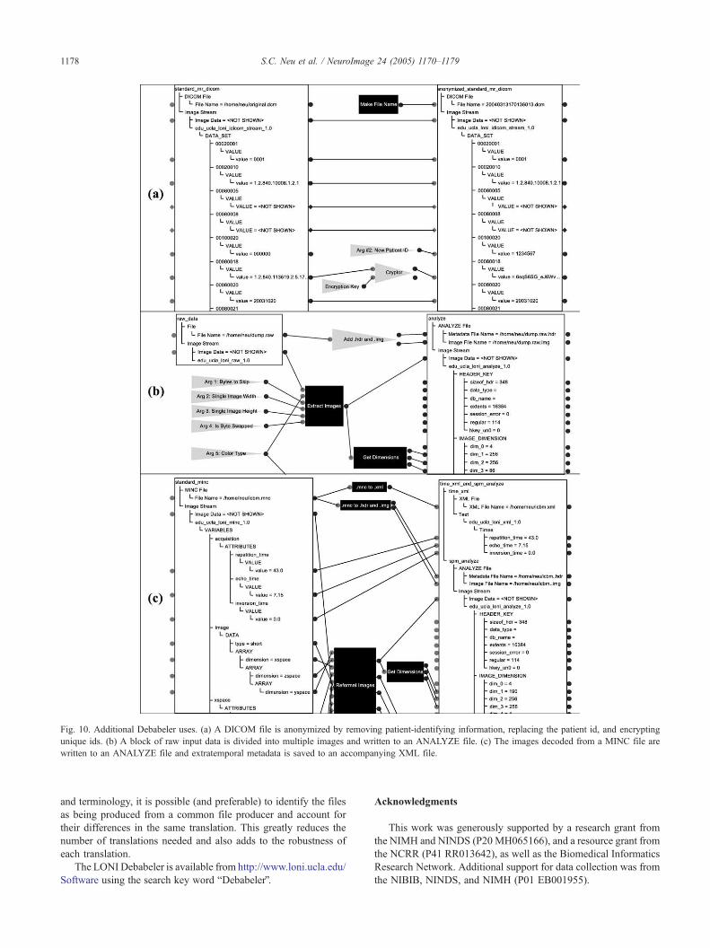
Fig. 10. Additional Debabeler uses. (a) A DICOM file is anonymized by removing patient-identifying information, replacing the patient id, and encrypting
unique ids. (b) A block of raw input data is divided into multiple images and written to an ANALYZE file. (c) The images decoded from a MINC file are
written to an ANALYZE file and extratemporal metadata is saved to an accompanying XML file.
S.C. Neu et al. / NeuroImage 24 (2005) 1170–11791178
and terminology, it is possible (and preferable) to identify the files
as being produced from a common file producer and account for
their differences in the same translation. This greatly reduces the
number of translations needed and also adds to the robustness of
each translation.
The LONI Debabeler is available from http://www.loni.ucla.edu/
Software using the search key word bDebabelerQ.
Acknowledgments
This work was generously supported by a research grant from
the NIMH and NINDS (P20 MH065166), and a resource grant from
the NCRR (P41 RR013642), as well as the Biomedical Informatics
Research Network. Additional support for data collection was from
the NIBIB, NINDS, and NIMH (P01 EB001955).

S.C. Neu et al. / NeuroImage 24 (2005) 1170–1179 1179
References
Bidgood, W.D., Horii, S.C., 1992. Introduction to the ACR-NEMA
DICOM standard. Radiographics 12 (2), 345–355.
Bidgood, W.D., Horii, S.C., et al., 1997. Understanding and using DICOM,
the data interchange standard for biomedical imaging. J. Am. Med.
Inform. Assoc. 4 (3), 199–212.
Cox, R.W., 1996. AFNI: software for analysis and visualization of
functional magnetic resonance neuroimages. Comput. Biomed. Res.
29 (3), 162–173.
De Cuyper, B., Nyssen, E., et al., 1991. Do you also have problems with the
file format syndrome? Med. Biol. Eng. Comput. 29 (6), 55–60.
Fissell, K., Tseytlin, E., et al., 2003. Fiswidgets: a graphical computing
environment for neuroimaging analysis. Neuroinformatics 1 (1),
111–126.
Rahm, E., Bernstein, P.A., 2001. A survey of approaches to automatic
schema matching. VLDB J. 10 (4), 334–350.
Rex, D.E., Ma, J.Q., et al., 2003. The LONI pipeline processing
environment. NeuroImage 19 (3), 1033–1048.
Robb, R.A., Hanson, D.P., et al., 1989. Analyze: a comprehensive,
operator-interactive software package for multidimensional medical
image display and analysis. Comput. Med. Imaging Graph. 13 (6),
433–454.
Todd-Pokropek, A., Cradduck, T.D., et al., 1992. A file format for exchange
of nuclear medicine image data: a specification of Interfile version 3.3.
Nucl. Med. Commun. 13 (9), 673–699.
Wiederhold, G., 1992. Mediators in the architecture of future information
systems. Computer 25 (3), 38–49.
Wiederhold, G., 2003. The impossibility of global consistency. Omics:
J. Integr. Biol. 7 (1), 17–20.